Note: Descriptions are shown in the official language in which they were submitted.
CA 02393607 2002-06-03
WO 01/46714 PCT/US00/42517
ULTRASONIC HORN ASSEMBLY
BACKGROUND OF THE INVENTION
This invention relates to ultrasonic vibration probes. More particularly,
this invention relates to such an ultrasonic probe or horn assembly which is
particularly useful in the simultaneous sonication of biological and cellular
materials disposed in multiple wells of a tray.
It has been well known for decades that a probe which vibrates at
ultrasonic frequencies (i.e. frequencies greater than 16,000 Hz) and has its
distal end submerged under fluids will create cavitation bubbles if the
amplitude
of vibration is above a certain threshold. Many devices have been
commercialized which take advantage of this phenomenon. An example of
such an ultrasonic cellular disrupter is disclosed in the SonicatorT"' sales
catalog
of Misonix Incorporated of Farmingdale, New York. In general, devices of this
type include an electronic generator for producing electrical signals with
frequencies ranging from 16 to approximately 100 KHz, a piezoelectric or
magnetostrictive transducer to convert the signal to mechanical vibrations and
a
probe (a.k.a. horn or velocity transformer) which amplifies the motion of the
transducer to usable levels and projects or removes the operating face away
from the transducer itself. The design and implementation of these
components are well known to the art.
The cavitation bubbles produced by such ultrasonic vibration devices
CA 02393607 2002-06-03
WO 01/46714 PCT/US00/42517
2
can be utilized to effect changes in the fluid or upon particles suspended
therein. Such changes include biological cell disruption, deagglomeration of
clumped particles, emulsification of immiscible liquids and removal of
entrained
or dissolved gases, among many others.
Cell disruption has been a particularly good application for probe type
devices, in that the cells may be disrupted without the heat or cellular
changes
which prevent further analysis by conventional methodology. Many scientific
protocols have been written which name the SonicatorT"~ (or similar devices)
as
the instrument of choice for the procedure.
One characteristic of the probe type ultrasonic vibration devices which
limit their use is the fact that the standard probes must be inserted directly
into
the fluid. Because the probe occupies volume as it is submersed, very small
samples cannot be processed. In addition, the probe becomes contaminated
with the fluid since the probe is in direct contact with the fluid. If the
probe is
subsequently dipped into another sample, contamination of that sample may
occur. In some cases, this cross contamination renders the second sample
unusable for analysis.
One way to mitigate these deficiencies is to have the probe tip separated
from the sample by a membrane or other solid surface. If liquid is present on
both sides of the membrane or surface, the acoustic waves will propagate
CA 02393607 2002-06-03
WO 01/46714 PCT/US00/42517
3
through the membrane and transfer the cavitation forces to the second liquid
volume without having the probe in direct contact with that second liquid
volume. This membrane does not have to be elastic. In fact, experience shows
that glass or hard plastic is an acceptable material. Consequently, glass and
plastic test tubes and beakers are routinely used in this service. Misonix
Inc.
produces and sells a device called the Cup HornT"" which uses this method of
acoustic wave transfer to allow the researcher to segregate the probe from the
sample.
One requirement for use of the Cup Horn is that the beaker or test tube
diameter be significantly smaller than the distal diameter of the Cup Horn
probe
itself. This allows the acoustic energy to be relatively uniform across the
diameter of the sample container. In addition, liquid is forced to surround
the
entire probe end in order to provide the transfer fluid for the acoustic wave.
Figure 1 shows the relationship of the Cup Horn probe 12, transfer fluid 14
and
sample test tube. A cup 16 having a cylindrical sidewall 18, an inwardly
extending annular flange 20 and a cylindrical sleeve 22 is mounted to the horn
or probe 12 via a coupling sleeve 24 and a pair of O-rings 26 disposed in a
region about a node of ultrasonic vibration of the probe. The transfer fluid
not
only covers a transverse end face 28 of probe 12, but also surrounds a
substantial portion of the cylindrical distal surface 30 of the probe.
The requirements of (a) the relative sizes of the probe 12 and the test
CA 02393607 2002-06-03
WO 01/46714 PCT/US00/42517
4
tube and (b) the surrounding of the probe end surface 30 by the transfer fluid
14 give rise to at least two problems. First, the size of the vessel is
limited to
that of the surface area of the probe 12 and second, the liquid 14 surrounding
the probe 12 places a great load upon the probe. The power required to
overcome this load is many times that needed for acoustic coupling into the
small sample. In some cases, as the probe has been made larger to
accommodate larger samples, the energy required has become greater than
the power capability of the electronic generators currently available. In such
cases, system overloads have occurred.
These limitations become especially apparent when the sample vessel
takes the form of a multi-well microtiter plate or tray. Such a plate is
typically
made from clear hard plastic such as polystyrene, polyvinylchloride or
acrylics.
The tray is fairly shallow and may contain up to approximately 96 depressions
(wells) into which the samples or specimens are placed. Each depression may
contain only a few microliters of sample. In most cases, the insertion of a
probe
device is problematic since each sample must be isolated from the others, the
wells are too small and the total processing time would be an unacceptable
multiple of the processing time of one cell. Therefore, most researchers would
prefer a device which would isolate the samples from the ultrasound probe and
process all cells simultaneously.
It would be obvious to most persons skilled in the art to simply enlarge
CA 02393607 2002-06-03
WO 01/46714 PCT/US00/42517
the diameter of the probe to allow the entire tray to be covered. However, as
previously stated, the probe becomes very large, leading to non uniformity in
the vibrational amplitude of the distal surface, very high power requirements
and high cost of manufacture. In the past, probes of smaller square section
5 were made which allow a quarter of the tray to be processed at a time, which
decreased processing time substantially. However, most researchers required
a further reduction in time in order to process their entire workload in one
day.
Also, the outer edges of the trays received irregular ultrasonic energy and
therefore inconsistent cell breakdown in successive samples.
OBJECTS OF THE INVENTION
An object of the present invention is to provide an ultrasonic device
which could treat a full microtiter tray simultaneously.
Another object of the present invention is to provide such an ultrasonic
device which increases the degree of uniformity of acoustic intensity across
the
cells of the microtiter tray.
A further object of the present invention is to provide such an ultrasonic
device which does not heat the fluid or the sample liquids, and which require
minimum energy to operate, thereby allowing the use of the device on existing
laboratory scale ultrasonic processors.
CA 02393607 2002-06-03
WO 01/46714 PCT/US00/42517
6
These and other objects of the present invention will be apparent from
the drawings and descriptions herein.
BRIEF DESCRIPTION OF THE INVENTION
The present invention is directed to an ultrasonic sonication device which
includes two basic components, namely, (1 ) a velocity transformer (or probe)
which, when coupled to a vibrating transducer of the piezoelectric or
magnetostrictive type, resonates in sympathy with the transducer and either
increases or decreases the magnitude of the transducer's vibration and 2) a
shallow cup assembly which holds a microtiter tray in a suitable orientation
and
contains an amount of liquid which provides efficient acoustic coupling.
An ultrasonic horn assembly comprises, in accordance with the present
invention, an ultrasonic horn or probe having an axis and a distal end with an
end face oriented substantially transversely to the axis. The end face of the
probe is disposed at least approximately at an antinode of ultrasonic
vibration of
the horn or probe. A cup member is attached to the horn or probe at least
approximately at the antinode so as to define a liquid reservoir covering the
end
face of the horn or probe. This attachment of the cup member at, or
approximately at, the antinode at the distal end of the probe enables the
formation of the reservoir as a shallow reservoir covering essentially only
the
end face of the probe. A small or marginal circumferential surface of the
probe,
contiguous with the end face thereof, may be submerged in the coupling liquid,
CA 02393607 2002-06-03
WO 01/46714 PCT/US00/42517
7
as well.
In an ultrasonic horn assembly in accordance with the present invention,
the load placed upon the probe is decreased owing to the reduction in the area
of contact between the coupling fluid and the probe. The power requirements
are accordingly reduced for a probe end face of a given area.
The cup member is attached to the horn or probe via a flexible coupling
element such as an O-ring or an annular elastomeric membrane. Where the
cup member includes a sidewall and a lower wall or flange extending inwardly
from the sidewall, the lower wall is provided with at least one port for
feeding
liquid to the reservoir. Preferably, the port is one of at least a pair of
ports
disposed on substantially opposite sides of the cup member. The feeding of
the coupling liquid through a lower wall of the cup member has advantages
detailed below.
The end face of the probe is disposed in a first plane and an upper
surface of the flange is disposed in a second plane spaced a first
predetermined distance from the first plane, so that a lower surface of a
specimen-containing tray resting on the upper surface of the flange is spaced
a
second predetermined distance from the probe end face. This spacing
optimizes the acoustic effects of the ultrasonic energy on specimens contained
in wells of a microtiter tray. To enable an optimal spacing, the probe end
face
CA 02393607 2002-06-03
WO 01/46714 PCT/US00/42517
8
is provided with a plurality of grooves for receiving peripheral lower edges
of the
tray so that contact between the tray and the vibrating probe is prevented.
Where the end face of the probe is circular, the end face has a diameter
larger than a largest dimension of the portion of the tray containing the
sample
wells. Thus, all of the sample wells are located over the end face of the
probe.
In accordance with another feature of the present invention, the probe is
provided at the distal end, proximately to the end face, with an annular
concavity for providing or enhancing uniformity of the ultrasonic wave field
generated in the coupling fluid reservoir.
An ultrasonic sonication device in accordance with the present invention
is an effective apparatus to acoustically treat or disrupt samples within a
multiwell microtiter tray.
BRIEF DESCRIPTION OF THE DRAWINGS
Figure 1 is a cross-sectional view, taken along an axial plane, of an
ultrasonic sonication device in accordance with the prior art.
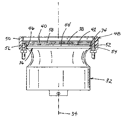
Figure 2 is a cross-sectional view, taken along an axial plane, of an
ultrasonic sonication device in accordance with the present invention.
CA 02393607 2002-06-03
WO 01/46714 PCT/US00/42517
9
Figure 3 is a cross-sectional view, taken along an axial plane, of another
ultrasonic sonication device in accordance with the present invention.
Figure 4 is a top plan view of the ultrasonic sonication device of Figure 2,
showing a microtiter tray in place on the probe.
Figure 5 is a partial cross-sectional view taken along line V-V in Figure 4.
Figure 6 is a detail, on a larger scale, of a portion VI of Figure 5.
Figure 7 is an enlarged top plan view similar to Figure 4, showing flow
paths for a transfer fluid.
DESCRIPTION OF THE PREFERRED EMBODIMENTS
As illustrated in Figure 2, an ultrasonic sonication device comprises a
horn or probe 32 having an axis 34 defining a direction of ultrasonic standing
wave propagation. Probe 32 has a distal end portion 36 formed with an active
end face 38 oriented transversely to axis 34 and provided with at least one
pair
of parallel grooves 40 and 42. Distal end portion 36 of probe 32 is further
formed with an annular groove 44 receiving an elastomeric O-ring seal 46.
The ultrasonic sonication device of Figure 2 additionally comprises a cup
member 48 having a vertical cylindrical sidewall 50 and a horizontal annular
CA 02393607 2002-06-03
WO 01/46714 PCT/US00/42517
flange 52 extending inwardly from a lower end of the sidewall. An inner
periphery of flange 52 is in fluid tight contact with an outer periphery of
distal
horn portion 36, through or over O-ring seal 46. Flange 52 is provided on
opposite sides with a pair of liquid ports or fittings 54 and 56 for the
continuous
5 introduction and removal, respectively, of a pressure-wave transfer fluid 58
from
a reservoir defined in part by probe end face 38 and cup member 48.
As depicted in Figure 3, a modified ultrasonic sonication device
comprises a cup member 60 having a sidewall 50' with a larger diameter than
sidewall 50 of cup member 48. An inner periphery of an annular flange 52' is
10 spaced from and connected to the outer periphery of distal horn portion 36
by
an annular elastomeric membrane 62. Membrane 62 is sealingly fixed along an
inner side to distal horn portion 36 and along an outer side to flange 52'.
Figures 4, 5, and 6 depict the use of the sonication device of Figure 2
with a microtiter tray or plate 64 having a plurality of specimen-receiving
wells
or cells 66 disposed in a rectangular array. Four corners 68 of tray 64 rest
on
flange 52 so that a bottom surface 70 (Figure 6) of the tray is disposed in a
plane P1 spaced a predetermined distance D from a plane P2 in which the
vibrating end face 38 of probe 32 is located. This distance D is selected to
optimize the transmission of ultrasonic wave energy from end face 38 through
fluid 58 and into tray 64.
CA 02393607 2002-06-03
WO 01/46714 PCT/US00/42517
11
Tray 64 is conventionally configured to have a peripheral lower rim 72
(Fig. 6) which extends below the plane P1 of bottom tray surface 70. This rim
72 is in contact with an upper surface 76 (Figures 4-6) of flange 52 and is
spaced from horn or probe 32 by virtue of grooves 40, 42, etc., provided in
end
face 38.
Probe 32 functions in part as a velocity transformer which amplifies the
motion of a piezoelectric or magnetostrictive transducer (not shown) to usable
levels. Probe 32 can be designed and constructed using standard techniques
known to the art. However, several important operating characteristics must be
obtained for probe 32 to be useful in this device. First, distal end face 38
of
probe 32 must be large enough to cover the entire area of bottom surface 70 of
microtiter tray 64. In the embodiment described herein, distal end face 38 is
circular and has a diameter of 5.25 in., but other diameters or geometric
shapes
may be employed as well. One important aspect regarding size is that
microtiter tray wells 66 must not be less than 0.125 inches from an outer edge
74 of probe end face 38. If a tray cell 66 is located at edge 74 or within
0.125
inches of that edge, acoustic input to the well will be decreased due to
ultrasonic edge effects. Second is that it is advantageous if a uniform
amplitude of vibration is generated across the entire end face 38 of probe 32.
If
significantly non-uniform vibrations are present, then non-uniformity of
processing in the microtiter wells 66 will result. In order to obtain this
uniform
vibration for the size of probe discussed herein, the shape of probe 32 must
be
CA 02393607 2002-06-03
WO 01/46714 PCT/US00/42517
12
as that shown in Figure 2. It should be noted that the dimensions given
describe a probe 32 which has a fundamental resonant frequency of
approximately 20 kc. Other frequencies of operation may be employed without
deviating from the scope of this disclosure.
Grooves or reliefs 40, 42, etc., are machined or otherwise formed in
probe end face 38 (Figure 6) to allow microtiter tray edge or rim 72 to sit in
these recesses. In this way, the bottom surface 70 of microtiter tray 64 sits
within 0.100 inches (preferably between about 0.001 and 0.100 inches) of the
vibrating probe end face 38. Controlling this distance D is of paramount
importance if enough acoustic energy is to be transmitted through the wall of
tray 64 to the samples contained in wells or cells 66 thereof. The geometry of
probe end face 38 is particularly shown in Figures 4-6. Of course, probe 32
must be manufactured from an acoustically efficient material such as aluminum,
titanium, certain stainless steels and certain ceramics. These materials are
all
known to the art. Harder materials such as titanium or ceramics will yield a
device which does not wear quickly due to cavitation erosion. Connection to
the
transducer (not shown) can be accomplished by a threaded stud (not shown) or
other techniques well known to the art.
The seal provided by O-ring 46 or membrane 62 is elastomeric to
provide a compliant joint between cup member 48 or 60 and probe 32. This
seal is liquid tight and yet isolates cup member 48 or 60 from the vibrations
CA 02393607 2002-06-03
WO 01/46714 PCT/US00/42517
13
transmitted by probe 32. This isolation prevents loading and possible detuning
of probe 32 while keeping acoustic power from being absorbed by cup member
48 or 60, preventing melting thereof if the cup member is manufactured from
thermoplastics. It is to be noted that O-ring 46 and membrane 62 are placed at
or near an anti-node (point of maximum displacement) of probe operation as
opposed to being placed at a node (point of no displacement) as is generally
practiced by the art. Since the node point is found approximately at the
midpoint of probe 12 (see Figure 1 ), placing the seal at the node would mean
that half of the probe would be submerged under cooling/coupling fluid 14.
Prior art, as shown in Figure 1, uses the node point sealing method, with all
of
the inherent problems as described above. Moving the seal position near the
antinode (and thus near probe end face 28) greatly reduces the power loading
and energy consumption of the device.
Cup members 48 and 60 are fabricated alternatively from clear acrylic
and clear polyvinylchloride. However, other materials such as thermoplastics,
metals, ceramics or thermosets may be used with equal results.
Several features of cup member 48 and 60 are important to the
operation of the device. First, cup members 48 and 60 must have an internal
diameter just slightly greater than the diagonal dimension of the microtiter
tray
64. This centers the tray 64 with respect to the end face 38 of probe 32, as
shown particularly in Figure 4. Upper surface 76 of flange 52, 52' must be
CA 02393607 2002-06-03
WO 01/46714 PCT/US00/42517
14
designed in conjunction with the dimensions of microtiter tray 64 in order to
hold
the tray off the probe end face 38 by the proper distance D. To that end, a
plane P3 in which surface 76 is disposed is located at a predetermined
distance
D2 (Figure 6) from the plane P2 of probe end face 38. Microtiter tray 64 sits
on
cup surface 76 and does not contact probe 32 at any point. If tray 64 is
allowed
to touch end face 38 of the probe, melting of the tray will result.
Next, cup member 48 or 60 must incorporate liquid fittings or ports 54
and 56, to allow coupling fluid 58 to be pumped in and out of the cup member.
If fluid transport is not provided, then heating of the fluid will result with
extended use. The temperatures generated may exceed the cytocoagulation
temperature of the biological samples in wells 66, effectively cooking the
specimens. A constant flow of fresh or cooled fluid obviates this eventuality.
Although the necessity for cooling is well known to the art, an improvement
disclosed herein is to place the fittings 54 and 56 so that the coupling fluid
or
liquid 58 is introduced and removed from under the microtiter tray 64. Figure
7
shows general paths 78 of fluid flow under microtiter tray 64 from one port or
fitting 54 to the other port 56. When the ports or fittings 54, 56 are
disposed on
opposite sides of cup member 48, 60 and along flanges 52, 52' thereof, the
coupling fluid 58 has maximum cooling effect and reduces or eliminates
splashing onto the top of the tray 64, thereby preventing contamination of the
samples. Another benefit is extremely important in that the liquid flow as
illustrated in Figure 7 will purge or flush trapped air from the underside or
CA 02393607 2002-06-03
WO 01/46714 PCT/US00/42517
bottom surface 70 of tray 64. Air bubbles, if present between the probe end
face 38 and the bottom surface 70 of the tray 64, will not allow acoustic
coupling to the tray wells 66 and no processing will result. Therefore,
bubbles
or air entrapment must be eliminated, something which this embodiment
5 accomplishes. In the disclosed embodiment, port elements 54, 56 are standard
liquid tubular fittings provided on the lower surface of the cup member 48,
60.
The coupling fluid or liquid can be plain tap water, saline, distilled water
or, if
sub freezing temperatures are desired, a solution of glycol and water may be
employed.
10 In operation, a thin plastic film (not shown) should be applied to the top
of microtiter tray 64 in a fashion known to the art. This thin film prevents
loss of
samples from the tray wells 66 during acoustic processing, from either
bubbling
or atomization. In addition, cross contamination of samples is eliminated.
Although when using non-ultrasonic techniques of sample preparation, this film
15 is optional, the film is deemed essential in use of the ultrasonic
sonication
devices disclosed herein.
Cup member 48, 60 must incorporate features such as a counterbore to
prevent slippage of the cup relative to probe 32. This prevents the cup from
lowering with respect to the probe end face 38 and maintains the clearance
between the bottom surface 70 of microtiter tray 64 and the probe end face.
CA 02393607 2002-06-03
WO 01/46714 PCT/US00/42517
1G
Although the invention has been described in terms of particular
embodiments and applications, one of ordinary skill in the art, in light of
this
teaching, can generate additional embodiments and modifications without
departing from the spirit of or exceeding the scope of the claimed invention.
Accordingly, it is to be understood that the drawings and descriptions herein
are
proffered by way of example to facilitate comprehension of the invention and
should not be construed to limit the scope thereof.