Note: Descriptions are shown in the official language in which they were submitted.
CA 02395492 2002-06-25
WO 01/48180 PCT/CA00/01561
METHOD AND APPARATUS FOR
TARGETING LOCALISED ELECTROPORATION
Field of the Invention
This invention relates to the fields of introduction of foreign compounds into
cells
by electroporation.
Background of the Invention
Current transgenic methods include viral techniques, lipofection,
microparticle
bombardment, and paddle electroporation (Leber et. al 1996; Methods Cell Biol.
51:
161-83; Muramatsu et. al 1997; Bioc Biophys Res Comm. 23: 376-80).
Electroporation is a versatile and popular method of transfecting cells by
briefly
subjecting them to an electric field, which forms pores in the lipid bilayer,
and allows
entry of compounds into the cytoplasm (Lurquin 1997; Mol Biotechnol. 7 (1 ): 5-
35).
Electroporation overcomes the disadvantages of viral techniques, such as small
insert
size and biohazard concerns (Leber et. al 1996; Methods Cell Biol. 51: 161-
83).
Additionally, electroporation has been shown to be more efficient than other
methods
of in ovo transfection, such as lipofection or microparticle bombardment
(Muramatsu
et. al 1997; Bioc Biophys Res Comm. 23: 376-80). However, previous
electroporation
efforts have had a number of limitations, including the inability to
effectively target the
electroporation, lack of reproducibility of results, and exposure of non-
targeted cells to
potentially damaging current. Specifically, the electroporation apparatuses of
the prior
art are generally paddle-like pairs of electrodes, having inherent limitations
as to how
small a target area would be electroporated and lacking integral means for
introducing
the foreign matter (commonly DNA) into the cells. Instead, the prior art
teaches
separate injection of the foreign matter into a localised target area, and
electroporation
of a larger region including and extending beyond the target area.
Accordingly, the
prior art teaches unnecessary electroporation of (and thereby potential
attendant injury
to) cells outside the target area.
SUBSTITUTE SHEET (RULE 26)
CA 02395492 2002-06-25
WO 01/48180 PCT/CA00/01561
Moreover, methods of electroporation in the prior art have proved inefficient
and
unreliable in localising to any degree the transfection events, and cannot
target a
specific region of an organ or tissue. Truly localised electroporation could
neither be
accomplished with prior methods, nor was it contemplated. Prior art methods of
in ovo
electroporation have involved subjecting the entire embryo to potentially
damaging
current and the effects have been difficult to reproduce. Although prior art
in vivo
efforts have included attempts to target on particular organs or tissues,
these efforts
have involved the injection of a DNA-containing solution into an organ or into
the blood
stream of an organism and the use of paddle-like pairs of electrodes for the
application
of the electric field. Cells and tissues subjected to these prior art methods
would be
not be uniformly electroporated, and as a result only random transfection
events would
occur within the selected areas. Introduction of foreign compounds other than
genetic
material by electroporation has likewise suffered from a high degree of
unpredictability,
due to the inefficiency and unreliability of prior art methods of
electroporation.
Summary of the Invention
The invention comprises methods and apparatus for targeting localised
electroporation. In one aspect of the invention, an electroporation apparatus
is
provided, comprising a fluid delivery means, an electrode means for
establishing an
electrical field in the target area, and a means for connecting the electrode
means to
a source of electricity. In preferred embodiments of the inventive apparatus,
the
electrode means may comprise a pair of conductors. The fluid delivery means
f~as an
upstream end, a downstream end, and a passageway fluidly connecting these
ends.
The upstream end has a receiving aperture for receiving a fluid, and the
downstream
end comprises a discharging aperture for discharging the fluid into a target
area. The
electrode means is located at least in part in the vicinity of the discharging
aperture of
the fluid delivery means. Although the electrode means may generally be
located
outside the fluid delivery means, in preferred embodiments, substantially the
entire
electrode means may be located within the fluid delivery means. Accordingly,
the
apparatus of the invention differs from those of the prior art in that the
former has a
fluid delivery means integrally associated with the electrode means.
2
SUBSTITUTE SHEET (RULE 26)
CA 02395492 2002-06-25
WO 01/48180 PCT/CA00/01561
In further preferred embodiments of the inventive apparatus, the following
features may be present:
~ means for controlling elE:ctroporation parameters, such as wave form, number
of trains, train duration, tr~~in polarity, pulse length, output voltage, and
pulse
frequency;
~ insulation of the electrode means, except at the portions of the electrode
means
that are in the vicinity of the discharging aperture;
~ a fluid source, such as the screw-drive syringe of Example 1 of the Detailed
Description, connected upstream to the fluid delivery means.
In at least one preferred embodiment of the apparatus, a double-barrelled tube
is used
both to maintain the conductors spaced apart from one another and to deliver
the fluid
to the target area.
The inventive apparatus may be used to both electroporate and apply foreign
matter to a target area having a diameter of at least as low as 100 ,um.
Where the target area is separated from the outer surface of an organism by
intervening matter (such as, for example, skin or some integumentary matter),
the
apparatus may advantageously be provided with a means for piercing through
such
intervening matter. Preferably, either the electrode means or the downstream
end of
the fluid delivery means is sharpened in order to be used to pierce the
intervening
matter. On the other hand, where the target area is at or near the surface of
the
organism, the surface downstream end of the fluid delivery means may
preferably be
polished smooth, so as to minimise the invasiveness of the electroporation.
Another preferred modification of the inventive apparatus is to provide it
with
means to maintain fluid-mediated contact between the electrode means and the
target
area, thereby obviating the need for direct physical contact between the
apparatus and
the target area. This may advantageously be accomplished by, as in the example
provided in the Detailed Description, providing the apparatus with a screw-
drive
syringe, which may both release the fluid into the fluid delivery means and
provide a
3
SUBSTITUTE SHEET (RULE 26)
CA 02395492 2002-06-25
WO 01/48180 PCT/CA00/01561
suction to maintain fluid-mediated contact between the electrode means and the
target
area.
The apparatus of the present invention may provide yet another advantage over
the apparatuses of the prior art. The electrode means of the prior art contact
the
target area, which may lead to greater cellular injury at the point of contact
than would
occur if the electrode means did not contact the target area. In the present
invention,
the electrode means need not contact the target area; instead, the ends of the
electrode means may be located upstream of the discharging aperture, such that
the
target area, if it were to come into contact with the inventive apparatus at
all, would
come into contact only with the discharging aperture. Furthermore, the need
for any
contact between the inventive apparatus and the target area may be obviated
entirely
by the presence of a sufficient quantity of electrolyte, which may either be
provided
through the discharging aperture of the inventive apparatus or may already
present in
the target area. In this connection, the apparatus may advantageously be
provided
with a fluid source for providing an electrolyte as well as the foreign matter
to be
introduced into the target area. Alternatively, the apparatus may be provided
with a
separate electrolyte source fluidly connected to the fluid delivery means,
upstream
from the discharging aperture.
Another aspect of the invention is a method for introducing foreign matter
into
living cellular material. The inventive method comprises selecting a target
area,
applying the foreign matter to the target area, and electroporating the target
area. In
contrast to the prior art, the method is characterised in that both the
electroporation
and the application of the foreign matter are substantially localised to the
target area,
thereby minimising electroporation injury to non-target cellular material. The
target
area has a diameter preferably no greater than about 0.5 mm, more preferably
no
greater than about 100 gym, and most preferably even less than about 100 gym.
The
target area may be located in ovo, in vivo, or in vitro, although clearly the
biggest
advantage provided by the invention is its suitability to in vivo and in ovo
target areas.
Moreover, it is far less invasive and with far lower potential for injury than
prior art
methods of in vivo and in ovo electroporation.
4
SUBSTITUTE SHEET (RULE 26)
CA 02395492 2002-06-25
WO 01/48180 PCT/CA00/01561
In the context of the target area in ovo, the inventive method preferably
further
comprises, as a step preceding the electroporation and application of foreign
matter,
creating an aperture in the covering surrounding the cellular material.
Similarly, where
the in vivo target area is separated from an outer surface of the organism by
some
intervening matter, such as, for example, skin, the method preferably
comprises, as
a step preceding the electroporation and foreign matter application, piercing
or creating
an aperture in the intervening matter.
The foreign matter introduced into the cellular material in accordance with
the
invention can be selected from, but is not restricted to, the class comprising
polynucleotides, polypeptides, lipids, immunogenic molecules, and so forth.
Where the
selected target area is located in an organism having an operational immune
system,
the selection of immunogenic molecules, preferably polynucleotides encoding
either
antigens, epitopic regions on immunogenic proteins, or immunogenic proteins
(or
polypeptides comprising either antigens, epitopic regions on immunogenic
proteins ,
or immunogenic proteins) for introduction into the target area by the
invention results
in an immune response. In this connection, the target area may be selected
from the
class comprising tissues having associated immune system components, most
preferably from the class comprising dermal, epidermal, and mucosal tissues.
In this
manner, the invention provides a highly efficient, effective, and relatively
non-invasive
means of immunisation. Where the selected foreign matter comprises immunogenic
polynucleotides, the invention provides the additional benefit of savings in
time, effort,
and cost associated with expression and purification of the counterpart
immunogenic
proteins.
In respect of all aspects of the invention, the inventors have found that
electroporation may be optimised in various respects (such as optimisation of
transfection in the target area and minimisation of cellular damage) by
controlling
electroporation parameters. These parameters include, but are not restricted
to, wave
form, number of trains, train duration, train polarity, output voltage, and
pulse
frequency. Regarding the first listed parameter, wave form, there has been
some
controversy in the prior art as to whether the "square wave" form is the most
suitable
5
SUBSTITUTE SHEET (RULE 26)
CA 02395492 2002-06-25
WO 01/48180 PCT/CA00/01561
for electroporation, which it may have been for the prior art methods and
apparatuses.
However, the inventors have found that other wave forms, such as the "radio
frequency
pulse" form, appear to be just as well-suited to the present invention as the
"square
wave" form. In general, it is to be understood that an empirical approach is
to be
taken.
This invention overcomes drawbacks of viral techniques, lipofection, and
previous attempts at in ovo electroporation, and, in doing so, may be
advantageously
employed in many contexts. For instance, it is of great utility to biologists
interested
in development or cellular studies of cell lineage, cell fate determination,
gene function,
especially studies of gene function involving mosaic analysis. It provides the
first
efficient and reliable method and apparatus for targeting localised
electroporation in
vivo, and so can be used as a relatively non-invasive, efficient, and
effective means
of DNA immunisation, as a platform for gene therapeutics, and for many other
applications.
6
SUBSTITUTE SHEET (RULE 26)
CA 02395492 2002-06-25
WO 01/48180 PCT/CA00/01561
Summary of the Diagrams
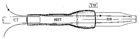
Figure 1. Schematic drawing cf the double-barreled suction electrode (not to
scale).
TW: tungsten wire, CT: capillary tube mounting shaft, HST: heat shrink tubing,
DB:
double-barreled capillary tube drawn and forged.
Figure 2. The effect of varying pulse frequency on mean number of avian embryo
cells
transfected. Six, one-second trains were administered and pulse length was
adjusted
to provide equal power output. Electrodes were approximately the same size and
2-5
embryos were analysed per condition. Bars indicate standard errors.
Figure 3. The effect of train number and polarity on mean number of avian
embryo
cells transfected. Trains were one second long and alternating train polarity
is
indicated by 3+3. All tests were performed at 500 pulses per second with a
pulse
duration of 1 ms. Electrodes were approximately the same size and 2-5 embryos
were
analysed per condition. Bars indicate standard errors.
Figure 4. Whole mount chicken embryos showing GFP-expressing cells 48 hours
post-electroporation. (A) Left side of head of embryo in which rhombomeric
neural
crest was targeted. GFP-expressing neural crest cells have migrated ventrally
and are
visible in the first branchial arch (Arrow). Inset shows magnified view of
fluorescent
cells. EY: eye, HE: heart, Bar: 500 um. (B) Confocal laser scanning image of
dorsal
head region of embryo after train polarity alternation. Arrows indicate barrel
locations.
Bar: 200 ,um. (C) GFP expression in cells comprising half the lens vesicle of
an
embryo in which presumptive lens epithelium was electroporated. Bar: 50 ,um.
(D)
Confocal image of cells within the neural tube expressing GFP after neural
plate was
electroporated. Bar: 25 gym. (E) Confocal image of GFP-expressing cells
migrating
from cranial neural folds. The movement of these cells away from the neural
tube
suggest they are neural crest derived. Bar: 50~m. (F) Confocal image of an
epithelial
cell showing cytoplasmic GFP expression. Bar: 10,um. (G) Depth coding
constructed
from a confocal stack showing epithelial cells and cells up to 15 ~m below the
point
of transfection expressing GFP. EC: epithelial cells, Arrow: deep cell, Bar:
25~m.
7
SUBSTITUTE SHEET (RULE 26)
CA 02395492 2002-06-25
WO 01/48180 PCT/CA00/01561
Figure 5. Fluorescent images of localised electroporation of mouse tail. (A)
Spot 2
days after electroporation (200V). (B) 7 days, the spot is larger and
fluorescence is
beneath the surface. (C) 15 days after electroporation, the cells expressing
GFP
formed a small, well demarked spot at the surface of the skin. Inset shows one
of
several hairs that were brightly fluorescent. (D) After 24 days, the cells
expressing
GFP appear to have been sloughed. (E) Control preparation in which a control
plasmid, incapable of mammalian expression was transfected. (F) Control in
which a
spot was only pierced with the electrode, no plasmid was used and no
electroporation
was done. (G) Spot in which GFP expression plasmid was released into an
incision,
but not electroporated. (H) Avian cells expressing GFP. (I) The same cells
prepared
for immunofluorescence with serum harvested after localised electroporation.
(J)
Control in which cells serum was used with cells not expressing GFP to
determine
background immunoreactivity.
SUBSTITUTE SHEET (RULE 26)
CA 02395492 2002-06-25
WO 01/48180 PCT/CA00/01561
Detailed Description of the Invention
The method of this invention is in principle applicable to all multicellular
organisms and cultures of unicellular organisms, but it is to be understood
that where
species selected are different from those mentioned in the following examples,
an
empirical approach to selection of wave form, number of trains, train
duration, train
polarity, pulse frequency, pulse length, and output voltage is recommended,
preferably
with a view to optimising electroporation while minimising cellular damage. A
variety
of capillary tubes, mounting shafts, conductors, and other components are
suitable for
constructing the inventive apparatus. so the particular choice is not expected
to be
important.
Example 1: Electroporation of avian embryos
An electroporation apparatus, back-filled with DNA containing solution and
driven by a conventional neurophysiological stimulator, was used to transfect
plasmids
containing green-fluorescent protein (GFP) into avian embryos. The
electroporation
apparatus was made from a 1.2 mm x 0.6 mm, 4 inch, double-barrelled glass
capillary
tube (catalog #6070, A-M Systems) which was drawn on a vertical pipet puller
(Model
700C, David Kopf Instruments), broken back to an inside diameter of 200-250
gym, and
forged. Tungsten wire (catalog #7960, A-M Systems). coated with Teflon as an
insulator and with the Teflon removed 5 mm from the ends, was inserted in each
barrel
to within 1 mm of the tip. A capillary tube mounting shaft was attached with
heat-
shrink tubing and sealed with epoxy (Figure 1 ). This apparatus was connected
with
polyethylene tubing to a screw-drive syringe, and was held in a
micromanipulator. The
tungsten wire leads were connected to the poles of a stimulus isolation unit
attached
to a square wave stimulator (Grass S48).
Plasmid (eGFP NI, Clontech) was diluted to 250 ng/ul in 0.85 M NaCI. The
double-barreled capillary tube was backfilled with DNA solution and positioned
in
contact with the surface of the embryo. Fertile chicken eggs (stage 10-13)
were
windowed and the vitelline layer over the target area was carefully reflected
with a fine
9
SUBSTITUTE SHEET (RULE 2G)
CA 02395492 2002-06-25
WO 01/48180 PCT/CA00/01561
tungsten needle (1,5). A seal was formed by applying a small amount of suction
to the
tube with the screw-drive syringe. One-second trains of pulses were delivered
and
pulse frequency, pulse length, train number, and train polarity were varied as
required.
Train length was always one second and a reciprocal relationship was
maintained
between the pulse frequency and pulse length, thus delivering identical total
power at
each setting. The voltage output of the stimulator was adjusted to the maximum
setting that did not cause visible tissue damage. This differed slightly for
each
apparatus, but generally was between 70 and 80 V. Immediately after
administration
of the electroporation trains, the apparatus was backed slightly away from the
embryo,
the suction released, and a small volume of DNA solution was injected over the
embryo. The egg was resealed with adhesive tape and returned to the incubator
for
48 hours. Whole embryos were removed into phosphate buffered saline (PBS) and
the amnion was removed. GFP-expressing cells were visualised using either an
epifluorescent compound microscope (Zeiss) or a confocal laser scanning
microscope
(Zeiss). If fixation was required, 2% paraformaldehyde in PBS for 5 min proved
to be
adequate without inducing auto-fluorescence.
Electroporation produced two scattered patches of GFP-expressing cells at the
point of electroporation. Patch diameter exceeded conductor size but was
approximately proportional to it. The patch associated with the negative pole
of the
electrode means normally contained the majority of GFP-expressing cells;
however,
with alternating train polarities, both patches were similar in size. This is
consistent with
observations of paddle electroporation producing transfected cells on the side
of the
neural tube nearest the positive paddle - in both cases DNA follows a cathode
to
anode path. Early GFP expression was visible less than two hours post-
electroporation, but did not reach maximum intensity until 24 to 48 hours.
Expression
continued until incubation was halted (>8 days), at which point thousands of
transfected cells were present. GFP intensity was slightly lower which is
consistent
with episomal plasmid dilution through mitotic division.
Increasing the number of trains provided an apparent increase in transfection
efficiency (Figure 3), with no decrease in survivorship. Alternating the
polarity between
SUBSTITUTE SHEET (RULE 26)
CA 02395492 2002-06-25
WO 01/48180 PCT/CA00/01561
trains increased the average cumber of cells transfected from 230 to almost
400,
though a total of six trains wa ~ delivered in each case. Six alternating
trains were
significantly more efficient than a single train (P<0.01 ).
Five hundred pulses per second (pulse length 1 ms) produced the optimal
number of transfected cells (Figure 2). Higher and lower frequencies resulted
in
approximately half the number of transfected cells. Transfection rates at the
lowest
frequency (5 pulses per second) with long pulse durations (100 ms) decreased
survivorship and increased the incidence of abnormal embryos. No deformities
were
observed at high pulse frequencies (low pulse duration).
Given the number of variables in electroporation systems, the above parameters
may require modification when the electroporation apparatus is adapted to
different
power sources or situations. For instance, when titrating the voltage, it is
advisable to
find the threshold at which scarring occurs and then decrease output by 10-
20V.
Multiple cell types and various targets were controllably electroporated.
Migration was not impeded and cells proceeded on predicted routes (Figure 4a).
Hundreds of cells could be transfected with large barrel diameters and
alternating train
polarity (Figure 4b). Cells of the lens vesicle (Figure 4c), neuronal cells
(Figure 4d),
neural crest cells (Figure 4e), and epithelial cells (Figure 4f), were
successfully
transfected. Surface epithelial cells and mesenchyme deep to the point of
electroporation expressed GFP (Figure 4g), indicating that direct contact with
electrode
is not necessary.
Example 2: In vivo mammalian electroporation
The electroporation apparatus was made from a 1.2 mm x 0.68 mm, 4 inch,
double-barreled borosilicate glass capillary tube (catalog #6350, A-M Systems)
which
was drawn on a vertical pipet puller (Model 700C, David Kopf Instruments),
broken
back to and ground to a bevel edge with a Narashige EG-4 electrode grinder.
Teflon-
coated tungsten wire (catalog #7960, A-M Systems), with the Teflon removed 5
mm
11
SUBSTITUTE SHEET (RULE 26)
CA 02395492 2002-06-25
WO 01/48180 PCTlCA00/01561
from the ends, was inserted in each barrel to within 1 mm of the tip. A
capillary tube
mounting shaft (catalog #6260, A-M Systems) was attached with heat-shrink
tubing
and sealed with hotmelt glue. The apparatus was held in a micromanipulator and
connected with polyethylene tubing to a screw-drive syringe. The tungsten wire
leads
were connected to the poles of a BioRad Gene Pulser II with RF Module.
Plasmid (eGFP N1, Clontech) was diluted to 250 ng/NI in 0.85 M NaCI. Female
Balb/C mice were restrained in a tube restraining device and their tails taped
to the
base. The tails were swabbed with ethanol and marked with an indelible pen.
The
double-barreled tube was backfilled with DNA solution and the skin of the
mouse's tail
was pierced. A small pool of DNA solution was released and electroporated.
Electroporations consisted of 10, 40 msec bursts at 30 KHz and bursts were 0.2
sec
apart with either 200. 300 or 400 Volts. The train polarity was then reversed
and a
second set of pulses applied. Immediately after administration of the
electroporation
trains, the apparatus was backed slightly away, and a small volume of DNA
solution
was released. Sterile water was used to rinse the electrode to prevent
clogging and
prolong its life. Mice were returned to the cage and examined at intervals
with a Leica
MZ Apo microscope fitted with a GFP module.
Serum was collected 23 days after electroporation and used in an indirect
immunofluorescence assay with cells transfected with a GFP expression vector.
Results and Discussion:
After 48 hours the small incision that was electroporated had a slight green
fluorescence that was distinguishable from the background fluorescence (Fig
1A).
After 1 week the spot that had been electroporated appeared slightly swollen
and
inflamed. With fluorescence microscopy, there was a bright fluorescent region
with
diffuse boundaries beneath the surface of the tail (Fig. 1 B). At 15 days
there was a
small, colourless scab at the point of electroporation. There was a well-
demarked spot
of fluorescence, and in some mice, there were hairs adjacent to the site of
transfection
that were brightly fluorescent throughout their Inrgtfi (Fig 1 C). By 24 days,
the
inflammation had subsided and there was a slightly ~~ink, hairless spot at the
site of
12
SUBSTITUTE SHEET (RULE 26)
CA 02395492 2002-06-25
WO 01/48180 PCT/CA00/01561
transfection.
In control preparations in which a non-expression plasmid was electroporated,
or a sham site in which there was no plasmid and no electroporation, or a site
at which
plasmid was released without electroporation, the site had healed over within
7 days
and no inflammation, nor fluorescence was detected (Figs. 1 E, F, G). In all
of the
voltages attempted there were fluorescent cells, and there did not appear to
be any
differences in the size of the region expressing GFP.
These experiments indicate that localised electroporation can be applied to
mammals. The use of the suction apparatus on the surface of the skin was not
successful, probably because the skin forms an effective barrier to the
aqueous DNA
solution. However, once the skin is broken, a small region of cells at the
point of
electroporation express the GFP reporter gene. The observation of hairs
expressing
GFP suggests that the cells of the hair follicle that proliferate to form the
hair shaft
were transfected in some instances. The size of the fluorescent spot produced
did not
appear to correlate with the voltage used, suggesting all were above the
threshold
required for efficient transfection.
As there appeared to be a localised inflammatory response to the transfection,
the immune response of the mice to the GFP reporter protein was assayed. A
sample
of blood was obtained from a transfected mouse and serum was used in an
indirect
immunofluorescence assay of avian cells transfected with a GFP expression
plasmid.
The cells that express GFP are also immunoreactive to the serum, indicating
the
presence of antibodies to GFP (Fig. 1 H, I). Controls included normal mouse
serum
or cells sham transfected and neither contained cells that were immunoreactive
(Fig.
1 J ).
These experiments indicate that mice can be immunised by localised
electroporation. Cells at the site of electroporation express the transgene
for about 3
weeks, and then appear to be sloughed from the skin. This invention has
considerable
potential for use by researchers. There is saving in time and effort if mice
can be
immunised with DNA encoding a protein of interest. There is no need to express
and
13
SUBSTITUTE SHEET (RULE 26)
CA 02395492 2002-06-25
WO 01/48180 PCT/CA00/01561
purify the protein as an immunogen. The fact that mice express the transgene
for only
a few weeks, means that this method of immunisation could, after safety tests,
be used
for immunisation of livestock, pets and potentially humans. The localised
electroporation apparatus provides a relatively non-invasive means of DNA
immunisation. In addition localised electroporation may also function as a
platform for
gene therapeutics, including for example transient gene therapeutics. There
are
situations where expression of a transgene ectopically in skin cells for a
short period
of time would be useful.
REFERENCES
1. Hamburger, V., & Hamilton, H.L. 1951. A series of normal stages in the
development of the chick embryo. J Morph. 88: 49-92
2. Leber, S.M., Yamagata, M., & Sanes, J.R. 1996. Gene transfer using
replication-defective retroviral and adenoviral vectors. Methods Cell Biol.
51:
161-83
3. Lurquin, P.F. 1997. Gene transfer by electroporation. Mol Biotechnol. 7
(1): 5-
4. Muramatsu, T., Mizuntani, Y., Ohmori, Y., & Okumura, J. 1997. Comparison of
three nonviral transfection methods for foreign gene expression in early
chicken
25 embryos in ovo. Bioc Biophys Res Comm. 23: 376-80
5. Selleck, M. A. 1996. Culture and microsurgical manipulation of the early
avian
embryo. Methods Cell Biol 51: 1-21
14
SUBSTITUTE SHEET (RULE 26)