Note: Descriptions are shown in the official language in which they were submitted.
CA 02397370 2002-08-06
WO 01/58373 PCT/USO1/04235
SURGICAL DEVICES AND METHODS FOR
USE IN TISSUE ABLATION PROCEDURES
TECHNICAL FIELD
The invention generally relates to surgical devices and, more particularly, to
surgical devices and methods for use in procedures performed on moving tissue.
BACKGROUND
Some forms of surgery involve ablation to kill tissue in an organ in order to
achieve a therapeutic result. Ablation can be achieved by various techniques,
including
1o the application of radio frequency energy, lasers, cryogenic probes, and
ultrasound. Thus,
the term "ablation," as used herein refers to any of a variety of methods used
to kill tissue
within an organ. To be successful, ablation treatment may require considerable
precision.
The surgeon must target a particular region, and be careful not to cause
unnecessary
trauma to outer areas of the patient's body near the target area. Just as
important, the
15 surgeon must be confident that the procedure within the target area has
been appropriately
performed. For example, the surgeon may need to determine whether the tissue
has been
ablated to an appropriate degree. The surgery may be made more difficult if
the target
area is moving.
One such surgical procedure in which a surgeon may wish to ablate moving
tissue
2o is an operation to correct an abnormal heartbeat. To function efficiently,
the heart atria
rriust contract before the heart ventricles contract. As blood returns to the
heart and enters
the atria, blood also flows through the atrioventricular (AV) valves and
partially fills the
ventricles. Following an electrical excitation by the sinoatrial (SA) node,
the atria
contract in unison, expelling blood into the ventricles to complete
ventricular filling. The
25 ventricles then become excited and contract in unison. Ventricular
contraction ejects the
blood out of the heart. Blood ejected from the right ventricle enters the
pulmonary
arteries for oxygenation by the lungs, and blood ejected from the left
ventricle enters the
main aorta and is distributed to the rest of the body. If the timing of
cardiac functions is
impaired, such as by the atria not contracting in unison or by the ventricles
contracting
3o prematurely, then the operation of the heart is impaired.
The synchxonization of heart functions is initiated by an excitation from the
SA
node, which is the heart's natural pacemaker. The excitation propagates along
an
-i-
CA 02397370 2002-08-06
WO 01/58373 PCT/USO1/04235
interatrial pathway, extending from the SA node in the right atrium to the
left atrium. The
excitation then spreads across gap junctions throughout the atria, causing the
atria to
contract in unison. The excitation further travels down an internodal pathway
to the AV
node, which transmits the excitation to the ventricles along the bundle of His
and across
the myocardium via the Purkinje fibers. In an aging heart, the atria may
stretch, and the
conduction paths by which the excitations travel may become lengthened. As a
result, the
excitations have a longer distance to travel, and this may affect the timing
of the heart
contxactions and may create an arrhythmia. The term "arrhythmia" is used to
describe
any variation from normal rhythm and sequence of excitation of the heart.
1o One form of arrhythmia is atrial fibrillation. Atrial fibrillation is
characterized by
chaotic and asynchronized atrial cell contractions resulting in little or no
effective blood
pumping into the ventricle. Ventricular contractions are not synchronized with
atrial
contractions, and ventricular beats may come so frequently that the heart has
little time to
fill with blood between beats. Atrial fibrillation may occur if conduction
blocks form
1s within the tissue of the heart, causing the electrical excitations to
degenerate into flurries
of circular wavelets, or "reentry circuits," which interfere with atrial
activity. Initiation or
maintenance of atrial fibrillation may be facilitated if atria become
enlarged. Atrial
enlargement increases the time required for the electrical impulse to travel
across the
atria. This allows sufficient time for the cells that contracted initially to
repolarize and
2o allows the re-entry circuit to be maintained.
One surgical procedure for treating some forms of arrhythmia is to disrupt
conduction paths in the heart tissue by severing the paths at selected regions
of the atrial
myocardium. Selective disruption of the conduction pathways permits impulses
to
propagate from the SA node to activate the atria and the AV node, but prevents
the
2s propagation of aberrant impulses from other anatomic sites in the atria.
Severing may be
accomplished, for example, by incising the full thickness of the myocardial
tissue
followed by closing the incision with sutures. The resultant scar permanently
disrupts the
conduction paths. As an alternative, permanent lesions, in which tissue is
killed, can be
created by ablation. The ablation process involves creating a lesion that
extends from the
3o top surface of the myocardium to the bottom surface (endocardial surface).
Thus, the
purpose of ablation is to create one or more lesions that sever certain paths
for the
excitations while keeping other paths intact. In the case of atrial
fibrillation, for example,
the lesions may interrupt the reentry circuit pathways while leaving other
conduction
pathways open. By altering the paths of conduction, the synchronization of the
atrial
-2-
CA 02397370 2002-08-06
WO 01/58373 PCT/USO1/04235
contractions with the ventricular contractions may be restored. A plurality of
lesions may
be needed to achieve the desired results.
Incision through the myocardium, referred to as the "maze procedure," requires
suturing to restore the integrity of the myocardium, and exposes the patient
to
considerable risk and morbidity. In contrast, thermal or other forms of
ablation can create
effective lesions without the need for sutures or other restorative
procedures.
Consequently, ablation can be performed more quickly and with far less
morbidity. For
these reasons, ablation is becoming a preferred method for severing conduction
paths.
The surgical ablation procedure may be performed during open-heart surgery. In
a
1o typical open-heart surgery, the patient is placed in the supine position.
The surgeon must
then obtain access to the patient's heart. One procedure for obtaining access
is the
median sternotomy, in which the patient's chest is incised and opened.
Thereafter, the
surgeon may employ a rib-spreader to spread the rib cage apart, and may incise
the
pericardial sac to obtain access to the cardiac muscle.
i5 For some forms of open-heart surgery, the patient is placed on
cardiopulmonary
bypass (CPB) and the patient's heart is arrested. CPB is preferred for many
coronary
procedures because the procedure is difficult to perform if the heart
continues to beat.
CPB, however, entails txauma to the patient with attendant side effects and
risks.
In some circumstances, the patient may be treated by a procedure less invasive
2o than the procedure described above. One such less invasive procedure may be
a lateral
thoracotomy. The heart may be accessed through a comparatively small opening
in the
chest and accessed through the ribs. In such a procedure, arrest of the
patient's heart may
not be feasible, and if the heart cannot be arrested, the surgery must be
performed while
the heart continues to beat. Other procedures for access to the heart include
sternotomy,
25 thoracoscopy, transluminal, or combinations thereof.
Once the surgeon has obtained access to the heart, ablation can be carried out
with
a probe that delivers ablative energy. The ablative energy may take the form
of
electromagnetic radiation generated by a laser or radio frequency antenna.
Other
techniques for achieving ablation include the application of ultrasound energy
or very low
so temperature. For the procedure to be successful, the created lesions should
sever the
targeted conduction paths. Typically, the surgeon must create a lesion of a
particular
length to create the desired severance. The surgeon must also create a lesion
of a.
particular depth in order to prevent the electrical impulses from crossing the
lesion. In
particular, when the myocardial tissue is ablated, the lesion must be
transmural, i.e., the
-3-
CA 02397370 2002-08-06
WO 01/58373 PCT/USO1/04235
tissue must be killed in the full thickness of the myocardium to prevent
conduction across
the ablation line.
SUMMARY
The present invention is directed to surgical devices and methods useful in
guiding surgical instruments during procedures on internal organs such as the
heart. The
device may take the form of a surgical "template" device that is attached to
the surface of
an organ. The device can be configured to facilitate surgical procedures such
as tissue
ablation. For example, a surgical template can be used as a guide for travel
of a surgical
or ablative probe along a path to aid a surgeon in ablation of tissue to sever
conduction
1o paths in the heart and thereby alleviate arrhythmia. A surgical template
device may be
especially useful in operations where the organ tissue being treated is
moving, e.g., for so-
called beating heart surgery. The surgical template device may be effective in
providing
local stabilization of the tissue to which the tissue ablation procedure is
directed. The
devices and methods also may find use in procedures in which the pertinent
organ is not
moving.
Alternatively, the device may be configured to provide little or no
stabilization,
but provide guide structure for placement of the ablation probe in the same
frame of
motion as the moving tissue. In some cases, the template may incorporate
hardware that
structurally supports the instrument for travel along the ablation path. The
template
2o devices and methods can be configured for application of other types of
therapeutic
devices, such as diagnostic probes, pacing leads, and drug delivery devices,
to the surface
of a moving organ. To promote adhesion, in some embodiments, the device may be
equipped with a compliant, tacky material that forms a seal for contact with
tissue. The
device also may be equipped with one or more vacuum ports that make use of
vacuum
pressure to enhance the attachment to the organ tissue. Adhesion refers to the
ability of
the device to hold fast to an organ on a temporary basis, either with the
benefit of an
adhesive or vacuum pressure or both. The present invention also is directed to
surgical
devices and methods useful in determining the effectiveness of a tissue
ablation
procedure. In some embodiments, a sensor may be integrated with a surgical
template
3o device as described above to assist the surgeon by making measurements that
gauge
whether the surgical procedure has been satisfactorily performed. For example,
the
CA 02397370 2002-08-06
WO 01/58373 PCT/USO1/04235
surgical device may be configured to measure the effectiveness of an ablation
procedure
in terms of ablation length, depth or width. For example, the sensor may
measure
electrical characteristics of the tissue proximate the target conduction
paths, e.g., tissue
impedance, tissue conduction velocity, or tissue conduction time, as an
indication of the
effectiveness of the procedure. The information obtained by the sensor can be
used as the
basis for feedback to the surgeon, e.g., in audible and/or visible form.
Moreover, the
sensor information can be used as feedback for the closed-loop control of the
tissue
ablation probe. The sensor may be employed independently of a surgical
template
device.
1o As a further aid to the surgeon, the surgical template device may include
indicators such as visible markings that show the targeted length of the
ablation. The
visible markings can be used as a reference by the surgeon during movement of
the
ablation probe within the template area provided by the device. Also, the
template device
may include a structure that physically restricts the length of travel of the
ablation probe,
15 as well as the shape of the path along which the probe travels. In
particular, the length
indicator may include a stop structure that extends into the path for travel
of the ablation
device and is oriented for abutment with the ablation device. In some
embodiments, for
example, the ablation template device may provide a linear path for travel of
the ablation
probe. In other embodiments, however, the template device may define a non-
linear, e.g.,
2o curved, path for travel of the ablation probe.
Further, the present invention is directed to surgical devices and methods for
manipulation of the heart and local stabilization of heart tissue for a tissue
ablation
procedure. In this aspect, the present invention may make use of a surgical
template
device that provides not only a guide for a tissue ablation procedure but also
a structure
25 that provides local stabilization of heart tissue within the operative
area. In some
embodiments, the ablation template device may be accompanied by a surgical
manipulation device that adheres to the heart tissue and enables manipulation
of the heart
to provide the surgeon with a desired access orientation for the procedure.
The
manipulation device may permit lifting, pushing, pulling, or turning of the
pertinent organ
3o to provide the surgeon with better access to a desired area. For both the
template and
manipulation device, to promote adhesion, a compliant, tacky interface
material can be
provided for contact with tissue, along with one or more vacuum ports for use
of vacuum
pressure.
-5-
CA 02397370 2002-08-06
WO 01/58373 PCT/USO1/04235
Tn addition to providing a guide for a procedure, a template device and
associated
methods can be arranged to provide structure that supports instruments such as
ablation
probes, diagnostic probes, pacing leads, and drug delivery devices, for
application to the
surface of a moving organ and active guidance along a path. For some surgical
procedures, it is necessary to bring surgical instruments into contact with
the surface of a
particular organ. In addition to the ablation application described above, one
example is
the placement of one or more electrodes within or in contact with organ tissue
to deliver
electrical impulses to the organ tissue for various purposes, such as a pacing
to control the
beating of the heart. Another example is the placement of a syringe needle to
deliver a
1o medicament to a specific location on an organ. Although all these
procedures could be
performed manually by the surgeon when the body cavity is opened during
surgery, each
is made more difficult when performed via a small opening in the body cavity,
usually
through an endoscopy port. Moreover, such procedures are particularly
complicated
when the surface of the pertinent organ is moving, as with a beating heart.
fs Recently, some types of cardiac surgery have been performed through access
ports
or rather small incisions in the rib cage, instead of in the open field
created by cutting
through the sternum (a sternotomy) and spreading open the rib cage with a
mechanical
device. In these situations, there are occasions when surgical devices
(diagnostic,
therapeutic, etc.) will need to be affixed to a particular location on the
heart surface
2o without direct contact of the human hand. This might also be done while the
heart is still
beating. There is an increasing frequency of coronary artery bypass surgery
done on
beating hearts to avoid the morbidity associated with stopping the heart and
placing the
patient on cardiopulmonary bypass. Some surgeries on the beating heart are
also
performed using the traditional sternotomy. Access procedures such as
sternotomy,
2s thoracotomy, thoracoscopy, and percutaneous transluminal are contemplated.
To facilitate such procedures, a template device is provided to fix a
particular
surgical tool or diagnostic or therapeutic device within a defined operative
path for the
tool or device. There are some surgical procedures performed on a beating
heart, or other
organ, that will require the fixation of a surgical instrument, diagnostic
device or
3o therapeutic device to accomplish a specific surgical procedure, diagnostic
measurement,
or delivery of some therapeutic product or method. This is particularly true
when such
procedures, measurements, or deliveries are performed under minimally invasive
conditions, such as through narrow tubes or ports that penetrate the skin and
enter the
abdominal or thoracic cavities. Template devices and associated methods, in
accordance
-6-
CA 02397370 2002-08-06
WO 01/58373 PCT/USO1/04235
with the present invention, are useful in guiding surgical instruments,
certain diagnostic
sensors, or mechanisms for delivery of medicaments on the surface of internal
organs,
such as the heart.
The template devices and methods are particularly useful in attaching such
instruments to the surface of the beating heart without any additional manual
assistance of
the surgeon, thereby facilitating certain procedures carried out both in open
and
minimally invasive procedures. Notable features of the template device include
conformability to the contours of tha organ, such as the heart, the ability to
fix the device
in place using vacuum, mechanical pressure, or adhesives, and atraumatic
attachment by
1o virtue of specific soft polymeric interfaces and shapes. The template
device can be
configured to attach to various surfaces of the heart using a vacuum seal.
This device
provides two or more vacuum ports surrounded by a conformable, compressible
silicone
gel or elastomer. As in the ablation template, these seals contain integrated
electrodes for
sending and receiving~an electrical signal for the purpose of measuring
impedance or
~5 conductance time or velocity across tissue in a treatment area. The
electrodes may be
surface or interstitial. Also, the electrodes may be multipolar, e.g.,
bipolar. In some
embodiments, a single electrode within the seal may be sufficient with a
reference
electrode located elsewhere. A vacuum port or other fluid removal device may
be
desirable to remove fluids from the chamber to avoid the effects of such
fluids on the
2o electrical performance of the electrodes) or electrical ablation devices.
The ports can be
attached to a single or multiple independent vacuum lines.
In some embodiments of the invention, ablation is performed on the interior
surfaces of the tissues. For example, an ablating instrument may be directed
transluminally, such as by way of a catheter, near the ostia of the pulmonary
veins in the
25 left atrium of the heart. Following the ablation and creation of a lesion,
electrodes
delivered by the catheter may be used to measure the efficacy of the ablation.
For radio frequency ablation, for example, enclosed in the body of the device
can
be a channel in which is located a moveable cable housing a radio frequency
(RF)
antenna for delivery of RF energy to the myocardium. The device allows the RF
antenna
3o to be moved by a remote control unit on the distal end of the cable. The
cable can be
moved through its channel by the controller in response to feedback from the
sensors on
the vacuum seals. As a lesion becomes transmural in one location, the sensors
detect
either decreases in impedance or increases in conduction time. This
information is
processed by the controller, and the RF antenna is moved by a motor that
advances the
CA 02397370 2002-08-06
WO 01/58373 PCT/USO1/04235
cable assembly along a track in the device. Such a device is suitable~for use
in both open
and minimally invasive procedures for the creation of linear transmural
lesions for the
treatment of atrial fibrillation.
Another embodiment is a similar device, which contains malleable metal
elements
that allow the device to be formed into an arc (like a shepherd's crook) whose
circumference can match the outer circumference of the base of the pulmonary
vein.
This device is similar in construction to the embodiment described above,
except that it is
attached to a rod suitable for insertion into a port access device for entry
into the thorax or
for manual manipulation by a surgeon in an open procedure. The device is
brought into
1 o contact with the base of the pulmonary vein, and vacuum is used to attach
it to a portion
of the basal circumference of the vein. RF energy is delivered controllably as
described
above. When a full thickness lesion is created on one side of the vein, the
vacuum is
released, and the device moved so that its arc rests over the side of the vein
that has not
been treated. A full thickness lesion can then be created on that side.
is For some applications, the surgeon may manually control advance of the
radio
frequency antenna within the template device, and control further movement
with a
remote control device. In particular, the surgeon can also utilize manual
movement of the
RF antenna assembly through a joystick or other actuation transducer that
advances the
RF antenna. The joystick is operated by the surgeon in response to an
indicator (light,
2o etc.) that responds to the appropriate decrease in impedance or increase in
conductance
time detected by the sensors mounted in the vacuum seals. As an alternative,
the surgeon
may simply monitor the advance of the radio frequency antenna visually, and
actuate a
joystick or similar device. In either case, the template device operates as
both a guide and
an automated actuator to translate the radio frequency antenna (or other
device) along a
2s desired path. Notably, the template device is affixed to the pertinent
tissue and provides
automated movement of the instrument, reducing motion problems relative to the
instrument offering enhanced precision.
In one embodiment, the present invention provides a surgical device for use in
a
tissue ablation procedure. The device includes a contact member that engages
the tissue
3o near a location where the tissue is to be ablated. The contact member
defines a guide that
indicates, upon engagement of the contact member with the tissue, the location
where the
tissue is to be ablated, and provides a path for travel of a tissue ablation
probe. The
contact member of the device may include a compliant and tacky interface
element for
engagement with the tissue. The device may further define an interior chamber,
and may
_g_
CA 02397370 2002-08-06
WO 01/58373 PCT/USO1/04235
include a vacuum port in fluid communication with the interior chamber. The
interior
chamber may be capable of delivering vacuum pressure to the contact member,
thereby
promoting vacuum-assisted adherence of the contact member to the tissue. In
addition,
the device may include a sensor that may indicate whether the desired degree
of tissue
ablation has been achieved.
In another embodiment, the present invention provides an apparatus for
determining whether conduction paths within heart tissue have been adequately
ablated
during a surgical procedure.. The apparatus includes a first electrode capable
of
transmitting a first electrical signal adjacent the tissue to be ablated, a
second electrode
1o capable of receiving a second electrical signal adjacent the tissue to be
ablated and a
measuring device electrically coupled to at least the second electrode to
receive the
second electrical signal from the second electrode. The measuring deviee may
determine
whether the extent to which the tissue has been ablated to a sufficient degree
based on the
second electrical signal. The apparatus further includes an output device that
provides an
15 indication of extent, e.g., depth, to which the tissue is ablated. In order
to measure
impedance when using RF ablation, it may be necessary to use an energy
frequency
outside of the ablation energy frequency range or pulse or ablation energy and
measure
impedance during the quiescent period between ablation pulses.
In another embodiment, the present invention provides a method for severing
2o conduction paths within tissue. The method involves placing a first device
near the target
conduction paths to be severed, using the first device as a guide to sever the
target
conduction paths, and with a second device, measuring to determine whether the
desired
severing has been achieved. In this embodiment, the target conduction paths
may be
severed by tissue ablation. Measurement may involve determining whether the
lesion
25 depth is sufficient to sever the target conduction paths.
BRIEF DESCRIPTION OF THE DRAWINGS
FIG. 1 is a perspective view of an ablation template device in accordance with
an
embodiment of the present invention placed on a heart for purposes of
illustration.
FIG. 2 is an enlarged perspective view of an ablation template device as shown
in
3o FIG. l, showing use of a surgical instrument.
-9-
CA 02397370 2002-08-06
WO 01/58373 PCT/USO1/04235
FIG. 3A is a top view of an ablation template device in accordance with an
embodiment of the invention.
FIG. 3B is a side view of an ablation template device in accordance with an
embodiment of the invention.
FIG 3C is a cross-sectional side view of the device of FIGS. 3A and 3B.
FIG. 4 is a conceptual diagram illustrating an ablation template device in
accordance with an embodiment of the invention.
FIG. 5 is another conceptual diagram illustrating an ablation template device
in
accordance with an embodiment of the invention.
1o FIG. 6 is a perspective view of an ablation template device in accordance
with an
alternative embodiment of the invention placed on a heart for purposes of
illustration.
FIG. 7 is a top view of an ablation template device in accordance with an
embodiment of the invention.
FIG. 8 is a top view of an ablation template device in accordance with an
~s embodiment of the invention.
FIG. 9A is a perspective top view of an ablation template device in accordance
with an embodiment of the invention.
FIG. 9B is a perspective bottom view of an ablation template device as shown
in
FIG, 9A.
2o FIG. 10 is a perspective view of an ablation template device in accordance
with an
embodiment of the invention.
FIG. I 1 is a perspective view of an ablation template device in accordance
with an
embodiment of the present invention, placed on a heart for purposes of
illustration, used
in cooperation with another device that permits manipulation of the heart.
25 FIG. 12 is a cross-sectional side view of a cup-like manipulation device.
FIG. 13 is a cross-section side view of another cup-like manipulation device.
FIG. 14 is a perspective view of an ablation template device incorporating
structure for accommodating an ablation probe;
FIG. 15 is a cross-sectional view of the device of FIG. 14, taken at point
145.
3o FIG. 16 is a cross-sectional view of a shaft incorporated in the device of
FIG. 14,
taken at point B.
FIG. 17 is a perspective view of an arcuate ablation template device
incorporating
structure for accommodating an ablation probe.
- l0-
CA 02397370 2002-08-06
WO 01/58373 PCT/USO1/04235
FIG. 18 is a perspective view of an added ablation template device
incorporating
structure for accommodating an ablation probe.
FIG. 19 is a cross-sectional view of the device of FIG. 18, taken along line
210-
210'.
FIG. 20 is a bottom view of the device of FIG. 18.
FIG. 21 is a perspective view of an ablation template device incorporating a
movable carnage for support of an ablation probe.
FIG. 22 is a cross-sectional view of the device of FIG. 21, taken along line
250-
250'.
1o FIG. 23 is a cross-sectional view of the device of FIG. 21, taken along
line 244-
244'.
FIG. 24 is a cross-sectional front view of an ablation template device having
an
internal ablation probe.
FIG. 25 is a cross-sectional side view of the ablation template device of FIG.
24.
FIG. 26 is a cross-sectional side view of a catheter-mounted ablation device.
FIG. 27 is a side view of a catheter-mounted ablation device.
FIG. 28 is a side view of a catheter-mounted ablation device.
FIG. 29 is a cross-sectional side view of a catheter-mounted ablation device.
FIG. 30 is a side view of a catheter-mounted ablation device.
2o DETAILED DESCRIPTION
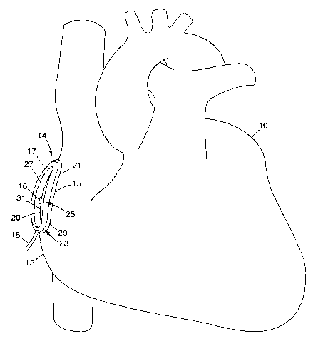
FIG. 1 is a perspective view of an ablation template device 14 in accordance
with
an embodiment of the present invention. In FIG. 1, ablation template device 14
is shown
placed on a heart 10 for purposes of illustration. In particular, heart 10 has
been exposed
by an open-chest surgical technique and ablation template device 14 has been
affixed to
the right atrium 12 of the heart. In some embodiments, ablation template
device 14
includes a contact member 17 that engages the tissue. In the example of FIG.
1, contact
member 17 takes the form of a substantially ovular ring. Inner and outer
diameters 20, 21
of the ring-like contact member 17 define an annular chamber for engagement
with tissue
on the surface of heart 10.
so Contact member 17 may be affixed to the surface 15 of atrium 12 in many
ways,
such as by application of an adhesive at the inner and outer diameters 20, 21,
or by
-11-
CA 02397370 2002-08-06
WO 01/58373 PCT/USO1/04235
application of vacuum pressure to the annular chamber. Another way to achieve
adherence between contact member I7 and the surface tissue 15 is to include a
seal
member 23 formed from an adhesive material in the contact member. One example
of an
adhesive material is a coating of compliant, tacky material, such as silicone
gel, at the
interface between the contact member 17 and the tissue on the surface 15 of
atrium 12. In
this case, contact member 17 may include a semi-rigid frame member 25 and a
compliant,
tacky seal member. The compliant, tacky seal member 23 provides intrinsic
adhesive
properties, and aids conformability and sealing to surface 15, while the frame
25 imparts
structural integrity to contact member 17. Each of frame 25 and seal member 23
has a
1o substantially annular shape. In particular, seal member 23 may include
inner and outer
portions 27, 29 disposed at the inner and outer diameters 20, 21 of contact
member 17.
With a silicone gel, intrinsic adherence of seal member 23 may be
sufficient that ablation template device 14 remains affixed to the heart 10 in
spite of
contractions of atrium 12 and in spite of the use of device 14 in surgical
procedures
described below. Nevertheless, application of vacuum pressure will be
desirable in many
applications to provide secure adherence. Although the adherence should be
secure, the
adherence preferably is not permanent. Rather, adherence between device 14 and
the
tissue may be discontinued as desired without serious trauma to the tissue,
and the device
repositioned and adhered anew at a different location. As an alternative,
ablation
2o template device 14 can be forced against atrium 12 to provide pressure
contact with heart
10. In such a case, ablation template device 14 may have a local stabilizing
effect on the
contact region of heart 10 despite continued beating of the heart. Ablation
template
device I4 may be sized or shaped to allow it to mold to the contours of the
atrium 12.
Ablation template device 14 can be made principally of nonconductive
materials, such as
polyurethane, silicone, or natural or synthetic rubber. Shore A 50-80 silicone
elastomer
may be used, for example, to form frame 25 of device 14. Metal such as
annealed
stainless steel or zinc or polymeric reinforcing members may be incorporated
in device
14, e.g., embedded within the molded elastomer, to resist excessive
deformation or
collapse during use. Shape memory alloys, in particular, may be useful in
imparting a
3o desired shape to device 14 during use, and permit collapse and unfolding to
the desired
position for endoscopic deployment in minimally invasive techniques.
An electrode I6 can be affixed to device 14, e.g., within seal member 23 or
frame
member 25, and placed in contact with the surface 15 of the heart 10. The
electrode 16
may send signals across the tissue of the heart 10 to be received by a second
electrode
-12-
CA 02397370 2002-08-06
WO 01/58373 PCT/USO1/04235
(not shown in FIG. 1). These signals will traverse the tissue area being
ablated. The
associated circuitry for the electrodes may reach device 14 by way of a
connective tube
18. As will be described, electrode 16 may form part of a sensor for
determining the
effectiveness of a tissue ablation procedure. In particular, the electrodes
can be used to
measure electrical properties (such as impedance, phase angle, conduction
time,
conduction velocity, capacitance) of the local tissue area being ablated, and
thereby
indicate whether an effective lesion has been formed in the tissue. In some
embodiments,
ablation template device I4 may have multiple sets of electrodes situated at
different
positions along the major axis of the device. In this case, such electrodes
may take the
1o same types of measurements at different positions, or different types of
measurements
such as impedance, conduction velocity, and conduction time.
If ablation template device 14 is attached with the assistance of vacuum
pressure,
connective tube 18 may also serve the purpose of attaching the interior
chamber formed
by contact member 17 to an external source of vacuum pressure (not shown).
Ablation
is template device 14 may be shaped to define an interior chamber that is
enclosed upon
engagement of the device with the tissue. In the example of FIG. 1, the
chamber is
substantially annular. Application of vacuum pressure. may cause the enclosed
chamber
to slightly deform, creating a vacuum seal and causing the device 14 to become
more
affixed to the tissue. With added compliance from seal member 23, in
particular, contact
2o member 17 can conform to tissue surface 15 to achieve an effective seal. At
the same
time, the compliant seal member 23 distributes sealing force across the tissue
to reduce
tissue trauma.
As shown in FIG, l, contact member 17 of ablation template device 14 generally
may have a somewhat annular shape, with substantially oval-shaped inner and
outer
2s diameters, and an opening 31 through which the tissue of atrium 12 may be
accessed.
The lengths of the major and minor axes of annular-shaped device 14 may vary
to provide
opening 31 with varying sizes according to the characteristics of the
particular procedure
to be performed. In some applications, opening 31 may define a narrow, linear
track for
travel of an ablation probe. In other applications, opening 31 may be much
wider or
3o define nonlinear tracks for travel of an ablation probe. Other shapes for
contact member
17 beside the annular shape may also be suitable.
A closer perspective view of ablation template device 14 appears in FIG. 2. In
FIG. 2, a surgeon's fingers 24 hold a surgical instrument shown as an ablation
probe 22
that may be used to ablate the tissue of the heart 10. Even though the heart
10 is beating,
-13-
CA 02397370 2002-08-06
WO 01/58373 PCT/USO1/04235
the surgeon 24 may position the probe 22 within the opening 31 with relative
ease. The
surgeon 24 may also use the probe 22 to ablate a particular area of the atrium
12, even
though the atrium 12 is in the process of contracting and relaxing, by using
the inside
edge 26 of the device 14 as a guide for travel of the probe. Again, opening 31
may define
a substantially linear path for travel of an ablation probe. Alternatively,
opening 31 can
be non-linear, e.g., curved, or have other shapes appropriate for given
surgical
applications. In either case, the surgeon may use opening 31 as a guide, even
resting the
ablation probe 22 against the inside edge 26 of contact member 17 in some
cases.
Because significant heat may be generated by RF, laser, and ultrasonic energy,
it may be
1o desirable to provide ablation probe 22 with a thermally insulative sleeve
that extends
downward to the tip of the probe, thereby protecting the inside edge 26 of
contact
member 17. Also, inner edge 26 of contact member 17 can be coated with or
coupled to
an insulative material for contact with ablation probe 22.
If ablation template device 14 is fixed to a point of reference, it may
provide a
15 Local stabilizing effect that holds the tissue within opening 31
substantially stationary, or
at least constrains the local area against excessive movement, despite
continued beating
of heart 10. For example, ablation template device 14 may be pushed against
heart 10 to
apply stabilizing pressure to the local area of contact. Alternatively,
ablation template
device 14 can make use of suction or adherence in combination with either a
pushing or
2o pulling force to provide a stabilizing effect.
Ablation probe 22 may use a number of methods to achieve ablation. The probe
22 may, for example, use a laser to ablate tissue. As another alternative, the
probe may
incorporate an antenna that emits radio frequency (RF) energy to ablate
tissue. The
amount of power delivered by the ablation probe may vary. A typical RF probe,
for
2s example, may deliver from 5 to 50 watts. In this alternative, the probe 22
may include an
electrode at its tip. An electrode can be provided within ablation template
device 14 to
provide circuit completion for a probe using RF energy. For example, a passive
electrode
forming part of the sensor described above could be used as the return
electrode. As a
further alternative, probe 22 could take the form of an ultrasound probe that
emits
3o ultrasound energy, or a cryosurgical probe that cools the tissue to ultra-
low temperatures.
Thermal, chemical, and mechanical probes for obtaining or incising tissue are
also
contemplated. In each case, opening 31 of ablation template device 14 provides
a guide
for travel of probe 22, enabling greater precision in the ablation of
conduction paths
within the heart tissue.
- 14-
CA 02397370 2002-08-06
WO 01/58373 PCT/USO1/04235
Other views of ablation template device 14 appear in FIGS. 3A and 3B. In these
views, the device is shown in a top view, FIG. 3A, and a side view, FIG. 3B.
FIG 3C is a
cross-sectional side view of the device of FIGS. 3A and 3B. Inner seal member
27 is
indicated by dashed line 33. The interior chamber of contact member 17 is
indicated by
reference numeral 35. Ablation template device 14 may be flexible, and its
relaxed shape
may be curved as shown in FIG. 3B to more readily conform to the surface of
the heart.
The exemplary annular shape allows first electrode 16 and second electrode 30
to be
located opposite to each other across the opening 31. The distance between the
electrodes
16, 30 may be a known, fixed distance. The interior edges 26, 32 of the
opening 31
1 o preferably have sufficient rigidity to serve as a guide for travel of a
probe or other
surgical instrument. Although seal member 23 may be substantially compliant
and
conformable, the inner edge of frame member 25 may provide the degree of
rigidity
desirable to support the probe. In addition, ablation template device 14 may
include one
or several length indicators in the form of visible markings 28, to assist the
surgeon in
forming a lesion of a desired length.
A surgeon desiring to make a lesion of a particular length may use the
markings
28 as a guide for manipulating the probe. Thus, the guide provided by opening
31 is
useful in guiding both the direction of travel of the probe and the extent of
travel. Also,
the template device 14 may include a structure that physically restricts the
length of travel
of the ablation probe, as well as the shape of the path along which the probe
travels.
Substantially straight ablation tracks ordinarily will be desirable.
Accordingly, the guide
surface on the interior of the opening may be substantially straight. In other
applications,
however, it may be desirable to effect a curved ablation track. Therefore, the
shape of the
guide within opening 31 may vary according to the application. Furthermore,
because
ablation typically causes a change in tissue color, the markings 28 may
provide the
surgeon with information as to the actual length of the lesion.
In one aspect, the invention can be useful in determining whether the
conduction
path has indeed been cut. Ordinarily, a surgeon cannot visually gauge the
depth of a
lesion. The guide defined by ablation template device 14 may provide an
indication of
3o the length of a lesion. A lesion of an insufficient depth may result in
currents that pass
under or over the lesion, however, and may thus be incapable of disrupting the
reentry
circuits or other undesirable current pathways. The myocardium consists of
interlaced
bundles of cardiac muscle fibers. Within the fibers, cardiac muscle cells are
joined by
intercalated discs, which include areas of low electrical resistance known as
gap
-15-
CA 02397370 2002-08-06
WO 01/58373 PCT/USO1/04235
junctions. Gap junctions permit excitations or action potentials to propagate
from one
cell to another. A lesion created by ablation may destroy the tissue and the
gap junctions,
effectively interrupting electrical conduction. Thus, determination of whether
the
conduction paths are indeed ablated may be crucial to a successful treatment.
As shown in FIGS. 3A and 3B, ablation template device 14 may include at least
two electrodes, 16, 30 that operate as part of a sensor. A sensor may be used
to indicate
to the surgeon whether a desired degree of tissue ablation has been achieved.
Electrodes
16, 30 preferably are integrated with ablation template device 14 to reduce
the number of
instruments that need to be introduced in to the surgical field. In
particular, electrodes 16,
1o 30 can be molded into the material forming seal member 23 or frame member
25, and
have conducting members that extend away from the tissue site via tube 1 ~. A
tip portion
of each electrode may be exposed beyond the surface of seal member 23 to
enable
sufficient electrical contact with the tissue to which contact member 17 is
attached.
In other embodiments, however, electrodes 16, 30 may be introduced
i5 independently of ablation template device 14. FIGS. 3A and 3B show an
exemplary
embodiment of the present invention, and other embodiments may incorporate
more than
two electrodes. After an ablation is performed inside the opening 31, and
during ablation,
electrodes 16 and 30 may be located on opposite sides of the lesion. The
distance
between electrodes 16 and 30 may be a known distance and relatively fixed. The
2o electrodes 16, 30 may be used to determine whether the conduction path has
been severed
by ablation to the desired degree.
One way to make the determination is to use the electrodes 16, 30 as probes
for an
impedance-measuring instrument. Electrodes 16, 30 may be electrically coupled
to the
impedance-measuring instrument. The impedance of the area of tissue may be
measured
25 before any ablation is made, and this measurement may be used as a
baseline. The
impedance may be measured again after the ablation is made and may be compared
with
the baseline measurement to determine whether the conduction path has been
severed.
Moreover, it may be desirable to measure impedance during an,ablation
procedure to
assess progress in producing an effective lesion. During ablation, impedance
measured
3o from one side of the lesion to the other side will decrease as ablation
ruptures cell
membranes, permitting dissolved ions to move with less restriction. Impedance
will
generally decrease until impedance reaches a minimum value when the lesion
becomes
transmural. One way to determine whether the ablation is complete is to look
for the
point at which the impedance measurement levels off. For example, a baseline
- 16-
CA 02397370 2002-08-06
WO 01/58373 PCT/USO1/04235
measurement on canine atrial myocardium may show an impedance of 240 ohms, but
measurements taken during the ablation may show a steady decline in impedance,
eventually leveling off at 150 ohms after about 90 seconds. It may also be
possible in
some circumstances to evaluate the ablation process on the basis of a
percentage change
of impedance or on the basis that a predetermined impedance value has been
reached.
Parameters such as the baseline value, the leveling off value and the time
needed to
produce a transmural lesion are dependent upon the patient being treated, the
tissue being
ablated, the distance of the electrodes, the thickness of the tissue, and
other factors. In the
case of the heart, for example, not all hearts have the same impedance, and
different
1o sections of a single heart may also have varying impedance. In such cases a
baseline
measurement may be desirable, with transmural penetration indicated by the
leveling off
of impedance measurements.
In addition to measuring impedance or as an alternative to measuring
impedance,
alternating current (ac) phase angle may be measured. In a capacitive circuit,
the voltage
15 lags the current, and the amount of lag is often expressed in the form of a
phase angle. In
a purely capacitive circuit, the voltage is 90° behind the current,
expressed as a phase
angle of -90°. A phase angle of 0° means the circuit is purely
resistive. A phase angle
between 0° and -90° means the circuit is partly resistive and
partly capacitive. Typically a
phase angle measurement across tissue will be between 0° and -
90°, indicating some
2o capacitive nature of the tissue. As ablation proceeds, cell membranes are
ruptured,
making the tissue less capacitive. Accordingly, the phase angle across the
ablative lesion
will become more positive (i.e., will approach zero) as cells die in the
lesion. One way to
determine whether the ablation is complete is to look for the point at which
the phase
angle measurement levels off. A baseline measurement of canine myocardium, for
25 example, may show a phase angle of -13.1°. Measurements taken during
the ablation
may show the phase angle becoming more positive, eventually leveling off at -
12° after
about 20 seconds. As with impedance measurements, phase angle measurements are
dependent upon many factors.
Another way to make the determination is to use the electrodes to.measure
3o conduction distance by measuring conduction time. A signal traveling on a
conduction
path propagates as an action potential and propagates via gap junctions. The
length of a
conduction path, the speed of conduction and the time taken for a signal to
travel the path
are related by the simple formula
D=RT
17-
CA 02397370 2002-08-06
WO 01/58373 PCT/USO1/04235
where D is the distance traveled by the signal, R is the rate of speed of the
signal, and T is
the time taken for the signal to travel the distance. In the case of an actual
operation, a
particular value of D or T may be desired. A value for R may be obtained by
sending a
test signal from one electrode, receiving it at the other electrode, the
distance between the
electrodes being known and relatively fixed, and measuring the time of
conduction. In
many cases, however, a relative measure of conductive velocity or time is
sufficient, and
therefore the distance between electrodes need not be known absolutely so long
as it
remains fixed. This measurement may then be used as a baseline measurement.
Again, a
baseline measurement may be desirable, because not all hearts have the same
conduction
to speed, and different sections of a single heart may also have varying
conduction speeds.
The time of conduction may be measured again after the ablation is made and
may be
compared with the desired value of D or T. In general, conduction time
increases and
conduction velocity decreases as the ablation proceeds, and one way to
determine
whether the ablation is complete is to look for the point at which the
measured quantity
levels off. For example, a conduction time of 15 ms may be measured as a
baseline.
During ablation, conduction time may increase, eventually leveling off at
around 30 ms.
The leveling off indicates the ablation is transmural.
In the case of measurement of conduction time, velocity, or distance,
electrode 30
may be a single electrode or a bipolar or multipolar electrode. Thus, in the
description of
2o this invention, it is to be understood that the transmitting electrode 16
positioned on one
side of the ablation track may be unipolar, while the measurement or
"recording"
electrode 30 positioned on the opposite side of the ablation track can be
unipolar, bipolar,
or multipolar, depending upon the electrical measurement that is utilized to
determine if
the conduction paths have been severed or ablation of the target tissue has
been
transmural, and desired precision. With a unipolar recording electrode 16, an
electrical
signal transmitted into the tissue by the transmitting electrode is first
sensed as an
electrical signal that is then followed by a depolarization wavefront that
propagates
through the cells disposed between electrodes 16, 30. It is the depolarization
wavefront
that is detected to measure conduction time.
3o A unipolar recording electrode 30 simply measures whether the
depolarization
wavefront exceeds a given threshold. With a bipolar recording electrode 30,
however, the
two electrodes can be used to measure current flow or a voltage potential
between them.
The two electrodes of the bipolar recording electrode 30 can be oriented in a
line
substantially parallel to the ablation track, and thereby form a "T" with the
transmitting
- 18-
CA 02397370 2002-08-06
WO 01/58373 PCT/USO1/04235
electrode 16. As the depolarization wavefront propagates through the cells
positioned
between transmitting electrode 16 and recording electrode 30, the cells
disposed between
two recording electrodes of bipolar recording electrode 30 depolarize,
producing a
difference in current flow between the two recording electrodes. This bipolar
'
arrangement enables measurement of an increase in the intensity of current
flow between
the two electrodes of bipolar recording electrode 30, and more precision in
the
measurement. In particular, an intensity threshold can be set. Conduction time
can be
measured between the time at which transmitting electrode 16 transmits the
initial signal
and the time at which current flow between the two electrodes of bipolar
recording
1o electrode 30 exceeds the threshold. Again, the initial signal transmitted
by transmitting
electrode 16 and sensed by the recording electrode 30 can be ignored. Rather,
the
depolarization wavefront typically will be the event of interest in
determining conduction
time.
A method of using measurement of impedance or conductance variables to'
~5 determine the transmurality of a lesion may also be employed using bipolar
radio
frequency electrosurgical ablation devices. For example, separate electrodes,
using an
electrical frequency different from the frequency used by the ablation device,
can be
mounted on the device and used to form a separate measuring circuit for
impedance for
the purpose of measuring the distance ablated. A typical bipolar device could
have two
2o electrode surfaces, one for one side of a tissue surface and one for the
other side of a
planar tissue surface, such as the myocardium, or a vascular structure. One
transmitting
electrode, or a plurality of electrodes, can be mounted with one of the
surgical electrodes,
and a receiving or "recording" electrode, which could be bipolar or
multipolar, or a
plurality of unipolar, bipolar, or multipolar electrodes, can be mounted on
the opposite
2s surgical electrode. Impedance or conductance, such as time, distance, or
velocity, can be
measured as described herein and can be used to determine transmurality, and
shut off
power to the ablation device as described. It is envisioned that one specific
application of
such a bipolar device would be for deployment through a puncture hole in the
myocardium. The ablation device could be equipped with "jaws" that carry the
3o electrodes. Entry of one of the "jaws" of the surgical RF device could be
either from the
endocardial or epicardial surfaces. After deployment, there would be a
surgical electrode
on both the epicardial surface and the endocardial surface. As RF power is
supplied to
the surgical ablation device, the tissue between the two surgical electrodes
is heated and
killed, creating a lesion for the purpose of interrupting conductance
pathways. The
- 19-
CA 02397370 2002-08-06
WO 01/58373 PCT/USO1/04235
transmurality of this lesion at different points along its length can be
measured
simultaneously or at time intervals during ablation using measurement of
impedance or
conductance variables with the separate circuits defined by the transmitting
and recording
electrodes placed along the path of the surgical electrodes and the underlying
lesion.
FIG. 4 shows a conceptual diagram of an implementation of an aspect of the
invention. Electrodes 16, 30 shown in FIG. 3 may serve as probes 34 for a
measurement
device 36. The measurement device 36 may measure a quantity related to
conduction,
such as impedance or conduction time or conduction velocity. Data measured by
measurement device 36 may be fed into a processor 38. Processor 38 may be in
the form
~o of a generalized computing device, such as a personal computer.
Alternatively, processor
38 may be in the form of a smaller and more specialized computing device, such
as a
microprocessor or an application-specific integrated circuit. As a further
alternative,
processor 38 could be realized by discrete logic circuitry configured
appropriately to
perform the necessary measurement control and processing functions.
Accordingly,
15 processor 38 need not be embodied by integrated circuitry, so long as it
capable of
functioning as described herein.
In addition, processor 38 may take an active role in the measurement process
and
may control measurements made by measurement device 36 through probes 34. In
particular, processor 38 may control a current or voltage source to apply
electrical current
20 or voltage to one of electrodes 16, 30. Two representative instances where
the processor
38 may actively control the measurement process are in the taking of a
baseline
measurement, and in the taking of periodic measurements during the ablation
procedure
to monitor progress. Processor 38 may further perform calculations as needed,
and may
provide output to the surgeon by way of an output device 40 such as a display.
In
2s addition, processor 38 may receive input from an additional input device
42, which may
include, for example, a keyboard or a touch screen. Using input device 42, the
surgeon
may, for example, input the length of a desired lesion, and the processor 38
may be able
to provide feedback to the surgeon via output device 40 as to whether the
desired lesion
has been created. Output device 40 may provide audible and/or visible output
such as
3o beeps, flashing light emitting diodes (LED's), speech output, display
graphics, and the
like, to provide feedback to the surgeon. Output device 40 can be mounted in a
housing
associated with processor 38, or integrated with the ablation probe 22. For
example, one
or more LED's could be mounted on the ablation probe in view of the surgeon.
- 20 -
CA 02397370 2002-08-06
WO 01/58373 PCT/USO1/04235
FIG. 5 shows another conceptual block diagram of an implementation of an
aspect
of the invention. FIG. 5 is similar to FIG. 4, except that the processor 38 is
connected to
the ablation device 44. Ablation device 44 may be any device intended to sever
conduction paths by killing tissue, such as the RF, laser, ultrasonic, or
cryogenic probe 22
depicted in FIG. 2. In each case, ablation device 44 may be in the form of a
powered
instrument such as a laser, RF, or ultrasonic electrosurgical probe, or be
coupled to a
cryogenic supply. Processor 38 may control ablation device 44 by, for example,
cutting
off power or supply to the ablation device once the desired lesion has been
created. In
this manner, the surgeon can take advantage of closed-loop, real-time control
of the
1o output of ablation device 44, ensuring ablation to a proper level of
effectiveness and
avoiding excessive ablation. The result may be the creation of an effective
lesion in a
shorter time period, reducing the time necessary for access to the patient's
heart tissue.
The system may be even more effective if multiple electrode pairs are mounted
along
opening 31 to measure the effectiveness of ablation in creating a lesion along
a
continuous track.
The system shown in FIG. 5 may be useful for dynamic monitoring and control of
the surgical procedure. The surgeon may choose an ablation device 44, such as
a laser,
that will not interfere with the operation of the probes 34. Alternatively, if
interference is
created by an RF probe, power can be intermittently turned off to enable
measurement.
2o By any combination of taking a baseline measurement or receiving input
through input
device 42, the processor 38 may determine what measurements received from
measurement device 36 will satisfy the conditions for a successful surgical
procedure.
Processor 38 may continuously or frequently monitor the measurements received
from
measurement device 36 to determine whether the criteria for a successful
surgical
procedure have been met. When those criteria have been met, processor 38 may
cut off
power to, or otherwise interrupt the operation of, ablation device 44. In
other words,
processor 38 may use a feedback system as part of its control of ablation
device 44 for
either automated control or manual control by the surgeon.
One advantage of this system is the speed by which the surgeon may perform the
3o ablation procedure. Speed is of a considerable advantage to the patient in
several
respects. First, risks attendant to surgery may be minimized if the time spent
on the
operating table is reduced. Second, a procedure performed on moving tissue
such as a
beating heart may be more efficient if done quickly.
-21-
CA 02397370 2002-08-06
WO 01/58373 PCT/USO1/04235
Once ablation template device 14 is placed into position, a baseline
measurement
may be taken, and the surgeon may then proceed to make the ablation, using
ablation
template device 14 as a template or a guide. Use of the device 14 as a
template or guide
is one factor enhancing the speed of the procedure. The surgeon may use
markings 28 on
ablation template device 14 to get a general idea of where to begin and end
the ablation.
The processor 38 may be used to suggest to the surgeon via output device 40
suitable
markings 28 for beginning and ending the ablation pass. The surgeon may then
make a
pass with the ablation device 44. If the pass is too long, the processor 38
may interrupt
the function of the ablation device 44 before the pass is completed. If the
pass is too
1o short, the processor 38 may assist the surgeon in determining the best
approach for a
second pass. Again, the length determination may be aided by the use of a
series of
electrode pairs along an ablation track. The use of dynamic processing and
feedback
further enhance the speed of the procedure. FIG. 6 is a perspective view of an
ablation
template device 50 in accordance with an alternative embodiment of the present
invention. Like ablation template device 14 in FIG. 1, ablation template
device 50 is
shown placed on the right atrium 12 of a heart 10 in FIG. 6 for purposes of
illustration. In
particular, heart 10 has been exposed and ablation template device 50 has been
affixed to
the right atrium 12 of the heart. Ablation template device 50 includes a
contact member
51 which may engage and may be affixed to the surface 15 of atrium 12 by being
pushed
2o against the heart. Because ablation template device 50 generally has a U-
shaped shape,
contact member 51 includes two contact tines or contact "feet" 53.
Electrodes used to take the measurements described herein may take the form of
discrete electrodes that operate in pairs to transmit and receive signals
across the ablated
tissue region. Alternatively, one or more of the electrodes may take the form
of bipolar or
mufti-polar electrodes that are integrated in a common electrode package and
positioned
in very close proximity to one another. With the closer spacing available in a
bipolar
package, for example, the signal transmitted by one electrode and received by
the other as
an EMG potential can be cleaner in terms of having a reduced degree of
background
noise due to surrounding electrical potentials produced by the heart. Instead,
the bipolar
so electrode is capable of more effectively measuring the local signal
conduction time.
Also, in some embodiments, series of electrodes on each side of the ablation
track can be
realized by a continuous electrode component that includes conductive
electrode regions
and insulating regions disposed therebetween. Again, this sort of component
can permit
closer electrode spacing. In this case, however, the closer spacing is not
between
-22-
CA 02397370 2002-08-06
WO 01/58373 PCT/USO1/04235
transmitting and receiving electrodes but between adjacent transmitting
electrodes and
adjacent receiving electrodes extending parallel to the ablation track. The
closer spacing
permits a higher degree of resolution in monitoring the progress of the
ablation procedure
along the ablation track, and thus the length of the resulting lesion. The
closer spacing
permits more precise feedback and control of the ablation probe by the surgeon
or by an
automated controller.
To maintain its position relative to the heart 10, ablation template device 50
may,
in addition, have a compliant, tacky material such as silicone gel at the
point of contact
between contact member 51 and the surface 15 of the atrium 12, providing a
compliant,
1o tacky interface. Ablation template device 50 may remain substantially
affixed to the heart
in spite of contractions of atrium 12 and in spite of the use of ablation
template device
SO in surgical procedures described such as those described above. By being
forced
against the heart, ablation template device 50 may have a stabilizing effect
on the contact
region of heart 10 despite continued beating of the heart. Shaft 52, made of a
rigid
i5 material and formed in any suitable shape, may be used to press ablation
template device
50 against atrium 12 and hold the device in place.
Although ablation template device 50 may be more rigid than ablation template
device 14 in FIG. 1, ablation template device 50 may be sized or shaped to
allow it to
mold to the contours of the atrium 12. Like ablation template device 14 in
FIG. 1,
2o ablation template device SO can be made (with the exception of the
compliant, tacky
interface) principally of substantially rigid, nonconductive materials, and
may include a
first electrode 56 and a second electrode (not shown in FIG. 6). The
associated circuitry
for the electrodes may reach ablation template device 50 by way of shaft 52.
The general
U-shape of ablation template device 50 includes an opening 54 through which
the tissue
25 of atrium 12 is accessible. The dimensions of ablation template device 50
and opening 54
may vary. Other shapes beside the U-shape may also be suitable for the device
50, such
as the annular shape, and the opening 54 may be in other suitable shapes as
well.
A top view of ablation template device SO appears in FIG. 7. The exemplary U-
shape allows first electrode 56 and second electrode 58 to be located opposite
to each
3o other across the opening 54. The distance between the electrodes 56, 58 may
be a known,
fixed distance. The interior edges 60, 62 of the opening 54 have sufficient
rigidity to
serve as a guide for travel of a probe or other surgical instrument. In
addition, like
ablation template device 14, ablation template device 50 may include several
length
indicators 64, to assist the surgeon in forming a lesion of a desired length.
-23-
CA 02397370 2002-08-06
WO 01/58373 PCT/USO1/04235
A top view of a variation of ablation template device SO appears in FIG. 8.
Ablation template device 50 is like the same device depicted in FIG. 7, except
the first
electrode 56 and second electrode 58 are not rigidly affixed to the body of
the device 50.
Electrodes 56, 58 are electrically coupled to ablation template device 50 by
way of
electrical connectors 66, 68. Electrical connectors 66, 68 may be flexible
wires, and may
allow a surgeon to place electrodes 56, 58 at a desired location on the tissue
or at a
desired distance apart. Alternatively, electrical connectors 66, 68 may be
spring-like
connectors, that may appear somewhat like insect antennae, and which may force
the
electrodes 56, 58 against the tissue when the ablation template device 50 is
pressed
1o against the tissue to enhance electrical coupling pressure and surface
area. As shown in
FIG. 8, electrodes 56, 58 may be deployed within the opening 54. Electrodes
56, 58 may
also be deployed at other locations as well.
FIGS. 9A and 9B show an ablation template device 69, which is similar to the
ablation template device 14 shown in FIG. 1. However, FIGS. 9A and 9B
illustrates a
frame member 7S and a seal member 77 in somewhat greater detail. FIG. 9A is a
perspective top view of device 69, while FIG. 9B is a perspective bottom view
of device
69. FIGS. 9A and 9B differ slightly in the shape of device 69. Specifically,
device 69 of
FIG. 9A is shown as having a somewhat curved contour for conformability to the
surface
of the tissue.
2o Frame member 75 can be formed from a semi-rigid material that lends
structural
integrity to contact member 73, while seal member 77 is formed from a more
compliant
material that facilitates conformance of the contact member to the tissue
surface and
promotes a seal that is generally atraumatic and more effective. Seal member
77 includes
an inner skirt-like member 70 coupled to and extending around the inner edge
of contact
member 73 that acts as an interface with the tissue. Skirt-like member 70 may
function in
part as a seal gasket. Ablation template device 69 also includes an outer
skirt-like
member 72, coupled to and extending around the outer edge of the contact
member 73.
Skirt-like members 70, 72 define annular vacuum chamber 76. Inside of skirt-
like
member 70, contact member 73 defines opening 81 for access to a tissue site.
Skirt-like
3o members 70, 72 may be composed of a material that is generally more
compliant and
conformable than the rest of contact member 73.
Use of Shore A 5-10 durometer silicone elastomer for the skirt-like member 70,
72 may be appropriate for some applications. Silicone gels are preferred,
however, due to
the intrinsic compliance and tackiness provided by such materials. Like
silicone
-24-
CA 02397370 2002-08-06
WO 01/58373 PCT/USO1/04235
elastomers, silicone gels can be manufactured with a range of crosslink
densities.
Silicone gels, however, do not contain reinforcing filler and therefore have a
much higher
degree of malleability and conformability to desired surfaces. As a result,
the compliance
and tackiness of silicone gel materials can be exploited in skirt-like members
70, 72 to
provide a more effective seal. An example of one suitable silicone gel
material is MED
6340, connmercially available from NUSIL Silicone Technologies, of
Carpinteria,
California. The MED 6340 silicone gel is tacky and exhibits a penetration
characteristic
such that a 19.5 gram shaft with a 6.35 mm diameter has been observed to
penetrate the
gel approximately S mm in approximately S seconds. This penetration
characteristic is
1o not a requirement, but merely representative of that exhibited by the
commercially
available MED 6340 material.
Metal or polymeric reinforcing tabs can be incorporated in skirt-like members
70,
72 to prevent collapse, and promote structural integrity for a robust seal.
Skirt-like
members 70, 72 can be compliant, tacky silicone gel molded about the
reinforcing tabs.
In particular, for manufacture, frame member 7S can be molded about
reinforcing tabs or
springs, allowing a portion of the tabs or springs to extend downward, to one
or both of
the inner diameter or outer diameter side'of the annular contact member. Then,
one or
both skirt-like members 70, 72 can be molded onto frame member 7S, encasing
the
exposed portions of the tabs or springs. In the example of FIG. 9, outer skirt-
like member
20 72 and the outer diameter side of frame member 7S are molded about and
encase a
continuous spring member, shown partially in FIG. 9 and indicated by reference
numeral
79. Spring member 79 can be shaped from a continuous length or one or more
segments
of spring steel, or other materials capable of exerting a spring bias on
contact member 73.
When ablation template device 69 is placed in contact with tissue, skirt-like
25 members 70, 72 may promote adherence between the tissue and the device.
Furthermore,
ablation template device 69 may include a vacuum port 74. When vacuum pressure
is
supplied by connective tube 71 to vacuum port 74, skirt-like members 70, 72
may
promote the creation of a seal, further enhancing the adherence of device 69
to the tissue.
Upon application of vacuum pressure, skirt-like members 70, 72 may deform
slightly,
3o conforming to the surface of the tissue and helping define a sealed vacuum
chamber 76
having a substantially annular shape. Skirt-like members 70, 72 may therefore
improve
adherence to the tissue in two ways: by being tacky and compliant, and by
assisting the
creation of a vacuum seal. Silicone gels, such as NuSil 6340, may be
especially well
-25-
CA 02397370 2002-08-06
WO 01/58373 PCT/USO1/04235
suited for this function, providing a quality of adherence and compressibility
appropriate
for the intended purposes.
FIG. 10 shows a perspective view of an ablation template device 80, which is
similar to ablation template device 50 shown in FIG. 6. The contact member 82
of the
device 80 has been supplied with a thin layer of a compliant, tacky substance
84 such as a
silicone gel. When ablation template device 80 is held by pressure against
tissue using
shaft 86, tacky layer 84 may provide added adherence between the device and
the tissue,
and may reduce the risk of slippage. The tacky material may be included at
every point
of contact between the tissue and contact member 82, or at selected sites of
contact.
1o FIG. 11 is a perspective view of an ablation template device 100, shown
placed on
a heart 10 for purposes of illustration. Ablation template device 100 is like
ablation
template device 69 shown in FIG. 9. Contact member 102 has been placed against
the
surface 15 of the right atrium 12. Inner skirt-like member 104, extending
around the
inner edge of contact member 102, and outer skirt-like member I06, extending
around the
15 outer edge of contact member 102, assist in substantially affixing device
100 to the heart
10. Vacuum pressure supplied to vacuum port 108 via connecting tube I10 may
promote
additional adherence between contact member 102 and heart surface 15.
It may be difficult for a surgeon to obtain direct access to the tissue of the
atrium
12 where ablation is to be performed. It may be necessary for the surgeon to
manipulate
20 or move the heart so that access may be obtained. FIG. l I illustrates the
use of a surgical
manipulating device 120, whereby the apex 122 of the heart 10 is held and
manipulated,
allowing the surgeon to obtain access to the desired site on the atrium 12. It
is known that
some significant portion of the aberrant impulses responsible for atrial
fibrillation can
originate in myocardial cells that have migrated to the inner base of the
pulmonary veins.
25 Accordingly, it is important that ablation lines be drawn in such a way as
to isolate the
pulmonary veins and prevent those impulses from traveling into the atrial
tissue.
Accomplishing this isolation requires that the ablation lines be drawn
relatively close to
the base of the pulmonary veins.
The use of surgical manipulating device 120 and similar devices described
herein
3o enables the surgeon to grasp the apex 122 of the beating or stopped heart
10 and access
the base of the pulmonary veins, e.g., by lifting, pulling, and/or turning the
beating heart
to expose the pulmonary veins. Important additional benefits of device 120 and
similar
devices described herein may include the ability to lift and manipulate the
heart 10
without causing significant trauma to the epicardium and with minimal or no
disturbance
-26-
CA 02397370 2002-08-06
WO 01/58373 PCT/USO1/04235
of hemodynamics, reducing the overall risk of the procedure to the patient.
The rigid
handle 127 on device 120 permits the surgeon to apply axial (i.e., along axis
from top of
heart to apex) tension to the beating heart while lifting the heart 10 from
the pericardial
cavity. Maintaining axial tension while lifting the heart from the supine
position to a
position 90-110 degrees from the spine prevents distortion of valves and the
decline in
cardiac output that occurs when the heart is lifted by the surgeon's hand
alone.
In some embodiments, two suction devices, e.g., like surgical manipulating
device
120, can be used to access the posterior of the heart and the base of the
pulmonary veins.
One device may be applied to the apex of the heart and the second device may
be applied
1o to a suitable location on the anterior surface of the heart, such as the
area between the
right and left ventricles (interventricular groove). Both devices can then be
manually
manipulated in concert so that the heart can be raised to a vertical position,
i.e., close to
90 degrees from its ordinary anatomic orientation, without distorting the axis
that runs
from the apex to the great vessels. In addition, manual manipulation of both
devices
simultaneously permits the surgeon to move the raised heart from left to right
inside the
thoracic cavity. The use of the second device on the anterior surface of the
heart keeps
the chambers and valves in the heart from being compressed or distorted, and
permits
elevation and rotation of the heart without compromising blood flow. No
decline in
blood pressure (measured just below the aortic arch with an intravascular
transducer) is
observed when these manipulations are performed with the two devices used in
concert.
The two devices (each of which may conform substantially to device 120) can
also be
secured by a suitable clamp or frame that is anchored to the operating table
or the chest
retractor.
Manipulating device 120, as shown in FIG. 11, may define a cup-like chamber
123 having a vacuum port 125 coupled to a vacuum tube 127. Chamber 123 can be
formed from a cup frame 121 formed with semi-rigid material and a compliant,
tacky
skirt-like member 129. Vacuum tube 127 may be coupled to an external vacuum
source
for delivery of vacuum pressure to the interior of chamber 123.
Compliant, tacky skirt-like member 129 can be formed, for example, from
silicone
3o gel, and can be attached to an outer wall defined by chamber 123 to provide
a sealing
interface with tissue at apex I22 of heart 10. Skirt member 129 can be molded,
cast,
deposited or otherwise formed about the wall of chamber 123, or adhesively
bonded to
the chamber wall. Although the tackiness of skirt member 129 promotes
adherence,
adherence may be improved by application of the vacuum pressure via tube 127
and port
- 27 -
CA 02397370 2002-08-06
WO 01/58373 PCT/USO1/04235
125. Upon application of vacuum pressure, at least a portion of the seal
member 129
deforms and substantially forms a seal against the surface
The semi-rigid chamber 123 imparts structural integrity to the device 120,
while
the tacky, deformable material forming the skirt-like member 129 provides a
seal
interface with the heart tissue that is both adherent and adaptive to the
contour of the
heart. Moreover, as the skirt-like member 129 deforms, it produces an
increased surface
area for contact with the heart tissue. The increased surface area provides a
greater
overall contact area for adherence, and distributes the coupling force of the
vacuum
pressure over a larger tissue area to reduce tissue trauma. In general, the
structure of
1o device 120 can be helpful in avoiding ischemia, hematoma or other trauma to
the heart
10. Device 120 provides a grasping point, however, for manipulation of heart
10 to
provide better access to a desired surgical site, e.g., by lifting, turning,
pulling, pushing,
and the like. Once the desired presentation of heart 10 is achieved using
device 120, the
heart can be held relatively stationary, e.g., by fixing vacuum tube 127 to a
more
stationary object such as a rib spreader. Device 120 and similar devices
described herein
can be used to stabilize the heart in a similar manner by grasping the apex
and/or other
suitable locations on the heart, such as the anterior interventricular groove,
and attaching
the device to a stationary object. In this manner, it is possible to use one
or more devices
such as device 120 and similar embodiments in concert with the various
embodiments of
2o tissue ablation templates described herein placed at a variety of suitable
locations on the
heart to create a relatively stable epicardial surface for ablation. Such
stabilization allows
the surgeon to complete the manual ablation or other surgical procedures more
easily and
more quickly than without stabilization. For example, using a first device 120
on a
suitable ventricular surface and a second device 120 on the apex permits the
surgeon to
elevate the heart and stabilize it to permit ablation with an ablation
template on the
posterior side of the heart. Addition of a flexible joint between vacuum tube
127 and
member 121 may allow the heart to maintain its normal movement resulting from
contraction further reducing trauma to the heart.
In some embodiments, device 120 and an ablation template device as described
3o herein may be appropriately miniaturized to permit deployment via port-
access methods,
such as small thoracotomies. An ablation template device as described herein
also could
be appropriately miniaturized for application on the endocardial surface of
the heart, e.g.,
using transluminal approaches. For endocardial application, an ablation probe
such as an
RF antenna can be integrated with the ablation template device, which could be
made
-28-
CA 02397370 2002-08-06
WO 01/58373 PCT/USO1/04235
substantially flexible but incorporate shape memory elements or elasticity to
expand
following transluminal deployment.
In alternative embodiments, no external vacuum pressure need be applied.
Instead, as shown in the cross-sectional side view of FIG. 12, a device 120'
can be
s configured to incorporate a mechanical structure that permits variation of
the volume
within the chamber 123', e.g., by actuation of a piston-like member or
modulation of a
fluid chamber. For example, a shaft 130 can be mounted within chamber 123'
substantially where vacuum port 125 and vacuum tube 127 are located in FIG.
11. A
distal end 131 of the shaft 130 is positioned to engage a flexible membrane
132 within
1 o chamber 123'. An attachment pad can be placed between distal end 131 of
shaft 130 and
flexible membrane 132 to permit adhesive or thermal attachment. Upon actuation
of the
shaft 130, the membrane 132 can be moved inward and outward relative to the
interior of
chamber 123', and thereby change the volume and, as a result, pressure within
the
chamber 123'.
15 As an illustration, upon engagement of seal member 129 with heart 10, shaft
130
and cup 121 are pushed onto heart surface 15. Retracting shaft 130 draws
membrane 132
and heart surface 15 into the chamber defined by cup 121. Upon release of
shaft 130,
elasticity of membrane 132 biases the membrane and shaft 130 back to their
original
positions, increasing the volume and decreasing the pressure within chamber
123'. As a
2o result, chamber 123' produces a suction effect without application of
external negative
pressure that enhances the seal provided by the tacky skirt-like member 129.
Thus, the
shaft 130 and membrane 132 can be used to create a negative pressure within
chamber
123' that serves to aid adhesion of the tacky skirt-like gasket member 129 to
apex 122
(shown in FIG. 11). FIG. 12 also illustrates internal attachment of skirt-like
member 129
2s with cup frame 121. In particular, as shown in FIG. 12, skirt-like member
129 can be
molded about the outer lip 133 of cup frame 121. Also, an insert 135 formed
from a
metal or polymeric material can be embedded within cup frame 121 and skirt-
like
member 129 to provide added structural integrity to device 120'.
FIG. 13 illustrates another embodiment of a device 120' incorporating a limpet-
30 like structure. In the example of FIG. 13, instead of a shaft 130 as shown
in FIG. 12,
chamber 123 receives a fluid tube 134 at port 125. Fluid tube 134 permits
inflow and
outflow of fluid 136 into the internal cavity 138 defined by membrane 132 and
the inner
wall 140 of chamber 123. In this case, internal cavity 138 can be normally
filled with a
fluid 136 such as saline. When fluid is drawn from device 120 through fluid
tube 134,
-29-
CA 02397370 2002-08-06
WO 01/58373 PCT/USO1/04235
membrane 132 is drawn toward port 125, decreasing the volume of the portion
138 of
chamber I23 that engages heart 10. In this manner, pressure within chamber 123
is
reduced, creating a suction effect that aids the sealing pressure of skirt-
like member 129
at apex 122. A stopping mechanism such as a valve or stopcock (not shown) may
be
employed to stop the flow of fluid through fluid tube 134, and thereby fixing
the sealing
pressure.
FIG. 14 depicts a device 141 that permits attachment of an antenna for
delivery of
radio frequency (RF) energy to the surface of a heart for the purpose .of
creating a linear
lesion of dead tissue that is transmural. FIG. 15 shows a cross section at
point 145 on
1 o device 140 of FIG. 14. The body 147 of the device 140 can be made of a
suitable flexible
polymeric material such as silicone elastomer. A shaft 142, made of either a
rigid or
flexible material, depending upon application, can be used to position the
device 140 in
either an open or minimally invasive surgical procedure. The diameter of shaft
142
would be sized differently for each of these applications. In the example of
FIGS. 14 and
15 15, shaft 142 also contains a moveable inner catheter 143 that contains the
RF antenna
and, if appropriate, a fluid delivery lumen 148. In addition to the catheter
143, shaft 142
can provide a vacuum connection to device 140, which may define one or more
inner
chambers. The device 140 can be attached to the heart using two vacuum ports
144, 146
connected to one or more seal members 149, 151. Vacuum pressure can be
provided to
2o ports 144, 146 via tubes 150, 152, which are coupled to an external vacuum
source and
branch off from shaft 142.
The body I47 of device 140 can be molded to define two vacuum chambers 154,
156 and a central lumen 158, which opens to a base side 160 of the device and
forms a
continuous track for accommodation of catheter 143. Malleable metal shafts
162, 163,
25 164 can be inserted into the body 147 to provide shaping capability and
added structural
integrity, but may not be necessary to achieve compatibility with all desired
contours and
positions on the heart. Vacuum pressure delivered through vacuum chambers 154,
156
via vacuum ports 144, 146 is used to attach the device 140 to the heart.
Flexible seal
members 166, 168, and 170, 172 are disposed adjacent each vacuum chamber 154,
156,
ao respectively, and conform to the surface of the heart and function as seals
149, 151. Seal
members 166, 168, 170, 172 can be made of silicone elastomers as soft as 5 on
the Shore
A scale, or can be made of silicone gel. A suitable silicone elastomer
material may have
a durometer, for example, in the range of 5 to 30 Shore A. An example of one
suitable
silicone gel material is MED 6340, commercially available from NUSIL Silicone
- 30 -
CA 02397370 2002-08-06
WO 01/58373 PCT/USO1/04235
Technologies, of Carpinteria, California. The MED 6340 silicone gel is tacky
and
exhibits a penetration characteristic such that a 19.5 gram shaft with a 6.35
mm diameter
has been observed to penetrate the gel approximately 5 mm in approximately 5
seconds.
This penetration characteristic is not a requirement, but merely
representative of that
exhibited by the commercially available MED 6340 material. These materials can
conform to the irregular shape of the myocardium under negative pressure
created by the
vacuum source and, if formed from silicone gel, may provide tackiness that
aids the seal.
The seal members 166, 168, 170, 172 can be partially shaped and stiffened, if
necessary by fins 174, 176, 178, 180, respectively, placed at different
intervals along the
length of the seal members. These fins can be made of flexible metal or can be
part of the
material forming body 147 of device 140 and integrally molded therewith. Seal
members
166, 168, 170, 172 and associated vacuum chambers 154, 156 may extend along
the
length of body 147, like central lumen 158, to define elongated tracks. Upon
application
of vacuum pressure to vacuum ports 144, 146, vacuum chambers 154, 156 serve to
hold
device 140 tightly against the surface of the heart. Device 140 may be sized
and
structured to provide a local stabilizing effect on the tissue to which the
device is
attached, e.g., for beating heart surgical applications. In many embodiments,
however,
stabilization will not be necessary. Rather, it is sufficient that device 140
fix a surgical
instrument, e.g., RF antenna 141, in the same frame of motion as the moving
tissue. In
2o this manner, an instrument can be applied with precision to the surface of
the heart
without significant relative motion.
In the central lumen 158 is inserted catheter 143, which, in the example of
FIGS.
14 and 15, contains RF antenna 141. Antenna 141 may, itself, enclose fluid
delivery
lumen 148. RF antenna 141 is shown in FIGS. 14 and 15 at the end of catheter
143,
where the antenna emerges at an angle to the catheter and protrudes through
the track
defined by central lumen 158 of device 140. By sliding catheter 143 along the
track
defined by lumen 158, the tip 182 of antenna 141 can move along the track and
deliver
energy to the tissue with which it is in contact, creating a lesion that can
extend the full
thickness of the myocardium. An RF antenna is one example of an ablation probe
so suitable for use with device 140 to ablate tissue. Other ablation
instruments could be
placed in catheter 143, however, including laser, ultrasonic, and cryogenic
probes, all, all
of which could create a lesion in a similar fashion.
In some embodiments, catheter 143 can be moved through lumen 158 either
manually by a surgeon by grasping the proximal end of the catheter or by a
mechanical
-31-
CA 02397370 2002-08-06
WO 01/58373 PCT/USO1/04235
device connected to the catheter, e.g., at its distal end. For example, a
variety of electrical
motors could be used to drive catheter 143 along central lumen 158, e.g.,
directly via a
worm gear drive or indirectly via pulley or gear arrangements. The motors can
be driven
either automatically, or at the direction of the surgeon using a joystick or
other manual
controls. Electrodes 184, 186 can be mounted on an inner surface of the
innermost seal
members 168, 170 for contact with the myocardium. Electrodes 184, 186 are
connected
to conductors 188, 190, respectively, which extend out of device body 147 and
continue
into shaft 142. Electrode 184 and conductor 188 on one side of the device 140
can be
used to send an electric signal across the lesion area formed by antenna 141
for detection
1o on the other side of the device by another electrode 186 and conductor 190.
FIG. 16 is a cross section at point B on shaft 142 of FIG. 14. Conductors 188,
190
can be connected via a cable 192 to appropriate instrumentation. Such
conductor/electrode sets can be used to measure impedance across the lesion or
conduction velocity across the lesion. These measurements can be used to
determine if
15 the lesion is truly transmural, that it extends the full thickness of the
myocardium.
Conductors 188, 190 can be ultimately connected to an external control unit
which is
capable of using impedance or conductance time or velocity measurements to
generate
either a signal observable by the surgeon or a signal for control of a device
responsible for
advancing catheter 143 along central lumen 1S8 when a transmural lesion has
been
2o created in one region. To that end, a plurality of electrodes 184, 186 can
be placed on
respective sides of central lumen 1S8 to take measurements at several
positions along the
length of the lesion track, thereby driving controlled advancement of catheter
143 as an
effective lesion is formed at each position. Again, advancement of catheter
143 can be
automated or manual. In either case the surgeon can be assured during the
procedure that
25 an effective lesion has been formed.
As shown in FIG. 16, outer shaft 142 may contain two separate lumens 194, 196,
which provide vacuum pressure to chambers 154, 1S6 via tubes 150, 152. FIG. 16
also
shows a cable with a wiring bundle including conductors 188, 190, for
electrical
communication with electrodes 184, 186 (FIG. 1S). The number of conductors may
be
3o dependent upon the number of electrodes placed on each side of the inner
sealing
members 168, 170. For example, each electrode 184, 186 preferably is coupled
to an
individual conductor 188, 190, respectively. Alternatively, a single
continuous electrode
could be disposed on one side of central lumen 1S8 and coupled to a single
conductor. In
this case, a series of electrodes at various positions on one side of central
lumen 1S8
- 32 -
CA 02397370 2002-08-06
WO 01/58373 PCT/USO1/04235
would transmit signals to the continuous electrode on the other side or vice
versa.
Catheter 143 fits in the central lumen 158 of shaft 142 and, in this example,
contains RF
antenna I41 and fluid lumen 148. Again, other embodiments could have different
types
of ablation probes built into catheter 143.
FIG. 17 shows a specialized form of a device 140' as shown in FIG. 14. In this
embodiment, the device body 147' is shaped in a substantially semicircular
form to
facilitate contact around the base of the pulmonary vein or similar structure.
Device body
147' is moved into position via shaft 142' and vacuum is used to affix it to
its first
location on the vein. In this case, a catheter is translated around the
arcuate path defined
1o by a central lumen. The catheter carries an RF antenna or other ablation
probe that is
exposed via opening for contact with the outer wall of the pulmonary vein.
Lesion
generation is earned out on the full thickness of the vein wall in one
location by
energization of the RF antenna or activation of other suitable probe. As shown
in FIG.
17, vacuum pressure can be applied via vacuum chambers 154', 156' with seal
members
15 166', 168', 170', 172' providing an effective seal. When vacuum pressure is
released,
device 140'can be moved via shaft 142' to another location to create a lesion
continuous
with the previous one until a circumferential lesion is created all the way
around the base
of the pulmonary vein. As in the example of FIGS. 14-16, device 140 can be
fixed in the
same frame of motion as the pulinonary vein, eliminating significant relative
motion to
2o enhance precision in creation of the lesion. The interior of device 140' is
identical to that
of device 140 as shown in FIG. 15, with two modifications. The malleable metal
inserts
162, 164 are replaced with shaped memory metal inserts, which cause 140' to
assume an
arcuate shape shown in FIG. 17. Malleable insert 163 is replaced with a semi-
rigid metal
rod which can be withdrawn through shaft 142' to allow elements 162, 164 to
assume
25 their arcuate shape and cause device 140' to also assume an arcuate shape.
Insertion of
the semi-rigid rod causes device 140' to straighten into a linear shape that
would permit
device 140' to entry into or withdraw from a tubular access port used in
minimally
invasive surgical procedures.
Although device I40 is depicted as having a "shepherd's crook" shape, that
shape
3o is merely an exemplary embodiment of the invention. The ablative device may
take other
forms such as a loop, hook, ess or snare. In any of these configurations,
electrode sets
may be placed on the device so as to have a one or more transmitting
electrodes on one
side of the lesion and one or more receiving electrodes on the opposite side
of the lesion
to measure the effectiveness of the ablation.
-33-
CA 02397370 2002-08-06
WO 01/58373 PCT/USO1/04235
FIGS. 18-20 illustrate another embodiment of an ablation template device 200.
FIG. 18 is a perspective side view of device 200. FIG. 19 is a cross-sectional
side view of
device 200 taken at line 210-210' in FIG. 18. FIG. 20 is a bottom view of
device 200. As
shown in FIGS. 18-20, device 200 includes a ring-like contact member 202
defining an
annular but generally oval-shaped chamber 204. Contact member 202 may include
a
frame 204 formed from a semi-rigid material, and seal members 206, 208 formed
at the
inner and outer diameters of frame 204. Seal members 206, 208 can be formed,
for
example, from a silicone gel material. A vacuum tube 212 is mounted in a
vacuum port
214 that communicates with an interior chamber 216 defined by frame 204 and
seal
1 o members 206, 208. A cover 218 can be mounted within the central aperture
220 defined
by frame 204, or integrally formed with the frame, e.g., by molding. Cover 218
includes
a slot-like track 222 that extends along the major axis of contact member 202.
Track 222
accommodates an ablation probe 224.
Ablation probe 224 may take the form of an RF, laser, ultrasonic, or cryogenic
probe, and includes upper and lower flanges 226, 228 that hold the probe
within track. In
particular, upper flange 226 bears on an upper surface of cover 218 adjacent
track 222,
while lower flange 228 bears on a lower surface of the cover. Ablation probe
224 is
slidable along track 222, however, to define a lesion path for an ablation
procedure. In
particular, a surgeon can simply slide ablation probe 224 along track 222.
Electrodes
230, 232 on opposite sides of track 222 can be electrically coupled to
electronics that
provide measurements, e.g., impedance, conduction velocity, and conduction
time, to
assess the effectiveness of the ablation procedure. In response to indications
provided
based on the electrode measurements, the surgeon advances ablation probe 224
along
track 222. Alternatively, ablation probe 224 can be advanced automatically
along track
222 in response to such indications. In some embodiments, tip 234 of ablation
probe 224
may contact tissue.
FIGS. 2I-23 illustrate another ablation template device 240. FIG. 21 is a
partial
perspective view of device 240. FIG. 22 is a partial cross-sectional side view
of device
240 of FIG. 21 taken at line 242-242'. FIG. 23 is a cross-sectional front view
of device
240 of FIG. 21 taken at line 244-244'. As shown in FIGS. 21-23, device 240
includes a
contact member 246 mounted on an elongated guide member 248 that extends
through
bore 249. Contact member 246 may be slidable along guide member 248 or fixed.
The
contact member includes a frame 250 formed of a flexible material, and a seal
member
252 formed from a compliant, tacky material such as silicone gel. The seal
member 252
- 34 -
CA 02397370 2002-08-06
WO 01/58373 PCT/USO1/04235
interfaces with tissue, e.g., on the surface of the heart. Frame 250 further
defines one or
more rails 254 that extend radially outward relative to contact member 246 and
longitudinally relative to guide member 248. A carnage 256 is mounted on rails
254,
e.g., via inner grooves that engage the rails, and defines a lateral flange
258 designed to
hold an ablation probe 260. As shown in FIGS. 21 and 23, in particular,
ablation probe
260 protrudes downward from lateral flange 258 for contact with organ tissue.
Ablation probe 260 can be molded into or otherwise encased in lateral flange
258
of carriage 256. A second lateral flange 262 (FIG. 23) can be provided, along
with a
counter probe 264, to contact tissue and thereby balance device 240 on a side
of carriage
1o 256 opposite lateral flange 258. Ablation probe 260 may take the form of an
RF, laser,
ultrasonic, or cryogenic probe designed to ablate tissue. Ablation probe 260
may have
electric conductors that run along the length of guide member 248 to an
external power
supply, in the case of an RF or ultrasonic probe. Alternatively, an optical
fiber or fiber
bundle may be coupled between ablation probe 260 and an external source of
laser
energy. As a further alternative, a fluid line may extend between ablation
stylus and a
cryogenic source. In each case, device 240 can be sized and arranged to permit
deployment by endoscopic or other minimally invasive techniques to an ablation
site, e.g.,
on the surface of the heart. Thus, in one application, device 240 can be
deployed and
affixed to the surface of a beating heart, and fix the ablation probe 260 in
the same frame
of motion as the heart.
Seal member 252 may define a plurality of vacuum ports 266 coincident with
vacuum ports in guide member 248. A vacuum tube resides within an inner lumen
270 of
guide member 248 and includes one or more output ports that apply vacuum
pressure to
vacuum ports 266. To perform an ablation procedure, device 240 is deployed to
a desired
site on the surface of an organ such as the heart. Vacuum pressure is applied
to affix
contact member 246 to the tissue surface via the seal interface provided by
seal member
252. At the same time, ablation probe 260 is brought in contact with the
tissue surface.
Ablation probe 260 is then energized to ablate the local tissue area proximate
the tip of
the probe. A guide wire or other elongated member can be coupled to carriage
256,
3o which preferably is slidable along rails 254 defined by contact member 252.
By
translating the guide wire, carriage 256 can .be moved relative to contact
member 252 and
thus relative to the tissue surface, thereby creating an ablation track. As in
other
embodiments, electrodes can be integrated with seal member 252 to measure the
extent of
-35-
CA 02397370 2002-08-06
WO 01/58373 PCT/USO1/04235
ablation. Again, the measurements can be used as the basis for manual or
automated
control of the guide wire, and resulting movement of carriage 256.
FIGS. 24 and 25 illustrate another ablation template device 272. FIG. 24 is a
cross-sectional front view of device 272, while FIG. 25 is a fragmentary cross-
sectional
side view. Device 272 is somewhat similar to device 240 of FIGS. 21-23.
However,
device 272 need not incorporate a carnage. Rather, device 272 provides an
internal
optical waveguide 274 mounted within a guide member 276 that transmits laser
radiation.
Waveguide 274 may be housed in a cannula 278. Waveguide 274 may incorporate a
reflector 280 at its distal end 282 that reflects laser energy downward
through a chamber
1o defined by seal member 284 to ablate tissue. Seal member 284 may be
substantially
compliant and tacky and may be attached to a semi-rigid frame 286 that is
coupled to or
integrated with guide member 276. Cannula 278 and waveguide 274 preferably are
movable along the length of guide member 276, as indicated by arrow 288.
Optical
waveguide 274 can be mounted within an outer vacuum lumen 290 that delivers
vacuum
pressure to affix device 272 to the tissue 292 via seal member 284. To form an
ablation
track, optical waveguide 274 can be translated within guide member 276, as
indicated by
arrow 288. Once again, electrodes can be integrated with seal member to enable
manual
or automated control of waveguide movement.
Ablation, and measurement of impedance or conduction time to assess ablation
lesion depth, can also be performed along the interior surfaces of a
structure. For
example, a linear RF electrode can be transluminally introduced via a catheter
into the
atria of the heart and positioned on the endocardium in appropriate locations.
Ablative
energy from the RF electrode can then be applied. Electrode sets used to
measure
impedance or conduction time or other electrical properties can be integrated
into the
catheter body parallel to but insulated from the active RF electrode at the
distal end of the
catheter. These electrode sets can be utilized as described above to both
measure lesion
depth (from the endocardial to the epicardial surface) and to control delivery
of energy.
Transluminal introduction, therefore, represents an additional way to create a
lesion around the base of the pulmonary veins, and thereby treat atrial
fibrillation. The
lesion may be created on the interior surfaces of the heart or pulmonary
veins, rather than
the heart's or veins' exterior surfaces. The treatment entails ablating the
endocardial
tissue near the ostia of the pulmonary veins in the left atrium. Typically the
ablation
apparatus is delivered to the site on the distal end of a steerable catheter
introduced into
-36-
CA 02397370 2002-08-06
WO 01/58373 PCT/USO1/04235
the atrium or the pulmonary veins, and is manipulated and controlled at the
proximal end
of the catheter.
FIG. 26 is a side view of an apparatus that may be directed transluminally
near the
ostia of the pulmonary veins in the left atrium. The device of FIG. 26 may
conform
substantially to the device shown in U.S. Patent No. 5,938,660 to Swartz et
al. In the
example of FIG. 26, however, the device has been adapted in accordance with
the present
invention to incorporate components for measurement of ablation depth or
effectiveness.
In particular, electrodes have been positioned on the device so as to come
into contact
with tissue on opposing sides of a lesion created by the ablative components.
1o FIG. 26 depicts a distal end of a catheter body 300, with balloons 302, 304
on the
catheter body 300 shown inflated. Fluid medium introduced through catheter
lumen 306
at the proximal end emerges at the distal end through openings 308, thus
inflating the
balloons 302, 304. Inflation causes balloons 302, 304 to lodge against the
tissue.
Catheter 300 may include a tip electrode 310 for sensing electrical activity.
Catheter 300
15 may also include RF electrode 312, which performs the actual ablation.
After balloons
302, 304 are inflated, ablation may be accomplished by introducing a
conductive media
through catheter 300, which emerges at the distal end through openings 318.
Application
of RF energy follows, and the tissue between the balloons 302, 304 is ablated.
Electrodes 314, 316 are mounted on the surface of the balloons 302, 304 at the
2o circumference of the balloons. Electrodes 314, 316 are insulatively
separated from RF
electrode 312 and tip electrode 310. Electrodes 314, 316 may be uni-polar or
mufti-polar.
Connecting leads 320 and 322 are coupled to electrodes 314 and 316
respectively. Leads
320, 322 may be wires or conductors printed on the surface of balloons, or a
combination
of both. Leads 320, 322 travel from electrodes 314, 316 toward proximal end of
catheter
25 300, and emerge from proximal end of catheter where leads are electrically
coupled to a
measuring device such as an impedance meter or conduction time measuring
device.
Following measurements that show a successful ablation, the conductive media
may be
withdrawn, balloons 302, 304 may be deflated, and the catheter may be
extracted.
Many variations are possible. For example, a plurality of electrodes can be
3o mounted on the surface of balloons 302, 304. Flexible disks or other
extendable members
could be used in place of balloons. The RF electrode may be extended or
unfolded from
the body of the catheter or otherwise steered into proximity with the tissue
surface.
Ultrasound energy or other energy forms may be used in place of RF. Sites
other than the
-37-
CA 02397370 2002-08-06
WO 01/58373 PCT/USO1/04235
ostium may be treated. In each of these variations, however, electrodes can be
used to
measure the efficacy of the treatment.
FIG. 27 is a side view of an additional apparatus that may be directed
transluminally near the ostia of the pulmonary veins in the left atrium. The
device of
FIG. 27 may conform substantially to the device shown in U.S. Patent No.
6,024,740 to
Lesh et al. and to the device shown in U.S. Patent No. 6,012,457 to Lesh. In
the example
of FIG. 27, however, the device has been adapted in accordance with the
present
invention to incorporate components for measurement of ablation depth or
effectiveness.
In particular, electrodes have been positioned on the device so as to come
into contact
with tissue on opposing sides of a lesion created by the ablation element.
FIG. 27 depicts a distal end of a catheter 330, with balloon 332 on the
catheter
body 330 shown inflated. Fluid medium introduced through catheter lumen 334 at
the
proximal end inflates balloon 332, causing balloon 332 to lodge against the
tissue,
preferably but not necessarily at the ostia of the pulmonary veins. Catheter
330 may also
include RF electrode 336, which contacts the tissue. Catheter 330 may further
include a
proximal perfusion port 338 and a distal perfusion port 340 connected by a
perfusion
lumen 342.
Electrodes 344, 346 are mounted on the surface of balloon 332, and contact the
tissue. Electrodes 344, 346 are insulatively separated from RF electrode 336.
Electrodes
344, 346 may be uni-polar or mufti-polar. A plurality of such electrode pairs
could be
employed. Connecting leads 348 and 350 are coupled to electrodes 344 and 346,
respectively, and travel from electrodes 344, 346 toward proximal end of
catheter 330.
At the proximal end of catheter, leads 348, 350 are electrically coupled to a
measuring
device such as an impedance meter or conduction time measuring device.
Following
measurements that show a successful ablation, the balloon 332 may be deflated
and the
catheter may be extracted. As with the apparatus shown in FIG. 26, many
variations are
possible.
FIG. 28 is a side view of a further apparatus that may be directed
transluminally to
various locations within either atrium. FIG. 28 depicts a distal end of a
catheter body
360. Catheter 360 is steerable, allowing it to be positioned against the
tissue. An energy
delivery means such as an RF electrode 362 performs the ablation.
Electrodes 364, 366 may be independently controlled from the proximal end of
the catheter and may be extended from or retracted into lumens 368, 370.
Electrodes 364,
366 may be uni-polar or mufti-polar. Electrodes 364, 366 extend toward
proximal end of
-38-
CA 02397370 2002-08-06
WO 01/58373 PCT/USO1/04235
catheter 360, where they are electrically coupled to a measuring device such
as an
impedance meter or conduction time measuring device. Electrode tips 372, 374
can be of
various shapes to facilitate insertion into the tissue. For example, electrode
tips 372, 374
may have needle-like shapes or screw-like shapes. Being independently
extendable and
retractable, electrodes 364, 366 may be directed to different sites along a
lesion and may
be used to make measurements at multiple locations along a lesion. There could
also be a
plurality of such electrodes to provide electrical measurements at various
sites along a
lesion.
FIG. 29 shows another apparatus that may be used transluminally in either
atrium.
1o The device of FIG. 29 may conform substantially to the device shown in U.S.
Patent No.
5,676,662 to Fleischhacker et al. In the example of FIG. 29, however, the
device has
been adapted in accordance with the present invention to incorporate
components for
measurement of ablation depth or effectiveness. In particular, electrodes have
been
positioned on the device so as to come into contact with tissue on opposing
sides of a
15 lesion created by the helical ablation element.
FIG. 29 shows a distal end of a catheter body 380. Catheter 380 is steerable,
allowing it to be positioned against the tissue. An RF electrode 382 in the
form of helical
coils 384 performs the ablation. Coils 384 are electrically isolated from each
other by an
insulating substance 386.
2o Electrodes 388, 390, which may be uni-polar or mufti-polar, are mounted on
opposing sides of catheter 380 and are electrically isolated from helical
coils 384.
Electrodes 388, 390 are connected to leads 392, 394, which extend toward
proximal end
of catheter 380. At the proximal end of catheter, leads 392, 394 are
electrically coupled
to a measuring device such as an impedance meter or conduction time measuring
device.
2s FIG. 30 is a side view of a further apparatus that may be directed
transluminally,
and may also be positioned on the atrial endocardium via thoracoscope or port
access.
The device of FIG. 30 may conform substantially to the device shown in U.S.
Patent No.
5,916,213 to Haissaguerre et al. In the example of FIG. 30, however, the
device has been
adapted in accordance with the present invention to incorporate components for
3o measurement of ablation depth or effectiveness. In particular, electrodes
have been
positioned on the device so as to come into contact with tissue on opposing
sides of a
lesion created by the ablation elements.
FIG. 30 depicts a distal end of a steerable catheter body 400. Catheter 400
includes two energy delivery surfaces 402, 404 such as RF electrodes, which
perform the
-39-
CA 02397370 2002-08-06
WO 01/58373 PCT/USO1/04235
ablation. Energy delivery surfaces 402, 404 are mounted on movable arms 406,
408
respectively. Arms 406, 408 can be manipulated through a yoke 410, which is
coupled to
a cable 412 leading to the proximal end of the catheter. By manipulation of
cable 412 and
yoke 410, arms 406, 408 can be drawn into the tip of catheter body 400 and
placed in a
closed position parallel to catheter body 400. Cable 412 may also be used to
supply
power to energy delivery surfaces 402, 404. Arms 406, 408 can be extended from
the tip
of catheter body 400 and placed in an open position perpendicular to catheter
body 400.
When arms 406, 408 are in the open position, catheter 400 can be steered to
press energy
delivery surfaces 402, 404 against the epicardium or endocardium. Once energy
delivery
1o surfaces 402, 404 are in place, energy may be applied to energy delivery
surfaces 402,
404 to effect the ablation and create a lesion.
Electrodes 414 and 416 are mounted on opposite sides of arm 406 and electrodes
418 and 420 are mounted on opposite sides of arm 408. Electrodes 414, 416,
418, 420
may be uni-polar or mufti-polar. Connecting leads 422, 424, 426 and 428 are
coupled to
electrodes 414, 416, 418 and 420 respectively, and travel from electrodes 414,
416, 418
and 420 toward proximal end of the catheter. At the proximal end of the
catheter, leads
422, 424, 426 and 428 are electrically coupled to one or more measuring
devices such as
an impedance meter or conduction time measuring device. Leads 422 and 424
carry
information pertaining to the lesion created by energy surface 402, and leads
426 and 428
2o carry information pertaining to the lesion created by energy surface 404.
Many of the devices described above, such as those depicted in FIGS. 28, 29
and
30, may be used with epicardial applications as well as endocardial
applications. The
devices described above may also be applied to tissues other than cardiac
tissues. The
electrode sets may be used with or without a surgical template. Although only
one set of
electrodes is shown in the figures for clarity, a plurality of electrode sets
can be used in
any embodiment. The electrode sets may be also be deployed independently of
the
ablative energy delivery system, and may be used with any ablative energy
delivery
system. Furthermore, in the devices described above, the electrode sets may be
used as
probes to control the delivery of energy as outlined in FIGS. 4 and 5. The
specific
3o embodiments described above are intended to be illustrative of the general
principle and
are not intended to be limited to a particular device or to a particular
template or to a
particular ablative energy delivery system.
A number of embodiments of the present invention have been described. Other
embodiments are within the scope of the following claims.
-40-