Note: Descriptions are shown in the official language in which they were submitted.
CA 02397659 2002-07-16
WO 01/52829 PCT/USO1/01888
STATIN-TYPE BONE GROWTH STIMULATORS
Technical Field
The invention relates to treatment of bone disorders in vertebrates including
fractures and cartilage disorders. In particular, the invention concerns
methods to
treat these conditions, and in particular to effect stimulation of osteoblasts
and bone
formation by use of a preferred group of statin-type compounds.
Background Art
Bone is subject to constant breakdown and resynthesis in a complex process
mediated by osteoblasts, which produce new bone, and osteoclasts, which
destroy
bone. The activities of these cells are regulated by a large number of
cytokines and
growth factors, many of which have now been identified and cloned.
There is a plethora of conditions which are characterized by the need to
enhance bone formation. Perhaps the most obvious is the case of bone
fractures,
where it would be desirable to stimulate bone growth and to hasten and
complete bone
repair. Agents that enhance bone formation would also be useful in facial
reconstruction procedures. Other bone deficit conditions include bone
segmental
defects, periodontal disease, metastatic bone disease, osteolytic bone disease
and
conditions where connective tissue repair would be beneficial, such as healing
or
regeneration of cartilage defects or injury. Also of great significance is the
chronic
condition of osteoporosis, including age-related osteoporosis and osteoporosis
associated with post-menopausal hormone status. Other conditions characterized
by
the need for bone growth include primary and secondary hyperparathyroidism,
disuse
osteoporosis, diabetes-related osteoporosis, and glucocorticoid-related
osteoporosis.
Statins in general, including, for example, mevastatin, lovastatin,
simvastatin,
and other known statin-type molecules have been shown to enhance directly the
production of bone by stimulating osteoblast proliferation and/or
differentiation, as
described in PCT publication WO 98/25460 published 18 June 1998 and
incorporated
herein by reference. This published application further describes the use of
these
members of the statin family in treatment of bone related disorders.
It has now been found that a specific subset of statin-type molecules is
particularly useful in treatment of bone conditions, and in particular in
enhancing
-1-
CA 02397659 2002-07-16
WO 01/52829 PCT/USO1/01888
bone formation by stimulating osteoblasts. The present application is directed
to
methods to treat bone disorders using this subset of the statin class.
Disclosure of the Invention
In one aspect, the invention provides statin-type compounds that can be
administered as ordinary pharmaceuticals and in particular have the metabolic
effect
of directly enhancing bone growth. These statins can be confirmed in this
property
using appropriate assays as described below. Thus, the invention is directed
to
methods and compositions for stimulating the growth of skeletal (bone) tissue,
which
methods and compositions use, as at least one of the active ingredients,
compounds of
the formula:
COOR'
OH
~,)
wherein R' is H, a cation, or alkyl (1-6C);
X is -CHZCHZ- or -CH=CH-;
ZisNorC,and
the symbol
-2-
CA 02397659 2002-07-16
WO 01/52829 PCT/USO1/01888
/Z
C
Y
represents an aromatic or heteroaromatic ring system which may optionally be
further
substituted in addition to the indicated p-fluorophenyl substituent. The
compounds of
the invention may also be in the form of their corresponding lactones.
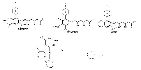
Particularly preferred in this group are the known statins cerivastatin,
atorvastatin, and NK-104.
Brief Description of the Drawings
Figure 1 shows the structures of cerivastatin, atorvastatin and NK-104.
Modes of Carryin~ Out the Invention
The ultimate goal of the methods and compositions of the invention is to treat
or ameliorate bone disorders in vertebrate subjects, particularly mammals, and
more
particularly humans.
As used herein, "treat" or "treatment" include a postponement of development
of bone deficit symptoms and/or a reduction in the severity of such symptoms
that
will or are expected to develop. The terms further include ameliorating
existing bone
or cartilage deficit symptoms, preventing additional symptoms, ameliorating or
preventing the underlying metabolic causes of symptoms, preventing or
reversing
bone resorption and/or encouraging bone growth. Thus, the terms denote that a
beneficial result has been conferred on a vertebrate subject with a cartilage,
bone or
skeletal deficit, or with the potential to develop such deficit.
By "bone deficit" is meant an imbalance in the ratio of bone formation to bone
resorption, such that, if unmodified, the subject will exhibit less bone than
desirable,
or the subject's bones will be less intact and coherent than desired. Bone
deficit may
also result from fracture, from surgical intervention or from dental or
periodontal
disease. By "cartilage defect" is meant damaged cartilage, less cartilage than
desired,
or cartilage that is less intact and coherent than desired. "Bone disorders"
includes
both bone deficits and cartilage defects.
-3-
CA 02397659 2002-07-16
WO 01/52829 PCT/USO1/01888
Representative uses of the compounds of the present invention include: repair
of bone defects and deficiencies, such as those occurnng in closed, open and
non-union fractures; prophylactic use in closed and open fracture reduction;
promotion of bone healing in plastic surgery; stimulation of bone ingrowth
into
non-cemented prosthetic joints and dental implants; elevation of peak bone
mass in
pre-menopausal women; treatment of growth deficiencies; treatment of
periodontal
disease and defects, and other tooth repair processes; increase in bone
formation
during distraction osteogenesis; and treatment of other skeletal disorders,
such as
age-related osteoporosis, post-menopausal osteoporosis, glucocorticoid-induced
osteoporosis or disuse osteoporosis and arthritis, or any condition that
benefits from
stimulation of bone formation. The compounds of the present invention can also
be
useful in repair of congenital, trauma-induced or surgical resection of bone
(for
instance, for cancer treatment), and in cosmetic surgery. Further, the
compounds of
the present invention can be used for limiting or treating cartilage defects
or disorders,
and may be useful in wound healing or tissue repair.
The compositions useful in the invention may be administered systemically or
locally. For systemic use, the compounds herein are formulated for parenteral
(e.g.,
intravenous, subcutaneous, intramuscular, intraperitoneal, intranasal or
transdermal)
or enteral (e.g., oral or rectal) delivery according to conventional methods.
Intravenous administration can be by a series of injections or by continuous
infusion
over an extended period. Administration by injection or other routes of
discretely
spaced administration can be performed at intervals ranging from weekly to
once to
three times daily. Alternatively, the compounds disclosed herein may be
administered
in a cyclical manner (administration of disclosed compound; followed by no
administration; followed by administration of disclosed compound, and the
like).
Treatment will continue until the desired outcome is achieved. In general,
pharmaceutical formulations will include a compound of the present invention
in
combination with a pharmaceutically acceptable vehicle, such as saline,
buffered
saline, 5% dextrose in water, borate-buffered saline containing trace metals
or the
like. Formulations may further include one or more excipients, preservatives,
solubilizers, buffering agents, albumin to prevent protein loss on vial
surfaces,
lubricants, fillers, stabilizers, etc. Methods of formulation are well known
in the art
and are disclosed, for example, in Remin~ton's Pharmaceutical Sciences, latest
-4-
CA 02397659 2002-07-16
WO 01/52829 PCT/USO1/01888
edition, Mack Publishing Co., Easton PA, which is incorporated herein by
reference.
Pharmaceutical compositions for use within the present invention can be in the
form
of sterile, non-pyrogenic liquid solutions or suspensions, coated capsules,
suppositories, lyophilized powders, transdermal patches or other forms known
in the
art. Local administration may be by injection at the site of injury or defect,
or by
insertion or attachment of a solid carrier at the site, or by direct, topical
application of
a viscous liquid, or the like. For local administration, the delivery vehicle
preferably
provides a matrix for the growing bone or cartilage, and more preferably is a
vehicle
that can be absorbed by the subject without adverse effects.
Delivery of compounds herein to wound sites may be enhanced by the use of
controlled-release compositions, such as those described in PCT application
WO 93/20859, which is incorporated herein by reference. Films of this type are
particularly useful as coatings for prosthetic devices and surgical implants.
The films
may, for example, be wrapped around the outer surfaces of surgical screws,
rods, pins,
plates and the like. Implantable devices of this type are routinely used in
orthopedic
surgery. The films can also be used to coat bone filling materials, such as
hydroxyapatite blocks, demineralized bone matrix plugs, collagen matrices and
the
like. In general, a film or device as described herein is applied to the bone
at the
fracture site. Application is generally by implantation into the bone or
attachment to
the surface using standard surgical procedures.
In addition to the copolymers and carriers noted above, the biodegradable
films and matrices may include other active or inert components. Of particular
interest are those agents that promote tissue growth or infiltration, such as
growth
factors. Exemplary growth factors for this purpose include epidermal growth
factor
(EGF), fibroblast growth factor (FGF), platelet-derived growth factor (PDGF),
transforming growth factors (TGFs), parathyroid hormone (PTH), leukemia
inhibitory
factor (LIF), insulin-like growth factors (IGFs) and the like. Agents that
promote
bone growth, such as bone morphogenetic proteins (U.S. Patent No. 4,761,471;
PCT
Publication WO 90/11366), osteogenin (Sampath, et al., Proc. Natl. Acad. Sci.
USA
(1987) 84:7109-13) and NaF (Tencer, et al., J. Biomed. Mat. Res. (1989) 23:
571-89)
are also contemplated. Biodegradable films or matrices include calcium
sulfate,
tricalcium phosphate, hydroxyapatite, polylactic acid, polyanhydrides, bone or
dermal
collagen, pure proteins, extracellular matrix components and the like and
-5-
CA 02397659 2002-07-16
WO 01/52829 PCT/USO1/01888
combinations thereof. Such biodegradable materials may be used in combination
with
non-biodegradable materials, to provide desired mechanical, cosmetic or tissue
or
matrix interface properties.
Alternative methods for delivery of compounds of the present invention
include use of ALZET osmotic minipumps (Alza Corp., Palo Alto, CA); sustained
release matrix materials such as those disclosed in Wang, et al. (PCT
Publication WO
90/11366); electrically charged dextran beads, as disclosed in Bao, et al.,
(PCT
Publication WO 92/03125); collagen-based delivery systems, for example, as
disclosed in Ksander, et al., Ann. Surg. (1990) 211(3):288-94; methylcellulose
gel
systems, as disclosed in Beck, et al., J. Bone Min. Res. (1991) 6(11):1257-65;
alginate-based systems, as disclosed in Edelman, et al., Biomaterials (1991)
12:619-26 and the like. Other methods well known in the art for sustained
local
delivery in bone include porous coated metal prostheses that can be
impregnated and
solid plastic rods with therapeutic compositions incorporated within them.
The compounds of the present invention may also be used in conjunction with
agents that inhibit bone resorption. Antiresorptive agents, such as estrogen,
bisphosphonates and calcitonin, are preferred for this purpose. More
specifically, the
compounds disclosed herein may be administered for a period of time (for
instance,
months to years) sufficient to obtain correction of a bone deficit condition.
Once the
bone deficit condition has been corrected, the vertebrate can be administered
an
anti-resorptive compound to maintain the corrected bone condition.
Alternatively, the
compounds disclosed herein may be administered with an anti-resorptive
compound
in a cyclical manner (administration of disclosed compound, followed by
anti-resorptive, followed by disclosed compound, and the like).
In additional formulations, conventional preparations such as those described
below may be used.
Aqueous suspensions may contain the active ingredient in admixture with
pharmacologically acceptable excipients, comprising suspending agents, such as
methyl cellulose; and wetting agents, such as lecithin, lysolecithin or long-
chain fatty
alcohols. The said aqueous suspensions may also contain preservatives,
coloring
agents, flavoring agents, sweetening agents and the like in accordance with
industry
standards.
-6-
CA 02397659 2002-07-16
WO 01/52829 PCT/USO1/01888
Preparations for topical and local application comprise aerosol sprays,
lotions,
gels and ointments in pharmaceutically appropriate vehicles which may comprise
lower aliphatic alcohols, polyglycols such as glycerol, polyethylene glycol,
esters of
fatty acids, oils and fats, and silicones. The preparations may further
comprise
antioxidants, such as ascorbic acid or tocopherol, and preservatives, such as
p-hydroxybenzoic acid esters.
Parenteral preparations comprise particularly sterile or sterilized products.
Injectable compositions may be provided containing the active compound and any
of
the well known injectable Garners. These may contain salts for regulating the
osmotic
pressure.
If desired, the osteogenic agents can be incorporated into liposomes by any of
the reported methods of preparing liposomes for use in treating various
pathogenic
conditions. The present compositions may utilize the compounds noted above
incorporated in liposomes in order to direct these compounds to macrophages,
monocytes, as well as other cells and tissues and organs which take up the
liposomal
composition. The liposome-incorporated compounds of the invention can be
utilized
by parenteral administration, to allow for the efficacious use of lower doses
of the
compounds. Ligands may also be incorporated to further focus the specificity
of the
liposomes.
Suitable conventional methods of liposome preparation include, but are not
limited to, those disclosed by Bangham, A.D., et al., JMoI Biol (1965) 23:238-
252,
Olson, F., et al., Biochim Biophys Acta (1979) 557:9-23, Szoka, F., et al.,
Proc Natl
Acad Sci USA (1978) 75:4194-4198, Kim, S., et al., Biochim Biophys Acta (1983)
728:339:348, and Mayer, et al., Biochim Biophys Acta (1986) 858:161-168.
The liposomes may be made from the present compounds in combination with
any of the conventional synthetic or natural phospholipid liposome materials
including phospholipids from natural sources such as egg, plant or animal
sources
such as phosphatidylcholine, phosphatidylethanolamine, phosphatidylglycerol,
sphingomyelin, phosphatidylserine, or phosphatidylinositol and the like.
Synthetic
phospholipids that may also be used, include, but are not limited to:
dimyristoylphosphatidylcholine, dioleoylphosphatidylcholine,
dipalmitoylphosphatidylcholine and distearoylphosphatidycholine, and the
corresponding synthetic phosphatidylethanolamines and phosphatidylglycerols.
_7_
CA 02397659 2002-07-16
WO 01/52829 PCT/USO1/01888
Cholesterol or other sterols, cholesterol hemisuccinate, glycolipids,
cerebrosides, fatty
acids, gangliosides, sphingolipids, 1,2-bis(oleoyloxy)-3-(trimethyl ammonio)
propane
(DOTAP), N-[1-(2,3-dioleoyl) propyl-N,N,N-trimethylammonium chloride
(DOTMA), and other cationic lipids may be incorporated into the liposomes, as
is
known to those skilled in the art. The relative amounts of phospholipid and
additives
used in the liposomes may be varied if desired. The preferred ranges are from
about
60 to 90 mole percent of the phospholipid; cholesterol, cholesterol
hemisuccinate,
fatty acids or cationic lipids may be used in amounts ranging from 0 to 50
mole
percent. The amounts of the present compounds incorporated into the lipid
layer of
liposomes can be varied with the concentration of the lipids ranging from
about 0.01
to about 50 mole percent.
The liposomes with the above formulations may be made still more specific
for their intended targets with the incorporation of monoclonal antibodies or
other
ligands specific for a target. For example, monoclonal antibodies to the BMP
receptor may be incorporated into the liposome by linkage to
phosphatidylethanolamine (PE) incorporated into the liposome by the method of
Leserman, L., et al., Nature (1980) 288:602-604.
Veterinary uses of the disclosed compounds are also contemplated. Such uses
would include treatment of bone or cartilage deficits or defects, i.e., bone
disorders, in
domestic animals, livestock and thoroughbred horses.
The compounds of the present invention may be used to stimulate growth of
bone-forming cells or their precursors, or to induce differentiation of bone-
forming
cell precursors, either in vitro or ex vivo. The compounds described herein
may also
modify a target tissue or organ environment, so as to attract bone-forming
cells to an
environment in need of such cells. As used herein, the term "precursor cell"
refers to
a cell that is committed to a differentiation pathway, but that generally does
not
express markers or function as a mature, fully differentiated cell. As used
herein, the
term "mesenchymal cells" or "mesenchymal stem cells" refers to pluripotent
progenitor cells that are capable of dividing many times, and whose progeny
will give
rise to skeletal tissues, including cartilage, bone, tendon, ligament, marrow
stroma and
connective tissue (see A. Caplan, J. Orthop. Res. (1991) 9:641-SO). As used
herein,
the term "osteogenic cells" includes osteoblasts and osteoblast precursor
cells. More
particularly, the disclosed compounds are useful for stimulating a cell
population
_g_
CA 02397659 2002-07-16
WO 01/52829 PCT/USOI/01888
containing marrow mesenchymal cells, thereby increasing the number of
osteogenic
cells in that cell population. In a preferred method, hematopoietic cells are
removed
from the cell population, either before or after stimulation with the
disclosed
compounds. Through practice of such methods, osteogenic cells may be expanded.
The expanded osteogenic cells can be infused (or reinfused) into a vertebrate
subject
in need thereof. For instance, a subject's own mesenchymal stem cells can be
exposed to compounds of the present invention ex vivo, and the resultant
osteogenic
cells could be infused or directed to a desired site within the subject, where
further
proliferation and/or differentiation of the osteogenic cells can occur without
immunorejection. Alternatively, the cell population exposed to the disclosed
compounds may be immortalized human fetal osteoblastic or osteogenic cells. If
such
cells are infused or implanted in a vertebrate subject, it may be advantageous
to
"immunoprotect" these non-self cells, or to immunosuppress (preferably
locally) the
recipient to enhance transplantation and bone or cartilage repair.
Within the present invention, an "effective amount" of a composition is that
amount which produces a statistically significant effect. For example, an
"effective
amount" for therapeutic uses is the amount of the composition comprising an
active
compound herein required to provide a clinically significant increase in
healing rates
in fracture repair; reversal of bone loss in osteoporosis; reversal of
cartilage defects or
disorders; prevention or delay of onset of osteoporosis; stimulation and/or
augmentation of bone formation in fracture non-unions and distraction
osteogenesis;
increase and/or acceleration of bone growth into prosthetic devices; and
repair of
dental defects. Such effective amounts will be determined using routine
optimization
techniques and are dependent on the particular condition to be treated, the
condition
of the patient, the route of administration, the formulation, and the judgment
of the
practitioner and other factors evident to those skilled in the art. The dosage
required
for the compounds of the invention (for example, in osteoporosis where an
increase in
bone formation is desired) is manifested as a statistically significant
difference in
bone mass between treatment and control groups. This difference in bone mass
may
be seen, for example, as a S-20% or more increase in bone mass in the
treatment
group. Other measurements of clinically significant increases in healing may
include,
for example, tests for breaking strength and tension, breaking strength and
torsion,
4-point bending, increased connectivity in bone biopsies and other
biomechanical
_9_
CA 02397659 2002-07-16
WO 01/52829 PCT/USO1/01888
tests well known to those skilled in the art. General guidance for treatment
regimens
is obtained from experiments carried out in animal models of the disease of
interest.
The dosage of the compounds of the invention will vary according to the
extent and severity of the need for treatment, the activity of the
administered
compound, the general health of the subj ect, and other considerations well
known to
the skilled artisan. Generally, they can be administered to a typical human on
a daily
basis as an oral dose of about 0.1 mg/kg-1000 mg/kg, and more preferably from
about
1 mg/kg to about 200 mg/kg. The parenteral dose will appropriately be 20-100%
of
the oral dose. While oral administration may be preferable in most instances
(for
reasons of ease, patient acceptability, and the like), alternative methods of
administration may be appropriate for selected compounds and selected defects
or
diseases. In comparative assays, positive control compounds or other bone-
active test
compounds may be administered subcutaneously, while statin-type test compounds
are administered orally.
Confirmator~Assays
The osteogenic activity of the compounds of formula 1 used in the methods of
the invention can be verified in various assay systems.
Neonatal Mouse Calvaria Assay~ln vitro)
An assay for bone resorption or bone formation is similar to that described by
Gowen M. & Mundy G., Jlmmunol (1986) 136:2478-82. Briefly, four days after
birth, the front and parietal bones of ICR Swiss white mouse pups are removed
by
microdissection and split along the sagittal suture. In an assay for
resorption, the
bones are incubated in BGJb medium (Irvine Scientific, Santa Ana, CA) plus
0.02%
(or lower concentration) (3-methylcyclodextrin, wherein the medium also
contains test
or control substances. The medium used when the assay is conducted to assess
bone
formation is Fitton and Jackson Modified BGJ Medium (Sigma) supplemented with
6 p,g/ml insulin, 6 ~.g/ml transfernn, 6 ng/ml selenous acid, calcium and
phosphate
concentrations of 1.25 and 3.0 mM, respectively, and ascorbic acid to a
concentration
of 100 p,g/ml is added every two days. The incubation is conducted at
37°C in a
humidified atmosphere of 5% COZ and 95% air for 96 hours.
-10-
CA 02397659 2002-07-16
WO 01/52829 PCT/USO1/01888
Following this, the bones are removed from the incubation media and fixed in
10% buffered formalin for 24-48 hours, decalcified in 14% EDTA for 1 week,
processed through graded alcohols; and embedded in paraffin wax. Three pm
sections of the calvaria are prepared. Representative sections are selected
for
histomorphometric assessment of bone formation or bone resorption. Bone
changes
are measured on sections cut 200 p,m apart. Osteoblasts and osteoclasts are
identified
by their distinctive morphology.
Other auxiliary assays can be used as controls to determine non-BMP
promoter-mediated effects of test compounds. For example, mitogenic activity
can be
measured using screening assays featuring a serum-response element (SRE) as a
promoter and a luciferase reporter gene. More specifically, these screening
assays can
detect signaling through SRE-mediated pathways, such as the protein kinase C
pathway. For instance, an osteoblast activator SRE-luciferase screen and an
insulin
mimetic SRE-luciferase screen are useful for this purpose. Similarly, test
compound
stimulation of cAMP response element (CRE)-mediated pathways can also be
assayed. For instance, cells transfected with receptors for PTH and calcitonin
(two
bone-active agents) can be used in CRE-luciferase screens to detect elevated
CAMP
levels. Thus, the BMP promoter specificity of a test compound can be examined
through use of these types of auxiliary assays.
In vivo Assay of Effects of Compounds on Murine Calvarial Bone Growth
Male ICR Swiss white mice, aged 4-6 weeks and weighing 13-26 gm, are
employed, using 4-5 mice per group. The calvarial bone growth assay is
performed as
described in PCT application WO 95/2421 l, incorporated by reference. Briefly,
the
test compound or appropriate control vehicle is injected into the subcutaneous
tissue
over the right calvaria of normal mice. Typically, the control vehicle is the
vehicle in
which the compound was solubilized, and is PBS containing 5% DMSO or is PBS
containing Tween (2 X1/10 ml). The animals are sacrificed on day 14 and bone
growth measured by histomorphometry. Bone samples for quantitation are cleaned
from adjacent tissues and fixed in 10% buffered formalin for 24-48 hours,
decalcified
in 14% EDTA for 1-3 weeks, processed through graded alcohols; and embedded in
paraffin wax. Three to five ~,m sections of the calvaria are prepared, and
representative sections are selected for histomorphometric assessment of the
effects
-11-
CA 02397659 2002-07-16
WO 01/52829 PCT/USO1/01888
on bone formation and bone resorption. Sections are measured by using a camera
lucida attachment to trace directly the microscopic image onto a digitizing
plate.
Bone changes are measured on sections cut 200 ~m apart, over 4 adj acent 1 x 1
mm
fields on both the injected and noninjected sides of the calvaria. New bone is
identified by its characteristic woven structure, and osteoclasts and
osteoblasts are
identified by their distinctive morphology. Histomorphometry software
(OsteoMeasure, Osteometrix, Inc., Atlanta) is used to process digitizer input
to
determine cell counts and measure areas or perimeters.
Additional In Vivo Assays
Compounds can be further confirmed to have the desired activity in intact
animals using an in vivo, dosing assay. Prototypical dosing may be
accomplished by
subcutaneous, intraperitoneal or oral administration, and may be performed by
injection, sustained release or other delivery techniques. The time period for
administration of test compound may vary (for instance, 28 days as well as 35
days
may be appropriate). An exemplary, in vivo oral or subcutaneous dosing assay
may
be conducted as follows:
In a typical study, 70 three-month-old female Sprague-Dawley rats are
weight-matched and divided into seven groups, with ten animals in each group.
This
includes a baseline control group of animals sacrificed at the initiation of
the study; a
control group administered vehicle only; a PBS-treated control group; and a
positive
control group administered a compound (non-protein or protein) known to
promote
bone growth. Three dosage levels of the compound to be tested are administered
to
the remaining three groups.
Briefly, test compound, positive control compound, PBS, or vehicle alone is
administered subcutaneously once per day for 35 days. All animals are injected
with
calcein nine days and two days before sacrifice (two injections of calcein
administered each designated day). Weekly body weights are determined. At the
end
of the 35-day cycle, the animals are weighed and bled by orbital or cardiac
puncture.
Serum calcium, phosphate, osteocalcin, and CBCs are determined. Both leg bones
(femur and tibia) and lumbar vertebrae are removed, cleaned of adhering soft
tissue,
and stored in 70% ethanol for evaluation, as performed by peripheral
quantitative
computed tomography (pQCT; Ferretti, J., Bone (1995) 17:3535-64S), dual energy
-12-
CA 02397659 2002-07-16
WO 01/52829 PCT/USO1/01888
X-ray absorptiometry (DEXA; Laval-Jeantet A., et al., Calcif Tissue Intl (
1995)
56:14-18; J. Casez, et al., Bone and Mineral (1994) 26:61-68) and/or
histomorphometry. The effect of test compounds on bone remodeling can thus be
evaluated.
Lead compounds can also be tested in acute ovariectomized animals
(prevention model) using an in vivo dosing assay. Such assays may also include
an
estrogen-treated group as a control. An exemplary subcutaneous dosing assay is
performed as follows:
In a typical study, 80 three-month-old female Sprague-Dawley rats are
weight-matched and divided into eight groups, with ten animals in each group.
This
includes a baseline control group of animals sacrificed at the initiation of
the study;
three control groups (sham ovariectomized (sham OVX) + vehicle only;
ovariectomized (OVX) + vehicle only; PBS-treated OVX); and a control OVX group
that is administered a compound known to promote bone growth. Three dosage
levels
of the compound to be tested are administered to the remaining three groups of
OVX
animals.
Since ovariectomy (OVX) induces hyperphagia, all OVX animals are pair-fed
with sham OVX animals throughout the 35 day study. Briefly, test compound,
positive control compound, PBS, or vehicle alone is administered orally or
subcutaneously once per day for 35 days. Alternatively, test compound can be
formulated in implantable pellets that are implanted for 35 days, or may be
administered orally, such as by gastric gavage. All animals, including sham
OVX/vehicle and OVX/vehicle groups, are injected intraperitoneally with
calcein
nine days and two days before sacrifice (two injections of calcein
administered each
designated day, to ensure proper labeling of newly formed bone). Weekly body
weights are determined. At the end of the 35-day cycle, the animals' blood and
tissues are processed as described above.
Compounds may also be tested in chronic OVX animals (treatment model).
An exemplary protocol for treatment of established bone loss in ovariectomized
animals that can be used to assess efficacy of anabolic agents may be
performed as
follows. Briefly, 80 to 100 six month old female, Sprague-Dawley rats are
subjected
to sham surgery (sham OVX) or ovariectomy (OVX) at time 0, and 10 rats are
sacrificed to serve as baseline controls. Body weights are recorded weekly
during the
-13-
CA 02397659 2002-07-16
WO 01/52829 PCT/USO1/01888
experiment. After approximately 6 weeks (42 days) or more of bone depletion,
10
sham OVX and 10 OVX rats are randomly selected for sacrifice as depletion
period
controls. Of the remaining animals, 10 sham OVX and 10 OVX rats are used as
placebo-treated controls. The remaining OVX animals are treated with 3 to 5
doses of
test drug for a period of 5 weeks (35 days). As a positive control, a group of
OVX
rats can be treated with an agent such as PTH, a known anabolic agent in this
model
(Kimmel, et al., Endocrinology (1993) 132:1577-84). To determine effects on
bone
formation, the following procedure can be followed. The femurs, tibiae and
lumbar
vertebrae 1 to 4 are excised and collected. The proximal left and right tibiae
are used
for pQCT measurements, cancellous bone mineral density (BMD) (gravimetric
determination), and histology, while the midshaft of each tibiae is subjected
to cortical
BMD or histology. The femurs are prepared for pQCT scanning of the midshaft
prior
to biomechanical testing. With respect to lumbar vertebrae (LV), LV2 are
processed
for BMD (pQCT may also be performed); LV3 are prepared for undecalcified bone
histology; and LV4 are processed for mechanical testing.
Statin Compounds Useful in the Invention
The compounds of the invention may be in the open chain form, as shown, or
may be in the form of a lactone. The compounds contain chiral carbons; all
stereoisomeric forms of these compounds and mixtures thereof are included.
The statin compounds useful in the methods of the invention contain an
aromatic or heteroaromatic system represented by:
/Z
C
Y
By aromatic or heteroaromatic system is meant a monocyclic or fused bicyclic
system
which is aromatic in nature and which contains 5-12 ring members. Typical
aromatic
systems are, of course, benzene and naphthalene. For heteroaromatic
embodiments,
the carbon in one or more positions may be replaced by nitrogen, sulfur, or
oxygen.
Replacement by nitrogen is preferred. Thus, the heteroaromatic system may
preferably be pyrrole, pyrazole, imidazole, pyridine, pyrazine, pyrimidine,
indole,
-14-
CA 02397659 2002-07-16
WO 01/52829 PCT/USO1/01888
purine, benzimidazole, quinoline, isoquinoline, quinazoline, and the like.
Particularly
preferred are pyrrole, quinoline, and pyridine. When the heteroaromatic system
is
pyrrole, it is preferred that Z occupies position l; when the heteroaromatic
system is
quinoline, it is preferred that Z is at position 3, and when the
heteroaromatic system is
pyridine, position 3 for Z is also preferred.
The heteroaromatic system is required to be substituted at the position
adjacent Z by p-fluorophenyl. Additional substituents may also be present.
These
substituents may contain additional aromatic groups, and may be alkyl,
including
cycloalkyl, OR, SR, NRz, and the like wherein R is H or alkyl (1-6C), halo,
and/or
phenyl. Substituents also may include those of the formula R"COO, R"CONH,
R"CO, R"HNCO, and R"OOC wherein R" is H, alkyl (1-6C) or phenyl. Particularly
preferred substituents are phenyl, lower alkyl, methoxy, and cycloalkyl.
The compounds of the invention which are useful in treating bone disorders by
enhancing bone growth are generally classified as "statins." Particularly
preferred for
use in the invention are cerivastatin, marketed under the name Baycol~ by
Bayer (See
U.S. patents 5,006,530 and 5,177,080), atorvastatin, marketed under the name
Lipotor by Warner-Lambert (See U.S. patent 5,273,995), and NK-104 developed by
NEGMA. (See Akiba, T et al., JToxicol Sci (1998) 23V:713-720.) All the above-
cited documents are incorporated herein by reference.
The compositions of the invention may also include the bisphosphonates and
their analogs. Typically, and preferably, the bisphosphonates are of the
formula
Rio
H \ ~ H
O ~ C P, O
HO I \0H
Rio
and the pharmaceutically acceptable salts, esters and amides thereof. Typical
salts are
those of the inorganic ions, such as sodium ion, potassium ion, calcium ion,
magnesium ion and the like; any pharmaceutically acceptable canon may be used.
Typical esters are the ethyl, methyl, isobutyl, ethylene glycol, and other
typical
pharmaceutically acceptable esters; typical amides are the unsubstituted -NHZ
amides
as well as the alkyl and dialkyl amides.
-15-
CA 02397659 2002-07-16
WO 01/52829 PCT/USO1/01888
Embodiments of R'° include halo, OR, SR, NR2, where R is H or alkyl
(1-6C)
or alkyl or arylalkyl with optional substitutions. Particularly preferred are
the
amino-substituted alkyl embodiments. Typically, both Rl° are not
identical, although
in some embodiments, such as clodronate, both Rl° are halo.
Particularly preferred
compounds among the bisphosphonates are risedronate, alandronate, pamidronate,
clodronate and in particular ibandronate. These compounds are particularly
useful in
combination with the statins.
In addition to the statins or other isoprenoid inhibiting compounds of the
invention, the compositions may also include other agents, including those
which
inhibit bond resorption such as estrogens or their analogs and compounds of
the
formula Ar-L-Ar wherein Ar represents an aryl substituent and L represents a
linker.
The following examples are intended to illustrate but not to limit the
invention.
Example 1
In vivo Calvarial Bone Growth Assay
Cerivastatin, atorvastatin and NK-104 are assayed in vivo according to the
procedure described previously (see "In vivo Assay of Effects of Compounds on
Murine Calvarial Bone Growth," supra). Vehicle control, bFGF and varying doses
of
test compound are tested.
Example 2
In vitro Bone Formation
Cerivastatin, atorvastatin and NK-104 and appropriate controls are assayed in
vitro (ex vivo) for bone formation activity (described above in "Neonatal
Mouse
Calvaria Assay (in vitro)"). Histomorphometrical assessments of ex vivo
calvaria are
carned out using an OsteoMetrics bone morphometry measurement program,
according to the manufacturer's instructions. Measurements are determined
using
either a 10- or 20-fold objective with a standard point counting eyepiece
graticule.
New bone formation is determined (using a l OX objective) by measuring the
new bone area formed in one field in 3 representative sections of each bone (4
bones
per group). Each measurement is carried out %z field distance from the end of
the
-16-
CA 02397659 2002-07-16
WO 01/52829 PCT/USO1/01888
suture. Both total bone and old bone area were measured. Data are expressed as
new
bone area in mm2.
Osteoblast numbers are determined by point counting. The number of
osteoblast cells lining the bone surface on both sides of the bone are counted
in one
field using a 20X objective. Data are expressed as osteoblast numbers/mm of
bone
surface.
Example 3
Effect on Resor~tion
Cerivastatin, atorvastatin and NK-104 and controls are tested in an
antiresorptive assay. Briefly, 15 day timed pregnant CD-1 female mice are
injected
with 45Ca (25 ~Ci/mouse). The calvaria from the 4 day old pups are dissected
out and
cut in half. The excised half calvaria are placed on metal grids (at the
surface) in 1 ml
of BGJ medium (Sigma) containing 0.1 % BSA with glutamine and Pen/Strep added.
The bones are incubated at 37°C in a 5% humidified incubator for a
period of 24 h,
and then transferred to wells containing 1 ml medium with factors added (IL-1,
PTH,
and/or test compounds). The treated bones are incubated under the above
conditions
for a further 72 h. After this incubation period, the bones are removed and
placed into
20% TCA in a scintillation vial for 1.5 h, and then counted with scintillation
fluid.
An aliquot of medium (0.4 ml) is also counted. The results are expressed as %
45Ca
release.
This assay may be modified by including test compounds/factors or control
compounds/factors in the preincubation medium (i.e., during the first 24 h).
Since
most of the osteoclasts are formed in the calvaria following the preincubation
period,
compounds or factors that affect osteoclast formation may have a greater
effect during
the preincubation period.
In this assay, compound toxicity is indicated by obvious death of the cells in
the periosteal region and within the marrow cavity of the bone organ cultures.
These
cells are characterized by pyknotic nuclei and vacuolated cytoplasm,
characteristic of
cell necrosis and distinct from apoptosis.
-17-
CA 02397659 2002-07-16
WO 01/52829 PCT/USO1/01888
Example 4
Systemic Administration of Statins in OVX Models
Cerivastatin, atorvastatin and NK-104 are also analyzed in vivo using an acute
OVX (prevention model) and/or chronic (treatment model) OVX model system, as
described above under "Additional In Yivo Assays".
Example 5
Statin-Mediated Fracture Repair Effects of Test Compounds
Cerivastatin, atorvastatin and NK-104 are examined for effects on surgical
defects in the rabbit radius. Healing of these defects may be assessed by X-
ray,
histology and biomechanical strength.
The test compound is weighed out in a microcentrifuge tube, and 50 ~1 of
1.5% sodium alginate solution is added as a carrier. This test sample is
vortexed to
wet all of the powder. The sample is sonicated for 20-30 min, and then
vortexed
again. Disks are created in the top of the microcentrifuge tube (by placing
the tube lid
or stopper-side down). The indent in the top of the stopper (i.e., lid) is
used to form
the disk (7.5 mm diameter). CaCl2 solution (100 p1 of 100 mM) is added to the
sodium alginate/drug solution. The samples are allowed to sit for 5-10 min,
and then
the calcium-alginate disks are carefully removed. The disks are rinsed in a
beaker
filled with water to rinse off the excess calcium solution, and are saved in
tubes using
water as vehicle. All solutions and containers are sterile, and all procedures
for
preparations of disks are performed under a laminar flow hood under sterile
conditions.
Bone healing is examined as follows. Briefly, six-month old male rabbits are
obtained, and are divided into 4 treatment groups (n=3 animals/group). The
treatment
groups received either: 1) placebo; 2) test compound (5 mg/disk); 3) test
compound
(10 mg/disk); or 4) an autologous bone graft. Animals are anesthetized with
rabbit
cocktail (1 m1/1.5 kg intramuscularly), and the right forelimb is clipped,
prepped and
draped for aseptic surgery. Anesthesia is maintained using isofluorane
delivered with
a face mask. To create a 20 mm gap defect in the right mid-radius, an incision
is
made over the lateral aspect of the forearm, and an osteotomy is performed
with an
oscillating bone saw. The cerivastatin, atorvastatin and NK-104 or vehicle is
applied
-18-
CA 02397659 2002-07-16
WO 01/52829 PCT/USO1/01888
to the defect and the defect is closed in layers. No external splinting is
needed, as the
radius is paired with the ulna, which functions to allow normal ambulation in
the
rabbit. Disks are cut on strips and inserted in the fracture to cover all the
defect.
Radiologic evaluation is performed at zero time and at 4 weeks.
Because the vehicle (placebo treatment group) prevented full healing in the
control group, only X-ray results are obtained and analyzed. Accordingly, X-
ray
analysis 4 weeks after initiation of treatment showed callus formation at the
bone
treatment site in the treated (both doses), but not the placebo (vehicle or
autologous
bone graft) groups.
From the foregoing, it will be appreciated that, although specific embodiments
of the invention have been described herein for purposes of illustration,
various
modifications may be made without deviating from the spirit and scope of the
invention. Accordingly, the invention is not limited except as by the appended
claims.
-19-