Note: Descriptions are shown in the official language in which they were submitted.
CA 02399765 2002-07-31
WO 01/68179 PCT/CA01/00304
1 "Transcutaneous Medical Device Dressings and Method of Use"
2
3 FIELD OF THE INVENTION
4 The invention relates to transcutaneous medical device dressings, and
processes for
their production and use, for controlling infections.
6 BACKGROUND OF THE INVENTION
7 Transcutaneous medical devices are catheters, pins, implants and the like
which pass
8 through the skin and are indwelling for some considerable time. Exeinplary
of
9 transcutaneous medical devices are central venous catheters, peripheral
venous catheters,
Swan-Gauz pulmonary catheters, central nervous system implants (ex. external
ventricular
11 drainage and ventricular reservoirs), peritoneal dialysis catheters, such
as for continuous
12 ambulatory peritoneal dialysis and continuous cyclic peritoneal dialysis,
hemodialysis
13 catheters, transvenous pacemaker leads and temporary orthopedic pins. All
of these
14 transcutaneous medical devices, when in place, have a portion of the device
which is external,
that is which is left protruding from the skin, and which can be the the cause
of infection.
16 The risk of acquiring infections from transcutaneous infections is very
high. For
17 instance, the risk of acquiring catheter-related bloodstream infection
ranges from 0.9 to 8%.
18 This nosocomial bloodstream infections cause a case fatality of more than
20%, and account
19 for an increase of thousands of dollars in hospital costs per infection, or
tens of thousands of
dollars per survivor in ICU needing an extra week of hospital stay. As for
peritoneal dialysis,
21 a very experienced center today still has a peritonitis rate of one episode
per 15 to 25 patient
22 months. The major sources of bacteria in these infections are from
surrounding skin.
23 To prevent infections associated with transcutaneous medical devices
antiseptic
24 preparation of insertion sites, including the initial application of
topical anti-microbial
solutions such as alcohol or iodine to the insertion sites is known. A further
topical ointment
26 after insertion of the device, such as an ointment containing neomycin,
polymyxin and
27 bactracin, has been shown to prevent catheter colonization/infection, but
it may increase the
28 risk of fungal infection. Ointments are also inconvenient, requiring
multiple replacements.
29 There have also been attempts to attach a cuff to the catheters, with an
anti-microbial agent
impregnated in the cuff. Efforts to coat the catheters with anti-microbial
agents are known.
31 However, none of these efforts has been completely successful in clinical
trials. Presently,
32 the most common catheter dressing used in hospitals comprises sterile gauze
or polyurethane
CA 02399765 2002-07-31
WO 01/68179 PCT/CA01/00304
1 film, which have limited infection control properties.
2 Recent efforts to replace gauze with a transparent film dressing to allow a
visual
3 check on the insertion site is known, see for instance US Patent No.
5,372,589, issued
4 December 13, 1994 to Davis. No anti-microbial control is taught with such a
dressing.
Johnson & Johnson Medical Inc. markets a product under the trade mark
BIOPATCH, which
6 is a chlorhexidine gluconate-impregnated catheter patch. An lodophor
transparent dressing
7 has also been suggested. However, to date, no completely effective anti-
microbial device for
8 use with transcutaneous medical devices is known.
9 A securement devices is taught for securing a intravenous device to the body
in US
Patent 3,918,446, issued November 11, 1975 to Buttaravoli. The device has an
upper and a
11 lower pad, between which the intravenous device is fixed. Since the
function of the device is
12 to secure the device to the body, there is a teaching to provide an
adhesive material to the
13 bottom of lower pad, and to the bottom of the top pad. There is a mention
of providing the
14 adhesive with an antibacterial agent. This device has the disadvantage of
using adhesives
with the antibacterial agent, which limits the effectiveness and long lasting
ability of the
16 antibacterial agent. Furthermore, the adhesive can be irritating next to
the skin, cause skin
17 damage and patient discomfort on removal, and inhibits the removal or
changing of the
18 device. Furthermore, many adhesives act as moisture barriers, which can
limit the
19 effectiveness of the antibacterial agent. Finally, the device of this
patent teaches including a
slit in the bottom pad of the dressing, which lies below the intravenous
needle or catheter
21 when the device is in place, allowing the intravenous device to remain in
contact with the
22 skin, and therefore limiting the infection control of the device.
23 SUMMARY OF THE INVENTION
24 In one broad aspect, the invention provides a transcutaneous device
dressing for use
with a transcutaneous medical device which has punctured the skin of a patient
and which has
26 a portion of the medical device protruding from the skin, comprising:
27 a top and a bottom dressing, both being formed from a flexible material and
having
28 upper and lower surfaces, with the lower surface being skin facing in use;
29 the bottom dressing having a slit formed therein extending from one edge
inwardly to
a termination point within the confines of the bottom dressing;
31 an anti-microbial material provided without adhesives at the upper and
lower surfaces
32 of the bottom dressing, and at least at the lower surface of the top
dressing;
2
CA 02399765 2002-07-31
WO 01/68179 PCT/CA01/00304
1 whereby, in use, the bottom dressing is placed next to the skin, the slit
allowing the
2 bottom dressing to surround the puncture site such that the lower surface of
the bottom
3 dressing is in contact with the skin and the upper surface of the bottom
dressing is in contact
4 with a portion of the medical device protruding from the skin, and the top
dressing is placed
above the puncture site such that its lower surface is in contact with a
portion of the medical
6 device protruding from the skin, thereby exposing a portion of the medical
device protruding
7 from the skin from above and below to the anti-microbial activity of the
anti-microbial
8 material.
9 In another broad aspect, the invention provides a method of dressing the
puncture site
of a transcutaneous medical device to limit infection by microorganisms from
the
11 surrounding skin and the portion of the medical device that protrudes from
the skin of a
12 patient, comprising:
13 providing a transcutaneous device dressing, comprising:
14 a top and a bottom dressing, both being formed from a flexible material and
having
upper and lower surfaces, the lower surfaces being skin facing when the
dressing is in use;
16 the bottom dressing having a slit forined therein extending from one edge
inwardly to
17 a termination point within the confines of the bottom dressing; and
18 an anti-microbial material provided without the use of adhesives at the
upper and
19 lower surfaces of the bottom dressing, and at least at the lower surface of
the top dressing;
sliding the bottom dressing in place next to the skin using the slit to allow
the bottom
21 dressing to surround the puncture site at the termination point such that
the lower surface of
22 the bottom dressing is in contact with the skin surrounding the puncture
site while the upper
23 surface of the bottom dressing is in contact with a portion of the medical
device protruding
24 from the skin;
applying the top dressing above bottom dressing such that its lower surface is
in
26 contact with a portion of the medical device protruding from the skin;
27 depending on the anti-microbial material, applying a water or alcohol based
28 electrolyte to the top and bottom dressings to release the anti-microbial
agent; and
29 fixing the top and bottom dressings to the skin, preferably with an
occlusive or semi-
occlusive layer such as an adhesive film.
31 The transcutaneous device dressing of this invention has the advantage of
ease of
32 placement and been demonstrated to be much more effective than disc type
dressings which
3
CA 02399765 2002-07-31
WO 01/68179 PCT/CA01/00304
1 are laid flat under the transcutaneous device, which have only a limited
portion, generally
2 only the thickness of the dressing (less than 3 mm), in contact with the
portion of the medical
3 device which protrudes from the skin.
4 Preferably, the dressing of this invention is formed such that the top and
bottom
dressings are joined along a fold line, that is they are formed from a unitary
dressing which is
6 folded over in use. The slit is preferably formed from the edge of the
bottom dressing which
7 is parallel to the fold. The dressing is preferably formed from
multilayered, laminated
8 dressing materials. The anti-microbial material is preferably a thin film of
an anti-microbial
9 metal, most preferably formed with atomic disorder so as to create an
effective anti-microbial
effect, and to create an interference colour so as to provide an indicator, as
described in
11 W098/41095, published September 24, 1998, and naming inventors R. E.
Burrell and R. J.
12 Precht.
13 The dressing of this invention has application to transcutaneous medical
devices such
14 as listed above, made from a wide variety of materials, for example metals,
including steel,
aluminum and its alloys, latex, nylon, silicone, polyester, polyurethane, and
other plastics and
16 rubbers. Such devices are generally made of a bioinert or Biocompatible
material. The
17 device may take a variety of shapes including rod or tube shapes, hollow or
solid, and may be
18 rigid or flexible, factors dictated by its intended utility.
19 As used herein and in the claims, the terms and phrases set out below have
the
meanings which follow.
21 "Metal" or "metals" includes one or more metals whether in the form of
substantially
22 pure metals, alloys or compounds such as oxides, nitrides, borides,
sulphides, halides or
23 hydrides.
24 "Anti-microbial metals" are metals whose ions have an anti-microbial
effect.
Preferably, the metal will also be biocompatible. Preferred anti-microbial
metals include Ag,
26 Au, Pt, Pd, Ir (i.e. the noble metals), Sn, Cu, Sb, Bi and Zn, with Ag
being most preferred.
27 "Biocompatible" means non-toxic for the intended utility. Thus, for human
utility,
28 biocompatible means non-toxic to humans or human tissues.
29 "Anti-microbial effect" means that atoms, ions, molecules or clusters of
the anti-
microbial metal (hereinafter "species" of the anti-microbial metal) are
released into the
31 alcohol or electrolyte which the material contacts in concentrations
sufficient to inhibit
32 bacterial (or other microbial) growth in the vicinity of the material. The
most common
4
CA 02399765 2002-07-31
WO 01/68179 PCT/CA01/00304
1 method of measuring anti-microbial effect is by measuring the zone of
inhibition (ZOI)
2 created when the material is placed on a bacterial lawn. A relatively small
or no ZOI (ex. less
3 than 1 mm) indicates a non useful anti-microbial effect, while a larger ZOI
(ex. greater than 5
4 mm) indicates a highly useful anti-microbial effect. One procedure for a ZOI
test is set out in
the Examples which follow.
6 "Sustained release" or "sustainable basis" are used to define release of
atoms,
7 molecules, ions or clusters of an anti-microbial metal that continues over
time measured in
8 hours or days, and thus distinguishes release of such metal species from the
bulk metal, which
9 release such species at a rate and concentration which is too low to achieve
an anti-microbial
effect, and from highly soluble salts of anti-microbial inetals such as silver
nitrate, which
11 releases silver ions virtually instantly, but not continuously, in contact
with an alcohol or
12 electrolyte.
13 "Atomic disorder" includes high concentrations of: point defects in a
ciystal lattice,
14 vacancies, line defects such as dislocations, interstitial atoms, amorphous
regions, gain and
sub grain boundaries and the like relative to its normal ordered crystalline
state. Atomic
16 disorder leads to irregularities in surface topography and inhomogeneities
in the structure on
17 a nanometer scale.
18 "Normal ordered crystalline state" means the crystallinity normally found
in bulk
19 metal materials, alloys or compounds formed as cast, wrought or plated
metal products. Such
materials contain only low concentrations of such atomic defects as vacancies,
grain
21 boundaries and dislocations.
22 "Diffusion", when used to describe conditions which limit diffusion in
processes to
23 create and retain atomic disorder, i.e. which freeze-in atomic disorder,
means diffusion of
24 atoms and/or molecules on the surface or in the matrix of the material
being formed.
"Alcohol or water-based electrolyte" is meant to include any alcohol or water-
based
26 electrolyte that the anti-microbial materials of the present invention
might contact in order to
27 activate (i.e. cause the release of species of the anti-microbial metal)
into same. The term is
28 meant to include alcohols, water, gels, fluids, solvents, and tissues
containing water,
29 including body fluids (for example blood, urine or saliva), and body tissue
(for example skin,
muscle or bone).
31 "Colour change" is meant to include changes of intensity of light under
32 monochromatic light as well as changes of hue from white light containing
more than one
5
CA 02399765 2002-07-31
WO 01/68179 PCT/CA01/00304
1 wavelength.
2 An "interference colour" is produced when light impinges on two or more
partly
3 reflective surfaces separated by a distance which bears the right
relationship to the
4 wavelength of the light to be removed by destructive interference.
"Partly reflective" when used to describe the base or top layer materials,
means that
6 the material has a surface which reflects a portion of incident light, but
which also transmits a
7 portion of the incident light. Reflection occurs when a ray of incoming
light encounters a
8 boundary or interface characterized by a change in refractive index between
two media. For
9 the top layer of the anti-microbial materials of this invention, that
interface is with air. For
the base layer, the interface is with the top layer. The reflectance of the
base and top layers is
11 balanced so as to generate an interference colour.
12 "Partly light transmissive" when used to describe a thin film of the top
layer material
13 means that the thin film is capable of transmitting at least a portion of
incident visible light
14 through the thin film.
"Detectable" when used to describe a colour change means an observable shift
in the
16 dominant wavelength of the reflected light, whether the change is detected
by instrument,
17 such as a spectrophotometer, or by the human eye. The dominant wavelength
is the
18 wavelength responsible for the colour being observed.
19 DESCRIPTION OF THE DRAWINGS
Figure 1 is a schematic sectional figure of a three layer transcutaneous
device dressing
21 in accordance with the present invention;
22 Figure 2 is a schematic perspective view of a three layer transcutaneous
device
23 dressing folded along a central line to form top and bottom dressings and
showing the slit for
24 placement around the transcutaneous medical device;
Figure 3 is a schematic sectional view of the folded transcutaneous device
dressing in
26 place with a catheter penetrating the skin of a patient;
27 Figure 4 is a plan view of the transcutaneous device dressing of this
invention,
28 showing the slit in the bottom dressing;
29 Figure 5 is a plan view of the transcutaneous device dressing slid in place
with a
catheter, such that the bottom dressing is in contact with a portion of the
catheter protruding
31 from the skin; and
32 Figure 6 is a plan view of transcutaneous device dressing folded such that
the top
6
CA 02399765 2002-07-31
WO 01/68179 PCT/CA01/00304
1 dressing is in contact with a portion of the catheter protruding from the
skin.
2 DESCRIPTION OF THE PREFERRED EMBODIMENTS
3 Transcutaneous Device Dressin~
4 The dressing in accordance with the invention includes at least one, and
preferably at
least two or three layers of medical dressing materials, laminated together by
known means
6 such as low temperature thermal fusing, stitching or, most preferably,
ultrasonic welding. A
7 three layer dressing in accordance with the invention is shown generally at
10 in Figure 1 to
8 include a first layer 12, which will be skin facing in use, a second layer
14, which preferably
9 forms an absorbent core, and a third layer 16. The layers 12, 14 and 16 are
shown to be
laminated together by ultrasonic welds 18 at intermittent locations across the
dressing 10.
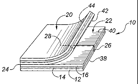
11 Figure 2 shows the dressing 10 to comprise a top dressing 20 and a bottom
dressing
12 22 formed from the three layers 12, 14 and 16. In Figure 2, the top and
bottom dressings 20,
13 22 are joined along a fold line 24, being formed from a unitary dressing
10. However, in
14 accordance with this invention, the top and bottom dressings 20, 22 may be
formed from
separately, from same or different medical dressing materials. If the top and
bottom dressings
16 20, 22 are provided separately, the top dressing 20 may include an
occlusive or semi-
17 occlusive layer such as an adhesive film (not shown) in order to secure the
dressing 10 in
18 place, and retain moisture for activation of the anti-microbial material. A
slit 26 is formed in
19 the bottom dressing 22, preferably extending from the edge of the bottom
dressing 22 which
is parallel to the fold line 24, and terminating at a termination point 28
which is preferably
21 about the center point of the bottom dressing 22.
22 The dressing 10 is shown in place against the skin 30 of a patient in
Figure 3, with a
23 catheter 32 protruding from the skin 30 at a penetration site 34. The
dressing is held in place
24 against the skin with an occlusive or semi-occlusive layer 36, such as
adhesive tape or
polyurethane film. The dressing is sized to cover a significant portion of the
catheter 32 that
26 protrudes from the skin 30, and not just the immediate skin area
surrounding the penetration
27 site. This aids in limiting infection, since bacteria are prevented from
migrating along the
28 catheter 32. A minimum dressing size will preferably provide at least 5 mm
coverage of the
29 protruding catheter 32, more preferably 1- 5 cm coverage.
Depending on the size of the transcutaneous medical device, the termination
point 28
31 of the slit 26, may include additional cuts, preferably a cross-cut, or a
penetrating hole (not
32 shown), to allow the medical device to fit through the dressing, while
still maintaining the
7
CA 02399765 2002-07-31
1 portions around the termination point in close contact with both the skin
and protruding
2 section of the medical device.
3 Figures 4, 5 and 6 demonstrate placement of the dressing 10 around a
catheter 32,
4 with the bottom dressing 22 sliding under the catheter 32 such that the
lower surface 38 (see
Figure 2) of the bottom dressing 22 contacts the patient's skin (not shown),
while the upper
6 surface 40 of the bottom dressing 22 contacts the catheter 32 protruding
from the skin. Once
7 the top dressing 20 is applied, by folding it over the bottom dressing 22,
the lower surface 42
8 (see Figure 2) of the top dressing 20 is in contact with the catheter 32
protruding from the
9 skin. The upper surface 44 of the top dressing 20 is then covered with the
occlusive or semi-
occlusive layer 36, as shown in Figure 3. As shown in Figures 4 and 5, when
the dressing 10
11 is formed from a unitary dressing, the lower surface 42 of the top dressing
20 and the upper
12 surface 40 of the bottom dressing 22, are one and the same layer,
represented as layer 16 in
13 Figure 1.
14 The lower and upper surfaces 38 and 40 of the bottom dressing 22, and at
least the
lower surface 42 of the top dressing 20 are provided with an anti-microbial
material in order
16 to limit infection. Anti-microbial materials for use with medical dressing
materials are well
17 known in the art. The anti-microbial material may be impregnated in one or
more of the
18 layers of the dressing 10, but will more preferably be provided as a thin
film of an anti-
19 microbial metal on those surfaces of the top and bottom dressings 20, 22
which will be skin
or catheter facing once the dressing is in place. Alternatively, the anti-
microbial material may
21 be an antibiotic composition or a composition formed from an anti-microbial
metal, as are
22 well known in the art.
23 The preferred and alternate compositions of the layers 12, 14 and 16,
together with the
24 preferred anti-microbial metal coatings are set out in further detail
below.
DressingMaterials
26 The first layer 12 of the dressing 10 is formed of a perforated, preferably
non-adherent
27 material which allows for fluids to penetrate or diffuse there through in
either or both
28 directions. The perforated material may be formed of a woven or non-woven,
non-woven
29 being preferred, fabric such as cotton, gauze, a polymeric net or mesh such
as polyethylene,
nylon, polypropylene or polyester, an elastomer such as polyurethane or
polybutadiene
31 elastomers, or a foam such as open cell polyurethane foam. Exemplary
perforated, non-
32 adherent materials useful for the dressing include non-woven meshes such as
DELNET""
8
CA 02399765 2002-07-31
WO 01/68179 PCT/CA01/00304
1 P530, which is a non-woven veil formed of high density polyethylene using
extrusion,
2 embossing and orientation processes, produced by Applied Extrusion
Technologies, Inc. Of
3 Middletown, Delaware, USA. This same product is available as Exu-Dry
CONFORMANT
4 2Tm Wound Veil, from Frass Survival Systems, Inc., Bronx, New York, USA as a
subset of
that company's Wound Dressing Roll (Non-Adherent) products. Other useful non-
woven
6 meshes include CARELLETM or NYLON 90TM, available from Carolina Formed
Fabrics
7 Corp., N-TE.RFACETM, available from Winfield Laboratories, Inc., of
Richardson, Texas,
8 USA. Exemplary woven meshes may be formed from fibreglass or acetate, or
cotton gauze.
9 An exemplary hydrophilic polyurethane foam is HYPOL'm, available from W.R.
Grace &
Co., New York, NY, USA.
11 For ease of ultrasonic welding for lamination, at least one of the first
and second
12 layers 12, 14 is preferably formed from a polymeric material which is
amenable to ultrasonic
13 welding, that is which will melt on the application of localized heat and
then fuse the layers
14 together on cooling.
The second, absorbent layer 14 is formed from an absorbent material for
holding
16 sufficient moisture next to the skin in order to activate the anti-
microbial metal coating, that
17 is to cause release of ions, molecules, atoms or clusters of the anti-
microbial metal in order to
18 cause an anti-microbial effect. Preferably, the absorbent material is an
absorbent needle
19 punched non-woven rayon/polyester core such as SONTARAT'' 8411, a 70/30
rayon/polyester blend commercially available from Dupont Canada, Mississauga,
Ontario,
21 Canada. This product is sold by National Patent Medical as an American
White Cross sterile
22 gauze pad. However, other suitable absorbent materials include woven or non-
woven
23 materials, non-woven being preferred made from fibers such as rayon,
polyester,
24 rayon/polyester, polyester/cotton, cotton and cellulosic fibers. Exemplary
are creped
cellulose wadding, an air felt of air laid pulp fibers, cotton, gauze, and
other well known
26 absorbent materials suitable for medical dressings.
27 The third layer 16 of the dressing 10 is preferably formed of perforated,
non-adherent
28 material such as used in the first layer 12. This allows moisture
penetration as sterile water
29 and the like are added in order to activate the anti-microbial metal
coating.
Additional layers (not shown) may be included between or above the first,
second and
31 third layers 12, 24, 16, as is well known in medical. Thus the use of the
terms first, second
32 and third layer, as used herein and in the claims is not meant to exclude
such additional
9
CA 02399765 2002-07-31
WO 01/68179 PCT/CA01/00304
1 layers.
2 The layers 12, 14, and 161aminated together at intermittent spaced locations
across
3 the dressing 10 by ultrasonic welds 18. Ultrasonic welding is a known
technique in the
4 quilting art, and thus will not be discussed at length. Briefly, heat
(generated ultrasonically)
and pressure are applied to either side of the dressing 10 at localized spots
through an
6 ultrasonic horn so as to cause melting of at least one of the plastic
materials in the first and
7 second layers 12, 14, and the subsequent bonding together of the layers on
cooling. The
8 welds appear at localized circular spots and are preferably less than 0.5 cm
in diameter. If the
9 third layer 16 is present, the ultrasonic welding can be performed from
either side of the
dressing, and will bind all three layers 12, 14 and 16 together.
11 The use of ultrasonic welding of the layers at spaced locations has the
advantage of
12 retaining the absorbent and moisture penetration properties of the layers
12, 14, while
13 retaining the conforming properties of the dressing. Edge seams, stitching
and adhesives
14 have the disadvantage of interfering with one or more of these desirable
properties of the
dressings. Furthermore, by spacing the welds 18 at intermittent locations
across the dressing,
16 the dressing 10 may be cut to smaller sizes, as needed, without causing
delamination.
17 Preferred spacings of about 2.5 cm between welds allows the dressing to be
cut down to
18 about 2.5 cm sizes, while maintaining at least one weld to hold the
laminated layers together.
19 Anti-Microbial Coatinj4
The dressing 10 of this invention preferably includes an anti-microbial
coating formed
21 from an anti-microbial metal. The coating is applied to one or more of the
layers 12, 14, 16,
22 but is most preferably applied at least to the first and third layers 12
and 16, so as to provide
23 the anti-microbial effect both against the skin and against the
transcutaneous medical device
24 held between the top and bottom dressings 20, 22.
The coating is most preferably formed with atomic disorder in accordance with
the
26 procedures set out above and as described in US Patent 5,454,886, and
W098/41095, both to
27 Burrell et al. Most preferably, the coating is formed as a multilayer anti-
microbial coating
28 having a top and a base layer, as set below, to produce an interference
colour. In this way, the
29 coating provides not only an anti-microbial effect to limit infection, but
also acts as an
indicator of activation of the dressing. As the top layer of the coating is
activated with an
31 alcohol or water-based electrolyte, such as sterile water or ethanol, even
minor dissolution of
32 the anti-microbial metal results in a detectable colour change, indicating
that an anti-
CA 02399765 2002-07-31
WO 01/68179 PCT/CA01/00304
1 inicrobial effect is being provided. If there is no colour change,
additional moisture might be
2 provided to the dressing by adding water, until a colour change is detected.
Once activated,
3 the dressing should be maintained in a moist condition by the addition of
sterile water if
4 necessary.
Sterilization
6 Dressings 10 with anti-microbial coatings of an anti-microbial metal formed
with
7 atomic disorder are preferably sterilized without applying excessive thermal
energy, which
8 can anneal out the atomic disorder, thereby reducing or eliminating a useful
anti-microbial
9 effect. Gamma radiation is preferred for sterilizing such dressings, as
discussed in US Patent
5,454,886.
11 It should be appreciated that the use of ultrasonic welding to laminate the
layers of
12 dressings with anti-microbial coatings formed from anti-microbial metals
with atomic
13 disorder is advantageous since it achieves bonding in localized spots and
avoids applying heat
14 to any significant portion of the dressing, thereby avoiding any
significant reduction in the
anti-microbial effect through annealing out of atomic disorder.
16 The sterilized dressings should be sealed in packaging which excludes light
17 penetration to avoid additional oxidation of the anti-microbial coating.
Polyester peelable
18 pouches are preferred. The shelf life of anti-microbial dressings thus
sealed is over one year.
19 Directions for Use of Dressings with Transcutaneous Devices
With transcutaneous devices such as flexible catheters, the dressing 10 is
placed on
21 the skin around the catheter 32 by passing the catheter 32 through the slit
26. The dressing 10
22 is rotated, if needed, to ensure that slit 26 is roughly perpendicular to
the long axis of the
23 catheter 32, thus ensuring that the portion of the catheter 32 protruding
from the skin is
24 contacted by the upper surface 40 of the bottom dressing 22. The top
dressing 20 is folded
over the bottom dressing 22 (or placed over, if the top and bottom dressings
are separate),
26 such that the lower surface 42 of the top dressing 20 is in contact with
the portion of the
27 catheter 32 protruding from the skin. If the anti-microbial material is an
anti-microbial metal
28 coating, the dressing is then moistened with drops of sterile water or 70%
ethanol, in order to
29 activate the coating for release of anti-microbial metal species. The
dressing 10 is then
secured in place with an occlusive or semi-occlusive layer 36, such as an
adhesive film,
31 which keeps the dressing in a moist environment.
32 If the transcutaneous device is rigid, such as a temporary orthopedic pin,
the bottom
11
CA 02399765 2002-07-31
WO 01/68179 PCT/CA01/00304
1 dressing 22 is put in place as set out above, but the top dressing 20 is
then folded and secured
2 around the portion of the pin protruding from the skin, in a tent-like
manner, since the pin
3 generally protrudes at an angle normal to the skin surface.
4 Animal trials with the dressing of the present invention, carrying a bi-
layer anti-
microbial coating formed with silver having atomic disorder, manufactured as
set out above
6 and as described in greater detail in Example 3, have shown excellent
results in controlling
7 infection. In use, the dressings are kept moist, at 100% relative humidity.
Adding sterile
8 water initially to activate the anti-microbial metal coating is needed, and
then as needed to
9 maintain the dressing in a moist condition. Dressings may be changed as
required for
observation and cleaning, but need not be changed more frequently than every 7
days, and
11 can provide an anti-microbial effect for a much longer period of time.
12 Multilayer Anti-Microbial Materials With Interference Colour
13 The dressings preferably include the anti-microbial metal coating formed
with at least
14 two metal layers, a base layer and a top layer over the base layer, so as
to produce an
interference colour, as set forth in W098/41095. Both layers are partly
reflective; the top
16 layer is partly light transmissive. The top layer is a thin film containing
at least one anti-
17 microbial metal formed with sufficient atomic disorder such that the top
layer, in contact with
18 an alcohol or water based electrolyte, releases ions, atoms, molecules or
clusters of the anti-
19 microbial metal at a concentration sufficient to provide a localized anti-
microbial effect on a
sustainable basis. In this way, the top layer, in contact with the alcohol or
electrolyte, will
21 undergo a change in optical path length, either by a change in thickness
resulting from some
22 dissolution, or through a change in the refractive index of the top layer
resulting from a
23 change in the composition of a newly formed thin layer formed on the top
layer. Either or
24 both of these results are sufficient to cause a detectable colour change,
thus providing an
indicator that the top layer has been activated.
26 Both the base layer and the top layer are formed from a partly reflective
material. In
27 this way, at least a portion of the incoming light is reflected from the
surface of the layer
28 while another portion is transmitted through the layer. The top layer is
partly light
29 transmissive to allow incident light to reach the interface with the base
layer. The top layer
thus cannot approximate 100% reflectivity, such as in pure Al or Ag, or
interference colours
31 cannot be generated, as is well known in the art. The materials for the top
and base layers
32 should be balanced in their reflectances in order to generate an
interference colour.
12
CA 02399765 2002-07-31
WO 01/68179 PCT/CA01/00304
1 Generally, the top layer is deposited as a thin film having a thickness
which maintains
2 adequate transmittance to generate an interference colour. Furthermore, the
refractive index
3 for the materials in layers is different, accomplished by differences in
their actual or effective
4 compositions. For instance different materials in the two layers will result
in the materials
having different actual refractive indexes. However, if it is desired to make
the layers from
6 the same material, the layers can be deposited with different porosities or
different
7 levels/types of atomic disorder, in order to achieve different effective
compositions, and thus
8 different refractive indexes.
9 In this manner, incoming light reflects off the interface of the base and
top layers.
Incoming light reflects from the interface of the top layer with air, and
interferes with the
11 light reflected from the interface with the base layer so as to generate an
"interference
12 colour". The particular colour which is generated and its brightness will
depend on the
13 properties of the layers, most importantly on the composition of the
layers, which determines
14 its transmittance and absorption properties, along with its refractive
index, and on the
thickness of the layers. Generally, it is desirable to generate first and
second order
16 interference colours, by limiting the thickness of the base layer and top
layers to minimize the
17 number of internal reflections. First and second order interference colours
are generally
18 brighter than third and fourth order etc. colours, making them more
aesthetically pleasing,
19 more consistently reproducible in manufacturing, and more susceptible to
detectable colour
change on variations in thickness on dissolution of even a minor amount of the
top layer.
21 The property which determines the particular colour which is generated is
the
22 effective optical thickness of the top layer, that is the product of the
refractive index of the
23 top layer material and the actual thickness of the top layer. Thus the
colour which is desired
24 can be altered by changing the actual thickness or the top layer or its
refractive index.
Preferably, the material in the base layer is a reflective metal. Such metals
are known
26 in the art and include, for example one or more of the valve metals, e.g.,
Ta, Nb, Ti, Zr and
27 Hf, as well as transition metals such as Au, Ag, Pt, Pd, Sn, Cu, V, W and
Mo, or the metal Al.
28 More preferably, the base material is formed from Ag, Au, Pt, Pd, Cu, Ta
and Al. Use of a
29 metal such as tantalum as the base layer may cause reduction of oxide
containing materials in
the top layer. To avoid this, a barrier layer (not shown), such as tantalum
oxide formed by
31 anodizing at least a portion of the top surface of the Ta metal, should be
included above a
32 tantalum layer. Preferred metals for the base layer are the anti-microbial
metals Au, Ag, Pt,
13
CA 02399765 2002-07-31
WO 01/68179 PCT/CA01/00304
1 Pd, Sn and Cu, more preferably Au, Pt and Ag, and most preferably Ag, in a
partly reflective
2 form.
3 The base layer may be formed by known techniques, such as the vapour
deposition
4 techniques of evaporation or physical vapour deposition. Preferably, the
base layer is formed
as a thin film by physical vapour deposition with atomic disorder, as set out
below and in US
6 5,454,889, in order to produce a sustainable anti-microbial effect when the
base layer is
7 ultimately exposed to an alcohol or water based electrolyte. The thickness
of the base layer is
8 generally not critical, provided that it is partly reflective. Preferred
thicknesses will vary
9 widely with the material composition and the desired colour. However, in
that the layer is
preferably a thin film formed by physical vapour deposition techniques, it
should be at least
11 about 25 nm thick to create a useful colour. To generate first and second
order interference
12 colours and to produce an anti-microbial effect, the base layer should be
greater than 60 nm
13 thick, more preferably 300 to 2500 nm thick, and most preferably 600 to 900
nm thick.
14 The top layer is formed of a partly reflective, partly light transmissive
thin film
containing at least one anti-microbial metal formed with atomic disorder so as
to produce a
16 sustainable anti-microbial effect, and ultimate colour change, when exposed
to an alcohol or a
17 water based electrolyte. The anti-microbial metal is preferably one or more
of Ag, Au, Pt,
18 Pd, Ir, Sn, Cu, Sb, Bi, and Zn in a partly reflective, partly transmissive
form. More
19 preferably, the anti-microbial metal is Ag, Au, Pt, Pd or Cu. The thickness
of the top layer
formed from these metals is preferably less than 400 nm in order to maintain
the preferred
21 level of light transmission. The desired thickness will vary with the
composition of the top
22 layer, and with the desired end colour and colour change. For first and
second order
23 interference colours, the thickness will generally be less than about 400
nm. More preferably,
24 the thickness will range from 5 to 210 nm, most preferably from 10 to 100
nm.
The top layer may be a thin film of the base layer material, formed with a
different
26 refractive index for instance by altering the deposition conditions to
change the porosity,
27 composition and/or degree of atomic disorder in the layers.
28 When the base layer is itself formed from an anti-microbial metal with
atomic
29 disorder, the top layer may be provided as an in situ generated top layer
by virtue of its
thickness and/or composition changing on contacting an alcohol or water based
electrolyte, so
31 as to produce an interference colour which differs from the initial colour
of the base layer.
32 Most preferably, the top layer is a thin film of a composite material
formed by co-,
14
CA 02399765 2002-07-31
WO 01/68179 PCT/CA01/00304
1 sequentially or reactively depositing an anti-microbial metal in a matrix
with atoms or
2 molecules of a different material to create atomic disorder in the matrix,
in the manner set out
3 below. The different material is selected from a) biocompatible metals, b)
oxygen, nitrogen,
4 hydrogen, boron, sulphur or halogens, or c) an oxide, nitride, carbide,
boride, halide, sulphide
or hydride of either or both of an anti-microbial metal or a biocompatible
metal. Most
6 preferably, the top layer material is a composite material containing
silver, and one or both of
7 silver oxide and atoms or molecules containing oxygen trapped or absorbed in
the silver
8 matrix. The term "silver oxide" is meant to include any oxide or mixture of
oxides of silver.
9 However, the top layer is preferably not formed solely of AgO and/or Ag20,
since the
solubility of these materials is low for providing a useful anti-microbial
effect.
11 Anti-Microbial Materials Containing Atomic Disorder
12 At least the top layer, and preferably also the base layer, is formed in a
crystalline
13 form from anti-microbial metals with atomic disorder so as to produce an
anti-microbial
14 effect. The production of atomic disorder through physical vapour
deposition techniques is
described in U.S. 5,454,886, and as outlined below.
16 The anti-microbial metal is deposited as a thin metallic film on one or
more surfaces
17 of the dressing 10, by vapour deposition techniques. Physical vapour
techniques, which are
18 well known in the art, all deposit the metal from the vapour, generally
atom by atom, onto a
19 substrate surface. The techniques include vacuum or arc evaporation,
sputtering, magnetron
sputtering and ion plating. The deposition is conducted in a manner to create
atomic disorder
21 in the coating as defined above. Various conditions responsible for
producing atomic
22 disorder are useful. These conditions are generally those which one has
been taught to avoid
23 in thin film deposition techniques, since the object of most thin film
depositions is to create a
24 defect free, smooth and dense film (see for example J.A. Thornton, supra).
While such
conditions have been investigated in the art, they had not been linked to
enhanced solubility
26 of the coatings so-produced prior to Applicants inventions.
27 The preferred conditions which are used to create atomic disorder during
the
28 deposition process include:
29 - a low substrate temperature, that is maintaining the surface to be coated
at a
temperature such that the ratio of the substrate temperature to the melting
point of the metal
31 (in degrees Kelvin) is less than about 0.5, more preferably less than about
0.35 and most
32 preferably less than about 0.3; and optionally one or both of:
CA 02399765 2002-07-31
WO 01/68179 PCT/CA01/00304
1 - a higher than normal working (or ambient) gas pressure, i.e. for vacuum
2 evaporation: e-beam or arc evaporation, greater than 0.01 mT, gas scattering
evaporation
3 (pressure plating) or reactive arc evaporation, greater than 20 mT; for
sputtering: greater than
4 75 mT; for magnetron sputtering: greater than about 10 mT; and for ion
plating: greater than
about 200 mT; and
6 - maintaining the angle of incidence of the coating flux on the surface to
be
7 coated at less than about 75 , and preferably less than about 30 .
8 The metals used in the coating are those known to release ions etc. having
an anti-
9 microbial effect, as set out above. For most dressings, the metal must also
be biocompatible.
Preferred metals include the noble metals Ag, Au, Pt, Pd, and Ir as well as
Sn, Cu, Sb, Bi, and
11 Zn or alloys or compounds of these metals or other metals. Most preferred
is Ag or Au, or
12 alloys or compounds of one or more of these metals.
13 For economic reasons, the thin metal film has a thickness no greater than
that needed
14 to provide release of metal ions on a sustainable basis over a suitable
period of time, and to
generate the desired interference colour. Within the preferred ranges of
thicknesses set out
16 above, the thickness will vary with the particular metal in the coating
(which varies the
17 solubility and abrasion resistance), and with the degree of atomic disorder
in (and thus the
18 solubility of) the coating. The thickness will be thin enough that the
coating does not
19 interfere with the dimensional tolerances or flexibility of the device for
its intended utility.
The anti-microbial effect of the material so produced is achieved when the
coating is
21 brought into contact with an alcohol or a water based electrolyte, thus
releasing metal ions,
22 atoms, molecules or clusters. The concentration of the metal species which
is needed to
23 produce an anti-microbial effect will vary from metal to metal. Generally,
anti-microbial
24 effect is achieved in body fluids such as plasma, serum or urine at
concentrations less than
about 0.5 - 5 g/ml.
26 The ability to achieve release of metal atoms, ions, molecules or clusters
on a
27 sustainable basis from a coating is dictated by a number of factors,
including coating
28 characteristics such as composition, structure, solubility and thickness,
and the nature of the
29 environment in which the device is used. As the level of atomic disorder is
increased, the
amount of metal species released per unit time increases. For instance, a
silver metal film
31 deposited by magnetron sputtering at T/Tm < 0.5 and a working gas pressure
of about 7
32 mTorr releases approximately 1/3 of the silver ions that a film deposited
under similar
16
CA 02399765 2002-07-31
WO 01/68179 PCT/CA01/00304
1 conditions, but at 30 mTorr, will release over 10 days. Films that are
created with an
2 intermediate structure (ex. lower pressure, lower angle of incidence etc.)
have Ag release
3 values intermediate to these values as determined by bioassays. This then
provides a method
4 for producing controlled release metallic coatings. Slow release coatings
are prepared such
that the degree of disorder is low while fast release coatings are prepared
such that the degree
6 of disorder is high.
7 For continuous, uniform coatings, the time required for total dissolution
will be a
8 function of film thickness and the nature of the environment to which they
are exposed. The
9 relationship in respect of thickness is approximately linear, i.e. a two
fold increase in film
thickness will result in about a two fold increase in longevity.
11 It is also possible to control the metal release from a coating by forming
a thin film
12 coating with a modulated structure. For instance, a coating deposited by
magnetron
13 sputtering such that the working gas pressure was low (ex. 15 mTorr) for
50% of the
14 deposition time and high (ex. 30 mTorr) for the remaining time, has a rapid
initial release of
metal ions, followed by a longer period of slow release. This type of coating
is extremely
16 effective on devices such as urinary catheters for which an initial rapid
release is required to
17 achieve immediate anti-microbial concentrations followed by a lower release
rate to sustain
18 the concentration of metal ions over a period of weeks.
19 The substrate tenlperature used during vapour deposition should not be so
low that
annealing or recrystallization of the coating takes place as the coating warms
to ambient
21 temperatures or the temperatures at which it is to be used (ex. body
temperature). This
22 allowable OT, that the temperature differential between the substrate
temperature during
23 deposition and the ultimate temperature of use, will vary from metal to
metal. For the most
24 preferred metals of Ag and Au, preferred substrate temperatures of -20 to
200V , more
preferably -10 C to 100 C are used.
26 Atomic order may also be achieved, in either or both of the base and top
layers by
27 preparing composite metal materials, that is materials which contain one or
more anti-
28 microbial metals in a metal matrix which includes atoms or molecules
different from the anti-
29 microbial metals.
The preferred technique for preparing a composite material is to co- or
sequentially
31 deposit the anti-microbial metal(s) with one or more other inert,
biocompatible metals
32 selected from Ta, Ti, Nb, Zn, V, Hf, Mo, Si, Al and alloys of these metals
or other metal
17
CA 02399765 2002-07-31
WO 01/68179 PCT/CA01/00304
1 elements, typically other transition metals. Such inert metals have a
different atomic radii
2 from that of the anti-microbial metals, which results in atomic disorder
during deposition.
3 Alloys of this kind can also serve to reduce atomic diffusion and thus
stabilize the disordered
4 structure. Thin film deposition equipment with multiple targets for the
placement of each of
the anti-microbial and inert metals is preferably utilized. When layers are
sequentially
6 deposited the layer(s) of the inert metal(s) should be discontinuous, for
example as islands
7 within the anti-microbial metal matrix. The final ratio of the anti-
microbial metal(s) to inert
8 metal(s) should be greater than about 0.2. The most preferable inert metals
are Ti, Ta, Zn and
9 Nb. It is also possible to form the anti-microbial coating from oxides,
carbides, nitrides,
sulphides, borides, halides or hydrides of one or more of the anti-microbial
metals and/or one
11 or more of the inert metals to achieve the desired atomic disorder.
12 Another composite material may be formed by reactively co- or sequentially
13 depositing, by physical vapour techniques, a reacted material into the thin
film of the anti-
14 microbial metal(s). The reacted material is an oxide, nitride, carbide,
boride, sulphide,
hydride or halide of the anti-microbial and/or inert metal, formed in situ by
injecting the
16 appropriate reactants, or gases containing same, (ex. air, oxygen, water,
nitrogen, hydrogen,
17 boron, sulphur, halogens) into the deposition chamber. Atoms or molecules
of these gases
18 may also become absorbed or trapped in the metal film to create atomic
disorder. The
19 reactant may be continuously supplied during deposition for codeposition or
it may be pulsed
to provide for sequential deposition. The final ratio of anti-microbial
metal(s) to reaction
21 product should be greater than about 0.2. Air, oxygen, nitrogen and
hydrogen are particularly
22 preferred reactants.
23 The above deposition techniques to prepare composite coatings may be used
with or
24 without the conditions of lower substrate temperatures, high working gas
pressures and low
angles of incidence previously discussed. One or more of these conditions are
preferred to
26 retain and enhance the amount of atomic disorder created in the coating.
27 Examples
28 Example 1
29 This example shows the preparation of a bilayer anti-microbial silver
coating on a
dressing material. A high density polyethylene dressing, DELNETrm or
CONFORMANT
31 2TM was coated with a silver base layer and a silver/oxide top layer to
generate a coloured
32 anti-microbial coating having indicator value. The coating layers were
formed by magnetron
18
CA 02399765 2002-07-31
WO 01/68179 PCT/CA01/00304
1 sputtering under the conditions set out in Table 1.
2 Table 1
3 SputteringConditions: Base La.Ter Top Layer
4 Target 99.99% Ag 99.99% Ag
Target Size 20.3 cm diameter 20.3 cm diameter
6 Working Gas 96/4 wt% Ar/O2 96/4 wt% Ar/02
7 Working Gas Pressure 40 mTorr 40 mTorr
8 Power 0.3 kW 0.15 kW
9 Substrate Temperature 200C 200C
Base Pressure 3.0 X 10"6 Torr 3.0 X 10⁻⁶ Torr
11 Anode/Cathode Distance 100mm 100mm
12 Sputtering Time 7.5 - 9 min 1.5 min
13 Voltage 369 - 373 V 346 V
14 The resulting coating was blue in appearance. A fingertip touch was
sufficient to
cause a colour change to yellow. The base layer was about 900 nm thick, while
the top layer
16 was 100 nm thick.
17 A zone of inhibition test was conducted. Mueller Hinton agar was dispensed
into
18 Petri dishes. The agar plates were allowed to surface dry prior to being
inoculated with a
19 lawn of Staphylococcus aureus ATCC#25923. The inoculant was prepared from
Bactrol
Discs (Difco, M.) Which were reconstituted as per the manufacturer's
directions.
21 Immediately after inoculation, the coated materials to be tested were
placed on the surface of
22 the agar. The dishes were incubated for 24 hr. at 37 C. After this
incubation period, the
23 zone of inhibition was calculated (corrected zone of inhibition = zone of
inhibition - diameter
24 of the test material in contact with the agar). The results showed a
corrected ZOI of about 10
mm.
26 The coating was analyzed by nitric acid digestion and atomic absorption
analysis to
27 contain 0.24 +/- 0.04 mg silver per mg high density polyethylene. The
coating is a binary
28 alloy of silver (>97%) and oxygen with negligible contaminants, based on
secondary ion
29 mass spectroscopy. The coating, as viewed by SEM, was highly porous and
consisted of
equiaxed nanocrystals organized into coarse columnar structures with an
average grain size of
31 10 nm. Silver release studies demonstrated that silver was released
continuously from the
32 coating until an equilibrium concentration of about 66 mg/L was reached
(determined by
33 atomic absorption), a level that is 50 to 100 times higher than i,s
expected from bulk silver
34 metal (solubility _ lmg/L).
By varying the coating conditions for the top layer to lengthen the sputtering
time to 2
19
CA 02399765 2002-07-31
WO 01/68179 PCT/CA01/00304
1 min, 15 sec., a yellow coating was produced. The top layer had a thickness
of about 140 nm
2 and went through a colour change to purple with a fingertip touch.
Similarly, a purple
3 coating was produced by shortening the sputtering time to 1 min, to achieve
a top layer
4 thickness of about 65 nm. A fingertip touch caused a colour change to
yellow.
Example 2
6 This example is included to demonstrate a multilayer transcutaneous device
dressing
7 in accordance with the present invention. High density polyethylene mesh
dressing material
8 DELNETTM or CONFORMANT 2TM dressing was coated with a bilayer blue anti-
microbial
9 coating as set forth in Example 1, using the sputtering conditions of Table
1. Two layers of
this coated dressing material were placed above and below an absorbent core
material formed
11 from needle punched rayon/polyester (SONTARATM 8411). With the silver
coating on both
12 the first and third layers, the dressing may be used with either the blue
coating side or the
13 silver side in the skin facing position. For indicator value, it might be
preferable to have the
14 blue coating visible. The three layers were laminated together by ultasonic
welding to
produce welds between all three layers spaced at about 2.5 cm intervals across
the dressing.
16 This allowed the dressing to be cut down to about 2.5 cm size portions for
smaller dressing
17 needs while still providing at least one weld in the dressing portion.
18 The coated dressings were sterilized using gamma radiation and a
sterilization dose of
19 25 kGy. The finished dressing was packaged in sealed individually polyester
peelable
pouches, and has shown a shelf life greater than 1 year in this form. The
coated dressings can
21 be cut in ready to use sizes, such as 5.1 x 10.2 cm strips, and slits
formed therein before
22 packaging. Alternatively, the dressings may be packaged with instructions
for the clinician to
23 cut the dressing to size and form the desired length of the slit for the
medical device.
24 Example 3
This animal study evaluated four prototype catheter dressings, one of which
was in
26 made in accordance with Example 2 above, the others being 3 cm disc shaped
catheter
27 dressings of laminated dressing materials, having a thickness less than
about 1 mm, formed
28 with a slit to their center to fit beneath the catheter, and being coated
with a silver coating
29 deposited as in Example 1. The silver coatings were prepared in a full
scale roll coater under
conditions to provide coatings having the same properties set out in Examples
2 and 3 above.
31 The prototype dressings were as follows:
32 1. Silver Catheter Dressing, prepared as in Example 2, with a top and a
bottom
CA 02399765 2002-07-31
WO 01/68179 PCT/CA01/00304
1 dressing sized 5.1 x 5.1 cm, with a 2.6 cm slit in the bottom dressing, as
an
2 example of this invention.
3 2. Silver Disc 1- A 3 cm disc of the dressing material of Example 2, but
used
4 only as a flat disc beneath the catheter (i.e., with no top dressing)
3. Silver Disc 2 - A 3 cm disc of the dressing material which included a first
6 layer of silver coated DELNET, as set out in Example 1, laminated to
7 STATEX, AET, 8.0NP2 A/QW, which is a layer of 100% rayon on a
8 polyurethane film, used as a flat disc beneath the catheter (i.e. with no
top
9 dressing)
4. Silver Foam Disc 3 - A 3 cm disc formed of three layers of silver coated
high
11 density polyethylene prepared as in Example 1, alternating with two layers
of
12 polyurethane foam, L-00562-6 Medical Foam, available from Rynel Ltd.,
13 Bootbay, Maine, USA, used as a flat disc beneath the catheter (i.e., with
no top
14 dressing).
Fifteen healthy New Zealand white rabbits weighing 2.6 to 2.9 kg were used.
16 Segments of polyurethane catheters (Arrow polyurethane indwelling central
venous catheters,
17 16 Ga, from Arrow International, Inc., Reading, PA, USA, cut to 5 cm long)
were implanted
18 subcutaneously in the back of the rabbits. The rabbits were anesthetized
with halothane
19 (University of Calgary, LESARC SOP A6 - Life & Environmental Sciences
Animal Resource
Centre, Standard Operating Procedures). The dorsal thorax and abdomen were
clipped and
21 scrubbed with non-antibiotic soap. A scalpel was used to make a cut on the
skin. A 5 cm
22 long catheter segment was then inserted into subcutaneous tissue space
perpendicular to the
23 spine. Six catheters were implanted in each rabbit. The catheter sites were
dressed with each
24 of the prototype catheter dressings or control gauzes, such that the
protruding portion of the
catheter passed through the slit of the disc or dressing. In the case of the
dressing of this
26 invention, Silver Catheter Dressing, the top layer was placed over the
catheter and pressed
27 down so that the dressing surrounded the segment of the catheter protruding
from the skin.
28 The catheters were sutured to the skin. Three rabbits were dressed with
each type of dressing
29 or control dressing.
Bacterial challenges of each site were made by placing bacterial suspension-
soaked
31 gauzes (10 x 12.5 cm) on the tops of the dressings. In this inoculation
technique the gauzes
32 used were larger than the test dressings and were in contact with the heads
of the catheters
21
CA 02399765 2002-07-31
WO 01/68179 PCT/CA01/00304
1 which protruded above the dressings, as a result the gauzes provided sources
of bacteria to the
2 surrounding skin, the catheter dressings and the catheter heads. Five
milliliters of bacterial
3 suspension (Stapliylococcus aureus ATCC 25923, grown in TSB (tryptic soy
broth)
4 overnight, washed with PBS (phosphate buffered saline) and re-suspended in
PBS,
concentration adjusted to 10' CFU/ml (colony forming unit)) was inoculated at
each catheter
6 site. An occlusive tape was then placed over the dressing to ensure a moist
environment
7 inside.
8 The rabbits were observed for seven days and then euthanized on Day 7. The
skin
9 was dissected and the inside portion of catheter was carefully exposed.
Catheter sections (1
cm) were cut from the proximal and distal sites to the skin entrance and
collected in tubes
11 containing 2 ml of STS (0.4% sodium thioglycolate, 0.85% sodium chloride
and 1% TweenTm
12 20). Adherent bacteria were recovered from the proximal and distal sites of
the catheters by
13 sonication and vortexing. The solutions were plated on TSA plates using a
drop-plate method
14 and bacterial counts were recorded.
During the whole test period, all dressings or discs remained in place,
although some
16 curling and folding occurred on three of the Disc 1 type discs. The results
of the colonization
17 rates and bacteria counts for the proximal and distal sites of the
implanted catheters are
18 presented in Table 2, with means of n (shown in parentheses) samples. The
numbers in
19 parenthesis following the colonization rates represent nuinbers of
colonized/total catheters.
Table 2 Catheter Colonization Rates and Bacterial Recovery from Catheters
21 Dressing Type Colonization Rate Bacterial Counts Bacterial Counts
(%) for the Proximal for the Distal Site
Site (CFU/cm) (CFU/cm)
22 Silver Catheter 0(0/18) 0 0
23 Dressing
24 Silver Disc 1 38.8 (7/18) 1.8 x 10³ (n=7) 5.8 x 10² (n=9)*
Silver Disc 2 47.1 (8/17) 4.6 x 10³ (n=8) 3.2 x 10² (n=6)
26 Silver Foam Disc 3 33.3 (6/18) 1.4 x 10³ (n=6) 3.0 x 10² (n=2)
27 Control 94.4 (17/18) T 7.6 x 10⁴ (n=17) 4.1 x 10³ (n=11)
28 * Two distal sites had 50 CFU/cm (only one colony was found in one of the
duplicate plates,
29 while the proximal sites of the same catheters had no bacteria). This may
have been because
of contamination. These two sites were not included when the colonization rate
was
22
CA 02399765 2002-07-31
WO 01/68179 PCT/CA01/00304
1 calculated for this group.
2 The data showed that with gauze coverage, 94.4 % of the catheters were
contaminated
3 with bacterial counts averaging over 104 CFU/cm. With regard to the Silver
Catheter
4 Dressing of this invention, it completely prevented bacterial colonization.
The disc type
dressings, Discs 1, 2 and 3 reduced catheter contamination rates to 38.8%, 47%
and 33.3 %,
6 respectively. It is expected that these disc dressing materials would reduce
catheter
7 contamination to a sufficiently low rate if a top dressing of the sanle
material was used with
8 the discs, in accordance with the present invention.
9 All publications mentioned in this specification are indicative of the level
of skill of
those skilled in the art to which this invention pertains. All publications
are herein
11 incorporated by reference to the same extent as if each individual
publication was specifically
12 and individually indicated to be incorporated by reference.
13 The terms and expressions in this specification are, unless otherwise
specifically
14 defined herein, used as terms of description and not of limitation. There
is no intention, in
usiiig such terms and expressions, of excluding equivalents of the features
illustrated and
16 described, it being recognized that the scope of the invention is defined
and limited only by
17 the claims which follow.
23