Note: Descriptions are shown in the official language in which they were submitted.
CA 02400460 2006-09-21
74667-201
MAPPING OF DIFFERENTIAL DISPLAY OF PROTEINS
FIELD OF THE INVENTION
The present invention relates to protein separation systems and methods
capable
of resolving and characterizing large numbers of cellular proteins. In
particular, the
present invention provides novel mass mapping systems and methods for the
differential display of proteins.
BACKGROUND OF THE INVENTION
As the nucleic acid sequences of a number of genomes, including the human
genome, become available, there is an increasing need to interpret this wealth
of
information. While the availability of nucleic acid sequence information
allows for the
prediction and identification of genes, it does not explain the expression
patterns of the
proteins produced from these genes. The genome does not describe the dynamic
processes on the protein level. For example, the identity of genes and the
level of
gene expression does not represent the amount of active protein in a cell nor
does the
gene sequence describe post-translational modifications that are essential for
the
function and activity of proteins. Thus, in parallel with the genome projects
there has
begun an attempt to understand the proteonie (i.e., the quantitative protein
expression
pattern of a genome under defined conditions) of various cells, tissues, and
species.
-1-
CA 02400460 2002-08-07
WO 01/59460 PCT/US01/03887
Proteome research seeks to identify targets for drug discovery and development
and
provide information for diagnostics (e.g., tumor markers).
An important aspect of genome and proteome analysis is the ability to
differentiate expression patterns between two related samples (e.g.,
differentiated and
undifferentiated cells, cancer cells and normal cells, drug-treated cells and
untreated
cells, etc.). The importance of such techniques can be seen by looking at the
example
of cancer cells. An important current area of research involves developing an
understanding of the mechanisms behind cancer progression. In order to follow
changes in cancer cells at the molecular level, methods are used that monitor
the
activation of different genes as the cancer process evolves. This is usually
performed
by monitoring mRNA expression using techniques such as differential display
(Liang
and Pardee, Science 257:967 [1992] and Miller et al., Electrophoresis 20:256
[1999])
and subtractive hybridization (Schweinfest and Papas, Intern. J. Oncol., 1:499
[1992]).
The differential display method is based upon the systematic amplification of
portions
of mRNAs, which are then resolved on a DNA sequencing gel. On the other hand,
the
subtractive hybridization method works by subtracting cDNAs reverse
transcribed from
mRNA from two physiological states. This allows for the isolation of
transcripts that
are differentially expressed. The isolated transcripts then undergo a series
of
hybridization reactions followed by selective amplification. Even though these
methods provide information on gene activation, there are inherent problems
with them
(Sturtevant, Clin. Micro. Rev., 13:408 [2000]). Since the methodology depends
upon
amplification of rare transcripts by PCR, results are semi-quantitative at
best, where
the ability to study quantitative changes is often important. Also, bands that
are
differentially displayed in one trial are often difficult to reproduce in a
second run and
differential expression is often difficult to confirm by Northern blotting.
However,
often the mRNA is altered without a corresponding change observed in protein
levels,
and protein levels are frequently altered without a corresponding change
observed in
mRNA levels (Russel et al., Oncogene 18:1983 [1999] and Ozturk et al., Anal.
Cell
Pathol. 16:201 [1998]).
2-
CA 02400460 2002-08-07
WO 01/59460 PCT/USO1/03887
The problems involved with correlating changes in cancer cells to mRNA
expression have led investigators to study altered protein expression in
cancer
progression. Since proteins are the basic entities that perform functions in
the cells, it
becomes logical to follow changes in protein expression as cells progress to
malignancy. This involves using methods to monitor changes in quantitative
expression of proteins and also structural changes in proteins during
progression. The
classic methods for following such changes in protein expression involve 1-D
and 2-D
polyacrylamide-gel electrophoresis. The 1-D gel method is generally a simple
method
used to achieve a crude separation of cell lysates where the most abundant
proteins can
be separated and detected. Although a relatively low resolution technique, 1-D
gel
method remains a general method for monitoring the more highly expressed
proteins in
cells. 2-D gel electrophoresis is a high resolution method capable of
separating out
hundreds of protein spots, where the spot pattenl is characteristic of the
cell protein
expression. 2-D gel patterns have been traditionally used to study changes in
proteins
that are peculiar to stages of cancer progression (Lopez, Electrophoresis
21:1082
[2000]; Langen, Electrophoresis 21:2105 [2000]; and Williams et al.,
Electrophoresis
19:333 [1998]).
Gel electrophoresis methods (1-D and 2-D) have certain fundamental limitations
for screening and identification of proteins from cells. Gel electrophoresis
separations
are slow, where even a 1-D gel requires nearly eight hours to run with bands
having
sufficient resolution to study protein changes. Also, gel electrophoresis only
provides
separation, where for proteins that change in expression, identification of
the proteins
is required. Although various procedures have been developed for identifying
proteins
based upon MALDI-MS of in-gel digests (Shevchenko et al., Anal. Chem., 68:850
[1996]; Courchesne et al., Electrophoresis 18:369 [1997]; Aebersold et al.,
Proc. Natl.
Acad. Sci. USA 84:6970 [1987]; Waltham et al., Electrophoresis 18:391 [1997];
and
Henzel et al., Proc. Natl. Acad. Sci., USA 90:5011 [1993]), the procedures
remain
rather labor intensive and laborious. In addition, direct determination of the
molecular
weight of intact proteins from gels remains difficult, although there have
been several
-3-
CA 02400460 2007-08-23
53039-2
new developments for molecular weight determination
(Loo et al., Anal. Chem., 68:1910 [1996]; Cohen and Chait,
Anal. Biochem., 247:257 [1997] and Liang et al., Anal.
Chem., 68:1012 [1996]). Another significant problem with
gel electrophoresis is quantitation, where small changes in
expression (plus or minus 10%) are often difficult to
observe with Coomassie* staining, and quantitation at any
level is difficult with silver staining (Rodriguez et al.,
Electrophoresis 14:628 [1993]). Other methods are required
to routinely screen for changes in protein expression and
identification. Thus, what is needed are new methods and
systems to allow efficient and informative comparison of
protein expression patterns between cells (e.g., cancer and
normal cells).
SUMMARY OF THE INVENTION
The present invention relates to protein
separation systems and methods capable of resolving and
characterizing large numbers of cellular proteins. In
particular, the present invention provides a novel mass
mapping system and methods for the differential display of
proteins.
The present invention provides a method for
producing a protein profile map, comprising: a) providing:
i) a first sample comprising a plurality of proteins; ii) a
second sample comprising a plurality of proteins; iii) a
separating apparatus, wherein said separating apparatus
separates proteins based on a physical property; iv) a mass
spectroscopy apparatus; and b) separating said first and
second samples with said separating apparatus to produce a
first separated protein sample and a second separated
*Trade-mark
- 4 -
CA 02400460 2007-08-23
53039-2
protein sample, wherein said first and second separated
protein samples are collected from said separating apparatus
in a plurality of fractions, each of said fractions defined
by a physical property; and c) analyzing said plurality of
fractions from each of said first and second separated
protein samples with said mass spectroscopy apparatus to
produce a protein profile map for each of said first and
second samples, wherein said protein profile maps display
each protein as a separate band corresponding to said mass
of each of said proteins in said first protein sample and
said second protein sample, and wherein the intensity of
said band corresponds to said protein abundance of said
protein represented by said band, and wherein said protein
profile maps represent said first protein sample and said
second protein sample; and wherein said protein profile maps
for each of said first and second samples are displayed side
by side.
In some embodiments, the methods of the present
invention further include an automated sample handling
device operably linked to the separating apparatus and the
mass spectroscopy apparatus, wherein the sample handling
device transfers the first and second samples to the
separating apparatus, and wherein the sample handling device
transfers the first and second separated protein samples
from the separating apparatus to the mass spectroscopy
apparatus. In some embodiments, the methods of the present
invention further comprise a centralized control network
operably linked to the automated sample handling device, the
separating apparatus, and the mass spectroscopy apparatus,
wherein the centralized control network controls the
operations of the automated sample handling device, the
separating apparatus, and the mass spectroscopy apparatus.
- 5 -
CA 02400460 2007-08-23
53039-2
In some embodiments, the centralized control network
comprises computer memory and a computer processor.
The present invention also provides a method of
producing protein profile maps, comprising: a) providing:
i) a first sample comprising a plurality of proteins; ii) a
second sample comprising a plurality of proteins; iii) a
separating apparatus, wherein said separating apparatus
separates proteins based on a physical property; iv) a mass
spectroscopy apparatus; and b) treating said first and
second samples with said separating apparatus to produce a
first separated protein sample and a second separated
protein sample, wherein said first and second separated
protein samples are collected from said separating apparatus
in a plurality of fractions, each of said fractions defined
by a physical property; c) analyzing said plurality of
fractions from each of said first and second separated
protein samples with said mass spectroscopy apparatus to
produce first and second protein profile maps for each of
said first and second protein samples, wherein said protein
profile maps display protein abundance and mass of each of
said proteins in said first protein sample and said second
protein sample; and d) displaying a differential display
protein map of said first and second protein profile maps,
wherein said differential display protein map displays the
difference in protein abundance versus mass between proteins
in said first and second protein samples, and wherein said
differential display protein profile map displays the
difference in protein abundance between each protein as a
separate band corresponding to said mass of said first
protein sample and said second protein sample, and wherein
the intensity of said band corresponds to the difference in
protein abundance.
- 6 -
CA 02400460 2007-08-23
53039-2
In some embodiments, the first sample comprises a cell lysate from a first
cell
type and the second sample comprises a cell lysate from second cell type. In
some
embodiments, the first cell type is a cancerous cell type and the second cell
type is a
non-cancerous cell type. In some embodiments, additional samples (e.g., third,
fourth,
fifth, etc.) are included. In some embodiments, the additional samples
comprise cell
lysates from additional cell types (e.g., including but not limited to, pre-
cancerous cells
and cekls from different stages of a cancer). In other embodiments, the
additional
samples comprise cell lysates from the same cell types that have each been
treated
with a different external agent (e.g., pharmacological agent or environmental
toxin).
In some embodiments, the protein profile map displays a comparison of protein
abundance and mass between the first protein sample and the second protein
sample.
In some embodiments, the protein profile map displays a comparison of the
additional
samples (e.g., third, fourth, fifth, etc.). In some embodiments, protein
abundance is
expressed as barid of varying intensity or different colors. In preferred
embodiments,
protein abundance and mass are indicative of the cell type of the protein
sample. In
some preferred embodiments, the protein profile map distinguishes between post-
translational modifications of the same protein (e.g., including, but not
limited to,
truncations, glycosylation, and phosphorylation). In some preferred
embodiments, the
methods of the present invention further comprise determining the identity of
individual bands on the protein profile map. In some embodiments, the first
sample is
treated with an external agent (e.g., a drug or an environmental toxin) prior
to treating
- 7 -
CA 02400460 2007-08-23
53039-2
the first and second samples with the separating apparatus. In some
embodiments, the
external agent is estradiol.
In some embodiments, the automated sample handling device comprises a
switchable, multi-channel valve. In some embodiments, the first and second
samples
further comprises a buffer, wherein the plurality of proteins are solubilized
in the
buffer and wherein the buffer is compatible with the separating apparatus and
the mass
spectrOscopy apparatus. In some embodiments, the buffer comprises a compound
of
the formula n-octyl SUGARpyranoside (e.g., n-octyl C6-C,Z glycopyranoside,
where
C6 C12 glycopyranoside is a six to twelve carbon sugar pyranoside). The
present
invention is not limited to any one buffer of the formula n-octyl
SUGARpyranoside.
Indeed, a variety of formulations are contemplated, including but not limited
to, n-
octyl f3-D-glucopyranoside and n-octyl B-D-galactopyranoside. In some
preferred
embodiments, the separating apparatus comprises a liquid phase separating
apparatus.
In some embodiments, the liquid phase separating apparatus comprises a reverse
phase
HPLC separating apparatus. In preferred embodiments, the reverse phase HPLC
comprises non-porous reverse phase HPLC.
In some embodiments, prior to said analyzing the first and second separated
protein samples by mass spectroscopy, the samples are divided into first and
second
portions and the second portions are subjected to enzymatic digestion. In some
embodiments, analyzing the first and second separated protein samples by mass
spectrometry comprises analyzing the samples by ESI oa TOF/MS. The present
invention is not limited to any one mass spectroscopy technique. Indeed, a
variety of
techniques are contemplated, including but not limited to, ion trap mass
spectrometry,
ion trap/time-of-flight mass spectrometry, quadrupole and triple quadrupole
mass
spectrometry, Fo'urier Transform (ICR) mass spectrometry, and magnetic sector
mass
spectrometry.
- 7a -
CA 02400460 2007-08-23
53039-2
The present invention also provides a method for
generating and comparing protein profile maps, comprising:
a) providing: i) a cell lysate derived from a cell of
unknown type, said cell lysate comprising a plurality of
proteins; ii) a first protein profile map generated by the
method of the present invention, wherein said first protein
profile map represents a first sample comprising a plurality
of proteins; iii) a separating apparatus, wherein said
separating apparatus separates proteins based on a physical
property; and iv) a mass spectroscopy apparatus; and
b) separating said cell lysate with said separating
apparatus to produce a separated protein sample; wherein
said separated protein sample is collected from said
separating apparatus in a plurality of fractions, each of
said fractions defined by a physical property; c) analyzing
said plurality of fractions from said separated protein
sample with said mass spectroscopy apparatus to produce a
second protein profile map, wherein said second protein
profile maps display each protein as a separate band
corresponding to said mass of each of said proteins in said
first protein sample and said second protein sample, and
wherein the intensity of said band corresponds to said
protein abundance of said protein represented by said band,
and wherein said first and second protein profile maps
represent said first protein sample and said second protein
sample; and d) comparing said first protein profile map and
said second protein profile map, wherein said first and
second protein profile maps are displayed side by side.
In some embodiments, the first protein profile map
displays protein abundance and mass from cell lysates of
several known cell types and the second protein profile map
displays protein abundance and mass from said cell lysate of
unknown type. In some embodiments, the known cell types are
- 7b -
CA 02400460 2007-08-23
53039-2
non-cancerous, pre-cancerous, and cancerous cell types. In
some embodiments, the protein abundance is expressed as
bands of varying intensity or of different colors. In some
embodiments, the protein abundance and mass are indicative
of the cell type of the protein sample. In some preferred
embodiments, the protein profile map distinguishes between
post-translational modifications of the same protein.
The present invention further provides a system
for generating a data representation of a protein profile
map, comprising: a) a non-porous reverse phase HPLC
separating apparatus; b) an automated sample handling
apparatus configured to receive separated proteins from said
reverse phase HPLC separating apparatus; c) a mass
spectroscopy apparatus configured to receive proteins from
said automated sample handling apparatus; d) a processor
configured to produce a data representation of a protein
profile map representing protein samples analyzed by said
mass spectroscopy apparatus, wherein said protein profile
map displays protein abundance and mass of a separated
protein sample, wherein said protein profile map displays
proteins as separate bands corresponding to said protein
abundance and mass of said separated protein sample, and
wherein the intensity of said bands corresponds to the
abundance of said proteins; and e) a display apparatus that
displays said protein profile map.
In some embodiments, the protein profile map
displays a comparison of protein abundance and mass between
the first protein sample and the second protein sample. In
some embodiments, the protein abundance is expressed as
bands of varying intensity. In some preferred embodiments,
the protein abundance is expressed as bands of different
colors. In some embodiments, the protein abundance and mass
- 7c -
CA 02400460 2007-08-23
53039-2
are indicative of the cell type of the protein sample. In
some preferred embodiments, the
- 7d -
CA 02400460 2002-08-07
WO 01/59460 PCT/US01/03887
processor is capable of determining the identity of individual bands on the
protein
profile map.
In some embodiments, the automated sample handling device comprises a
switchable, multi-channel valve. In some embodiments, the mass spectrometry
apparatus comprises a ESI oa TOF/MS apparatus. The present invention is not
limited
to any one mass spectroscopy technique. Indeed, a variety of techniques are
contemplated, including but not limited to, ion trap mass spectrometry, ion
trap/time-
of-flight mass spectrometry, quadrupole and triple quadrupole mass
spectrometry,
Fourier Transform (ICR) mass spectrometry, and magnetic sector mass
spectrometry.
DESCRIPTION OF THE FIGURES
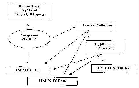
Figure 1 shows an overview of the methodology of multidimensional non-
porous LC-MS protein analysis methods used in some embodiments of the present
invention.
Figure 2 shows a 2-D image of NP-RP-HPLC-ESI-oaTOF total ion
chromatogram profiles of (a) Ca1dCL1, (b) ATIE, (c) ATI, (d) 10A, and (e) SUM-
149
human breast whole cell lysates. Peak intensity is depicted in different
shades of gray.
The inset shows the chromatogram for (a) Ca1dCL1.
Figure 3 shows a 1-D image of protein molecular weight for (a) Ca1dCLl, (b)
AT1E, (c) AT1, (d) 10A, and (e) SUM-149 human breast whole cell lysates. The
right bar shows the molecular weight scale (kDa) and the peak intensity is
depicted in
a color-coded mass map, where the intensity increases from shades of violet to
indigo,
then from shades of blue to green.
Figure 4 shows 2-Column NP-RP-HPLC protein profiles of (a) AT1E and (b)
AT 1 whole cell lysates.
Figure 5 shows a zoom-in 1-D image of protein molecular weight for (a)
Ca1dCLland (b) SUM-149 malignant human breast whole cell lysates. The right
bar
shows molecular weight scale (kDa) while the peak intensity is depicted in a
color-coded mass map.
8-
CA 02400460 2006-09-21
74667-201
Figures 6A and 6B show the identity and molecular weight of proteins
identified
from tryptic peptide maps using PDE-MALDI-TOF MS for ATlE lysates.
GENERAL DESCRIPTION OF THE INVENTION
The present invention relates to protein separation systems and methods
capable
of resolving large numbers of cellular proteins. The methods of the present
invention
provide protein profile maps for imaging and comparing protein expression
patterns.
The present invention provides alternatives to traditional separation methods
for the
screening of protein profiles. For exaniple, in some embodiments of the
present
invention, non-porous reverse-phase HPLC is used to separate and analyze
proteins as
an alternative to 1-D gels. Such methods are described herein, demonstrating
their
effectiveness for comparing expression profiles between cells.
For example, data produced using the systems and methods of the present
invention has provided accurate and informative expression information from
whole
cell lysates of human breast cancer cell lines. A series of cell lines
representing
sequential stages in the development of breast cancer (MCF10 model) were
examined.
These cell lines have been developed from spontaneously immortalized breast
epithelial cells obtained from a patient with fibrocystic disease (Soule et
al., Cancer
Research 50:6075 [1990]) and include premalignant (Miller et al., J. Natl.
Cancer Inst.,
85:1725 [1993]) and Dawson et al., Am. J. Pathol., 148:313 [1996]) as well as
malignant cell lines (Santner et al., Proc. Am. Assoc. Cancer Res., 39:202
[1998]). As
all stages are derived from a single patient, differences in background gene
expression
are minimized. Using the systems and methods of the present invention, it was
shown
that elevated levels of proteins or the appearance of new proteins can be
observed in
malignant cells as compared to premalignant or normal cells. Moreover, a mass
map
of intact proteins from cell lysates can be obtained. This mass map can be
used for
differential display of protein molecular weights in order to observe
differences in
quantitative expression and changes in structure due to post translational
modifications.
In addition, proteins can be collected in the liquid phase and identified by
mass
9-
CA 02400460 2002-08-07
WO 01/59460 PCT/US01/03887
spectroscopy tryptic mapping procedures. Of great relevance, it is shown that
distinct
changes in phosphorylation patterns are observed during neoplastic
progression.
The systems and methods of the present invention may be used to analyze any
protein-containing sample and to compare the protein content of the sample to
other
desired samples (e.g., sample from another cell or reference sample that
represent a
known condition or status). A major advantage of the systems and methods of
the
present invention over traditional techniques is the rapid assay times and
amenability
to automation. For example, in some preferred embodiments of the present
invention,
proteins are processed in the liquid phase to allow automated transfer of the
analyzed
sample from one apparatus (e.g., a separation column) to another apparatus
(e.g., mass
spectrometer). In recent work, several liquid phase based techniques have been
developed for separation of proteins (Yang et al., Anal. Chem., 70:3235
[1998];
Opitek et al., Anal. Biochem., 258:344 [1998]; Ayala et al., Appl. Biochem.
Biotech.,
69:11 [1998]; Hayakawa et al., Anal. Chim. Acta 372:281 [1998]; Nilsson et
al.,
Electrophoresis 20:860 [1999]; Nilsson et al., Rapid Comm. Mass Spec., 11:610
[1997]; Davidsson et al., Anal. Chem., 71:642 [1999]). Of note has been the
use of a
nonporous (NP) silica based media for separation of proteins in reversed-phase
HPLC.
This media has been used for separation of proteins from whole cell lysates of
bacterial cells and various mammalian cells (Wall et al., Anal. Chem., 71:3894
[1999]
and Chong et al., Rapid Commun. Mass Spec., 13:1808 [1999]). These NP packing
materials have been shown to provide important advantages in the separation of
protein
mixtures where separations of whole cell lysates can be performed in 15-30
minutes
with excellent resolution. The use of these NP materials in reverse phase HPLC
avoids the problems of proteins sticking inside the pores of the porous
materials and
results in considerably improved resolution and protein recovery. Of great
importance
is that the ability to separate and isolate proteins in the liquid phase
allows easy
interfacing of the separation methods to mass detection techniques for
identification
and molecular weight analysis.
-10-
CA 02400460 2002-08-07
WO 01/59460 PCT/USO1/03887
DEFINITIONS
To facilitate an understanding of the present invention, a number of terms and
phrases are defined below:
As used herein, the term "multiphase protein separation" refers to protein
separation comprising at least two separation steps. In some embodiments,
multiphase
protein separation refers to two or more separation steps that separate
proteins based
on different physical properties of the protein (e.g., a first step that
separates based on
protein charge and a second step that separates based on protein
hydrophobicity).
As used herein, the term "protein profile maps" refers to representations of
the
protein content of a sample. For example, "protein profile map" includes 1-
dimensional displays of total protein expressed in a given cell. In some
embodiments,
protein profile maps may also display subsets of total protein in a cell.
Protein profile
maps may be used for comparing "protein expression patterns" (e.g., the amount
and
identity of proteins expressed in a sample) between two or more samples. Such
comparing find use, for example, in identifying proteins that are present in
one sample
(e.g., a cancer cell) and not in another (e.g., normal tissue), or are over-
or under-
expressed in one sample compared to the other.
As used herein, the term "separating apparatus capable of separating proteins
based on a physical property" refers to compositions or systems capable of
separating
proteins (e.g., at least one protein) from one another based on differences in
a physical
property between proteins present in a sample containing two or more protein
species.
For example, a variety of protein separation columns and composition are
contemplated including, but not limited to ion exclusion, ion exchange,
normal/reversed phase partition, size exclusion, ligand exchange, liquid/gel
phase
isoelectric focusing, and adsorption chromatography. These and other
apparatuses are
capable of separating proteins from one another based on a "physical
property."
Examples of physical properties include, but are not limited to, size, charge,
hydrophobicity, and ligand binding affinity. Such separation techniques yield
fractions
or subgroups of proteins "defined by a physical property," i.e., separated
from other
- 11 -
CA 02400460 2002-08-07
WO 01/59460 PCT/US01/03887
proteins in the sample on the basis of a difference in a physical property,
but with all
of the proteins in the fraction or subgroup sharing that physical property.
For
example, all of the proteins in a fraction may elute from a column at a
defined
solution condition (e.g., salt concentration) or narrow range of solution
conditions,
while other proteins not in the fraction remain bound to the column or elute
at
different solution conditions.
A "liquid phase" separating apparatus is a separating apparatus that utilizes
protein samples contained in liquid solution, wherein proteins remain
solubilized in
liquid phase during separation and wherein the product (e.g., fractions)
collected from
the apparatus are in the liquid phase. This is in contrast to gel
electrophoresis
apparatuses, wherein the proteins enter into a gel phase during separation.
Liquid
phase proteins are much more amenable to recovery/extraction of proteins as
compared
to gel phase. In some embodiments, liquid phase proteins samples may be used
in
multi-step (e.g., multiple separation and characterization steps) processes
without the
need to alter the sample prior to treatment in each subsequent step (e.g.,
without the
need for recovery/extraction and resolubilization of proteins).
As used herein, the term "displaying proteins" refers to a variety of
techniques
used to interpret the presence of proteins within a protein sample. Displaying
includes,
but is not limited to, visualizing proteins on a computer display
representation,
diagram, autoradiographic film, list, table, chart, etc. "Displaying proteins
under
conditions that first and second physical properties are revealed" refers to
displaying
proteins (e.g., proteins, or a subset of proteins obtained from a separating
apparatus)
such that at least two different physical properties of each displayed protein
are
revealed or detectable. For exanlple, such displays include, but are not
limited to,
tables including columns- describing (e.g., quantitating) the first and second
physical
property of each protein and two-dimensional displays where each protein is
represented by an X,Y locations where the X and Y coordinates are defined by
the
first and second physical properties, respectively, or vice versa. Such
displays also
- 12 -
CA 02400460 2002-08-07
WO 01/59460 PCT/USO1/03887
include multi-dimensional displays (e.g., three dimensional displays) that
include
additional physical properties.
As used herein, the term "detection system capable of detecting proteins"
refers
to any detection apparatus, assay, or system that detects proteins derived
from a
protein separating apparatus (e.g., proteins in one or fractions collected
from a
separating apparatus). Such detection systems may detect properties of the
protein
itself (e.g., UV spectroscopy) or may detect labels (e.g., fluorescent labels)
or other
detectable signals associated with the protein. The detection system converts
the
detected criteria (e.g., absorbance, fluorescence, luminescence etc.) of the
protein into
a signal that can be processed or stored electronically or through similar
means (e.g.,
detected through the use of a photomultiplier tube or similar system).
As used herein, the term "buffer compatible with an apparatus" and "buffer
compatible with mass spectrometry" refer to buffers that are suitable for use
in such
apparatuses (e.g., protein separation apparatuses) and techniques. A buffer is
suitable
where the reaction that occurs in the presence of the buffer produces a result
consistent
with the intended purpose of the apparatus or method. For example, a buffer
compatible with a protein separation apparatus solubilizes the protein and
allows
proteins to be separated and collected from the apparatus. A buffer compatible
with
mass spectrometry is a buffer that solubilizes the protein or protein fragmeni
and
allows for the detection of ions following mass spectrometry. A suitable
buffer does
not substantially interfere with the apparatus or method so as to prevent its
intended
purpose and result (i.e., some interference may be allowed, but not enough to
prevent
an accurate determination of mass).
As used herein, the term "automated sample handling device" refers to any
device capable of transporting a sample (e.g., a separated or un-separated
protein
sample) between components (e.g., separating apparatus) of an automated method
or
system (e.g., an automated protein characterization system). An automated
sample
handling device may comprise physical means for transporting sample (e.g.,
multiple
- 13 -
CA 02400460 2002-08-07
WO 01/59460 PCT/USO1/03887
lines of tubing connected to a multi-channel valve). In some embodiments, an
automated sample handling device is connected to a centralized control
network.
As used herein, the term "switchable multi channel valve" refers to a valve
that
directs the flow of liquid through an automated sample handling device. The
valve
preferably has a plurality of channels (e.g., 4 or more, and preferably, 6 or
more). In
addition, in some embodiments, flow to individual channels is "switched" on an
off.
In some embodiments, valve switching is controlled by a centralized control
system.
A switchable multi-channel valve allows nlultiple apparatus to be connected to
one
automated sample handler. For example, sample can first be directed through
one
apparatus of a system (e.g., a first chromatography apparatus). The sample can
then
be directed through a different channel of the valve to a second apparatus
(e.g., a
second chromatography apparatus).
As used herein, the terms "centralized control system" or "centralized control
network" refer to information and equipment management systems (e.g., a
computer
processor and computer memory) operably linked to multiple devices or
apparatus
(e.g., automated sample handling devices and separating apparatus). In
preferred
embodiments, the centralized control network is configured to control the
operations of
the apparatus and device linked to the network. For example, in some
embodiments,
the centralized control network controls the operation of multiple
chromatography
apparatus, the transfer of sample between the apparatus, and the analysis and
presentation of data.
As used herein, the terms "computer memory" and "computer memory device"
refer to any storage media readable by a computer processor. Examples of
computer
memory include, but are not limited to, RAM, ROM, computer chips, digital
video
disc (DVDs), compact discs (CDs), hard disk drives (HDD), and magnetic tape.
As used herein, the term "computer readable medium" refers to any device or
system for storing and providing information (e.g., data and instructions) to
a computer
processor. Examples of computer readable media include, but are not limited
to,
- 14-
CA 02400460 2002-08-07
WO 01/59460 PCT/US01/03887
DVDs, CDs, hard disk drives, magnetic tape and servers for streaming media
over
networks.
As used herein, the terms "processor" and "central processing unit" or "CPU"
are used interchangeably and refers to a device that is able to read a program
from a
computer memory (e.g., ROM or other computer memory) and perform a set of
steps
according to the program.
As used herein, the term "sample" is used in its broadest sense. In one sense
it
can refer to a cell lysate. In another sense, it is meant to include a
specimen or culture
obtained from any source, including biological and environmental samples.
Biological
samples may be obtained from animals (including humans) and encompass fluids,
solids, tissues, and gases. Biological samples include blood products (e.g.,
plasma and
serum), saliva, urine, and the like and includes substances from plants and
microorganisms. Environmental samples include environmental material such as
surface matter, soil, water, and industrial samples. These examples are not to
be
construed as limiting the sample types applicable to the present invention.
DETAILED DESCRIPTION OF THE INVENTION
The present invention provides a novel separation methods for the detection of
differential expression of proteins in two or more cell types (e.g., in
cancerous and
non-cancerous cell lines). The present invention is not limited by the type of
samples
being compared. The methods of the present invention are suitable for use in
any
situation where it is advantageous to determine the difference in protein
expression
between two or more samples. The present invention thus provides methods
suitable
for a variety of diagnostic, screening (e.g., drug screening), and research
uses,
including, but not limited to, those disclosed herein.
In some preferred embodiments, the present invention provides methods of
separating proteins using any suitable protein separation technique (e.g., non-
porous
RP-HPLC) linked to mass spectroscopy to generate a protein mass map, and
comparing expression patterns among one or more samples. The following
discussion
15-
CA 02400460 2002-08-07
WO 01/59460 PCT/USO1/03887
is provided in two sections: I) separation and mass spectroscopic analysis;
and II)
differential protein expression in hunian breast cancer cell lines.
1. Separation and Analysis
In some embodiments, the present invention provides methods of separating
and analyzing protein expression in one or more cell lines or types. Cells are
lysed
using any suitable method, including but not limited to, those disclosed
herein.
Following lysis, cell extracts are first separated based on a physical
property. The
present invention is not limited to separation based on any particular
property. Nor is
the present invention limited to any particular separation method.
Following separation, the mass, abundance, and identity of proteins in the
different cell samples being analyzed is determined (e.g., using mass
spectroscopy).
The present invention in not limited to any particular detection or mass
spectroscopy
technique. Any suitable mass spectroscopy technique may be utilized, including
but
not limited to, those disclosed herein. In some embodiments, following mass
spectroscopy, a 1-D protein map is generated that compares the protein
expression
levels of the various samples being analyzed.
In some embodiments of the present invention, protein separation and analysis
is automated. In some embodiments, the process is controlled by a centralized
control
network including an automated sample handling device and a centralized
control
network.
A. Separation
In preferred embodiments, prior to analyzing protein mass and expression
patterns, proteins are separated based on one or more physical properties. For
example, in some embodiments of the present invention, proteins are separated
by
hydrophobicity using non-porous (NP) reversed phase (RP) HPLC (See e.g., Liang
et
al., Rap. Comm. Mass Spec., 10:1219 [1996]; Griffin et al., Rap. Comm. Mass
Spec.,
9:1546 [1995]; Opiteck et al., Anal. Biochem. 258:344 [1998]; Nilsson et al.,
Rap.
- 16 -
CA 02400460 2002-08-07
WO 01/59460 PCT/USO1/03887
Comm. Mass Spec., 11:610 [1997]; Chen et al., Rap. Comm. Mass Spec., 12:1994
[1998]; Wall et al., Anal. Chem., 71:3894 [1999]; Chong et al., Rap. Comm.
Mass
Spec., 13:1808 [1999]). Illustrative Example 2 provides a description of one
NP-
HPLC method suitable for use in the present invention. One skilled in the art
recognizes that other NP-HPLC or separation methods may be utilized in the
methods
of the present invention.
The present invention provides the novel combination of employing non-porous
RP packing materials (Eichrom) with a RP HPLC compatible detergent (e.g., n-
octyl
f3-D-galactopyranoside) to facilitate the separation and mass detection
methods of the
present invention. This detergent is also compatible with mass spectrometry
due to its
low molecular weight. These columns are well suited to this task as the non-
porous
packing they contain provides optimal protein recovery and rapid efficient
separations.
It should be noted that though several detergents are disclosed herein for
increasing
protein solubility while being compatible with RP HPLC there are many other
different detergents (e.g., low molecular weight non-ionic) that could be used
for this
purpose.
This method provides for exceptionally fast and reproducible high-resolution
separations of proteins according to their hydrophobicity and molecular
weight. The
non-porous silica packing material used in these reverse phase separations
eliminates
problems associated with porosity and low recovery of larger proteins, as well
as
reducing analysis times by as much as one third. Separation efficiency remains
high
due to the small diameter of the spherical particles, as does the loadability
of the NP
RP HPLC columns.
In some embodiments, proteins are reduced and alkylated (e.g., with DTE and
iodoacetamide respectively) prior to the NP-HPLC step. This step insures that
all
disulfide bonds are broken and optimal proteolysis is produced. This
derivatization
step can be added to the NP RP HPLC method by performing the reduction and
alkylation step prior to NP RP HPLC or during cell lysis.
- 17-
CA 02400460 2002-08-07
WO 01/59460 PCT/US01/03887
The present invention is not limited to any one separation technique. Indeed,
a
variety of separation techniques are contemplated, including, but not limited
to, 1-D
SDS PAGE lane gels and various chromatography techniques.
In some preferred embodiments, the separation is performed in the liquid
phase.
Separation in the liquid phase facilitates efficient analysis of the separated
proteins and
enables products to be fed directly into additional analysis steps (e.g.,
directly into
mass spectrometry analysis). In sonie preferred embodiments involving
separation in
the liquid phase, sample handling is automated. For example, an automated
sample
handler is utilized to transfer samples to the HPLC apparatus, collect peak
fractions,
and transfer fractions to the mass spectroscopy analysis step.
B. Mass Spectroscopy Analysis
In preferred embodiments of the present invention, separation (e.g., by NP-
HPLC) is followed by mass spectroscopy analysis. In some embodiments, the
eluent
from NP-RP-HPLC is analyzed directly with ESI-oaTOF MS for on-line molecular
weight determination as well as relative peak abundance in the sample. In
other
embodiments, the proteins are separated and detected by UV absorption. In yet
other
embodiments, the eluting proteins are collected and the fractions digested
with trypsin
so that the resulting tryptic peptides can be mapped with MALDI-TOF MS or
ESI-QIT-reTOF MS. In still further embodiments, the protein fraction are also
sized
on MALDI-TOF MS for protein molecular weight.
The present invention is not limited by the nature of the mass spectrometry
technique utilized for such analysis. For example, techniques that find use
with the
present invention include, but are not limited to, ion trap mass spectrometry,
ion
trap/time-of-flight mass spectrometry, quadrupole and triple quadrupole mass
spectrometry, Fourier Transforni (ICR) mass spectronietry, and niagnetic
sector mass
spectrometry. Those skilled in the art will appreciate the applicability of
other mass
spectroscopic techniques to such methods.
For example, in some embodiments, proteins are analyzed simultaneously to
determine molecular weight and identity. A fraction of the effluent from the
- 18 -
CA 02400460 2006-09-21
74667-201
separation step is used to determine molecular weight by either MALDI-TOF-MS
or
ESI oa TOF (LCT, Micromass) (See e.g., U.S. Pat. No. 6,002,127.
The remainder of the eluent is used to determine the
identity of the proteins via digestion of the proteins and analysis of the
peptide mass
map fingerprints by either MALDI-TOF-MS or ESI oa TOF. The molecular weight
protein map is matched to the appropriate digest fingerprint by correlating
the
molecular weight total ion chromatograms (TIC's) with the UV-chromatograms and
by
calculation of the various delay times involved. The UV-chromatograms are
automatically labeled with the digest fingerprint fraction number. The
resulting
molecular weight and digest mass fingerprint data can then be used to search
for the
protein identity via web-based programs like MSFit (UCSF).
In some embodiments, proteins are transferred to the mass spectroscopy step
via an automated sample handling system. In some embodiments, data is
automatically transferred to analysis software for the generation of protein
profile
maps.
C. Software and Data Presentation
The data generated by the above listed techniques may be presented as 1-D
mass maps of intact proteins. In some embodiments, MaxEnt (version 1) software
and
Mass Lynx version 3.4 (Micromass) are used to analyzed mass spectroscopy data.
The
protein molecular weights are determined by MaxEnt deconvolution of multiply
charged protein umbrella mass spectra that are obtained by combining anywhere
from
10 to 60 seconds of data from the initial total ion chromatogram (TIC). All
deconvoluted mass spectra from a given TIC are added together to produce one
mass
spectrum for each TIC.
In some embodiments, the data generated in the mass spectroscopy analysis
(e.g., TIC's or integrated and deconvoluted mass spectra) are converted to
ASCII
format and then plotted vertically, using a 256 step gray scale, such that
peaks are
represented as darkened bands against a white background.
19-
CA 02400460 2002-08-07
WO 01/59460 PCT/US01/03887
In other embodiments, a color coded 1-D protein profile mass map is generated
from differential display of protein molecular weights. In some embodiments,
the
image is displayed by a computer system as a color-coded mass map, where the
intensity of the protein bands corresponds to colors of the rainbow,
increasing from
blue to green to yellow to red. Thus, the image provides a protein expression
pattern
that can be used to locate proteins that are differentially displayed in
different samples
(e.g., cells representing different stages of a cancer). Naturally, the image
can be
adjusted to show a more detailed zoom of a particular region or the more
abundant
protein signals can be allowed to saturate thereby showing a clearer image of
the less
abundant proteins. As the image is automatically digitized it may be readily
stored
and used to analyze the protein profile of the cells in question. Protein
bands on the
image can be hyper-linked to other experimental results, obtained via analysis
of that
band, such as peptide mass fingerprints and MSFit search results. Thus all
information
obtained about a given 1-D image, including detailed mass spectra, data
analyses, and
complementary experiments (e.g., immuno-affinity and peptide sequencing) can
be
accessed from the original image.
The data generated by the above-listed techniques may also be presented as a
simple read-out. For example, when two or more samples are compared (e.g.,
cancerous and non-cancerous cells), the data presented may detail the
difference or
similarities between the samples (e.g., listing only the proteins that differ
in identity or
abundance between the samples). In this regard, when the differences between
samples (e.g., cancerous and non-cancerous cells) are indicative of a given
condition
(e.g., cancer cell), the read-out may simply indicate the presence or identity
of the
condition. In one embodiment, the read-out is a simple +/- indication of the
presence
of particular proteins or expression patterns associated with a specific
condition that is
to be analyzed.
A useful feature of the liquid phase method of the present invention is the
capability of the high resolution mass spectrometry to quantitate which allows
the
observer to record relative levels of each form of a given protein.
Consequently, it is
- 20 -
CA 02400460 2002-08-07
WO 01/59460 PCT/US01/03887
contemplated that one can determine the relative abundances of the
phosphorylated and
non-phosphorylated forms of a given protein. In addition, post-translational
modifications such as phosphorylation can be found by searching the data for
intervals
of some integer value times 80 Da.
With a mass resolution of 5000 Da, a 50000 Da protein can be resolved from a
50010 Da protein. Clearly, single phosphorylations on entire proteins can be
observed
with this level of resolution. Quantitative comparison between 1-D images can
be
achieved by spiking samples with known amounts of standard proteins and
normalizing
images through landmark proteins. Thus, the observer can detect significant
abundance changes in the protein profiles of different samples.
D. Automation
In some embodiments of the present invention, one or more (e.g., all) of the
above described steps are automated, for example, into one discrete
instrument. In
preferred embodiments, an automated on-line sample handling system fully
integrates
the separation and analysis steps of the methods of the present invention. The
sample
flows directly from the separation phase (e.g., NP-RP HPLC) to the mass
spectrometer. The automation of protein separation increases efficiency and
speed as
well as decreases sample loss or potential contamination that may occur
through
handling.
In some embodiments of the present invention, sample analysis is automated
and integrated with the centralized control network. For example, mass
spectroscopy
data is transferred to an integrated computer system containing software for
the
generation of 1-D protein maps. The integrated computer system is also capable
of
searching databases and generating a report. The report is provided to the
operator in
a format that is customized to the particular application. For example, the
report may
identify specific proteins that are present in one sample (e.g., a cancer cell
line) and
absent in another (e.g., a control non-cancerous cell line) or are present at
different
abundances between the two samples.
-21 -
CA 02400460 2002-08-07
WO 01/59460 PCT/US01/03887
E. Presentation of Results
In some preferred embodiments of the present invention, the information
generated by the protein profile display is distributed in an coordinated and
automated
fashion. In some embodiments of the present invention, the data is generated,
processed, and/or managed using electronic communications systems (e.g.,
Internet-
based methods).
In some embodiments, a computer-based analysis program is used to translate
the raw data generated by the protein profile map (e.g., identity and
abundance of
proteins in a sample) into data of predictive value for the clinician (e.g.,
the existence
of a malignancy, the probability of pre-cancerous cells becoming malignant, or
the
type of malignancy). The clinician (e.g., family practitioner or oncologist)
can access
the predictive data using any suitable means. Thus, in some preferred
embodiments,
the present invention provides the further benefit that the clinician, who is
not likely to
be trained in molecular biology or biochemistry, need not understand the raw
data of
the protein profile map. The data is presented directly to the clinician in
its most
useful form. The clinician is then able to immediately utilize the information
in order
to optimize the care of the subject.
The present invention contemplates any method capable of receiving,
processing, and transmitting the information to and from medical personal and
subject.
For example, in some embodiments of the present invention, a sample (e.g., a
biopsy)
is obtained from a subject and submitted to a protein profiling service (e.g.,
clinical
lab at a medical facility, protein profiling business, etc.) to generate raw
data. Once
received by the protein profiling service, the sample is processed and a
protein profile
is produced (i.e., protein expression data), specific for the condition being
assayed
(e.g., presence of specific cancerous or pre-cancerous cells).
The protein profile data is then prepared in a format suitable for
interpretation
by a treating clinician. For example, rather than providing raw protein
profile data,
the prepared format may represent a risk assessment or probability of
developing a
malignancy that the clinician may use or as recommendations for particular
treatment
- 22 -
CA 02400460 2002-08-07
WO 01/59460 PCT/US01/03887
options (e.g., surgery, chemotherapy, or observation). The data may be
displayed to
the clinician by any suitable method. For example, in some embodiments, the
protein
profiling service generates a report that can be printed for the clinician
(e.g., at the
point of care) or displayed to the clinician on a computer monitor.
In some embodiments, the protein profile information (e.g., protein profile
map) is first analyzed at a point of care or at a regional facility. The raw
data is then
sent to a central processing facility for further analysis into clinician. The
central
processing facility provides the advantage of privacy (all data is stored in a
central
facility with unifonn security protocols), speed, and uniformity of data
analysis. For
example, using an electronic communication system, the central facility can
provide
data to the clinician, the subject, or researchers. The use of an electronic
communications system allows protein _profile data to be viewed by clinicians
at any
location. For example, protein profile data could be accessed by a specialist
in the
type of disease (e.g., cancer) that the subject is affected with. This allows
even
remotely located subjects to have their protein profiles analyzed by the
leading experts
in a particular field. The present invention thus provides a coordinated,
timely, and
cost effective system for obtaining, analyzing, and distributing life-saving
information.
H. Differential Protein Expression in Human Breast Cancer Cell Lines
In some embodiments, the present invention provides methods of utilizing the
methods of the present invention to rapidly separate proteins from whole cell
lysates of
human breast cancer cells and detect the protein molecular weiglits on-line
(e.g., using
an ESI-oaTOF MS). In some embodiments, the present invention provides methods
of
detecting proteins that are more highly expressed in certain malignant and pre-
malignant cancers. In some embodiments, the molecular weight profiles are
displayed
as a mass map analogous to a virtual "1-D gel" and differentially expressed
proteins
are compared by image analysis. In other embodiments, the separated proteins
are
detected by LTV absorption and differentially expressed proteins are
quantitated. In yet
- 23 -
CA 02400460 2002-08-07
WO 01/59460 PCT/US01/03887
other embodiments, the eluting proteins are collected in the liquid phase, and
the
molecular weight and peptide maps determined by MALDI-TOF identification.
Illustrative Example 3 demonstrates the use of the methods of the present
invention to identify proteins differentially expressed in human breast cancer
cell lines.
Example 3A describes separation of proteins from various cancerous and pre-
cancerous
human breast cancer cell lines by HPLC and on-line detection by ESI-oa-TOF MS.
Figure 2 shows a 1-D image of the nonporous separation of five different whole
cell
lysates of human breast cancer cell lines. The intensity of the protein peaks
is shown
in different shades of gray so that the images provide a differential display
of key
oncoproteins according to their relative abundance.
In Figure 3 is shown a 1-D image of the proteins from the various breast
cancer cells lines displayed by molecular weight as determined by the LCT.
This
figure is very much an analogue to a 1-D gel, but provides very accurate
molecular
weight information with much improved resolution compared to a gel. The image
is
displayed by the computer as a color-coded mass map, where the intensity
increases
from shades of violet to indigo, then from shades of blue to green. The image
provides
a means of directly comparing protein expression in different cell lines with
respect to
quantitative expression and changes in protein structure through changes in
molecular
weight. The 1-D column separation methods of the present invention thus
provide a
means of rapidly monitoring changes in proteins that are highly expressed in
cancerous
cell lines.
Illustrative Example 3B provides methods for determining the identify of
differentially expressed proteins by using UV detection. The point in the
gradient at
which each peak is detected is highly reproducible. The molecular weights
determined
were correlated with the gradient of the separation, and the proteins were
collected in
the liquid phase at the corresponding point in the gradient. The proteins were
then
digested via trypsin or CNBR and analyzed by MALDI-MS. In Table 1 are listed a
selection of the key proteins and their molecular weight as determined by
MALDI-MS.
The present invention also provides methods of assaying the effects of various
compounds (e.g., hormones or environmental toxins) on the protein expression
patterns
-24-
CA 02400460 2002-08-07
WO 01/59460 PCT/US01/03887
of cancer cell lines. Previous studies have shown that estrogens stimulate the
proliferation of many breast tumors and cell lines derived from them
(Maggiolini et
al., Cancer Research 59:4864 [1999]). Estrogens also stimulate growth of
normal and
malignant breast cells in tissue culture (Thomas et al., J. Nat Cancer Inst.,
69:1017
[1982]). Further studies have also shown that estrogen is associated with a
significant
increase in breast cancer risk. These data taken together with other
epidemiological
data and laboratory evidence suggest that estrogen is a promoter of mammary
tumors
(Mils et al., Cancer 64:591 [1989]). In addition, estradiol-induced
inactivation of p53
may be involved in the tumorigenesis of estrogen-dependent neoplasm (Molinari
et al.,
Cancer Research 60:2594 [2000]).
Illustrative Example 3C describes the effects of estradiol exposure on AT1
cells. Proteins from cells exposed to estradiol and control cells not exposed
were
separated analyzed for molecular weight by MALDI-MS. In addition, part of the
fraction was digested by trypsin or CNBR for identification by MALDI-MS and
database searching. The protein profiles observed in Figure 4 are clearly
different
between the AT1 and AT1E samples. A list of some of the more abundant proteins
that have been identified by peptide mapping and MALDI-MS are listed in Table
2.
There are several proteins for which expression is induced by estradiol,
including PS2
estrogen inducible protein, estradiol 17 (3-dehydrogenase 7 and ERRI estrogen
receptor-like 1. Other proteins such as HSP 27 become much more highly
expressed in
response to estradiol.
Recent studies (Tesarik et al., Steroids, 64:22 [1999]) have shown that
estrogen/estradiol stimulates cell proliferation in breast tumors and cell
lines derived
from them, thus accelerating these cells towards malignancy. Indeed, in this
example,
the expression of key oncoproteins in AT1E starts to resemble those of the
highly
malignant cell line CaldCLI. This change in expression is evident in the
online
ESI-TOF-MS protein profile of Figure 3 and also in the UV chromatogram protein
profile. As expected the malignant and premalignant protein profiles vary
markedly
from the normal (immortalized) cell line MCF 10A. The present invention thus
- 25 -
CA 02400460 2002-08-07
WO 01/59460 PCT/US01/03887
provides methods of monitoring pre-cancerous cells for their level of
malignancy in
response to certain external stimulants such as estrogen. For example, the
protein
expression pattern of pre-cancerous cells identified in a patient could be
monitored
more closely if they were taking a compound known to effect cell
proliferation.
The over-expression of the c-src oncogene has been observed in several types
of cancers including breast and colon cancer (Rosen et al., J. Biol. Chem.,
261:13754
[1986]; Ottenhoff-Klaff et al., Cancer Res. 52:4773 [1992]; Brown et al., M.
T.;
Cooper, J. A., Biochimica et Biophysica acta 1287:121 [1996]; Mao et al.,
Oncogene
15:3083 [1997]; and Egan et al., Oncogene 18:1227 [1999]). Elevated levels of
c-src
kinase activity have been attributed to changes in phosphorylation patterns at
Tyr 530
(Brown et al., Biochimica et Biophysica Acta, 1287:121[1996]; Egan et al.,
Oncogene
18:1227 [1999]). C-src kinase activity has been implicated in tumorigenesis
and
metastasis in these cancers (Mao et al., Oncogene 15:3083 [1997]). It is also
suspected that c-src is responsible for phosphorylating other proteins, thus
changing
their functions in cell cycle regulation (Brown et al., Biochimica et
Biophysica Acta,
1287:121 [ 1996]).
Illustrative Example 3C (Figure 3) demonstrates that the molecular weight of
c-src in AT1E is 60,540 Da while that in CaldCLl is 62,780 Da. The database
value
is 59,835 Da. The two malignant cell lines, CaldCLl and SUM-149, also show
distinct differences in protein expression as seen in Figures 2 and 3. Figure
5 shows a
zoom-in 1-D image (from Figure 3) comparing Cal dCL I and SLTM- 149. The
molecular weight of c-src in SUM- 149 is 61,860 Da.
Illustrative Example 3C further describes the study of differences between c-
src
in the AT 1 and AT 1 E cell lines. More than 45 peptides from c-src were
detected and
analyzed and as expected most of them are the same between ATI and AT1E cell
lines. Several peptides were identified that are modified differently between
AT1 and
AT1E. It appears that there are differences in the phosphorylation patterns of
the
peptides detected. It is contemplated that the shift in molecular weight and
the change
in phosphorylation pattern as a function of cancer progression may be related
to
-26-
CA 02400460 2002-08-07
WO 01/59460 PCT/US01/03887
changes in protein structure and function that affect protein cascades leading
to
tumorigenesis and metastasis (Brown et al., Biochimica et Biophysica Acta,
1287:121[1996]; Egan et al., Oncogene 18:1227 [1999]). The present invention
thus
provides methods of identifying modifications (e.g., phosphorylation) present
or absent
only in pre-cancerous or cancerous cells.
It should be noted that other important proteins also show changes in
molecular
weight as a function of cancer progression. In particular, p-53 is a tumor
suppressor
protein that is involved in controlling the cell cycle. Wild-type p-53 is
involved in
maintaining genomic integrity and stability, where the p-53 searches for
mutations in
the DNA sequence (Gottleib and Oren, Biochimica et Biophysica Acta 1287:77
[1996];
"Tumor Suppressor Genes" in Cancer Biology, 3rd Ed., by Raymond W. Ruddon,
Oxford University Press, N. Y. 1995, pgs.318-340). If such mutations are found
a
series of events either leads to DNA repair or if repair is not effected then
to cell death
(Gottleib and Oren, Biochimica et Biophysica Acta 1287:77 [1996]; "Tumor
Suppressor Genes" in Cancer Biology, 3rd Ed., by Raymond W. Ruddon, Oxford
University Press, N. Y. 1995, pgs.318-340). This mechanism prevents the build-
up of
mutations in normal cells. However, if the p-53 is phosphorylated in critical
sites then
it does not function as a tumor suppressor and the cell divides without
control or
becomes immortalized ("Tumor Suppressor Genes" in Cancer Biology, 3rd Ed.,
Raymond W. Ruddon, Oxford University Press, N. Y. 1995, Ch. 8 pp. 318-340).
The
measured molecular weight of p-53 in Figure 3 as a function of progression
indicates
changes in structure that may affect its function.
Another protein associated with various types of cancer is Hsp 27 (Tetu et
al.,
Breast Cancer Research & Treatment 36:93 [1995]). Studies have shown that Hsp
27
can be induced or activated by excess estrogen/estradiol (Porter et al.,
Molecular
Endocrinology 10:1371 [1996]). In Figure 2 there are both changes in
expression and
molecular weight observed in HSP 27 as a function of progression.
- 27 -
CA 02400460 2002-08-07
WO 01/59460 PCT/US01/03887
The 1-D images generated by the methods of the present invention provide a
direct method of comparing the more highly expressed proteins in different
cell lines
at different stages of neoplastic progression.
It is demonstrated by illustrative Example 3 that the expressed protein
profiles
change during neoplastic progression and that many oncoproteins are readily
detected.
It is also shown that the response of premalignant cancer cells to estradiol
can be
rapidly screened by this method demonstrating significant changes in response
to an
external agent. Ultimately, the proteins can be studied by peptide mapping to
search
for post-translational modifications of the oncoproteins accompanying
progression.
The present invention thus provides improved methods for the study the
response of
cells in terms of protein expression to such external stimulants. In addition,
the
present invention provides methods of identifying pre-cancerous cells based on
protein
expression patterns, thus providing for intervention before malignancies have
developed. Early detection allows for increased treatment options, decreased
morbidity, and decreased mortality.
The present invention also provides the ability to monitor changes in protein
expression in cancer cells in response to pharmacological, environmental or
chemotherapeutic agents. The use of the 1-D liquid separation can provide
identification of the major changes in protein expression due to such external
agents.
III. Drug Screening
In some embodiments, the systems and methods of the present invention find
use in drug screening applications. For example, in some embodiments, the
effect of
one or more test compounds (e.g., pharmacological agents or environmental
toxins) on
the level of expression of one or more specific protein species is
investigated. In some
embodiments, the phosphorylation state of one or more proteins in the presence
or
absence of the test compound is investigated. In some embodiments, a protein
profile
map that highlights only the specific protein(s) of interest is generated.
In other embodiments, the effect of one or more compounds on the global
expression pattern of one or more samples (e.g., cell types) is investigated.
Protein
- 28 -
CA 02400460 2002-08-07
WO 01/59460 PCT/US01/03887
profile maps can be compared to maps generated from known cell types (e.g.,
differentiated or non-differentiated cell types or cancerous or non-cancerous
cell types)
in order to determine the state of the samples following exposure to the
research
compound.
The drug screening methods of the present invention are amenable to high-
throughput screening analysis. The computer generated protein profile maps of
the
present invention allow for the efficient analysis and comparison of large
numbers of
samples.
EXPERIMENTAL
The following examples serve to illustrate certain preferred embodiments and
aspects of the present invention and are not to be construed as limiting the
scope
thereof.
In the experimental disclosure which follows, the following abbreviations
apply: N (normal); M (molar); mM (millimolar); M (micromolar); mol (moles);
mmol (millimoles); mol (micromoles); nmol (nanomoles); pmol (picomoles); g
(grams); mg (milligrams); g (micrograms); ng (nanograms); 1 or L (liters); ml
(milliliters); l (microliters); cm (centimeters); mm (millimeters); m
(micrometers);
nm (nanometers); C (degrees Centigrade); PBS (phosphate buffered saline); and
Geno
Technology (Geno Technology Inc., St. Louis, MO).
Example 1
MCF10 Cell Line
This example describes the properties, growth procedures, and lysis procedures
of cell lines used in the following experiments. The MCF10 cell lines that
were used
in these experiments were obtained from spontaneously immortalized breast
epithelial
cells from a patient with fibrocystic disease (Soule et al., Cancer Research
50:6075
[1990]). The MCF10AT1 cell line produces xenograft lesions in immune deficient
mice that resemble high risk proliferative breast disease in women. These
lesions
spontaneously progress to invasive carcinoma at about 25% incidence during the
life of
- 29 -
CA 02400460 2002-08-07
WO 01/59460 PCT/USO1/03887
the host mouse (Miller et al., J. NatL Cancer Inst., 85:1725 [1993]; Dawson et
al.,
Am. Journal of Pathology 1996, 148, 313-319.). Progression of the MCF10AT1
lesions in mice is accelerated by estradiol (Shekhar et al., Int. J Oncology
13:907
[1998]). Because exposure to estrogen is a generally accepted risk factor for
breast
cancer development, MCF10AT1 serves as an important model to test the effect
of
estrogen on the development of human breast cancer.
A. Cell growth
MCF10AT1 cells are grown in monolayer on plastic in DMEM/F12 medium
(1:1 mixture of Dulbecco's modified Eagle's medium and Ham's F-12 medium)
supplemented with 5% hourse serum, 10 g/ml insulin, 20 ng/ml epidermal growth
factor, and 0.5 g/ml hydrocortisone. Approximately 50% confluent cell
monolayers
were treated with 10-' estradiol for 24 hours, collected by scraping, washed
two times
by centrifugation in phosphate buffered saline, and stored at -70 C. Estradiol
was
dissolved in absolute ethanol and controls were treated with the same volume
of
ethanol so that the final concentration of ethanol during treatment was 1%. A
fully
malignant metastatic variant, MCFIOCaIdCLI, was derived from premalignant
MCFIOAT xenografts (Santner et al., Proc. Am. Assoc. Cancer Res. 39:202
[1998]).
Cells were maintained in a humidified CO2 incubator at 37 C, and adherant
cells
harvested in log phase (75-80% confluence). In order to harvest the cells, the
growth
media was aspirated and the cells gently washed with PBS, prior to scraping
with a
rubber policeman. The cells were immediately frozen (-70 C) upon removal from
the
tissue culture dishes.
Protein profiles were also examined for SUM-149, which is a recently
developed cell line form a primary infiltrating ductal carcinoma of the breast
from a
patient with locally advanced disease. The culture medium for SUM-149
consisted of
Ham's F-12 with 5% fetal bovine serum, insulin, and hydrocortisone.
B. Cell lysis
- 30 -
CA 02400460 2006-09-21
74667-201
Proteins were extracted from cells using a chemical lysis procedure. The lysis
buffer contained 6M guanidine-HCL, 20 mM n-octyl P-D-glucopyransoside and 50
mM Tris. The cells were vortexed vigorously and stored overnight at -20 C. The
cells were then centrifuged at 17,000 rpm for 20 minutes. The supernatant was
removed from the cellular material and re-centrifuged at high speed to remove
any
particulate. Lysate was preferably used within 48 hours. Protein concentration
was
assayed using the protein dot metric kit (Geno Technology).
EXAMPLE 2
Methods
This example illustrates some of the experimental methods utilized in the
development of certain embodiments of the present invention.
A. Chemicals
The chemicals used in the following examples were used without prior
purification. Acetone (HPLC grade) was obtained from Fisher (Fair Lawn, NJ).
Acetonitrile, guanidine hydrochloride (gu-HCI), a-cyano-4hydroxycinnamic acid
((x-
CHCA) trifluoroacetic acid (TFA), formic acid (FA), and octyl glucopyranoside
(OCG) were from Aldrich (Milwaukee, WI). Trypsin was acquired from Promega
(Madison, WI). Distilled and deionized water was obtained from Milli-Q reagent
grade purification system from Millipore (Bedford, MA). The nitrocellulose
(NC)
~
used, Immobilin-NC pure was from Millipore.
B. HPLC
A Beckman (Fullerton, CA) System Gold HPLC was utilized. The pump
(model 128) has a gradient solvent delivery module with built-in system
controller.
The detector was a programmable detector module (Model 166) with an analytical
flow cell. The deuterium lamp provided a wide rage of detection from 190 to
700 nm.
All separations in this work were monitored at 214 nrn.
*Trade-mark
- 31 -
CA 02400460 2006-09-21
74667-201
ODSIIIE and ODSI NP RP HPLC columns (Eichrom Technologies, Darien, IL)
contained 1.5 m C18 (ODSI) non-porous silica beads. Column dimensions were
4.6
* 33 mm (ODSIIIE) and 4.6 * 14 mm (ODSI). The RP-HPLC separations of proteins
in the tumor cell lysate was performed via gradient elution of two solvents
(Solvent A:
Milli-Q water with 0.1%TFA; Solvent B:ACN with 0.1% TFA) with a flow rate of
1.0
mL/minute. The column was placed in a Timberline column heater and maintained
at
60 C. The gradient profile used was as follows: 1) 0% for 1.5 min; 2) 0 to 10%
acetonitrile (solvent B) in 2 minutes; 3) 10 to 60% B in 25 minutes; 4) 60 to
80% B
in 5 minutes; 5) 80 to 100% B in 1 minute; 6) 100% B for 2 minutes; 7) 100 to
0 %
B in 1 minute. In order to obtain a reproducible separation profile, the
sample was
"conditioned" to the column environment by mixing the sample with an
equivalent
amount of water (0.1% TFA) in a 1:1 ratio. This acidifying step was performed
prior
to sample injection. Each injection contained an average of 20-30 g of
protein. The
fractions collected were subsequently subjected to MALDI analysis to size the
protein
masses. Each of the peaks contained an average of 0.5-2.5 ug available for
analysis
after collection. The fractions were then digested by trypsin before
undergoing pulse-
delayed extraction (PDE) MALDI-TOF analysis to obtain their peptide maps.
C. MALDI-TOF MS
The TOF mass spectrometer employed in these studies was a modified Wiley-
McLareri design with a four-plate acceleration stage (Whittal and Li, Anal
Chem. 67:
1950 [1995]). It was capable of high voltage acceleration up to +/- 20kV (R.M.
Jordan Co., Grass Valley, CA). The laser source used to produce MALDI was a
MINILITE 10 Hz Nd:YAG laser system (Continuum, Santa Clara, CA). All mass
spectra were obtained using 355 nm radiation. The laser power density was
estimated
at 5x10G to 1x10' W/cmZ. the detector was a triple microchannel plate (MCP)
detector
(R.M. Jordon) which adapted a CuBe conversion dynode with post-acceleration
(PA)
capability up to +/- 12 kV in front of the MCP. The total ion acceleration
across the
TOF device may thus be > 30kV. The PA stage enhances the detection of heavy
species, but at the expense of resolution. In addition, pulsed delayed
extraction (PDE)
*Trade-mark
- 32 -
CA 02400460 2006-09-21
74667-201
was used to enhance the resolution for the analysis of the tryptic digests.
The 1-m
long flight tube was pumped to a base pressure of 8x10"' to 1x10"6 Torr by a
diffusion
pump (Varian Inc, Lexington, MA). Data was recorded using a LeCroy 9310AM (400
MHz) digital oscilloscope (LeCroy Corp., Chesnut Ridge, NY) and was processed
on a
Gateway 586 computer.
D. ESI-oaTOF MS Analysis
An LCT (Micromass, Ltd., Manchester, UK) was used for online
NP-RP-HPLC-ESI-oaTOF MS analysis. The MS parameters were set as follows:
Source - Capillary = 3000 V; Sample Cone 45 V, RF lens = 800 V; Extraction
Cone =
2 V; Desolvation Temperature = 300 C, and Source Temperature = 120-150 C. The
Beckman HPLC system (as described above) was interfaced with the LCT using the
NP column separations. The solvents for the mobile phase were (solvent A)
Milli-Q
water with 0.1% TFA + 0.2 to 0.3% FA and (solvent B) acetonitrile with 0.1%
TFA +
0.2 to 0.3% FA with a flow rate of 0.5 mLimin where the temperature of the NP
column was maintained at 65 C in a Timberline column heater. The gradient
profile
used for solvent B was generally as follows: 5% for 1.5 min; 5 to 20% in 2
min; 201
to 30% in 4 min; 30 to 45% in 10 min; 45 to 60% in 7.5 rnin; 60 to 70% for 4
min;
70 to 100% in 1 min, 100% for 2 min, 100 to 5% in I min, 5% for 2 min. The 0.5
mL/min was split to a 1:1 ratio before entering the electrospray source. The
chromatograms generated were deconvoluted using MaxEnt software (Micromass).
E. ESI-QIT-reTOF MS Analysis
The experimental setup consists of an HPLC separation system (Star 9012,
Varian Associates, Inc., Walnut Creek, CA) interfaced to an electrospray
ionization,
source with detection using a quadrupole ion trap reflectron time-of-flight
mass
spectrometer (Model C-1251, R. M. Jordan Co., Grass Valley, CA). This hybrid
mass
spectrometer has been described in detail in previous work (Michael et al.,
Anal
Chem., 65:2614 [1994]). Mass spectra were acquired using a DOS-based Borland
Pascal software program written in-house (Li et al., J. Am. Soc. Mass Spec.,
9:701
*Trade-mark
- 33 -
CA 02400460 2006-09-21
74667-201
[1998]), and digitization of the mass spectrum was performed by an 8-bit 250
MHz
analog bandwidth transient recorder (Model 9846, Precision Instruments,
Knoxville,
TN). Ions were accumulated for 333 ms and subsequently ejected by applying a
+2000V dc pulse to the entrance endcap (DEI GRX-3.OK-H, Directed Energy, Fort
Collins, CO).
The liquid chromatography system was operated at 200 L/min with a prime/
purge valve located immediately before the injection valve to split the mobile
phase in
a 3:1 ratio. The 10 cm x 250 m i.d. column was packed with porous 5 m C18
particles (Vydac, Hesperia, CA) in-house using the slurry packing method (Qian
et al.,
Anal Chem., 67:2870 [1995]). Mobile phase A consisted of Milli-Q H20 with 0.1%
formic acid and mobile phase B of acetonitrile with 0.1% formic acid. The
separation
gradient for mobile phase B was as follows: 5% for 5 min, 5% to 20% in 5 min,
20%
to 60% in 25 min, 60% to 100% in 15 min, 100% for 5 min, 100% to 5% in 5 min,
and 5% for 15 minutes.
F. Database Searching Procedure for Protein Identification
The MS-Fit sequence database located in the Protein Prospector program was
used for protein identification by entering the peptide masses generated by
tryptic
digestion. The program is available on the Internet at http://prospector.ucsf
edu.
Subsequently, other relevant parameters such as protein species, molecular
weight and
pl range are also entered in order to narrow down the search. In the
illustrative
examples of the present invention, Homo sapiens was chosen as the species.
Since
these proteins were obtained from HPLC, no isoelectric point (pl) information
was
available. Thus, the pI range was set between 3 and 10. The range ofmolecular
weight values for each search was determined by MALDI-TOF or ESI-TOF analysis.
The tolerance for the search of peptides. against the database was set at 2 Da
for
MALDI-MS spectra and 0.5 Da for QIT-reTOF-MS spectra.
*Trade-mark
34-
CA 02400460 2002-08-07
WO 01/59460 PCT/US01/03887
EXAMPLE 3
Mass Mapping of Proteins from Premalignant and Malignant Cell Lines
This Example describes multidimensional NP-RP-HPLC-MS analysis of human
breast cell lines representing different stages of neoplastic progression. An
overview
of the methodology is shown in Figure 1. The cell lines utilized included
MCF10A,
which is a "normal," but immortalized, cell line where the cell line keeps
dividing but
the phenotype is non-tumorigenic. The AT1 sample is considered a"premalignant"
stage in progression. The AT 1 E lysate is the AT 1 cell line that has been
exposed to
estradiol. The Ca1dCL1 is a highly malignant, tumorigenic cell line. These
four cell
lines have developed from a common precursor with essentially the same genetic
background. The SUM-149 sample is a highly malignant cell line that has been
developed from breast cancer tissue from a different patient and is included
for
comparison.
A. NP-HPLC and ESI-oa-TOF MS Analysis
An ODS2 nonporous column was used to separate the protein content of the
cell with on-line detection by ESI-oa-TOF MS using the Micromass LCT. The
total
ion chromatogram (TIC) mode of operation was used to collect the data. Figure
2
shows a 1-D image of the nonporous separation of five different whole cell
lysates of
human breast cell lines. A typical TIC of the nonporous separation of the
Ca1dCLI
cell line is shown in the inset of Figure 2. The y-axis in the 1-D image of
Figure 2
represents the elution time of each peak in the chromatogram. Each of the
bands in
the 1-D image corresponds to an eluting protein peak. The intensity of the
protein
peaks is shown in different shades of gray so that the images provide a
differential
display of key oncoproteins-according to their relative abundance.
In Figure 3 is shown a 1-D image of the proteins displayed by molecular
weight as determined by the LCT. In Figure 2, the bands represent the TIC,
where the
corresponding ESI mass spectra are ladders of multiply charged peaks generated
in the
electrospray process. These ladders are processed by the MaxEnt program to
provide
the molecular weights, which correspond to the protein bands of Figure 3. The
- 35 -
CA 02400460 2002-08-07
WO 01/59460 PCT/US01/03887
intensity of the protein peaks has been normalized relative to common peaks in
each
sample. The image is displayed by the computer as a color-coded mass map,
where
the intensity increases from shades of violet to indigo, then from shades of
blue to
green. The image provides a means of directly comparing protein expression in
different cell lines with respect to quantitative expression and changes in
protein
structure through changes in molecular weight. This is shown in Figure 3 in
comparison of the bands for c-src and p53 where large changes in expression
are
observed and where shifts in molecular weight were also detected.
In the images of Figures 2 and 3, approximately 75-80 unique protein masses
over a mass range of 5 to 90 kDa were determined using the MaxEnt software for
each cell line. Due to the dynamic range of the 1-D image in Figure 3, only
the more
highly expressed proteins appear in bands whereas the dark areas represent
protein
bands in extremely low intensity. It should also be noted in the TIC of Figure
2 that
the baseline of the separation never returns to zero. The mass spectrum shows
that
there are protein peaks everywhere (i.e., in both the peaks and the valleys).
This is to
be expected since there are thousands of proteins expressed in these cells.
The limited
number of peaks observed is either due to the fact that many of the lower
level
proteins are lost during the MaxEnt process or that many of the peaks in the
baseline
have not been analyzed. The results of this experiment (Figure 2) show that a
variety
of proteins are expressed very differently in the progression of cancer.
B. NP-HPLC with UV Detection and MALDI-MS Analysis
The data in Figures 2 and 3 provide maps from which protein expression can
be compared, but they do not in themselves provide protein identification. In
order to
obtain such identification, the nonporous separation was performed using UV
detection. The point in the gradient at which each peak is detected is highly
reproducible. The molecular weight of the proteins detected by the LCT during
the
on-line separation is not known since only multiply charged envelopes are
obtained,
and is determined later using MaxEnt. The molecular weights determined were
correlated with the gradient of the separation, and the proteins were
collected in the
- 36 -
CA 02400460 2002-08-07
WO 01/59460 PCT/USOl/03887
liquid phase at the corresponding point in the gradient. The proteins were
then
digested via trypsin or CNBR and analyzed by MALDI-MS. In Table 1 are listed a
selection of the key proteins and their molecular weight as determined by
MALDI-MS.
It should be noted that MALDI and ESI methods are complementary for
determination
of molecular weight in these samples. Some proteins are detected by both
methods;
however, some proteins are detected only by off-line liquid collection and
MALDI-MS, and others are detected by on-line ESI-MS. The results of this
experiment indicate that it is possible to determine the identity of proteins
detected by
on-line ESI-MS.
- 37 -
CA 02400460 2002-08-07
WO 01/59460 PCT/US01/03887
Table 1. Proteins identified in ATIE that are differentially expressed
compared to AT1:
Molecular Weight (Da): Detected
Protein Name SwissProteiri Database AT1E ATl Ca1dCL1
H-ras Transforrning protein P21 21298 21700 not detected 21695
PS2 Estrogen-inducible protein 9149 (unprocessed precursor) 8960 not detected
not detected
HS27 Heat shock protein 22327 22620 22560 not detected
Estradiol 17 P-dehydrogenase 7 38206 38220 not detected 38440
O-Actin or y-Actin 41737, 41793 42010 41710 42100
P53 Cellular tumor antigen 43653 44380 not detected 44880
ERRI Estrogen receptor-likel 55439 55960 55770 55640
C-src Tyrosine-protein kinase 59703 60540 60060 61860
Triosephosphate isomerase TIM 26670 26940 not detected 26850
- 38 -
CA 02400460 2002-08-07
WO 01/59460 PCT/US01/03887
C. The Effect of Estrogen on Protein Expression
This example describes the effects of estradiol exposure on protein expression
in AT 1 cells. Figures 4A and 4B show the chromatograms obtained by nonporous
separation of whole cell lysates of ATl and AT1E with UV detection at 214 mn.
These separations were performed with a 2-column tandem system: an ODSIIIE
column followed by an ODSI column. This method is used in order to optimize
the
loadability and the amount of sample collected for detailed sequencing
experiments.
The 2-column separation was performed at the expense of resolution in the
separations.
The proteins were collected in the liquid phase using a fraction collector and
analyzed for molecular weight by MALDI-MS. In addition, part of the fraction
was
digested by trypsin or CNBR for identification by MALDI-MS and database
searching.
The protein profiles observed in Figure 4 are clearly different between the
AT1 and
AT1E samples. A list of some of the more abundant proteins that have been
identified
by peptide mapping and MALDI-MS are listed in Table 2. There are several
proteins
in which expression is induced by estradiol, including PS2 estrogen-inducible
protein,
estradiol 17 P-dehydrogenase 7 and ERRI estrogen receptor-like 1. Other
proteins such
as HSP 27 become much more highly expressed in response to estradiol. The
change
in protein expression between ATl and AT1E is clearly evident as shown in
Figures 2-
4. In addition, the expression of key oncoproteins in AT1E starts to resemble
those of
the highly malignant cell line Ca1dCLI. This change in expression is evident
in the
online ESI-TOF-MS protein profile of Figure 3 and also in the UV chromatogram
protein profile. As expected the malignant and premalignant protein profiles
vary
markedly from the normal (immortalized) cell line MCFIOA.
The use of nonporous separations with online ESI-MS detection in Figure 3
clearly shows that the molecular weight of c-src in AT1E is 60,540 Da while
that in
Ca1dCL1 is 62,780 Da. The database value is 59,835 Da. Similar molecular
weights
were also determined by MALDI-MS for c-src. The two malignant cell lines,
Ca1dCL1 and SUM-149, also show distinct differences in protein expression as
seen in
Figures 2 and 3. Figure 5 shows a zoom-in 1-D image (from Figure 3) comparing
Cal
dCL I and SLTM- 149. The molecular weight of c-src in SUM- 149 is 61,860 Da.
- 39 -
CA 02400460 2002-08-07
WO 01/59460 PCT/US01/03887
In order to study differences between c-src in the ATI and AT1E cell lines,
detailed analysis of the proteins collected in the liquid phase by the tandem
column
separation were performed using capillary LC-MS, CE-MS and MALDI-MS of the
protein digests. The capillary LC-MS was performed using the LCT-MS and the
IT-reTOF-MS. The CE-MS was performed on the IT-reTOFMS. The coverage of the
c-src sequence was >50% using these methods with trypsin and CNBR digests.
More
than 45 peptides from c-src were detected and analyzed using these methods and
as
expected most of them are the same between ATI and AT1E cell line. However, as
shown in Table 2 for c-src, there are several peptides that are modified
differently
between AT 1 and AT 1 E. It appears that there are differences in the
phosphorylation
patterns of the peptides detected. In addition, Figure 2 shows changes in
expression
and molecular weight observed in HSP 27 as a function of cancer progression.
-40-
CA 02400460 2002-08-07
WO 01/59460 PCT/USO1/03887
Table 2
A comparison of modified tryptic peptides between AT 1 and AT 1 E
Amino Acid Masses Modifications
start end Experimental Database Peptide sequence ATI ATl P'
I 9 887.16 887.4951 (-)MGSNKSKPK(D) Acet N Acet N, 2PO4
14 655.96 656.2405 (K)DASQR (R ) not modified 1P0,
156 159 545.24 545.3524 (K)ITRR(E) not modified 1P0,
159 163 756.50 756.6895 (R)RESER(L) not modified 1P04
210 220 1215.52 1215.601 (K)LDSGGFYITSR(T) not modified 1P0,
244 260 1853.67 1854.0768 (R)LTTVCPTSKPQTQGLAK(D) not modified 1P0.
355 362 1082.95 1083.4277 (K)GETGKYLR(L) 2P0, 1PO.
363 382 2277.90 2276.5384 (R)LPQLVDMAAQIASGMAYVER(M) IMet-ox 2Met-ox, IPO
383 388 898.62 899.3599 (R)MNYVHR(D) 1P0, iMet-ox
423 430 872.21 871.5042 (R)QGAKFPIK(W) pyroGlu not modified
-41 -
CA 02400460 2006-09-21
74667-201
Various modifications and variations of the described
method and system of the invention will be apparent to those skilled in the
art without
departing from the scope and spirit of the invention. Although the invention
has been
described in connection with specific preferred embodiments, it should be
understood
that the invention as claimed should not be unduly limited to such specific
embodiments. Indeed, various modifications of the described modes for carrying
out
the invention which are obvious to those skilled in the art are intended to be
within the
scope of the following claims.
-42-