Note: Descriptions are shown in the official language in which they were submitted.
CA 02400559 2009-06-05
WO 01/62801 PCT/USOI/06191
HUMANIZED ANTIBODIES THAT SEQUESTER AP PEPTIDE
S
Technical Field
The invention relates to humanized antibodies that bind to an epitope between
amino acids 13 and 28 of the A(3 peptide and to preventive and therapeutic
treatment of
conditions associated with beta amyloid, such as Alzheimer's disease, Downs
syndrome,
and cerebral amyloid angiopathey. More specifically, it concerns use of
humanized
monoclonal antibodies to sequester amyloid beta (A43) peptide in plasma,
brain, and
cerebrospinal fluid to prevent accumulation or to reverse deposition of the
A(3 peptide
within the brain and in the cerebrovasculature and to improve cognition.
Background Art
A number of symptomologies which result in cognitive deficits, stroke, brain
hemorrhage, and general mental debilitation appear to be associated with
neuritic and
cerebrovascular plaques in the brain containing the amyloid beta peptide
(A(3). Among
these conditions are both pre-clinical and clinical Alzheimer's disease,
Down's syndrome,
and pre-clinical and clinical cerebral amyloid angiopathy (CAA). The amyloid
plaques are
formed from amyloid beta peptides. These peptides circulate in the blood and
in the
cerebrospinal fluid (CSF), typically in complexed form with lipoproteins. The
A(3 peptide
in circulating form is composed of 39-43 amino acids (mostly 40 or 42 amino
acids)
resulting from the cleavage of a common precursor protein, amyloid precursor
protein,
often designated APP. Some forms of soluble AP are themselves neurotoxic and
may
determine the severity of neurodegeneration and/or cognitive decline (McLean,
C. A., et
al., Ann. Neurol. (1999) 46:860-866; Lambert, M. P., et al. (1998) 95:6448-
6453; Naslund,
J., J. Am. Med. Assoc. (2000) 283:1571).
Evidence suggests that AP can be transported back and forth between brain and
the
blood (Ghersi-Egea, J-F., et al., J. Neurochem. (1996) 67:880-883; Zlokovic,
B. V., et al.,
Biochem. Biophys. Res. Comm. (1993) 67:1034-1040; Shibata M, et al., J. Clin.
Invest.
1
CA 02400559 2009-06-05
WO 01/62801 PCT/US01/06191
(2000) 106:1489-1499). Further A(3 in plaques is in an equilibrium with
soluble AD in the
brain and blood (Kawarabayashi T, et al., J. Nerlrosci. (2001) 21:372-381).
As described in PCT publication No. WO 2001/049875 and U. S. Patent No.
6,465,195,
total circulating levels of A(3 peptide in CSF are similar
in normal individuals and individuals predisposed to exhibit the symptoms of
Alzheimer's.
However, AP42 levels are lower on average in individuals with Alzheimer's
disease (Nitsch,
R. M., et al., Ann. Neurol. (1995) 37:512-518). It is known that AP42 is more
prone to
aggregate than is A(340, and when this happens, adverse consequences such as
A(3
deposition in amyloid plaques, conversion of AR to toxic soluble forms, nerve
cell damage,
and behavioral impairment such as dementia ensue (Golde, T.E., et al.,
Biochem. Biophys.
Acta. (2000) 1502:172-187).
Methods to induce an immune response to reduce amyloid deposits are described
in
PCT publication W099/27944 published 10 June 1999. The description postulates
that
full-length aggregated AR peptide would be a useful immunogen. Administration
of a M
fragment (amino acids 13-28) conjugated to sheep anti-mouse IgG caused no
change in
cortex amyloid burden, and only one in nine animals that received injections
of the Aa 13-
28 fragment-conjugate showed any lymphoproliferation in response to AD40. The
application also indicates that antibodies that specifically bind to AR
peptide could be used
as therapeutic agents. However, this appears to be speculation since the
supporting data
reflect protocols that involve active immunization using, for example, A1342.
The peptides
are supplied using adjuvants and antibody titers formed from the immunization,
as well as
levels of AD peptide and of the precursor peptide, are determined. The
publication strongly
suggests that AD plaque must be reduced in order to alleviate Alzheimer's
symptoms, and
that cell-mediated processes are required for successful reduction of A(3
plaque.
WO 99/60024, published 25 November 1999, is directed to methods for amyloid
removal using anti-amyloid antibodies. The mechanism, however, is stated to
utilize the
ability of anti-An antibodies to bind to pre-formed amyloid deposits (i.e.,
plaques) and
result in subsequent local microglial clearance of localized plaques. This
mechanism was
not proved in vivo. This publication further states that to be effective
against AR plaques,
anti-An antibodies must gain access to the brain parenchyma and cross the
blood brain
barrier.
2
CA 02400559 2009-06-05
WO 01/62801 PCT/USOI/06191
Several PCT applications that relate to attempts to control amyloid plaques
were
published on 7 December 2000. WO 00/72880 describes significant reduction in
plaque in
cortex and hippocampus in a transgenic mouse model of Alzheimer's disease when
treated
using N-terminal fragments of A(3 peptides and antibodies that bind to them,
but not when
treated with the AP 13-28 fragment conjugated to sheep anti-mouse IgG or with
an
antibody against the 13-28 fragment, antibody 266. The N-terminal directed
antibodies
were asserted to cross the blood-brain barrier and to induce phagocytosis of
amyloid
plaques in in vitro studies.
WO 00/72876 has virtually the same disclosure as WO 00/72880 and is directed
to
immunization with the amyloid fibril components themselves.
WO 00/77178 describes antibodies that were designed to catalyze the hydrolysis
of
j3-amyloid, including antibodies raised against a mixture of the phenylalanine
statine
transition compounds CysAf10-25, statine Phe19-Phe20 and Cys-Af 10_25 statine
Phe20-
Ala21 and antibodies raised against A1310-25 having a reduced amide bond
between Phe 1 g
and Phe20. This document mentions sequestering of AR, but this is speculation
because it
gives no evidence of such sequestering. Further, the document provides no in
vivo
evidence that administration of antibodies causes efflux of A(3 from the
central nervous
system, interferes with plaque formation, reduces plaque burden, forms
complexes between
the antibodies and A(3 in tissue samples, or affects cognition.
It has been shown that one pathway for A4 metabolism is via transport from CNS
to
the plasma (Zlokovic, B.V., et al., Proc. Natl. Acad. Sci (USA) (1996) 93:4229-
4234;
Ghersi Egea, 7-F., et al., J. Neurochem. (1996) 67:880-883). Additionally, it
has been
shown that Ali in plasma can cross the blood-brain-barrier and enter the brain
(Zlokovic, B.
V., et al., Biochem. Biophys Res. Comm. (1993) 67:1034-1040). It has also been
shown
that administration of certain polyclonal and monoclonal AR antibodies
decreases AR
deposition in amyloid plaques in the APPW'7r transgenic mouse model of
Alzheimer's
disease (Bard, F., et al., Nature Med. (2000) 6:916-919); however, this was
said to be due
to certain anti-A13 antibodies crossing the blood-brain-barrier stimulating
phagocytose of
amyloid plaques by microglial cells. In Bard's experiments, assays of brain
slices ex vivo
showed that the presence of added AP antibody, along with exogenously added
microglia,
induced phagocytosis of AP, resulting in removal of AP deposits.
3
CA 02400559 2009-06-05
WO 01/62801 PCT/USOI/06191
The levels of both soluble A f) a and Al)1 in CSF and blood can readily be
detected
using standardized assays using antibodies directed against epitopes along the
Al) chain.
Such assays have been reported, for example, in U.S. patents 5,766,846;
5,837,672;
and 5,593,846. These patents describe the production of murine monoclonal
antibodies to
the central domain of the AP peptide, and these were reported to have epitopes
around and
including positions 16 and 17. Antibodies directed against the N -terminal
region were
described as well. Several monoclonal antibodies were asserted to immunoreact
with
positions 13-28 of the Al) peptide; these did not bind to a peptide
representing
positions 17-28, thus, according to the cited patents, establishing that it is
this region,
including positions 16-17 (the a-secretase site) that was the target of these
antibodies.
Among antibodies known to bind between amino acids 13 and 28 of AP are mouse
antibodies 266,4G8, and 1 C2.
We have now unexpectedly found that administration of the 266 antibody very
quickly and almost completely restores cognition (object memory) in 24-month
old
hemizygous transgenic mice (APPv"7 ). Yet, the antibody does not have the
properties
that the art teaches are required for an antibody to be effective in treating
Alzheimer's
disease, Down's syndrome, and other conditions related to the Al) peptide. To
our further
surprise, we observed that antibodies that bind Al) between positions 13 and
28 (266 and
4G8) are capable of sequestering soluble forms of Al) from their bound,
circulating forms
in the blood, and that peripheral administration of antibody 266 results in
rapid efflux of
relatively large quantities of Al) peptide from the CNS into the plasma. This
results in
altered clearance of soluble Al), prevention of plaque formation, and, most
surprisingly,
improvement in cognition, even without necessarily reducing Al) amyloid plaque
burden,
crossing the blood brain barrier to any significant extent, decorating plaque,
activating
cellular mechanisms, or binding with great affinity to aggregated Al).
Disclosure of the Invention
The invention provides humanized antibodies, or fragments thereof, that
positively
affect cognition in diseases and conditions where Al) may be involved, such as
clinical or
pre-clinical Alzheimer's disease, Down's syndrome, and clinical or pre-
clinical cerebral
amyloid angiopathy. The antibodies or fragments thereof need not cross the
blood-brain
barrier, decorate amyloid plaque, activate cellular responses, or even
necessarily reduce
4
CA 02400559 2009-06-05
WO 01/62801 PCT/US01/06191
amyloid plaque burden. In another aspect, this invention provides humanized
antibodies
and fragments thereof that sequester A(3 peptide from its bound, circulating
form in blood,
and alter clearance of soluble and bound forms of AR in central nervous system
and
plasma. In another aspect, this invention provides humanized antibodies and
fragments
thereof, wherein the humanized antibodies specifically bind to an epitope
between amino
acids 13 and 28 of the AR molecule. In another aspect, the invention provides
humanized
antibodies and fragments thereof, wherein the CDR are derived from mouse
monoclonal
antibody 266 and wherein the antibodies retain approximately the binding
properties of the
mouse antibody and have in vitro and in vivo properties functionally
equivalent to the
mouse antibody (sequences SEQ ID NO:1 through SEQ ID NO:6). In another aspect,
this
invention provides humanized antibodies and fragments thereof, wherein the
variable
regions have sequences comprising the CDR from mouse antibody 266 and specific
human
framework sequences (sequences SEQ ID NO:7 - SEQ ID NO:10), wherein the
antibodies
retain approximately the binding properties of the mouse antibody and have in
vitro and in
vivo properties functionally equivalent to the mouse antibody 266. In another
aspect, this
invention provides humanized antibodies and fragments thereof, wherein the
light chain is
SEQ ID NO:11 and the heavy chain is SEQ ID NO: 12.
Also part of the invention are polynucleotide sequences that encode the
humanized
antibodies or fragments thereof disclosed above, vectors comprising the
polynucleotide
sequences encoding the humanized antibodies or fragments thereof, host cells
transformed
with the vectors or incorporating the polynucleotides that express the
humanized antibodies
or fragments thereof, pharmaceutical formulations of the humanized antibodies
and
fragments thereof disclosed herein, and methods of making and using the same.
Such humanized antibodies and fragments thereof are useful for sequestering AR
in
humans; for treating and preventing diseases and conditions characterized by
AP plaques or
A(3 toxicity in the brain, such as Alzheimer's disease, Down's syndrome, and
cerebral
amyloid angiopathy in humans; for diagnosing these diseases in humans; and for
determining whether a human subject will respond to treatment using human
antibodies
against A.
Administration of an appropriate humanized antibody in vivo to sequester A(3
peptide circulating in biological fluids is useful for preventive and
therapeutic treatment of
conditions associated with the formation of A(3-containing diffuse, neuritic,
and
cerebrovascular plaques in the brain. The humanized antibody, including an
5
CA 02400559 2009-06-05
WO 01/62801 PCTIUS01/06191
immunologically reactive fragment thereof, results in removal of the AR
peptide from
macromolecular complexes which would normally be relevant in transporting it
in body
fluids to and from sites where plaques can form or where it can be toxic. In
addition,
sequestering of plasma AD peptide with the antibody or fragment thereof
behaves as a
"sink," effectively sequestering soluble AD peptide in the plasma compartment,
and
inducing AD to enter the plasma from locations in the central nervous system
(CNS). By
sequestering AD in the blood, net efflux from the brain is enhanced and
soluble AD is
prevented from depositing in insoluble plaques and from forming toxic soluble
species in
the brain. In addition, insoluble AD in plaques which is in equilibrium with
soluble AD can
be removed from the brain through a sequestering effect in the blood.
Sequestering the AD,
peptide with the antibody also enhances its removal from the body and inhibits
toxic effects
of soluble AD in the brain and the development and further accumulation of
insoluble AD as
amyloid in plaques. The antibodies useful in the invention do not cross the
blood-brain
barrier in large amounts (<<0.1% plasma levels). In addition, humanized
antibodies used in
the invention, when administered peripherally, do not need to elicit a
cellular immune
response in brain when bound to Aft peptide or when freely circulating to have
their
beneficial effects. Further, when administered peripherally they do not need
to appreciably
bind aggregated AD peptide in the brain to have their beneficial effects.
Thus, in one aspect, the invention is directed to a method to treat and to
prevent
conditions characterized by the formation of plaques containing beta-amyloid
protein in
humans, which method comprises administering, preferably peripherally, to a
human in
need of such treatment a therapeutically or prophylactically effective amount
of humanized
monoclonal antibody or immunologically reactive fragment thereof, which
antibody
specifically binds to the mid-region of the 4 peptide. In another aspect, the
invention is
directed to a method to inhibit the formation of amyloid plaques and to clear
amyloid
plaques in humans, which method comprises administering to a human subject in
need of
such inhibition an effective amount of a humanized antibody that sequesters AD
peptide
from its circulating form in blood and induces efflux out of the brain as well
as altered AD
clearance in plasma and the brain. In additional aspects, the invention is
directed to such
humanized antibodies, including immunologically effective portions thereof,
and to
methods for their preparation.
6
CA 02400559 2009-06-05
WO 01/62801 PCTIUS01/06191
The invention also includes methods of reversing cognitive decline, improving
cognition, treating cognitive decline, and preventing cognitive decline in a
subject
diagnosed with clinical or pre-clinical Alzheimer's disease, Down's syndrome,
or clinical
or pre-clinical cerebral amyloid angiopathy, comprising administering to the
subject an
effective amount of a humanized antibody of the invention.
The invention also includes use of a humanized antibody of the invention for
the
manufacture of a medicament, including prolonged expression of recombinant
sequences of
the antibody or antibody fragment in human tissues, for treating, preventing,
or reversing
Alzheimer's disease, Down's syndrome, or cerebral amyloid angiopathy; for
treating,
preventing, or reversing cognitive decline in clinical or pre-clinical
Alzheimer's disease,
Down's syndrome, or clinical or pre-clinical cerebral amyloid angiopathy; or
to inhibit the
formation of amyloid plaques or the effects of toxic soluble AR species in
humans.
The invention is related to the surprising observation that within a short
period of
time after administration of an antibody of the present invention, relatively
large quantities
of A(3 efflux from the central nervous system to the blood. Thus, this
invention includes
methods to assess the response of a human subject to treatment with an
antibody that binds
A(3 or a fragment thereof, comprising: a) administering the antibody or a
fragment thereof
to the subject; and b) measuring the concentration of A(3 in the subject's
blood.
The invention also includes a method of treating a human subject with an
antibody
that binds A13 or a fragment thereof, comprising: a) administering a first
amount of the
antibody or fragment thereof to the subject; b) within 3 hours to two weeks
after
administering the first dose, measuring the concentration of A(3 in the
subject's blood; c) if
necessary, calculating a second amount of antibody or fragment thereof based
on the result
of step b), which second amount is the same as or different than the first
amount; and
d) administering the second amount of the antibody or fragment.
The invention also includes a method of assessing in a human subject the
efficacy
of an antibody that binds to A(3, or a fragment thereof, for inhibiting or
preventing AP
amyloid plaque formation, for reducing A(3 amyloid plaque, for reducing the
effects of
toxic soluble AP species, or for treating a condition or a disease associated
with AP plaque,
comprising: a) obtaining a first sample of the subject's plasma or CSF; b)
measuring a
baseline concentration of A(3 in the first sample; c) administering the
antibody or fragment
thereof to the subject; d) within 3 hours to two weeks after administering the
antibody or
fragment thereof, obtaining a second sample of the subject's plasma or CSF;
and e)
7
CA 02400559 2009-06-05
WO 01/62801 PCT/US01/06191
measuring the concentration of AD in the second sample; wherein, efficacy is
related to the
quantity of AD bound to the antibody in the blood and the concentration of AR
in the CSF.
Brief Description of the Drawings
Figure 1 shows the percentage of the Al) peptide withdrawn from human
cerebrospinal fluid through a dialysis membrane by Mab 266 as a function of
the molecular
weight cutoff of the dialysis membrane.
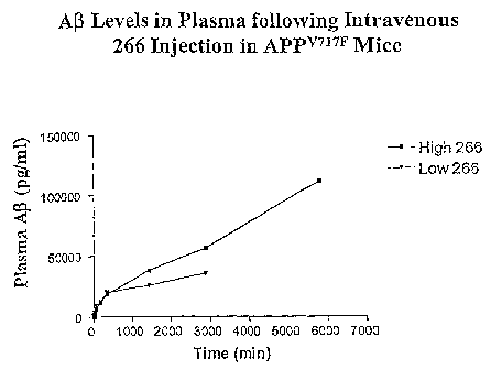
Figure 2 shows the concentration of AlTotal found in the plasma of an APPV717F
transgenic mouse after injection with either 200 p.g or 600 g of Mab 266 as a
fimction of
time.
Figure 3A shows the quantity of AD peptide deposition in the cortex in
APPv717F
transgenic mice treated with saline, mouse IgG, or Mab 266. Figure 3B shows
correlation
of these results with parental ori gin.
Figure 4 shows the polynucleotide sequences for expressing humanized 266 light
chain from plasmid pVk-Hu266 and the single amino acid codes for the expressed
humanized 266 light chain (corresponding to SEQ ID NO: 11 when mature).
Figure 5 shows the polynucleotide sequences for expressing humanized 266 heavy
chain from plasmid pVgl-Hu266 and the single amino acid codes for the
expressed
humanized 266 heavy chain (corresponding to SEQ ID NO:12 when mature).
Figure 6 is a plasmid map of pVk-Hu266.
Figure 7 is a plasmid map of pVgl-Hu266.
Modes of Carrying Out the Invention
The A(3 peptides that circulate in human biological fluids represent the
carboxy
terminal region of a precursor protein encoded on chromosome 21. It has been
reported
from the results of in vitro experiments that the Al) peptide has poor
solubility in
physiological solutions, since it contains a stretch of hydrophobic amino
acids which are a
part of the region that anchors its longer precursor to the lipid membranes of
cells. It is
thus not surprising that circulating Al) peptide is normally complexed with
other moieties
that prevent it from aggregating. This has resulted in difficulties in
detecting circulating
Al) peptide in biological fluids.
The above-mentioned patent documents (U.S. patents 5,766,846; 5,837,672 and
5,593,846) describe the preparation of antibodies, including a monoclonal
antibody,
8
CA 02400559 2009-06-05
WO 01/62801 PCTIUS01/06191
designated clone 266 which was raised against, and has been shown to bind
specifically to,
a peptide comprising amino acids 13-28 of the AP peptide. The present
applicants have
found that antibodies that bind within this region, in contrast to antibodies
that bind
elsewhere in the amino acid sequence of A(3, are able to sequester the soluble
A(3 peptide
very effectively from macromolecular complexes. This sequestration will effect
net A(3
peptide efflux from the CNS, alter its clearance in CNS and plasma, and reduce
its
availability for plaque formation. Thus, antibodies of this specificity,
modified to reduce
their immunogenicity by converting them to a humanized form, offer the
opportunity to
treat, both prophylactically and therapeutically, conditions that are
associated with
formation of beta-amyloid plaques. These conditions include, as noted above,
pre-clinical
and clinical Alzheimer's, Down's syndrome, and pre-clinical and clinical
cerebral amyloid
angiopathy.
As used herein, the word "treat" includes therapeutic treatment, where a
condition
to be treated is already known to be present and prophylaxis - i.e.,
prevention of, or
amelioration of, the possible future onset of a condition.
By "monoclonal antibodies that bind to the mid-region of A(3 peptide" is meant
monoclonal antibodies (Mab or Mabs) that bind an amino acid sequence
representing an
epitope contained between positions 13-28 of A. The entire region need not be
targeted.
As long as the antibody binds at least an epitope within this region
(especially, e.g.,
including the a-secretase site 16-17 or the site at which antibody 266 binds),
such
antibodies are effective in the method of the invention.
By "antibody" is meant a monoclonal antibody per se, or an immunologically
effective fragment thereof, such as an F b, F8b=, or F(ab=)2 fragment thereof.
In some
contexts, herein, fragments will be mentioned specifically for emphasis;
nevertheless, it
will be understood that regardless of whether fragments are specified, the
term "antibody"
includes such fragments as well as single-chain forms. As long as the protein
retains the
ability specifically to bind its intended target, and in this case, to
sequester A(3 peptide from
its carrier proteins in blood, it is included within the term "antibody." Also
included within
the definition "antibody" for example, are single chain forms, generally
designated Fõ
regions, of antibodies with this specificity. Preferably, but not necessarily,
the antibodies
useful in the invention are produced recombinantly, as manipulation of the
typically murine
or other non-human antibodies with the appropriate specificity is required in
order to
convert them to humanized form. Antibodies may or may not be glycosylated,
though
9
CA 02400559 2009-06-05
`glycosytated antibodies are preferred. Antibodies are properly cross-linked
via disulfide
bonds, as is well-known.
The basic antibody structural unit is known to comprise a tetramer. Each
tetramer is
composed of two identical pairs of polypeptide chains, each pair having one
"light" (about
25 kDa) and one "heavy" chain (about 50-70 kDa). The amino-terminal portion of
each
chain includes a variable region of about 100 to 110 or more amino acids
primarily
responsible for antigen recognition. The carboxy-terminal portion of each
chain defines a
constant region primarily responsible for effector function.
Light chains arc classified as gamma, mu, alpha, and lambda. Heavy chains are
classified as gamma, mu, alpha, delta, or epsilon, and define the antibody's
isotype as IgG,
IgM, IgA, IgD and IgE, respectively. Within light and heavy chains, the
variable and
constant regions are joined by a "I" region of about 12 or more amino acids,
with the heavy
chain also including a "D" region of about 10 more amino acids.
The variable regions of each light/bcavy chain pair form the antibody binding
site.
Thus, an intact antibody has two binding sites. The chains all exhibit the
same general
structure of relatively conserved framework regions (FR) joined by three
hypervariable
regions, also called complementarily determining regions or CDRs. The CDRs
from the
two chains of each pair are aligned by the framework regions, enabling binding
to a
specific epitopc. From N- terminal to C-terminal, both light and heavy chains
comprise the
domains F R I, CDR], FR2, CDR2, FR3, CDR3 and FR4. The assignment of amino
acids
to each domain is in accordance with well known conventions [Kabat "Sequences
of
Proteins of Immunological Interest" National Institutes of Health, Bethesda,
Md., 1987 and
1991; Chothia, et al., J. Mol, Biol. 196:901-917 (1987); Chothia, et al.,
Nature 342:878-883
(1989)].
As is well understood in the art, monoclonal antibodies can readily be
generated
with appropriate specificity by standard techniques of immunization of
mammals, forming
hybridomas from the antibody-producing cells of said mammals or otherwise
immortalizing them, and culturing the hybridomas or immortalized cells to
assess them for
the appropriate specificity. In the present case such antibodies could be
generated by
immunizing a human, rabbit, rat or mouse, for example, with a peptide
representing an
epitope encompassing the 13-28 region of the AP peptide or an appropriate
subregion
thereof. Materials for recombinant manipulation can be obtained by retrieving
the
nucleotide sequences encoding the desired antibody from the hybridoma or other
cell that
CA 02400559 2009-06-05
WO 01/62801 PCTIUS01/06191
produces it These nucleotide sequences can then be manipulated to provide them
in
humanized form.
By "humanized antbody" is meant an antibody that is composed partially or
fully
of amino acid sequences derived from a human antibody germline by altering the
sequence
of an antibody having non-human complementarity determining regions (CDR). The
simplest such alteration may consist simply of substituting the constant
region of a human
antibody for the marine constant region, thus resulting in a human/murine
chimera which
may have sufficiently low immunogenicity to be acceptable for pharmaceutical
use.
Preferably, however, the variable region of the antibody and even the CDR is
also
' humanized by techniques that are by now well known in the art. The framework
regions of
the variable regions are substituted by the corresponding human framework
regions leaving
the non-human CDR substantially intact, or even replacing the CDR with
sequences
derived from a human genome. Fully human antibodies are produced in
genetically
modified mice whose immune systems have been altered to correspond to human
immune
systems. As mentioned' above, it is sufficient for use in the methods of the
invention, to
employ an immunologically specific fragment of the antibody, including
fragments
representing single chain forms.
A humanized antibody again refers to an antibody comprising a human framework,
at least one CDR from a non human antibody, and in which any constant region
present is
substantially identical to a human immunoglobulin constant region, i.e., at
least about 85-
90%, preferably at least 95% identical. Hence, all parts of a humanized
antibody, except
possibly the CDRs, are substantially identical to corresponding parts of one
or more native
human immunoglobulin sequences. For example, a humanized immunoglobulin would
typically not encompass a chimeric mouse variable region/human constant region
antibody.
Humanized antibodies have at least three potential advantages over non-human
and
chimeric antibodies for use in human therapy:
1) because the effector portion is human, it may interact better with the
other parts
of the human immune system (e.g., destroy the target cells more efficiently by
complement-dependent cytotoxicity (CDC) or antibody-dependent cellular
cytotoxicity
(ADCC)).
2) The human immune system should not recognize the framework or C region of
the humanized antibody as foreign, and therefore the antibody response against
such an
11
CA 02400559 2009-06-05
WO 01/62801 PCTIUS01/06191
injected antibody should be less than against a totally foreign non human
antibody or a
partially foreign chimeric antibody.
3) Injected non-human antibodies have been reported to have a half-life in the
human circulation much shorter than the half-life of human antibodies.
Injected humanized
antibodies will have a half-life essentially identical to naturally occurring
human
antibodies, allowing smaller and less frequent doses to be given.
The design of humanized immunoglobulins may be carried out as follows. When
an amino acid falls under the following category, the framework amino acid of
a human
immunoglobulin to be used (acceptor immunoglobulin) is replaced by a framework
amino
acid from a CDR-providing non-human immunoglobulin (donor immunoglobulin):
(a) the amino acid in the human framework region of the acceptor
immunoglobulin
is unusual for human immunoglobulin at that position, whereas the
corresponding amino
acid in the donor immunoglobulin is typical for human immunoglobulin at that
position;
(b) the position of the amino acid is immediately adjacent to one of the CDRs;
or
(c) any side chain atom of a framework amino acid is within about 5-6
angstroms
(center-to-center) of any atom of a CDR amino acid in a three dimensional
inununoglobulin model [Queen, et al., op. cit., and Co, et al., Proc. Natl.
Acad. Sci. USA
88, 2869 (1991)]. When each of the amino acid in the human framework region of
the
acceptor immunoglobulin and a corresponding amino acid in the donor
immunoglobulin is
unusual for human immunoglobulin at that position, such an amino acid is
replaced by an
amino acid typical for human immunoglobulin at that position.
A preferred humanized antibody is a humanized form of mouse antibody 266. The
CDRs of humanized 266 have the following amino acid sequences:
light chain CDR1:
1 5 10 15
Arg Ser Ser Gin Ser Leu Ile Tyr Ser Asp (fly Asn Ala Tyr Lau His
(SEQ ID NO:1)
light chain CDR2:
1 5
Lys Val Ser Asn Arg Phe Ser (SEQ ID NO:2)
light chain CDR3:
1 5
Ser Gin Ser Thr His Val Pro Trp Thr (SEQ ID NO:3)
heavy chain CDR1:
1 5
Arg Tyr Ser Met Ser (SEQ ID NO:4)
12
CA 02400559 2009-06-05
WO 01/62801 PCT/USOI/06191
heavy chain CDR2:
1 5 10 15
Gln Ile Asn Ser Val Gly Asn Ser Thr Tyr Tyr Pro Asp Thr Val Lys Gly (SEQ
ID NO:5)
and, heavy chain CDR3:
1
Gly Asp Tyr (SEQ ID NO:6).
A preferred light chain variable region of a humanized antibody of the present
invention
has the following amino acid sequence, in which the framework originated from
human
germline Vk segments DPKl8 and J seqment Al, with several amino acid
substitutions to
the consensus amino acids in the same human V subgroup to reduce potential
immunogenicity:
1 5 10 15
Asp Xaa Val Met Thr Gin Xaa Pro Leu Ser Leu Pro Val Xaa Xaa
25 30
Gly Gln Pro Ala Ser Ile Ser Cys Arg Ser Ser Gln Ser Leu Xaa
35 40 45
Tyr Ser Asp Gly Asn Ala Tyr Leu His Trp Phe Leu Gin Lys Pro
50 55 60
Gly Gln Ser Pro Xaa Leu Leu Ile Tyr Lys Val Ser Asn Arg Phe
65 70 75
Ser Gly Val Pro Asp Arg Phe Ser Gly Ser Gly Ser Giy Thr Asp
80 85 90
Phe Thr Leu Lys Ile Ser Arg Val Glu Ala Giu Asp Xaa Gly Val
95 100 105
Tyr Tyr Cys Ser Gln Ser Thr His Val Pro Trp Thr Phe Giy Xaa
110
Gly Thr Xaa Xaa Glu Ile Lys Arg (SEQ ID NO:7)
wherein:
Xaa at position 2 is Val or Ile;
Xaa at position 7 is Ser or Thr;
Xaa at position 14 is Thr or Ser;
Xaa at position 15 is Leu or Pro;
Xaa at position 30 is Ile or Val;
Xaa at position 50 is Arg, Gln, or Lys;
Xaa at position 88 is Val or Leu;
13
CA 02400559 2009-06-05
WO 01/62801 PCT/US01/06191
Xaa at position 105 is Gln or Gly;
Xaa at position 108 is Lys or Arg; and
Xaa at position 109 is Vat or Leu.
A preferred heavy chain variable region of a humanized antibody of the present
invention has the following amino acid sequence, in which the framework
originated from
human germline VH segments DP53 and J segment JH4, with several amino acid
substitutions to the consensus amino acids in the same human subgroup to
reduce potential
immunogenicity.
1 5 10 15
Xaa Val Gin Leu Val Glu Xaa Gly Gly Gly Leu Val Gln Pro Gly
25 30
Gly Ser Leu Arg Leu Ser Cys Ala Ala Ser Gly Phe Thr Phe Ser
35 40 45
Arg Tyr Ser met Ser Trp Val Arg Gln Ala Pro Gly Lys Gly Leu
50 55 60
Xaa Leu Val Ala Gln Ile Asn Ser Val Gly Asn Ser Thr Tyr Tyr
65 70 75
Pro Asp Xaa Val Lys Gly Arg Phe Thr Ile Ser Arg Asp Asn Xaa
80 85 90
Xaa Asn Thr Leu Tyr Leu Gin Met Asn Ser Leu Arg Ala Xaa Asp
95 100 105
Thr Ala Val Tyr Tyr Cys Ala Ser Gly Asp Tyr Trp Gly Gln Gly
110
Thr Xaa Val Thr Val Ser Ser (SEQ ID NO:8)
wherein:
Xaa at position 1 is Glu or Gln;
Xaa at position 7 is Ser or Leu;
Xaa at position 46 is Glu, Val, Asp, or Ser,
Xaa at position 63 is Thr or Ser;
Xaa at position 75 is Ala, Ser, Val, or Thr,
Xaa at position 76 is Lys or Arg;
Xaa at position 89 is Glu or Asp; and
Xaa at position 107 is Leu or Thr.
14
CA 02400559 2009-06-05
WO 01/62801 PCTIUS01/06191
A particularly preferred light chain variable region of a humanized antibody
of the
present invention has the following amino acid sequence, in which the
framework
originated from human germline Vk segments DPK18 and J segment Jkl, with
several
amino acid substitutions to the consensus amino acids in the same human V
subgroup to
reduce potential immunogenicity:
1 5 10 15
Asp Val Val Met Thr Gln Ser Pro Leu Ser Leu Pro Val Thr Leu
20 25 30
Gly Gln Pro Ala Ser Ile Ser Cys Arg Ser Ser Gln Ser Leu Ile
35 40 45
Tyr Ser Asp Gly Asn Ala Tyr Leu His Trp Phe Leu Gln Lys Pro
50 55 60
Giy Gln Ser Pro Arg Leu Leu Ile Tyr Lys Val Ser Asn Arg Phe
65 70 75
Ser Gly Val Pro Asp Arg Phe Ser Gly Ser Gly Ser Gly Thr Asp
80 85 90
Phe Thr Leu Lys Ile Ser Arg Val Glu Ala Glu Asp Val Gly Val
95 100 105
Tyr Tyr Cys Ser Gln Ser Thr His Val Pro Trp Thr Phe Gly Gln
110
Gly Thr Lys Val Glu Ile Lys Arg (SEQ ID NO:9).
A particularly preferred heavy chain variable region of a humanized antibody
of the
present invention has the following amino acid sequence, in which the
framework
originated from human germline VH segments DP53 and J segment JH4:
1 5 10 15
Glu Val Gln Leu Val Glu Ser Gly Gly Gly Leu Val Gln Pro Gly
20 25 30
Gly Ser Leu Arg Leu Ser Cys Ala Ala Ser Gly Phe Thr Phe Ser
35 40 45
Arg Tyr Ser Met Ser Trp Val Arg Gln Ala Pro Gly Lys Gly Leu
55 60
Glu Leu Val Ala Gin Ile Asn Ser Val Gly Asn Ser Thr Tyr Tyr
65 70 75
Pro Asp Thr Val Lys Gly Arg Phe Thr Ile Ser Arg Asp Asn Ala
80 85 90
Lys Asn Thr Leu Tyr Leu Gin Met Asn Ser Leu Arg Ala Glu Asp
95 100 105
Thr Ala Val Tyr Tyr Cys Ala Ser Gly Asp Tyr Trp Gly Gln Gly
CA 02400559 2009-06-05
WO 01/62801 PCT/US01/06191
110
Thr Leu Val Thr Val Ser Ser (SEQ ID NO:10).
A preferred light chain for a humanized antibody of the present invention has
the
amino acid sequence:
1 5 10 15
Asp Val Val Met Thr Gln Ser Pro Leu Ser Leu Pro Val Thr Leu
20 25 30
Gly Gin Pro Ala Ser Ile Ser Cys Arg Ser Ser Gln Ser Leu.Ile
35 40 45
Tyr Ser Asp Gly Asn Ala Tyr Leu His Trp Phe Leu Gln Lys Pro
50 55 60
Gly Gln Ser Pro Arg Leu Leu Ile Tyr Lys Val Ser Asn Arg Phe
65 70 75
Ser Gly Val Pro Asp Arg Phe Ser Gly Ser Gly Ser Gly Thr Asp
80 85 90
Phe Thr Leu Lys Ile Ser Arg Val Glu Ala Glu Asp Val Gly Val
95 100 105
Tyr Tyr Cys Ser Gln Ser Thr His Val Pro Trp Thr Phe Gly Gln
110 115 120
Gly Thr Lys Val Glu Ile Lys Arg Thr Val Ala Ala Pro Ser Val
125 130 135
Phe Ile Phe Pro Pro Ser Asp Glu Gin Leu Lys Ser Gly Thr Ala
140 145 150
Ser Val Val Cys Leu Leu Asn Asn Phe Tyr Pro Arg Glu Ala Lys
155 160 165
Val Gln Trp Lys Val Asp Asn Ala Leu Gln Ser Gly Asn Ser Gin
170 175 180
Glu Ser Val Thr Glu Gln Asp Ser Lys Asp Ser Thr Tyr Ser Leu
185 190 195
Ser Ser Thr Leu Thr Leu Ser Lys Ala Asp Tyr Glu Lys His Lys
200 205 210
Val Tyr Ala Cys Glu Val Thr His Gln Gly Leu Ser Ser Pro Val
215
Thr Lys Ser Phe Asn Arg Gly Glu Cys (SEQ ID NO:11).
A preferred heavy chain for a humanized antibody of the present invention has
the
amino acid sequence:
1 5 10 15
Glu Val Gln Leu Val Glu Ser Gly Gly Gly Leu Val Gln Pro Gly
16
CA 02400559 2009-06-05
WO 01/62801 PCTIUS01/06191
20 25 30
Gly Ser Leu Arg Leu Ser Cys Ala Ala Ser Gly Phe Thr Phe Ser
35 40 45
Arg Tyr Ser Met Ser Trp Val Arg Gln Ala Pro Gly Lys Gly Leu
50 55 60
Glu Leu Val Ala Gin Ile Asn Ser Val Gly Asn Ser Thr Tyr Tyr
65 70 75
Pro Asp Thr Val Lys Gly Arg Phe Thr Ile Ser Arg Asp Asn Ala
80 85 90
Lys Asn Thr Leu Tyr Leu Gln Met Asn Ser Leu Arg Ala Glu Asp
95 100 105
Thr Ala Val Tyr Tyr Cys Ala Ser Gly Asp Tyr Trp Gly Gln Gly
110 115 120
Thr Leu Val Thr Val Ser Ser Ala Ser Thr Lys Gly Pro Ser Val
125 130 135
Phe Pro Leu Ala Pro Ser Ser Lys Ser Thr Ser Gly Gly Thr Ala
140 145 150
Ala Leu Gly Cys Leu Val Lys Asp Tyr Phe Pro Glu Pro Val Thr
155 160 165
Val Ser Trp Asn Ser Gly Ala Leu Thr Ser Gly Val His Thr Phe
170 175 180
Pro Ala Val Leu Gln Ser Ser Gly Leu Tyr Ser Leu Ser Ser Val
185 190 195
Val Thr Val Pro Ser Ser Ser Leu Gly Thr Gln Thr Tyr Ile Cys
200 205 210
Asn Val Asn His Lys Pro Ser Asn Thr Lys Val Asp Lys Lys Val
215 220 225
Glu Pro Lys Ser Cys Asp Lys Thr His Thr Cys Pro Pro Cys Pro
230 235 240
Ala Pro Glu Leu Leu Gly Gly Pro Ser Val Phe Leu Phe Pro Pro
245 250 255
Lys Pro Lys Asp Thr Leu Met Ile Ser Arg Thr Pro Glu Val Thr
260 265 270
Cys Val Val Val Asp Val Ser His Glu Asp Pro Glu Val Lys Phe
275 280 285
Asn Trp Tyr Val Asp Gly Val Glu Val His Asn Ala Lys Thr Lys
290 295 300
Pro Arg Glu Glu Gln Tyr Asn Ser Thr Tyr Arg Val Val Ser Val
305 310 315
Leu Thr Val Leu His Gln Asp Trp Leu Asn Gly Lys Glu Tyr Lys
17
CA 02400559 2009-06-05
WO 01/62801 PCTIUS01/06191
320 325 330
Cys Lys Val Ser Asn Lys Ala Leu Pro Ala Pro Ile Glu Lys Thr
335 340 345
Ile Ser Lys Ala Lys Gly Gln Pro Arg Glu Pro Gln Val Tyr Thr
350 355 360
Leu Pro Pro Ser Arg Asp Glu Leu Thr Lys Asn Gln Val Ser Leu
365 370 375
Thr Cys Leu Val Lys Gly Phe Tyr Pro Ser Asp Ile Ala Val Glu
380 385 390
Trp Glu Ser Asn Gly Gln Pro Glu Asn Asn Tyr Lys Thr Thr Pro
395 400 405
Pro Val Leu Asp Ser Asp Gly Ser Phe Phe Leu Tyr Ser Lys Leu
410 415 420
Thr Val Asp Lys Ser Arg Trp Gln Gln Gly Asn Val Phe Ser Cys
425 430 435
Ser Val Met His Glu Ala Leu His Asn His Tyr Thr Gln Lys Ser
440
Leu Ser Leu Ser Pro Gly Lys (SEQ ID NO:12).
Other sequences are possible for the light and heavy chains for the humanized
antibodies of the present invention and for humanized 266. The immunoglobulins
can have
two pairs of light chain/heavy chain complexes, at least one chain comprising
one or more
mouse complementarity determining regions functionally joined to human
framework
region segments.
In another aspect, the present invention is directed to recombinant
polynucleotides
encoding antibodies which, when expressed, comprise the heavy and light chain
CDRs
from an antibody of the present invention. As to the human framework region, a
framework or variable region amino acid sequence of a CDR providing non-human
immunoglobulin is compared with corresponding sequences in a human
immunoglobulin
variable region sequence collection, and a sequence having a high percentage
of identical
amino acids is selected. Exemplary polynucleotides, which on expression code
for the
polypeptide"chains comprising the heavy and light chain CDRs of monoclonal
antibody
266 are given in Figures 4 and 5. Due to codon degeneracy and non-critical
amino-acid
substitutions, other polynucleotide sequences can be readily substituted for
those
sequences. Particularly preferred polynucleotides of the present invention
encode
antibodies, which when expressed, comprise the CDRs of SEQ ID NO: 1 - SEQ ID
NO:6,
or any of the variable regions of SEQ ID NO:7 - SEQ ID NO:10, or the light and
heavy
chains of SEQ ID NO:11 and SEQ ID NO: 12.
18
CA 02400559 2009-06-05
WO 01/62801 PCT/USOI/06191
The polynucleotides will typically further include an expression control
polynucleotide sequence operably linked to the humanized immunoglobulin
encoding
sequences, including naturally-associated or heterologous promoter regions.
Preferably,
the expression control sequences will be eukaryotic promoter systems in
vectors capable of
transforming or transfecting eukaryotic host cells, but control sequences for
prokaryotic
hosts may also be used. Once the vector has been incorporated into the
appropriate host
cell line, the host cell is propagated under conditions suitable for high
level expression of
the nucleotide sequences, and, as desired, the collection and purification of
the light chains,
heavy chains, lightlheavy chain dimers or intact antibodies, binding fragments
or other
immunoglobulin forms may follow.
The nucleotide sequences of the present invention capable of ultimately
expressing
the desired humanized antibodies can be formed from a variety of different
polynucleotides
(genomic or cDNA, RNA, synthetic oligonucleotides, etc.) and components (e.g.,
V, J. D.
and C regions), as well as by a variety of different techniques. Joining
appropriate genomic
and synthetic sequences is a common method of production, but cDNA sequences
may also
be utilized.
Human constant region DNA sequences can be isolated in accordance with well
known procedures from a variety of human cells, but preferably from
immortalized B-cells.
The CDRs for producing the immunoglobulins of the present invention will be
similarly
derived from non-human monoclonal antibodies capable of binding to an epitope
between
amino acids 13 and 28 of the Ap peptide, which monoclonal antibodies are
produced in any
convenient mammalian source, including, mice, rats, rabbits, or other
vertebrates capable of
producing antibodies by well known methods, as described above. Suitable
source cells for
the polynucleotide sequences and host cells for immunoglobulin expression and
secretion
can be obtained from a number of sources well-known in the art.
In addition to the humanized immunoglobulins specifically described herein,
other
"substantially homologous" modified immunoglobulins can be readily designed
and
manufactured utilizing various recombinant DNA techniques well known to those
skilled in
the art. For example, the framework regions can vary from the native sequences
at the
primary structure level by several amino acid substitutions, terminal and
intermediate
additions and deletions, and the like. Moreover, a variety of different human
framework
regions may be used singly or in combination as a basis for the humanized
immunoglobulins of the present invention. In general, modifications of the
genes may be
19
CA 02400559 2009-06-05
WO 01/62801 PCT/US01/06191
readily accomplished by a variety of well-known techniques, such as site-
directed
mutagenesis.
Alternatively, polypeptide fragments comprising only a portion of the primary
antibody structure may be produced, which fragments possess one or more
immunoglobulin activities (e.g., complement fixation activity). These
polypeptide
fragments may be produced by proteolytic cleavage of intact antibodies by
methods well
known in the art, or by inserting stop codons at the desired locations in
vectors using site-
directed mutagenesis, such as after CHI to produce Fab fragments or after the
hinge region
to produce F(ab')2 fragments. Single chain antibodies maybe produced by
joining VL and
VH with a DNA linker.
As stated previously, the encoding nucleotide sequences will be expressed in
hosts
after the sequences have been operably linked to (i.e., positioned to ensure
the functioning
of) an expression control sequence. These expression vectors are typically
replicable in the
host organisms either as episomes or as an integral part of the host
chromosomal DNA.
Commonly, expression vectors will contain selection markers, e.g.,
tetracycline or
neomycin, to permit detection of those cells transformed with the desired DNA
sequences.
E.coli is a prokaryotic host useful particularly for cloning the
polynucleotides of
the present invention. Other microbial hosts suitable for use include bacilli,
such as
Bacillus subtilus, and other enterobacteriaceae, such as Salmonella, Serratia,
and various
Pseudomonas species. In these prokaryotic hosts, one can also make expression
vectors,
which will typically contain expression control sequences compatible with the
host cell
(e.g., an origin of replication). In addition, any of a number of well-known
promoters may
be present, such as the lactose promoter system, a tryptophan (trp) promoter
system, a beta-
lactamase promoter system, or a promoter system from phage lambda. The
promoters will
typically control expression, optionally with an operator sequence, and have
ribosome
binding site sequences and the like, for initiating and completing
transcription and
translation.
Other microbes, such as yeast, may also be used for expression. Saccharomyces
is
a preferred host, with suitable vectors having expression control sequences,
such as
promoters, including 3-phosphoglycerate kinase or other glycolytic enzymes,
and an origin
of replication, termination sequences and the like as desired.
In addition to microorganisms, mammalian tissue cell culture may also be used
to
express and produce the polypeptides of the present invention. Eukaryotic
cells are
CA 02400559 2009-06-05
WO 01/62801 PCT/US01/06191
actually preferred, because a number of suitable host cell lines capable of
secreting intact
in munoglobulins have been developed in the art, and include the CHO cell
lines, various
COS cell lines, Syrian Hamster Ovary cell lines, HeLa cells, preferably
myeloma cell lines,
transformed B-cells, human embryonic kidney cell lines, or hybridomas.
Expression
vectors for these cells can include expression control sequences, such as an
origin of
replication, a promoter, an enhancer, and necessary processing information
sites, such as
ribosome binding sites, RNA splice sites, polyadenylation sites, and
transcriptional
terminator sequences. Preferred expression control sequences are promoters
derived from
immunoglobulin genes, SV40, Adenovirus, Bovine Papilloma Virus,
cytomegalovirus and
the like.
The vectors containing the nucleotide sequences of interest (e.g., the heavy
and
light chain encoding sequences and expression control sequences) can be
transferred into
the host cell by well-known methods, which vary depending on the type of
cellular host.
For example, calcium chloride transfection is commonly utilized for
prokaryotic cells,
whereas calcium phosphate treatment or electroporation may be used for other
cellular
hosts.
Once expressed, the whole antibodies, their dimers, individual light and heavy
chains, or other immunoglobulin forms of the present invention can be purified
according
to standard procedures of the art, including ammonium sulfate precipitation,
ion exchange,
affinity, reverse phase, hydrophobic interaction column chromatography, gel
electrophoresis and the like. Substantially pure immunoglobulins of at least
about 90 to.
95% homogeneity are preferred, and 98 to 99% or more homogeneity most
preferred, for
pharmaceutical uses. Once purified, partially or to homogeneity as desired,
the
polypeptides may then be used therapeutically or prophylactically, as directed
herein.
The antibodies (including immunologically reactive fragments) are administered
to
a subject at risk for or exhibiting A(3-related symptoms or pathology such as
clinical or pre-
clinical Alzheimer's disease, Down's syndrome, or clinical or pre-clinical
amyloid
angiopathy, using standard administration techniques, preferably peripherally
(i.e. not by
administration into the central nervous system) by intravenous,
intraperitoneal,
subcutaneous, pulmonary, transdermal, intramuscular, intranasal, buccal,
sublingual, or
suppository administration. Although the antibodies may be administered
directly into the
ventricular system, spinal fluid, or brain parenchyma, and techniques for
addressing these
locations are well known in the art, it is not necessary to utilize these more
difficult
21
CA 02400559 2009-06-05
WO 01/62801 PCT/USOI/06191
procedures. The antibodies of the invention are effective when administered by
the more
simple techniques that rely on the peripheral circulation system. The
advantages of the
present invention include the ability of the antibody exert its beneficial
effects even though
not provided directly to the central nervous system itself Indeed, it has been
demonstrated
herein that the amount of antibody which crosses the blood-brain barrier is
<0.1% of
plasma levels and that the antibodies of the invention exert their ability to
sequester AP in
the peripheral circulation as well as to alter CNS and plasma soluble AP
clearance.
The pharmaceutical compositions for administration are designed to be
appropriate
for the selected mode of administration, and pharmaceutically acceptable
excipients such as
dispersing agents, buffers, surfactants, preservatives, solubilizing agents,
isotonicity agents,
stabilizing agents and the like are used as appropriate. Remington's
Pharmaceutical
Sciences. Mack Publishing Co., Easton PA, latest edition,
provides a compendium of formulation techniques as are generally known to
practitioners.
It may be particularly useful to alter the solubility characteristics of the
antibodies of the
invention, making them more lipophilic, for example, by encapsulating them in
liposomes
or by blocking polar groups.
Peripheral systemic delivery by intravenous or intraperitoneal or subcutaneous
injection is preferred. Suitable vehicles for such injections are
straightforward. In addition,
however, administration may also be effected through the mucosal membranes by
means. of
nasal aerosols or suppositories. Suitable formulations for such modes of
administration are
well known and typically include surfactants that facilitate cross-membrane
transfer. Such
surfactants are often derived from steroids or are cationic lipids, such as
N [l-(2,3-dioleoyl)propyl-N,N,N-trimethylammoniumchloride(DOTMA) or various
compounds such as cholesterol hemisuccinate, phosphatidyl glycerols and the
like.
The concentration of the humanized antibody in formulations from as low as
about
0.1% to as much as 15 or 20% by weight and will be selected primarily based on
fluid
volumes, viscosities, and so forth, in accordance with the particular mode of
administration
selected. Thus, a typical pharmaceutical composition for injection could be
made up to
contain 1 mL sterile buffered water of phosphate buffered saline and 1-100 mg
of the
humanized antibody of the present invention. The formulation could be sterile
filtered after
making the formulation, or otherwise made microbiologically acceptable. A
typical
composition for intravenous infusion could have a volume as much as 250 mL of
fluid,
such as sterile Ringer's solution, and 1-100 mg per mL, or more in antibody
concentration.
22
CA 02400559 2009-06-05
WO 01/62801 PCT/US01/06191
Therapeutic agents of the invention can be frozen or lyophilized for storage
and
reconstituted in a suitable sterile carrier prior to use. Lyophilization and
reconstitution can
lead to varying degrees of antibody activity loss (e.g. with conventional
immune globulins,
IgM antibodies tend to have greater activity loss than IgG antibodies).
Dosages may have
to be adjusted to compensate. The pH of the formulation will be selected to
balance
antibody stability (chemical and physical) and comfort to the patient when
administered.
Generally, pH between 4 and 8 is tolerated.
Although the foregoing methods appear the most convenient and most appropriate
for administration of proteins such as humanized antibodies, by suitable
adaptation, other
techniques for administration, such as transdermal administration and oral
administration
may be employed provided proper formulation is designed.
In addition, it may be desirable to employ controlled release formulations
using
biodegradable films and matrices, or osmotic mini-pumps, or delivery systems
based on
dextran beads, alginate, or collagen.
In summary, formulations are available for administering the antibodies of the
invention and are well-known in the art and may be chosen from a variety of
options.
Typical dosage levels can be optimized using standard clinical techniques and
will
be dependent on the mode of administration and the condition of the patient.
The following examples are intended to illustrate but not to limit the
invention.
The examples hereinbelow employ, among others, a murine monoclonal antibody
designated "266" which was originally prepared by immunization with a peptide
composed
of residues 13-28 of human A(3 peptide. The antibody was confirmed to
immunoreact with
this peptide, but had previously been reported to not react with the peptide
containing only
residues 17-28 of human A(3 peptide, or at any other epitopes within the A(3
peptide. The
preparation of this antibody is described in U.S. patent 5,766,846.
As the examples here describe experiments conducted in murine systems, the
use of murine monoclonal antibodies is satisfactory. However, in the treatment
methods of
the invention intended for human use, humanized forms of the antibodies with
the
immunospecificity corresponding to that of antibody 266 are preferred.
23
CA 02400559 2009-06-05
WO 01/62801 PCT/USOI/06191
Example 1
Sequestration of Added AJ Peptide in Human Fluids
Samples of human cerebrospinal fluid (CSF) (50 l) and human plasma (50 l)
were incubated for 1 hour at room temperature as follows:
1. alone;
2. along with 5 ng A1340 peptide; or
3. 5 ng A(3 40 peptide plus 1 mg monoclonal antibody 266 (described, for
example, in U.S. patent 5,766,846 incorporated herein by reference).
The samples were then electrophoresed on a 4-25% non-denaturing gradient gel,
i.e., non-denaturing gradient electrophoresis (NDGGE) and transferred to
nitrocellulose.
The blots were then stained with Ponceau S or, for Western blot, probed with
biotin labeled
monoclonal antibody (3D6) which is directed against the first five amino acids
of AR
peptide, developed with streptavidin horse radish peroxidase and detected by
enhanced
chemiluminescence (ECL). The hydrated diameters of the materials contained in
bands on
the blots were estimated using Pharmacia molecular weight markers. Thus, if
the AB
peptide is bound to other molecules, it would run at the size of the resulting
complex.
Western blots of CSF either with or without 5 ng M peptide shows no evidence
of
the AR peptide in response to detection mediated by antibody 3D6. Similar
results are
obtained for human plasma. This was true despite the fact that AP peptide
could be
detected by SDS-PAGE followed by Western blot using the same technique and on
the
same CSF samples. Presumably, the detection of Ap peptide was prevented by
interactions
between this peptide and other factors in the fluids tested. However, when Mab
266 is
added to the incubation, characteristic bands representing sequestered AP
peptide
complexed to the antibody are present both in plasma and in CSF. The major
band is at
approximately 11 nm hydrated diameter, corresponding to antibody monomer with
an
additional smaller band at 13 nm corresponding to antibody dimer.
Example 2
Specificity of the Sequestering Antibody
Samples containing 50 pl of human CSF or 10 pi of APPV717F CSF were used.
APP~v"7F are transgenic mice representing a mouse model of Alzheimer's disease
in which
24
CA 02400559 2009-06-05
WO 01/62801 PCT/US01/06191
the human amyloid precursor protein transgene with a familial Alzheimer's
disease
mutation is expressed and results in the production of human Al) peptide in
the central
nervous system.
The samples were incubated with or without various Mabs (1 g) for 1 hour at
room
temperature and then electrophoresed on a 4-25% NDGGE and blotted onto
nitrocellulose
as described in Example 1. The antibodies were as follows:
Mab 266 (binds to positions 13-28);
Mab 4G8 (binds to positions 17-24);
QCBpan (rabbit polyclonal for positions 1-40);
mouse IgG (non-specific);
Mab 3D6 (binds to positions 1-5);
Mab 21F12 (binds to positions 33-42):
Mab 6E10 (binds to positions 1-17); and
QCB40,42 (rabbit polyclonals to AP40 and A(342).
Detection of the Al) peptide antibody complex was as described in Example 1-
biotin labeled 3D6 (to the AP peptide N-terminus) followed by streptavidin-HRP
and ECL.
Similar detection in human CSF incubated with Mab 266, in some instances
substituted
QCB40, 42, which binds to the carboxyl terminus of Al) peptide, for 3D6.
The results showed that of the antibodies tested, only Mab 4G8 and Mab 266
permitted the detection of Al) peptide.
The results showed that for human CSF, only Mab 266 and Mab 4G8 were able to
sequester in detectable amounts of an antibody Al) complex (again, without any
antibody,
no Al) is detected). Mab 266 was also able to produce similar results to those
obtained with
human CSF with CSF from APP"717F transgenic mice. AP peptide could be
sequestered in
human CSF using Mab 266 regardless of whether 3D6 or QCB4o, 42 antibody was
used to
develop the Western blot.
Example 3
Demonstration of AR Peptide -266 Complex by Two-Dimensional Electrophoresis
A sample containing 50 ng Al4o peptide was incubated with 2 g Mab 266 at 37 C
for 3 hours. A corresponding incubation of Mab 266 alone was used as a
control.
The samples were then subjected to 2-dimensional gel electrophoresis.
CA 02400559 2009-06-05
WO 01/62801 PCTIUS01/06191
In the first dimension, the incubated samples were subjected to NDGGE as
described in Example 1. The polyacrylamide gel was then cut into individual
lanes
perpendicular to the direction of the first dimensional flow and gel
separation under
denaturing/reducing conditions by SDS-PAGE (Tricine urea gel) was performed in
the
second dimension. The presence of the bands was detected either by Ponceau-S
staining
(any protein) or by specific development using 6E1 0 Mab (Senetek, Inc.) and
biotinylated
anti-mouse AP in the HRP-based detection system.
Ponceau-S staining of the nitrocellulose blots after transfer permitted
visualization
of the heavy and light chains of Mab 266 alone. It was confirmed that A(3
peptide was in a
complex with Mab 266 as a band at 4 kD was observed that aligns with the size
of full-
length Mab 266 seen after the first dimension NDGGE.
Example 4
Demonstration of Non-Equivalence of Binding and Sequestration
A(3 peptide as it circulates in plasma and CSF is thought to be contained in a
complex with proteins, including apolipoprotein E. The present example
demonstrates that
antibodies to apoE, while able to bind to the complex, do not sequester apoE
from the
remainder of the complex.
ApoE complexes (500 ng) were incubated with Mab or polyclonal antibodies to
apoE (2 g) at 37 C for one hour. The incubated samples were then subjected to
NDGGE
using the techniques described in Example 1. Following NDGGE, Western blotting
was
performed with affinity purified goat anti-apoE antibodies with detection by
ECL. When
no antibody is present, apoE can be detected at 8-13 nm consistent with its
presence in
lipoprotein particles. The presence of monoclonal or polyclonal antibodies to
apoE results
in a population shift of apoE to a larger molecular species, a "super shift".
This
demonstrates that the antibodies to apoE did not sequester, i.e., remove apoE
from a
lipoprotein particle, rather they bind to apoE on the lipoproteins creating a
larger molecular
species.
26
CA 02400559 2009-06-05
WO 01/62801 PCT/US01/06191
Example 5
Sequestration of AD is Not Perturbed by Anti-apoE Antibodies
A sample of 100 i d human CSF was incubated either with Mab 266 alone, or with
polyclonal anti-apoE, or with both antibodies for 60 minutes at 37 C. The
samples were
then analyzed by NDGGE as described in Example 1 and the detection of bands
performed
as described in Example 1.
The results show that as long as Mab 266 was added to the sample, the band at
approximately 11 rim diameter characteristic of the sequestered 266-An peptide
complex
was visible. This is the case whether or not anti-apoE is present. This band,
demonstrating
sequestered A13, also appears if 50 ng of AO peptide is added to the
incubation mixture in
the presence of Mab 266. Thus, alteration of the molecular weight of apoE by
the presence
.of anti-apoE antibodies does not interfere with sequestration of AR peptide
by Mab 266.
Example 6
Sequestration of a Peptide In Vivo
A. Transgenic APPvlI7I mice, also termed PDAPP mice, over-express a
mutant form of human APP protein. These mice produce human A(3 in the CNS and
have
elevated levels of human Ali peptide circulating in the CSF and plasma. Eight
month old
mice were injected intravenously with saline or 100 g of Mab 266. They were
bled
10 minutes after initial injection and again at 20 hours after initial
injection.
Samples containing 20 pl of plasma from each animal were analyzed by NDGGE
and Western blot with antibody 3D6 as described in Example 1. The saline
injected
animals did not show the presence of the characteristic 11 nm sequestered A(3
peptide band
either after 10 minutes or 20 hours. However, the two animals that were
injected with
Mab 266 did show the appearance of this band after 20 hours.
B. Two month old APPv"7F mice were used in this study. At day zero, the
mice received either no Mab 266, 1 mg Mab 266, or 100 g of this antibody.
Plasma
samples were taken two days prior to administration of the antibodies and on
days 1, 3, 5
and 7. The plasma samples were subjected to NDGGE followed by Western blotting
and
detection with 3D6 as described in Example 1. At all time points following
administration
of Mab 266, the 266/An complex was detected unless the plasma sample had been
treated
27
CA 02400559 2009-06-05
WO 01/62801 PCT/USO1/06191
with protein G, which binds to immunoglobulin, thus effectively removing the
Mab 266.
Consistent levels of complex over the time period tested were found except for
a slight
drop-off at day seven in animals injected with 100 pg of Mab 266; in general,
the levels in
animals administered 100 gg were consistently lower than those found in the
mice
administered 1 mg of this antibody.
C. Two two-month old APPV717F mice were administered 1 mg of Mab 266
intravenously and a 25 l plasma sample was taken from each. The plasma sample
was
subjected to NDGGE followed by Western blot as described above except that
binding with
biotinylated 3D6 was followed by detection with streptavidin' I(Amersham) and
exposure
to a phosphorimaging screen. The level of complex was estimated in comparison
to a
standard curve using known amounts of A04o complexed with saturating levels of
Mab 266
and detected similarly. The amount of AP peptide bound to Mab 266 was
estimated at
approximately 100 ng/ml, representing an increase of approximately 1,000-fold
over
endogenous Ap peptide in these mice which had been determined to be about 100
pg/ml.
This is also similar to the level of AR peptide in APP"117F brain prior to AD
deposition
(50-100 ng/g); human APP and human AP in APP"717 Tg mice are produced almost
solely
in the brain. Thus, it appears that the presence of Mab 266 in the plasma acts
as an AD
peptide sink facilitating net efflux of AR peptide from the CNS into the
plasma. This
increased net efflux likely results from both increasing AD efflux from CNS to
plasma and
also from preventing AR in plasma from re-entering the brain.
The correct size for the sequestered AR peptide was confirmed by running 20 L
of
plasma samples obtained from APPv717F mice 24 hours after being injected with
1 mg
Mab 266 on TRIS-tricine SDS-PAGE gels followed by Western blotting using anti-
AD
antibody 6E10 prior to, or after, protein G exposure using protein G-bound
beads. A band
that was depleted by protein G was detected at 4-81cD, consistent with the
presence of
monomers and possibly dupers of AD peptide.
D. Two month old APPV717F mice were treated with either PBS (n=7) or 500 g
biotinylated Mab 266 - i. e., m266B (n=9) intraperitoneally. Both prior to and
24 hours
after the injection, plasma was analyzed for total AD peptide using a
modification of the
ELISA method of Johnson-Wood, K., et al., Proc. Natl. Acad. Sci. USA (1997)
94:1550-
1555; and Bales, K.R., et al., Nature Genet (1997) 17:263-264. Total AD bound
to m266B
was measured by using 96-well Optiplates (Packard, Inc.) coated with m3D6.
Diluted
* Trade-mark 28
CA 02400559 2009-06-05
WO 01/62801 PCTIUS01/06191
plasma samples and standards (varying concentrations of A1340 and m266B) were
incubated
overnight in the coated plates and the amount of total A(3/m266B complex was
determined
with the use of 125I-Streptavidin. In addition, at the 24-hour time point, the
plasma samples
were first treated with protein G to quantitate A(3 peptide not bound to Mab
266, and
APT w and AP42 were determined by ELISA in the CSF. In PBS injected animals,
plasma
4 peptide levels were 140 pg/mI both before and after injection. Plasma levels
were
similar in the Mab 266-injected mice prior to injection, but levels of AP
peptide not bound
to Mab 266 were undetectable at 24 hours post injection.
Levels in the CSF were also measured, CSF represents an extracellular
compartment within the CNS and concentration of molecules in the CSF reflects
to some
extent the concentration of substances in the extracellular space of the
brain. CSF was
isolated from the cisterna magna compartment. Mice were anesthetized with
pentobarbital
and the musculature from the base of the skull to the first vertebrae was
removed. CSF was
collected by carefully puncturing the arachnid membrane covering the cistern
with a
micro needle under a dissecting microscope and withdrawing the CSF into a
polypropylene
micropipette. At 24 hours post injection, an increase in total AP peptide in
the CSF of
Mab 266-injected mice was found, and an approximately two-fold increase in
A1+42 as
compared to PBS injected mice was obtained in the CSF. This was confirmed
using
denaturing gel electrophoresis followed by Western blotting with A042-specific
antibody 21F12.
In an additional experiment, three month old APP"'' Tg mice were injected with
either PBS or Mab 266 intravenously and both A(340 and AR42 levels were
assessed in the
CSF as follows:
For measurement of A(34o, the monoclonal antibody m2G3, specific for Auto was
utilized. The ELISA described (Johnson-Wood, K., et al., Proc. Natl. Acad.
Sci. USA
(1997) 94:1550-1555) was modified into an RIA by replacing the Streptavidin-
HRP
reagent with 1251-Streptavidin. For plasma and CSF samples, the procedure was
performed
under non-denaturing conditions that lacked guanidine in the buffers. For
assessment of
carbonate soluble and insoluble Aft in brain homogenate, samples were
homogenized with
100 mM carbonate, 40 mM NaCl, pH 11.5 (4 C), spun at 10,000 x g for 15 min,
and Aft
was assessed in the supernatant (soluble) and the pellet (insoluble) fractions
as described
(Johnson-Wood, K., et al., Proc. Natl. Acad. Sci. USA (1997) 94:1550-1555) and
listed
29
CA 02400559 2009-06-05
WO 01/62801 PCT/US01/06191
above. The measurement of A(3/Mab 266 complex in plasma was performed by a
modified
RTA. Mice were injected with biotinylated Mab 266 (Mab 266B) and plasma was
isolated
at multiple time points. Total A(3 bound to Mab 266 was measured by using 96-
well
Optiplates (Packard, Inc.) coated with m3D6. Diluted plasma samples and
standards
(varying concentrations of Af34o and Mab 266B) were incubated overnight in the
coated
plates and the amount of total A(3/Mab 266B complex was determined with the
use of 1 uI-
Streptavidin.
Three hours following the intravenous injection of Mab 266, there was a two-
fold
increase in CSF Ap40 levels and a non-significant increase in A(342. However,
at both 24
and 72 hours there was a two to three-fold increase in both A(340 and Ar42 in
the CSF.
Similar results were obtained using denaturing gel analysis followed by AJ
Western
blotting of pooled CSF. The efflux of AR through brain interstitial fluid,
which is reflected
to some degree by CSF levels, likely accounts for the observed increase in CSF
ABB.
It is significant that the change in CSF AR peptide levels cannot be due to
entry of
Mab 266 into the CSF since the levels measured 24 hours after injection, which
are less
than 0.1 % plasma levels of Mab 266, are insufficient to account for the
changes. These
results suggest A(3 peptide is withdrawn from the brain parenchyma into the
CSF by the
presence of the antibody in the bloodstream.
Forms of A(3 peptide which are soluble in PBS or carbonate buffer were
measured
in cerebral cortical homogenates in the same mice which had been injected with
Mab 266
and in which the CSF was analyzed as described above. Similar increases in
these soluble
forms in the cortical homogenates were observed.
Example 7
Mab 266 Acts as an A(3 Peptide Sink In Vitro
A dialysis chamber was constructed as an in vitro system to test the ability
of
Mab 266 to act as a sink for A( peptide. One mL of human CSF was placed in the
top
chamber of a polypropylene tube separated by a dialysis membrane with a
specified cutoff
in the range 10-100 kD from a bottom chamber containing 75 L PBS with or
without 1 g
of Mab 266.
It appeared that equilibrium was reached after 3 hours, as determined by
subjecting
material in the bottom chamber to acid urea gels followed by Western blotting
for AR
CA 02400559 2009-06-05
WO 01/62801 PCT/USOI/06191
peptide with 6E10 at various time points. Samples were denatured in formic
acid to a final
concentration of 80% (vol/vol) and reduced with j3-mercaptoethanol (1%).
Samples were
electrophoresed (anode to cathode) in a 0.9 M acetic acid running buffer
through a 4% to
35% polyacrylamide gradient gel containing 6 M urea, 5% (vol/vol) glacial
acetic acid, and
2.5% TEMED. The acidic pH of the gel was neutralized prior to transfer to
nitrocellulose.
Subsequently, standard Western blotting techniques-were used to identify AP.
The bands
detected correspond to 4 kD.
The amount of AR removed from the top chamber was thus determined by ELISA
analysis of both top and bottom chambers (n=4) after 3 hours. The results for
various
molecular weight cutoffs in the presence and absence of Mab 266 are shown in
Figure 1.
As shown, while only minimal amounts of A(3 peptide crossed the membrane when
PBS
was placed in the bottom chamber, 50% of the AR peptide was sequestered in the
bottom
chamber when Mab 266 was present and the molecular weight cutoff was 25 kD;
increasing amounts crossed as the molecular weight cutoff increased to 100 kD,
when
almost 100% of the A(3 peptide was drawn across the membrane.
It was also observed that the anti-N-terminal AP antibodies 3D6 and 1OD5 were
able to draw A(3 peptide across the membrane in this system, though not able
to sequester
AP peptide in the assays described in Example 1. These results show that
antibodies to the
A(3 peptide have sufficient affinity under these conditions to sequester the
peptide in
physiological solutions away from other binding proteins, but that Mabs such
as 266 which
are immunoreactive with an epitope in positions 13-28 are substantially more
efficient and
bind with higher affinity.
In similar assays, astrocyte-secreted apoE4 which was purified as described by
DeMattos, R.B., et al., J. Biol.. Chem. (1998) 273:4206-4212; Sun, Y., et al.,
J. Neurosci.
(1998) 18:3261-3272, had a small by statistically significant effect in
increasing the mass
of AP peptide in the bottom chamber. No apparent affect was observed when
polyclonal
IgG or BSA was substituted for Mab 266.
Example 8
Flux of AD Peptide into Plasma from the CNS
A. One g of AJ 0 was dissolved into 5 pL of rat CSF to keep it soluble and
was then injected into the subarachnoid space of the cisterna magna of wild-
type Swiss-
31
CA 02400559 2009-06-05
WO 01/62801 PCT/US01/06191
Webster mice which had previously received N injections of either PBS (n=3) or
200 pg
of biotinylated Mab 266 (n---3). At different time-points following treatment,
A(3Tooi in the
plasma of the mice was determined by AP ELISA, using 3D6 as the coating
antibody and
standards of AP mixed with an excess of biotinylated 266. Each plasma sample
was spiked
with an excess of biotinylated 266 after removal from each animal for AP
detection in the
ELISA. In the PBS-injected mice, minimally detectable amounts of the peptide
at levels of
0.15 ng/ml were detected as peak values after 30-60 minutes, after which the
levels were
essentially zero. In the mice administered Mab 266, however, plasma AP peptide
reached
levels 330-fold higher than those detected in PBS-injected mice after 60
minutes
(approximately 50 ng/ml) and reached values of approximately 90 ng/ml after
180 minutes.
B. This procedure was repeated using either 200 g (n=3) or 600 g (n=3)
injected IV into two-month-old APPV717F mice. Mab 266 was injected i.v. into 3
month old
APPV717F +/+ mice with the above doses. Prior to and at different time-points
following i.v.
injection, the plasma concentration of AR bound to Mab 266 was determined by
RIA. The
detailed results from one illustrative mouse are shown in Figure 2.
It was found that the concentration of AP bound to the monoclonal antibody
Mab 266 increased from basal levels of 150 pg/ml to levels of over 100 nghnl
by four days.
By analyzing early time points on the curve, it was determined that the net
rate of entry of
AJ Totat into plasma of the APP;v717F Tg mice was 42 pg/ml/minute in the
presence of
saturating levels of the antibody.
The effects of Mab 266 on plasma A3 levels in both wild type and APP`1717F Tg
mice as well as the effects of the antibody on AR concentration in CSF show
that the
presence of circulating Mab 266 results in a change in the equilibrium of A(3
flux or
transport between the CNS and plasma.
Example 9
Mab 266 Effect on AD in the Brain
Four month old APPv717F+/+ mice were treated every 2 weeks for 5 months with
IP
injections of saline, Mab 266 (500 g), or control mouse IgG (100 g,
Pharmigen). The
mice were sacrificed at nine months of age, and AP deposition in the cortex
was
determined. The % area covered by A3-immunoreactivity, as identified with a
rabbit pan-
M antibody (QCB, Inc.), was quantified in the cortex immediately overlying the
dorsal
32
CA 02400559 2009-06-05
WO 01/62801 PCT/USOI/06191
hippocampus as described by Holtzman, D.M., et al., Ann. Neurol. (2000)
97:2892-2897.
The results are shown in Figure 3A. At this age, about half of each group has
still not
begun to develop AP deposition. However, the % of mice with >50% AP burden in
the
cortex was significantly less (P=0.02, Chi-square test) in the 266-treated
group. While
APP"7l7F mice can develop large amounts of AR deposits by nine months, there
is great
variability with about 50% showing no deposits and about 50% showing
substantial
deposits. In PBS and IgG treated animals, 6/14 and 5/13 mice had greater than
50% of the
cortex covered by A(3 staining, while only one of 14 mice treated with Mab 266
had this
level of staining. Almost 50% of the animals in all groups still had not
developed A(3
deposition by 9 months of age. The latter appears to be due to parental origin
of individual
mice in our cohort since even though all mice studied were confirmed to be
APPv717r" / ,
high levels of A(3 deposition was observed only in mice derived from 4/8
breeding pairs
(High pathology litters). Mice derived from the other 4 breeding pairs were
virtually free
of A13 deposits (Low pathology litters). Using parental origin as a co-
variate, there was a
strong, significant effect of m266 in reducing AP deposition (p=0.0082, Fig.
3B).
Example 10
Peripherally injected Mab 266 does not bind to plaques in APPV717F Tg mice
To determine whether Mab 266 injected i.p. over 5 months was bound to AR in
brain, brain sections from 9 month old APPV717' Tg mice which contained AP
deposits
and had been treated with either Mab 266, saline, or control IgG were
utilized. Tissue
processing and immunostaining was performed as described (Bales, K.R., et al.,
Nature
Genet. (1997) 17:263-264). Tissue from all groups of animals was incubated
with
fluorescein-labeled anti-mouse IgG (Vector, Inc.) and then examined under a
fluorescent
microscope. No specific staining of A13 deposits was seen in any of the
groups. In
contrast, when applying Mab 266 to sections prior to incubation of the
sections with anti-
mouse IgG, A43 deposits were clearly detected.
33
CA 02400559 2009-06-05
WO 01/62801 PCT/USO1/06191
Example 11
Effect of administration of antibody 266 on cognition in 24-month old
transnenic,
iemizygous PDAPP mice
Sixteen hemizygous transgenic mice (APP"71) were used. The mice were
approximately 24 months old at the start of the study. All injections were
intraperitoneal
(i.p.). Half the mice received weekly injections of phosphate buffered saline
(PBS,
"Control") and the other half received 500 micrograms of mouse antibody 266
dissolved in
PBS. Injections were made over a period of seven weeks (42 days) for a total
of six
injections. Three days following the last injection, the behavior of the
animals was
assessed using an object recognition task, essentially as described in J.-C.
Dodart, et al.,
Behavioral Neuroscience, 113 (5) 982-990 (1999). A recognition index (B x
100)/(TB-
TA) was calculated. Results are shown below in Table 1.
Table 1. Descriptive statistics for recognition index
Reco 'lion Index minutes
Mean Standard Standard
N Deviation Error
Control (PBS) 8 71.2 8.80 3.11
Antibody 266 8 54.35 7.43 2.62
** p=0.0010
Administration of 500 micrograms of antibody 266 weekly to 24 month old,
hemizygous, transgenic mice was associated with a significant change in
behavior.
Antibody treated transgenic mice had recognition indices which were similar to
wildtype
control animals [J.-C. Dodart, et alj. The difference in the recognition index
was
statistically significant at the 0.001 probability level. The increased
recognition index is an
indication that treatment with an antibody that binds to the beta amyloid
peptide in the
region of amino acids 13-28 will reverse the behavioral impairments that had
been
documented in this mouse model of Alzheimer's Disease. Therefore, the
administration of
antibodies that bind beta amyloid peptide in the region of amino acids 13-28
will treat
diseases such as Alzheimer's disease and Down's syndrome and will halt the
cognitive
decline typically associated with disease progression.
34
CA 02400559 2009-06-05
WO 01/62801 PCTIUSOI/06191
The amyloid burden (% area covered by immunoreactive material after staining
with anti-AP antibodies 3D6 or 21F12) was quantified in the cortex immediately
overlying
the hippocampus including areas of the cingulate and parietal cortex from the
brains of the
24 month-old animals treated with mouse antibody 266 for seven weeks, as
described
above. The results are presented in the table below. The differences between
the treatment
groups are not statistically significant.
Table 2. Amyloid plaque burden in APP v7i7F +' mice following treatment with
mouse 266 anti-Af antibody
Plaque Burden 0/U
Using 3D6 Using 21F12
Mean Standard Mean Standard
N Error Error
Control (PBS) 7 44.3 5.93 0.77 0.14
Antibody 266 8 38.0 2.96 0.93 0.11
For these very old animals, treatment with mouse antibody 266 did not result
in a
significantly different amyloid burden compared with the PBS-treated group,
measured
using either 3D6 or using 21F12. Furthermore, the Aft burden was substantially
greater
and significantly increased compared with the amyloid burden in younger
animals (see
below) who were not able to discriminate a novel object from a familiar one in
the object
recognition task. Most surprisingly, these results demonstrate that anti-At
antibodies can
reverse cognitive deficits without the need to reduce amyloid burden per se.
After 7 weeks of treatment, the recognition index of the m266-treated group
was not
significantly different than what would be expected for a wild type cohort of
24 month old
mice! This indicates a complete reversal of cognitive decline in these
transgenic animals.
Example 12
Effect of administration of antibody 266 on cogniti_o n young transgenic,
hemizygous PDAPP mice
Fifty-four (54) homozygous, transgenic mice (APPV717) were used. Twenty-three
(23) mice were approximately two months old at the start of the study. The
remaining mice
were approximately four months old at the start of the study. The duration of
treatment
was five months. Thus, at study termination, the mice were either
approximately seven (7)
months old or approximately nine (9) months old.
CA 02400559 2009-06-05
WO 01/62801 PCTIUS01/06191
All injections were intraperitoneal (i.p.). Each mouse in "PBS" control groups
received a weekly injection of phosphate buffered saline (PBS; 200 L). Each
mouse in
the "IgG" control groups received a weekly injection of IgGlxl isotype control
(100 g/mouselweek). Each mouse in the "High Dose" groups received a weekly
injection
of 500 microgram of antibody 266 dissolved in PBS C BD'). Each mouse in the
`Low
Dose" group received a weekly injection of 100 micrograms of antibody 266
dissolved in
PBS ("LD"). Three days following the last injection, the behavior of the
animals was
assessed using an object recognition task, as described in Example 10 above,
and a
discrimination index was calculated as the difference between the time spent
on a novel
object and the time spent on a familiar object. Results are shown below in
Table 3. The
data are grouped by the age of the mice at the end of the study.
Table 3. Descriptive statistics for discrimination index
Discrimination Index minutes
Mean Standard Standard
N Deviation Error
7 months old
PBS 7 2.12 4.22 1.59
IgG 8 0.81 3.64 1.29
HD 8 10.04* 6.52 2.30
9 months old
PBS 7 1.87 3.54 1.34
I 8 0.96 3.51 1.24
LD 8 10.75* 6.44 2.28
HI) 8 12.06*** 7.82 2.76
*p<0.05
***p<0.0001
Taken together these data support the conclusion that administration of
antibody
266, an antibody directed against the central domain of A13, attenuates plaque
deposition in
7-9 month old APPv"'transgenic mice, as well as reverses the behavioral
impairments
previously characterized. Treatment of patients with an antibody directed
against the
central domain of the AP peptide will inhibit or prevent cognitive decline
typically
associated with disease progression, and will reverse it.
36
CA 02400559 2009-06-05
WO 01/62801 PCTIUS01/06191
The discrimination index for treated animals was not significantly different
than
what would be expected for wild type mice of the same age. Thus, just as in
older animals
(Example 11), treatment with m266 completely reversed cognitive decline in
these younger
transgenic animals.
Example 13
Synthesis of Humanized Antibody 266
Cells and antibodies. Mouse myeloma cell line Sp2/0 was obtained from ATCC
(Manassas, VA) and maintained in DME medium containing 10% FBS (Cat #
SH32661.03,
HyClone, Logan, UT) in a 37 C CO2 incubator. Mouse 266 hybridoma cells were
first
grown in RPMI-1640 medium containing 10% FBS (HyClone), 10 mM HEPES, 2 mM
glutamine, 0.1 mM non-essential amino acids, l mM sodium pyruvate, 25 g/ml
gentamicin, and then expanded in serum-free media (Hybridoma SFM, Cat # 12045-
076,
Life Technologies, Rockville, MD) containing 2% low Ig FBS (Cat # 30151.03,
HyClone)
to a 2.5 liter volume in roller bottles. Mouse monoclonal antibody 266 (Mu266)
was
purified from the culture supernatant by affinity chromatography using a
protein-G
Sepharose column. Biotinylated Mu266 was prepared using EZ-Link Sulfo-NHS-LC-
LC-
Biotin (Cat # 21338ZZ, Pierce, Rockford, IL).
Toning of variable region cDNAs. Total RNA was extracted from approximately
10' hybridoma cells using TRlzol reagent (Life Technologies) and poly(A)+ RNA
was
isolated with the PolyATract*mRNA Isolation System (Promega, Madison, WI)
according
to the suppliers' protocols. Double-stranded cDNA was synthesized using the
SMARTRACE cDNA Amplification Kit (Clontech, Palo Alto, CA) following the
supplier's protocol. The variable region cDNAs for the light and heavy chains
were
amplified by polymerase chain reaction (PCR) using 3' primers that anneal
respectively to
the mouse kappa and gamma chain constant regions, and a 5' universal primer
provided in
the SMARTRACE cDNA Amplification Kit. For VL PCR, the 3' primer has the
sequence:
5'-TATAGAGCTCAAGCTTGGATGGTGGGAAGATGGATACAGTTGGTGC-3'
[SEQ ID NO:13]
* Trade-mark 37
CA 02400559 2009-06-05
WO 01/62801 PCTIUSOI/06191
with residues 17- 46 hybridizing to the mouse Ck region. For VH PCR, the 3'
primers
have the degenerate sequences:
A G T
5'- TATAGAGCTCAAGCTTCCAGTGGATAGACCGATGGGGCTGTCGTTTTGGC-3'
T
[SEQ ID NO:141
with residues 17 - 50 hybridizing to mouse gamma chain CH1. The VL and VH
cDNAs
were subcloned into pCR4Blunt-TOPO vector (Invitrogen, Carlsbad, CA) for
sequence
determination. DNA sequencing was carried out by PCR cycle sequencing
reactions with
fluorescent dideoxy chain terminators (Applied Biosystems, Foster City, CA)
according to
the manufacturer's instruction. The sequencing reactions were analyzed on a
Model 377
DNA Sequencer (Applied Biosystems).
Construction of humanized 266 (Hu266variable regiops. Humanization of the
mouse antibody V regions was carried out as outlined by Queen et al. [Prot.
Natl. Acad.
Sci. USA 86:10029-10033 (1988)]. The human V region framework used as an
acceptor for
Mu266 CDRs was chosen based on sequence homology. The computer programs ABMOD
and ENCAD [Levitt, M., J. Mol. Biol. 168:595-620 (1983)] were used to
construct a
molecular model of the variable regions. Amino acids in the humanized V
regions that
were predicted to have contact with CDRs were substituted with the
corresponding residues
of Mu266. This was done at residues 46, 47, 49, and 98 in the heavy chain and
at residue
51 in the light chain. The amino acids in the humanized V region that were
found to be
rare in the same V-region subgroup were changed to the consensus amino acids
to
eliminate potential immunogenicity. This was done at residues 42 and 44 in the
light chain.
The light and heavy chain variable region genes were constructed and amplified
using eight overlapping synthetic oligonucleotides ranging in length from
approximately 65
to 80 bases [He, X. Y., et al., J. Immunol.160: 029-1035 (1998)). The
oligonucleotides
were annealed pairwise and extended with the Klenow fragment of DNA polymerase
I,
yielding four double-stranded fragments. The resulting fragments were
denatured,
annealed pairwise, and extended with Klenow, yielding two fragments. These
fragments
were denatured, annealed pairwise, and extended once again, yielding a full-
length gene.
The resulting product was amplified by PCR using the Expand High Fidelity PCR
System
(Roche Molecular Biochemicals, Indianapolis, IN). The PCR-amplified fragments
were
gel-purified and cloned into pCR4Blunt-TOPO vector. After sequence
confirmation, the
38
CA 02400559 2009-06-05
WO 01/62801 PCT/US01/06191
VL and VH genes were digested with MIuI and Xbal, gel-purified, and subcloned
respectively into vectors for expression of light and heavy chains to make pVk-
Hu266 and
pVgl-Hu266 (see Figures 6 and 7, respectively, herein) [Co, M. S., et al., J.
Immunol.
148:1149-1154 (1992)]. The mature humanized 266 antibody expressed from these
plasmids has the light chain of SEQ ID NO:11 and the heavy chain of SEQ ID
NO:12.
Stable transfection. Stable transfection into mouse myeloma cell line Sp2/0
was
accomplished by electroporation using a Gene Pulser apparatus (BioRad,
Hercules, CA) at
360 V and 25 F as described (Co et al., 1992). Before transfection, pVk-Hu266
and
pVgl -Hu266 plasmid DNAs were linearized using FspI. Approximately 107 Sp2/0
cells
were transfeced with 20 g of pVk-Hu266 and 40 g of pVgl-Hu266. The
transfectod
cells were suspended in DME medium containing 10% FBS and plated into several
96-well
plates. After 48 hr, selection media (DME medium containing 10% FBS, HT media
supplement, 0.3 mg/ml xanthine and 1 pg/ml mycophenolic acid) was applied.
Approximately 10 days after the initiation of the selection, culture
supernatants were
assayed for antibody production by ELISA as shown below. High yielding clones
were
expanded in DME medium containing 10% FBS and further analyzed for antibody
expression. Selected clones were then adapted to growth in Hybridoma SFM.
Measurement of antibody expression by A. Wells of a 96-well ELISA plate
(Nunc-Immuno plate, Cat # 439454, NalgeNunc, Naperville, IL) were coated with
100 l
of 1 gg/ml goat anti-human IgG, Fey fragment specific, polyclonal antibodies
(Cat # 109-
005-098, Jackson ImmunoResearch, West Grove, PA) in 0.2 M sodium carbonate-
bicarbonate buffer (pH 9.4) overnight at 4 C. After washing with Washing
Buffer (PBS
containing 0.1 % Tweed 20), wells were blocked with 400 l of Superblock
Blocking
Buffer (Cat # 37535, Pierce) for 30 min and then washed with Washing Buffer.
Samples
containing Hu266 were appropriately diluted in ELISA Buffer (PBS containing I%
BSA
and 0.1% Tween 20) and applied to ELISA plates (100 gl per well). Asa
standard,
humanized anti-CD33 IgGI monoclonal antibody HuM195 (Co, et at., 1992, above)
was
used. The ELISA plate was incubated for 2 hr at room temperature and the wells
were
washed with Wash Buffer. Then, 100 l of 1/1,000-diluted HRP-conjugated goat
anti-
human kappa polyclonal antibodies (Cat # 1050-05, Southern Biotechnology,
Birmingham,
AL) in ELISA Buffer was applied to each well. After incubating for 1 hr at
room
temperature and washing with Wash Buffer, 100 l of ABTS substrate (Cat #s
507602 and
506502, Kirkegaard and Perry Laboratories, Gaithersburg, MD) was added to each
well.
* Trade-mark 39
CA 02400559 2009-06-05
WO 01/62801 PCT/USOI/06191
Color development was stopped by adding 100 l of 2% oxalic acid per well.
Absorbance
was read at 415 nm using an OPTImax microplate reader (Molecular Devices,
Menlo Park,
CA).
Purification of Hu266. One of the high Hu266-expressing Sp2/0 stable
transfectants (clone 1D9) was adapted to growth in Hybridoma SFM and expanded
to 2
liter in roller bottles. Spent culture supernatant was harvested when cell
viability reached
10% or below and loaded onto a protein-A Sepharose column. The column was
washed
with PBS before the antibody was eluted with 0.1 M glycine-HC1 (pH 2.5), 0.1 M
NaCI.
The eluted protein was dialyzed against 3 changes of 2 liter PBS and filtered
through a 0.2
m filter prior to storage at 4 C. Antibody concentration was determined by
measuring
absorbance at 280 nm (1 mg/ml =1.4 A 2W). SDS-PAGE in Tris-glycine buffer was
performed according to standard procedures on a 4-20% gradient gel (Cat #
EC6025,
Novex, San Diego, CA). Purified humanized 266 antibody is reduced and run on
an SDS-
PAGE gel. The whole antibody shows two bands of approximate molecular weights
25 kDa
and 50 kDa. These results are consistent with the molecular weights of the
light chain and
heavy chain or heavy chain fragment calculated from their amino acid
compositions.
Example 14
In vitro binding properties of humanized 266 antibody
The binding efficacy of humanized 266 antibody, synthesized and purified as
described above, was compared with the mouse 266 antibody using biotinylated
mouse 266
antibody in a comparative ELISA. Wells of a 96-well ELISA plate (Nunc-Immuno
plate,
Cat # 439454, NalgeNunc) were coated with 100 l of 3-amyloid peptide (1-42)
conjugated
to BSA in 0.2 M sodium carbonate/bicarbonate buffer (pH 9.4) (1014g/n2L)
overnight at
4 C. The API-42-BSA conjugate was prepared by dissolving 7.5 mg of Af I..42-
43 (C-
terminal cysteine Ali 1-42, AnaSpec) in 500 L of dimethylsulfoxide, and then
immediately
adding 1,500 L of distilled water. Two (2) milligrams of maleimide-activated
bovine
serum albumin (Pierce) was dissolved in 200 L of distilled water. The two
solutions were
combined, thoroughly mixed, and allowed to stand at room temperature for two
(2) hours.
A gel chromatography column was used to separate unreacted peptide from AR
1.42-Cys-
BSA conjugate.
CA 02400559 2009-06-05
WO 01/62801 PCT/US01/06191
After washing the wells with phosphate buffered saline (PBS) containing 0.1%
Tween 20 (Washing Buffer) using an ELISA plate washer, the wells were blocked
by
adding 300 L of SuperBlock reagent (Pierce) per well. After 30 minutes of
blocking, the
wells were washed Washing Buffer and excess liquid was removed.
A mixture of biotinylated Mu266 (0.3 #g/ml final concentration) and competitor
antibody (Mu266 or Hu266; starting at 750 g/ml final concentration and serial
3-fold
dilutions) in ELISA Buffer were added in triplicate in a final volume of
1001.11 per well.
As a no-competitor control, 100 l of 0.3 pg/ml biotinylated Mu266 was added.
As a
background control, 100 p1 of ELISA Buffer was added. The ELISA plate was
incubated
at room temperature for 90 min. After washing the wells with Washing Buffer,
100 pl of 1
g/ml HRP-conjugated streptavidin (Cat # 21124, Pierce) was added to each well.
The
plate was incubated at room temperature for 30 min and washed with Washing
Buffer. For
color development, 100 l/well of ABTS Peroxidase Substrate (Kirkegaard &
Perry
Laboratories) was added. Color development was stopped by adding 100 l/well
of 2%
oxalic acid. Absorbance was read at 415 nm. The absorbances were plotted
against the log
of the competitor concentration, curves were fit to the data points (using
Prism) and the
IC50 was determined for each antibody using methods well-known in the art.
The mean IC50 for mouse 266 was 4.7 pg/mL (three separate experiments,
standard
deviation =1.3 g/mL) and for humanized 266 was 7.5 gg/niL (three separate
experiments,
standard deviation =1.1 pg/mL). A second set of three experiments were carried
out,
essentially as described above, and the mean IC50 for mouse 266 was determined
to be
3.87 g/mL (SD = 0.12pg/mL) and for human 266, the IC50 was determined to be
4.0
p.ghnL (SD = 0.5 g(mL). On the basis of these results, we conclude that
humanized 266
has binding properties that are very similar to those of the mouse antibody
266. Therefore,
we expect that humanized 266 has very similar in vitro and in vivo activities
compared with
mouse 266 and will exhibit in humans the same effects demonstrated with mouse
266 in
mice.
Example 15
In vitro binding properties of mouse antibodies 266 and 4G8
Antibody affinity (KD = Kd / Ka) was determined using a BlAcore biosensor 2000
and data analyzed with BlAevaluation (v. 3.1) software. A capture antibody
(rabbit anti-
41
CA 02400559 2009-06-05
WO 01/62801 PCTIUSOI/06191
mouse) was coupled via free amine groups to carboxyl groups on flow cell 2 of
a biosensor
chip (CM5) using N-ethyl-N-dimethylaminopropyl carbodiimide and N
hydroxysuccinimide (EDC/NHS). A non-specific rabbit IgG was coupled to flow
cell 1 as
a background control. Monoclonal antibodies were captured to yield 300
resonance units
(RU). Amyloid-beta 1-40 or 1-42 (Biosource International, Inc.) was then
flowed over the
chip at decreasing concentrations (1000 to 0.1 times KD). To regenerate the
chip, bound
anti-4 antibody was eluted from the chip using a wash with glycine-HC1 (pH 2).
A
control injection containing no anyloid-beta served as a control for baseline
subtraction.
Sensorgrams demonstrating association and dissociation phases were analyzed to
determine
Kd and Ka. Using this method, the affinity of mouse antibody 266 for both AP 1-
40 and for
AP 1-42 was found to be 4 pM. The affinity of 4G8 for AD1-40 was 23 nM and for
AP 1-42
was 24 W. Despite a 6000-fold difference in affinities for AD, both 266 and
4G8, which
bind to epitopes between amino acids 13 and 28 of AD, effectively sequester
A(3 from
human CSF. Therefore, the location of the epitope is paramount, rather than
binding
affinity, in determining the ability of an antibody to sequester AP and to
provide the
beneficial and surprising advantages of the present invention.
Example 16
Epitgpe mapping of mouse antibody 266 using Biacore methodoogy and soluble
peptides
The BlAcore is an automated biosensor system for measuring molecular
interactions [Karlsson R., et al. J. Immunol. Methods 145:229-240 (1991)]. The
advantage
of the BlAcore over other binding assays is that binding of the antigen can be
measured
without having to label or immobilize the antigen (i.e. the antigen maintains
a more native
conformation). The BlAcore methodology was used to assess the binding of
various
amyloid-beta peptide fragments to mouse antibody 266, essentially as described
above in
Example 12, except that all dilutions were made with HEPES buffered saline
containing
Tween 20, a variety of fragments of AD (BioSource International) were
injected, and a
single concentration of each fragment was injected (440 nM).
Amyloid beta fragments 1-28, 12-28, 17-28 and 16-25 bound to mouse antibody
266, while AR fragments1-20, 10-20, and 22-35 did not bind. Fragments 1-20, 10-
20 and
22-35 bound to other MAbs with known epitope specificity for those regions of
Aft. Using
this methodology, the binding epitope for the mouse antibody 266 appears to be
between
42
CA 02400559 2009-06-05
WO 01/62801 PCTIUSOI/06191
amino acids 17 and 25 of A. Since binding usually occurs with at least 3
residues of the
epitope present, one could further infer that the epitope is contained within
residues 19-23.
Example 17
In vitro binding properties of humanized antibody 266
The affinity (KD = Kd / Ka) of humanized 266 antibody, synthesized and
purified
as described above, was determined essentially as described above in Example
15. Using
this method, the affinity of humanized 266 for AP1-42 was found to be 4 pM.
43