Note: Descriptions are shown in the official language in which they were submitted.
CA 02400867 2002-08-21
WO 01/63267 PCT/GBO1/00351
Method of Screening Compounds for Biological Activity
The present invention relates to the use of nuclear magnetic resonance (NMR)
to
screen and/or identify compounds which bind to specific target molecules, for
use
especially in screening libraries of ligands and their binding to target
molecules so as
to assist in rational drug design.
Background to the Invention
The various genome sequencing projects currently underway are generating data
at an
enormous rate. The three-dimensional structures of the target molecules
encoded by
the relevant gene sequences are a suitable platform for rational drug design,
i.e. the
design of compounds that bind to target molecules, for example as agonists or
antagonists of a natural ligand, as an inhibitor, a substrate or a target
vector. For the
purpose of rational drug design it is even more beneficial to have a three-
dimensional
structure at atomic resolution of the complex between the target molecule and
the
natural ligand. However, the complexity of the energetics of the binding
process are
currently insufficiently understood to enable rational drug design using this
information alone.
It is commonplace to design a large number of compounds (a 'library') based
upon a
common chemical theme, or a reduced number of compounds (a 'focussed libray')
whose common structural skeleton is inferred from upon the three dimensional
structure of the complex. These compounds are screened either individually or
as
mixtures in a chemical or biological assay designed to detect the desired
activity (a
'positive'). However, a problem associated with assays of this nature concerns
the
number of 'false positives' and 'false negatives'. A false positive can arise
where a
member of the library binds non-specifically to the target molecule in a
position other
than the site of the binding (the 'binding site'), whereas a false negative
can arise
where a member of the library has an affinity for the target molecule which is
too low
1
CA 02400867 2002-08-21
WO 01/63267 PCT/GBO1/00351
to enable detection in the assay procedure. False results can be costly for
the
pharmaceutical industry both in research and development time and money.
It is known from the prior art to use protein crystallography to determine the
three-
s dimensional structures of target molecules and their complexes with ligands.
However, the problem associated with this method is that a crystal of the
target
molecule-ligand complex is required for every member of the library. and the
growth
of such crystals is largely trial-and-error even for one skilled in the art.
Thus it will
be apparent that crystallography does not represent a method suitable for
rapid
screening.
Another technique that has been tried is NMR. NMR requires materials to be in
solution and in principle, more than one member of a given library can be
screened
simultaneously. It is known from the prior art, as disclosed in US Patent Nos
5,698,401, 5,804,390 and US 5,891,643 to use NMR to screen libraries of
putative
ligands so as to identify the compound or compounds that bind to the target
molecule. Each of the above techniques is based on generating a first two
dimensional 'SN/'H NMR correlation spectrum from an isotopically enriched
protein
and a second ''N/'H NMR correlation spectrum from the isotopically enriched
protein/ligand complex. The protein spectrum changes are then used to identify
the
binding site. In other words the prior art technique can only give information
as to
the location of the binding site on the protein and whether a ligand has
actually
bound to the protein. Moreover the technique is restricted to isotopically
enriching
the protein with I SN.
The problem associated with the prior art NMR technique is that it is not
possible to
gain information as to orientation of members of the ligand family being
screened.
The prior art techniques can neither give information as to the relative
orientation of
the ligand family members i.e. the technique is not capable of comparative
identification of the best candidates) from a library/set, nor is the
technique able to
2
CA 02400867 2002-08-21
WO 01/63267 PCT/GBO1/00351
give information as to the absolute orientation of the ligand with respect to
the
protein.
The present invention mitigates or overcomes these difficulties by providing a
method which (a) enables the detection of a member or members of a library
whose
affinity or affinities are too weak to detect by conventional assays, and (b)
allows
discrimination between two or more members of a given library that bind with
the
same or different relative orientations with respect to the target molecule.
We have used a completely different approach to the problem of NMR screens for
ligands based on a chemical shift approach. We have used a method based on the
energy of interaction between two magnetic moments in an applied magnetic
field
which is dependent upon the distance between the moments and the angle formed
between the vector joining the moments and the magnetic field. In the NMR
spectrum of atomic nuclei that possess such moments, this energy of
interaction is
manifest as a 'splitting' of the resonance line corresponding to each nucleus.
The
magnitude of this splitting measured in Hertz is known as the dipolar coupling
constant. The dipolar coupling constant is not observed for atomic nuclei in
molecules that tumble rapidly in solution and have no net orientation
('isotropic'
tumbling), since the average value of the angular term averages to zero.
Conversely,
large dipolar couplings (typically kilohertz) are observed between atoms in
molecules
that are rigidly aligned with respect to the applied field, i.e. the solid
state.
In the present invention we describe the use of residual dipolar coupling ( 1
) which
has particular advantage in identifying weakly oriented target molecules and
their
complexes (2). A ligand bound to a target molecule will adopt the same degree
of
alignment as the target molecule, and will possess the same orientation with
respect
to the applied magnetic field. This contrasts with the free ligand in
solution, which
will possess a much smaller degree of alignment by virtue of its much smaller
size.
3
CA 02400867 2002-08-21
WO 01/63267 PCT/GBO1/00351
We have used our unexpected observations to overcome the problems associated
with the prior art so as to advantageously improve the sensitivity of a ligand
screen,
and to provide immediate information on the disposition of 'positives' with
respect to
the target molecule, thus enabling the detection of 'positives' that bind in
the correct
binding site. The present invention allows simultaneous data to be generated
regarding both the ligand binding affinity and its orientation with respect to
the target
molecule. We believe the method of screening ligands of the present invention
will
be of particular use to the pharmaceutical industry.
Statement of the Invention
In its broadest aspect, the invention provides a method of screening compounds
to
identify ligands that bind to specific target molecules using the measurement
of
residual dipolar couplings.
According to a first aspect of the invention there is provided a method of
identifying
a ligand or ligands that bind to a specific target molecule comprising the
steps of:
(i) placing at least one ligand in a liquid crystalline solution; ;
(ii) generating a first one-, two- or multidimensional high resolution NMR
correlation spectrum of said at least one ligand, so as to observe one-
two- or multiple bond scalar couplings;
(iii) adding a sample of the specific target molecule to the at least one
ligand in solution;
(iv) generating a second one-, two- or multidimensional high resolution
NMR correlation spectrum of the at least one ligand and;
4
CA 02400867 2002-08-21
WO 01/63267 PCT/GBO1/00351
(v) comparing said first and second high resolution NMR correlation
spectra so as to identify differences in splitting of resonance lines
assigned to particular pairs of nuclei within the at least one ligand.
Preferably, the first and/or second high resolution NMR correlation spectra
relate to
chemical shifts of NMR active nuclei of any element which occurs in the
specific
target molecule.
Preferably, the second high resolution NMR correlation spectrum of the ligand
is
obtained under identical conditions as those for obtaining the first of said
spectra so
as to ensure accurate comparisons between the two can be made.
Preferably, the specific target molecule is a protein or polypeptide,
optionally the
target molecule may be a membrane protein in, for example, a detergent
solution.
Thus it will be appreciated that the invention provides a one-, two- or
multidimensional high resolution NMR correlation spectrum of the 'natural
ligand',
ligand library or selected members thereof, the ligand being provided in any
dilute
liquid crystalline medium. The high resolution NMR correlation spectrum is
obtained in a manner that permits the observation of one- two- or multiple
bond
scalar couplings. The spectrum will typically correlate the chemical shifts of
NMR
active nuclei such as'H,'3C,''N or 31P, but is not restricted to these nuclei
and may
be correlated to any other element of the specific target molecule. The method
of the
present invention is applicable to any target macromolecule.
The composition of the liquid crystalline medium is well known to those
skilled in
the art, and is not intended to limit the scope of the application.
Nonetheless, suitable
examples include any one of the following:
(i) dimyristoyl phosphatidylcholine : dihexanoylphosphatidylcholine,
preferably at a concentration of 2.9:1 (mol/mol) in aqueous solution
(5-15% w/v);
5
CA 02400867 2002-08-21
WO 01/63267 PCT/GBO1/00351
(ii) ditridec~rlphosphatidylcholine : dihexylphosphatidylcholine,
preferably at a concentration of 3.0:1 (mol/mol) in aqueous solution
(5-15% w/v) or;
(iii) aqueous solution of cetylpyridinium chloride : hexanol, preferably at a
concentration of 1-5% (w/w) 1:1 (w/w) in 0.2 M NaCI.
Once a sample of the specific target molecule/macromolecule is added to the
ligand
in the dilute liquid cr~-stalline medium, a second correlation spectrum is
acquired
under conditions that are otherwise identical with the first. The differences
in
splittings of the resonance lines are assigned to particular pairs of nuclei
within the
ligand or ligands, by conventional methods. Ligand library members that are
'positives' are identified by changes in the splittings of their resonance
lines, and
'positives' that bind in the same binding site and with the same relative
disposition are
identified by splittings that change in the same ratio when compared over all
nuclear
pairs.
The present invention makes use of the molecule existing in a state
intermediate
between the fully aligned and isotropic case, i.e. partially aligned. This
latter state is
induced by dissolving the molecule in any liquid-crystalline medium, that
imparts a
small net degree of order on the molecule. In this way the residual dipolar
couplings
are scaled relative to their maximum values, and give rise to splittings on
the order of
tens of hertz. The scaling of the splittings considerably simplifies spectral
interpretation, a task which is practically impossible for more than a dozen
nuclei in
the fully aligned state.
The use of residual dipolar couplings derives from the realisation that a
ligand that is
bound to a target molecule will adopt the same degree of alignment as the
target
molecule, and will possess the same orientation with respect to the applied
magnetic
field. This contrasts with the free ligand in solution, which will possess a
much
smaller degree of alignment by virtue of its much smaller size. Moreover,
members
of a library that bind to the target molecule in the same binding site and in
a similar
6
CA 02400867 2002-08-21
WO 01/63267 PCT/GBO1/00351
manner to the 'natural ligand' will exhibit the same or very similar residual
dipolar
couplings. The binding phenomenon is manifest in a change in the size of the
splitting of the resonance lines of atoms within the 'natural ligand' or of
members of
the library that possess activity. Typically the resonance line is also split
by the
scalar spin-spin coupling interaction. Since the size of this coupling is
constant and
does not depend on alignment, the residual dipolar coupling can be measured as
the
difference between the size of the scalar splitting in the absence of
alignment
compared with its value in the partially aligned state.
In one embodiment of the invention, the method further includes the step of
isotopically enriching both the ligand or a ligand library and the specific
target
molecule, or alternatively the ligand or a ligand library alone, with an NMR
active
stable isotope prior to generating the high resolution NMR correlation
spectra. Such
a step offers the further advantage of improving the sensitivity by virtue of
the
I S increased number of stable isotopic nuclei per unit volume of the sample.
However,
it will also be appreciated that this additional step is not required in order
for
comparable high resolution spectra to be produced. it merely offers a method
of
further increasing sensitivity.
In the instance of isotopically enriching the specific target molecule, either
alone or
with the ligand or ligand library, there is a yet further advantage to the
invention in
that parameters relevant to the extent and degree of alignment of the target
molecule
(the components of the alignment tensor) can be extracted by conventional
procedures, and used to assist in the construction of high-resolution three-
dimensional structures of target molecule-ligand complexes.
Preferably, the enriching NMR active stable isotope is selected from the group
consisting of: I'C, I'N, 31P or ZH, or a mixture of such isotopes or
radioactive
isotopes thereof in any combination, or any other NMR active stable isotope or
unstable isotope thereof which occurs in the ligand.
7
CA 02400867 2002-08-21
WO 01/63267 PCT/GBO1/00351
In another embodiment of the invention, the target molecule is biochemically
derivatised such that it is bound strongly to the chemical species that
comprise the
matrix of the liquid-crystalline medium, or possesses the inherent capacity to
do so. It
is recognised that certain proteins may inherently contain suitable
derivatives, for
example membrane proteins. The derivitisation can take many forms and it is
not
intended to limit the scope of the application. Nonetheless, suitable examples
include any one of the following:
(i) myristoylation at one or more positions on the protein;
(ii) presence of a membrane spanning domain or glycosyl-
phosphatidylinositol membrane anchor either inherently by genetic
engineering of an over-expressed protein or;
(iii) covalent attachment of the protein to functional groups on the liquid
crystalline medium by chemical means.
This embodiment offers the further advantage that the target molecule will
adopt a
high degree of alignment, such that the resonance lines of ligands which bind
only
weakly to the target molecule (dissociation constants > 10-6 molar) will show
significant splitting due to residual dipolar couplings.
According to a second aspect of the invention there is provided the method of
the
first aspect of the invention for use in screening a library of ligands so as
to select a
candidate therapeutic comprising a ligand or ligands with appropriate
biological
activity.
Preferably, the method further includes mixing the selected ligand or ligands
identified as a candiadate therapeutic, or derivative or homologue thereof
with a
pharmaceutically acceptable carrier.
Preferably, the method further includes any one or more of the preferred
features
herein before described.
8
CA 02400867 2002-08-21
WO 01/63267 PCT/GBO1/00351
According to a third aspect of the invention there is provided a method for
the
production of a pharmaceutical composition comprising identifying an agent
ligand
or ligands by the method as herein described, and furthermore miring the agent
identified, or derivative or homologue thereof with a pharmaceutically
acceptable
carrier..
Brief Description of the Drawings
The invention will now be described by way of example only, with reference to
the
accompanying Figures wherein:
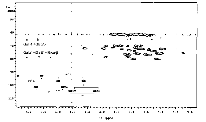
Figure 1 illustrates 13C-IH Heteronuclear Single Quantum Correlation (HSQCj
spectrum of a mixture of lactose (Gal(31-4Glc) and globotriaosylceramide
oligosaccharide (Galal-4Gal(31-4Glc), in the absence (bold lines) and presence
(faint
lines) of the receptor B-subunit derived from the Escherichia coli 0157 toxin.
Only
the resonances of Galal-4Ga1~31-4Glc, the natural ligand, are shifted in the
presence
of the receptor. The resonances of Gal(31-4Glc, which is not a ligand for the
protein.
are unchanged.
Detailed Description of the Embodiments
To a pre-weighed pre-washed glass septum vial (Pierce No. 13804) was added 840
ml dihexanoylphosphatidylcholine (DHPC) in chloroform solution (Sigma P4148)
by
use of a 250 ~l Hamilton microsyringe. Chloroform was evaporated in a stream
of
dry nitrogen gas for 15 minutes, followed by lyophilisation for at least two
hours. The
vial was re-weighed to determine the exact amount of DHPC (22 mg). To the
dried
DHPC was added 785 ~1 deuterium oxide (Aldrich), followed by 95.8 mg
dimyristoylphosphatidylcholine (DMPC, Sigma P6392), to give a 15% solution of
DHPC:DMPC 1:2.9 (mol/mol). Once dissolution was complete, a further 785 ul
deuterium oxide was added to give a 7.5% solution. To 650 pr of this solution
w-as
added uniformly 13C-enriched globotriaosylceramide oligosaccharide and ~'C-
9
CA 02400867 2002-08-21
WO 01/63267 PCT/GBO1/00351
enriched lactose, prepared as described (3) to final concentrations of 0.28 mM
and
0.14 mM respectively. A '3C-'H HSQC spectrum was recorded on this solution at
308 K and pH 7.0 without broadband '3C decoupling in the F2 dimension. in
order
that the one-bond 1'C-1H splittings could be observed. The relevant resonances
are
shown in boldface in figure 1. A total of 2.7 mg. lyophilised E. coli 0157 B
subunit
receptor was then dissolved in the above solution, and a second HSQC spectrum
was
recorded under otherwise identical conditions to the first. The relevant
resonances
are shown in normal face in Figure 1.
15
10
CA 02400867 2002-08-21
WO 01/63267 PCT/GBO1/00351
References
1. Tjanda N and Bax A, Science, 278, 1111-1114 (1997).
2. Shimizu H, Donohue-Rolfe A and Homans S W, J. Am. Chem. Soc.,
121 5815-5816 (1999).
3. Shimizu H, Brown J M, Homans S W and Field R A, Tetrahedron, 54, 9489-
9506, (1998).
11
