Note: Descriptions are shown in the official language in which they were submitted.
CA 02401234 2002-08-23
WO 01/78587 PCT/CA01/00585
TISSUE VIABILITY/HEALTH MONITOR UTILIZING NEAR INFRARED
SPECTROSCOPY
FIELD OF THE INVENTION
The present invention relates generally to the field of medical devices. More
specifically, the present invention relates to a device that non-invasively or
with minimal
invasion to the body can be used to determine the viability, heath or status
of tissue by
using visible and near infrared light.
BACKGROUND OF THE INVENTION
The present and accepted standard for determining the status of tissue
relies on visual inspection of the tissue. Based on the surface appearance of
the tissue,
medical personnel will make an assessment of the tissue and proceed to a
course of
action or treatment. Visual inspection of tissue is central to many areas of
clinical
medicine, and remains a cornerstone of dermatology, reconstructive plastic
surgery, and
in the management of chronic wounds, and burn injuries. For example, in
plastic surgery,
it is extremely important to assess the status of the tissue prior to surgery,
during surgery
and following surgery. Detection of complications or tissue compromise before
the onset
of irreversible tissue damage is paramount. Early detection of tissue
compromise following
surgery enables a more effective course of intervention to be taken in order
to salvage
tissue which is at risk of failing. The monitoring of tissue viability or
status during and
following surgery ensures the efficacy of surgical procedures and non-surgical
means of
intervention can be determined prior to irreversible damage to the tissue.
Unfortunately,
visual manifestations of tissue compromise generally become apparent several
hours after
the onset of the complication. Thus, current clinical assessment methods based
on visual
examination of the tissue provide an indication of tissue compromise well
after the onset of
the problem. This delays possible corrective action, which in turn impacts the
clinical
outcome of the affected tissue. Poor blood supply to the extremities is a
common problem
among the elderly and diabetic populations. Poor peripheral circulation is the
leading
cause of amputation in these populations. Poor peripheral blood supply is a
major
underlying contribution in persistent or chronic wounds of the lower legs and
feet. These
wounds are difficult to heal and can become infected and gangrenous if not
assessed and
treated as early as possible. Clinical evaluation of thermal injuries is made
to determine if
the standard wound care practises will be sufficient to heal the injury or
whether there is
the need for surgical intervention. The course of action based on visual
assessment of the
CA 02401234 2002-08-23
WO 01/78587 PCT/CA01/00585
2
injury is generally made two to three days after the injury and the initial
visual inspection.
Even with this delayed evaluation the assessment of the injury is only
slightly better than
the initial guess.
The prior art teaches a number of devices intended to assess tissue
viability or status, as discussed below.
Laser Doppler Flowmetry is used to estimate blood flow in the skin. The
method has the appeal of an easy-to-use instrument that is minimally invasive.
The
instrument collects a profile of Doppler shifted wavelengths, which it then
fits to a velocity
distribution. The relationship between the Doppler profile and the velocity
distribution
derived for tissue is based on two major assumptions: (1 ) photons are
randomly scattered
by the tissue medium; and (2) photons undergo a single collision event before
capture by
the detector. Based on these assumptions, the fit of the Doppler profile to
the velocity
distribution provides the rms velocity of the particles that are moving within
the tissue that
is being probed by the laser light. Anything that perturbs the laser Doppler
profile will affect
the calculated rms velocity (laser Doppler flux). Thus the instrument is
extremely sensitive
to motion, be it motion of the probe or motion of the subject. Furthermore,
the major
drawback to laser Doppler is the enormous variation in the laser Doppler flux
from
comparable sites between subjects, from different sites in the same subject,
and even
from the same site in the same individual at intervals of minutes, hours and
days. Also, the
apparatus attempts to determine the blood flow and makes no endeavour in the
assessment of oxygen delivery or utilization in tissue.
Fluorescence dyes can be used to determine the extent of blood perfusion
in tissue and vessels. The method involves injecting a dye into the systemic
circulation. The
dye is then carried to the site of interest by the blood stream. The area of
interest is
illuminated with light of a suitable wavelength to excite dye fluorescence. If
fluorescence is
detected, the site is receiving a supply of blood. If only weak fluorescence
or no fluorescence
is measured, the site is not receiving an adequate supply of blood. The method
has
demonstrated success in qualitatively assessing blood flow and the extent of
perfusion in
compromised tissue. However, this method is invasive as it involves the
injection of a dye.
Furthermore, the extended washout times of the dye limits the frequency with
which these
methods can be applied to the site of interest. Again, this method was
primarily used to
measure perfusion, which in turn is used indirectly to assess the status of
the tissue.
Transcutaneous Oxygen Pressure Measurement (TCOM) consists of
placing a heated oxygen specific electrode on the skin to measure the oxygen
diffusing
CA 02401234 2002-08-23
WO 01/78587 PCT/CA01/00585
3
across the skin. The hot TCOM probe is generally not placed directly on the
compromised
tissue to avoid further injury to the tissue. Oxygen delivery to the
compromised tissue is
inferred by measuring the healthy tissue surrounding the compromised tissue.
The heating
of the tissue beneath the electrode increases tissue perfusion. Thus, the
oxygenation of
the heated, healthy tissue must be extrapolated to give an indication of the
oxygen
delivery at a neighbouring injured or compromised site. TCOM, when combined
with a
standard measurement protocol, is an effective and non-invasive means of
identifying
tissues and wounds receiving inadequate levels of oxygen. However, the TCOM
measurement protocol is time consuming, requiring approximately 1 h per
patient, and is
difficult for a non-specialist to perform.
Thermography consists of observing and detecting the emitted irradiance
from an object, in this case tissue. The method attempts to assess tissue
perfusion based
on the surface temperature of normal and suspicious tissue. It is of note that
thermograhic
methods applied to tissue probe only the first few microns (<100 microns) of
the tissue,
and room and patient temperature variations cause havoc on the measured
values.
Magnetic Resonance Imaging can be applied to examine a variety of
disorders, ranging from skin lesions to leg ulcers, by examining metabolism in
vivo in a
non-invasive manner. However, the time necessary to acquire an image, the
total cost of a
single unit and the limited mobility and portability make this method
clinically impractical in
this field of use.
Photoplethysmography is defined as the continuous acquisition of the
intensity of light scattered from a given source by the tissues and collected
by a photo-
detector. Photoplethysmography measures changes in blood volume by monitoring
intensity changes in the observed signal that arise from the pulsatile change
in blood
volume in the blood vessels. Tissues that have a reflected light signal with a
large pulsatile
modulation are assumed to have a good arterial supply of blood. This technique
has been
primarily used to determine the sufficiency of arterial blood supply to the
extremities,
particularly the toes and fingers. The method measures the strength of the
pulsatile
modulation of the optical reflectance signal, which in turn is related to a
change in blood
volume. This measure is extrapolated as an indicator of blood supply to the
tissue. The
method does not report information related to oxygenation, a vital parameter
in tissue
health and viability and the technique is dependent on the tissue having a
distinct pulsatile
modulated blood volume which is typical only of highly vascularized tissue.
US Patents 4,223,680 and 4,281,645 both to Jobsis describe a method and
CA 02401234 2002-08-23
WO 01/78587 PCT/CA01/00585
4
apparatus for in vivo monitoring metabolism in body organs using near infrared
light. This
is accomplished by measuring the absorption characteristics associated with
the cellular
metabolism of cytochrome aa3. However, this apparatus uses a particular set of
measuring
and reference wavelengths to measure changes and trends in the metabolic
activity of an
internal body organ. Jobsis also specified in both patents that the near
infrared light must
span a relatively long path (several centimeters) through bone, skin and
tissue to the
organ of interest for his invention to work.
US Patents 5,161,531 and 5,127,408 both to Parsons, et al, describe an
invasive method and apparatus for in vivo monitoring of internal body organs
such as
heart, brain, liver and kidneys with the use of fiber optic probes and an
elongated catheter.
Specifically, the apparatus makes measurements pertaining to the oxygen
availability and
utilization in internal body organs and not cutaneous (skin) tissue. Likewise,
US Patent
4,513,751 by Abe et al. describes an invasive method and apparatus that
follows oxygen
metabolism in an internal organ.
PCT Patent 9608201A by Vari and Maarek describe a non-invasive
spectroscopic apparatus and method to assess burn injuries. The apparatus
depicted
specifically targets the use of selected wavelengths to assess the burn injury
by evaluating
the intensity of the fluorescence and tissue attenuation at these specific
wavelengths. The
device described therein does not acquire a multitude of discrete wavelengths
comprising
a spectroscopic response for a given wavelength range; rather, the
aforementioned device
looks at the intensity (or counts) and compares this to a database of normal
tissue to
assess the injury. In other words, the device lacks the ability to look at the
attenuation as
related to tissue absorption to delineate tissue viability. Burn injuries are
classified
according to the depth of the burn injury, no mention of the burn depth is
disclosed by Vari
and Maarek.
W092/15008 to Rava et al teaches using laser light for diagnosis as well as
treatment and/or removal of tissue. Specifically, the described device
includes a laser
catheter for removing plaques from a vessel wall as a method for treating
atherosclerosis.
W096/07889 to Vo-Dinh teaches a method of laser-induced synchronous
luminescence for analyzing tumors and other tissues using dyes.
W099/22640 teaches a device for the detection of various tissue states by
observing various optical phenomena (emission and reflectance) using various
illumination
sources (UV, IR, far IR, and lasers). It is further stated that the device
will use a database
containing previous spectra for comparison purposes when determining tissue
status.
CA 02401234 2002-08-23
WO 01/78587 PCT/CA01/00585
However, no clear outline of how this will be accomplished or a description of
the device is
provided. Furthermore, no indication of the processing methods or algorithms
is provided,
nor is any data shown. In addition, W099/22640 does not consider the need to
distinguish
between surface and subsurface tissue absorptions or describe any steps for
enhancing
and analyzing data obtained from the spectra.
As discussed above, prompt and effective assessment of tissue following
surgery or injury promotes a proper course of action, reduces the need of
unnecessary
medical attention, and aids in the restoration of the damaged tissue. Clearly,
an apparatus
that provides an early means of determining the status of tissues that are
potentially
threatened as a result of trauma, a chronic condition, disease state or a
surgical procedure
is required. The apparatus would preferably determine the status of tissue in
a non-
invasive, and non-subjective manner. The apparatus can also provide long term
non-
subjective re-assessment of tissue during the recovery process. This long term
usage is
essential in areas such as chronic wounds where the healing process can span
several
months or years. The apparatus can also be used at the time of surgery to
determine the
efficacy of a surgical procedure.
In view of this, Sowa et al (W098/44839) describes a method of using near
infrared spectroscopic imaging to assess tissue viability. Specifically,
visible and near-
infrared spectroscopy is used to analyze tissue hydration and oxygenation. The
data are
acquired simply, rapidly and non-invasively. Furthermore, the data from a
single spectrum
is sufficient, using the method described therein, to predict tissue
viability, obviating the
need to continuously monitor trends. The relative change and distribution of
the levels of
oxyhemoglobin (Hb02), and deoxyhemoglobin (Hb) in tissue is examined and used
to
predict tissue viability. The near-IR and visible absorption spectra of Hb,
Hb02 and water
are well understood and the differential absorption by these chromophores can
be
distinguished at certain characteristic wavelength regions (Eaton and
Hofrichter, 1981,
Meth Enz 76:175-261 ). However, there are several factors which must be taken
into
consideration and several limitations overcome when designing a device to
carry out this
method. Specifically, the light source must have sufficient light in the vis-
near infrared
range and the source must be stable. Corrections for curved surfaces and
translational,
rotational and scaling corrections for image registration must also be taken
into account.
Components capable of distinguishing between tissue surface and subsurface
phenomena and detecting and differentiating between small signals must be
designed.
Furthermore, the device must be arranged to carry out a number of tasks,
including, for
CA 02401234 2002-08-23
WO 01/78587 PCT/CA01/00585
6
example, tissue assessment at multiple points and at multiple depths, as well
as two-
dimensional imaging of an injured area.
SUMMARY OF THE INVENTION
According to a first aspect of the invention, there is provided a device for
single or
multiple point spectroscopy for determining status of a tissue portion at the
surface of the
tissue comprising: a light source emitting energy in the wavelength region
between 400-
2500 nm; an illuminator delivering light from the light source to the tissue
surface; a
collector receiving remitted light from the tissue surface; a detector
measuring wavelength
data from the remitted light; an analyzer analyzing the data from the detector
for
measuring tissue viability; and a display unit displaying results from the
analyzer.
The device may include a wavelength sensitive element for dispersing the
collected remitted light into wavelength dependent components. As will be
apparent to one
knowledgeable in the art, this includes dispersive and non-dispersive elements
such as
gratings, prisms, acousto-optical tunable filters (AOTFs), liquid crystal
tunable filters
(LCTFs) and the like.
The device may include at least one optical path router for switching between
single and multiple illuminators and collectors.
The optical path router may be connected to the detector for switching between
a
single collector at a single tissue site and multiple collectors detecting
several tissue sites.
The optical path router may be connected to the light source for switching
between
a single illuminator illuminating one tissue site and multiple illuminators
illuminating several
tissue sites.
The optical path router may be connected to the detector for switching to
obtain a
sample of the illumination source.
The optical path router may be connected to the detector for switching to
obtain a
sample of the illumination source passing through a wavelength calibration
standard.
An optical path router or shutter may be connected to the detector for
switching to
obtain a dark spectrum when no light is transmitted.
The optical path router or shutter may be connected to the illumination source
for
switching the transmission of source illumination off. This may be used to
troubleshoot
ambient light problems or probe/tissue contact problems.
The collector and the illuminator may be at a fixed distance relative one
another for
determining optical depth.
CA 02401234 2002-08-23
WO 01/78587 PCT/CA01/00585
7
The detector may include a two dimensional detector.
According to a second aspect of the invention, there is provided a device for
imaging spectroscopic analysis of a tissue portion comprising: a light source
emitting
energy in the wavelength region between 400-2500 nm; an illuminator delivering
light to
the tissue portion; a collector receiving remitted light from the tissue
portion; a detector
having a two dimensional sensor array for acquiring images at selected
wavelengths from
the remitted light; imaging devices detecting wavelength-dependent images from
the
detector; an analyzer processing the images into parameters for assessing
tissue status;
and a display unit for displaying the parameters.
The device may include an optical path router mounted to the collector for
receiving remitted light from multiple sites within the tissue portion.
The device may include an optical path router mounted to the illuminate a
single
and multiple site within the tissue portion.
According to a third aspect of the invention, there is provided a device for
multiple
point spectroscopic analysis of a tissue portion comprising: a light source
emitting energy
in the wavelength region between 400-2500 nm; a probe head having mounted
thereon:
an illuminator illuminating the tissue portion; collectors gathering remitted
light from the
tissue portion, each of said collectors being mounted on the probe head at a
position distal
to the illuminator and one another for acquiring spectral information at a
given tissue
depth; a plurality of detectors dispersing the remitted light gathered by the
collectors into
wavelength dependent components, each detector being linked to a respective
one of the
collectors; an analyzer processing the wavelength dependent components into
parameters
for assessing tissue status; and a display unit for showing the parameters.
The light source may be modulated.
The detectors may detect the remitted light in the time or frequency domain as
the
light source modulates.
BRIEF DESCRIPTION OF THE DRAWINGS
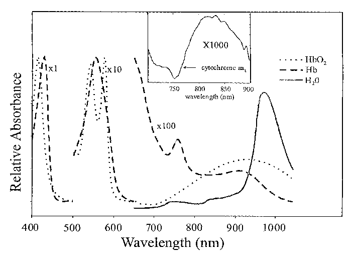
Figure 1 shows the spectrum of the Hb, Hb02, water, and the difference
spectrum of oxidized minus reduced cytochrome aa3, to describe the
chromophores or
parameters one can obtain with the apparatus.
Figure 2 shows a diagram of depth spectroscopy setup a) general
instrument diagram b) sampling depth with multi-point spectroscopy.
Figure 3 is a typical response for the depth spectroscopy apparatus a)
response of an optical standard b) dark noise response c) reflectance response
from the
CA 02401234 2002-08-23
WO 01/78587 PCT/CA01/00585
surface of normal skin.
Figure 4 shows the basic apparatus concepts.
Figure 5 shows embodiments of the apparatus wherein a modulated light
source is utilized.
DESCRIPTION OF THE PREFERRED EMBODIMENTS
Unless defined otherwise, all technical and scientific terms used herein
have the same meaning as commonly understood by one of ordinary skill in the
art to
which the invention belongs. Although any methods and materials similar or
equivalent to
those described herein can be used in the practice or testing of the present
invention, the
preferred methods and materials are now described. All publications mentioned
hereunder
are incorporated herein by reference.
DEFINITIONS
As used herein, tissue viability refers to the state of the tissue with
regards
to whether or not the tissue will survive if no further action is taken.
As used herein, tissue health refers to the state of the tissue with regards
to
proper tissue perfusion, oxygenation saturation, oxygen consumption, and water
content.
As used herein, tissue status refers to the current state of the tissue with
respect to the current status of the tissue chromophores, health and
viability.
As used herein, abnormal or compromised tissue refers to tissue in some
sort of flux or perturbation from its original status prior to injury,
disease, the onset of a
condition, or surgical procedure.
As used herein, thermal injury refers to an injury caused by either extreme
cold or heat which alters or damages the tissue, chemical or electrical burn
which alters or
damages the tissue, or chemical or electrical trauma which alters or damages
the tissue.
As used herein, systemic refers to the entire system or whole body, for
instance systemic oxygenation refers to the oxygenation status of the blood
circulating
through-out the body.
As used herein, tissue oxygenation refers to oxygenated hemoglobin ratio
of blood contained in the arteries, veins and capililary compartments of the
sampled tissue
volume.
As used herein, oxygenation refers to the ratio of hemoglobin carrying
oxygen to the amount of hemoglobin that is oxygen depleted. Tissue oxygenation
refers to
the ratio of oxygenated to total hemoglobin in the blood contained in the
arteries, veins
and capillary compartments of the sampled tissue volume.
CA 02401234 2002-08-23
WO 01/78587 PCT/CA01/00585
9
As used herein, blood volume or total hemoglobin refers to a combined
measure of oxygenated and deoxygenated hemoglobin, which can be used as an
indicator
of tissue perfusion.
As used herein, hydration refers to amount of fluid present both lack of or
accumulation resulting in a significant decrease or increase in tissue volume.
As used herein, chronic wound refers to a medical state wherein there is a
persistent injury and the normal healing process is impaired.
As used herein, contact refers to a state of interaction or touching of the
tissue with the apparatus.
As used herein, non-contact refers to a state of immediate proximity without
touching or disturbing the tissue.
As used herein, non-invasive refers to a procedure whereby the tissue is
unaltered from it's present state and non-intrusive
As used herein, minimally invasive refers to a procedure whereby the tissue
is minimally and unnoticeably adjusted to permit the apparatus to obtain
meaningful
measurements.
Described herein is a device for use in assessing tissue viability, status and
health, as shown in Figure 4. The device comprises a light source, an
illuminator/collector,
a detector, an analyzer and a display unit. The apparatus provides information
on tissue
viability in a non-invasion manner utilizing visible and near infrared
absorption reflection
spectroscopy. The tissue viability is based on measures of the chromophores
deoxyhemoglobin (Hb), oxyhemoglobin (Hb02), water (H20) and others that may be
present in the tissue, as taught in W098/44839, which is incorporated herein
by reference.
Figure 4 describes the general concept of the apparatus broken down into
the various components: Light source, illuminator/collector, detector,
analyser, and
display.
The light source provides light illumination to the tissue. The light source
may comprise, for example, a light source emitting energy in the visible and
infrared range
encompassing the wavelength region between 400 and 2500 nm.
The illuminator delivers the light to the tissue and the collector receives
information from the surface and from this gathers the spectroscopic
information from the
tissue. The collector may, for example, collect the wavelength dependent
components of
the remitted light, collect remitted light at selected wavelengths for
developing images or
collect wavelength components of remitted light from the tissue surface at
several radial
CA 02401234 2002-08-23
WO 01/78587 PCT/CA01/00585
positions away from the illumination source, as described below.
The collector may, for example, collect the wavelength dependent
components of the light that are remitted from the tissue. Selected discrete
wavelengths of
the remitted light can be collected for developing multi-spectral images or
responses. A
continuum of wavelengths can also be collected to provide hyperspectral and
spectroscopic image data or spectra. The remitted light can be collected from
one or more
radial positions away from the illumination source, as described below.
The detector unit disperses the light into the various wavelength
components and tracks/records the intensity of the various wavelength
components from
the collector. The detector unit may, for example, detect reflected light
energy from one or
more tissue sites or from areas of tissue. The detector unit may also detect a
portion of the
light source for determining the system or instrument response. The detector
unit may be
an optical detector, a fiber optic detector, or a lens based optical system,
as described
below.
The analyzer receives the spectroscopic information from the detector unit
and analyzes this data using computation formulas to provide a meaningful
measure of
tissue viability. The analyzer may, for example, process the wavelength
dependent
spectroscopic profiles, or wavelength-dependent images into sets of parameters
used to
assess the status, viability or health of the tissue in near real time.
The display unit displays the information from the analyzer on either a
visual display or as a printout. This information may be displayed in near
real-time.
An example of the field of use of the apparatus by a medical practitioner is
in the assessment of the condition of tissues in various conditions of health
ranging from
tissue which is healthy through tissues which are at risk of becoming
necrotic. A
comparison of healthy and near necrotic tissue shows a stark contrast in their
near
infrared spectra, and the oxygenation and total blood volume and hydration
parameters
derived from the near infrared spectra.
In one embodiment of the invention, there is provided an apparatus for
single or multiple point spectroscopy for determining the status of tissue. In
this
embodiment, the light source produces light to illuminate the tissue, the
illuminator/collector delivers and collects the remitted light from the tissue
surface as well
as a means of collecting a portion of the light source to determine the system
response.
The detector unit is a spectroscopic optical means, that measures the
wavelength
dependent components of the remitted light from the tissue. The wavelength
dependent
CA 02401234 2002-08-23
WO 01/78587 PCT/CA01/00585
11
components are used to obtain parameters used in the determination of tissue
status. The
analyzer processes the wavelength dependent spectroscopic profiles into
parameters
used to assess the status, viability, or health of the tissue in near real
time, the results of
which are shown on the display unit. In this embodiment, the device also
includes a
wavelength selector that disperses the remitted and reference light into its
wavelength
dependent components. The wavelength dependent components are dependent on the
state of the light source, the apparatus and status of the tissue.
Specifically, in this embodiment, the device comprises a light source
emitting energy in the visible and infrared range encompassing the wavelength
region
between 400 and 2500 nm, an optical means of illuminating the tissue, and an
optical
means of collecting the light that is reflected, transmitted or scattered from
the tissue site.
In this embodiment there is a means whereby the distance separating the
delivery and
collection optics can vary by some known distance on the surface of the
tissue. In this
embodiment there is a spectroscopic system designed for dispersing the electro-
magnetic
spectrum into its respective wavelength components and converting this
information into
parameters that are related to the status of tissue. In some embodiments, this
requires
digitization of the spectrum but in other embodiments, may include optical
computations
as well.
The spectroscopic system must meet the criteria of producing a spectrum
of sufficient wavelength resolution, of sufficient signal to noise ratio and a
spectrum
containing sufficient regions) of wavelength information.
In this embodiment, the device also includes an optical path router (OPR)
capable of switching between multiple optical inputs and outputs either using
a single or
plurality of these. The OPR can function in a reversible manner as well, in
that it may have
a single input and multiple output switched paths. This reversibility of,the
OPR allows the
device to be situated either before the detector unit to select from multiple
inputs, or after
the light source to direct the light to several output targets. The OPR's may
exist in the
various permutations in the apparatus to provide both selection of the
illumination target
and/or selection of the reception site. In some embodiments, there may be
provided a
device without an OPR. It is of note that the OPR can be capable of
transmitting a point
source, one dimensional linear source, or two dimensional imaging source.
Also, more
than one OPR many be chained together to increase permutations of
functionality.
In this embodiment, there is provided a controller/analyzer unit which
controls the various sub-assemblies in the device, controlling the detector
and obtaining
CA 02401234 2002-08-23
WO 01/78587 PCT/CA01/00585
12
spectra from it, controlling the OPR('s) if the apparatus contains one,
monitoring the
illumination source, and the analyzer processes the obtained spectra into
medical
meaningful data.
In this embodiment, the analyzer calculates medical diagnostic
parameters) on tissue viability from the spectroscopic information acquired by
the
detector, for example, data relating to hydration, hemoglobin, oxygen bound
hemoglobin,
oxygen saturation as well as total hemoglobin.
The display unit allows for visualization of the calculated information.
In some embodiments, the device may include an input device such as a
keyboard to allow for user interaction with the apparatus.
As a result of this arrangement, the apparatus provides information on
tissue viability in a non-invasion manner utilizing visible and near infrared
absorption
reflection spectroscopy. The tissue viability is based on measures of the
chromophores
deoxyhemoglobin (Hb), oxyhemoglobin (Hb02), water (H20) and others that may be
present in the tissue.
As discussed above, the apparatus operates by delivering and receiving
visible and near infrared light in the range 400 to 2500nm to the tissue site
of interest.
Light in this region of the electromagnetic spectrum is ideal for tissue
viability and health
assessment due to the attributes of weak absorption by the tissue and high
forward
directed scattering by tissue constituents. This combination of weak
absorption and high
scattering permits the light to penetrate a substantial distance within
tissue. Since tissue is
a highly scattering medium, there is strong inverse correlation between the
amplitude of
the returned signal and depth of tissue sampled. This decrease in reflected
signal strength
and the detection limit of the detector unit limits the maximum depth of
tissue that may be
probed. The spacing between the fiber optics of the illuninator and collector
determines
the mean optical depth that light can penetrate into the tissue. The reflected
light provides
input for the detector unit, which may contain a grating spectrometer known in
the art,
which disperses the light of the electro-magnetic spectrum into its wavelength
components. This dispersed spectrum is then directed on a linear array
detector, which
converts the light into an electrical signal. This electrical signal is then
digitized and
transferred to the analyzer creating a digital spectrum of the tissue. The
spectrum is then
processed using computational algorithms described in W098/44839 and the
results are
displayed. A keyboard may also be provided to allow for user interaction with
the
apparatus.
CA 02401234 2002-08-23
WO 01/78587 PCT/CA01/00585
13
It is worth noting that the optical path router (OPR) is capable of switching
between multiple optical inputs and directing either a single or plurality of
these to an
output port that can be positioned at the entrance of the detector unit. The
selection of
multiple inputs allows for the selection of reflected light from one or more
tissue sites, light
directly from the illumination source, or no input light to measure the dark
or null response
of the apparatus. These spectra allow for the calculation of an optical
density (OD)
spectrum given by the relationship, OD=logo ( (illumination spectrum-dark
spectrum) /
(tissue spectrum-dark spectrum) ). The ratioed responses provide information
related to
the attenuation of light at the particular wavelength, which is ultimately
related to the
absorption of the tissue chromophore at that particular wavelength.
Furthermore, the dark
response of the system may be used to further correct for the instrument
response,
thereby improving the quality of the tissue spectrum
The apparatus may also have multiple tissue probes with the OPR acting
as a multiplexer allowing the apparatus to scan several tissue sites. These
sites may
include both tissue sites of suspicious health and sites of healthy tissue. In
addition, the
inputs may include: light from the source passing through a wavelength
calibration
standard, light from a single or multiple discrete sources) such as a laser
diode for
calibration purposes, multiple tissue sites which may include reference sites,
a second
apparatus or another patient. Multiple or combinational OPR's may exist in the
apparatus
to provide both selection of illumination target and selection of the
reception site.
The OPR can be capable of transmitting either a point source, a one-
dimensional source, or a two-dimensional source. In a point source
configuration, the
organization and orientation of the light transmission is not important and a
simple
detector unit utilizing either a linear detector or a column binned two-
dimensional detector
or area detector. The one-dimensional configuration allows for multiple
channel
throughputs into a detector unit equipped with an area detector. This allows
for several
channels to be recorded simultaneously. The optical isolation of the channels
is retained
through the OPR and detector unit so that each channel can be distinguished
when
imaged on to the detector. It is of note that such a system with an area
detector could be
used without an OPR and still sample several sources including tissue sites,
source
reference, backgrounds and others using other forms of channel multiplexing.
An area
detector may require a shutter during readout or for background spectrum
collection. In
place of the area detector and shutter, a frame transfer detector may also be
used. It is of
note that in some embodiments, a device may exist with a combination of OPR(s)
and/or
CA 02401234 2002-08-23
WO 01/78587 PCT/CA01/00585
14
multiple detector units. The OPR may also transmit two dimensional source
imagery which
may be used in imaging systems, discussed below. A system may also exist
without an
OPR or channel multiplexer.
The OPR differs between the conventional fiber optic switches used in fiber
optic communication networks in that the OPR transmits broadband light,
usually using
multimode fiber optic cable. The OPR is usually involved in the transmission
and coupling
of bundles containing multiple fiber optics. The concept of the OPR includes:
reproducible
switching between inputs or outputs, high coupling efficiency and minimal
throughput
loses of the transmitted signal(s), minimization of channel cross-talk or
interference and
minimization of light leakage when OPR is set to transmit no signal.
It is of note that when a system is constructed without an OPR, several
challenges are presented. For example, the light source is either monitored
through some
other mechanism or it is assumed to be stable during the tissue assessment. In
addition,
the collection of a reference spectra is done manually, which opens up the
possibility of
user error in the collection of the reference. Furthermore, the lack of an OPR
limits the
device to processing difference values for monitored tissue parameters and the
system to
a single probe if the detector unit is equipped with a single element or one-
dimensional
detector. These drawbacks may be managed through the use of multiple fibers
and an
area detector assuming the response on the detector is known and stable.
Thus, a system that incorporates an OPR is considerably more flexible.
Firstly, the OPR allows for collection of a reference spectrum at any time
point. This is
important not only in the computation of OD spectra but also as self-
diagnostic or system
troubleshooting algorithms. In a similar fashion, the incorporation of a
wavelength
calibration standard as an input channel can also be used for apparatus
diagnostic
purposes and performance tracking. As a result of this arrangement, the system
can
check the dark spectrum for system performance in an apparatus diagnostic
methodology
and use this information to self-correct system performance. The OPR combined
with a
single element or one-dimensional detector enables the usage of multiple
sampling sites
or depths. When combined with a two-dimensional detector and the appropriate
illuminator/collector the same system may either collect multiple optical
channels with a
two-dimensional detector or capture a single weak spectrum using the detector
in a binned
format. When an OPR is coupled to a light source, the apparatus can then
sample multiple
sites while maintaining maximum light delivery to the current site of queue
and therefore
maximum signal quality.
CA 02401234 2002-08-23
WO 01/78587 PCT/CA01/00585
The sampling probe contained in the illuminator/collector can be as simple
as separate delivery and reception fiber optics at a known or controlled
separation.
Usually, the delivery and reception fibers will be integrated into a single
probe head with a
set spacing. The probe may also contain a pressure and/or shear stress sensor
linked
back to the apparatus to indicate the force being applied to the tissue
beneath the probe.
Pressure or shear stress forces involve movement of interstitial fluids and
blood of the
tissue, which affect the concentration of chromophores being detected from the
tissue
beneath the probe.
The sampling probe may contain a temperature sensor to account for
temperature variation of the tissue. The optical and spectroscopic properties
of tissue are
temperature dependent. Tissue can be thought of consisting of a medium
composed of
microscopic constituents of varying refractive index. Since the refractive
index and density
of a medium are strongly temperature dependent, changes in the observed
response are
expected. With an increase or decrease in temperature, the density and optical
properties
of the medium are altered, thus changing the response from the apparatus.
Temperature
also plays a major role with respect to oxygen diffusion in tissue and can
provide
erroneous results if these effects are not accounted for in the analysis.
Alternatively,
temperature may be calculated from the tissue spectrum. Water and its
temperature
dependent spectral variation has been a field of interest in biomedical tissue
spectroscopy.
The extinction coefficients of water are extremely temperature dependent, such
that a
temperature variation will cause a spectral shift in the spectrum. A
temperature increase
will cause the water spectrum to shift to shorter wavelengths. Therefore, the
temperature
of the tissue can be monitored using the response in the water spectral region
The illuminator/collector probe may also contain a reference fiber set for the
correction of fiber absorbances and loses caused by fiber core material, fiber
impurities,
fiber cladding, mechanical stress or other sources. This reference fiber set
may contain a
loop in the probe head or may contain a reference standard to reflect off of.
The reference
fiber set may consist of one or more fibers with input from the illumination
source and
output back to the apparatus. The purpose of the reference fiber is to provide
a reference
spectrum of the illumination source and a spectrum of the result of the
illumination light's
interaction with the fiber. The spectrum collected from this reference fiber
when used in
the computation of optical density as stated above should account for the
fiber and system
effects in the resulting tissue sample spectrum. Other methods of collecting a
reference
spectrum, disparate from the probe reference method, may include a separate
fiber optic
CA 02401234 2002-08-23
WO 01/78587 PCT/CA01/00585
16
within the device of similar fiber characteristics with input from the source
and output to
spectroscopic section of the device. A manual reference can also be acquired
by manually
placing the probe on a reflectance standard. Another configuration may involve
monitoring
the source at one or more discrete wavelengths and using this information to
account for
source stability, although this method does not account for effects that the
fiber optic may
have on the attenuation of the signal. If changes in tissue condition or
differences between
tissue sites are being monitored, then a sample from a previous measurement or
other
physical site may be used to ratio out the system's effect on the tissue
sample site of
interest.
The probe may also contain a tag, which is capable of identifying the probe
to the main device unit. This tag may contain information such as probe model
number, a
unique serial number, and information about the probe or configuration
information for the
device. The tag may also contain write-able memory, which may contain
information on
probe usage or other updateable information.
With the flexibility provided by the OPR, the apparatus may also be
composed of more than one probe head to sample more than one tissue site. The
sites
may be composed of various permutations involving tissue of suspicious
viability and
healthy tissue or other configurations to provide the medical practitioner
with the data
stream they desire. An application of this would be to compare and contrast
the suspicious
tissue sites against a known healthy tissue site. An alternate probe
configuration may
consist of multiple illuminator and collector probes separated at various
distance from one
another. This permits sampling of multiple depths into the tissue and the
investigation of
the various layers of tissue beneath that which is visible. An application of
this would be to
profile the depth of thermal damaged tissue to differentiate intermediate
partial thickness
burns from deep partial thickness burns, as described below. The probe
arrangement may
involve a combination of discrete probe sites and depth profiling sites.
An additional embodiment of the apparatus may include a high rate
scanning mode. This mode is intended to distinguish and identify the extent of
the
abnormal tissue across the surface. The high rate scan mode consists of moving
the
probe across the surface of the suspicious and normal tissue and collecting,
processing
and displaying the acquired information. In this mode, the boundaries between
healthy
and abnormal tissue can be quickly identified. This high rate scan mode may be
accomplished manually, mechanically, optically, or by some other means. In
order to
enable this mode of operation, the detector exposure time per spectrum must be
CA 02401234 2002-08-23
WO 01/78587 PCT/CA01/00585
17
shortened to the point that motion artifacts are minimized in the spectrum. To
accomplish
the short exposure time and maintain a reasonable signal level a detector of
higher
efficiency is required and a detector cooler may also benefit the system by
reducing dark
noise. In addition, processing algorithms can detect artifacts from excessive
motion and
disregard those spectrum and signal the system that the motion is causing
artifacts. The
algorithms may be altered to deal with increased data flow, changed spectrum
characteristics and other differences from standard operation. The system may
also
incorporate a trigging device for the user to capture data at their
discretion.
A major benefit of using broadband spectroscopy is that the scattering
contribution to the light attenuation by tissue may be determined and
corrected over
narrow regions of the spectrum. Scatter correction is not available to
discrete wavelength
systems simply because they do not provide sufficient data as function of
wavelength to
afford a reliable correction. The ability to qualify the scattering is what
gives this apparatus
the ability to give reproducible results even after repeated probe removals
and re-
attachments, and to account for patient and probe movement. Both of these
factors are a
major failing of prior art devices in this area.
In addition to the techniques discussed in W098/44839, the following
technique may also be used to extract information about the tissue from the
measured
(recorded) spectra. Herein, hydration values are obtained from the 980nm water
band
region and hemoglobin values are obtained from the 700-840 nm region. The
relative
concentrration of these chromophores are derived from the specified regions of
the
spectrum by a least squares fit of the chromophore extinction coefficients to
the measured
spectrum. In addition, possible water effects can be removed from the results
by using
prior calculated hydration values, and then carrying out a least square fit of
the oxy-
hemoglobin, deoxy-hemoglobin and other optical effects. The chromophore
concentrations
derived form the tissue spectrum may now be used to calculate the ratio of
oxygenated to
deoxygenated hemoglobin, the combined oxygenated and deoxygenated hemoglobin
or
total hemoglobin and the ratio of oxygenated hemoglobin to total hemoglobin.
In a further embodiment, the device is arranged for imaging spectroscopy
to determine the status of tissue. In this embodiment, the light source
produces light to
illuminate the tissue used to measure the absorption or reflectance from the
tissue to
provide a tissue spectrum. The collector receives the remitted light from the
tissue surface
using a lens-based optical system. The detector unit selects the wavelengths
using a non-
disperse device to collect remitted light from the tissue in an imaging
fashion using a two-
CA 02401234 2002-08-23
WO 01/78587 PCT/CA01/00585
18
dimensional sensor array to acquire images at selected wavelengths. The
analyzer
processes the wavelength dependent images into parameters used to assess the
status,
viability, or health of the tissue in near real-time on a display.
As discussed above, the point spectroscopic apparatus provides an
excellent means of evaluating tissue viability, status and health when the
area being
assessed is clearly known or only a select few areas are necessary. However,
in some
cases, the dividing line between health and necrosis or disease is not easily
distinguishable i.e. there are varying degrees of poor perfusion or damage
across the
surface of the tissue. Several apparatus can be used to obtain total tissue
surface health
information. One approach involves sampling multiple locations on the tissue
surface. A
second approach comprises the use of multiple optical fibers at multiple
locations across
the tissue site of interest. An alternative to both these techniques employs
an apparatus
capable of obtaining spectroscopic information all at once in an imaging
fashion. Such a
method has the advantage of acquiring spatial tissue health information
instantaneously.
Imaging devices are the most well known and used of the photonic
detectors. In general, imaging devices/detectors convert light or photons into
an electrical
charge that is collected and stored in a metal oxide semiconductor. The
accumulated
charge or response is a linear function of the incident and exposure time to
the light
energy. The response from an imaging device represents the reflected spatial
light
intensity of a given object. In conventional imaging devices, the detected
response
observed is the accumulated intensity for a broad range of wavelengths.
Therefore, an
imaging device for in vivo tissue applications is of little use without some
means of
wavelength selection. Several methods can be applied to coupling a wavelength
selection
device to an imaging detector to provide spectroscopic spatial information to
assess tissue
viability, status or health.
An extension of the point apparatus is a system capable of providing spatial
information on tissue viability in a non-invasive and non-contact manner using
visible and
near infrared reflectance/absorption spectroscopy. This can be accomplished
using a
spectroscopic imaging apparatus utilizing: an illumination source(s), a two
dimensional
detector and a wavelength selection system capable of passing two-dimensions
of spatial
information. One variation of the imaging apparatus is to use an imaging
spectrometer and
to allow a single column of a multicolumn image into the imaging spectrometer
in a
stepped fashion until all columns in the image are collected. Therefore, the
image
acquired consists of one spatial and one wavelength dimension. Another
possibility would
CA 02401234 2002-08-23
WO 01/78587 PCT/CA01/00585
19
be the use of a point spectrometer in which the image is generated by scanning
the input
over the surface area. The result in all these methods is a three-dimensional
cube of data
consisting of x and y dimensions of spatial information and a z dimension
containing
wavelength information. The formats may include both micro and macro imaging,
as well
as varying degrees of image pixel density from those tight enough to create a
visually
recognizable image to someone trained in the art to a more sparsely spaced
grid
approaching that of a random discrete point map. In some formats the apparatus
may take
the form of a contacting probe system while still maintaining non-
invasiveness.
The apparatus utilizes: an illumination source(s), a wavelength selection
unit, a optics system to form an image, a two dimensional detector, an
analyzer/controller,
a display system, and a keyboard for user interaction. The device may also
contain: a
point spectrometer and OPR to monitor the source(s), a reference system to
obtain
information on detection system characteristics, a system to model contours of
imaged
surface such as laser scanner or stereoscopic images, a system to over-lay
images
captured from different surface and camera viewpoint configurations and
possible different
time points, a system to calculate relevant medical diagnostic images of
tissue viability
and a system to account for uneven illumination of scene due to shadowing or
contour
effects, image registration to correct for translational, rotational, and
scaling uncertainties
and the use of polarizers to distinguish between surface and sub surface
phenomena.
An application of the imaging spectroscopy is in the field of tracking and
measuring the status of chronic wounds under treatment. One of the methods of
treatment
for chronic wounds is hyperbaric oxygen therapy. The increase in oxygenation
results in a
contrast enhancement for spectroscopic imaging. The contrast is provided by
increased
oxygen inhalation by the patient. The increased contrast allows for increased
differentiation of healthy and tissue which is at risk in both point and
imaging applications.
In another embodiment, there is provided an apparatus for multiple point
spectroscopy to determine the status of tissue comprising: a source for
producing light to
illuminate the tissue used to measure the absorption or reflectance from the
tissue to
provide a spectrum; an optical means (fiber optics) of illuminating the tissue
using a light
source; an optical means (fiber optic) to collect the remitted light from the
tissue surface at
several radial positions away from the illumination source as a method to
acquire spectral
information at various depths into the tissue; a device to disperse the
remitted light
collected at several positions into its wavelength dependent components which
provides
spectral information on the status of the tissue at various depths into the
tissue; a device
CA 02401234 2002-08-23
WO 01/78587 PCT/CA01/00585
to detect the wavelength dependent components of the remitted light collected
at several
positions away from the source to obtain parameters used in the determination
of tissue
status; and a computational method to process the wavelength dependent
spectroscopic
profiles into parameters used to assess the status, viability, or health of
the tissue in near
real-time on a display.
The determination of tissue viability, status, and health following
reconstructive surgery and/or a tissue-altering insult relies on the ability
to accurately
assess the tissue below its superficial layer. Such alterations modify the
physical and
optical properties of the tissue from the surface to deep within the tissue.
As discussed
above, the spectroscopic properties of the tissue can be acquired using either
point
spectroscopy or spectroscopic imaging. However, these methods are limited to
sampling
very shallow depths into the tissue therefore probing a thin portion of the
tissue. In many
cases however, the tissue assessment relies on the determination of the extent
of viable
tissue below the surface. Depth dependent tissue assessment can be
accomplished by
acquiring spectroscopic tissue responses at various depths into the tissue.
The transport of light through tissues is governed by the absorption of light
by the tissue chromophores as well as the light-scattering interactions in the
medium.
Scattering of light occurs as a direct result of the interaction of light with
random variations
in the refractive index or small particles in the medium, resulting in a
dispersion of the light
in all directions. In the reflectance geometry of Figure 2, a small fraction
of the light
penetrates into the tissue and is remitted out back to the surface. This
remitted or diffusely
reflected light collected by the detector has been attenuated as a result of
the scattering
as well as through absorption by the chromophores in the tissue. A measure of
the
reflected light provides spectral information on the scattering by the tissue
and absorption
by the tissue chromophores. Figure 2 also depicts the detected light path
through skin at
various source-detector separation distances. When light enters a scattering
media such
as tissue, the light is preferentially scattered in a forward direction. In
order for light to
reach a detector a set distance away from the source, the light must traverse
a path
through the media, denoted by the shaded region. As the source-detector
separation
increases, the path increases and the depth sampled into the medium also
increases.
These paths have been described by a number of authors using both Monte Carlo
simulations and single photon time correlated spectroscopic techniques.
Essentially,
collecting spectroscopic absorption spectra at various source-detector
separations
provides information on the tissue alteration or status at several depths into
the tissue.
CA 02401234 2002-08-23
WO 01/78587 PCT/CA01/00585
21
As shown in Figure 4, the present apparatus permits the evaluation and
assessment of tissue health, status and viability deep within the tissue. The
apparatus
employs visible and infrared light to monitor and evaluate the condition of
the tissue based
on the tissue optical changes encountered. Proper evaluation of the tissue
following a
surgical procedure or an insult to the tissue will allow for the correct
course of action to be
taken. This may result in one of two basic options: 1 ) to perform a surgical
procedure to
correct the problem; or 2) to allow the tissue to heal without surgical
intervention. This
apparatus is directed towards an unbiased non-invasive method to assess deep
tissue
viability.
The device shown in Figure 4 includes a visible-infrared light source, a
detector
unit consisting of a wavelength dispersive instrument or module, and an area
array
detector or sensor, an analyzer consisting of a data acquisition and
processor, and several
optical fibers for the illuminator and collector. The detector for this
application may be
more sensitive since the signal levels at the deeper tissue levels will be
lower. It is of note
that in some embodiments, the detector may require cooling to decrease
electrical noise
levels to maintain good spectral signal to noise ratios.
In another embodiment, there is provided an apparatus for multiple point
spectroscopy to determine the status of tissue in a given area comprising: a
source for
producing light to illuminate the tissue used to measure the absorption or
reflectance from
the tissue to provide an absorption spectrum; an optical means (fiber optics)
of illuminating
the tissue using a pulsed or modulated light source; an optical means (fiber
optic) to
collect the remitted light from the tissue surface at several radial positions
away from the
illumination source as a method to acquire spectral information at various
depths into the
tissue; a device to detect the remitted light collected at several positions
away from the
source to obtain parameters used in the determination of tissue status; a
means of
detecting the remitted light in the time or frequency domain as related to the
illumination
source to obtain tissue parameters associated with health or injury; and a
means to
display the processed time or frequency response to assess the status,
viability, or health
of the tissue in near real-time.
In general, as light enters into tissue two processes occur, absorption and
scattering of the light. When tissue is illuminated with near infrared light,
some of the light
is absorbed by the tissue chromophores while a large portion of the light is
diffusely
scattered. Scattering of light occurs as a direct result of the interaction of
light with random
variations in the refractive index or small particles in the medium, resulting
in a dispersion
CA 02401234 2002-08-23
WO 01/78587 PCT/CA01/00585
22
of the light in all directions. The observed or detected light is related to
the concentration
of scattering centers. Scattering alters the straight-line direction of the
path that light
propagates through tissue. Thus scattering results in an increase in the path-
length that
light travels through tissue relative to the straight-line path that light
travels in a non-
scattering medium. The increased path traveled by the light results in a
greater attenuation
of the light intensity due to an increased chance of light absorption by a
chromophore
compared to a non-scattering medium with comparable chromophore concentration.
As a
whole, the reflected light observed from the tissue is a function of the
absorption by the
chromophores and scattering from the constituents. Assessing tissue viability,
health and
status often requires the scattering contribution of the detected light to be
distinguished or
separated from the absorption contribution prior to the analysis and display
processes.
The common approach to scatter correction in spectroscopy has been to use the
entire spectrum or spectra in such routines as internal standards, second
derivative data
processing, and multiplicative signal correction to decrease the variability
resulting from
scattering. An alternative approach to scatter correction in tissue uses
either photon time
or frequency resolved techniques to obtain information on the mean paths light
travels
through tissue. Photon time-of-flight methods, a time resolved technique, use
ultra-short
pulses of illumination in which the light is diffusely scattered in the
tissue. Semiconductor,
dye, or solid state lasers produce the ultra-short pulses at discrete
wavelengths in time-of-
flight instruments. Photon paths through tissue can be obtained using these
ultra short
laser pulses and electronics to digitize and collect the response for a single
photon event.
Using a large number of photons, an intensity distribution is constructed of
the sum of the
number of photons with various times through the sample. A measurement of the
photon
time distribution effectively probes a series of pathlengths through the
sample, which is
related to absorption and scattering properties of the tissue. Frequency
domain or intensity
modulated techniques also use a laser as source except the intensity is
modulated at
radio frequencies with measurements of the intensity and phase made through
the tissue
sample. Knowledge of the phase shift and the modulation frequency can be used
to
determine the mean photon pathlength through the tissue sample. Photon time
and
frequency responses inherently contain information on the optical properties
of the tissue.
Applying the results from either technique to a modified Beer-Lambert
relationship
provides a means to reduce or correct for the scatter contributions in the
multi-wavelength
responses applied to tissue viability, health and status. Time and frequency
resolved
techniques are used as a correction methodology which use absorption and
scattering
CA 02401234 2002-08-23
WO 01/78587 PCT/CA01/00585
23
determined from time or frequency resolved distributions to reduce the
spectral variations
resulting from the medium.
In some embodiments, the light source may be a laser, for example, an
ultra-short pulse solid state, semiconductor laser or a solid state or
semiconductor laser
output capable of being modulated.
In some embodiments, the detector may have ultra-fast rise and fall times
and be capable of detecting low light levels. These may include, for example,
photomultipliers (PMT), microchannel plate PMT, streak cameras, photodiodes,
PIN
photodiodes, and avalanche photodiodes.
In some embodiments, the collector may include, for example, mirrors, fiber
optics, and OPR to direct the light to the tissue surface and detector.
In some embodiments, the analyzer may include, for example, electronics
to detect, amplify, acquire, and process the responses from the laser and
detector to a
meaningful result to be used to correct for scattering
While the preferred embodiments of the invention have been described
above, it will be recognized and understood that various modifications may be
made
therein, and the appended claims are intended to cover all such modifications
which may
fall within the spirit and scope of the invention.