Note: Descriptions are shown in the official language in which they were submitted.
CA 02401996 2002-09-03
WO 01/66043 PCT/US01/06974
BULBOUS VALVE AND STENT FOR TREATING VASCULAR REFLUX
Field of the Invention
The present invention relates to venous valve replacement and, in particular,
to
replacement venous valves to lower extremities and a therapeutic method of
treating venous
circulatory disorders.
Background of the Invention
Chronic venous insufficiency (CVI) of the lower extremities is a common
condition
that is considered a serious public health and socioeconomic problem. In the
United States,
approximately two million workdays are lost each year, and over 2 million new
cases of
venous. thrombosis are recorded each year. About 800,000 new cases of venous
insufficiency
syndrome will also be recorded annually. Ambulatory care costs of about
$2,000, per patient,
per month' contribute to the estimated U.S. cost of $16,000,000 per month for
the treatment
of venous stasis ulcers related to CVI.
It is estimated that greater thari 3 /o of the Medicare population is
afflicted by a
degree of CVI manifested as non-healing ulcers. Studies have indicated that
about 40% of
seriously-. affected individuals cannot work or even leave the house except to
obtain medical
care: It is estimated that 0.2% of the American work force is afflicted with
CVI.
Chronic venous insufficiency arises from long duration venous hypertension
caused
by-, valvular insufficiency and/or venous obstruction secondary to venous
thrombosis. Other
primary causes of CVI include varicosities of long duration, venous hypoplasia
and
arteriovenous fistula: The signs and symptoms of CVI have been used to
classify the degree
of severity of the disease., and reporting standards have been published.
Studies demonstrate
that deterioration of venous hemodynamic status correlates with disease
severity. Venous
reflux, measured by ultrasound
studies, is the method of choice of initial evaluation of patients with pain
and/or swelling in
the lower extremities. In most serious cases of CVI, venous stasis ulcers are
indicative of
incompetent venous valves in all systems, including superficial, common, deep
and
communicating veins. This global involvement affects at least 30 /a of all
cases. Standard
principles of treatment are directed at elimination of venous reflux. Based on
this
observation, therapeutic intervention is best determined by evaluating the
extent of valvular
incompetence, and the anatomical distribution of reflux. Valvular
incompetence, a major
CA 02401996 2002-09-03
WO 01/66043 PCT/USO1/06974
-2-
component of venous hypertension, is present in about 60% of patients with a
clinical
diagnosis of CVI.
Endovascular valve replacement refers to a new concept and new technology in
the
treatment of valvular reflux. The concept involves percutaneous insertion of
the prosthetic
device under fluoroscopic guidance. The device can be advanced to the desired
intravascular
location using guide wires and catheters. Deployment at a selected site can be
accomplished
to correct valvular incompetence. Percutaneous placement of a new valve
apparatus provides
a less invasive solution compared to surgical transposition or open repair of
a valve.
The modem concept of a stent was introduced in the 1960s. Subsequently, it has
been
successfully incorporated in the treatment of arterioral aneurysms and
occlusive disease. The
use of endovascular stents represents one of the most significant changes in
the field of
vascular surgery since the introduction of surgical graft techniques in the
early 1950s.
Initially, the dominant interest of vascular specialists was application of
stents in the
arterial system. The venous system and venous disease were not considered an
arena for stent
application. The utilization of endovascular treatment in venous disease was
initially confined
to the treatment of obstruction, in the pelvic veins [for CVI] as well as
treatment of
obstructed hemodialysis access grafts and decompression of portal hypertension
(TIPS).
Although these procedures enjoy widespread application, the actual number of
patients
involved is relatively low compared to the number afflicted with CVI and
related syndrome.
Thus, the necessity for therapy using endovascular technology for the
treatment of venous
disease arose. The prevalence of CVI and the magnitude of its impact demand
development
of an effective altemative therapy.
Brief Description of the Drawings
Figure 1 is a schematic representation of a portion of a venous system.
Figure 2 is a schematic representation of a section view of a portion of a
venous
system at a closed venous valve.
Figure 3 is a schematic representation of a sectional view of a portion of a
venous
system.
Figure 4 is a schematic representation of a portion of a venous system.
Figure 5 is a schematic representation of a section view of a portion of a
venous
system at an open venous valve.
CA 02401996 2002-09-03
WO 01/66043 PCT/USO1/06974
-3-
Figure 6 is a schematic representation of a section view of a portion of a
venous
system showing a deployment system for a device of the invention.
Figure 7 is a schematic representation of a section view of a portion of a
venous
system showing a deployed device of the invention.
Figure 8 is a schematic view of one embodiment of the invention.
Figure 9 is a schematic view of one embodiment of the invention.
Figure 10 is a schematic view of one embodiment of the invention illustrating
angular
relationships of components.
Figure 11 is a top plan view taken along line 11-11 of Figure 9.
Figure 12 is a schematic elevation view of one embodiment of the invention.
Figure 13 is a schematic view of various valve material placement embodiments
of
the invention.
Figure 14 is a schematic view of a multiple stage embodiment of the invention.
Figure 15 is a side elevation view of a six strut dual stage embodiment of the
invention.
Figure 16 is a side elevation view of a six strut dual stage truncated cone
embodiment
of the invention.
Figure 17 is a photo image of an embodiment of the invention in vivo.
Figure 18 is a photo image of an embodiment of the invention in vivo.
Figure 19 is a photo image of an embodiment of the invention in vivo.
Figure 20 is a photo image of an embodiment of the invention in vivo.
Figure 21 is a photo image of an embodiment of the invention in vivo.
Figure 22 is a photo image of an embodiment of the invention in vivo.
Figure 23 is a photo image of an embodiment of the invention in vivo. Figure
24 is a
perspective view of one embodiment of the invention.
Figure 25 is a flow diagram depicting one embodiment of the invention.
Figure 26 is a flow diagram depicting one embodiment of the invention.
Figure 27 is a side elevation depiction of another embodiment of the
invention.
Figure 28 is a representative sizing view of the invention according to Figure
27.
Summary of the Invention
A replacement valve assembly designed for optimized shaping and fit is
provided that
is configured for implantation within a vascular lumen. The valve assembly
comprises a
CA 02401996 2007-10-29
-4-
plurality of flexible members, with each flexible member arranged to cooperate
with at
least one other flexible member to unidirectionally admit vascular fluid
through the valve
assembly. In one embodiment, at least a portion of one of the flexible members
includes
natural sclera tissue. In other embodiments, the flexible members include at
least a
portion of either SIS or other known biocompatible material. Methods of
manufacturing
the flexible members and of assembling and delivering the assembly to the
patient's
venous system are also provided.
According to an aspect of the present invention, there is provided a self-
expanding replacement valve assembly having a bulbous-shaped portion and which
is
configured for implantation within a vascular lumen, the valve assembly
comprising a
plurality of flexible members, each flexible member conformed to cooperate
with at least
one other flexible member to unidirectionally admit vascular fluid through the
valve
assembly and to prevent retrograde flow of the vascular fluid through the
valve assembly.
According to another aspect of the present invention, there is provided a
stent and
valve assembly for use in a vascular lumen, comprising:
a. a flexible and resilient structure of a plurality of struts designed as a
variably diametered tubular shape; and
b. a plurality of valve leaflets formed from either SIS or sclera material and
attached along designated edge portions to a plurality of the struts to
enable opening and closing of free edge portions to emulate the operation
of a naturally occurring vascular valve.
According to a further aspect of the present invention, there is provided a
method
of making a replacement valve assembly for implantation into a vascular lumen
and to
function as a check valve, the valve assembly comprising a plurality of
flexible members,
each flexible member conformed to cooperate with the other at least one
flexible member
to unidirectionally admit vascular fluid through the valve assembly, the
method
comprising the steps of:
providing a flexible biocompatible material;
constructing a plurality of flexible members from the flexible material; and
disposing the flexible members in a bulbous-shaped portion of a tubular member
having at least one stage so as to function as a unidirectional flow valve.
According to another aspect of the present invention, there is provided use of
a
replacement valve assembly to treat chronic vascular insufficiency, the valve
assembly
CA 02401996 2007-10-29
-4a-
shaped with a bulbous portion for implantation within a venous lumen, the
valve
assembly comprising a plurality of flexible members, each flexible member
shaped to
cooperate with the other at least one flexible member to unidirectionally
admit vascular
fluid through the valve assembly; at least one of the replacement valve
assemblies capable
of being introduced into a venous lumen generally proximate an insufficient
vascular
valve; and the replacement valve assembly capable of being fixed in the venous
lumen by
actuating a self expanding portion of the valve assembly to engage the inner
lumenal wall
of the venous lumen.
According to a further aspect of the present invention, there is provided use
of a
mammalian sclera or small-intestine sub-mucosa (SIS) to make a vascular valve
member
assembly, the assembly fashioned from the mammalian sclera or SIS obtained
from a
mammalian tissue source.
Detailed Description of the Preferred Embodiments
Within the field of endovascular treatment, no previous technology has
effectively
combined a replacement valve and a stent in a percutaneously located assembly.
Indeed,
recognition of the need for such a device, system and method of employment has
been
laclcing. Attempts at venous valve repair are not common. Indeed, minimally
invasive
repair or replacement procedures are quite uncommon. This is due, in part, to
the poor
availability of properly sized and properly designed prosthetic venous valves.
United
States Patent 5,500,014 has an excellent discussion of the different attempts
to provide
prosthetic venous valves. For the anatomy of venous valves, an excellent
reference
includes Venous Valves, by R. Gottlub and R. May, published by Springer
Verlag,
Austria, 1986.
The inventors have devised a device, system and method of deployment for a
stent and valve assembly utilizing various materials having excellent cost,
biocompatibility, and ease of use. In one embodiment, a stent is assembled
having
excellent length and stability characteristics, as well as an improved profile
for ease of
placement and automatic deployment at a deployment site. The assembly does not
rely
on placement at a previous valvular site but may be utilized either proximate
or distal to
the incompetent valve site due to the self-expanding features and improved
anti-migration
characteristics of the assembly.
The use of the material chosen for endovascular valve replacement in this
assembly represents a unique application of a biocompatible substance. Whether
the
CA 02401996 2007-10-29
- 4b -
material is formed of elastomer, sclera, small intestine sub-mucosa (SIS),
other
mammalian tissue, or other suitable material, the venous stent device of this
invention
will serve as a substitute for deteriorated venous valves which have been
altered by
thrombosis or congenital hypoplasia. The valve prosthesis within the self-
expanding stent
will be percutaneously introduced with a small sized catheter delivery system.
Justification for development of this invention is based
CA 02401996 2002-09-03
WO 01/66043 PCT/US01/06974
-5-
on the incidence of venous disorders that lack adequate endovascular therapy.
Patients who
are treated surgically undergo a more invasive method that involves greater
costs and more
numerous potential complications. The minimally invasive technique of this
invention will
decrease length of hospital stay, lower over-all costs and permit an almost
immediate return
to normal activity. Indeed, it is believed that the availability of this
treatment will
dramatically alter the lives of many people, including those who might not
have been able to
undergo previous surgical techniques for the repair or replacement of damaged
venous
valves.
Figure 1 is a schematic representation of an exemplary portion 10 of a human
venous
system. In venous system portion 10, a representative venous valve 15 is
illustrated and
shown in a closed position. As is well understood, the flow of blood through
venous system
10 is in the direction of arrows 17, with the dominant pressure illustrated by
a symbol P,.
Although the venous system is designed to ensure flow of blood from
extremities back to the
heart, Figure 1 also illustrates the phenomenon of retrograde flow and
retrograde pressure
which exists in the venous system and which is illustrated by symbol P2. The
design of
competent human venous valves takes into account this retrograde pressure.
Accordingly, the
configuration of bicuspid venous valve 15 accommodates the pooling of the
blood at a
plurality of sites each known as a valvular sinus 22. The temporal pooling of
blood in each
sinus or pocket creates retrograde pressure against the valve leaflets and
facilitates closure of
the free borders 27 of the valve cusp. Although the clear majority of human
venous valves are
of the bicuspid variety, it is noted that certain venous valve formations in
humans may also
include other than bicuspid configurations.
Figure 2 is a sectional view taken along line 2-2 of Figure 1. In Figure 2 it
may be
seen that the free borders 27 of cusp 29 of valve 15 are essentially closed,
and are facilitated
in maintaining that closure by the pressure of blood pooling in the valvular
sinus areas 22. It
is recognized that the free borders 27 of the valve cusp may actually present
as an undulating
shape rather than merely a substantially straight shape across the diameter of
the valve when
viewed from section As shown in the healthy venous valve schematically
represented in
Figure 3, the vertical length L of valve 15 cusp 29 is often at least about
twice the diameter d
of the respective blood vessel. This relationship, though not absolute, is
quite common. Also,
the free borders 27 of the valvular cusps of bicuspid valve 15, when closed,
may contact each
other over a length corresponding to approximately 1/5 to 1/2 of the venous
diameter d at the
site of the particular valve. Thus, the natural human bicuspid venous valve,
in a competent
CA 02401996 2002-09-03
WO 01/66043 PCT/US01/06974
-6-
state, utilizes both the axial and retrograde pressure of the blood in the
valvular sinus, as well
as the contact of the lengthy free ends of the valve cusps to maintain
closure. In other words,
the contact of the free ends is further enhanced by the axial pressure created
by the weight
and volume of the pooled blood in the sinus areas.
Replication of this phenomenon has generally been beyond the technical ability
of
known devices or prostheses. The challenge is particularly formidable in view
of the anatomy
of the venous valve system and in particular the nature of veins themselves.
One example of
the challenge attendant to venous valve replacement relates to the shape of
the veins in the
venous system. Indeed, inside the body, veins will have cross-sections of
elliptic shape,
particularly at the venous valve locations. This is due to the interaction of
the skin, the
subcutaneous fascia, and other tissue that presses the veins toward the
muscles, or the
muscles pressing the veins toward the bone. This results in the free ends of
the valvular cusps
being generally aligned along the longitudinal axis of the above-described
ellipse. Therefore,
proper insertion of or repair to venous valves involves precise orientation
within the vessel.
As appreciated from the above description, the optimum apposition of the free
ends of venous
valve cusps is achieved when the valvular cusps are aligned with the longest
diameter of the
ellipse. The venous system also includes, as shown in Figure 3, a slight
thickening of the
vessel wall proximate each venous valve. Figure 4 illustrates venous system
portion 10,
corresponding to that shown in Figure 1, but with venous valve 15 in an open
configuration
and normal blood flow proceeding through the valve. Figure 5 illustrates,
similar to Figure 2,
the action of the free ends 27 of valve 15 cusps.
Figure 6 illustrates one embodiment of a deployment technique for deploying a
valve
and stent into a venous system according to the invention. In this figure,
catheter means 38
comprises a portion of an interventional system facilitating, through various
guiding
technologies, placement and deployment of a stent and valve device 43 at an
optimum
location within representative venous system 10. It is understood that the
optimum location
for placement of stent and valve device 43 is generally proximate to existing
sites of venous
valves in the patient receiving the stent and valve device. However, it is
recognized that by
using the teachings of this invention it is possible to further optimize and
possibly customize
a stent and valve device 43 suitable for placement at various locations
according to the
anatomy of the patient's vein at the specific locations. Further discussion of
this feature of the
invention is included below. Figure 6 illustrates the stent and valve device
43, with the stent
portion partially deployed from the catheter means 38.
CA 02401996 2002-09-03
WO 01/66043 PCT/USO1/06974
-7-
Figure 7 is a representative, schematic, illustration of a venous portion 10,
as shown
in Figure 6, with a fully deployed stent and valve device 43 therein. In this
embodiment, the
stent portion 51 of stent and valve device 43 comprises a functionally unitary
mesh-type
construction. As is understood in the art, stent material may vary accordinQ
to the lumen or
other tissue structure for which it is designed to provide support. In this
instance, stent
portion 51 accommodates the inner lumen of venous portion 10 sufficient to
allow valve
portion 55 sufficient diameter to properly function as an artificial venous
valve. In Figure 7,
valve portion 55 is shown in a closed position. However, the inventors have
discovered
certain optimal features and properties for stent and valve device 43, which
although they
may vary according to design and patient need, may represent further
improvements over the
embodiment illustrated in Figure 7.
The size of a preferred stent and valve device 43 is determined primarily by
the
diameter of the vessel lumen (preferably for a healthy valve/lumen
combination) at the
intended implant site, as well as the desired length of the overall stent and
valve device. This
latter feature is for optimum placement by achieving the best stability during
the
employment. Thus, an initial assessment of the location of the natural venous
valves in the
patient is determinative of several aspects of the prosthetic design. For
example, the location
will determine the number of support struts, the type of valve material
selected, the size of
deployment vehicle (French size of catheter or other deployment means) and the
characteristics of the valvular sinus-like pockets. These and other factors
must be considered
according to the patient need. In one embodiment, the inventors have utilized
algorithmic
means for determining proper fit and customization of valves suitable for
replacement of
incompetent or insufficient valves in the patient. Once again, further
discussion of this
method is discussed herein below.
Another representative stent and valve device is shown in Figure 8. In this
embodiment, the stent and valve device 61 is simplified to demonstrate the 4-
point
connection of the selected valve material 73 at connection sites 80 on stent
frame 84. Once
again, stent frame 84 is shown in very simplified form but is adequate to
demonstrate the
challenge of having only a very minimum number of connection sites 80. This is
challenging
because it is important that the valvular sinuses retain the blood above the
valve when the
valve is in the closed position. Otherwise, a condition known as reflux
exists. ObviouslNI, a
single point connection to the stent frame portion adjacent the lumenal wall
probably will not
provide adequate sealing of the valve material to the wall to prevent
retrograde flow of blood
CA 02401996 2002-09-03
WO 01/66043 PCT/USOI/06974
-8-
past the valve. Indeed, what has been determined is the need for multiple
point connection of
the valve material to the stent structure to properly emulate the natural
competent valve.
Referring to Figures 9 and 10, an exemplary single-stage stent and valve
device,
referred to in this embodiment as device 86, comprises multiple connection
points 91 for the
selected valve material 89 along various struts 93 of stent frame structure
95. The number of
struts may vary between merely several struts to upwards of eight to ten
struts or even more,
as appropriate, according to the lumen size of the vein. For example, in the
embodiment of
Figure 9, using valve material comprising either naturally occurring sclera
tissue or naturally
occurring small intestine sub-mucosa (SIS) or other comparable materials, or a
combination
thereof, it is possible to utilize between about six to twelve struts and
deploy the stent and
valve device 86 utilizing an approximately ten to fourteen French deployment
catheter
system.
Another consideration in the design and construction of stent and valve device
86
relates to the angle at which the valve material extends from the
circumferential wall, i.e., the
inner venous wall. In Figure 10, a partial stent frame structure is shown as a
vertical wall strut
101 corresponding to the elastic membrane and endothelial cells of the inner
wall of a venous
blood vessel. Valve material 105 is shown extending from a portion of strut
101 with a first
side 107 corresponding to the lumenal part facing the lumen of the vessel and
a parietal part
109 facing the wall of the vessel. Thus, the angle formed between strut 101
(corresponding to
the venous wall) and valve material 105 is defined as angle V as shown in
Figure 10. The
normal flow of blood through the stent and valve device 86 in the embodiment
depicted in
Figure 10 is in the direction of arrow F. Thus the angle V corresponds to the
angle at which
the venous valve structure extends from the lumenal wall of a natural venous
valve. Although
various connection angles occur, it is believed that in the region of the
natural valvular agger
connection area (corresponding to area 113 of Figure 10) angle V is in a range
of between
about 35 to 70 . It should also be recognized that the lumenal part of a
natural venous valve
in a human patient comprises a plurality of crypt-like crevices that further
provide means for
capturing and collecting the blood pooling in the valvular sinus areas. These
crypts do not
occur on the parietal side of the valve. Thus, in addition to whatever angle
is selected for an
artificially manufactured venous valve, it is important to note that there is
no disclosure in
any known prior artificial valve system to accommodate the angle V and the
crypt structure.
However, to the extent that a naturally occurring and non-thrombolytic
substance may be
used for valve material, it is possible that the structure may include
substructures that act
CA 02401996 2002-09-03
WO 01/66043 PCT/US01/06974
-9-
similar to the collection features of the naturally occurring crypts. For
example, if valve
material 105 is manufactured utilizing natural tissue such as the above-
referenced SIS or
sclera tissue, rather than a plastic or elastomer material, then the increased
benefits of the
tissue structure acting as pseudo-crypts may in fact provide unrealized
advantages in a
venous valve structure. It should also be appreciated that such advantage may
be more
accurately emulated subject to the cost limitations and manufacturing
techniques attendant to
manufacture of inventions disclosed herein. It is worth noting that this and
other features of
the invention may also be appropriate for placement into a non-venous valve
device. Figure
11 illustrates a top plan view of Figure 9, in which the points of attachment
are indicated and
the free ends 27 of the valve material cusps are shown in apposition.
Figures 12 and 13 illustrate the optional radius R which may be formed at the
free
ends 27 of the valve material 89. A certain amount of radius allows improved
functionality
for a valve and stent device, subject to the size of the device and the
location of use. Figure
13 also indicates several options for attachment locations for free ends 27 on
stent frame
members. Any of these options may be selected, although a preferred embodiment
may also
be selected from other figures herein. It is noted that for certain uses
valvular sinuses may be
either deep or shallow, and the free ends of the valve material may be either
centered or offset
from a diameter when attached to the stent frame struts or other structure.
Figure 14 illustrates another embodiment of stent and valve device 133 of the
invention. The inventors realized that during deployment, under certain
conditions, the
self-expanding frame structure 137 and marginal retaining members 140 are
inadequate to
prevent momentary lack of control. As shown, frame structure 137 will expand
and contract
according to the pressure applied to the frame in axial directions, as shown
by symbols E and
C in Figure 14. In particular, when a single stack device is allowed to exit
or otherwise be
liberated from a deployment means, the device may expand at an undesired rate.
This may
result in lack of stability during and after deployment. In order to overcome
this concern, a
double stacked device 133 is provided. As shown, device 133 is configured with
valve
material 146 arranged so that free ends 153 are proximate an end 149 of the
device, rather
than lower within the volume of the device. As noted in relation to Figure 13,
it is possible
within the scope of this invention to alter the location of the valve
material, as appropriate.
The double stack feature of this device allows for deployment of one stack,
and engagement
and stability of the deployed stack to occur prior to liberating the second
stack. However, the
CA 02401996 2002-09-03
WO 01/66043 PCT/US01/06974
-10-
second stack is held is place pre-liberation by the deployment means, e.g. a
catheter
deployment means.
Figures 15 and 16 illustrate further embodiments of a stent and valve device
167,
similar to that shown in Figure 14, but having only six struts 174 per stack
or stage. These
devices are configured with marginal wires or other thin retaining means 181
providing
connection through eye-loops 184 on each strut. The truncated cone arrangement
of Figure 16
may be particularly useful in certain geometries of vein locations. Figures 15
and 16 each
disclose an excellent embodiment for employment as a modular design for
controlled
deployment. Indeed, such a design as shown in Figure 15 has been tested in
vivo, with
excellent results for stability and valve operation.
Example 1
Figure 17 is an in vivo photo image taken of porcine subject #5020 with the
Emitron
Corporation DigiMed IITM imaging system of a venous system portion in which a
device
according to the invention is being deployed. Stent and valve device 202 is
shown in its
compressed configuration within the deployment catheter. Device 202 is
approximately 2 cm
in length, and is about 15 mm in fully extended diameter. In this example,
valve material
comprising SIS is used, although sclera was used successfully in similar
trials. Figure 18
shows device 202 having deployed first stage 205 to establish a stable
platform, and second
stage 208 (with the valve material therein) in the process of deployment.
Figure 19 shows the
fully expanded device 202 which has accommodated the internal lumen of the
venous site
and has placed the valve material in position. Figure 20 is a further view of
device 202 during
the systolic flow of blood through the device 202, and with the imaging system
measuring
gage 213 shown in a verification mode to ensure proper deployment.
Verification of valve
functionality is also shown in Figure 21. In that Figure, the venous portion
is shown in
diastole, with the blood pooled in valvular sinus areas 220 and 221 (partially
hidden due to
orientation of image). Figure 21 clearly illustrates the anti retrograde
feature of device 202
according to several of the teachings of the invention.
Example 2
Figure 22 is an in vivo photo image taken of porcine subject #5022 with the
Emitron
Corporation DigiMed IITM imaging system of a venous system portion in which a
device
according to the invention is being deployed. Stent and valve device 202 is
shown in its
CA 02401996 2002-09-03
WO 01/66043 PCT/US01/06974
-11-
partially deployed configuration within the deployment catheter. Device 202 is
approximately
2 cm in length, and is about 15 mm in fully extended diameter. In this
example, valve
material comprising SIS is used, although sclera was used successfully in
similar trials.
Figure 22 shows device 202 having deployed first stage 205 to establish a
stable platform,
and second stage 208 (with the valve material therein) in the process of
deployment. Figure
23 shows the fully expanded device 202 which has accommodated the internal
lumen of the
venous site and has placed the valve material in position. Verification of
valve functionality
was demonstrated in similar manner to that shown in Figures 20 and 21 of
Example 1.
Example 3
The feasibility of a stent-valve combination was studied in the laboratory and
in a porcine
model. A modified self-expanding stent was combined with a biocompatible
material to
assess the efficacy, thrombogenicity and histocompatibility of a new
prosthesis. The material
was configured in a spherical shape and fashioned into adjacent leaflets as a
bi-valve desip.
Leaflets were secured to the stent with 7-0 nylon interrupted sutures.
Hydrodynamic and
barometric tests were conducted in clear tubular apparatus with variable
pulsatile flow. Upon
confirmation of valvular integrity, a pilot animal study was conducted. Under
general
anesthesia, prostheses having a tradename of ValvestentTM were implanted, from
a jugular
approach, in the distal IVC of 4 six-month old swine. Animals were maintained
on warfarin
anticoagulant to reduce the risk of embolism.
Following a 30-day observation, with no mortality or extremity edema, a second
set of 14
swine underwent baseline phlebography and ValvestentTM prosthesis placement.
Follow-up
studies were performed at 30, 60 and 180 days consist of phlebography,
perfusion retrieval of
IVC and iliac veins for histological analysis, and autopsy examination for
pulmonary
embolus.
Initial hemodynamic testing revealed 10-20% reflux, which was corrected with
design
modifications. The valve opens with low pressure and maintains shape with
elevated
hydrostatic pressure above. All animals rapidly recovered from the
implantation procedure
with no ill effects. Thirty-day mortality is 78% (14/18). One animal died of
malignant
hyperthermia during surgery, and three animals died at 6-8 days due to
internal bleeding
CA 02401996 2007-10-29
-12-
related to prolonged prothrombine time. Primary patency of the prostheses at
30 days is
100%. One pilot stent migrated to the pulmonary artery, but remained patent.
The combination of a self-expanding stent and biocompatible material suitable
for
formation of durable, flexible and non-thrombogenic valve substitute, which
does not
reflux, appears feasible. Percutaneous delivery of such a ValvestentTM
prosthesis
assembly would permit a minimally invasive treatment for lower extremity
valvular
insufficiency.
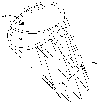
Figure 24 illustrates an alternate embodiment stent and valve device 234.
Device
234 has a two stage stent 238 configuration, with valve materia1241 arranged
both inside
the lumen and outside the structure of the generally tubular shaped device.
This example
is of a relatively shallow sinus variety, and may be one of several
embodiments which
have dual application to both venous and other vascular uses, including, e.
g., an arterial-
venous fistula treatment device.
Figure 25 is a flow diagram of a method of configuring a sheet or other
portion of
valve material for use in stent and valve devices according to the various
embodiments of
this invention. Block 263 illustrates obtaining basic tissue or other suitable
material for
use as valve material and providing it in a generally planar form 266 for
later processing.
In block 272, the material is further shaped over convex/concave shaping means
to
provide optimum concavity for use in the appropriately sized and shaped
valvular sinus
configuration. The final shaping and cutting is performed in block 279 at
which the
precise shape for use in a valve material leaflet is accomplished, including a
plurality of
arcuate and possibly other edge portions. As disclosed herein, various forms
of sclera
may be used in the embodiments of this invention. It has excellent features in
most
respects and is readily harvested at very low cost. Also discussed herein is
the use of the
known material made of small intestine sub-mucosa, also referred to as SIS.
Examples of
this material, though not in this use and application, are found in United
States Patents
No. 4,902,508, 4,956,178, 5,516,533 and 5,641,518, for the teachings of SIS
related
manufacture and principles of use.
Figure 26 illustrates an optional technique of manufacturing the proper stent
and
valve device of this invention according to its intended placement in a
specific patient. In
this technique, it is possible to utilize either some or all steps. In a full
utilization of this
methodology, a patient is designated 301 for sizing. The insufficient or
incompetent
valve site or sites are identified 305 using imaging means, such as that
identified herein or
other systems having highly accurate capabilities. Sizing values for optimum
stent and
CA 02401996 2007-10-29
-13-
valve configurations are obtained 308 using the imaging means, and the values
are then
either stored or otherwise transferred 311 to stent and valve device
manufacturing means.
Molds or other tools may be effectively utilized in this process. In order to
further
customize or render more effective in some manner the manufacture of the valve
material,
it is desired to either select or obtain 315 a tissue sample from the patient
or an
appropriate subject. The tissue sample may then be utilized in known manner to
construct or grow 319 a customized valve portion or portions for later use by
the
designated patient. Teaching examples of this tissue engineering technology
are found in
United States Patent Nos. 4,996,154, 5,326,357, 5,902,741, and 5,902,829.
Following
proper growth of the valve material, the material is then assembled 323 with a
properly
sized stent, and then placed 327 in the patient at the specifically targeted
site. A regimen
of monitoring and follow up 331 continues as appropriate. It is believed that
the
teachings of this method of manufacture and use of the devices herein will
greatly
facilitate the treatment of many people for a medical problem of great
severity and which
little history of remedy.
Figure 27 illustrates yet another advancement in design stents according to
the
principles of the invention. As discussed above, the proper placement and
accommodation of a replacement venous valve is enhanced by use of valves which
are
matched to each patient's physiology. Figure 27 shows one embodiment of stent
and
valve device 411 having a compound diameter with a first region Ll having a
length
corresponding generally to that depicted in Figure 3 as the customized length
of the
specific human valve cusp area. Region Ll has, in this embodiment, a varying
diameter
bulbous-shape formed by struts 419 of frame structure 425. It is appreciated
that Figure
27 illustrates a portion of the valve schematically, and that the shape
depicted will be
arrayed fully about the circumference of the device. Second portions L2 are
sized and
designed using the fit and customization techniques herein to contact those
portions of the
inner lumen of the vein adjacent the primary valve implant site.
Figure 28 is a representative sizing example of a stent and valve device
according
to the embodiment shown in Figure 27. It is recognized that each human valve
is
different, and thus the importance of this invention, but this example shows
one type of
ratios useful for shaping the optimum frame structure. As shown, Figure 28
corresponds
to Figure 27, and is
CA 02401996 2002-09-03
WO 01/66043 PCT/US01/06974
-14-
a side elevation view with D1 about 1.0 cm, D2 about 1.25 cm, H1 about 1.25
cm, and H2
about 2.0 cm.
The valves for placement within the frame structures of Figures 27 and 28 may
be
made from any known technique, although a preferred structure or mode of valve
construction and assembly is as shown throughout this entire disclosure.
Because numerous modifications may be made of this invention without departing
from the spirit thereof, the scope of the invention is not to be limited to
the embodiments
illustrated and described. Rather, the scope of the invention is to be
determined by appended
claims and their equivalents.