Note: Descriptions are shown in the official language in which they were submitted.
CA 02404088 2002-09-23
WO 01/70985 PCT/USO1/09284
BI-FUNCTIONAL CANCER TREATMENT AGENTS
FIELD OF THE INVENTION
The present invention relates generally to the methodology of preparing
and using fusion molecules to treat cancer.
BACKGROUND OF THE INVENTION
Human breast cancer is the predominant malignancy and the leading cause of
cancer death in women from Western society, as reported by Miller et al. ,
(eds)
BIOLOGY OF FEMALE CANCERS, 31-42 (CRC Press, 1997). According to recent
estimation by the American Cancer Society, one in every eight U.S. women will
have
~ o breast cancer and the disease will kill 43,500 women in 1998.
Several lines of evidence have strongly linked prolactin (PRL) to breast
cancer
development. It has been reported that the expression level of prolactin
receptors
(PRLR) is higher in human breast cancer cells compared to normal breast
epithelial
cells (Reynolds et al. , 1997), as well as in surgically removed breast cancer
tissues
~ s (Touraine, Martini P. et al. , Increased Expression Of Prolacti~z Receptor
Gene In
Human Breast Tumors Versus Contiraguous Normal Breast Tissues, (Abstract)
79'''
Annual Meeting of Endocrine Society, p.113, (1997)). The PRLR levels in
malignant
breast tissue can be five folds higher over its surrounding normal tissue (see
Touraine et
al. (1997), supra, making these cells highly sensitive to the stimulation of
hPRL.
zo Additionally, it has been suggested that one mechanism of the mitogenic
action of
estrogen in breast may influence the production and secretion of human
prolactin
(hPRL), since there is a positive correlation between PRLR, estrogen receptors
(ER) or
progesterone receptor levels (Sirbasku, 1978; Dixon and Lippman 1986; Lippman
an
Dickson, 1989). Taken together, these findings lead to a hypothesis that hPRL
serves
2s as an autocrine/paracrine growth factor that plays an important role in
mammary
-1-
CA 02404088 2002-09-23
WO 01/70985 PCT/USO1/09284
carcinogenesis (Clevenger, et al., Am. J. Pathology, 146: 695-705 (1995);
Ginsburg, E. et al., CanceY Res., 55: 2591-2595 (1995)).
An association between PRL expression and prostate disease has also been
proposed in Wennbo et al., Erzdocrinol. I38: 4410-4415 (I997). PRL receptors
are found in prostate tissue as reported Aragona et al. , E>zdocri>zol. 97:
677-684
(1975), and Leake et al., J. Erzdocrinol., 99: 321-328 (1983). In addition,PRL
levels has observed that can increase with age (Hammond et al. , Clirz.
EfzdocYiyzol. , 7: 129-135 (1977), Vekemans et al. , Br. Med. J. 4: 738-739
(1975)) coincident with the development of prostate hyperplasia. Transgenic
mice
overexpressing the PRL gene developed dramatic enlargement of the prostate
gland. (see Wennbo et al. (1977), supra).
In view of its link to both breast and prostate cancer, PRL signaling
represents an attractive target for therapeutic intervention. Heretofore,
however,
no suitable medicaments have been available for this purpose.
Immunological approaches hold great promise in treating cancer. There is
ample evidence that cancers express tumor-specific antigen and patients have T
cells that can respond to these antigens (Boon, Toward T., A Genetic Analysis
of
Human Tumor Rejection Antigens, Advances in Cancer Research, 58: 177-210
(1992); Urban, JL et al., Tunzor Ahtige>zs, Annu. Rev. Immuno. 10: 617-644
(1992)). Yet, these T cells, in many instances, are anergic or otherwise
ineffective in combating the cancer. Thus far, the main effort in
immunological
approaches for tumor therapy is to augment weak host inmlune responses to
tumor antigens such as by exogenously administering cytokines to the patients.
Among many cytokines used, interleukin 2 (IL-2) has been demonstrated
to have promising results. IL-2 is the principal cytokine responsible for
progression of T lymphocytes from the Gl to S phase of the cell cycle (see
Morgan et al., Science 193: 1007-1008 (1979). The principal actions of IL-2 on
lymphocytes are as follows: (1) IL-2, as the major autocrine growth factor for
T
_2_
CA 02404088 2002-09-23
WO 01/70985 PCT/USO1/09284
lymphocytes, determines the magnitude of T cell-dependent immune response.
(2) IL-2 stimulates the growth of natural killer (NK) cells and enhances their
cytolytic effect, as reported in Hendrzak et al. , EXPERIMENTAL AND
CLINICAL AGENTS, 263-282 Humana Press I~ac. (I997).
However, it has been reported that cancer patients receiving systemic IL-2
often experience potentially life-threatening side effects that limits the
total
amount that can be administered which, in turn, directly affects the efficacy
of
treatment. (see Rosenberg et al., N. Engl. J. Med. 319: 1676-1680 (1988);
Maas, Immunobiology 188: 281-292 (1993)). The main efforts regarding the use
of IL-2 in tumor therapy, therefore, have been concentrated on ways and means
to balance the side effect and the effective dose i. e. , increase the
specificity of
administered IL-2 (target the IL-2 precisely at the tumor site), thereby
dramatically decreasing the side effects induced by high systemic dosage.
Forni G., et al., J. Immuho~l. 138: 4033-4041 (1987) demonstrated that
injection of a physiological dose of IL-2 directly into tumor caused
suppression
of their growth. The major advantage of this in situ application is that it
decreases toxicity associated with the systemic use of cytokines, but it has
the
disadvantage of needing to know the exact location of all tumors, which is
particularly problematic in patients with widespread metastases.
Further efforts to decrease toxicity have shown that the injection of
transfected tumor cells which secrete IL-2 can induce specific T cell-
dependent
immunity on subsequent challenges by unmodified tumor cells, as reported in
Gansbacher et al. , J. Exp. Med. 172: 1217-1224 (1990); Fearon et al. , Cell
60:
397-403 (1990); and Pardoll, D.M., h~amuta. Today 14: 310-316 (1993).
However, Reisfeld et al., Cur. Top. Microbiol. Imnaunol. 213: 27-53 (1996)
note that clinical application of such an approach will be both time consuming
and costly, since it will involve the isolation, transfection, and re-
administration
of an individual patient's tumor cells.
-3-
CA 02404088 2002-09-23
WO 01/70985 PCT/USO1/09284
Recently, an alternative approach of using the binding specificity of anti-
tumor monoclonal antibodies (mAb) to direct cytokines to tumor sites has been
introduced. See Reisfeld et al. (1996), supra. This approach combines the
unique targeting ability of a rnAb with the multifunctional activities of
cytokines,
therefore, achieving an effective concentration of IL-2 in the tumor
microenviroment. Targeted IL-2 therapy can completely eradicate disseminated
pulmonary and hepatic murine melanoma metastases in immunocompetent,
syngeneic mice, as shown in Gillies et al. , PPOC. Natl. Acad. Sci. USA 89:
1428-
1432 (1992); and Sabzevari et al. , Proc. Natl. Acad. Sci. USA 91: 9626-9630
(1994).
There are advantages of this targeted IL-2 therapy. For instance, this
therapy does not require the mAb-IL-2 fusion protein to reach all target cells
to
achieve the maximum effects as in the case of other mAb targeted therapies
since
it is not a direct cytotoxic reaction. Reisfeld et al. (1996), supYa. Most
importantly, the therapeutic effect of targeted IL-2 therapy is associated
with the
induction of a long-lived and transferable, protective tumor immunity. This
mAb
targeted IL-2 therapy is also different and advantageous from ex vivo transfer
of
cytokine genes, since it concentrates IL-2 in the tumor environment in a non-
personalized, making this approach more clinically feasible.
Although the targeted immunotherapy approach shows promise in treating
cancer, the therapeutic benefits of combining the effects of antagonizing PRL
and
targeted IL-2 is unknown in treating cancer. There is, therefore, an unmet
need
to develop agents and therapies for simultaneously antagonizing the role of
PRL
in cancer maintenance or proliferation and augmenting the patient's immune
response to the cancer.
SUMMARY OF THE INVENTION
Accordingly, it is an object of the invention to provide a medicament that
is capable of interfering with the prolactin signaling mechanism in a cancer
cell.
-4-
CA 02404088 2002-09-23
WO 01/70985 PCT/USO1/09284
b
It is yet another object of the invention to provide a medicament that
induces apoptosis in a cancer cell.
It is a further object of the invention to provide a medicament that
contains a receptor antagonizing domain and a positive immunomodulating
domain.
It is still another object of the invention to provide a method for treating a
patient suffering from cancer by simultaneously antagonizing a receptor
present
in a targeted cancer cell and augmenting the patient's immune response to the
cancer.
It is another object of the invention to provide a method of treating cancer
by employing the medicaments described herein.
These and other objects which will be more readily apparent upon reading
the following disclosure may be achieved by the present invention.
In a composition of matter aspect, the present invention relates to
substantially to a protein comprising a receptor antagonizing domain and a
positive immunornodulator domain. The invention further provides that the
receptor antagonizing domain can be an apoptosis-promoting domain, while the
positive imrnunomodulator domain can be an interleukin. The receptor
antagonizing domain also can be the amino acid sequence SEQ ID NO: 1 or
conservative variants thereof.
In a methodological aspect, the present invention relates to a method for
treating cancer, comprising administering to a patient an effective amount of
a
protein having a receptor-antagonizing domain and a positive immunomodulator
domain. The invention further provides a methodology for administering to a
patient any of the proteins described herein.
-5-
CA 02404088 2002-09-23
WO 01/70985 PCT/USO1/09284
BRIEF DESCRIPTION OF THE DRAWINGS
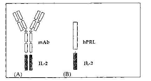
Fig. 1 is a schematic representation of an inventive bi-functional molecule.
(A)
mAb-IL-2 fusion protein model proposed in previous studies by Reisfeld et al.
(I996), supra. and (B) hPRLA-IL-2 fusion protein according to the invention.
Fig. 2 is a schematic representation of a proposed mechanism of action for an
inventive bi-functional fusion protein. PRL produced by breast cancer cell is
prevented from reaching the PRLR due to the occupancy of the fusion protein
(PRLA). At the same time, IL-2 portion of the fusion protein stimulates the
anti
tumor T cell reaction.
Fig. 3 illustrates the dose-response inhibitory effects of hPRI-G129R and
stimulatory effects of hPRL in T-47D human breast cancer cells using co-
culture
method. The x-axis represents the co-cultured L cell (control, L-PRL or L-
hPRL-G129R cell numbers. Each data point represents a mean of at least three
independent experiments with triplicate wells.
Fig. 4 shows the dose-response inhibitory effects of hPRL-G129R and its
additive effects with 4-OH-Tamoxifen in T-47D human breast cancer cell
proliferation assay. The x-axis represents the hPRL-G129R concentration either
in the absence (open bars) or presence of 4-OH-Tamoxifen. Each data point
represents a mean of at least three independent experiments with triplicate
wells.
Fig. 5 is a schematic representation of cloning and construction of the
expression
plasmid of pUCIG-MT-hPRL-IL-2 fusion protein cDNA.
DETAILED DESCRIPTION OF THE PREFERRED EMBODIMENTS
It has been discovered by the present inventors that the combined effects
of endocrine-based and targeted cytokine therapies greatly enhance the
treatment
of cancer. For instance, products and methods of treatment herein disclosed
act
to inhibit the autocrine/paracrine effects of endogenous PRL by blocking the
PRLR, typically resulting in apoptosis. In addition, this approach positively
-6-
CA 02404088 2002-09-23
WO 01/70985 PCT/USO1/09284
modulates an immune response, thereby inducing tumor-specific T lymphocyte
cytotoxicity specifically at the malignant tissue.
As used herein, "apoptosis" refers to a process whereby developmental or
environmental stimuli activate a genetic program to implement a specific
series of
events that culminate in the death and efficient disposal of a cell. The
morphological changes in the cell include dramatic shrinkage of cell volume,
accompanied by dilation of endoplasmic reticulum and convolution of the plasma
membrane. In turn, this causes the cell to break up into a series of membrane-
bounded bodies, containing structurally normal, yet compacted, organelles. The
nucleus undergoes discontinuous chromatin condensation and nuclease-mediated
DNA fragmentation occurs, degrading chromosomal DNA into small
oligonucleosomal fragments. The nucleus and cytoplasm condense and the dying
cell ultimately fragments into membrane-bound apoptotic bodies that are
rapidly
phagocytosed and digested by macrophages or by neighbouring cells.
The present invention combines the benefits associated with blocking the
PRLR and positively modulating an immune response by utilizing a multi-domain
molecule, each domain having the ability to carry out one of these functions.
Typical molecules have a "receptor-antagonizing domain" or an "apoptosis-
promoting domain, " combined with a "positive immunomodulator domain. "
As used herein, a "receptor-antagonizing domain" is a ligand that
specifically binds to a receptor that is associated with a disorder like
cancer,
whereupon binding to the receptor, the receptor-antagonizing domain acts to
inhibit one or more cellular processes, thereby interrupting the etiology or
maintenance of the disease. Such a domain that induces apoptosis is herein
referred to as the "apoptosis-promoting domain," while a "positive
irninunodulator domain" is one that augments an immune response, preferably
enhancing the immune response against an abnormal cell, like a cancer cell.
CA 02404088 2002-09-23
WO 01/70985 PCT/USO1/09284
Such an immune response typically involves recruiting T-cells and enhancing
_- their, for example, cytotoxic function.
The benefits of a fusion protein having these characteristics are immense.
For example, carcinogenic tissues are often characterized by increased levels
of
one or more protein receptors. A fusion protein containing a domain that is
specific to one of these receptors will be able to specifically target the
cancer
tissue. Where the receptor antagonizing domain disrupts the etiology of the
cancer, or disrupts cancer maintenance, as is the case of an apoptosis-
promoting
domain, the receptor antagonizing portion of the molecule has a direct
therapeutic
effect. In addition, due to the presence of the positive immunomodulator
domain, the molecule has a secondary therapeutic effect by inducing the
patient's
own immune system to respond specifically against the diseased tissue.
Accordingly, candidates to receive the therapy according to this invention
include individuals who suffer from malignant tumors those of which are
characterized by the presence of at least one receptor related to tumor
maintenance or proliferation. In a preferred embodiment, the receptor-
antagonizing domain of the fusion protein is an apoptosis-promoting domain,
which binds to a targeted membrane-bound receptor. Such binding induces
apoptosis; simultaneously, the positive immunomodulator domain induces tumor-
specific recruitment and enhancement of T lymphocyte cytotoxicity.
The Inventive Bi-Functional Protein:
In accordance with the invention, bi-functional proteins are contemplated
that have unique dual therapeutic effects on malignant tissue, namely (a)
receptor-antagonizing and/or apoptosis-promoting (which may be one and the
same) and (b) positive immunomodulating. The invention also contemplates
nucleic acids (e.g. DNA or RNA) encoding the inventive bi-functional proteins.
_g_
CA 02404088 2002-09-23
WO 01/70985 PCT/USO1/09284
Receptor-antagonizing domain
The invention contemplates a first domain that, in one aspect, will localize
the effects of the positive immunomodulator domain to the diseased tissue. For
example, carcinogenic tissues are often characterized by increased levels of
one
or more protein receptors. A fusion protein containing a domain that is
specific
to one of these receptors will be able to specifically target the cancer
tissue,
resulting in a localized tumor cytotoxicity reaction directed to the targeted
tissue.
In one embodiment, the domain that targets a particular receptor site is a
receptor-antagonizing domain, which, as its name suggests, binds to and
antagonizes its cognate receptor. In a preferred embodiment, the receptor-
antagonizing domain is an apoptosis-promoting domain. This targeted therapy
approach, utilizing a receptor antagonizing domain, is designed to provide
dramatically decreased systemic concentrations of the positive immunomodulator
domain (e.g., IL-2), thereby reducing its toxicity in vivo.
An additional therapeutic benefit of this dual-function molecule is that the
receptor-antagonizing domain typically has endocrine-blocking ability. Thus,
where the receptor-antagonizing domain, for example, is a prolactin
antagonist,
the normal endocrine function of prolactin will be disrupted. As a consequence
of this endocrine-blocking, in the case of prolactin and similar molecules,
for
instance, apoptosis of the targeted cells can result. In that case, the
receptor-
antagonizing domain is also an apoptosis-promoting domain.
In the case of an apoptosis-promoting domain, such a domain generally is
designed by creating antagonists of the normal function of a cellular
component
that is involved in preventing apoptosis. In both breast and prostrate cancer
tissue, for example, carcinogenesis and malignant cell proliferation is
stimulated,
at least in part, by increased levels of PRLR. Signaling via the PRLR is known
to be mediated by dimerization of the prolactin receptor, which is itself
mediated
by dimerization of receptor-bound prolactin molecules. The binding of
-g_
CA 02404088 2002-09-23
WO 01/70985 PCT/USO1/09284
endogenous PRL to two PRLRs induces PRLR dimerization, thereby triggering
signal transduction into the cancer cells. Accordingly, one embodiment of the
invention entails antagonizing the normal apoptosis-inhibiting function of
prolactin using a prolactin antagonist (PRLA) (i. e. , a prolactin antagonist
domain).
Signal transduction in the PRLR signaling pathway involves signal
transducers and activators of transcription (STAT) phosphorylation, which is
involved in preventing or blocking apoptosis, the normal result of PRLR
agonism. Thus, G129R antagonist promotes apoptosis by inhibiting STAT 5
phosphorylation in human breast cancer cells. Accordingly, blocking the PRLR
inhibits the autocrine/paracrine effects of endogenous PRL, which involves
STAT 5, and results in apoptosis. Thus, one class of apoptosis-promoting
compounds contemplated by the invention is one that can inhibit STAT 5
phosphorylation.
A suitable PRLA contemplated by the invention generally will retain the
characteristic of specific binding to the PRLR, yet will have some structural
deficiency that disrupts the normal PRL apoptosis-blocking mechanism. Such a
structural deficiency includes those that disrupt PRL(and thus PRLR)
dimerization.
In one preferred embodiment, shown in SEQ ID NO: 1, this structural
deficiency is a substitution of Gly to Arg at a position corresponding to 129
in
hPRL (denoted as hPRL-G129R). Figures 3 and 4, as well as the cell-based
assays presented in Examples 4, 5 and 6 demonstrate that this mutated hPRL
acts
as a true hPRLR antagonist. Accordingly, a receptor-antagonizing domain such
as hPRL-G129R can serve as a therapeutic medicament for treating certain types
of cancer.
-10-
CA 02404088 2002-09-23
WO 01/70985 PCT/USO1/09284
This embodiment is supported by Chen et al. , Clira. Cafa. Res. 5: 3583-93
(1999), who disclose a species comparison of amino acid sequences within the
third a-helical region of PRLs, shown in Table 1.
Table 1
Species Domain Peptide Sequence129 Pep. Seq.
Human PRL IEEQTKRLLR G MELIVS-QVHP
Rat PRL IEEQNKRLLE G IEKIIG-QAYP
Mouse PRL IEEQNKQLLE G VEKIIS-QAYP
Hamster PRL IGEQNKRLLE G IEKILG-QAYP
Fin whale PRL EEEENKRLLE G MEI<IVG-QVHP
Mink PRL IEEENRRLLE G MEI<IVG-QVHP
Cattle PRL IEEQNKRLIE G MEMIFG-QVIP
Sheep PRL EEEENKRLLE G MENIFG-QUIP
Pig PRL IEEQNKRLLE G MEKTVG-QVHP
Camel PRL IEEQNKRLLE G MEKIVG-QVHP
Horse PRL EIEQNRRLLE G MEKIVG-QVQP
Elephant PRL VKEENQRLLE G IEKIVD-QVHP
Ancestral mammal PRL IEEENKRLLE G MEKIVG-QVHP
Chicken PRL IEEQNKRLLE G MEKIVG-RVHS
Turkey PRL IEEQDKRLLE G MEKTVG-RIHS
Sea turtle PRL IEEQNKRLLE G MEKIVG-QVHP
Crocodile PRL IEEQNKRLLE G MEKTIG-RVQP
Alligator PRL IEEQNKRLLE G MEKVIG-RVQP
Ancestral amniote PRL IEEQNKRLLE G MEKIVG-QVHP
Xenopus PRL VEEQNKRLLE G MEKIVG-RIHP
Bullfrog PRL VEEQTKRLLE G MERITG-RIQP
Lungfish PRL VEDQTKQLIE G MEKILS-RMHP
Tilapia PRL MQQYSKSLKD G LD-VLSSKMGS
Tilapia PRL MQEHSKDLKD G LD-ILSSKMGP
Common carp PRL LQENINSLGA G LEHVF-NKMDS
Bighead carp PRL LQDNINSLGA G LERVV-HKMGS
Silver carp PRL LQDNINSLVP G LEHVV-HKMGS
Chun salmon PRL LQDYSKSLGD G LD-IMVNKMGP
Chinook salmon PRL LQDYSKSLGD G LD-IMVNKMGP
Trout PRL LQDYSKSLGD G LD-IMVNKMGP
Species Domain Peptide Sequence120 Pep. Seq.
Human GH VYDLLKDLEE G IQTLMRELEDG
Bovine GH VYEKLKDLEE G ILALMRELEDG
According to Table 1, it is clear that Gly 129 of hPRL is invariable
among PRLs, suggesting an important role in its function. Thus, substituting
any
amino acid for Gly 129 should produce PRLA in each of these species (Chen et
al., Molec. Endocrinol. (1995)). In one embodiment, an antagonist is created
by
substituting a relatively bulky side chain amino acid, such as Arg for Gly
129.
-11-
CA 02404088 2002-09-23
WO 01/70985 PCT/USO1/09284
Accordingly, one aspect of the invention contemplates conservative variants of
P1ZL that are characterized by the presence of a relatively small side-chain
amino
acid (i.e. Gly) at a specific position, such that substituting the small side-
chain
amino acid for a bulky side-chain amino acid will result in an antagonistic
form
of the protein.
The receptor-antagonizing domain of present invention also includes
conservative variants of receptor antagonizing domains discussed herein. The
overall structure and composition of the inventive domains, in that respect,
are
important only insofar as they confer the appropriate functional
characteristics, i. e. ,
receptor antagonism, apoptosis induction, positive immunomodulation.
Conservative variants according to the invention generally conserve the
overall molecular structure of the protein domains. Given the properties of
the
individual amino acids comprising the disclosed protein products, some
rational
substitutions will be apparent. Amino acid substitutions, i. e. "conservative
substitutions," may be made, for instance, on the basis of similarity in
polarity,
charge, solubility, hydrophobicity, hydrophilicity, and/or the amphipathic
nature of
the residues involved. '
For example: (a) nonpolar (hydrophobic) amino acids include alanine,
leucine, isoleucine, valine, proline, phenylalanine, tryptophan, and
methionine; (b)
polar neutral amino acids include glycine, serine, threonine, cysteine,
tyrosine,
asparagine, and glutamine; (c) positively charged (basic) amino acids include
arginine, lysine, and histidine; and (d) negatively charged (acidic) amino
acids
include aspartic acid and glutamic acid. Substitutions typically may be made
within
groups (a)-(d). In addition, glycine and proline may be substituted for one
another
based on their ability to disrupt a-helices. Similarly, certain amino acids,
such as
alanine, cysteine, leucine, methionine, glutamic acid, glutamine, histidine
and
lysine are more commonly found in a,-helices, while valine, isoleucine,
phenylalanine, tyrosine, tryptophan and threonine are more commonly found in
(3-
-12-
CA 02404088 2002-09-23
WO 01/70985 PCT/USO1/09284
pleated sheets. Glycine, serine, aspartic acid, asparagine, and proline are
commonly found in turns. Some preferred substitutions may be made among the
following groups: (i) S and T; (ii) P and G; and (iii) A, V, L and I. Given
the
known genetic code, and recombinant and synthetic DNA techniques, the skilled
scientist readily can construct DNAs encoding the conservative amino acid
variants.
Conservative variants specifically contemplate truncations of the presently
described receptor antagonizing domains. Truncations may be made from the N-
or
C-terminus, but generally do not entail deleting more than about 30 % of the
native
molecule. More preferably, less than about 20 % , and most preferably, less
than
about 10 % , of the native molecule is deleted.
In general, both the DNA and protein molecules of the invention can be
defined with reference to "sequence identity. " Some molecules have at least
about
50 % , 5S % or 60 % identity. Preferred molecules are those having at least
about
65 % sequence identity, more preferably at least 65 % or 70 % sequence
identity.
Other preferred molecules have at least about 80 % , more preferably at least
80 % or
85 % , sequence identity. Particularly preferred molecules have at least about
90
sequence identity, more preferably at least 90% sequence identity. Most
preferred
molecules have at least about 95 % , more preferably at least 95 % , sequence
identity. As used herein, two nucleic acid molecules or proteins are said to
"share
significant sequence identity" if the two contain regions which possess
greater than
85% sequence (amino acid or nucleic acid) identity.
"Sequence identity" is defined herein with reference the Blast 2 algorithm,
which is available at the NCBI (http://www.ncbi.nlm.nih.gov/BLAST), using
default parameters. References pertaining to this algorithm include: those
found
at http://www.ncbi.nlm.nih.gov/BLAST/blast references.html; Altschul, S.F.,
Gish, W., Miller, W., Myers, E.W. & Lipman, D.J. (1990) "Basic local
alignment search tool." J. Mol. Biol. 215: 403-410; Gish, W. & States, D.J.
(1993) "identification of protein coding regions by database similarity
search."
-13-
CA 02404088 2002-09-23
WO 01/70985 PCT/USO1/09284
Nature Genet. 3: 266-272; Madden, T.L., Tatusov, R.L. & Zhang, J. (1996)
"Applications of network BLAST server" Meth. Enzymol. 266: 131-141;
Altschul, S.F., Madden, T.L., Schaffer, A.A., Zhang, J., Zhang, Z., Miller,
W. & Lipman, D.J. (1997) "Gapped BLAST and PSI-BLAST: a new generation
of protein database search programs." Nucleic Acids Res. 25: 3389-3402; and
Zhang, J. & Madden, T.L. (1997) "PowerBLAST: A new network BLAST
application for interactive or automated sequence analysis and annotation."
Genome Res. 7: 649-656. Accordingly, the prolactin peptide sequences from
different species, which include those listed in Table 1, can be aligned,
using
standard computer programs like BLAST, to inform further variation in
prolactin-derived receptor-antagonizing domains that preserve their essential
function.
In addition to proteins that are conservative variants of those disclosed
herein, the invention also contemplates the use of proteins that play a role
in
inducing tumor proliferation, wherein an amino acid substitution will inhibit
the
protein's ability to induce this proliferation. For example, Gly 119 and Gly
120
of bovine growth hormone (bGH) and hGH, respectively, play critical roles in
the action of GH in stimulating growth enhancement. Growth hormone receptor
(GHR) dimerization is thought to be a key step for HG signal transduction.
Accordingly, any amino acid substitution (other than Ala), especially one with
a
bulky side chain such as Arg at these respective positions will prevent
receptor
dimerization, resulting in a growth hormone antagonist (GHA). Thus,
antagonists
such as GHA are contemplated by the invention.
In addition to antagonizing the normal function of a cellular component
involved in preventing apoptosis, the invention further comprehends, in the
context of apoptosis-promoting domains, agents that induce apoptosis by
positive
means. That is, such agents do not work by antagonizing an anti-apoptotic
pathway; rather they induce an apoptotic pathway. Examples of such agents are
protein kinase C (PKC) inhibitors, including chelerythrine.
-14-
CA 02404088 2002-09-23
WO 01/70985 PCT/USO1/09284
The benzophenanthridine alkaloid chelerythrine (I,2-dimethoxy-12-
methyl[1,3]benzodioxolo[5,6-c]phenanthridinium; Cz~HraNOa), also known as
toddaline, is extractable either in pure form or as a mixture with other
benzophenanthridine alkaloids from Chelidoniuna majus L., Zanthoxyluna
simulans, SanguinaYia candensis (or bloodroot), Macleaya coYdata, Carydali
sevctocozii, Ca~ydali ledebouni, Chelidonium majusm and other members of
Papaveracaceae.
Inhibitors of PKC can interact with the substrate binding site (ATP or
protein) or with the regulatory domain where activation occurs (diacylglycerol
or
phorbol ester binding site). Chelerythrine interacts directly with the
catalytic
domain of PKC. It is one of the most potent inhibitors of PKC identified and
does not appear to inhibit any other protein kinases. For example,
chelerythrine
shows potent cytotoxic effects against L-1210 tumor cells with an IC50 value
of
0.053 p,M by inhibiting cell growth and differentiation, as discussed by
Herbert
et al., Bioechem. Biophys. Res. Cornmun. 172: 993 (1990). Chelerythrine
induces apoptosis by specifically inhibiting PKC in a concentration-dependent
manner and strongly inhibiting platelet aggregation induced by strong
aggregation
inducers, such as arachidonic acid and collagen.
Thus, upon introduction to tumor ~ cells, chelerythrine chloride can
decrease the apoptotic threshold and trigger apoptosis therein. This is
particularly true when chelerythrine therapy is used in conjunction with other
methods of treatment. Accordingly, a fusion molecule that includes
chelerythrine
fused to another molecule used to combat cancer, for example a positive
immunomodulator domain, is contemplated by the invention. A molecule
containing chelerythrine can be fused to another molecule (i.e.. a domain as
described herein) by conventional chemical means, using multifunctional cross-
linkers, for example. Protein-based PKC inhibitors may be made as fusion
proteins.
-15-
CA 02404088 2002-09-23
WO 01/70985 PCT/USO1/09284
Positive inamunonaodulator domain
The invention also contemplates an additional, yet separate, domain that
acts as a positive immunomodulator. Preferred immunomodulator domains
support a tumor-directed positive immune response. An example of a suitable
positive immunomodulator includes a cytokine that can xecruit T lymphocytes to
the tumor, thereby inducing tumor specific T lymphocyte cytotoxicity at the
malignant tissue. In a preferred embodiment, the positive irnmunomodulator is
IL-2, which is characterized by its ability to control the magnitude of T cell-
dependent immune response. IL-2 also has activity on macrophages and
monocytes. In addition, IL-2 stimulates the growth of natural killer (NK)
cells
and enhances their cytolytic effect.
In addition to IL-2, the invention contemplates other molecules, including
additional cytokines, having these or similar properties. For example, IL-12
can
represent the positive immunomodulator domain. IL-12 is a key cytokine for
directing the T cell response to that of a Th1 type. IL-12 is made by B cells
and
monocytes/macrophages and acts synergistically with IL-2 to induce IFNy
production by T cells and NK cells. It also enhances the cytotoxicity activity
of
both T cells and NK cells. The invention also includes conservative variants
(as
detailed above) of the aforementioned positive immunomodulator domains.
Other suitable candidates for the positive immunomodulator domain
include the interferons (IFN). For example, IFN-(3, by itself, is known to
inhibit
tumor cell proliferation. IFN-~ is a universal macraphage activating agent for
antitumor activity. Accordingly, a fusion molecule containing IFN-~3 bound to
an apoptosis promoting domain would provide localized positive
hnmunomodulating therapy to a targeted tissue.
-16-
CA 02404088 2002-09-23
WO 01/70985 PCT/USO1/09284
Prepaf-ing Exefnplary Bi-Functzonal Molecules:
A bi-functional protein contemplated by this invention is one that contains
each of the previously mentioned domains, namely receptor-antagonizing (which
also may be apoptosis-promoting) and positive immunomodulating, wherein upon
such fusing, both domains substantially retain their associated
characteristics,
independent of the other. Figure 2 discloses one embodiment of the invention,
according to these characteristics. Although typically produced as fusion
proteins, the domains also may be fused by conventional chemical means, using
multifunctional cross-linkers, for example. When fusion proteins are made,
either domain may be placed C-terminal or N-terminal to the other.
In one embodiment, the fusion protein is a hPRLA-IL-2 protein, as shown
in Figure 1. This fusion protein can be integrated into an expression vector,
as
shown in example 1 and figure 5. The generated expression vector can then be
transfected into a stable cell line to subsequently produce a purified
protein.
Examples 2 and 3 are non-limiting procedures for carrying out the vector
transformation and purification processes. This fusion protein has the C-
terminus of PRLA fused to the N-terminal side of IL-2, which is shown in
Figure 5. However, the invention also contemplates any fusion protein having
domains as described herein.
Suitable methods for creating the fusion protein should be ones that do not
substantially change the biological activity of either of these domains. For
example, it has been demonstrated that fusion of the N-terminal of IL-2 to the
C-
terminal end of an antibody does not change the biological activity of IL-2
Reisfeld et al. (1996), supra. Therefore, a similar strategy can be adopted to
produce a fusion protein according to the invention. This process includes
designing a cDNA encoding a fusion protein which links the N-terminus of the
positive immunomodulator domain to the C-terminus of receptor-antagonizing
domain.
-17-
CA 02404088 2002-09-23
WO 01/70985 PCT/USO1/09284
There is evidence, moreover, that the C-terminal ends of hGH (we
deleted up to 10 amino acids) are not important for growth promoting
activities in
transgenic mice (Chen et al. , 1993) and, based on structural similarity,
fusion of
a positive modulator to the C-terminal end of other receptor-antagonizing
domains, such as hPRLA, should not alter the binding affinity of these
domains.
The present invention is not limited to any particular method of producing
the desired fusion protein contemplated herein. According to the contemplated
recombinant methods of production, however, the invention provides recombinant
DNA constructs comprising one or more of the nucleotide sequences of the
domains
described in the present invention. The recombinant constructs of the present
invention comprise a vector, such as a plasmid or viral vector, into which a
DNA
or DNA fragment, typically bearing an open reading frame, is inserted, in
either
orientation. The invention further contemplates cells containing these
vectors.
Recombinant protein production is well known in the art and is outlined
briefly below.
Bacterial Exp~essioh
Useful expression vectors for bacterial use are constructed by inserting a
structural DNA sequence encoding a desired protein together with suitable
translation initiation and termination signals in operable reading phase with
a
functional promoter. The vector will comprise one or more phenotypic
selectable
markers and an origin of replication to ensure maintenance of the vector and,
if
desirable, to provide amplification within the host. Suitable prokaryotic
hosts for
transformation include E. coli, Bacillus subtilis, Salmonella typhimurium and
various species within the genera Pseudomonas, Streptomyces, and
Staphylococcus, although others may, also be employed as a matter of choice.
In
a preferred embodiment, the prokaryotic host is E. coli.
Bacterial vectors may be, for example, bacteriophage-, plasmid- or
cosmid-based. These vectors can comprise a selectable marker and bacterial
-18-
CA 02404088 2002-09-23
WO 01/70985 PCT/USO1/09284
origin of replication derived from commercially available plasmids typically
containing elements of the well known cloning vector pBR322 (ATCC 37017).
Such commercial vectors include, for example, GEM 1 (Promega Biotec,
Madison, WI, USA), pBs, phagescript, PsiX174, pBluescript SK, pBs KS,
pNHBa, pNHl6a, pNHlBa, pNH46a (Stratagene); pTrc99A, pKK223-3,
pKK233-3, pKK232-8, pDR540, and pRIT5 (Pharmacia). A preferred vector
according to the invention is THE Pt71 expression vector (Paris et al. ,
Biotechnol. Appl. Biochem. 12: 436-449 (1990)).
These "backbone" sections are combined with an appropriate promoter
and the structural sequence to be expressed. Bacterial promoters include lac,
T3,
T7, lambda PR or PL, trp, and ara. T7 is the preferred bacterial promoter.
Following transformation of a suitable host strain and growth of the host
strain to an appropriate cell density, the selected promoter is
derepressed/induced
by appropriate means (e.g., temperature shift or chemical induction) and cells
are
cultured for an additional period. Cells are typically harvested by
centrifugation,
disrupted by physical or chemical means, and the resulting crude extract
retained
for further purification.
Euka~-yotic Expression
Various mammalian cell culture systems can also be employed to express
recombinant protein. Examples of mammalian expression systems include
selected mouse L cells, such as thymidine kinase-negative (TK) and adenine
phosphoribosul transferase-negative (APRT) cells. Other examples include the
COS-7 lines of monkey kidney fibroblasts, described by Gluzman, Cell 23: 175
{1981), and other cell lines capable of expressing a compatible vector, for
example, the C127, 3T3, CHO, HeLa and BHK cell lines. Mammalian
expression vectors will comprise an origin of replication, a suitable promoter
and
enhancer, and also any necessary ribosome binding sites, polyadenylation site,
splice donor and acceptor sites, transcriptional termination sequences, and 5'
-19-
CA 02404088 2002-09-23
WO 01/70985 PCT/USO1/09284
flanking non-transcribed sequences. DNA sequences derived from the SV40
viral genome, for example, SV40 origin, early promoter, enhancer, splice, and
polyadenylation sites may be used to pxovide the required non-transcribed
genetic
elements .
S Mammalian promoters include CMV immediate early, HSV thymidine
kinase, early and late SV40, LTRs from retrovirus, and mouse metallothionein-
I.
Exemplary mammalian vectors include pWLneo, pSV2cat, pOG44, pXTl, pSG
(Stratagene) pSVK3, pBPV, pMSG, and pSVL (Pharmacia). In a preferred
embodiment, the mammalian expression vector is pUCIG-MET. Selectable
20 markers include CAT (chloramphenicol transferase).
In mammalian host cells, a number of viral-based expression systems may
be utilized. In cases where an adenovirus is used as an expression vector, the
coding sequence of interest may be ligated to an adenovirus
transcription/translation control complex, e.g., the late promoter and
tripartite
1S leader sequence. This chimeric gene may then be inserted in the adenovirus
genome by ih vitro or in vivo recombination. Insertion in a non-essential
region
of the viral genome (e.g., region E1 or E3) will result in a recombinant virus
that
is viable and capable of expressing a,target protein in infected hosts. (E.g.,
See
Logan et al. , 1984, Proc. Natl. Acad. Sci. USA 81: 36SS-3659).
20 Therapeutic Compositions:
The proteins of the present invention can be formulated according to
known methods to prepare pharmaceutically useful compositions, whereby the
inventive molecules, or their functional derivatives, are combined in
admixture
with a pharmaceutically acceptable carrier vehicle. Suitable vehicles and
their
2S formulation, inclusive of other human proteins, e.g., human serum albumin,
are
described, for example, in Remingto~i's PhaYmaceutical Sciences (16th ed.,
Osol,
A., Ed., Mack, Easton PA (1980)). In order to form a pharmaceutically
acceptable composition suitable for effective administration, such
compositions
-20-
CA 02404088 2002-09-23
WO 01/70985 PCT/USO1/09284
will contain an effective amount of one or more of the proteins of the present
invention, together with a suitable amount of carrier vehicle.
Pharmaceutical compositions for use in accordance with the present
invention may be formulated in conventional manner using one or more
physiologically acceptable carriers or excipients. Thus, the bi-functional
molecules and their physiologically acceptable salts and solvate may be
formulated for administration by inhalation or insufflation (either through
the
mouth or the nose) or oral, buccal, parenteral or rectal administration.
For oral administration, the pharmaceutical compositions may take the
form of, for example, tablets or capsules prepared by conventional means with
pharmaceutically acceptable excipients such as binding agents (e. g. ,
pregelatinised maize starch, polyvinylpyrrolidone or hydroxypropyl
methylcellulose); fillers (e.g., lactose, microcrystalline cellulose or
calcium
hydrogen phosphate); lubricants (e.g., magnesium stearate, talc or silica);
disintegrants (e. g. , potato starch or sodium starch glycolate); or wetting
agents
(e, g. , sodium lauryl sulphate) . The tablets may be coated by methods well
known in the art. Liquid preparations for oral administration may take the
form
of, for example, solutions, syrups or suspensions, or they maybe presented as
a
dry product for constitution with water or other suitable vehicle before use.
Such
liquid preparations may be prepared by conventional means with
pharmaceutically acceptable additives such as suspending agents (e.g.,
sorbitol
syrup, cellulose derivatives or hydrogenated edible fats); emulsifying agents
(e.g., lecithin ox acacia); non-aqueous vehicles (e.g., almond oil, oily
esters,
ethyl alcohol or fractionated vegetable oils); and preservatives (e.g., methyl
or
propyl-p-hydroxybenzoates or sorbic acid). The preparations may also contain
buffer salts, flavoring, coloring and sweetening agents as appropriate.
Preparations for oral administration may be suitably formulated to give
controlled release of the active compound. For buccal administration the
-21-
CA 02404088 2002-09-23
WO 01/70985 PCT/USO1/09284
composition may take the form of tablets or lozenges formulated in
conventional
manner.
For administration by inhalation, the bi-functional molecules for use
according to the present invention are conveniently delivered in the form of
an
aerosol spray presentation from pressurized packs or a nebuliser, with the use
of
a suitable propellant, e. g. , dichlorodifluoromethane,
trichlorofluoromethane,
dichlorotetrafluoroethane, carbon dioxide or other suitable gas. In the case
of a
pressurized aerosol the dosage unit may be determined by providing a valve to
deliver a metered amount. Capsules and cartridges of, e.g. gelatin for use in
an
inhaler or insufflator may be formulated containing a powder mix of the
compound and a suitable powder base such as lactose or starch.
The bi-functional proteins may be formulated for parenteral
administration by injection, e.g., by bolus injection or continuous infusion.
Formulations for injection may be presented in unit dosage form, e.g., in
ampules or in multi-dose containers, with an added preservative. The
compositions may take such forms as suspensions, solutions or emulsions in
oily
or aqueous vehicles, and may contain formulatory agents such as suspending,
stabilizing and/or dispersing agents. Alternatively, the active ingredient may
be
in powder form for constitution with a suitable vehicle, e. g. , sterile
pyrogen-free
water, before use.
The compounds may also be formulated in rectal compositions such as
suppositories or retention enemas, e. g. , containing conventional suppository
bases such as cocoa butter or other glycerides.
In addition to the formulations described previously, the bi-functional
molecules may also be formulated as a depot preparation. Such long acting
formulations may be administered by implantation (for example subcutaneously
or intramuscularly) or by intramuscular injection. Thus, for example, the
compounds may be formulated with suitable polymeric or hydrophobic materials
-22-
CA 02404088 2002-09-23
WO 01/70985 PCT/USO1/09284
(for example as an emulsion in an acceptable oil) or ion exchange resins, or
as
sparingly soluble derivatives, for example, as a sparingly soluble salt.
The compositions may, if desired, be presented in a pack or dispenser
device which may contain one or more unit dosage forms containing the active
ingredient. The pack may for example comprise metal or plastic foil, such as a
blister pack. The pack or dispenser device may be accompanied by instructions
for administration.
The compositions, since they are useful in cancer treatment, may be
formulated in conjunction with conventional chemotherapeutic agents.
Conventional chemotherapeutic agents include alkylating agents,
antimetabolites,
various natural products (e.g., vinca alkaloids, epipodophyllotoxins,
antibiotics,
and amino acid-depleting enzymes), hormones and hormone antagonists.
Specific classes of agents include nitrogen mustards, alkyl sulfonates,
nitrosoureas, triazenes, folic acid analogues, pyrimidine analogues, purine
analogs, platinum complexes, adrenocortical suppressants,
adrenocorticosteroids,
progestins, estrogens, antiestrogens and androgens. Some exemplary compounds
include cyclophosphamide, chlorambucil, methotrexate, fluorouracil,
cytarabine,
thioguanine, vinblastine, vincristine, doxorubincin, daunorubicin, mitomycin,
cisplatin, hydroxyurea, prednisone, hydroxyprogesterone caproate,
medroxyprogesterone, megestrol acetate, diethyl stilbestrol, ethinyl
estradiol,
tomoxifen, testosterone propionate and fluoxymesterone. In treating breast
cancer, for example, tamoxifen is particularly preferred.
Methods of the Invention:
Treatf~ae~at Methods
The inventive therapeutic methods according to the invention generally
utilize the bi-functional proteins identified above. The domains of the fusion
proteins share the ability to specifically target a specific tissue and/or
augment an
immune response to targeted tissue. A typical method, accordingly, involves
-23-
CA 02404088 2002-09-23
WO 01/70985 PCT/USO1/09284
binding a receptor of a targeted cell to the receptor-antagonizing domain of
the
fusion protein and/or stimulating a T-cell dependent immune response via the
positive immunomodulator domain.
Therapeutic methods involve administering to a subject in need of
treatment a therapeutically effective amount of a fusion protein.
"Therapeutically
effective" is employed here to denote the amount of fusion proteins that are
of
sufficient quantity to inhibit or reverse cancer growth (e.g., induce
apoptosis).
Some methods contemplate combination therapy with known cancer medicaments
or therapies, for example, chemotherapy (preferably using compounds of the
sort
. listed above) or radiation. The patient may be a human or non-human animal.
A
patient typically will be in need of treatment when suffering from a cancer
characterized by increased levels of receptors that promote cancer maintenance
or
proliferation.
Administration during in vivo treatment may be by any number of routes,
including parenteral and oral, but preferably parenteral. Intracapsular,
intravenous, intrathecal, and intraperitoneal routes of administration may be
employed, generally intravenous is preferred. The skilled artisan will
recognize
that the route of administration will vary depending on the disorder to be
treated.
Determining a therapeutically effective amount of the bi-functional
protein, according to this invention, largely will depend on particular
patient
characteristics, route of administration, and the nature of the disorder being
treated. General guidance can be found, for example, in the publications of
the
International Conference on Harmonisation and in REMINGTON'S
PHARMACEUTICAL SCIENCES, chaptexs 27 and 28, pp. 484-528 (Mack
Publishing Company 1990).
Determining a therapeutically effective amount specifically will depend on
such factors as toxicity and efficacy of the medicament. Toxicity may be
determined using methods well known in the art and found in the foregoing
-24-
CA 02404088 2002-09-23
WO 01/70985 PCT/USO1/09284
references. Efficacy may be determined utilizing the same guidance in
conjunction with the methods described below in the Examples. A
pharmaceutically effective amount, therefore, is an amount that is deemed by
the
clinician to be toxicologically tolerable, yet efficacious. Efficacy, for
example,
can be measured by the induction or substantial induction of T lymphocyte
cytotoxicity at the targeted tissue or a decrease in mass of the targeted
tissue.
Suitable dosages can be from about lmglkg to lOmg/kg.
Screerziyag Assays to determine tlae biological activities of tlae fusion
proteifz
The present invention also provides cell-based assay systems that can be
used to compare the biological activities of the apoptosis-promoting domain,
positive immunomodulating domain, and/or a fusion protein comprising each of
these domains. To this end, a cell proliferation assay is used to ensure that
the
fused domains of the fusion protein each retain a function similar to the
respective domain when it is not fused (i.e. not part of a fusion protein).
In one embodiment, the biological activity of the fusion protein will be
determined by introducing the protein to two separate types of cell lines in
vitro:
each cell line determining the activity of a specific domain. For example, a
cell
line that is a reliable indicator of the biological activities of the
apoptosis-
promoting domain should be used to test the effects of that domain, while a
cell
line capable of indicating positive immunomodulating domain should be used to
monitor the activity of the other domain.
By introducing to a cell line various concentrations of a particular domain
in its antagonized, non-antagonized, and fused forms, one of skill in the art
could
determine the biological activity of the apoptosis-promoting domain of the
fused
protein vis-a-vis the same domain in its non-fused state. There are numerous
ways to measure apoptosis. These methods include, but are not limited to the
following techniques: (1) Loss of cell viability - measured by a failure to
either
exclude vital dye or uptake MTT (3-(4,5-dimethylthiazol-2-yl)-2,5-
-25-
CA 02404088 2002-09-23
WO 01/70985 PCT/USO1/09284
diphenyltetrazolium bromide), or MTS-PMS; (2) DNA fragmentation - assayed
by agarose gel electrophoresis, PFG electrophoresis, in situ terminal
transferase
labelling (TUNEL); Cell and nuclear morphology - employing microscopy to
visualize chromatin condensation, DNA organization, and cytoplasmic integrity;
and Cysteine protease activation assay - utilizing caspase activation assays
combined with colorimetric or fluorescent readouts, poly (ADP-ribose)
polymerise (PARP) or laminin cleavage by western blot or
imrnunohistochemisrty.] Examples 4 and 9, which use a human breast cancer cell
line (T-47D), provide non-limiting examples of acceptable assay systems to
this
extent.
Likewise, a cell line that can measure activity of the positive
immunomodulating domain should be similarly used to monitor the activity of
this domain of the fusion protein. Example 9, which uses a murine T cell line
(HT-2), is one possible, yet non-limiting method to determine biological
activity
of the positive immunomodulating domain in the fusion protein.
Another preferred method for determining activity of the fusion protein
according to the invention is the conduct tests in vivo. A suitable host for
this
test would be a mammalian host containing cancer tissue capable of binding the
apoptosis-promoting domain of the fusion protein. A suitable control can be
any
cell line characterized by few or no receptor sites for the chosen apoptosis-
promoting domain. Example 10 provides a non-limiting example of an in vivo
study, using mouse cells.
-26-
CA 02404088 2002-09-23
WO 01/70985 PCT/USO1/09284
The following examples are'intended to be illustrative and not limiting
EXAMPLES
Example 1: cloning ayad construction of the expression plasmid p IICIG-MT
Hprla-IL2 fusion protein cDNA.
PCR fragments from hPRLA cDNA (from restriction site XbaI to the
sequence just before translational stop codon) and IL-2 cDNA (purchased from
ATCC) from amino acid sequence + 1 site to translational stop codon,
respectively, were separately amplified. Next, these fragments were ligated
into.
pUCIG-MET, a mammalian expression vector, to generate an expression vector
that has incorparated these fragments (i.e. pUCIG-MET-hPRLA-IL2). A BamHl
restriction site was added for cloning purposes between hPRLA and IL-2 cDNA,
which resulted in two extra amino acid residues (Gly and Ser) at the junction
of
the fusion protein.
Example Z: trarcsfectircg an expression plasmid into a stable cell line.
We have already generated hPRL-IL-2, and hPRLA-IL-2 pooled stable
mouse cells for our preliminary iu vitro studies. In producing a fusion
protein
according to this invention, mouse L cells are used. These cells are first co-
transfected with DNA molecules encoding hPRLA-IL-2 cDNA driven by mouse
metallothionien regulatory sequence, along with the herpes viral TK gene and
the
hamster APRT gene (Leung et al. , 1985) by using lipofectin method (Gibco
BRL, Gaithersburg, MD). Following double selection for the TK+, the
APRT+ phenotypes, stable hPRL or hPRL analog-secreting mouse L cell lines
are established and maintained in Dulbecco Modified Eagles Medium (DMEM,
Gibco BRL, Gaithersburg, MD) plus 10 % Nu-serum (Collaborative Research,
Bedford, MA), l5,ug/ml hypoxanthine, l,ug/ml aminopterin, l5,ug/ml thymidine
and SO,ug/ml gentamicin. Cells are passed into T150 flasks until they reach
90%
confluency. At that point, serum free conditioned media is collected every 24
-27-
CA 02404088 2002-09-23
WO 01/70985 PCT/USO1/09284
hours and pooled. Pooled medium is frozen at -20 C until further purification
is
accomplished.
Example 3: puYifying the fusion pYOteih.
To obtain an increased yield of fusion proteins, it is desired to first purify
them, according to procedures that axe well known in the art. These steps
include: removing the cells from culture via centrifugation, followed by
precipitation, crossflow ultrafiltration, and chromatography methodologies,
such
as low pressure SEC and preparative RP-HPLC chromatography. These steps
are followed by: buffer exchange, depyrogenation, and lyophilation.
Step One: Cell Removal - 80-100 % of the conditioned medium is
centrifuged at 6000 x g for 30 min and at 4° C. The starting cell
culture medium
has a volume of 80-100 liters. All the hPRL-G129R is recovered in this step.
Step Two: Precipitation - The precipitation procedure is carried out by
gradually adding 3.5 kg ammonium sulfate to 10 liters of cell culture medium
with constant stirring. The solution is placed at 4° C for 12 hours.
The
supernatant is centrifuged off. The precipitate is then dissolved in 2 liters
of
distilled water. The non-dissolved impurities are removed by centrifugation.
All the centrifugation steps are operated at 6000 x g for 30 min and at
4° C. The
final volume of solution is about 5 liters.
Step three: Volume Reduction using Crossflow Ultrafiltration - A lOK
molecular cut-off membrane in a 2000 ml stirred cell is used to reduce the
volume of the solution, so that it reaches 10 % of the bed volume of the SEC
size-
exclusive column. This membrane filtration step is operated at 55 psi.
Step four: Low Pressure SEC - The purpose of this low pressure SEC
step is to remove the large size impurities and small size impurities that may
contaminate the preparative RP-HPLC column. Low pressure SEC is performed
in a 44 x 1000 mm Amicon glass column packed with Bio-Rad P60 gel. The
bed volume of the gel is 1231 ml. O.OSM ammonium sulfate is flowed through
-28-
CA 02404088 2002-09-23
WO 01/70985 PCT/USO1/09284
the column at a flow rate of 0.5 ml/min. The entire setup is placed in a
refrigerator and maintained at 4° C.
Step five: Preparative RP-HPLC - In this work, a Waters (Millipore
Corp., Bedford, MA) preparative HPLC system with a UV-visible detector is
used. A preparative- scale Dynamax C, RP-BPLC column (21.4 x 250 mm,
Sgm, 300A pore size) from Rainin Instrument, Inc. (Woburn, MA) is used to
obtain the hPRL-G129R product with high purity. A linear gradient, from 40%
acetonitrile (ACN) (v/v) + 0.1 % trifluoroacetic acid (TFA) to 80 % ACN +
0.1 % TFA, was used to achieve the separation. The linear gradient is
established
over 60 min, and the mobile phase flow rate is 5 ml/min. The UV detector is
set
at of 220 nm.
Step Six: Buffer Exchange - The remaining organic solvent is removed
by buffer exchange, using membrane dialysis. Buffer exchange is carried out in
a 50 ml stirred cell with an Amicon YMIO membrane. The ultrafiltration system
is prepared according to the depyrogenation protocol before loading the
protein
solution. The organic solvent is diafiltered by non-pyrogenic distilled water
after
several runs. hPRL-G129R is retained in the 50 ml retentate solution.
Step Seven: Depyrogenation - Since these products will be used in vivo, a
non-pyrogenic product is preferred. Therefore, 100K membrane filtration step
is
used to remove pyrogens. Depyrogenation is accomplished in a 50 ml stirred
cell
with a 100K membrane. The stirred cell is treated with O.1N NaOH solution
according to the depyrogenation protocol. The retentate is washed with non
pyrogenic water three times, and the hPRL-G129R is collected in the permeate.
The volume of the permeate is ~ 100 ml. The concentration of hPRL or hPRL
G129R is determined by a Radio-immuno-matrix Assay (RIMA) assay.
Step eight: Lyophilization - Lyophilization is required for the storage of
the final product. hPRL-G129R is more stable in the lyophilized form. All the
liquid solvent is removed in a lyophilization equipment with a centrifugal
vacuum
-29-
CA 02404088 2002-09-23
WO 01/70985 PCT/USO1/09284
evaporator. The lyophilized hPRL-G129R sample is then stored in N2 in a -
20° C
freezer.
Example 4: Testing the biological activities of puYifzed hPRL and hPRL-G129R
via Radioreceptor binding assay:
Radioreceptor binding assays are performed as previously described in
Chen et al., Proc. Natl. Acad. Sci USA 87: 5061 (1991), except that PRL is
substituted fox GH. Briefly, T-47D cells are grown in six-well tissue culture
plates until 90% confluent ( " 105 cells/well). Monolayers of cells are
starved in
serum-free RPMI-1640 medium for 2h. The cells are then incubated at room
temperature in serum-free RPMI-1640 containing 8x10⁴ cpm 1251 hPRL (Specific
Activity = 30 ~.Ci/~.g; NEN Dupont, Boston, MA) with or without various
concentrations of hPRL (from NIH as standard) and hPRL-G129R. Cells are
then washed three times in serum-free RPMI-1640 and solubilized in O.SmI of
O.1N NaOH/1 %SDS, and the bound radioactivity is determined by a Gamma
counter (ICN Biomedical, model 4/600p1us; Costa Mesa, CA). ECso values of
hPRL and hPRL-G129R are then determined and expressed as mean ~ SD.
Comparison is made by Student's t-test.
Example 5: Testing the biological activities of purified hPRL arad hPRL-G129R
via STAT 5 PhosphorylationllnamunopYecipitation Assay:
T-47D cells are grown in RPMI-1640 medium containing 10 % Charcoal
Stripped Fetal Bovine Serum (CSFBS; growth medium). For each experiment,
cells are passed into 6 well culture plates in growth medium till reach 90 %
confluence. On the day of the experiment, cells are depleted in serum free
media
for one hour and incubated in hPRL, hPRL-G129R or combination of two for 30
min. After treatment, T47-D cells are washed once with ice cold PBS and
collected by gentle scraping in 1m1 ice cold lysis buffer [20mM Tris-Cl (pH
7.4),
100 mM NaCI, 2mM EDTA, 1 % NP-40, 1mM phenylinethylsulfonyl fluoride,
10 ug/ml aprotinin, 10 ug/ml leupeptin] . The lysis mixture is then passed
through a 22 gauge needle several times avoiding air bubbles and spin at
-30-
CA 02404088 2002-09-23
WO 01/70985 PCT/USO1/09284
maximum speed for 20 minutes. The supernatant is then transferred to a new
microcentrifuge tube. Five p,g of STATS monoclonal antibody is then added to
100 microliters (200-500 micrograms total protein) of cell lysate along with
400
microliters of ddH20 and 500 microliters of 2X IP buffer [1 % Triton X-100,
150 mM NaCI, 10 mM Tris pH 7.4, 1 mM EDTA, 1mM EGTA, 0.2 mM
sodium vanadate, 0.2mM PMSF, 0.5 % NP-40] to each reaction. After overnight
incubation at 4° C and gentle rotation, 50 microliters of prewashed (1X
IP buffer)
protein A agarose beads are added to each IP reaction and continue the
Incubation for another 2 hours at 4C. At the end of incubation, the agarose
beads are washed 3X with 1X IP buffer and the protein are then eluted by
resuspending the protein A agarose beads in 50 microliters of 1X SDS PAGE
loading buffer. Samples are then subjected to 4-12.5 % SDS-PAGE and immune
blot
analysis using horse radish peroxidase (HRP)-conjugated anti-phosphotyrosine
antibody PY20 and ECL reagent kit (Amersham, IL). Blots are then exposed to X-
ray
films and developed using standard procedures (Kodak, Rochester, NY). The
results
using hPRL and hPRL-G129R on T-47D human breast cancer cells have demonstrated
that hPRL-G129R is able to block the signal transduction induced by hPRL,
which
suggesting its antagonistic effects.
Example 6: Testing the biological activities of puYifzed hPRL and hPRL-G129R
via TIlNEL Assay:
This assay (Fluorescein Apoptosis detection system, Promega Corp.)
works by labeling the nicks of the fragmented DNA at the 3-OH ends. The
fluorescein labeled dUTP is incorporated at the 3-OH ends by terminal
deoxynucleotidyl transferase. T47-D human breast cancer cells are used. Before
the assay, the breast cancer cells are switched to 10 % Charcoal-striped Fetal
Bovine Serum (CCS) for a week. Subsequently, the cells are plated onto an 8
chambered slide system (Lab TekII) at a confluence of 60-70 % per chamber.
The next day the breast cancer cells are treated with various concentrations
of
hPRL-G129R in conditioned medium (0.5% CSS). After about 24 to 48 hours,
-31-
CA 02404088 2002-09-23
WO 01/70985 PCT/USO1/09284
the chambers are dismantled and the assay is performed as per the
manufacturer's
instructions. The slides are examined under a FITC filter using an Olympus IX
70 microscope system.
Example 7: Determination of the concentration of the hPRLA-IL-2 Fusion
Protein:
SDS-PAGE and immune blotting analysis is performed SDS-PAGE and
immune blotting analysis is performed to further ensure that the expressed
hPRLA-IL-2 fusion protein possesses the appropriate molecular mass. The
culture fluid from transiently transfected mouse L cells is collected and
subjected
to 15 % SDS-PAGE that is performed routinely in our laboratory using a Bio-Rad
Protean II or Mini-Protean II system (Bio-Rad, Hercules, CA). Following
protein transfer, the nitrocellulose paper is blocked with 2 % gelatin in TBS
with
gentle agitation for 1 hour at room temperature, and then washed three times
with 0.05 % Tween 20 in TBS (5 min per wash). Polyclonal rabbit anti-hPRL
(from BioDesign International, Kennebunk, Maine, I :200 dilution) in 1
gelatin/TBS is added to the nitrocellulose membrane and incubated overnight at
room temperature with gentle agitation. After removing the primary antibody,
the
nitrocellulose paper is washed three times with 0.05 % Tween 20 in TBS and
subsequently incubated for 2 hours at room temperature in the presence of a
goat
anti-rabbit TgG horseradish peroxidase (HRP) conjugate (Boehringer Mannheim
Biochemicals) in 1 % Gelatin/TBS. Following incubation with secondary
antibody, the nitrocellulose is washed three times with 0.05% Tween 20 in TBS.
To visualize the protein bands, the nitrocellulose paper is incubated fox 10
min in a mixture of 50 ml of 0.018 % HzOz (v/v) in TBS and ZO ml of methanol
containing 30 mg of HRP color development reagent (Bio Rad). The
nitrocellulose paper is rinsed with water, air-dried and photographed.
Purified
hPRL, and IL-2 (Accurate Chemical & Scientific Corp. Westbury, NY) is used
to quantify the expressed hPRLA-IL-2 level by photographic and densitometric
methods (Fernadez and Kopchick, 1990).
-32-
CA 02404088 2002-09-23
WO 01/70985 PCT/USO1/09284
Example 8: Determining the Binding Features of hPRLA-IL2 Fusion Protein
using a Radioreceptor Bindirag Assay and Human Breast Cancer Cells:
The main purpose of this experiment is to compare the binding affinity of
hPRL, hPRLA and the hPRLA-IL-2 fusion protein using human breast cancer
cells to confirm that the fusion of hPRLA to IL-2 does not affect its binding
ability to hPRLR in breast cancer cells.
Radioreceptor binding assays are performed as described in Chen et al. ,
Proc. Natl. Acad. Sci. USA 87: 5061 (1991). Briefly, T-47D cells were grown
in six-well tissue culture plates until 90% confluent ( ~ 105 cells/well).
Monolayers of cells were starved in serum-free RPMI-1640 medium for 2h. The
cells were then incubated at room temperature in serum-free RPMI-1640
containing 8x10⁴ cpm lzsl hPRL (Specific Activity = 30 ~,Ci/~g; NEN Dupont,
Boston, MA) with or without various concentrations of hPRL (from NIH as
standard) and hPRL-G129R. Cells were then washed three times in serum-free
RPMI-1640 and solubilized in 0.5m1 of O.1N NaOH/1 % SDS, and the bound
radioactivity was determined by a Gamma counter (ICN Biomedical, model
4/600p1us; Costa Mesa, CA). ECso values of hPRL and hPRL-G129R were then
determined and expressed as mean ~ SD. Comparison was made by Student's t
test. Non-specific binding is determined by adding 1 ,ug/ml of unlabeled hPRL+
hug of IL-2.
Example 9: Comparison of Biological Activities of IL-2, hPRLA and hPRLA-IL-
2 Fusiofa Protein using Cell Proliferation Assays:
Two types of cell proliferation assays are used in this study to make sure
that hPRLA-IL2 fusion protein retains IL-2 like activity as well as hPRLA-like
activity. Murine T cell line (HT-2 cells) is a IL-2-responsive cell line,
which
typically has been used to examine the biological activities of recombinant
mouse
and human IL-2, is used to test the IL-2-like activity of the fusion protein
(Taniguchi et al. , 1983; Rosenberg et al. , 1984). In addition, the fusion
protein
is tested for its potential antagonistic activity using human breast cancer
cells.
-33-
CA 02404088 2002-09-23
WO 01/70985 PCT/USO1/09284
A human breast cancer cell line (T47-D) is used (from ATCC). The cells
are grown in corresponding culture media according to ATCC recommendations.
The assay conditions are described by Ginsburg and Vonderharr (1995) and may
be modified according to each cell line. In general, cells are maintained at
37° C
in a humidified atmosphere of 5 % COz in air. For individual growth
experiments, cells are plated in 12-well culture plates at a density of
approximately 2x10⁴ /ml/well. Cells are allowed to attach for one day and the
media are removed and changed to serum-free conditions with media containing
ITS + (insulin-transferrin-selenium-BSA-linoleic acid culture supplement;
Collaborative Research Bedford, MA). Various concentrations of hPRL,
hPRLA, hPRLA-IL-2 or in combination of two (hPRLA: hPRL or hPRLA-IL-
2:hPRL at 1:1, 5:1, 10:1 etc.) are added. After an additional three days in
culture, cells are harvested after brief trypsinization and counted in a cell
counter.
Example 10: In vivo Studies using Syyzgerzeic Mouse Models to Test the A>zti-
TumoY Activities of hPRLA-IL-2 Fusio>z PYOteih:
The ultimate test of the anti-tumor effects of the hPRLA-IL-2 is the i>2
vivo test. For this purpose, syngeneic immune competent C3H mice and breast
tumor cells derived from the same strain of mice to test the potential anti-
tumor
activities of the fusion protein are used.
In particular, CRL-6326 and/or CRL-6378 mouse mammary gland cancer
cells are used. To ascertain the status of the PRLR on those cells, a
radioreceptor binding assay is performed on these cells to ensure that they
contain PRLR. Two cell lines are used as positive and negative controls. One
cell line is C3H-derived mouse L cells purchased from ATCC. These
transformed fibroblasts induce tumors if injected subcutaneously. Since GHR or
PRLR are non-detectable on the L cell surface (data not shown), the L cells
are
used as non breast cancer controls. A mouse L cell line that is stably
transfected
with hPRLR cDNA is also used to induce tumor in C3H mice. The tumor
-34-
CA 02404088 2002-09-23
WO 01/70985 PCT/USO1/09284
induced by these cells is considered as a positive control due to the high
levels of
hPRLR on the cell surface.
Subcutaneous tumors is induced by inoculation of 5x10⁶ cancer cells to
serve as a cancer model. It induces tumors to grow to a volume of 25,u1 within
10 days Reisfeld et al. (1996), supYa. At that point, animals are treated by
iv.
administration of IL-2, PRLA, and PRLA-IL-2 fusion protein for 7 days. Two
doses (5~cg and 25~cg per injection) for each group are used. At the end of
the
treatment, the animals are sacrificed and the tumor weight between animals
receiving either no treatment or treatment with IL-2, hPRLA or hPRLA-IL-2
fusion proteins are measured and the statistical analysis is applied.
Tumor immunohistological evaluation is also carried out to examine the
evidence of cellular infiltration in situ. Briefly, frozen sections are fixed
in cold
acetone for 10 min followed by removal of endogenous peroxidase with 0.03
H2O2 and blocking of collagenous elements with 10 % serum in 1 % BSAIPBS.
The CD45 specific antibody is then overlayed onto serial sections at
predetermined dilutions (~ 20~,g/ml) and the slides are incubated in a humid
chamber for 30 min. With PBS washes between every step, a biotinylated
secondary antibody is applied for 10 min followed by alkaline phosphatase
linked to streptavidin for 10 min. After another wash, the substrate is added
and
the slides are incubated in the dark for 20 min. After a wash in PBS, the
slides
are counter stained, mounted, and viewed using Olympus (New Hyde Park, NY)
BH2 microscope.
The following table shows the experimental design of using syngeneic
mice and tumor cells to test the biological activities of hPRLA-IL2 fusion
protein.
-35-
CA 02404088 2002-09-23
WO 01/70985 PCT/USO1/09284
C3H Mouse C3H Mouse C3H Mouse
Group/Tumor Mouse L cells HpRLR+/Mouse Mouse Breast
cell L cells
In'ection Cancer Cells
Treatment Control Control Control
A
Treatment IL-2 IL-2 IL-2
B (5~g,
25pg)
Treatment HPRLA HPRLA hPRLA
C (S~g,
25pg)
Treatment hPRLA-IL2 HPRLA-1L2 hPRLA-IL2
D (Sltg,
25
-36-
CA 02404088 2002-09-23
WO 01/70985 PCT/USO1/09284
SEQ ID NO:l
(MNIKGSPWKGSLLLLLVSNLLLCQSVAP)LPICPGGAARCQVTLRDLFDR
AVVLSHYIHNLSSEMFSEFDKRYTHGRGFITKAINSCHTSSLATPEDKEQA
QQMNQKDFLSLIVSILRSWNEPLYHLVTEVRGMQEAPEAILSKAVEIEEQT
KRLLE G
MELIVSQVHPETKENEIYPVWSGLPSLQMADEESRLSAYYNLLHCLRRDS
HKIDNYLKLLKCRIIHNNNC