Note: Descriptions are shown in the official language in which they were submitted.
CA 02405301 2002-10-03
WO 01/78008 PCT/EPO1/04001
COMPUTER CONTROLLED MICROSCOPE
Computer controlled microscopes allow users to set
various parameters via a user interface and to initiate
the acquisition of image data on the basis of parameter
sets. The image data and the associated set of
parameters can be stored on a storage device, for
example a computer hard disc or a network server.
In the following, a set of control parameter data, for
example size and position of an imaging area,
resolution, illumination intensity, detection
sensitivity and time data, will be called a recording.
A recording defines a sequence of operations and/or the
status of a microscope and can be loaded and used to
configure the microscope, enabling, for example, the
acquisition of image data under identical conditions,
in comparison with the acquisition of image data using
the same set of control parameter data before in a
different location or at a later time.
In general, a microscope can be used to perform sample
illumination and/or sample observation. A recording
defines both sample illumination and/or sample
observation by defining the operational status of the
microscope. Consequently, the execution of a recording
is to be understood as the process of sample
illumination or the process of sample observation, as
well as the combination of a plurality of these two
processes.
CA 02405301 2002-10-03
WO 01/78008 PCT/EPO1/04001
2
The recent trend in the life sciences away from the
observation of "fixed" samples, towards the observation
of live specimens requires observations at discrete
time points over an extended period of time, maybe
days. Specimen preparation usually results in a
coverslip's worth of cells covering an area far wider
than may be observed by a single recording. Observation
of a single cell at discrete time points over several
hours means the microscope is actually working for only
a small amount of the total experiment time, and that
the microscope is missing the chance to observe many
other cells, a serious under utilization of sample,
microscope and scientists. Moreover, manual input over
such long experiment times can easily result in simple
user errors.
It is an object of the present invention to provide a
computer controlled microscope with improved usability,
which allows a user to quickly build, modify and reuse
complex sample illumination and/or observation
processes.
This object is achieved by introducing a recordings
hierarchy in which a recording can be both a parent
recording of one or more child recordings and a child
recording to a single parent recording. A recordings
hierarchy according to the present invention allows
control parameter data to be inherited from a parent
recording to a child recording. The group of child
recordings linked to a parent recording is called a
recordings collection. Viewed together, recordings and
recordings collections form a tree-like hierarchy.
Single load and store functions associated with a
CA 02405301 2002-10-03
WO 01/78008 PCT/EPO1/04001
3
recording permit the entire recording and child
recordings hierarchy to be stored on a storage device
and reloaded into the computer of a computer controlled
microscope. In other words, initiating a save function
for a single recording causes not only the root
recording, but all child recordings and recordings
collections to be saved also to the storage device.
Initiation of a load operation causes a root recording
(i.e. a recording that has no parent recording) and
child recordings and recordings collections to be
created in the computer memory of the computer of the
computer controlled microscope.
Each recordings collection may contain functions
enabling recordings (and implicitly their child
recordings) to be added to, removed from, and reordered
within the collection, as well as functions enabling
the recordings hierarchy to be traversed.
The standard microscope execute function is enhanced to
allow the recordings hierarchy to be worked through
with a single function call, passing as a parameter the
highest level recording in the hierarchy, which is to
be executed. In other words, a specific function
"recording execute" first executes tasks specified by
its own parameters, then loops through all recordings
contained within its collection of child recordings,
calling the same "recording execute" function on each
of these recordings. Furthermore, any recording may be
enabled or disabled with respect to the "recording
execute" function, i.e. may be marked such that the
"recording. execute" function of the microscope will
only initiate execution of those recordings which are
enabled. The enabling or disabling of a recording may
CA 02405301 2002-10-03
WO 01/78008 PCT/EPO1/04001
4
be achieved by introducing an "execution enabled"
indicator as a further parameter of the recording.
Image data produced by each recording can be displayed
in either a separate window, or the same window for a
whole recordings hierarchy.
The recording execute function is tolerant of child
recordings in which some parameters or groups of
parameters are undefined. These recordings then inherit
the undefined parameters from parent recordings, for
example, either by copying the respective parameter
values of the parent recording or by referring to the
respective parameter value of the parent recording.
It should be noted that a computer controlled
microscope may comprise a computer to control its
settings and/or operations. A computer controlled
microscope may also be linked to an external computer
controlling its settings and/or operations, for example
a personal computer, or to a network of computers,
which need not be located next to the microscope.
Further, it should be noted that the term "microscope"
relates not only to a microscope as such but also to
any auxiliary device linked to or cooperating with the
microscope in the process of image acquisition and
specimen or sample handling. These devices may include
but are not limited to heating or cooling units, gas or
liquid supply units, power supply units, sample
manipulators etc.
In the following, the invention will be described in
greater detail with reference to the drawings in which
CA 02405301 2002-10-03
WO 01/78008 PCT/EPO1/04001
Fig. 1 shows a schematic diagram of a computer
controlled microscope according to the invention;
Fig. 2 shows the basic structure of a recording
according to the invention;
Fig. 3 shows an example of an recording hierarchy
according to the invention;
Fig. 4 shows an example of a user interface of a
computer controlled microscope according to the
invention; and
Fig. 5 shows another example of a user interface of
a computer controlled microscope according to the
invention.
In Fig. 1, components of a computer controlled
microscope are schematically shown and will be
described in some detail in order to facilitate the
understanding of the invention.
The computer controlled microscope comprises a stage 1
on which a sample can be placed and which can be moved
in a plane indicated by X and Y. The sample is
illuminated by means of an illuminating device 3. An
imaging device 4 is located such that an image of the
sample can be acquired, i.e. such that the sample 2 is
placed within the imaging area 5 of the imaging device
4_. The illumination device 3 and the imaging device 4
are located in the same housing as shown in Fig. 1 or
may be provided as separate devices. In the microscope
shown in Fig. 1, stage 1, illumination device 3 and the
imaging device 4 are linked to a controller 6,
CA 02405301 2002-10-03
WO 01/78008 PCT/EPO1/04001
6
preferably a computer. The controller 6 controls the
operation and/or status of the microscope, i.e. the
positioning device 1, the illuminating device 3, and
the imaging device 4 shown in Fig. 1. In order to
control the microscope a control program is executed by
computer 6 after having been loaded into the memory
(not shown) of computer 6. The control program is
permanently stored and loaded from a storage device 7.
The user of the computer controlled microscope
interacts with the system through input devices like
keyboard device 8, pointing device 9, for example a
computer mouse, a trackball, a touch screen or the
like, and a microphone 10 and through output devices
like display 11 of which more than one, as shown in
Fig. 1, may be linked to the computer 6. On a display
surface 12 of output device 11, a picture of sample 2
as recorded by imaging device 4 is displayed for
inspection by the user and in addition to control
elements of the control program executed by computer 6.
Computer 6 controls the operation and/or status of the
microscope, for example by controlling the position of
the positioning device 1, by controlling the kind and
intensity of the illumination provided by illuminating
device 3 and by controlling the shape, size and
position of the imaging area 5 or imaging region 5a
grasped by imaging device 4. Further computer 6
receives image data from imaging apparatus 4 and
displays the image of sample 2 on output device 11
and/or stores the image on storage devices 7.
As mentioned before, the above description of a
computer controlled microscope as schematically shown
CA 02405301 2002-10-03
WO 01/78008 PCT/EPO1/04001
7
in Fig. 1 is given only to facilitate a better
understanding of the invention described further below
but is not intended to limit the scope of the invention
to a computer controlled imaging device as shown in
Fig. 1. However, the computer controlled imaging device
as shown in Fig. 1 clearly indicates that a plurality
of parameters of the imaging device are controlled by
computer 6. These parameters include but are not
limited to shape, size, orientation and position of the
imaging region, resolution of the imaging device,
illumination intensity, observation sensitivity, and
time data (like start-time and duration) etc.
The major improvement provided by the invention is
achieved by introducing a recording hierarchy into the
way the computer works i.e. processes and
stores/retrieves parameter data. The invention provides
a computer controlled microscope with additional
functionality and, therefore expands its usability as a
technical scientific instrument.
In Fig. 2 the structure of a single recording according
to the invention is shown, which may both a parent
recording and a child recording in a recording
hierarchy described in greater detail further below. As
can be seen in Fig. 2, the recording comprises a set of
parameters, for example shape, size and position of an
imaging region, resolution, illumination intensity,
detection sensitivity, time data (like start-time and
duration) and other data related to the process of
sample illumination or sample observation (image
acquisition) or plurality of these processes.
CA 02405301 2002-10-03
WO 01/78008 PCT/EPO1/04001
8
It should be noted that a recording may comprise a
plurality of each kind of parameter data. For example,
a recording may comprise two or more parameters
defining different imaging regions with respect to
shape, size and position. Also, a recording may
comprise different illumination and/or detection
parameters.
Furthermore, a recording may comprise, as an additional
parameter, an indicator as to whether or not the
recording will be executed if a "recording execute"
function of the computer controlled microscope is
initiated. If set, the execution indicator will cause
the "recording execute" function of the microscope to
also execute the specific recording of which it is a
parameter.
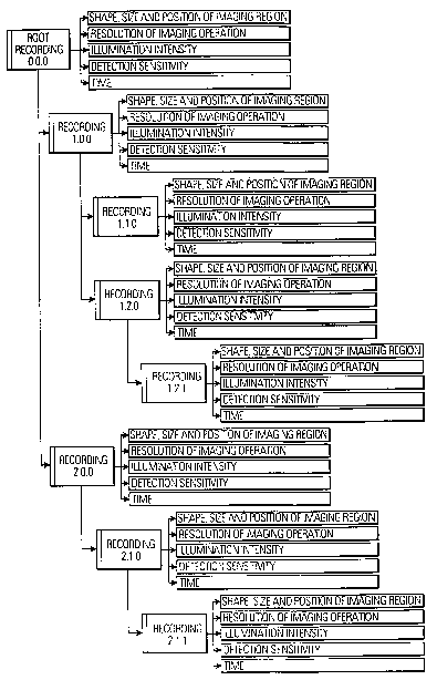
In Fig. 3 an example of a recordings hierarchy is
shown. Each of the recordings in this exemplary
hierarchy comprises a set of parameters as discussed
with respect to Fig. 2. However, it is obvious to the
person skilled in the art that recordings within a
recordings hierarchy according to the invention may
comprise additional parameters or may be limited to
fewer or different parameters.
A root recording 0Ø0 as shown in Fig. 3 is
characterized in that it has no parent recording. The
root recording 0Ø0 comprises control parameter data
such as the shape, the size and the position of an
imaging region, resolution of an imaging operation,
illumination intensity, detection sensitivity and time.
In the example of Fig. 3 this set of control parameter
data is used for each recording shown in the figure.
CA 02405301 2002-10-03
WO 01/78008 PCT/EPO1/04001
9
The root recording 0Ø0 has two child recordings, i.e.
recording 1Ø0 and 2Ø0, both of which comprise the
set of control parameter data as mentioned above.
Recording 1Ø0 is a parent recording to recordings
1.1.0 and 1.2.0, of which recording 1.2.0 is a parent
recording for recording 1.2.1. Similarly, recording
2.1.0 is a child recording of recording 2Ø0 on one
hand and a parent recording for recording 2.1.1 on the
other hand.
It is apparent to a person skilled in the art that this
hierarchy can be extended by adding further recordings
being child recordings to any one of the recordings
already being or becoming a part of the hierarchy shown
in Fig. 3.
According to the invention, the control program of a
computer controlled microscope comprises a function for
creating a recording hierarchy by allowing the creation
of a root recording and adding further recordings as
child recordings of the root recording or of child
recordings created in a previous step. The step of
creating child recordings may also be understood as a
function of adding recordings to the hierarchy. In a
preferred embodiment, the control program of the
computer controlled microscope further comprises the
function of deleting a recording andlor the function of
reordering the recordings in the recording hierarchy.
A benefit of introducing a recording hierarchy into the
control program of a computer controlled microscope is
that it introduces the possibility to inherit control
parameter data from a parent recording to a child
recording or group of child recordings.
CA 02405301 2002-10-03
WO 01/78008 PCT/EPO1/04001
For example, if a user has defined in a first step the
root recording 0Ø0 shown in Fig. 3 control parameter
data like shape, size and position of the imaging area,
resolution of the image acquisition process,
illumination intensity, detection sensitivity and time
have been defined in order to fully describe the status
and/or operation of the computer controlled microscope.
If the user defines in a second step child recording
1Ø0 at least some of the controlled parameter data
previously defined or set for root recording 0Ø0 are
used for the definition of the control parameter data
of child recording data 1Ø0. Similarly, control
parameter data of the root recording 0Ø0 is inherited
during the creation of child recording xØ0.
Of course, the control program of the computer
controlled microscope according to the invention allows
the user to override inherited control parameter data,
for example by redefining the shape, size and/or
position of the imaging region. With or without
limitations based on the control parameter data of the
parent recording, the user may change the control
parameter data of the child recording.
The user may for example reduce the size of the imaging
region of a child recording on one hand and increase
the resolution of the imaging operation on the other
hand.
Preferably, when the user creates a further child
recording, the control parameter data of the parent
recording are initially inherited by the child
recording created.
CA 02405301 2002-10-03
WO 01/78008 PCT/EPO1/04001
11
The user gains access to the improved technical
functions of the computer controlled microscope via a
graphical user interface displayed on an output device.
In the following, an example of such a user interface
will be described. Obviously, the user interface may
have a different look and arrangement of information.
In Fig. 4 an example of a graphical user interface is
shown, which is displayed on the display device (see
Fig. 1) of the computer controlled microscope, and
which not only displays the image 13 of the sample 2
under observation, but also comprises graphical control
elements 14 enabling the user to control the execution
of the control program in the computer of the
microscope.
The example in Fig. 4 shows the overview image 13, as
defined by a root recording according to the invention,
and one smaller imaging region 15 defined by a
respective child recording according to the invention.
The user may define the shape, the size, the
orientation and the position of the smaller imaging
region by means of the pointing device (see Fig. 1),
for example a computer mouse, in that he selects a
draw-tool from the palette 14 and "draws" the imaging
region within the boundaries of the overview image 13,
i.e. the root recording. This technique is basically
known from other computer applications and is adopted
for the invention to allow a facilitated creation of a
child recording.
In Fig. 5 another example of a graphical user interface
is shown, which is displayed on the display device (see
CA 02405301 2002-10-03
WO 01/78008 PCT/EPO1/04001
12
Fig. 1) of the computer controlled microscope, and
which shows the recording hierarchy 16 in a user
friendly format as well as graphical control elements
17 enabling the user to control the execution of the
control program in the computer of the microscope. The
diagram 16 represents the recordings hierarchy. A root
recording 18 and a single child recording 19 are shown
as an example of a recordings hierarchy according to
the invention. Basically the structure of the hierarchy
shown in Fig. 3 can be easily identified in the diagram
of Fig . 5 .
In the following some usage examples of the invention
will be described to give a better understanding of the
scope of the invention and the advantages achieved.
Example 1 .
Observation of a field of cells spread over a wide area
Whilst continuously scanning the imaging area with the
microscope (i.e. continuously executing the "recording
execute" function), the user varies the scan stage
position and recording parameters until a single cell
of interest is found. Once found, other parameters (for
example, illumination intensity and detection
sensitivity) can be adjusted until an optimal image is
obtained. A new imaging window is created, leaving the
image of the cell (and associated parameters) on the
computer screen in the old window. The user repeats the
process of finding cells. When an appropriate number of
cells have been found, the user selects each imaging
window in turn, and presses a button on the window
control bar, which causes the recording contained
CA 02405301 2002-10-03
WO 01/78008 PCT/EPO1/04001
13
within the window to be added to the root recording.
The root recording thus will contain a number of
recordings.
When data acquisition is restarted, each recording in
the root recording will be scanned in turn, reusing the
scan windows that were used to find the cells.
The recording collection can be scanned at timed
intervals, so that many spatially remote cells may be
observed time-multiplexed.
Example 2 .
Observation of a field of cells spread over a small
area
A low magnification scan is made, creating an image in
which a large number of cells can be identified. Using
draw tools selected from a scan window control menu,
cells of interest can be drawn on the display, the
drawn regions automatically defining child recordings,
which are zoomed versions of the main recording, to
which they are added. Initiation of image acquisition
(recording execution) causes the root (overview)
recording, and then all child (cell) recordings to be
scanned in sequence.
Acquisition of any one of the recordings can be
disabled, for example, such that only the cells are
scanned and not the overview recording. The graphical
objects defining the cells in the overview window can
be manipulated during a scan to adjust the size
parameters of the child recordings as they are scanned.
CA 02405301 2002-10-03
WO 01/78008 PCT/EPO1/04001
14
Example 3 .
Combination of wide area and small area observation
A basic recording comprising child recordings can be
added to another recording. Thus a recordings hierarchy
can be built containing clusters of recordings, within
each of which the scan stage doesn't have to be moved
(e. g. since this is slow).
Example 4 .
Optimal observation of different regions within a
single cell
Cells marked with fluorescent probes may have some
structures containing a high density of fluorophore,
and other structures with lower densities. An initial,
non-optimal scan can be made, upon which the areas to
be optimised are drawn, the drawn regions automatically
defining child recordings, which are unzoomed versions
of the root recording. Each child recording, by default
inherits parameters (other than size) from the parent
recording. Thereby, the user can optimise illumination
intensity and detection sensitivity for each region.
Example 5 .
Combination of three-dimensional, two-dimensional and
one-dimensional data acquisition
A confocal fluorescence microscope is able to measure
fluorescence intensity within a three-dimensional
CA 02405301 2002-10-03
WO 01/78008 PCT/EPO1/04001
volume. A single recording might constitute a single
sample or line. A two-dimensional confocal image can
represent a slice through a sample. A stack of slices
thus constitutes a three-dimensional data
representation of the sample. For such a machine, each
recording within the recordings hierarchy is defined in
three dimensions. A recordings hierarchy may therefore
be constructed, which contains recordings of different
dimensionality. Examples might include:
- An overview two-dimensional slice through a field
of cells, containing child recordings, which are three-
dimensional stacks.
- A recording creating a single, overview two-
dimensional image of a single cell (Nomarski contrast),
and a child recording, which defines stack of
fluorescent images.
- A root recording might define a slice in one
direction through a sample, e.g. in a plane orthogonal
to the optical axis. Drawing a line onto the overview
image causes a child. recording to be created. The
extent of such a child recording can be extended so
that it constitutes a plane orthogonal to its parent
Example 6 .
"Illumination-only" processes
There are many instances in which only illumination of
a sample, and not observation is interesting:
a) Photobleaching
CA 02405301 2002-10-03
WO 01/78008 PCT/EPO1/04001
16
A common technique in cell biology for observing cargo
movements with cells is photobleaching. A cell
containing proteins of interest marked with fluorescent
dyes is prepared. An initial image of the cell is
acquired, after which a high power laser scan is used
to bleach areas of fluorophore within the cell. The
microscope's light detectors may be turned off during
the bleach phase, since the image data collection is
not required. After the bleach, a timed sequence of
images is then taken to observe transport of
fluorophore back into the bleached region.
A simple bleaching experiment might include a root
recording, which defines a scan over the whole cell,
and one or more child recordings, which are the bleach
regions. The data created when scanning the bleach
regions is a kind of dummy, since the experimenter is
often only interested in controlling the amount of
energy hitting the sample, and the area over which
illumination occurs. The user simply initiates a timed
sequence of scans, upon which the root recording is
scanned. Whilst the bleach region is being scanned, the
user can toggle the "acquire" option on the bleach
region, so that this region is only scanned once in the
timed sequence.
The bleach region may be defined with exactly the same
parameters as all other recordings, so it is guaranteed
that the user has precise information about the shape
and position of the bleach region, as well as input
energy and the precise time at which the bleach
occurred.
CA 02405301 2002-10-03
WO 01/78008 PCT/EPO1/04001
17
b) Photoactivation (Uncaging)
This is basically the opposite of photobleaching,
whereby light is used to activate a fluorophore. The
parameters describing precisely when, where and how the
compound was activated are neatly contained within a
recording object.
c) Inhibition
This is similar to photobleaching. Compounds can be
caged, or their biological function switched off by
illumination with a characteristic wavelength.
Example 7:
"Observation-only" processes
In some experiments, samples can show Bioluminescence
(Chemiluminescence), i.e. samples emit light without
the need for illumination light.