Note: Descriptions are shown in the official language in which they were submitted.
CA 02405710 2008-08-05
50871-5
METHOD AND APPARATUS FOR CREATING INTRAUTERINE ADHESION$
BACKGROUND OF THE INVENTION
Menstrual bleeding is a part of normal life for women. The onset of
menstruation, termed menarche, usually occurs at the age of 12 or 13. The
length of
a woman's monthly cycle may be irregular during the first one to two years.
Once the
menstrt ral c:ycle stahilizr s H normal nyr;le may range. frorri 20 to 40
days, with 28 days
commonly being an average. Age, weight, athletic activity and alcohol
consuil'iptiart
are several factors that affect menstrual cycles. For example, younger women
(under
the age of 21) and older women (over the age of 49) tend to have longer cycle
times,
generally averaging 31 days and over. Siniilarly, women who are very thin or
athletic
also have longer cycles. In contrast, women who consume alcohol on a regular
basis
tend to have shorter cycle times.
Nearly all women, at some time during their reproductive life, experience some
type of menstrual disorder. These disorders range from mild to severe, often
resulting
in numerous lost work hours and the disruption of personal/family life each
month. In
general, physical symptoms such as bloating, breast tenderness, severe
cramping
(dysmenorrhea) and slight, temporary weight gain frequently occur during most
menstrual cycles. In addition to physical symptoms, emotional hypersensitivity
is also
very common. Women report a wide range of emotional symptoms, including
depression, anxiety, anger, tension and irritability. These symptonis are
worse a
week or so before a woman's menstrual period, generally resolving afterward.
Many women also suffer from a condition called menorrhagia (heavy bleeding).
Menorrhagia is a clinical problem characterized by extremely heavy
flow/bleeding and
major discomfort characterized by blood loss exceeding 80 cc/month. It is
estimated
that 1 in 5 women between the ages of 35 and 50, or approximately 6.4 million
-1-
CA 02405710 2002-10-08
WO 01/80788 PCT/US01/13169
women in the United States alone, are affected by menorrhagia. Fibroids,
hormonal
imbalance and certain drugs, such as anticoagulants and anti-inflammatory
medications, are common causes of heavy bleeding.
Women diagnosed with menorrhagia or dysmenorrhea have limited treatment
options available to them. Currently, other than hormone therapy and a few
experimental pain management techniques, hysterectomy (removal of the uterus)
and
endometrial ablation/resection (destruction of the lining of the uterus) are
the clinically
accepted treatment modalities for menorrhagia. Both of these surgical
procedures
eliminate the possibility of childbearing. Further, hysterectomy requires up
to a six
week recovery time and a lifetime of hormone therapy when the ovaries are
removed.
Endometrial ablation has a low success rate at achieving amenorrhea (cessation
of
menstrual bleeding). As a result, many of the women affected by menorrhagia
are
driven to make lifestyle-altering decisions.
Over 600,000 hysterectomies are performed each year in the United States. It
is estimated that 1 in 3 women in the U.S. have a hysterectomy before the age
of 65.
Menorrhagia is the most common reason why hysterectomies are performed.
Several
studies have estimated that menorrhagia is the cause of 30% (some studies as
high
as 50%) of the 600,000 annual hysterectomies, resulting in a basis of 180,000
to
300,000 procedures annually. Financially, these numbers translate into annual
hospital costs that exceed $5 billion per year.
Based on these statistics, hysterectomy is a very common operation. In
general, there are three types of hysterectomies: partial, total and radical.
As shown
in Figure 1, a partial hysterectomy involves removal of the upper portion 10
of the
uterus 12 (whereby the dotted lines in the figure indicated the area removed),
leaving
the cervix 14 and the base 16 of the uterus 12 intact. Figure 2 illustrates a
total
hysterectomy whereby the entire uterus 12 and cervix 14 are removed. A radical
hysterectomy, shown in Figure 3, entails removal of the uterus 12, both
Fallopian
tubes 18, both ovaries 20, and the upper part of the vagina 22. Each of the
above
three procedures may be performed via an abdominal incision (abdominal
hysterectomy) or through a vaginal incision (vaginal hysterectomy).
After the operation, the hospital stay is generally less than a week,
depending
on the type of hysterectomy and whether there are any complications. Since a
hysterectomy is a major operation, discomfort and pain from the surgical
incision are
-2-
CA 02405710 2002-10-08
WO 01/80788 PCT/US01/13169
most pronounced during the first few days after surgery. Medication is
available to
minimize these symptoms. By the second or third day, most patients are up
walking.
Normal activity can usually be resumed in four to eight weeks and sexual
activity can
usually be resumed in six to eight weeks.
Since the 1800's, attempts using various treatments have been made to control
uterine bleeding by means other than hysterectomy. Alternative methods include
chemicals, steam, ionizing radiation, lasers, electrocautery, cryosurgery and
others.
The long-term risk for such procedures is quite high and may lead to other
more
serious complications such as mixed mesodermal tumors or uterine cancer.
Typical therapy or treatment options include drug therapy followed by dilation
and curettage (D & C) and, as a last resort, hysterectomy. Drug therapy is
generally
the first treatment option employed to treat excessive bleeding. Birth control
pills,
progestin, danazol and gonadotropin-releaseing hormone (GnRH) are a few
examples of drug treatments prescribed to reduce bleeding. In general, birth
control
pills contain synthetic forms of estrogen and progesterone, which prevent
ovulation
and, thereby, reduce endometrial build-up or thickness. As a result, pill
users
normally have lighter or minimal menstrual bleeding. Progestin, another
synthetic
form of progesterone, balances the effects of estrogen normally produced by
the body
and, similar to the pill, reduces endometrial growth. Often, Danazol and other
GnRH
agents are prescribed to suppress estrogen production and ovulation. As a
result,
menstrual bleeding stops or is significantly reduced. However, side-effects of
such
treatments may include bloating, breast tenderness, increased risk of
osteoporosis
and high cholesterol.
D & C, frequently a second treatment option for excessive bleeding, is a very
common, minor surgical procedure that is generally performed on an outpatient
basis
in a hospital. Usually, the patient is given a general anesthetic, although
the
procedure occasionally is performed using only a local anesthetic. The
dilation step
of the procedure involves dilating or stretching the cervix, which is the
lower part of
the uterus. Once the cervix is appropriately dilated, the curettage step can
then be
performed. During curettage, a curette (a spoon-shaped instrument) is inserted
through the vagina, past the cervix and into the uterus. The curette is then
used to
scrape and/or collect tissue from the inside surfaces of the uterus.
-3-
CA 02405710 2002-10-08
WO 01/80788 PCT/US01/13169
Endometrial ablation has become more popular and has been offered as
another alternative treatment to hysterectomy for patients suffering from
menorrhagia.
In 1996, 179,000 ablation procedures were performed, up from 49,000 in 1993.
This
technique is intended to permanently ablate all layers of the endometrium and
allow
the cavity to become lined with fibrous tissue.
In general, endometrial ablation is less costly and requires less recovery
time for
the patient. However, the procedure has received mixed results for controlling
bleeding, depending on the technique used, and has a limited success rate of
no
greater than 20% when defined as complete cessation of bleeding. During one
five-
year study of 525 women with an average age of 42, endometrial ablation
completely
stopped uterine bleeding only 26% to 40% of the time. However, approximately
79%
to 87% of the women were satisfied with the surgery. About 16% of the women
required a repeat ablation to stop bleeding and 9% of the women ultimately
opted for
a hysterectomy. Research has also shown that the effectiveness of endometrial
ablation may decline over years, with menstruation returning in about one-
third of
women.
It should be noted, however, that the goal of endometrial ablation was never
to
create amenorrhea (cessation of menstrual periods). This procedure was
originally
developed as a less invasive alternative to hysterectomy in order to return
women
with menorrhagia to a normal menstrual flow.
In either endometrial abiation or resection, an attempt is made to remove or
destroy the entire lining of the uterus (the endometrium). Endometrial
resection, first
described in 1983 by De Cherney et al., involves the use of a resectoscope-
cutting
loop to perform endometrial ablation to remove the lining of the uterus. In
contrast,
ablation generally uses either vaporization, coagulation or some other thermal
energy
source to destroy the uterine lining.
Although ablation and resection procedures are often discussed as if they are
the same, they differ significantly. For example, some physicians argue that
resection
is more difficult. However, when it is performed skillfully, resection has
much better
results (control of bleeding in up to 88% of patients) than roller ball
ablation (40% to
55%) and newer ablation techniques (3% to 30%).
There are various methods by which an endometrial ablation procedure may be
performed. These methods include roller ball electrocautery, cryo-
cauterization,
-4-
CA 02405710 2002-10-08
WO 01/80788 PCT/US01/13169
microwave, free circulating water, vaporization, balloon ablation and
photodynamic
therapy. In general, these procedures are performed in a hospital or surgery
center,
not in the physician's office, due to the need for anesthesia.
Referring to Figure 4, conventional endometrial ablation, commonly referred to
as "roller ball" ablation, uses a device 24 that looks like a tiny
steamroller. This device
24 applies heat and, thereby, destroys endometrial tissue 26 (whereby the
destroyed
tissue is shown in the figure by a dotted line) as it rolls across the uterine
wall 28.
Endometrial ablation usually takes 15 to 45 minutes and the patient can go
home the
same day, although a general anesthetic is usually required.
Another type of ablation procedure is vaporization. This technique involves
vaporizing uterine tissue using a thin powerful laser beam or high electric
voltage.
Visualization of the uterine cavity is made possible by filling the cavity
with fluid. If
any resection or cauterization is performed, a special substance, such as
glycine,
sorbitol or mannitol, is used so that the fluid does not conduct electricity.
This
prevents accidental burn injuries to the rest of the uterus. Because this
procedure
involves removing or destroying the endometrium using a simple, rapid
technique, it is
often referred to as "global" endometrial ablation.
The NovaSure System is one example of a global endometrial ablation device
used to perform ablation via controlled vaporization of the endometrium. The
patient
is sedated using a local anesthesia with IV sedation and the cervix is
dilated. A gold-
plated mesh triangle is delivered via a slender tube and expanded into the
uterus of
the patient. The shape of the mesh is configured to generally resemble the
profile of
the uterine cavity. Prior to energizing the mesh, suction is applied to bring
the uterine
cavity into close contact with the mesh. After energy has been delivered to
the
endometrial lining via the mesh for one to two minutes, the mesh is retracted
and the
tube removed from the patient's body.
In 1994, Singer et al. reported preliminary experience with an ablation system
incorporating an intrauterine balloon. As shown in Figure 5, balloon ablation
utilizes a
balloon 30 at the tip 32 of a catheter tube 34 that is filled with fluid and
inflated until it
conforms to the walls of the uterus 28. A probe in the balloon (not shown)
heats the
fluid to destroy the endometrial lining. After eight minutes, the fluid is
drained out and
the balloon 30 is removed. Pregnancy is possible if some of the lining is
maintained,
but the risk to mother and child is considerable.
-5-
CA 02405710 2002-10-08
WO 01/80788 PCT/US01/13169
Photodynamic therapy is another type of ablation method. A light-sensitive
agent (photofrin II) that contains a cell-killing substance is given
intravenously and is
absorbed by the endometrium. A light anesthetic is administered and the
physician
then inserts a small probe into the uterine cavity, through which laser light
is
transmitted for a few minutes. The light activates the photofrin II, which
causes
destruction of the endometrium. Early results show reduced bleeding without
significant side effects.
Other ablation methods to treat menorrhagia, such as microwave and freezing
(cryoablation) techniques, are currently being investigated. However, long-
term
studies using these treatments to determine their effectiveness at producing
amenorrhea and any potential side effects are still needed.
Although ablation and resection procedures are less invasive than
hysterectomies, there are various complications that may occur. Examples of
possible complications include perforation of the uterus, injury to the
intestine,
hemorrhage or infection. Another concern associated with ablation treatment
involves
the risk of cancer. Since ablation does not remove the uterus, women still are
at risk
for developing endometrial cancer (although the risk is reduced; however, no
clinical
proof is currently available). Further, because endometrial ablation alters
the wall of
the uterus, early detection of cancerous changes may be difficult to identify.
Other potential side effects of ablation procedures are infections caused by
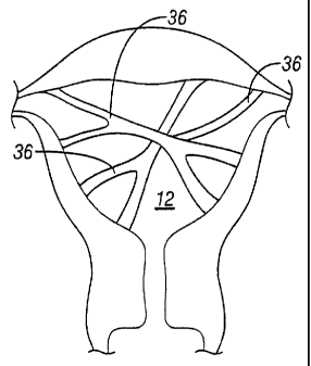
ablation or similar procedures and intrauterine adhesions. Intrauterine
adhesions or
synechiae are described as scar tissue inside the uterine cavity. Termed
Asherman's
Syndrome, intrauterine adhesions 36, as shown in Figure 6, are band-like
formations
that develop as a result of injury or trauma to the uterus 12 (due to, for
example, over-
vigorous curettage to the uterus 12) or can also happen simultaneously.
In 1894, Heinrich Fritsch was the first to describe amenorrhea resulting from
traumatic obliteration of the uterine cavity following puerperal curettage.
However, it
was not until 1948, that knowledge about uterine adhesions was first
disseminated in
medical journals by Joseph G. Asherman, for whom the condition is named. In
1957,
the 17th Congress of the Federation of French Speaking Societies of Gynecology
and
Obstetrics proposed the following classification of uterine synechiae:
Traumatic Synechiae connected with surgical or obstetrical
evacuation of the uterus
-6-
CA 02405710 2002-10-08
WO 01/80788 PCT/US01/13169
Spontaneous synechiae of tuberculosis origin
Synechiae occurring after myomectomy
Synechiae secondary to the attack of chemical or physical
agents and likewise those resulting from atrophic changes
In general, two types of traumatic synechiae are currently recognized. The
first
type is stenosis or obliteration of the cervical canal. The second type of
traumatic
synechiae is partial or complete obliteration of the uterine cavity by
conglutination of
the opposing walls.
Other terms, such as endometrial sclerosis, traumatic uterine atrophy, uterine
artesia, uterine synechiae and adhesive endometriosis, have also been used to
describe the phenomena of Asherman's Syndrome. The severity of adhesion is
generally classified into one of the following three groups or classes: Class
I
represents adhesions occurring in less than one-third of the uterine cavity
with both
ostia (i.e. openings of the Fallopian tubes) visible; Class II represents
adhesions
occurring in one-third to one-half of the uterine cavity with one ostium
visible; and
Class III represents adhesions occurring in greater than one-half of the
uterine cavity
with no ostium visible.
Although Asherman's Syndrome has been studied extensively and numerous
articles and papers have been written on the topic, uncertainty still exists
as to the
predominant causative factor(s) and biological mechanism(s). A general diagram
illustrating the process of adhesion formation after trauma is illustrated in
Figure 7. It
is believed that if the endometrium is severely damaged, it may be replaced by
granulation tissue, When this happens, the opposing uterine walls adhere to
one
another and form scar tissue. In particular, adhesions form and transluminally
bridge
the anterior and posterior surfaces of the uterus. The adhesions or tissue
that is
formed between the walls comprises connective tissue that is, typically,
avascular.
Soon after, the tissue may be infiltrated by myometrial cells and, later,
covered by
endometrium.
Conventionally, intrauterine adhesions have been regarded as undesirable
conditions (for example U.S. Patent No. 6,211,217, issued to Spinale et al,
U.S.
Patent No. 6,136,333, issued to Cohn et al. and U.S. Patent No. 6,090,997,
issued to
Goldbert et al.). Indeed, in several known treatment methods for menorrhagia,
it has
been encouraged to avoid the creation of adhesions. Even in those
circumstances
-7-
CA 02405710 2002-10-08
WO 01/80788 PCT/US01/13169
where clinicians have experimented with adhesion formation, the results have
not
proved promising. For example, in the March 1977 edition of the Israel Journal
of
Medicine, an article by J.G. Schenker, entitled Induction of Intrauterine
Adhesions in
Experimental Animals and Women, described an experiment in which surgical
sponges were implanted into the subcutaneous wall of the patient. The sponges
remained in the subcutaneous wall until fibroblasts, or connective-tissue
cells, were
formed within the sponges. Next, the sponges were then removed and implanted
into
the uterus of the same patient.
Schenker observed that, after a period of time, adhesions were formed in the
areas adjacent to the location of the implanted fibroblast bearing sponge. No
adhesions were observed in areas that did not have contact with the fibroblast
bearing sponge. These experiments were carried out in several animal models
(for
example, rabbit, rat and primates) and humans. Schenker concluded that it was
possible to artificially create adhesions within the uterus, but that such a
procedure
was not practical.
In view of the above, there is a need for a minimally invasive device and
method
to treat abnormal intrauterine bleeding. In particular, it is desirable that
the device
have a high success rate at treating menorrhagia and have minimal to no side-
effects
or related complications. Such a device must also be biocompatible and non-
toxic. In
addition, the related treatment methods should reduce patient recovery times
and
hospital costs. Overall, the method of treatment should also improve the
quality of life
for patients.
BRIEF SUMMARY OF THE INVENTION
In general, the present invention contemplates an implantable device for
treating
excessive bleeding in a body cavity. The device comprises a biocompatible
material
that is deliverable into the body cavity. The biocompatible material contains
an
attribute that promotes tissue growth that results in adhesion formation
within the
body cavity. The attribute of the biocompatible material is defined by at
least one of a
mechanical component of the biocompatible material and a non-cultured biologic
component of the biocompatible material.
The present invention also contemplates a method of creating adhesions in a
body cavity. In general, the method comprises inserting an implantable device
within
-8-
CA 02405710 2002-10-08
WO 01/80788 PCT/US01/13169
the body cavity. The method also includes locating the implantable device at
an
optimal site within the body cavity, wherein the optimal site promotes
effective
adhesion formation to control bleeding.
The present invention further contemplates a pretreatment device for creating
trauma to a tissue within a body cavity. The pretreatment device generally
includes a
stem section and a trauma-inducing section adjacent to the stem section. In
another
embodiment, the pretreatment device comprises a pretreatment fluid and a
flexible
tube housed within a catheter and used to insult the tissue with the
pretreatment fluid.
The present invention also contemplates a method of contraception. In general,
the method comprises inserting an implantable device within a uterus and
locating the
device at an optimal site within the uterus. The optimal site promotes
adhesion
formation and prevents conception.
In addition, the present invention aiso contemplates a tool used to deploy an
implantable device within a uterus. In one embodiment, the tool comprises a
cervical
cap and a guide located on a proximal end of the cervical cap. In an alternate
embodiment, the tool comprises one or more expanding elements attached to the
implantable device and one or more manipulator elements. In another
embodiment,
the tool is used to deploy an implantable device and comprises a guide
directed for
placement of the implantable device within the uterus to create adhesions.
The present invention also contemplates a device for monitoring the tissue of
a
uterus comprising at least one imagable marker. The marker has a size that is
less
than a size of an unexpanded uterus and a surface for adhering the marker to a
uterine wall. In addition, the marker is composed of a biocompatible material
suitable
for permanent implantation is the uterus.
The present invention also contemplates method of monitoring tissue of the
uterus comprising introducing at least one imagable marker into the interior
of the
uterus and allowing the at least one marker to become embedded in tissue
formed on
the interior of the uterus. The method also includes using the at least one
marker as
a reference location to evaluate tissue features on the interior of the
uterus. In
addition, the at least one imagable marker is introduced into the interior
during a
procedure wherein the uterus is being treated for a condition of menorrhagia.
Alternatively, the method may also include at least two imagable markers that
are
-9-
CA 02405710 2006-04-21
50871-5
introduced into the uterus and wherein the at least two
imageable markers provide a two dimensional frame of
reference.
The present invention also contemplates an implant
for treating a uterus of a female patient comprising: a
substance configured for causing a tissue response in
uterine tissue; said substance being sized and shaped for
placement in a lower Y of a uterine cavity of said uterus;
and, wherein said substance is configured for causing a
tissue response that eliminates menorrhagia.
The present invention also contemplates an implant
for changing the gynecological state of a female comprising:
a self-contained presterilized substance disconnectable from
a delivery tool; said self-contained substance configured
for causing a tissue response in uterine tissue; and, said
self-contained substance sized and shaped for sufficiently
contacting uterine tissue such that uterine walls of said
uterine tissue adhere together and thereby cause a
gynecological change in said female.
The present invention also contemplates an implant
for changing the gynecological state of a female comprising:
a presterilized substance in the form of a mesh material;
said substance configured for causing a tissue response in
uterine tissue; and, said substance sized and shaped for
sufficiently contacting uterine tissue such that walls of
said uterine tissue adhere together and thereby cause a
gynecological change in said female.
The present invention also contemplates an implant
for changing the gynecological state of a female comprising:
a presterilized substance having a frame, at least a portion
-10-
CA 02405710 2008-08-05
50871-5
of which is covered by a mesh material; said substance
configured for causing a tissue response in uterine tissue;
and, said substance sized and shaped for sufficiently
contacting uterine tissue such that menorrhagia is
eliminated in said female.
The present invention also contemplates a device
for occluding fluid flow from a uterus through a cervix
comprising: a first end having a first diameter; a second
end having a second diameter smaller than the first
diameter; and a narrow portion interposed between the first
end and the second end having a third diameter smaller than
the second diameter.
The present invention also contemplates a device
for occluding fluid flow from a uterus through a cervix
comprising: a rim; an end portion having a first diameter;
and, a narrow portion associating the rim to the end portion
and having a second diameter smaller than the first
diameter.
The present invention also contemplates a device
for occluding a uterine cavity comprising: a body having a
collapsed state and an expanded state; wherein said body is
passable through a cervix into a uterus via a catheter in
said collapsed state; wherein when said body is in said
expanded state, said body has a first end and a second end;
said first end having a dimension greater than a dimension
of the second end; and said second end contacting a cervix
without extending through the cervix to a vagina when in the
expanded state in a uterus.
-l0a-
CA 02405710 2008-08-05
50871-5
BRIEF DESCRIPTION OF THE DRAWINGS
Other features and advantages of the present
invention will be seen as the following description of
particular embodiments progresses in conjunction with the
drawings, in which:
Figure 1 is a sectional view of an embodiment of a
hysterectomy;
Figure 2 is a sectional view of another embodiment
of a hysterectomy;
Figure 3 is a sectional view of yet another
embodiment of a hysterectomy;
Figure 4 is a perspective view of one embodiment
of an ablation procedure;
Figure 5 is a perspective view of another
embodiment of an ablation procedure;
Figure 6 is a perspective view illustrating
intrauterine adhesions associated with Asherman's Syndrome;
Figure 7 is a general diagram illustrating the
process of adhesion formation;
Figure 8 is a perspective view of an embodiment of
the intrauterine implant device in accordance with the
present invention;
Figure 9A is a front perspective view of a uterine
cavity in a non-distended state;
Figure 9B is a side perspective view of a uterine
cavity in a non-distended state;
-10b-
CA 02405710 2008-08-05
50871-5
Figure 10 is a perspective view of an embodiment
of the intrauterine implant device in accordance with the
present invention;
Figure 11 is a perspective view illustrating an
embodiment of a random fiber bundle in accordance with the
present invention;
Figures 12A-12F illustrate various embodiments of
a pretreatment device in accordance with the present
invention;
Figures 13A-13C illustrate alternate embodiments
of a pretreatment device in accordance with the present
invention;
Figures 14A-14C illustrate various views of
another embodirnent of a pretreatment device in accordance
with the present invention;
Figure 15 illustrates a sectional view of another
embodiment of a pretreatment device in accordance with the
present invention;
Figures 16A-16D illustrate various views of a
cervical cap used with a delivery tool in accordance with an
embodiment of the present invention;
-10c-
CA 02405710 2002-10-08
WO 01/80788 PCT/US01/13169
Figure 17 illustrates a perspective view of the distal end of a catheter in
accordance with an embodiment of the present invention;
Figures 18A-18B illustrate an embodiment of the deployment tool in accordance
with the present invention;
Figures 19A-19B illustrate another embodiment of the deployment tool in
accordance with the present invention;
Figures 20A-20B illustrate yet another embodiment of the deployment tool in
accordance with the present invention;
Figures 21 A and 21 B illustrate another embodiment of the deployment tool in
accordance with the present invention;
Figures 22A-22B illustrate perspective views of embodiments of a self-
deploying
implant structure in accordance with the present invention;
Figure 23 illustrates an alternate embodiment of the implant in accordance
with
the present invention;
Figure 24 illustrates another embodiment of the implant in accordance with the
present invention;
Figure 25 illustrates an embodiment of a deployment device and implant in
accordance with the present invention;
Figure 26 shows an alternate embodiment of a deployment device and implant
in accordance with the present invention;
Figure 27 illustrates an alternate embodiment of the implant in accordance
with
the present invention;
Figure 28A-28C illustrate an embodiment of a catheter used in accordance with
the present invention;
Figure 29 shows another embodiment of the implant in accordance with the
present invention;
Figures 30A-30C illustrate an alternate embodiment of a deployment tool in
accordance with the present invention;
Figures 31 A-31 B illustrate an embodiment of a tool used in accordance with
the
present invention;
Figure 32 shows an embodiment of a marker in accordance with the present
invention;
-11-
CA 02405710 2002-10-08
WO 01/80788 PCT/US01/13169
Figure 33 shown an alternate embodiment of a marker in accordance with the
present invention; and
Figure 34 illustrates a perspective view of an implanted marker in accordance
with an embodiment of the present invention.
DETAILED DESCRIPTION OF THE INVENTION
Referring to Figure 8, an embodiment of the intrauterine implant device 40 in
accordance with the present invention is shown deployed within a uterus 42.
The
uterus 42, or womb, is part of the female internal genitals. The uterus 42. is
a hollow,
muscular organ approximately four inches long and three inches wide and is
generally
shaped like an upside-down pear. It should be noted that the uterus 42
depicted in
Figure 8 is in a distended state to clearly show the uterine cavity 44.
However, it is
understood that the uterine cavity is normally in a collapsed state, as shown
in
Figures 9A and 9B.
Two openings 46 located at the upper end of the uterus 42 lead to the
Fallopian
tubes that are connected to the ovaries (not shown). Opposite to the upper end
openings 42 is a lower, narrow open end 48 that forms the cervix 50 of the
uterus 42
and extends to the vagina 52. The thick walls of the uterus 42 are comprised
of three
layers of tissue and muscle: the inner endometrial layer, the middle
myometrial layer
and the outer perimetrial layer. It is the inner endometrial layer or lining
that
separates from the uterus 42 and leaves the body as the menstrual flow during
a
woman's menstrual period.
Excessive menstrual flow or bleeding, termed menorrhagia, is indicative of
abnormal sloughing of the endometrial tissue layer. Unlike conventional
therapies
such as hysterectomy or ablation/resection procedures, as described above, the
device 40 of the present invention achieves amenorrhea (i.e. cessation of
bleeding)
by way of an implant or substance that promotes formation of intrauterine
adhesions.
The intrauterine adhesions cause cessation of bleeding by a deactivation of
the
endometrial tissue, due to possibly a pressure gradient or neuro-modulating
effect.
Occlusion or obliteration of the uterine cavity may result. It is important to
note that
the endometrial tissue is deactivated through means other than the direct
destruction
of the lining, and that endometrial deactivation has been seen even in the
presence of
a small number of adhesions.
-12-
CA 02405710 2002-10-08
WO 01/80788 PCT/US01/13169
In general, the device of the present invention comprises a biocompatible
material that is deliverable within a body cavity, such as the uterus. The
material
contains an attribute promoting tissue growth that results in adhesion
formation within
the body cavity. The attribute may be defined by a mechanical component and/or
a
non-cultured biologic component, further described below. Although the
invention as
disclosed herein generally refers to a uterus, other body cavities, such as
cavities
within a heart, abdomen or other similar cavities, are also included within
the scope of
the present invention.
As shown in Figures 8 and 10, one embodiment of the device 40 of the present
invention comprises a sterile material generally shaped or having physical
properties
to conform to the internal structure of the uterine cavity 44. In general, the
device
material may be flexible, rigid or semi-rigid and sized to fit within the
uterus 42 of a
patient. As such, the device 40 should be more or less triangularly shaped,
having a
height, X, of approximately 7 inches (17.78 cm) and a base, Y, of
approximately 4
inches (10.16 cm). In an alternate embodiment (not shown), the device 40
comprises
a flowable liquid or material that conforms to the uterine anatomy following
device
delivery.
Device Materials
The device 40 of the present invention can be made from a wide variety of
materials including, but not limited to, mesh, suture, gel, porous, allograft,
protein,
hydrogel, liquid sealant, glue, cellulose, alginate, tissue, kitosan,
particulate, foam and
any combination of materials. The properties or characteristics of these
materials
may be non-absorbable, temporary/absorbable, whereby the material is broken
down
by the body through any means including enzymatic, hydrolytic, mechanical,
etc. and
excreted, or permanent/resorbable, whereby the material is remodeled through
some
process to form host or other similar tissue. In addition, the device material
should be
biocompatible, non-toxic and, preferably, one that is approved/cleared by the
Food
and Drug Administration (FDA) and has been used for a long period of time in
humans with the purpose of creating adhesions. Further, for embodiments of the
device 40 having a mechanical configuration, it is desirable that the material
be
capable of conforming to irregular volumes and/or shapes. In general, the
device 40
should be designed such that it can be placed in, stored in and deployed from
a
catheter or similar device delivery tool.
-13-
CA 02405710 2002-10-08
WO 01/80788 PCT/US01/13169
In one embodiment, the material is a woven, surgical mesh. Alternatively, the
mesh can be braided, spun, knitted, non-woven and any structural combination
thereof. Examples of representative surgical meshes include GORE-TEX
(manufactured by W.L. Gore & Associates, Arizona), Marlex (manufactured by C.
R.
Bard, New Jersey), Mersilene (manufactured by Johnson & Johnson, New Jersey),
Prolene (manufactured by Johnson & Johnson, New Jersey), Surgipro
(manufactured by US Surgical, Connecticut), Surgisis (manufactured by SIS
Technology Cook Group, Indiana), Vicryl (manufactured by Johnson & Johnson,
New Jersey) and Atrium Surgical Mesh (manufactured by Atrium, New Hampshire).
Specific references for these materials may be found in the manufactures'
product
catalogues. Additional surgical mesh materials such as polyester, felt,
polyethylene
fiber, non-absorbable mesh, PTFE (Polytetrafluoroethylene), absorbable mesh
and
other mesh materials not specifically disclosed herein may also be used to
create or
enhance the development of intrauterine adhesions 36.
In general, these materials are typically used for creation of adhesions or
tissue
repair within other regions of the body. One example of such use is hernia
repair,
whereby a specialized mesh or screen is used to hold the hernia in place. For
this
application, the material acts like a plug and soon becomes incorporated by
the
surrounding tissue to strengthen the weakened area.
Although select literature references describe some of the materials as being
adhesion barriers, these materials are in fact very good at creating adhesions
under
specific circumstances. One such example is Surgicel oxidized regenerated
cellulose (manufactured by Johnson & Johnson, New Jersey), which is considered
an
adhesion barrier material and, in certain circumstances, an adhesion
creator/promoter. Therefore, both adhesion barrier and adhesion promoter
materials
may be used for the device 40 of the present invention.
In another embodiment of the invention, the implant 40 is made of a woven
material, such as a fabric with a specific weave, that is also biocompatible.
In this
configuration, the material of the device creates a lattice-like structure
(having
openings or pores) that promotes infiltration of fibrous tissue, resulting in
adhesions
36. The material may be metallic, polymeric or a bio-material (including
combinations
of materials) and can be absorbable or non-absorbable, depending on the
physical
and procedural requirements. Additional material specifications or variables
may
-14-
CA 02405710 2002-10-08
WO 01/80788 PCT/US01/13169
include type of weave (such as plain, open, closed, twill, dutch, reverse
dutch, twill
dutch, or taffeta, including combinations of weaves), mesh count, fiber
diameter,
filament type (such as monofilament fiber or multi-filament fiber) or whether
there are
interconnection of weave points. A reference containing additional
specifications,
variables and general information on woven materials is Sefar America, Inc.,
Depew,
New York (sales literature booklet, dated 1998), which is incorporated herein
by
reference.
Alternatively, the device 40 of the present invention can also be made of non-
woven materials. One type of non-woven material is a random fiber bundle 54.
The
fiber bundle 54 may be a thin mat, similar to a woven mesh, with an irregular
fiber
pattern. Examples of materials having an irregular fiber pattern include
Scotchbrite
or Brillo pad materials. In addition, the material may be fabricated from any
monofilament or multi-filament material. An example of a monofilament material
that
can be used for the implant is suture material, such as Prolene or Vicryl
(manufactured by Johnson & Johnson, New Jersey). Although the fibers of the
non-
woven material are arranged in a random orientation, the configuration of the
fibers
produces an associated effective pore size 56, shown in Figure 11. Additional
examples of non-woven materials include all the materials listed above, since
materials fabricated into a woven product can also be manufactured into a
random
fiber bundle 54.
Numerous manufacturing methods and associated techniques may be used to
fabricate the woven and non-woven materials used in the device 40 of the
present
invention. For example, in one embodiment, a monofilament having a thickness
within the range of 0.003 to 0.007 inch (0.00762 cm to 0.01778 cm) is cut into
0.118
inch to 0.197 inch (0.3 cm to 0.5 cm) segments. The segments are then shaped
into
a predetermined configuration, such as a sphere or a cube. The porous
individual
shapes are then arranged into the final material design. The porosity of the
resultant
material is dependent on the size and shape of the fibers and the amount of
compression (density) of the fibers. Examples of other manufacturing
techniques
within the scope of the present disclosure include heating, ultrasonic
cutting, cold
cutting, ultrasonic welding, injection molding, compression molding, stamping,
drawing, forming and other techniques not specifically disclosed, but well
known in
the art.
-15-
CA 02405710 2002-10-08
WO 01/80788 PCT/US01/13169
In another embodiment, the device 40 of the present invention is made of
porous materials. Examples of such porous materials include, but are not
limited to,
ceramics, alumina, silicon, powdered metals, Nitinol , stainless steel,
titanium, porous
polymers, such as polypropylene, polyethylene, acetal, nylon, polyester, and
any
combination of such materials. Although these materials (and others not
specifically
described, but included in the scope of the claimed invention) may not be
inherently
porous, various manufacturing and processing techniques may be used to give
the
materials selective porosity characteristics.
In addition, one or more of these materials may be further incorporated into a
mesh matrix (i.e. porous fibers woven into a mesh or configured in a random
orientation). Alternatively, the materials can be configured as many.
particles of equal
or different size or shape that are constructed into a matrix. In another
embodiment,
the polymeric materials may be manufactured to form a sponge-like material
with
open pore cells. This sponge-like configuration not only promotes adhesions
36, but
also allows the implant 40 to better conform to the internal area of the
uterine cavity
44. Specific examples of such materials include Ivalon, a polyvinyl sponge
(manufactured by C. R. Bard, New Jersey) and Surgifoam (manufactured by
Johnson & Johnson, New Jersey). However, it should be noted that other
materials
not specifically listed herein may also be used.
Both the size of the pores of the material as well as the material's physical
characteristics have an impact on the effectiveness of the implant 40. These
material
attributes determine the type of tissue that will develop or grow into the
mesh and,
ultimately, the type of adhesion 36 that will form within the uterus 42. The
direct
correlation between these parameters for treatment of menorrhagia can be
determined from existing material classifications or types based on tissue in-
growth,
such as those used for hernia repair. For example, Type I includes materials
with
pore sizes greater than 75 microns which allows for growth of macrophages,
fibroblasts (fibroplasias), blood vessels (angiogenisis) and collagen fibers
into the
pores. This pore size is similar to the pore sizes found in Prolene
(manufactured by
Johnson & Johnson, New Jersey), Marlex (manufactured by C. R. Bard, New
Jersey) and other meshes described above. As such, Type I materials are
suitable
device materials.
-16-
CA 02405710 2002-10-08
WO 01/80788 PCT/US01/13169
Type II materials are micro-porous meshes with pore sizes less than 10 microns
in at least one of three dimensions. Materials such as GORE-TEX (manufactured
by
W.L. Gore & Associates, Arizona), PTFE and other surgical membranes are
typical
examples of these meshes. Thus, Type II materials are also appropriate device
materials.
Type III materials contain multi-filaments and include macro-porous and/or
micro-porous components. In general, Type III materials have varying pore
sizes and
are a combination of Type I and Type II materials. Several examples of Type
III
materials include Dacron mesh (such as Mersilene , manufactured by Johnson &
Johnson, New Jersey), braided polypropylene mesh (such as Surgipro ,
manufactured by US Surgical, Connecticut), and perforated PTFE (such as GORE-
TEX MYCROMESH , manufactured by W.L. Gore & Associates, Arizona). These
and other Type III materials not specifically listed herein may also be used
for the
device 40 of the present invention.
In another embodiment of the invention, the device or implant 40 is fabricated
from a liquid sealant or glue, such as collagen, tissue/collagen, thrombin,
polymer,
fibrin-based sealants and any combination thereof. In general, these materials
are
typically configured in a liquid format. However, collagen is a very common
substance and may be found in numerous configurations, including flour,
compressed
mat pad, non-woven fiber or other molded, extruded or compressed shapes with
varying density and/or porosity. Examples of collagen and tissue/collagen
materials
contemplated herein include Avitene (manufactured by C.R. Bard, New Jersey),
Helitene (manufactured by Integra LifeSciences Corporation, New Jersey),
Dermalogen , DermaplantTM (manufactured by Collagenesis, Inc, Massachusetts),
Apligraf , Engineered Collagen MatrixT"" and VitrixTM (manufactured by
Organogenesis Inc., Massachusetts). The collagen may be synthesized or derived
from bovine, porcine or human sources.
An example of a collagen-thrombin sealant is Costasis . Costasis ,
manufactured by Cohesion Technologies, California, is a collagen-thrombin
composite for use as a hemostatic agent to arrest or control bleeding at
various sites
within the patient's body. This material is comprised of bovine fibrillar
collagen and
bovine thrombin suspended in calcium chloride. At the time of application,
fibrinogen
(taken, for example, from the patient's plasma) is mixed with the Costasis ,
thereby
-17-
CA 02405710 2002-10-08
WO 01/80788 PCT/US01/13169
providing fibrinogen that is cleaved by the thrombin to form a collagen-
reinforced
liquid hemostat. The resultant liquid material may then be applied to the
target site to
control bleeding.
Alternatively, the physical properties of the liquid sealants may be altered
to
create hemostatic solids of specific shapes or pliable geometries. In one
embodiment, the sealant material may be placed in a carrier matrix that has
specific
flow requirements and may be activated by heat or moisture to change the
sealant's
physical characteristics. An example of an appropriate carrier matrix is
thrombin-
based CoStop , also manufactured by Cohesion Technologies, California.
However,
unlike Costasis , CoStop does not require plasma from the patient. Simply
combining the patient's blood with the thrombin-based CoStop is sufficient to
cause
platelet activation. As soon as the combination of blood and thrombin causes
platelet
activation, the thrombin further catalyzes the mixture to form a fibrin clot.
As such,
platelet activation initiates clot formation. A collagen-fibrin matrix
develops, forming
the basis or support-structure for the tissue that will be created at the
target site.
Thus, when used to treat menorrhagia, CoStop is placed within the uterus 42
of the
patient and forms the collagen-fibrin matrix. The newly formed tissue bridges
together the posterior and anterior walls of the uterus 42, thereby creating
an
adhesion 36 and promoting amenorrhea.
Alternative methods and components may also be used to modify the physical
properties of the liquid sealants. Although not specifically mentioned herein,
these
methods and components are well known in the applicable art and, therefore,
are
within the scope of the present disclosure and claimed invention.
In another embodiment, the device 40 of the present invention is made of
allograft materials (i.e. a graft of tissue taken from a donor of the same
species as the
recipient). These materials use the structure and properties of the allograft
tissue as
a matrix for new tissue formation. OsteofilTM (manufactured by Regeneration
Technologies Inc., Florida) is an example of one such material. The Osteofil
is
placed within the uterus 42 of the patient and a fibrous tissue is formed
within the
matrix. This new tissue forms the basis for the adhesion 36. The allograft
tissue from
Regeneration Technologies Inc. is initially contemplated as de-mineralized
bone;
however, other tissues derived from animals or humans may also be used. In
addition to Osteofil , other similar materials including, but not limited to,
Natural
-18-
CA 02405710 2002-10-08
WO 01/80788 PCT/US01/13169
Matrix (Xenograft), such as OsteoGraf N-Block (manufactured by Cera Med
Dental,
LLC, Colorado) and other tissues available from various accredited tissue
banks are
also within the scope of the claimed invention.
In yet another embodiment, protein materials are used to fabricate the device
40
of the present invention. Various companies and organizations have studied the
use
of proteins for creating both non-stick and attachable surfaces. One such
company is
Protein Polymer Technology located in San Diego, California. Protein Polymer
Technology creates synthetic genes using recombinant DNA technology. In
particular, Protein Polymer Technology is able to configure small protein
building
blocks into high molecular weight polymers.
Another company that uses proprietary technology to create application
specific
proteins is Gel-Del Technologies (St. Paul, Minnesota). Gel-Del Technologies,
like
Protein Polymer Technology, and other similar companies process proteins using
various methods. The physical structure and composition of the protein are
modified
to create a wide variety of properties for the protein. For example, proteins
have been
created that have cellular receptors, which promote active association or
adhesion 36.
The physical characteristics (for example, shape) of the protein and its side
chain
elements influence the development of a fibrous response and the formation of
the
desired adhesions 36. In particular, the available side chain elements
regulate
selective infiltration of tissue into the protein structure, thereby producing
adhesions
36 at the tissue target site.
In general, proteins may be developed into a wide variety of formats. Examples
of various protein formats include small beads, sheets, strips or other
regular or
irregular shaped configurations. The protein format allows the protein to be
implanted
in, for example, the uterus 42 to create the response necessary for adhesion
formation.
In another embodiment of the invention, the device or implant 40 is fabricated
from hydrogel materials. Hydrogels are coherent three-dimensional polymeric
networks that can absorb large quantities of water without dissolution of the
polymer
network. Classes of hydrogels, based on their method of preparation, include
homopolymer hydrogels, copolymer hydrogels, multipolymer hydrogels and
interpenetrating hydrogels. In general, hydrogels are hydrophilic polymers
incorporating Chitson derivatives or polyethylenimine together with
-19-
CA 02405710 2002-10-08
WO 01/80788 PCT/US01/13169
polyvinylpyrrolidone (PVP). Hydrogels may also include cellulose derivatives,
polyvinyl alcohol (PVA) or polyethylene glycol (PEG). An example of one common
hydrogel is polyHEMA (poly(2-hydroxyethyl) methacrylate) These highly
compatible
water-soluble polymer systems naturally combine with each other to form gels
possessing excellent physical properties. These properties may be varied by
the
chemistries of the gel (i.e. compounding), active ingredients and
biomolecules, which
can be readily incorporated without impairing biological activity. Virtually
any material
that can be dissolved, emulsified, or suspended can be added prior to gel-
formation
and evenly distributed in the finished gel.
The hydrogel AquatrixTM II (manufactured by Hydromer, New Jersey) is an
example of one such hydrogel product. The gel may be loaded with any of the
above-
mentioned materials, such as Marlex (manufactured by C. R. Bard, New Jersey),
Mersilene (manufactured by Johnson & Johnson, New Jersey), Surgipro
(manufactured by US Surgical, Connecticut), Surgisis (manufactured by SIS
Technology Cook Group, or any other material that is pulverized, ground, etc.
and
combined with the hydrogel material. In this configuration, the hydrogel is
acting as a
carrier material to allow for dispensing of the scaffold or lattice material.
The material
can then be delivered as a flowable liquid with a suspension of particles.
Further, the
gel may be formulated to be absorbed or resorbed by the body within 30 to 60
days.
However, the particle/mesh would remain, forming the desired adhesion 36 at
the
target site. In an alternate embodiment, the gel may be formulated to be non-
absorbable. In the case of a non-absorbable gel, the gel may be placed at the
target
site and then blown with a gas to form small pores. The pores function in a
manner
similar to the mesh openings or pores, allowing in-growth of tissue and,
ultimately,
forming adhesions 36.
In general, the materials used with the device 40 of the present invention may
be comprised of a combination of absorbable and/or non-absorbable materials or
components. In one embodiment, the absorbable material may be comprised of a
radio-opaque marker, or any other type of imagable marker, that allows the
target site
to be imaged. In another embodiment, the absorbable material may be used to
fixate
the non-absorbable material at the target site in the patient. For example,
the
absorbable material may be configured as a cervical cap. The cervical cap is
inserted
at the time of implantation of the device 40 and holds the device 40 in-place
within the
-20-
CA 02405710 2002-10-08
WO 01/80788 PCT/US01/13169
uterus 42. Within approximately 8 weeks, adhesions 36 form and the body of the
patient absorbs the absorbable material of the device 40.
Method of Use
Many methods for creating intrauterine adhesions 36 are contemplated herein.
Each methodology has a slightly different mechanism for creating adhesions 36.
As
explained above, only a limited understanding of the actual mechanism for
adhesion
creation is currently known. However, it seems that intrauterine adhesions 36
perform the same function of producing amenorrhea.
In one embodiment, the size and/or configuration of the device 40 is optimized
to promote effective adhesion development within the uterus 42. In another
embodiment, the device 40 is configured to contact substantially the entire
area of the
endometrium to maximize the yield (i.e. up to 100% coverage) of adhesions
within the
uterus 42. Alternatively, there may be optimal locations within the uterus 42
for site-
specific deployment and/or placement of the device 40. As such, the implant 40
need
only contact specific or discrete areas of the endometrium for effective
adhesion
formation (i.e. adhesions in less than 100% of the endometrium). For example,
the
device 30 may be located in the cervical canal, and not the uterus 42, to
produce
sufficient adhesions 36 to control bleeding. As another example, the device 40
may
be positioned at a specific site only within the uterus 42. Alternatively, a
combination
of uteral and cervical locations may be used for beneficial adhesion
formation.
In another embodiment of the invention, the implant or device 40 may include a
means for assessing and determining specific areas in the uterus 42 that are
areas of
excessive bleeding. These discrete areas may then be treated to specifically
form
adhesions 36 at these target sites. This approach allows the endometrium to
remain
viable and, simultaneously, reduces and/or controls bleeding. Since the
endometrium
is not completely obliterated, this method may allow for reversal of the
procedure (i.e.
removal of adhesions). Research has shown that adhesions 36 may be removed
and, thus, uterine viability restored.
In general, the method of use or treatment system of the present invention is
contraceptive in nature. The device or implant 40 creates intrauterine
adhesions 36
which deactivate the endometrial tissue. In addition, the adhesions 36 may
also
obstruct the Fallopian tubes and/or the entrance to the Fallopian tubes. This,
in turn,
eliminates the possibility of pregnancy (i.e. prevents conception) and
childbearing.
-21-
CA 02405710 2002-10-08
WO 01/80788 PCT/US01/13169
However, if only a limited area within the uterus 42 requires formation of
adhesions
36, the procedure and its associated effects may be reversed. Therefore, if
the
adhesions 36 are not extensive, it is possible to restore menstrual function,
and the
potential of pregnancy, to the uterus.
Adhesion formation or coverage is important not only in placement of the
coverage (which is related to device placement) but also percentage of
coverage.
Although the device 40 and methods referenced herein are directed at creating
100%
coverage of adhesions 36 over the entire area of the endometrium, it should be
understood that alternative device configurations and methods of use relating
to less
than 100% adhesion and/or endometrial area coverage are also contemplated
herein.
For example, in general, the percent of coverage must be greater than 75%
and/or
the placement of coverage should be within the lower two-thirds of the uterus
and/or
the entire cervical canal. Other coverage options, though not specifically
described
herein, are also included within the scope of the claimed invention.
The general adhesion formation is a Vocaiized response to an inflammatory
condition and foreign body (i.e. intrauterine device 40). The curettage or
other
pretreatment (further discussed below) causes trauma to the endometrium and
initiates an inflammatory response. The body then begins to create fibroblasts
(connective tissue) at the injury sight as means to heal the trauma). These
fibroblasts
continue to respond to the localized inflammation creating more and more
fibrous
tissue. As the fibrous tissues are created, the posterior and anterior walls
of the
uterus 42 are joined more closely by the scar tissue. The myometrial cells
eventually
infiltrate the scar tissue and the surrounding tissue reabsorbs the
endometrium.
Long-term deactivation of the endometrial tissue is believed to be due in part
to the
increased intrauterine pressure created by the adhesions 36. There are also
other
modulating factors, not specifically described herein, that may contribute to
the
deactivation of the endometrium.
In addition to creating permanent intrauterine adhesions 36 and treating
menorrhagia, the intrauterine device 40 of the present invention can also be
used on
a temporary basis or as a lifestyle choice. The disclosed implant
device/system 40
could also offer women the option of whether or not to have periods. Thus, the
device
may be used as a convenience to women to end their menstrual periods without
having to undergo major surgery. Further, the device 40 may be used to
eliminate
-22-
CA 02405710 2002-10-08
WO 01/80788 PCT/US01/13169
painful menstrual cycles or premenstrual symptoms. As yet another alternative,
the
device 40 may be used as a temporary means of contraception. When a woman is
ready to have children, the procedure could be reversed, whereby the adhesions
36
are removed and menstruation returns.
Pretreatment
In one embodiment of the invention, the methodology used to create adhesions
36 involves pretreatment of the endometrium prior to placement of the device
40 in
the uterus 42. The pretreatment method may be either direct or indirect.
Generally,
indirect pretreatment occurs during the time period prior to the procedure. In
contrast,
direct pretreatment occurs during the actual procedure.
Direct pretreatment (via mechanical means, chemical means or a combination
thereof) is performed in order to invoke a healing response from the uterus
42. One
type of direct pretreatment involves creating trauma to the endometrium prior
to
deployment of the device 40. Methods to achieve this trauma and, in some
instances,
necrosis of tissue may include curettage or a form of endometrial ablation.
These
methods may be performed with a sharp or blunt curette (vacuum curettage),
roller
ball electrocautery device, thermal energy device (such as re-circulating hot
water),
hot water filled balloon, radio-frequency (RF) energy director, microwave,
cryogenic
device (to freeze the tissue), cytotoxic agent, intense LASER light and other
devices
capable of imparting trauma to the endometrial lining, including combinations
of such
devices.
One embodiment of a pretreatment device 60 in accordance with the present
invention is shown in Figures 12A and 12B. The pretreatment device 60 is
configured
similar to that of a bottle-brush and can also function as the implant. In
this
configuration, the pretreatment device 60 includes a stem section 62 and an
adjacent
trauma-inducing section 64 consisting of bristles or spike-like projections.
The stem
62 of the device may be made of rigid polymers, ABS, nylon, PVC, metallics,
such as
stainless steel or aluminum, and any combination of such materials. The
bristles 64
can be fabricated from semi-rigid polymers, nylon or polyethylene.
Alternatively, the
trauma-inducing section 64 can be configured as a Brillo pad-like structure
66
capable of scouring tissue, as shown in Figure 12C. The Brillo portion 66 may
be
comprised of collagen coated with a sclerosing agent and can self-expand when
deployed from the catheter 68.
-23-
CA 02405710 2002-10-08
WO 01/80788 PCT/US01/13169
Prior to delivery, the brush or bristle portion 64 of the device 60 is
contained in
the distal section 70 of a catheter or other similar type of delivery tool 68.
Preferably,
the outer surface of the delivery tool 68 is smooth and/or lubricious to allow
for easy
insertion into the patient. During the delivery and treatment procedure, the
delivery
tool 68 is inserted transcervically into the patient and the distal section 70
is
positioned within the uterus 42. The pretreatment device 60 is maneuvered so
that
the bristle portion 64 of the device 60 is deployed at the target site. The
device 60 is
further manipulated, for example, rolled, twisted, pushed and/or pulled, so
that the
brush portion 64 inflames the endometrium tissue. The brush portion 64 is then
disconnected from the pretreatment device 60 and left in the uterus 42 of the
patient.
The bristles 64 of the device 60 allow in-growth of fibrous tissue and promote
adhesion formation. After the implant 40 is deployed, the catheter 68 is
removed from
the patient.
Figure 12D illustrates an alternate embodiment of the bottle-brush
pretreatment
device 60. The distal section 70 of the device 60 comprises a multipie-pronged
brush
72. This configuration reduces the amount of manipulation required to produce
sufficient trauma to the tissue and, also, allows for greater coverage of the
endometrium. '
In yet another embodiment of the bottle-brush pretreatment device 60, the
distal
section 70 of the device 60 includes a wire with sharp protrusions 74, similar
to a
barbed-wire, as shown in Figure 12E. As with the bottle-brush configuration,
the
barbed-wire end 74 of the pretreatment device 60 is rolled, twisted, pushed
and/or
pulled across the surface of the tissue to cause sufficient insult to the
endometrium.
Alternatively, an electric current can be applied to the wire 74 as another
means of
further abrading the endometrial tissue.
Referring to Figure 12F, the bottle-brush portion of the pretreatment device
60
can also be designed in a cap or collagen plug 76 configuration. The coliagen
plug
76 has a naturally abrasive surface and is attached to a stylet 78 for
manipulation and
deployment. The stylet 78 is rotated, pushed or pulled to create sufficient
trauma to
the endometrium. Alternatively, a crystalline material could be embedded in
the
collagen of the plug 76 to create greater surface roughness. After the
pretreatment
procedure is complete, the plug 76 is unscrewed from stylet 78 and remains in
the
uterus 42 to promote adhesion formation.
-24-
CA 02405710 2008-08-05
50871-5
Referring to Figures 13A and 13B, an alternate embodiment of the pretreatment
device 60 comprises a wire-forn-red distal end 80. The distal section of the
device 60
is comprised of one or more wires 82 that are configured to forni an egg-
beater or
whisk-like design. NitinolTM, stainless steel, titanium and other siniilar
materials,
including combinations of such materials, may be used to fabricate the wires
82 of the
device 60. Prior to insertion through the cervix 50 and into the uterus 42 of
the
patient, the wires 82 are retracted into the cannula 68 of the delivery tool.
The wires
82 may fold together, similar to closing an unibrella, or twist toward the
center axis of
the device 60, as shown in Figure 13C, in order to fit within the cannula 68.
After the
device 60 is positioned within the uterus 42, the wire-formed distal end 80 is
deployed
causing the wire form to radially expand from the center axis of the device
60. The
device 60 is then manipulated in a curettage-like action to scrape or insult
the
endometrium. After the pretreatment procedure is complete, the wire-formed
distal
end 80 is retracted and the device 60 is removed from the patient.
In another errrbodimEnt of the Inventlon, a balloon B4 having a rough external
surface is used to induce trauma to the endometrium. As shown in Figures 14A
and
14B, the balloon 84 is deflated and housed within the lumen of a cannula 68
prior to
deployment. After the distal portion of the pretreatment device 60 is
accurately
positioned within the uterus 42, the balloon 84 is deployed or radially
inflated to fill the
uterine cavity. The external surface of the balloon 84, shown in Figure 14C,
may
include various fine wires, blades, small bristles or other abrasive textures
capable of
abrading tissue. The device 60 is then manipulated, for example, rotated,
pushed
and/or pulled, or repeatedly inflated and deflated, so that the abrasive
surface of the
balloon 84 inflames the endonietrium. The balloon 84 is deflated and retracted
into
the cannula 68 and the device 60 is removed from the uterus 42 of the patient
after
the pretreatment procedure is completed.
Referring to Figure 15, the pretreatment device 60 of this embodiment of the
invention utilizes a type of "sandblasting" or liquid abrasion technique to
create
trauma to or insult the endometrium. A flexible tube 86 having a curved,
steerable tip
housed within a catheter 68 is used to deliver the pretreatment fluid 88 and,
thereby,
insult the tissue. In one embodiment, the fluid 88 comprises a crystalline
salt that is
suspended in or pulled into .the water stream. Prior to delivery, the uterus
42 of the
patient may be distended utilizing a gas, such as carbon dioxide (C0Z), or a
-25-
CA 02405710 2002-10-08
WO 01/80788 PCT/US01/13169
mechanical spreader to fully expose the entire surface area of the
endometrium. The
pretreatment device 60 is then inserted through the cervix 50 and into the
uterus 42 of
the patient. After the device 60 is properly located in the uterus 42, the
steerable tip
is maneuvered within the uterus 42 so that the fluid suspension 88 impinges on
the
endometrium and blasts away the tissue. The pretreatment device 60 is removed
from the patient after the procedure is completed. Although a flexible tube
having a
curved, steerable tip housed within a catheter is one embodiment, other tube
and/or
catheter configurations and steering/guiding means, though not specifically
described
herein, are also included within the scope of the claimed invention.
Indirect pretreatment involves the use of drugs or the patient's own
biological
timing cycle. In one embodiment, a hormonal drug therapy is used to help
reduce the
thickness of the endometrium and down regulate the patient prior to the
procedure.
Drugs such as depolupron (luprolide acetate) may be used to stimulate such a
response when given in dosages of 3.75 gm/month. This drug therapy may be
initiated up to sixty (60) days prior to receiving the device 40. In addition,
this
treatment may continue for a period post-implant to ensure complete acceptance
of
the device 40 at the target site. Alternatively, other hormone-altering
medications
(such as progesterone, estrogen), antibiotics, drugs or other indirect
pretreatment
preparations may also be used prior to implantation of the device 40 within
the
patient's uterus 42.
In an alternate embodiment, indirect pretreatment involves timing the device
implantation procedure to the patient's normal menstrual cycle. For example,
for
some patient's, optimal timing is defined as the point in time when the
patient's
endometrium is in a specific state or condition. Generally, the endometrium is
at its
thinnest at the beginning and end of the menstrual cycle. In particular, the
fourth or
fifth day after the initiation of bleeding is known to be when the endometrium
is at its
thinnest and beginning to reform (i.e. also known as the proliferative stage).
As such,
the endometrium is most vulnerable to insult and should be in an optimal state
for
adhesion formation. Therefore, it would be most advantageous to perform the
procedure using the intrauterine device 40 of the present invention during
these time
periods. In addition, timing could also be used in conjunction with drug
therapy to
further optimize the endometrial lining.
-26-
CA 02405710 2002-10-08
WO 01/80788 PCT/US01/13169
Another embodiment of a pretreatment method uses drugs, hormones or other
chemicals either alone or in conjunction with the mechanical pretreatment
devices 40
previously disclosed. For example, the mechanical devices 40 may be coated
with
the chemicals configured in a dry format. The chemicals are hydrolyzed and,
thereby,
activated when they come in contact with the patient's body fluids and/or
tissues.
Alternatively, the chemical(s) may be dispensed in a liquid format at the
treatment site
and allowed to act upon the tissue for a specified time period. At the end of
the time
period, the implant may be deployed or, as an alternative, the reaction is
stopped
prior to the implant being deployed.
Examples of appropriate chemicals include weak acids, weak bases, saline (with
a high concentration of salt to create an osmotic effect), silver nitrate,
quinine solution,
sodium morrhuate, sodium tetrade, alcohols, alcohols with formalin (i.e.
formaldehyde) and other similar sclerosing/necrosing agents or chemicals that
cause
insult to the endometrium. This pretreatment procedure may also require post-
procedure neutralization of the chemicals followed by a lavage of the uterine
cavity to
allow proper adhesion formation.
Post-Treatment
After the device 40 is implanted at the target site in the patient's uterus
42, the
patient may be placed on antibiotics to treat possible infections that may
occur within
the uterus 42. Although it may not be desirable to eliminate a low grade
infection
since this may be one of the factors that allows for the successful creation
of uterine
adhesions, long-term unresolved infections are undesirable and should be
treated.
Alternatively, additional hormone therapy, drugs or chemicals may also be
given to
the patient as post-treatment (to down regulate the patient) or for a
prescribed period
of time after the procedure.
Method of Device Deployment
The preferred method for deployment of the device 40 of the present invention
is
trans-vaginally and trans-cervically, without the need for surgical
intervention, and,
therefore, can be performed aseptically. In general, a catheter, cannula or
similar
device 68 is inserted through the cervix 50 and into the uterus 42 of the
patient. A
fluid, gas or mechanical means may be used to distend the uterus 42, thereby
facilitating delivery of the device 40. The device or implant 40 is then
deployed
through the catheter 68 with or without the use of additional tools, out the
distal end of
-27- 1
CA 02405710 2009-04-22
50871-5
the catheter 68, and into the uterine cavity 44. After device delivery, the
catheter 68
is removed from the patient and the uterus 42 is subsequently allowed to
contract or
collapse to its natural state, whereby all of the uterine walls are brought
into contact
with the device 40. In most cases, the procedure is performed without the use
of a
hysteroscope or other imaging device. Thus, the procedure is performed without
direct visualization of the uterine cavity. However, if necessary, imaging
techniques
such as ultrasound and fluoroscopy may be used. In general, the procedure of
the
present invention allows the patient to be treated in an out-patient setting
and requires
minimal pain management and time.
In an alternate embodiment, an additional apparatus or tool is used with the
implant 40 of the present invention. The additional apparatus is a hollow tube
or
guide that forms a pathway to the cervix 50. The guide is implanted, either
permanently or temporarily, within the patient. Alternatively; the apparatus
may also
be a lumen, channel or other similarly configured component. The pathway
formed
by the apparatus not only enables easy insertion of the device 40 but also
allows for
drainage of the uterine cavity 44. In addition, the pathway may also be used
for post-
procedure therapy or future diagnosis of the uterine cavity 44. For example;.
if
required, a biopsy of the uterine tissue may be performed using the channel as
an
access port.
-28-
CA 02405710 2009-04-22
50871-5
Alternatively, a cervical cap can also be used in
conjunction with a deployment or delivery tool. As one of
ordinary skill in the art could reasonably infer from
Figures 16A-C, the device 90 has a first end, a second end,
and a mid-portion interposed between the first and second
ends. The first end has an outer diameter that is
substantially greater than an outer diameter of the second
end, and the second end has an outer diameter substantially
greater that an outer diameter of the mid-portion. As shown
in Figures 16A and 16B, the cervical cap 90 comprises a one-
way valve device 92 that is deployed on the cervix 50 of the
patient. In addition to the one-way valve 92, the cap 90
also includes a hollow tube or guide 94 located on the
proximal end of the cap. In the embodiment shown in
Figure 16A, the guide 94 allows access for a tool, such as a
catheter 96. Initially, the cap 90 is installed on the
catheter/delivery tool 96. One or more slits 98 located on
the valve or duck-bill portion of the cap 90 open to allow
passage of the catheter 96 therethrough, as shown in
Figure 16C. When the catheter 96 is inserted through the
cervix 50 and into the uterus 42 of the patient, the cap 90
is deployed onto the cervix 50. After the implant 40 is
delivered and the catheter 96 removed from the uterus 42,
the cap 90 remains attached to the cervix 50.
In an alternate embodiment, the cervical cap forms
a cup-shaped device 100 attache.d to a hollow tube or
catheter body 96. Referring to Figure 16D, the cup 100
-28a-
CA 02405710 2009-04-22
50871-5
attaches to the cervix 50 of the patient (not shown) and includes = one or
more
lumen/ports for dispensing fluid, creating vacuum, delivering tools (such as a
current
wire) and deploying the implant_ In addition, the deployment tool may also be
configured to include a speculum 102 to dilate the vagina and allow free
movement
there-through of the catheter portion of the tool.
In another embodiment of the present invention, device deployment is
conducted via a cannula or catheter 96 inserted through the vaginal opening
and the
cervix 50. The cannula provides a means of access to the inside of the uterus
42. As
shown in Figure 17, the distal end 104 of the- cannula or catheter 96
comprises an
atraumatic, blunt tip 106. The tip 106 is made of a low durometer material,
such as
silicone, low durometer PVC, polyurethane, thermoplastic elastomers (TPEs) or
other
materials comprising a shore hardness of less than 50 durometers. In order to
withstand insertion forces, the body of the catheter 96 is made of higher
durometer
materials, such as Polyvinyl Chioride (PVC), PolypropylenP, iirethane,
polyethylene or
other similar materials. Using this access nieans, the physician can then
dispense
the material or deploy the. device 40 using either direct mechanical motion
(stylet
movement) or pressure/force created in a device external to the patient (for
example,
a syringe or spray-can type device).
Numerous methods for dispensing various types of implants 40 may be used in
conjunction with the cannula or catheter 96. For example, in one embodiment,
the
intrauterine device 40 is comprised of a mesh sheet that is cut to a
predetermined
size. Basing the dimensions on a large population sample can be used to
optimize
dimensions of the implant size. Alternatively, the mesh sheet can be custom
sized to
the patient's uterus 42 by pre-imaging the uterus to determine its shape and
then
using that image to cut the mesh into the appropriate configuration. The mesh
sheet
is loaded into the cannula 96 and the cannula 96 is manipulated through the
cervix 50
and into the uterus 42. A stylet, wire or other type of tool is used to push
the mesh
out of the cannula 96 and into the uterine cavity. The mesh unfolds, due to
either
physician manipulation or the material characteristics of the mesh, and covers
the
posterior surface of the uterine cavity.
Several embodiments of various deployment tools are shown in the following
Figures. In general, the deployment tools include one or more expanding
elements
attached to the implant. The expanding elements may be absorbable, resorbable
or
-29-
CA 02405710 2002-10-08
WO 01/80788 PCT/US01/13169
non-absorbable. In the non-absorbable configuration, the expanding elements
are
detached from the implant after deployment and removed from the patient. The
expanding elements are generally attached to the implant in such a way as to
cause
the implant to unfold, spread and/or expand within the body cavity. In
addition, one or
more manipulator elements can also be used to motivate, urge or manipulate the
expanding elements into a configuration that stretches and expands the implant
within
the body cavity. The manipulator elements may also be absorbable, resorbable
or
non-absorbable.
Referring to Figures 18A and 18B, one embodiment of the deployment tool
comprises a wire hook 110 and one or more wires 108 that are attached to the
implant 40 via a one-way barb 109. During deployment of the device 40, the
barbed-
wires 108 are advanced through the distal end 104 of the catheter 96 and into
the
uterus 42 of the patient. The wire-hook 110 is then retracted causing the
implant 40
to expand and spread into the uterine cavity. In general, the barbed-wires 108
are
designed and fabricated to expand into a fan-shape when extended from the
catheter
lumen. However, the particular location of attachment of the barbed-wires 108
to the
mesh material also promotes further stretching or expansion of the implant 40
in both
vertical and horizontal directions. After the implant 40 is properly
positioned in the
uterus 42, the wire-hook 110 is advanced and used to release the barbed-wires
108
from the implant 40 and retract the wires 108 back into the lumen of the
catheter 96
for subsequent removal from the patient.
Another embodiment of a deployment tool is shown in Figures 19A and 19B.
The deployment tool and delivery method of this embodiment are similar to that
of an
intrauterine device (IUD). The deployment tool comprises a catheter 96 and
three
rigid lumens or arms 112 that are attached to the implant 40. For an implant
40
configured similar to an inverted triangle, two of the lumens or arms 112a are
attached to equal halves of the base 114 of the triangularly-shaped implant 40
and
the third lumen 112b is attached to and runs along the height 116 of the
triangularly-
shaped implant 40. A string or monofilament 118 is housed within the lumen and
attached to the outer end 120 of each of the arms 112a. The strings 118 are
threaded through the lumen that runs along the height 116 of the implant 40
and into
the catheter 96 for manipulation by the physician. Prior to delivery, the arms
11 2a are
retracted adjacent to the third lumen 112b. After the tool is inserted through
the
-30-
CA 02405710 2002-10-08
WO 01/80788 PCT/US01/13169
cervix 50 and into the uterus 42, the three rigid lumens 112 are positioned
within the
uterine cavity using a stylet or similar device. The strings 118 are pulled by
the
physician in a proximal direction, thereby causing the arms 112a to deploy the
implant
40. After the device 40 is implanted in the uterus 42, the catheter 96 is
removed from
the patient. The biocompatible lumens 112 and strings 118 remain in the uterus
42
and/or are absorbed by the tissues.
In an alternate embodiment (not shown), a hinge located at the junction of the
three lumens 112 replaces the function of the monofilament 118. In this
configuration,
the hinge, in a spring-like fashion, automatically deploys the arms 112a of
the tool as
soon as they are released from the catheter 96 into the uterus 42 of the
patient. This
configuration and method is also similar to the IUD design and method of
deployment:
Referring to Figures 20A and 20B, the deployment tool comprises an elastic
membrane 122, such as a balloon, coated with mesh, mesh particulate or other
adhesion creating substance 124. The substance 124 forms a brittle coating on
the
external surface of the membrane 122. During deployment, the catheter 96 is
placed
in the uterus 42 of the patient and the balloon 122 is advanced and inflated
via an
inflation lumen. The expanded membrane 122 causes the particulate 124 to break
off
the surface of the balloon 122 and coat the endometrium. The balloon 122 is
then
deflated and removed from the uterus 42 of the patient. Alternatively, the
balloon 122
can be made of a biocompatible material and, therefore, can remain within the
uterine
cavity.
Another embodiment of the deployment tool of the present invention is shown in
Figures 21 A and 21 B. The deployment tool comprises a catheter 96 and an
inflatable
tube 126 attached to the perimeter of the device or implant 40. The inflatable
tube
126 includes an inflation lumen 128 that is connected to the tube 126 and runs
aiong
the length of the catheter 96. The device 40 is deployed in the uterine cavity
44
according to the above-described methods. The tube 126 is then inflated,
causing the
implant 40 to conform to the internal geometry of the uterus. The tube 126 may
either
be sealed (at its connection 130 with the inflation lumen 128) or disconnected
from
the inflation lumen 128 by twisting the catheter 96. Therefore, the tube
portion 126 of
the deployment tool remains in the uterus 42 in either an inflated or deflated
state.
Alternatively, the implant 40 may have a self-expanding structure attached to
its
perimeter to motivate it to unfold. This structure may consist of a material
that has a
-31-
CA 02405710 2008-08-05
50871-5
memory and/or spring-like structure or behavior (i.e. elastic properties).
Examples of
representative materials include, but are not limited to, metallics, such as
NitinolTM or
stainless steel, and polymerics, such as nylon, acetal or propylene.
In one embodiment, shown in Figure 22A, the self-deploying structure is a wire
132 that is attached to a portion of the perimeter of the implant 40. The wire
132 is
made of a thermally activated material, such as NitinolTM, that expands in
response to
the patient's body temperature. Alternatively, the frame of the device
comprises a
very compliant, tightly wound spring 134. Referring to Figure 22B, the wire
diameter
of the device, d, is approximately 0.001 inch (0.025 cm) and the spring
diameter, D, is
about 0.010 inch 0.25 cm). However, other spring configurations may also be
used
with the present invention. The garter-like spring 134 self-expands upon
deployment
from the catheter 96 and spreads the device 40 within the uterine cavity.
In an alternate embodiment, shown in Figure 23, a spring 136, located near the
base 138 of the implant 40, forces the wire frame 140 of the device 40 into an
expanded configuration after the device 40 is deployed from the catheter 96.
In yet
another embodiment, the wire frame 142 of the device 40 includes a pivot point
144
near its base. Referring to Figure 24, in this configuration, the wire frame
142 forms
an "X"-shaped device 40, with the mesh 146 attached to the upper-half of the
"X" and
the spring 148 attached to the opposite, lower half of the "X." As in the
previous
embodiment, after the device 40 is deployed from the catheter 96, the spring
148
forces the lower half of the frame 142 to expand, consequently forcing the
upper half
to also expand. This, in turn, causes the mesh portion 146 of the device 40 to
evenly
spread within the uterine cavity.
In an alternate enibodiment, the device 40 is comprised of mesh strips 150 cut
into small, thin rectangles, ovals or other various shapes. The mesh strips
150 are
loaded into the cannula 96 and deployed by the physician in a manner similar
to those
described above. The configuration of the device 40 together with the
deployment
method allows for accurate placenient of the device 40 within the uterine
cavity 44, as
shown in Figure 25_ In particular, the mesh strips 150 can be placed adjacent
to each
other, thereby creating a uniform covering. This method allows the physician
to
deploy the device 40 to specific areas or sites within the uterus (similar to
the method
by which carpeting is laid in a room or icing is piped onto a cake).
-32-
CA 02405710 2002-10-08
WO 01/80788 PCT/US01/13169
In yet another embodiment, shown in Figure 26, a tweezer-like device 152 is
used to place the strips of mesh 150 into the uterine cavity. The physician
manipulates the tweezer 152 to grab onto one end of a mesh strip 150. With the
catheter 96 properly positioned transcervically within the patient's uterus,
the tweezer
152, together with the mesh strip 150, are inserted into the catheter 96. The
physician manipulates the tweezer 152 to accurately position the strip 150
onto the
endometrium. The strip 150 is then released from the tweezer 152 and the
process
repeated until the uterine cavity is covered with strips of mesh 150. As
described
above, this method is similar to a carpeting-type approach.
Alternatively, the mesh strips 150 of the carpeting technique can be
configured
as continuously deployable strips (not shown). In particular, the strips are
loaded into
the cannula 96 of the deployment tool such that as each strip is deployed, the
next
strip is automatically exposed or positioned for deployment. This technique is
similar
to the method by which Kleenex is automatically dispensed from a tissue
dispenser.
In addition to mesh strips, the implant 40 can be configured as
threads/monofilaments
154 loaded with an adhesion creating substance 156. The substance 156 may be a
coating, beads or other components adhered to the threads, as shown in Figure
27.
In yet another embodiment, the device 40 of the present invention is comprised
of mesh particles. The mesh particles may be created by chopping or grinding
the
mesh material to a predetermined size. The mesh particles are then loaded into
the
cannula 96 and deployed as described above. This deployment method is similar
to
the manner by which insulation is blown into an attic or other open space. In
an
alternate embodiment, the mesh particles can be configured as atomized micro-
particles, semi-rigid foam, suspended aggregate, particulates, powder or other
similar
forms, including combinations thereof.
Alternatively, the mesh particles may be suspended in a liquid, gas, foam or
other flowable substance that uniformly disperses the mesh particulate when
injected
by the cannula or catheter 96 into the uterus 42. Preferably, the flowable
substance
is biocompatible and capable of being absorbed by the body. When configured in
a
liquid or semi-liquid form, the device 40 is dispensed and spread within the
uterus 42
using a syringe, instead of a stylet, and a fluid-dispensing catheter 96. As
shown in
Figures 28A-28C, the catheter includes a cannula or lumen 158 capable of
dispersing
fluid in the uterus 42 via one or more holes/ports located in the cannula 158.
The
-33-
CA 02405710 2002-10-08
WO 01/80788 PCT/US01/13169
dispensing cannula 158 may be configured in the form of a bend, curve, pig-
tail,
open-loop, closed-loop or other similar configuration. In addition, the
cannula 158
may also be designed to advance beyond the distal end of the catheter 96
during the
deployment procedure and retract into the catheter at the conclusion of the
procedure. The curved, distal end of the cannula 158 may be flexible so that
it
straightens as it is retracted into the catheter 96. Further, a guide-wire
(not shown)
may also be formed within the cannula 158 to steer and/or guide the cannula
158
within the uterus 42 of the patient.
In one embodiment, the particular composition or material make-up of the
flowable substance is such that its viscosity can be modified through thermal
changes. The thermal changes may include those produced externally or
generated
by the patient's own body temperature. One example of a thermally-sensitive
material is a polymer substance. However, it should be noted that other
thermally-
sensitive materials not specifically disclosed, but well known in the art, may
also be
used with the present invention.
In an alternate embodiment, the adhesion forming material is encapsulated in a
hydrophillic membrane, elastomeric compound, a gelatin, or other simi.lar
dissolvable
material. Referring to Figure 29, one or more capsules 162 are placed in the
uterus
42 of the patient using a catheter, cannula or other type of dispensing device
96. A
stylet (not shown) may be used to push and position the capsule(s) 162 in the
uterine
cavity. Contact with the uterine tissue causes the membrane of the capsule 162
to
dissolve and, thereby, deploy the encapsulated material. Alternatively, the
capsule
162 could be irrigated with a separate solution dispensed from the catheter 96
to
accelerate the dissolution process. The encapsulated material may be a self-
expanding or self-spreading material, such as a liquid, gel, foam, shaped-foam
or
other similar material. The self-expanding material may be absorbed by the
tissues
during adhesion formation. Alternatively, the self-expanding material may be
non-
absorbable and, thereby, forms the scaffolding or structure for tissue in-
growth and
subsequent adhesion formation.
In an alternate embodiment, shown in Figures 30A and 30B, the deployment tool
comprises a para-tube 164 housed within a catheter 96 and a funnel 166. The
funnel
166 functions to dilate the cervix and split the para-tube 164 into a fan-
shaped
configuration as it is advanced through the funnel 166 and into the uterus 42
of the
-34-
CA 02405710 2002-10-08
WO 01/80788 PCT/US01/13169
patient. The expanded structure of the para-tube 164 dispenses the fluid, gas
or
foam-type implant 40 throughout the uterine cavity. After the implant 40 is
dispensed
within the uterus 42, the para-tubes 164 are retracted back into the catheter
96 and
the tool is removed from the patient. Referring to Figure 30C, in another
embodiment,
hooks 168 located on the distal end of the para-tubes 164 can be used to catch
or
grab onto the mesh of the implant 40 and spread the device 40 within the
cavity of the
uterus 42. The retraction and removal methods are similar to those previously
described.
After insertion of the device 40 is completed, a vacuum can be applied to the
uterine cavity via the cervical canal. Alternatively, the implant or device 40
itself may
also have a filtering component (compatible with the uterine tissue)
integrated into the
device 40 that permits the evacuation of air via a vacuum applied to the
filtering
component. As such, a mating device is attached to the cannula 96/filtering
component and is used to create a seal on the cervix 50. Applying a light
vacuum via
the mating device produces the seal. The vacuum helps the surrounding uterine
tissue make better contact with the inserted device 40 and, thereby, assists
in luminal
bridging or adhesion creation between the uterine surfaces. The length of time
that
the vacuum is applied to the uterine cavity is dependent upon the condition of
the
patient and the procedure being performed; however, vacuum is generally
applied for
a time period of several minutes.
A plug or cap, similar to those previously described, may be inserted into or
placed over the cervix to also help contain the device 40. The cap prevents
movement of the device 40 or migration of the material, especially when the
material
is in a liquid configuration. The cap may be made of a material that, over a
period of
time, is absorbed by the surrounding tissue. This configuration of the cap
eliminates
the need to remove the cap at a later time and/or during a secondary
procedure.
An additional tool or device may also be used in conjunction with the device
of
the present invention. Referring to Figures 31A and 3113, the tool comprises a
cannula, catheter or lumen-based device 170 having graduated indicators or
markings 172 along the length of the device 170. During use, the distal end
174 of
the tool 170 is inserted into the vagina 176 of the patient. When the tip of
the catheter
170 reaches the cervical opening 178, the catheter marking visible at the
location of
the vaginal opening (i.e. reference point 1) is noted by the physician/user.
The
-35-
CA 02405710 2002-10-08
WO 01/80788 PCT/US01/13169
catheter 170 is further inserted through the cervix and into the uterus 42 of
the patient
until it abuts the rear wall fundus 180 of the uterus 42. When the tip of the
catheter
170 abuts the rear wall 180 of the uterus 42, the catheter marking visible at
the
vaginal opening (i.e. reference point 2) is also noted by the physician. As
such, the
physician would then use the two reference points to determine the length of
the
uterus 42 and/or fill volume of the uterine cavity.
In general, the markings on the device 170 allow the physician to determine
the
two points of reference within the filling range of the uterus 42. During use,
the
physician would begin to dispense the fluid-implant when the catheter 170 was
at its
maximum depth in the uterus 42. The physician would continue to dispense the
fluid-
implant and simultaneously retract the catheter 170 until the tip of the
catheter 170
reached the minimal depth (indicated by reference point 2). In this
configuration, the
markings act as a guide to give the physician an understanding of where to
fill.
Further, the markings may also enable the physician to determine the shape of
the
uterine cavity and the amount of material to dispense (via depth measurements
and,
possibly, an associated algorithm or chart).
Marker Technology
In addition to reducing and/or eliminating menorrhagia, the device 40 of the
present invention can also be used as a uterine marker. The marker provides
the
physician with the ability to visualize and quantify any endometrial growth or
abnormality, such as endometrial hyperplasia and/or endometrial cancer. In
this
regard, the marker may be used as an absolute reference from which the
physician
may gage the difference of other features (growths or other irregularities).
The
marker of the present invention may also be used to assist the physician in
determining the plane or location of view (e.g. determines the depth of the
imaging
plane) such that the cross-section or outside/inside diameters of the uterus
may be
determined and compared with subsequent diagnostic procedures. The marker may
also be used by the physician when performing a non-invasive biopsy, using the
marker as a landmark for guidance to the site under an imaging technique.
Therefore, the marker acts as a landmark to assist the physician in
determining visual
or dimensional differences in the uterus.
In general, the marker component is biocompatible and stable when embedded
or implanted over long periods of time (i.e. permanently) within tissue formed
on the
-36-
CA 02405710 2002-10-08
WO 01/80788 PCT/US01/13169
interior of the uterus 42. As such, the marker material should have good
dimensional
stability and allow for visualization when imaged using ultrasound, magnetic
resonance imaging (MRI), computed tomography (CT), x-ray or other common
imaging technique, including any combination of such techniques. The marker
can be
incorporated into the implant device 40, deployment device, and/or
pretreatment
device previously described, or can be provided as a stand-alone device.
When combined with or within the implant 40, the marker allows the physician
to
determine placement of the implant 40 (i.e. coverage, position, etc.), both
short term
and long term, and track/assess changes in the surrounding tissue. In one
embodiment, shown in Figure 32, the mesh or other adhesion promoting substance
of
the device 40 is connected to the marker 182 (configured as a bead) to ensure
that
the marker 182 remains in a fixed, known location. Referring to Figure 33, in
another
embodiment of the invention, the marker 182 is suspended in the adhesion
promoting
substance 40.
As referenced above, the marker may be used either as a stand-alone device
(i.e. diagnostic tool) or in combination with other procedures or implants
previously
described. In general, the stand-alone marker design would be intended as a
diagnostic tool and used on normal/healthy women for assisting in detection of
potential abnormalities within the uterus (such as fibroids, cancer, or other
abnormalities). The stand-alone marker device may vary in size from that of an
IUD
to a bead as small as 0.04 inch (0.1 cm) in diameter. In general, the marker
device
should be small enough to allow it to pass through the catheter or deployment
device
and be imaged via ultrasound or other means. Further, the size and/or shape of
the
marker device may also be adjustable to allow the marker to conform to the
configuration of the uterus. Therefore, the size of the marker device may
either be
selected to correspond to one of a predetermined range of uterus sizes or
adjusted at
the time of insertion to conform to the patient's particular uterus
size/shape.
In another embodiment of the invention, the marker may be a component that is
used to hold the device/implant in place, thereby acting as an anchor point.
The
marker may be anchored to the walls or fundus of the uterus to maintain its
position
and minimize the possibility for expulsion. The marker may be delivered with
a.
device, such as a catheter or other type of deployment device, that allows for
-37-
CA 02405710 2008-08-05
50871-5
attachment of the marker to the uterine walls. Alternatively, the marker could
also be
a feature of the implant 40 and delivered via an iniplant deployment
tool/device.
The ideal marker allows for imaging to be performed at any angle and the
marker to always be viewed at true length_ An example of such a design is a
marker
with a spherical shape. Alternatively, a hollow, equilateral triangular shape
or design
may also be used. In this configuration, the viewing angle is in true position
when all
sides of the triangle are of equal length. However, numerous other shapes may
also
be used to image both two-dimensional and three-dimensional views. Examples of
applicable marker shapes or designs include sphere, tube, donut, hollow
sphere,
curved object or any other geometric shape of a known size.
In addition, the marker may consist of a series of spaced spheres/markers to
provide the physician with a multiple number of nodes or test sights to
measure the
thickness of the myometrium, endometrium, or other desired tissue or site. The
multiple markers may be equally spaced or spaced at intervals that are
critical points
of ineasurement. Further, the multiple markers may be the same shape/size or
different shapes/sizes to differentiate between the individual markers. The
physician
will be able to know which marker the measurements are taken at to create a
repeatable measurement at each site that can be monitored during check-ups.
As previously described, the marker is externally imaged using any of the
above-mentioned techniques. For example, the ultrasound and x-ray based
imaging
techniques rely on different material densities for detection and/or imaging.
Accordingly, the marker should have a density that is different from the
density of the
surrounding tissue. The density difference allows for the imaging device to
precisely
indicate the location of the marker and its relative distance to various
features within
the uterus, myometrium, cervix, etc. As such, the greater the density
difference, the
greater the image intensity.
A very broad range of materials may be used for the marker, since any material
that has a density different from water (i.e. tissue) would be acceptable.
Examples of
these materials include, but are not limited to, polypropylene, ethylene,
titanium,
urethane, nylon, GORE-TEX , PTFE, NitinolTM, stainless steel, proteins, or any
type of
biological material that is stabile (not resorbed or absorbed by the body;
i.e. bone,
teeth, etc.). In addition, the marker may be configured as either a solid or
hollow
component.
-38-
CA 02405710 2002-10-08
WO 01/80788 PCT/US01/13169
Surface finish of the marker may also be an important feature for
visibility/imaging and tissue growth into the marker. For example, a rough
surface
finish is more readily viewable under ultrasound due to the
defraction/deflection of
sound waves. In addition, a marker configured with surface barbs or undercuts
not
only securely affixes the marker to the uterine tissue but also may promote
tissue in-
growth. Thus, overall device structure and material composition may enhance
both
imaging and in-growth of surrounding tissue into the marker.
Although only one marker is required, multiple markers may also be used. In
particular, multiple markers may have the added benefit of allowing for more
exact
measurement or better visualization, depending on the placement of the marker
to the
area of interest.
In another embodiment of the invention, the marker may be made of a
combination of materials to allow for multiple imaging modes. For example, the
marker material may be a polymeric substance with metallics suspended in the
resin.
This configuration gives the marker combined polymeric and metallic
characteristics
which allow imaging of the marker in numerous formats. Alternatively, the
marker
may also be a hollow member that is filled with a liquid (for example, a drug)
or gas
capable of permeating the hollow member. The hollow member is then imaged to
determine the amount of drug released and, thereby, act as a marker. In an
alternate
embodiment, after all of the liquid is diffused, the hollow member can be
refilled using
a syringe. This refilling technique would be similar to those used with
subcutaneous
access devices used in the IV drug dispensing industry.
In another embodiment of the invention, the marker comprises one or more
coatings applied to the implant/device 40. Examples of such a device include a
dyed
mesh or coated ball. In addition, the coating may be dissolvable, thereby
permitting
the coated device to be initially imaged using one type of imaging technique
and, after
the coating is completely dissolved, imaged using an alternate, long-term
imaging
technique. One example of this type of marker configuration is a polymer bead
coated with radiopaque ink that is water-soluble. The bead is initially imaged
using x-
ray fluoroscopy. After the ink is dissolved (generally within one to three
days), the
bead may be subsequently imaged using ultrasound. Further, the ink may be
designed to dissolve due to the in-growth or formation of tissue on the
marker. This
would enable the physician to accurately determine when the adhesion has been
-39-
CA 02405710 2002-10-08
WO 01/80788 PCT/US01/13169
sufficiently formed, since the ink/dye would be completely dissolved when the
marker
is fully surrounded with connective tissue (i.e. adhesions).
In an alternate embodiment, the marker may be a biological material that is
reactive to any or specific cancerous cells and/or tissues. The reaction would
cause
the biological material to change its properties (such as density), making it
imagable
with an ultrasonic device or other imaging mechanism. Alternatively, the
biological
material may be a substance coated on the exterior portion of the marker that
prevents liquid/moisture penetration. When the biological material contacts
specific
tissues/cells, the resulting reaction causes the protective coating to become
porous.
As the porosity increases, imaging contrast decreases. Thus, when the disease
(specific tissues/cells) is in an advanced state, the marker is barely, if at
all, imagable.
In another embodiment, as the porosity increases, body fluids/liquid/moisture
penetrate into the marker device, causing the physical structure of the marker
to
change. For example, the structure may be distorted, deformed, uniformly
expanded
in overall size, uniformly reduced in size, randomly expanded in size or
randomly
reduced in size. The degree of structural change may be used to indicate how
advanced the disease state is.
Alternatively, the biological material may also cause a specific reaction when
cancerous cells are encountered. For example, the reaction may create a
response
that promotes secretions through the cervix. The secretions could then be
detected
during patient examination or diagnosis using an appropriate swab or assay
test.
With the above parameters in mind, one preferred example of a uterine marker
182 is shown in Figure 34, in conjunction with the uterus 42. With this one
preferred
embodiment, the uterine marker 182 consists of two marker components that are
secured to the uterine walls at known locations. In this configuration, the
uterine
marker 182 can be formed into the delivery material of the implant/device 40
previously described. Alternatively, the uterine marker 182 can be deployed
independently. Regardless, once in place, the uterine marker 182 provides the
ability
to externally image and, therefore, monitor one or more specific uterine
locations and
characteristics, for example the thickness of the endometrial tissue. The
uterine
marker 182 effectively provides a"baseline" of endometrial tissue thickness.
This
baseline value can be compared to subsequent readings to evaluate and quantify
any
changes in the endometrial tissue. For example, with the one preferred
embodiment,
-40-
CA 02405710 2002-10-08
WO 01/80788 PCT/US01/13169
the marker components can be initially imaged and a distance between the two
components determined (i.e. calculated) and stored. At a later date, a new
distance
value can be determined by re-imaging the uterus 42, including the uterine
markers
182. When a change in distance is found, an early identification of potential
abnormalities is given, such as the formation of cancerous tissue.
Another example of a uterine marker comprises a bead formed of imagable
material and secured to the endometrial tissue. The bead has a known diameter
or
thickness that is viewable by the imaging device. Additionally, the imaging
equipment
also provides an indication of the endometrial tissue level, typically in the
form of
different image densities (e.g. endometrial tissue appears lighter in contrast
than a
remainder of the uterine walls). By providing the bead with a known thickness,
a
relationship between the observed endometrial tissue thickness and the actual
bead
diameter/thickness can be made and noted. Subsequent
observations/relationships
can be noted and compared to this baseline measurement. Any changes can
provide
a preliminary indication of uterine abnormalities. Additional examples of the
uterine
marker include markers comprising three or more components (such as a T-shaped
device) to provide an additional spatial orientation of the uterus.
Regardless of the exact form, the uterine marker greatly aids in the early
detection of uterine cancer or other abnormalities, and offers a major benefit
not
available with conventional diagnostic techniques or procedures. With the
uterine
marker, any physician can easily and quickly evaluate the patient and image
and
measure the uterine marker locations and related attributes (such as distances
between marker components) using conventional imaging equipment.
Other Applications
The above disclosed technology may also be used in the veterinary science
field
for treatment of similar disease states within animals. The use of the implant
may be
particularly beneficial to breeders, especially in treatment of large animals
such as
horses. Also, this implant system may have application in the treatment of
similar
disease states in primates or may be used to study the biology of intrauterine
adhesions and its tissue morphology.
In addition to providing an effective means of treating uterine disorders, the
device and method of use of the present invention effectively reduce pain,
infections
-41-
CA 02405710 2002-10-08
WO 01/80788 PCT/US01/13169
and post operative hospital stays. Further, the various treatment methods also
improve the quality of life for patients.
Although the invention has been described in terms of particular embodiments
and applications, one of ordinary skill in the art, in light of this teaching,
can generate
additional embodiments and modifications without departing from the spirit of
or
exceeding the scope of the claimed invention. Accordingly, it is to be
understood that
the drawings and descriptions herein are proffered by way of example to
facilitate
comprehension of the invention and should not be construed to limit the scope
thereof.
-42-