Note: Descriptions are shown in the official language in which they were submitted.
CA 02407275 2002-10-21
WO 01/82844 PCT/US01/13824
1
ROTATING, LOCKING INTER-
VERTEBRAL DISK STABILIZER
CROSS REFERENCE TO RELATED APPLICATIONS
The present application is a continuation-in-part of co-pending application
Serial
No. 09/561,483, filed April 28, 2000 and entitled ROTATING, LOCKING
INTERVERTEBRAL DISK STABILIZER. Serial No. 09/561,483 is a continuation-in-
part of co-pending application Serial No. 09/290,831, filed April 13, 1999
entitled
ROTATING, LOCKING INTERVERTEBRAL DISK STABILIZER AND
APPLICATOR. Serial No. 09/290,831 was a continuation of application Serial No.
08/900,174, filed July 25, 1997, also entitled ROTATING, LOCKING
INTERVERTEBRAL DISK STABILIZER AND APPLICATOR, and which is now
issued as Patent No. 5,893,890. Serial No. 08/900,174 was a continuation-in-
part of then
co-pending application Serial No. 08/475,211, filed June 6, 1995 and entitled
ROTATING,
LOCKING, MIDDLE-EXPANDED INTERVERTEBRAL DISK STABILIZER (as
amended) and now issued as Patent No. 5,658,336. Serial No. 08/475,211 was a
continuation-in-part of International Application No. PCT/US95/03374 entitled
MIDDLE
EXPANDED, REMOVABLE, INTERVERTEBRAL DISK IMPLANT AND METHOD
OF LUMBAR INTERVERTEBRAL DISK STABILIZATION filed on March 17, 1995.
International Application No. PCT/US95/03374 was itself a continuation-in-part
of U.S.
application Serial No. 08/210,229, filed March 18, 1994 and having that same
title and now
issued as Patent No. 6,093,207.
BACKGROUND OF THE INVENTION
The present invention relates to an intervertebral disk stabilizing implant
for
stabilizing two adjacent vertebrae. More specifically, the present invention
relates to
rectangularly-shaped disk implants which are expanded in the middle portion
and are used
for spinal fusion.
CA 02407275 2002-10-21
WO 01/82844 PCT/US01/13824
2
Treatment of a herniated disk in the neck and in the lumbar region continues
to be a
challenging field of medicine. The classical treatment for a ruptured disk is
diskectomy, i.e.,
removal of the disk from between the vertebrae. In this process, all or a
portion of the
intervertebral disk is removed, leaving a defect which continues to bother the
patients
throughout the rest of their lives. An additional procedure is to replace the
disk space with
a bone graft, usually bone chips cut from the patient's iliac crest, bringing
about fusion of
the vertebrae above and below the disk and eliminating the empty space between
the
vertebrae.
Diskectomy with fusion is not ideal because the replaced bone does not have
the
function of the cartilaginous tissue of the disk, i.e. no cushioning effect,
and has
complications because of several factors. First, conventional bone plugs used
to pack the
disk space do not conform to the space of the disk because the disk bulges
maximally in the
center. The disk space is wider in the middle and narrower at its anterior and
posterior
ends. For this reason, the various bone plugs which are currently available
commercially
have only four contact points, i.e. at the front and back of the disk space.
Secondly, access
to the disk is from the side of the dorsal spine of the adjacent vertebrae,
leaving a space that
is "off-center" relative to the bodies of the adjacent vertebrae such that the
stability of the
implant is even more problematical than might be apparent from the limited
contact
resulting from the shape of the intervertebral space. Another complication is
the possibility
of infection or other conditions that may require removal of the implant.
Also, if the bone
pieces do not fuse, they may eventually extrude out of the disk space, causing
pressure on
the nerve roots.
Various prosthetic disk plugs, or implants, are disclosed in the art, but all
are
characterized by limitations of not conforming to the shape of the disk space,
lack of
stability when inserted off-center, inability to be removed, or other
disadvantages. For
instance, U.S. Patent No. 4,863,476 (and its European counterpart, EP-A-
0260044)
describes an elongated body divided longitudinally into two portions having a
cam device
movable therebetween for increasing the space between the two body portions
once
inserted into the disk space. However, that device is generally cylindrical in
shape such that
the only contact points between the device and the vertebral bodies are at the
front and
CA 02407275 2009-12-18
3
back of the disk space, creating increased likelihood of instability and
generally rendering that
device unsuitable for use after partial diskectomy. The art also discloses
intervertebral disk
prostheses (e.g., U. S. Patent Nos. 3,867,728, 4,309,777, 4,863,477 and
4,932,969 and French
Patent Application No. 8816184) which may have more general contact with the
adjacent
disks, but which are not intended for use in fusion of the disks. The art also
includes spinal
joint prostheses such as is described in U.S. Patent No. 4,759,769, which is
again not indicated
for use when fusion is the preferred surgical intervention.
Published PCT Application W096/40016 discloses an intervertebral disk
stabilizer
having a lock that engages the end of the implant. There are, however,
indications for use of
an intervertebral stabilizer in which an increased likelihood of successful
fusion can be
obtained.
There is, therefore, a need for a device capable of stabilizing the vertebrae
adjacent an
intervertebral disk, but which is also removable, for use in spinal fusion.
There is also a need
for a method of implanting such a stabilizer.
Because of its desirable properties when used for fusion of adjacent
vertebrae, there is
also a need for an implant having these advantages but which also has a
construction, or shape,
that lends itself to being fabricated from bone.
SUMMARY OF THE INVENTION
These needs are met in the present invention by providing a vertebral disk
stabilizer
comprising an elongate implant of minimal height defined by first and second
sides and
maximal width defined by third and fourth sides, the third and fourth sides
being arched
between one end of the implant and the other to provide the portion
intermediate the ends with
a width larger than the width of the implant at the ends thereof. Also
provided is a locking
piece having an implant bearing surface formed thereon for engaging either the
first or second
sides of the implant to resist rotation of the implant relative to the
stabilizer after the implant is
inserted into the disk space between two adjacent vertebrae with the first and
second sides of
the implant contacting the bodies of the adjacent vertebrae and then rotated
so that the third
and fourth sides of the implant contact the adjacent vertebrae and the locking
piece is likewise
positioned in the disk space and a vertebral body bearing surface oriented at
approximately
CA 02407275 2009-12-18
4
90 to the implant bearing surface for engaging the body of the adjacent
vertebrae when
positioned in the disk space to resist rotation of the locking piece in the
disk space.
The implant is inserted into the disk space with the implant oriented so that
the first
and second sides engage the bodies of the adjacent vertebrae. The implant is
then rotated
approximately 90 in the disk space so that the third and fourth sides contact
the bodies of the
adjacent vertebrae. The locking piece is then inserted into the disk space
with the implant
bearing surface engaging either the first or second sides of the implant to
resist rotation of the
implant relative to the stabilizer. The vertebral body bearing surface bears
against the body of
the adjacent vertebrae to resist rotation of the locking piece relative to the
body of the adjacent
vertebrae against which the surface of the locking piece bears.
In another aspect, the present invention provides an apparatus for use in
fusing
adjacent vertebrae comprising an elongate implant with two substantially
planar faces and two
opposed edges, each edge being arched from one end of the implant to the
other, and a locking
piece dimensioned to be located adjacent a single one of the planar faces and
to resist rotation
of the implant after the implant and the locking piece are inserted into a
space between two
vertebrae. At least one of the implant and the locking piece are made of bone.
The implant is
preferably comprised of cortical bone and the locking piece is preferably
comprised of a
central core of cancellous bone surrounded on three sides by a layer of
cortical bone.
Also provided is a method of making an apparatus for use in fusing two
vertebrae
comprising the steps of forming a "blank" from cortical bone harvested from
the femur by
making two substantially parallel cuts in a section of the shaft of the femur
and then forming
the insert from the blank.
In another aspect, the present invention provides an apparatus for use in
fusing
adjacent vertebrae, the apparatus comprising an elongate implant to be located
between
adjacent vertebrae, the implant being of elongate form and having opposed top
and bottom
sides, each having an arched section, the opposed sides being such that an end
of the implant
is flared so that a portion of the implant away from the flared end is of
lesser width than the
width of the flared end.
CA 02407275 2002-10-21
WO 01/82844 PCT/US01/13824
BRIEF DESCRIPTION OF THE DRAWINGS
Figure 1 is a lateral view of a portion of a human spinal column having a
vertebral
disk stabilizer constructed in accordance with the teachings of the present
invention inserted
therein and having a portion of the bodies of the vertebrae adjacent the
implant shown cut
5 away and/or in shadow lines to show the engagement of the vertebral bodies
by the
vertebral disk stabilizer.
Figure 2 is an enlarged, perspective, partially schematic view of the
vertebral disk
stabilizer of Fig. 1 in place between two adjacent vertebral bodies.
Figure 3 is a perspective view of the implant of the vertebral disk stabilizer
of Figs.
1 and 2 and an applicator to which the implant is mounted for placing the
implant in the
position shown in Fig. 1.
Figure 4 is a perspective view of a human femur cut in cross-section to show a
method of fabricating the implant of the vertebral disk stabilizer in
accordance with the
method of the present invention is fabricated.
Figure 5A is a partially schematic lateral elevational view of a portion of
the human
skeleton comprising the hip joint, the ilium, and a portion of the femur to
show a method of
fabricting the locking piece of the vertebral disk stabilizer in accordance
with the method of
the present invention.
Figure 5B is a perspective view of a portion of the iliac crest after cutting
along the
shadow lines in Fig. 5A.
Figures 6A, 6B, and 6C are elevational views of a second embodiment of an
implant constructed in accordance with the teachings of the present invention.
Figure 7 is an elevational view of a third embodiment of an implant
constructed in
accordance with the teachings of the present invention.
Figure 8 is a perspective view of a fourth embodiment of an implant
constructed in
accordance with the teachings of the present invention.
DETAILED DESCRIPTION OF THE PREFERRED EMBODIMENTS
Referring now to the figures, a first embodiment of a disk stabilizer
constructed in
accordance with the teachings of the present invention is shown implanted in a
human
CA 02407275 2002-10-21
WO 01/82844 PCT/US01/13824
6
spinal column in Fig. 1. The vertebral disk stabilizer, indicated generally at
reference
numeral 10, is implanted between the bodies 12 and 14 of two adjacent
vertebrae 16 and
18, respectively, in the disk space (not numbered) from which a portion of the
intervertebral
disk 20 is removed, i.e. by simple diskectomy and small laminotomy.
Referring now also to Figs. 2 and 3, the preferred embodiment of the vertebral
disk
stabilizer 10 of the present invention shown is comprised of an elongate
implant 22 and
locking piece 24. In more detail, implant 22 is comprised of first and second
sides 32 and
third and fourth sides 34 providing a substantially rectangularly shaped cross-
section. The
height H of the rectangularly shaped cross-section is defined by first and
second sides 32
and the width W is defined by the third and fourth sides 34 and, as is
apparent by
comparison of H and W, the height of H of implant 22 is less than the width W.
As will be
explained below, H is minimized to facilitate insertion of the second end 36
into, and
positioning of implant 22 in, the disk space from which a portion of the
intervertebral disk
was removed and W is maximized to provide the desired stabilization to
adjacent
15 vertebrae 16 and 18. Third and fourth sides 34 are arched from one end of
implant 22 to
the other to provide the portion of implant 22 intermediate the ends 25 and 36
with a width
W which is larger than the width W' and W" at the ends 25 and 36,
respectively. By
comparison of the widths at the ends and middle portions of implant 22, it can
be seen that
in the embodiment shown in Fig. 3, the width W at the end 25 of implant 22 is
less than the
20 width W" at the end 36 of implant 22. Because the sides 32 of implant 22
are substantially
flat and the sides 34 are arched from one end 25 to the other end 36, implant
22 is described
as being a bi-planar, bi-convex implant.
In the embodiment shown in Figs. 1-3, both the implant 22 and locking piece 24
are
comprised of bone that is harvested and fabricated in the manner described
below. Those
skilled in the art who have the benefit of this disclosure will recognize that
implant 22 and
locking piece 24 may also be comprised of materials such as metal or polymeric
materials
so long as the material comprising implant 22 and locking piece 24 is
biologically inert
and/or minimally objectionable to the body's normal physiological processes.
Although not
shown, in one embodiment, the bi-convex sides 34 of implant 22 are provided
with a
plurality of teeth for biting into the adjacent vertebrae 16 and 18. So as to
provide
CA 02407275 2002-10-21
WO 01/82844 PCT/US01/13824
7
additional resistance to anterior-posterior (forward or backward) movement of
implant 22
in the disk space, the teeth located closest to the end 25 of implant 22
(e.g., the teeth in the
distal portion of implant 22) may be oriented at a slant toward the end 25 and
the teeth
closest to the end 36 of implant 22 may be oriented at a slant toward the end
36. The teeth
in the middle portion of implant 22, e.g., between the two sets of slanted
teeth, are then
oriented vertically. It will also be recognized that the sides 34 of implant
22 need not be
provided with the serrations or teeth to bite into the adjacent vertebrae.
This biting
function can also be accomplished by providing the sides 34 with multiple
steps formed in
right angles from the narrowest portions at the ends 25 and 36 to the widest
portion in the
approximate middle of implant 22 (i.e., from the dimension Wto W to W").
Alternatively,
because in the preferred embodiment shown the implant 22 is comprised of bone
(see
below), the surfaces of the sides 34 of implant 22 are scored, grooved, or
even just
"roughened up" either by the person fabricating the implant or by the surgeon
at the time of
implantation. Locking piece 24 can also be provided with teeth, serrations, or
other
structure to decrease the likelihood of extrusion of locking piece 24 from the
disk space.
Those skilled in the art who have the benefit of this disclosure will
recognize from
the preceding paragraph that the sides 34 of implant 22 need not define an
arch which is
symmetrical from the end 25 to the end 36 of implant 22. Regardless, the end
25 of implant
22 is formed in a blunt, or rounded shape to reduce the likelihood of injury
to the nerves of
the spinal cord during insertion into the disk space.
In the embodiment shown, locking piece 24 is substantially square when viewed
from the end 40 along the longitudinal axis 28. The sides of the square end 40
of locking
piece 24 comprise the surfaces 50 for bearing against the bodies 12 and 14 of
adjacent
vertebrae 16 and 18 as also explained in more detail below. It will be
recognized by those
skilled in the art who have the benefit of this disclosure that the bearing
surfaces 50 need
not be flat and that the end 40 of locking piece 24 need not be square. Other
shapes and
configurations may be utilized as needed to insure that movement of locking
piece 24 is
resisted by the bodies of the adjacent vertebrae 16 and 18. Best results are
obtained,
however, when at least the vertebral bearing surface is oriented at an angle
of
approximately 90 to the implant bearing surface. The surfaces 42 of the
locking piece 24
CA 02407275 2002-10-21
WO 01/82844 PCT/US01/13824
8
are substantially flat for contacting the first and second sides 32 of implant
22 to prevent
rotation of implant 22 relative to locking piece 24 when locking piece 24 and
implant 22
inserted into the disk space.
The purpose of the bi-planar, middle expanded, bi-convex implant 22 is to
enable
insertion of the implant 22 into the disk space and turning by approximately
90 to increase
the disk height and stabilize the disk space. The purpose of locking piece 24
is to lock
implant 22 against instability when in the vertical position, e.g., with the
sides 32 parallel to
the axis of the patient's spinal column, so as to maintain the disk height
thereafter.
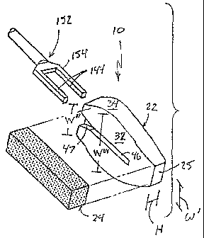
The sides 32 of implant 22 are provided with a keyway 46. Referring now to
Fig. 3, an applicator for use in connection with the present invention is
shown at 152
and is provided with an end 154 shaped in the form of a pair of prongs 144.
The
prongs 144 are formed in a size and shape substantially complementary to the
size and
shape of the keyway 46 of implant 22. Applicator 152 is mounted to implant 22
by
inserting the prongs 144 into the keyways 46 formed on the sides 32 of implant
22.
In this manner, the prongs seat the implant 22 on the end 154 of applicator
152 and
resist relative rotational movement between implant 22 and applicator 152 when
applicator 152 is used to rotate implant 22 in the disk space.
Although not shown in the figure, those skilled in the art who have the.
benefit
of this disclosure will recognize that the end of the keyways 46 may be
extended along
the sides 32 of implant 22 further than is necessary to receive the prongs 144
on
applicator 152 and that the extra length of the keyways 46 may be of gradually
reducing dimension so that the prongs 144 of applicator 152 are received in a
friction
fit in the keyways 46 to help affirmatively mount implant 22 thereto. Other
structure
for achieving this same result includes a detent or serrations formed in the
keyways 46.
The mouth 45 of the keyway 46 on the side 32 of implant 22 at the first end 36
of the
implant is wider than the width of the keyway 46 in the portion of the slot
intermediate
the ends 25 and 36 of implant 22 to facilatate insertion of the prongs 144 of
applicator
152 into the keyway 46. The funnel-shaped portion 47 of the keyway 46 behind
the
mouth 45, which gradually decreases in width, acts to increase the ease with
which
implant 22 is mounted to applicator 152 by insertion of the prongs 144 into
the
CA 02407275 2002-10-21
WO 01/82844 PCT/US01/13824
9
respective keyways 46 and helps to seat implant 22 thereon. Those skilled in
the art
who have the benefit of this disclosure will recognize that the keyway 46 may
be
located on the applicator 152 and a key 48 may be located on implant 22
without any
difference in the manner in which those component parts function to retain
implant 22
on the end 154 of applicator 152.
When the end 154 of applicator 152 is seated all the way into the keyways 46
of
implant 22, so as to prevent relative rotational movement therebetween,
implant 22 is
inserted into the disk space and rotated therein using applicator 152 as
explained below.
Applicator 152 is then detached from implant 22 simply by withdrawing the
applicator 152
from the disk space, the friction exerted by the adjacent vertebrae preventing
the
withdrawal of the implant 22. It will be apparent to those skilled in the art
who have the
benefit of this disclosure that the applicator 152 is of little assistance in
removing the
implant 22 from the disk space even if the keyways 46 of implant 22 are
provided with a
detent or other structure to engage the prongs 144 of applicator 152 to retain
the implant
22 thereon. As noted above, in the preferred embodiment described herein,
implant 22 is
comprised of bone. In alternative embodiments, implant 22 is comprised of
metal or inert
polymeric material and, when comprised of such material, the implant may also
include a
bore (not shown) in the end 36 of implant 22 for receiving a complementary
threaded end
(not shown) on applicator 152. The advantage to using such materials is that,
in the event
the implant 22 needs to be removed from the disk space, an applicator of the
type shown in
Patent No. 5,658,336, which disclosure is incorporated in its entirety as if
fully set forth
herein by this specific reference thereto, is screwed into the bore to allow
the implant 22 to
be pulled from the disk space. If implant 22 is comprised of bone as
contemplated herein
and it is necessary to remove the implant 22, the implant is simply drilled
out with a
conventional bone drill or burr and the pieces drawn back out of the disk
space with
forceps or other suitable instrument to be replaced with another implant as
needed.
The use of the stabilizer 10 of the present invention in, for instance, a
method of
lumbar intervertebral disk stabilization is illustrated in Fig. 1. Surgery is
performed as in a
simple diskectomy and the intervertebral disk 20 is exposed through a small
laminotomy.
The disk material is removed and any nerve root compression is corrected. The
posterior
CA 02407275 2002-10-21
WO 01/82844 PCT/USO1/13824
longitudinal ligament (not shown) and disk cartilage are removed until the
surface of the
bodies 12 and 14 of adjacent vertebrae 16 and 18, respectively, are exposed
above and
below the disk space.
Using spreaders such as those disclosed in International Application No.
5 PCT/US95/00347, which reference is hereby incorporated into this
specification in its
entirety by this specific reference thereto, the vertebrae 16 and 18 are
distracted to open the
disk space, and once the desired "spread" is achieved, the middle portion of
the disk space
is packed with cancellous bone chips. Because the posterior longitudinal
ligament is left
intact to the opposite side and to the center of the disk space, the bone
chips are held in
10 place in the disk space.
An implant 22 having a height H and width W selected to fit the disk space is
then
mounted to the prongs 144 of applicator 152. The appropriately-sized implant
22 is then
inserted into the disk space using the applicator 152 with the implant 22
oriented so that the
top and bottom thereof, i.e., the first and second sides 32, engage the bodies
12 and 14 of
adjacent vertebrae 16 and 18, respectively. Using the applicator 152, implant
22 is
positioned in the disk space by anterior-posterior movement to a position in
which the
expanded, middle portion and the smaller width ends 25 and 36 of the third and
fourth sides
34 of implant 22 contact the respective lower and upper surfaces of the bodies
12 and 14 of
the adjacent vertebrae 16 and 18 when rotated by approximately 90 using the
applicator
152. The respective lower and upper surfaces of the vertebral bodies 12 and 14
are slightly
concave such that the larger width middle portion W" of implant 22 allows the
implant 22
to engage substantially more of the respective surfaces of the vertebral
bodies 12 and 14
than conventional prosthetic devices, thereby providing increased stability to
the fusion
once further rotation of implant 22 in the disk space is prevented as
described below.
Once positioned in the disk space so as to provide maximum stabilization, the
applicator 152 is then detached from the implant 22 by backing out of the
incision in the
patient. Locking piece 24 is then inserted through that same incision and
pressed or
impacted into place in the disk space adjacent and lateral to implant 22 with
the implant
bearing surface 42 of locking piece 24 juxtaposed and/or engaging the surface
32 of
implant 22. Positioning the locking piece 24 adjacent implant 22 in this
manner resists
CA 02407275 2002-10-21
WO 01/82844 PCT/US01/13824
11
relative rotation between implant 22 and locking piece 24. Because the bearing
surfaces 50
of locking piece 24 bear against the bodies 12 and 14 of the adjacent
vertebrae 16 and 18 to
resist rotation of the locking piece 24 relative to the adjacent vertebrae 16
and 18 against
which the bearing surfaces 50 bear, rotation of the implant 22 is also
resisted. Those skilled
in the art who have the benefit of this disclosure will recognize that the
bearing surfaces 50
bear against the cortical end plate of the respective vertebral bodies 12 and
14, which is
comprised of cortical, non-cancellous bone, and provides a hard, relatively
smooth surface
against which the bearing surfaces 50 bear. The end 40 of locking piece 24,
which is also
preferably comprised of bone, may then be shaped to different sizes and shapes
(other than
the square shaped end 40 shown in the figures) so as to allow the surgeon to
select an
appropriately size and shape that provides a close fit with the space between
vertebral
bodies.
If necessary, a small amount of a physiologically compatible adhesive of a
type
known in the art is applied over the cancellous bone chips just medial to the
implant to
close off the remaining portion of the opening into the disk space. The
patient should be
able to ambulate soon after the procedure because of the stability imparted to
the spinal
column by the implant of the present invention. Before narrowing of the disk
space occurs,
the cancellous bone chips will have started the fusion process.
The stabilizer 10 is also used to advantage to perform, for instance, a
posterior
lateral intertransverse fusion. The implant 22 is inserted into the region of
the disk space
from which a portion of the disk has been removed as described above with the
locking
piece 24 inserted and the posterior lateral fusion performed. Because the
implant 22
provides stability to the spine until the posterior lateral fusion is solid,
the patient is
generally able to ambulate soon after surgery. This procedure also prevents
the narrowing
of the disk space, which is a common problem with posterior lateral fusion.
Referring now to Fig. 4, one preferred method for fabricating the implant 22
will
now be described. Fig. 4 shows a perspective view of a cross-sectioned portion
of the shaft
of the human femur 200. Femur 200 is comprised of hard cortical bone 202
surrounding a
central core of softer, cancellous bone 204. Implant 22 is fabricated from the
cortical bone
202 of the piece of femur 200 by making a first vertical cut along the
longitudinal axis of
CA 02407275 2002-10-21
WO 01/82844 PCT/US01/13824
12
the femur 200 in the area of the cortical bone 202 as shown at reference
numeral 206 and a
second vertical cut along the longitudinal axis of the femur 200 in the
cortical bone 202,
closer to the cancellous bone 204 in the center axis of femur 200 along the
line indicated at
208. The resulting piece of the cortical bone 202 comprising the femur 200,
identified by
reference numeral 210 in Fig. 4 serves as a "blank" for making the implant 22.
Using
conventional bone saws and shaping techniques, the blank 210 is shaped to
include the bi-
convex first and second sides 32, rounded end 36, and so on as described
above. Although
the lines 206 and 208 representing the first and second vertical cuts are
shown substantially
parallel to each other, those skilled in the art who have the benefit of this
disclosure will
recognize that lines 206 and 208 need not be parallel and that there is even
certain
advantages to be gained by not making the cuts parallel as, for instance, when
it is desired
to make an implant that is wedge-shaped as described below. Those skilled in
the art who
have the benefit of this disclosure will also recognize that there are other
bones in the body
from which a piece of cortical bone can be harvested that is big enough to
serve the same
role as the blank 210, the femur 200 being described herein merely for the
purpose of
enabling the present invention as required by the Patent Statute.
Referring now to Figs. 5A and 5B, Fig. 5A is a schematic representation of the
human iliac 212 and femur 200 in the area of the hip. In the presently
preferred
embodiment described herein, locking piece 24 is fabricated from a portion of
the iliac 212.
Implant 22 is fabricated from the hard cortical bone 202 of the femur 200 to
give the
implant 22 the strength to properly support the load of the spinal column. By
contrast,
because it is desired that the iliac 212 have properties that both impart
strength to locking
piece 24 and facilitate its fusion with the cancellous bone chips packed
around the implant
22 in the disk space as well as the adjacent vertebral bodies 16 and 18, the
locking piece 24
is preferably fabricated from a combination of cortical and cancellous bone
and that
combination is found in the iliac 212. Specifically, a first cut is made that
is orthogonal to
the long axis of the iliac (the latter being represented by the shadow line
216 in Fig. 5A) in
the so-called "wing" of the iliac 212 as shown at line 214. A second
orthogonal cut 218,
substantially parallel to the first cut 214, is then made across the wing of
the iliac 212 to
produce a piece of bone 220 that is in effect a cross-sectional piece of the
iliac. The piece
CA 02407275 2002-10-21
WO 01/82844 PCT/US01/13824
13
of bone 220 is then squared up at one end by cutting along the line 222.
Because the cuts
214, 218, and 222 can be made in any order such that the piece of bone 220 may
not be
removed from the iliac 212 when cut 222 is made, cut 222 is described herein
as being
substantially parallel to the long axis 216 of the iliac 212.
The piece of bone 220 is shown removed from the iliac 212 and in perspective
view
in Fig. 5B. When viewed in this manner, it will be apparent that the piece 220
of bone
harvested from the iliac 212 in the above-described manner is comprised of
both hard
cortical bone as shown at reference numeral 224 and softer cancellous bone as
shown at
226. To make two of the locking pieces 24 of the present invention, a cut 228
is made
through the piece of bone 220. Again, it will be recognized by those skilled
in the art that
the cut 228 can be made in any order relative to the cuts 214, 218, and 222
such that the
cut 228 can be described as being substantially parallel to the cuts 214 and
218 and
orthagonal to the longitudinal axis 216 of the iliac 212. The cortical bone
224 forms a
relatively thin outer layer around the two sides and one end of each of the
stabilizers 230A
and 230B that are fabricated from piece 220, such that the stabilizer of the
present
invention is referred to as being "tri-cortical," and will form the implant
bearing surface 50
and end 40 of stabilizer 24 as shown in Figs. 1-3. The central core of
cancellous bone 226
promotes the fusion of the locking piece 24 in the disk space as described
above, but is not
a necessary element of the invention, it being apparent from this disclosure
that a locking
piece can be fabricated entirely from cortical bone or cancellous bone, the
tri-cortical
locking piece described herein being only a preferred embodiment of the
locking piece.
Those skilled in the art who have the benefit of this disclosure will also
recognize that there
are other bones in the body from which a piece of cortical bone can be
harvested that is big
enough to serve the same role as the piece 220, the iliac 212 being described
herein as the
source of the piece 220 for the purpose of enabling the present invention as
required by the
Patent Statute.
In the embodiments described herein, the implant 22 and locking piece 24 are
fabricated to certain preferred dimensions, but those skilled in the art will
recognize that it is
desirable to fabricate them in several dimensions so as to facilitate a close
fit in the disk
space. For instance, a presently preferred size for implant 22 is for implant
22 to have a
CA 02407275 2002-10-21
WO 01/82844 PCT/US01/13824
14
height H of approximately 5 mm, a width W of about 10 mm at the ends 25, 36
increasing
to a maximum of about 15 mm, and a length (along the longitudinal axis of
implant 22) of
about 27 mm. A locking piece 24 intended for use with an implant of this 15 X
5 X 27 size
is about 10 mm X 10 mm X 27 mm. Other implant, and corresponding locking
piece, sizes
are also contemplated, it being understood that because they are comprised of
bone in this
preferred embodiment, the bone can be harvested in the desired shape and size
from several
locations of the skeleton.
Referring now to Fig. 6A, another embodiment of an implant constructed in
accordance with the present invention is shown. Specifically, the implant 322A
is
substantially rectangularly shaped in cross-section and, like implant 22 in
Figs. 1 - 3, is
comprised of first and second sides 332 defining the height H and third and
fourth sides 334
defining width W, the height dimension H being greater than the width
dimension W. Third
and fourth sides 334 are arched from one end of implant 322 to the other to
provide the
portion of implant 322 intermediate the ends 325 and 336 with a width W that
is larger than
the width W' and W" at the ends 325 and 336, except that the end 325 is flared
as at 360 to
provide the implant 322A with a width W at the end 325 that is approximately
equal to the
width of implant 322A at the maximum width W. The two sides 34 therefore
comprise
two opposed edges, each of arched form, the opposed edges being shaped such
that the
end 325 is flared so that a portion of the implant 322A between the flared end
part 360 and
the remainder of the implant is of lesser width than the width W' of the flare
360 in the end
325 of implant 322A. This portion of lesser width of implant 322A forms
upwardly and
downwardly opening recesses 362 and is preferably formed as a smoothly curved
recess to
interact with and provide stability for the adjacent vertebrae, the
articulating surfaces of
which are concave in shape. As noted above in connection with the description
of the
embodiment shown in Figs. 1-3, implants constructed in accordance with the
teachings of
the present invention may be provided with means for resisting anterior-
posterior
movement of the implant in the disk space. In the embodiment shown in Fig. 6,
implant
322 is provided with a plurality of teeth 338 for this purpose. As also noted
above, the
teeth near the ends of the implant may be slanted toward the ends of the
implant so as to
provide better contact with the adjacent vertebrae, but in the case of the
implant 322 shown
CA 02407275 2002-10-21
WO 01/82844 PCT/US01/13824
in Fig. 6, it has been found that better contact is obtained by slanting the
teeth in the flared
portion 360 away from the end 325.
As shown in Figs. 6B and 6C, the width W of the ends 325 of implants 322B and
322C is progressively greater than the width of implants 322B and 322C at the
5 approximate midpoint along the longitudinal axes of implants 322B and 322C
shown as the
maximim width W. Stated another way, the difference in the widths W and W of
implants
322B and 322C is defined by the angle a formed by extending lines from the
midpoint of
the implant 322B and 322C representing width W to the tip of the flare 360 at
the end 325
of each respective implant representing width W'. It is envisioned that
several implants 322
10 would be provided with different angles a to accommodate specific patient
needs; for
instance, in one patient, the curvature of the spine, or lordosis, may require
that an implant
having an angle a that provides a width W that is 5 mm larger than the width W
be utilized
between two vertebrae and that a second implant having an angle a that
provides a width
W that is 10 mm larger than the width W may be needed between the next two
vertebrae
15 to maintain the proper lordosis of the spine. Of course the angle a can be
any angle, but it
is generally preferred that an angle a of between 0 (those skilled in the art
will recognize
that the angle (x of the embodiment shown in Fig. 6A is 0 ) and 30 will
provide a sufficient
range of width W' dimensions to accommodate most patient needs.
Figure 7 shows an alternative embodiment of the implant 322 shown in Fig. 6
wherein the sides 332 of the implant 322 are provided with keyways 46 having a
funnel-
shaped mouth 47 for receiving the prongs 144 of applicator 152 as described
above. In
Fig. 7, the implant 322 is provided with an angle a of 0 such that the
dimensions W and
W are approximately equal, but those skilled in the art will recognize that
the implant
shown in Fig. 7 could have been formed with a flared end such that the angle a
could have
been any angle from about 0 to about 30 .
If the alternative embodiment shown in Fig. 8, is utilized, the surgeon need
not
select an implant having a desired angle a. Instead, the implant 422 shown in
Fig. 8 is
provided with a pair of blocks 464 movable relative to the implant in a
direction having a
component that is perpendicular to the longitudinal axis of implant 422 for
changing the
width W' of the flare 460 at the end 425 of implant 422. Blocks 464 are
threadably
CA 02407275 2002-10-21
WO 01/82844 PCT/US01/13824
16
engaged to a spindle 466 that is journaled in implant 422 and means is
provided, in the form
of the wheel 468 integral with spindle 466, for rotating the spindle 466 to
spread the blocks
464 apart, or to move the blocks 464 closer to the mid-line, or longitudinal
axis, of implant
422. A track 470 is formed in implant 422, and the surface of the blocks 464
adjacent the
track 470 is formed in a complementary shape (not shown) for engaging the
track 470 to
define the direction of in which the blocks 464 slide along track 470 relative
to implant 422.
Those skilled in the art who have the benefit of this disclosure will
recognize that the
direction of movement of blocks 464 relative to implant 422 need not be
perpendicular to
the longitudinal axis of implant 422 and that it may even be advantageous to
move blocks
464 in a direction defined by an angle that is not a 90 angle relative to the
longitudinal axis
of implant 422. For this reason, the movement of the blocks 464 is described
herein as
being in a direction "having a component that is perpendicular" to the
longitudinal axis of
implant 422 rather than in a direction that is perpendicular to the
longitudinal axis of
implant 422.
Although described in terms of the preferred- embodiment shown in the figures,
this
embodiment is shown to exemplify the present invention, it being recognized by
those
skilled in the art that certain changes can be made to the specific structure
of the preferred
embodiment shown and described without departing from the spirit of the
present
invention. In the case of one such change, the first and second sides of the
implant are
substantially flat but not parallel along their longitudinal axes so that the
implant is wedge-
shaped. The wedge shape of the implant facilitates insertion of the implant
into the disk
space, the rounded end of the implant reducing the likelihood of injury to the
nerves of the
spinal cord during insertion into the disk space. Likewise, the width at one
end of the
implant can be less than the width at the end, both widths, however, being
less than the
width in the middle, expanded portion of the implant. All such modifications,
and other
modifications which do not depart from the spirit of the present invention,
are intended to
fall within the scope of the following claims.