Note: Descriptions are shown in the official language in which they were submitted.
CA 02409353 2009-10-06
WO 01/94918 PCT/EP01/06365
REFERENCE DEVICE FOR EVALUATING THE PERFORMANCE OF A
CONFOCAL LASER SCANNING MICROSCOPE, AND A METHOD AND SYSTEM
FOR PERFORMING THAT EVALUATION
Field of the Invention
The invention concerns a reference device for evaluating the
performance of a confocal laser scan microscope of the kind
used for performing a two dimensional quantitative
fluorescence measurement of test matter distributed on a
flat surface of a first glass substrate in particular a DNA
binding array or the like, e.g. a DNA binding array of the
type described in U.S. Patent No.5,143,854.
The invention also concerns a method for evaluating the
performance of a confocal laser scan microscope of the above
mentioned kind.
The invention more in particular concerns an evaluation
method enabling the characterization of a confocal laser
scan microscope of the above mentioned kind in terms of
quantitative signal detection sensitivity, limit of
detection, uniformity of the confocal volume over the scan
field of view, spatial resolution of the scanning process
and dynamic behavior of the measured signal over the scan
field of view, said measured signal corresponding to the
fluorescent light received.
The invention further concerns a system for evaluating the
performance of a confocal laser scan microscope which is apt
to be used for performing a two dimensional quantitative
fluorescence measurement of test matter distributed on a
flat surface of a substrate.
CA 02409353 2002-11-15
WO 01/94918 PCT/EP01/06365
- 2 -
Background
The principle of confocal laser scan microscopy for two-
dimensional, quantitative fluorescence measurement is
illustrated in Figs. 1 and 2. Figure 1 shows the optical
setup of a 2-D flying spot confocal laser scan microscope,
using for fluorescence excitation a laser beam 11, a
dichroic beam splitter 12, a 2-dimensional scan engine 13
for spatial beam deflection in two orthogonal directions (X-
Y) and a lens 14 for focusing the laser beam into an object
plane 15. Fluorescent light of a longer wavelength than the
excitation laser 11 is generated by exciting fluorescent
molecules in the object plane 15.
Fluorescent light emitted by fluorophores located in the
object plane 15 of the scanned area is collected by lens 14
and then transmitted by means of the scan engine 13 and the
dichroic beam splitter 12 as a fluorescent light beam 17
which is focused by lens 18 into a pinhole aperture 19 in a
conjugate plane 21 in front of a photodetection device 22.
The concept of confocal imaging, which is currently used to
discriminate the generally weak fluorescence signal from
background radiation, is illustrated in Fig. 2. Only optical
radiation from within the confocal volume Vc, i.e., the
fluorescence signal, is detected by the photodetector 22. Vc
is defined by the optical transfer function of the detection
optics (OTFem) and the size of the detector pinhole 19 in
the conjugate plane 21. Higher background suppression rates
result for smaller confocal volumes Vc.
The size of the scan field of view is typically in the order
of 20 x 20 square millimeter. The confocal volume is
generally in the order of Vc = 5 x 5 x 50 cubic micrometer,
where Ac = 5 x 5 square micrometer and zc = 50 micrometer is
CA 02409353 2002-11-15
WO 01/94918 PCT/EP01/06365
- 3 -
approximately the spot size and the Rayleigh range of the
focused laser beam, respectively. The pixel size of the scan
engine 13 for scanning the laser beam 11 in the field of
view is typically 1 to 20 micrometer.
DNA binding arrays, e.g. those of the type described in U.S.
Patent No.5,143,854, consist of a glass chip carrying a
chemical system subdivided in adjacent cells, commonly
called features. The features are characterized by specific
probes. Specific nucleic acid sequences are immobilized
(captured) by the probes and labeled with a fluorescent dye.
The amount of captured nucleic acid on individual features
is detected using quantitative fluorescence measurement (the
fluorescent dye emits light when excited by light energy of
a given wavelength) by sequential pixel reading (scanning)
of the features. The features are spatially over-sampled by
the scanning procedure.(i.e. number of pixels > number of
features) for accurate spatial referencing of the glass chip
by numerical data analysis and for increased feature signal
quality by averaging physically measured light intensities.
Typical pixel sizes are in the order of 1 to 20 micrometer.
The ratio of the scan field of view to the cross-section Ac
of the confocal volume is typically high in confocal laser
scan microscopy, i.e. "scan field of view"/"cross-section Ac
of the confocal volume" >> 1, which readily leads to a x-y
position depending optical transfer function OTF(x, y)
OTFex * OTFem, where OTFex and OTFem are the optical
transfer functions of the excitation and emission optics,
respectively. The x-y position dependence is mainly due to
mechanical misalignment and imperfections of optical and
opto-mechanical components, such as e.g. the scan engine
used for scanning. It causes an inhomogeneous sensitivity
over the scan field of view, as schematically sketched in
Figures 3a, 3b and 3c and this in turn leads to erroneous
CA 02409353 2002-11-15
WO 01/94918 PCT/EP01/06365
- 4 -
quantitative fluorescence measurements. As an example, Fig.
4 schematically shows the scanned image of a DNA binding
array, e.g. of the type described in U.S. Patent
No.5,143,854, which array has a chess-board pattern. As
described hereinafter with reference to Fig. 4 the scanned
image has a lower signal level in the top right corner, due
to either inhomogeneous fluorophore density in the scanned
object or inhomogeneous sensitivity of the confocal laser
scan microscope over the scan field of view.
There is therefore a need for a reliable quantitative
measurement and evaluation of the sensitivity over the scan
field of view of a confocal laser scan microscope of the
above described type.
The availability of an appropriate reference standard target
object would allow to discriminate between instrument- and
scanned object (e.g. a DNA binding array of the type
described in U.S. Patent No.5,143,854) contributions to the
observed non-uniformity in Fig. 4. However, no reference
fluorescing target objects for characterizing key
performances of a confocal laser scan microscope, i.e.,
sensitivity, limit of detection, uniformity-, spatial
resolution- and signal dynamic behavior over the scan field
of view, have been reported yet.
There is therefore a need for an appropriate reference
standard target object that allows one to discriminate
between instrument- and scanned object contributions to a
non-uniformity of the type represented in Fig. 4.
Summary of the Invention
The aim of the invention is therefore to provide a reference
device, a method and a system of the above mentioned kinds
~---;
CA 02409353 2008-05-07
-5-
that make it possible to evaluate quantitatively the
performance of a confocal laser scan microscope for
performing two-dimensional, quantitative fluorescence
measurements.
According to a first aspect of the invention this aim is
attained with a reference device for evaluating the
performance of a confocal laser scan microscope. The
reference device comprises: (a) a substrate, and (b)
reference fluorescent matter distributed over a surface
of the substrate, the reference fluorescent matter
having a predetermined spatial distribution over the
surface and being dissolved in a liquid in a
predetermined concentration.
According to a second aspect of the invention the above
mentioned aim is attained with a method for evaluating
the performance of a confocal laser scan microscope of
the kind used for performing a two dimensional
quantitative fluorescence measure of test matter
distributed on a flat surface of a first substrate. The
method comprises performing a two-dimensional
quantitative fluorescence measurement of a reference
device using the microscope to obtain measured values for
one or more quantitative parameters, the reference device
comprising: (a) a second substrate, and (b) reference
fluorescent matter distributed over a surface of the
second substrate, the reference fluorescent matter having
a predetermined spatial distribution over the surface and
being dissolved in a liquid in a predetermined
concentration.
CA 02409353 2008-05-07
-5a-
According to a third aspect of the invention the above
mentioned aim is attained with a system for evaluating
the performance of a confocal laser scan microscope. The
system comprises: a confocal laser scan microscope, and a
reference device according to the above mentioned first
aspect of the invention.
The main advantages attained with a reference device,
method, and system according to the invention are that
they allow a quantitative and highly accurate evaluation
of the performance of a confocal laser microscope for
scanning DNA binding arrays of the above mentioned kind,
and that this evaluation makes it possible to evaluate
quantitatively measurement results obtained by scanning
with such a microscope, e.g. a sample DNA binding array
to be analyzed. In this context it is important to note
that the evaluation performed according to the invention
includes the measurement of the following
characteristics:
a) quantitative signal detection sensitivity,
b) quantitative signal detection limit,
c) uniformity of the confocal volume over the scan
field of view,
d) spatial resolution of the scanning process, and
e) dynamic behavior of the measured signal over
the scan field of view, said measured signal
corresponding to
CA 02409353 2002-11-15
WO 01/94918 PCT/EP01/06365
- 6 -
the fluorescent light received.
Brief Description of the Drawings
Preferred embodiments of the invention are described
hereinafter with reference to the accompanying drawings
wherein:
Fig. 1 shows a schematic representation of the basic setup
of a confocal laser scan microscope for performing a
two-dimensional, quantitative fluorescence
measurement,
Fig. 2 schematically shows a confocal volume Vc in an
object plane,
Figures 3a, 3b and 3c show schematic representations of
various forms of non-uniformity of the scanned
confocal volume over the scan field of view,
Fig. 4 shows a schematic representation of a scanned image
of a DNA binding array
Fig. 5a shows a top view of a first embodiment of a
reference device according to the invention,
Fig. 5b shows a cross-section through a plane A-A of the
embodiment shown by Fig. 5a,
Fig. 6 shows the shape of a representative electrical
signal obtained by measuring fluorescent light
emitted by fluorescent zones located in a row of the
array represented in Figures 5a and 5b.
Fig. 7a shows a top view of a second embodiment of a
CA 02409353 2002-11-15
WO 01/94918 PCT/EP01/06365
- 7 -
reference device according to the invention,
Fig. 7b shows a cross-section of the embodiment shown by
Fig. 7a.
Detailed Description of the Invention
The subject invention will now be described in terms of its
preferred embodiments. These embodiments are set forth to
aid the understanding of the invention, but are not to be
construed as limiting.
Fig. 1 schematically shows a basic setup of a confocal laser
scan microscope for two-dimensional, quantitative
fluorescence measurement in case of a two-dimensional flying
spot. An excitation laser beam 11, which is transmitted
through a dichroic beam splitter 12, is spatially scanned by
means of a two-axis scan engine 13, e.g., a galvo-scanner,
in two axis X, Y, perpendicular to each other, and is
focused by a lens 14 into an object plane 15 which is
parallel to a X-Y-plane defined by the axis X and Y, and
which is perpendicular to a third axis Z which is
perpendicular to the X-Y-plane.
Fluorophores within the confocal volume Vc in the object
plane 15 are excited by the focused laser spot 16 and the
fluorescent light 17 generated by excitation of those
fluorophores is collected and imaged by a lens 18 into a
detector pinhole 19 in the conjugate plane 21 and detected
by photodetector 22.
Fig. 2 schematically shows a confocal volume Vc (with Vc =
Ac * zc) in the object plane 15. Confocal volume Vc is
ideally a cylindrical volume having a rotation axis parallel
to the Z axis and a circular cross-section. The confocal
CA 02409353 2002-11-15
WO 01/94918 PCT/EP01/06365
- 8 -
volume Vc is defined by the optical transfer function
(OTFem) of the detection optics and the size and the shape
of the detector pinhole 19 in the conjugate plane 21. Only
optical radiation from within the confocal volume Vc is
detected by the photodetector 22. The concept of confocal
imaging allows high background suppression rates for
detecting weak signal levels, as is commonly the case in
fluorescence measurements.
Figures 3a, 3b and 3c show schematic representations in the
plane Y-Z of various forms of non-uniformity of the scanned
confocal volume Vc, that is of deviations of the shape of
this volume from the ideal shape represented in Fig. 2.
These deviations cause inhomogenities of the amplitude of
the fluorescent light intensity signal measured over the
scanned area.
Fig. 3a shows a scanned confocal volume 24 which is tilted
with respect to an ideal or nominal confocal volume 23. Fig.
3b shows a scanned confocal volume 25 which has not a
constant width and which is thus non-uniform compared with
the nominal confocal volume 23. Fig. 3c shows a scanned
confocal volume 26 having a shape which is distorted with
respect to the nominal confocal volume 23.
Mechanical misalignment and imperfection of optical and
opto-mechanical components used are the main reasons for the
non-uniformities of the confocal volumes represented in
Figures 3a, 3b and 3c.
Fig. 4 shows a schematic representation of a scanned image
of DNA binding array 31 of the type described in U.S. Patent
No.5,143,854. Array 31 has a chess-board array of
fluorescent points 32 apt to emit fluorescence light when it
is irradiated with excitation light. For the purpose of the
CA 02409353 2002-11-15
WO 01/94918 PCT/EP01/06365
- 9 -
following description it is convenient to distinguish two
zones of array 31: a first zone 33 which extends from a
diagonal 34 to the top right corner of array 31, and a
second zone 35 which extends from diagonal 34 to the low
left corner of array 31. Due to either inhomogeneous
fluorophore density in the scanned object or inhomogeneous
sensitivity of the confocal laser scan microscope over the
scan field of view, the image represented by Fig. 4 shows up
that fluorescent points 32 located in zone 33 of array 31
provide fluorescent light of lower intensity than
fluorescent points 32 located in zone 35. The purpose of a
reference device, a method and a system according to the
invention is to evaluate quantitatively to which extent such
variations of the intensity of the fluorescence light
detected are due specifically to inhomogeneous sensitivity
of the confocal laser scan microscope over the scan field of
view.
Fig. 5a shows a top view and Fig. 5b a cross-section of a
first embodiment of a reference device 41 according to the
invention for characterizing quantitatively the homogeneity
and sensitivity of a confocal laser scan microscope for two-
dimensional quantitative fluorescence measurement. In
Figures 5a and 5b dimensions are indicated in millimeters.
The reference device 41 shown by Figures 5a and 5b consists
of a top glass plate 42 bonded onto a glass substrate 43.
The 16 x 16 square millimeter glass substrate (thickness = 1
millimeter) has a 5 micrometer etched planar cavity 44
(hatched area). The glass top plate 42 (thickness = 0.7
millimeter) has two drilled holes 45 respectively 46 for
fluid in- and outlet. In another possible embodiment, a top
glass plate 42 without drilled holes 45 and 46 may not be
bonded onto a glass substrate 43.
CA 02409353 2002-11-15
WO 01/94918 PCT/EP01/06365
- 10 -
Glass substrate 43 has e.g. identical dimensions and
preferably the same optical properties as the substrate of
DNA binding array 31 described above with reference to Fig.
4.
The cavity 44 of the reference device is filled with
dissolved fluorophores with a predetermined concentration
leading to a predetermined spatial distribution of
fluorophores over the scanned area. Since the fluorophores
are dissolved uniformly, this spatial distribution is
determined by the structure of cavity 44, and not by the
dissolved fluorophores themselves. In the example described
with reference to Figures 5a and 5b the cavity 44 is planar
and therefore the spatial distribution of the fluorophores
is a uniform one over the whole area of cavity 44.
Glass cover 42 of reference device 41 shown by Figures 5a
and 5b has identical dimensions and optical properties as a
glass substrate of a given DNA binding array of the type
described above with reference to Fig. 4. Reference device
41 can therefore be scanned by the confocal laser scan
microscope under the same optical conditions. The well
defined size of the cavity and the controlled fluorophore
concentration having a predetermined spatial distribution
over the scanned area allows investigation of the
measurement signal with respect to sensitivity (limit of
detection) and homogeneity over the scan field of view. As
mentioned above the spatial distribution of the dissolved
fluorophores is determined by the structure of cavity 44,
and not by the uniformly dissolved fluorophores themselves.
Using a solution of dissolved fluorophores has the advantage
that it can be prepared just before the fluorescence
measurement is performed. Thus, the reference device filled
with a liquid of dissolved fluorophores is not affected by a
CA 02409353 2002-11-15
WO 01/94918 PCT/EP01/06365
- 11 -
possible bleaching effect, which otherwise could arise if it
has been stored for a longer time and which could lead to a
non-quantitative capability of fluorescence of the reference
device over the scan field of view. In addition, various
solutions with different concentrations of dissolved
fluorophores can easily be prepared, which allows for
fluorescence measurements at various intensity levels. In
particular, it is possible to determine the limit of
detectable intensity level.
The height of cavity 44 is much smaller than the depth of
the confocal volume Vc given by the Rayleigh range zc, such
that the thin planar layer of fluorophores in the cavity 44
allows for a reliable evaluation of e.g. possible
displacements of the confocal volume Vc in the Z-direction
(perpendicular to the scan field of view) as the reference
device 41 is being scanned over the whole scan field of
view.
As an example Fig. 6 shows the signal profile of row 588 of
1024 of the scanned image obtained with the reference device
shown by Figures 5a and 5b: the cavity 44 of the device has
a constant thickness over its whole extension and is filled
with 200 mg/ml fluorescein TRIS solution (Fluorescein in
aqueous 0.1 molar TRIS buffer pH 8.3). The image can be
analyzed for sensitivity and for uniformity of the confocal
laser scan microscope, as shown in Fig. 6 for the line
profile of row 588 of 1024 (field of view = 10 x 10 square
millimeter, pixel size = 10 micrometer, the resolution of
the scanning is 1024 x 1024 pixel, the scan time is 126
seconds, the detection sensitivity of the transimpedance
amplifier used is 10 microampere per volt, the cutoff or 6
dB frequency of the low-pass filter used with the
transimpedance amplifier is 30 kHz, the integration interval
tdwell has a duration of 40 microseconds). As can be
CA 02409353 2002-11-15
WO 01/94918 PCT/EP01/06365
- 12 -
appreciated from Fig. 6 some noise signal is superposed on
the line profile obtained. In Fig. 6 signal intensity is
indicated in arbitrary units.
The deviation of the signal intensity of each pixel from a
predetermined value, which is given e.g. by the signal
intensity averaged over the whole scanned image, gives a
quantitative parameter for the performance of a confocal
laser scan microscope.
As a further application it is possible to calibrate the
signal intensity level. The number of fluorophores per area
can be evaluated from the concentration of dissolved
fluorophores. Therefore, the signal intensity measured and
the measurement sensitivity are related to the number of
fluorophores per area in a quantitative manner.
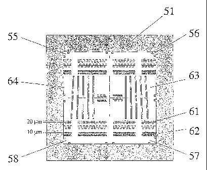
Fig. 7a shows a top view and Fig. 7b a cross-section of a
second embodiment of a reference device according to the
invention for a quantitative evaluation of the homogeneity
and spatial resolution of a confocal laser scan microscope.
In Figure 7a some dimensions in micrometer are indicated. In
Figure 7b some dimensions in millimeters are indicated.
The reference device 51 shown by Figures 7a and 7b consists
of a top glass plate 52 on a glass substrate 53 having an
area of 16 x 16 square millimeter. The upper surface of
glass substrate 53 (thickness = 1 millimeter) has a 5
micrometer etched, microstructured depression forming a
cavity 54 having a bottom inner surface. Cavity 54 is filled
with uniformly dissolved fluorophores having a predetermined
concentration, which leads to a predetermined spatial
distribution of fluorescent zones over the area of cavity
54. This spatial distribution is not determined by the
CA 02409353 2002-11-15
WO 01/94918 PCT/EP01/06365
- 13 -
uniformly dissolved fluorophores themselves, but by the
structure of cavity 54. Cavity 54 is covered by the glass
top plate 52 which has a lower outer surface. The space
comprised between the lower outer surface of plate 52 and
the bottom inner surface of depression 54 has a thickness D
which varies according to a predetermined function of the
form D- f(x,y) over the entire area of depression 54. The
latter space is at least partially filled with dissolved
fluorophores.
Glass substrate 53 has e.g. identical dimensions and
preferably the same optical properties as the substrate of
DNA binding array 31 described above with reference to Fig.
4.
Glass cover 52 of reference device 51 has identical optical
properties as a glass substrate of a given DNA binding array
31 of the kind described above with reference to Fig. 4.
Reference device 51 can therefore be scanned by a confocal
laser scan microscope under the same optical conditions.
The microstructured cavity 54 comprises different patterns
of fluorescent zones. In Fig. 7a each non-fluorescent zone
is represented by a shaded surface.
A first pattern of fluorescent zones comprises just two non-
fluorescent zones each represented in Fig. 7 by a shaded
square. In Fig. 7a fluorescent zones having this first
pattern are located at each of the corner zones 55, 56, 57,
58 of reference device 51. The measured signals
corresponding to the intensity of fluorescent light emitted
from these corner zones are evaluated in order to assess the
degree of uniformity over the scan field of view of the
scanning performed with a confocal laser scan microscope.
CA 02409353 2002-11-15
WO 01/94918 PCT/EP01/06365
- 14 -
Zones 61 and 62 located at different rows of reference
device 51 have a second pattern of spatial distribution of
fluorescent features. The measured signals corresponding to
fluorescent light emitted from zones like 61 and 62 are
evaluated in order to assess the resolution of the scanning
performed with a confocal laser scan microscope. In
addition, since the dimensions of the zones 61 and 62 are
known, the accuracy of the scan steps performed by the
scanning engine can be assessed. As shown in Fig. 7a the
zones 61 and 62 is subdivided such that the cavity forms one
connected reservoir which is filled by the dissolved
fluorophores.
Zones 63, 64 having the appearance of a group of bars having
different inclination angles represent a third pattern of
fluorescent features available on reference device 51. The
measured signals corresponding to fluorescent light emitted
from a zone like zone 63 is evaluated in order to assess the
dynamic signal behavior associated to the scanning performed
with a confocal laser scan microscope.
The present invention relates to the reference devices
described above and shown in Figures 5a, 5b and 7a, 7b
respectively, which are used for a quantitative evaluation
of a confocal laser scan microscope for two-dimensional,
quantitative fluorescence measurement. Typical
characteristics determined with a reference device,
respectively a method, according to the invention are
a) quantitative signal detection sensitivity,
b) quantitative signal detection limit,
c) uniformity of the confocal volume over the scan
field of view,
d) spatial resolution of the scanning process, and
e) dynamic behavior of the measured signal over the
scan field of view, said measured signal corresponding to
CA 02409353 2002-11-15
WO 01/94918 PCT/EP01/06365
- 15 -
the fluorescent light received.
The above mentioned use of the invention for evaluating the
performance of a confocal laser scan microscope
substantially comprises
scanning a reference device according to the invention
with a microscope to be evaluated in order to obtain a first
set of measurement values,
processing said first set of measurement values in
order to obtain correction factors,
storing said correction factors,
scanning a sample, e.g. a DNA binding array, with the
evaluated microscope in order to obtain a second set of
measurement values, and
correcting said second set of measurement values with
said correction factors in order to obtain a third set of
values which are free from deviations due to the performance
of the scanner and which therefore more accurately
correspond to characteristics of the particular sample
examined.
Although preferred embodiments of the invention have been
described above using specific terms, such description is
for illustrative purposes only, and it is to be understood
that changes and variations may be made without departing
from the spirit or scope of the claims of this patent
application.
List of reference numbers
11 excitation laser beam
12 dichroic beam splitter
13 two-axis scan engine
14 lens
15 object plane
CA 02409353 2002-11-15
WO 01/94918 PCT/EP01/06365
- 16 -
16 focused laser spot
17 fluorescent light
18 lens
19 detector pinhole
21 conjugate plane
22 photodetector
23 nominal confocal volume (cross-section in plane z-y)
24 scanned confocal volume (cross-section in plane z-y)
25 scanned confocal volume (cross-section in plane z-y)
26 scanned confocal volume (cross-section in plane z-y)
31 DNA binding array (located in object plane)
32 fluorescent point or fluorescent feature
33 zone
34 diagonal
35 zone
41 first embodiment of reference device
42 top plate
43 bottom plate
44 cavity
45 hole
46 hole
51 second embodiment of reference device
52 top plate
53 bottom plate
54 cavity
55 zone having a first pattern of fluorescent features
56 zone having a first pattern of fluorescent features
57 zone having a first pattern of fluorescent features
58 zone having a first pattern of fluorescent features
61 zone having a second pattern of fluorescent features
62 zone having a second pattern of fluorescent features
63 zone having a third pattern of fluorescent features
64 zone having a third pattern of fluorescent features
- - - - -