Note: Descriptions are shown in the official language in which they were submitted.
CA 02409773 2002-11-15
WO 01/89418 PCT/USO1/14620
Title: CARDIAC PROSTHESIS FOR HELPING IMPROVE OPERATION OF A
HEART VALVE
Technical Field
The present invention relates to an implantable cardiac prosthesis and,
more particularly, to a prosthesis that may be implanted at an annulus of a
heart
valve to help improve operation of a defective or damaged valve.
Backuround
A heart valve may become defective or damaged, such as resulting from
congenital malformation, disease, or aging. When the valve becomes defective
or damaged, the leaflets may not function properly. One common problem
associated with a degenerating heart valve is an enlargement of the valve
annulus (e:g., dilation). Other problems that may result in valve dysfunction
are
chordal elongation and lesions developing on one or more of the leaflets.
The bicuspid or mitral valve is located in the left atrioventricular opening
of the heart for passing blood unidirectionally from the left atrium to the
left
ventricle of the heart. The mitral valve is encircled by a dense fibrous
annular
ring and includes two valve leaflets of unequal size. A larger valve leaflet,
which is known as the anterior leaflet, is located adjacent the aortic
opening.
The smaller leaflet is the posterior leaflet.
When a mitral valve functions properly, for example, it prevents
regurgitation of blood from the ventricle into the atrium when the ventricle
contracts. In order to withstand the substantial backpressure and prevent
regurgitation of blood into the atrium during the ventricular contraction, the
cusps are held in place by fibrous cords (cordae tendinae) that anchor the
valve
cusps to the muscular wall of the heart.
By way of example, if an annulus enlarges or dilates to a point where the
attached leaflets are unable to fully close (malcoaptation), regurgitation or
valve
prolapse may occur. Adverse clinical symptoms, such as chest pain, cardiac
arrhythmias, dyspnea, may manifest in response to valve prolapse or
regurgitation. As a result, surgical correction, either by valve repair
procedures
or by valve replacement, may be required.
CA 02409773 2002-11-15
WO 01/89418 PCT/USO1/14620
Surgical reconstruction or repair procedures may include plication,
chordal shortening, or chordal replacement. Another common repair procedure
relates to remodelling of the valve annulus (e.g., annuloplasty), which may be
accomplished by implantation of a prosthetic ring to help stabilize the
annulus
and to correct or help prevent valvular insufficiency which may result from
defect or dysfunction of the valve annulus. Properly sizing and implanting the
annuloplasty ring can substantially restore the valve annulus restored to its
normal, undilated, circumference. In situations where the valve leaflets
exhibit
lesions, it also may be necessary to reconstruct one or more valve leaflets by
securing grafts or patches to the leaflets, such as over lesions or holes
formed
in the leaflet. The repair or reconstruction of the leaflets may be
complicated
and time consuming, the results of which are not readily reproducible.
Summary
The present invention relates to a cardiac prosthesis that may be
implanted at an annulus of a heart valve to help improve operation of a
defective
or damaged valve. The apparatus includes a base portion, which may be
attached to the valve annulus for providing support at the annulus. A buttress
portion extends from the base portion, such as in a radially inwardly and
generally axially manner. When the apparatus is implanted at an annulus of a
heart valve, the buttress provides a surface against which one or more
leaflets
(depending on the type of heart valve) may move into and out of engagement.
When the leaflet engages or coapts with the buttress, flow of blood through
the
apparatus and valve is inhibited, thereby mitigating regurgitation. The
apparatus
also permits the flow of blood through the apparatus and valve as the leaflet
is
urged away from the buttress.
An aspect of the present invention provides an apparatus for helping
improve operation of a heart valve. The apparatus includes a generally annular
base. A buttress extends generally axially from and inwardly relative to an
arc
portion of the base for, when implanted, providing a surface with which a
leaflet
of the heart valve may move into and out of engagement for controlling blood
flow relative to the apparatus.
CA 02409773 2002-11-15
WO 01/89418 PCT/USO1/14620
Another aspect of the present invention provides an apparatus for helping
improve operation of a heart valve. The apparatus includes a frame having a
generally annular base portion and a support portion extending generally
axially
and inwardly relative to the base portion. The support portion terminates at a
distal end spaced from the base portion. A sheath of a flexible material
covers
the frame to form a buttress extending between the base portion and the distal
end of the support portion. As a result, when the apparatus is implanted, the
buttress provides a surface with which a leaflet of the heart valve may move
into
and out of engagement.
To the accomplishment of the foregoing and related ends, the invention,
then, comprises the features hereinafter fully described and particularly
pointed
out in the claims. The following description and the annexed drawings set
forth
in detail certain illustrative aspects of the invention. These aspects are
indicative, however, of but a few of the various ways in which the principles
of
the invention may be employed. Other objects, advantages and novel features
of the invention will become apparent from the following detailed description
of
the invention when considered in conjunction with the drawings.
Brief Description of the Drawings
Fig. 1 is an isometric view of an apparatus in accordance with the present
invention;
Fig. 2 is an outflow view of an apparatus in accordance with the present
invention;
Fig. 3 is a side elevation of an apparatus for supporting a heart valve in
accordance with the present invention, taken along line 3-3 of Fig. 2;
Fig. 4 is a cross-sectional view of the apparatus taken along line 4-4 of
Fig. 2;
Fig. 5 is an isometric view of an apparatus for supporting a heart valve in
accordance with another aspect of the present invention;
Fig. 6 is a view of the apparatus taken along line 6-6 of Fig. 5;
Fig. 7 is an isometric view of an apparatus in accordance with another
aspect of the present invention;
CA 02409773 2002-11-15
WO 01/89418 PCT/USO1/14620
Fig 8 is a cross-sectional view of the apparatus taken along line 8-8 of
Fig. 7;
Fig. 9 is an isometric view of a support frame for an apparatus in
accordance with another aspect of the present invention;
Fig. 10 is an isometric an apparatus in accordance with another aspect of
the present invention, which also may be employed as a frame for an apparatus;
Fig. 11 is a cross-sectional view of part of a heart in which an apparatus,
in accordance with the present invention, is mounted at a heart valve,
illustrating
a first condition of the heart valve; and
Fig. 12 is a cross-sectional view of the heart and apparatus, similar to Fig.
11, illustrating a second condition of the heart valve.
Description
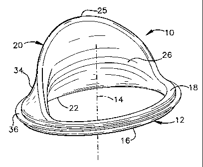
Figures 1-4 illustrate an apparatus 10, in accordance with an aspect of the
present invention, for helping improve operation of a heart valve. The
apparatus
10 includes a generally annular base portion 12, which may be an oval shape,
egg-shaped or another suitable shape dimensioned and configured for
attachment at an annulus of a heart valve. A central axis 14 extends through
the
apparatus 10 substantially transverse to a plane extending through the base
portion 12. The base portion 12 has an inflow side 16 and an outflow side 18.
The base portion 12 may be formed of a generally rigid or flexible
material, such as depending on the desired amount of support for a valve
annulus to which the apparatus 10 is to be mounted. For example, the base
portion 12 may be a plastic-like material, a metal, or other material suitable
for
implantation into a patient. The base portion 12 provides the benefits of an
annuloplasty ring (e.g., it helps support a valve annulus at a desired
orientation
at systole).
The apparatus 10 also includes a buttress 20 that is attached to and
extends from the base portion 12 for providing a surface against which a
leaflet
of a heart valve may engage. The buttress 20 is connected to the base portion
12 along a circumferentially extending arc length of the base portion. The arc
length of the base portion 12 may approximate the length of annular attachment
CA 02409773 2002-11-15
WO 01/89418 PCT/USO1/14620
for a defective or damaged valve leaflet for which the buttress 20 (when the
apparatus is implanted) is intended to function.
By way of example, when the apparatus 10 is to be implanted at the
annulus of a mitral valve and function in place of a posterior leaflet, the
S circumferential arc may approximate the length of the annulus adjacent the
posterior leaflet of the valve. Additionally, the circumferential length of
the
sidewall of the buttress 20 approximates the posterior leaflet.
The buttress 20 extends generally axially from and radially outwardly
relative to the outflow side 18 of the base portion 12. An axial length of a
portion
22 of the buttress 20 proximal the base portion 12 extends radially inwardly
toward the axis 14 and generally axially away from the base portion. A
distally
extending portion 24 of the buttress 20 extends from the proximal portion 22
and
curves radially outwardly therefrom for the remaining length of the buttress
to
terminate in a distal end 25. The buttress 20 has a radially inner surface 26
that
provides a surface against which a leaflet (e.g., an anterior leaflet of a
mitral
valve) may coapt at systole. As shown in Figs. 3 and 4, a radially outer
surface
28 of the buttress 20 at the distally extending portion 24 has a generally
convex
or an inverted C-shaped cross-section.
In the example of the apparatus 10 shown in Figs. 1-4 (having a complete
annular base portion 12), an aperture extends axially through the apparatus 10
between another arc length of the base portion 12 and the buttress itself. The
aperture provides an opening or orifice to permit the passage of blood through
the apparatus 10, such as during diastole. The buttress 20 in conjunction with
the leaflet (or leaflets) also inhibits the flow of blood when the valve is in
a closed
position, such as during ventricular contraction at systole.
The apparatus 10 shown in Figs. 1-4 includes a support frame 32 that is
dimensioned and configured to provide a desired shape for the apparatus 10.
The frame 32 provides a support mechanism that forms the base portion 12 and
the buttress 20. The frame 32, for example, may be formed of a resilient
and/or
flexible material, such as a plastic, metal or other material suitable for
implantation into a human. The rigidity or flexibility of each part of the
frame may
vary depending upon the amount of support desired at the annulus (by the base
CA 02409773 2002-11-15
WO 01/89418 PCT/USO1/14620
portion) as well as the amount of flexibility desired during engagement
between
a leaflet and the buttress 20.
Alternatively, the underlying support frame 32 of the buttress 20 and/or
the base portion 12 may be formed of a substantially inelastically deformable
material (e.g., it is bendable to and remains at a desired position), such as
a
metal wire. As a result, a surgeon implanting the apparatus 10 may reorient
the
buttress 20 and/or the base portion 12 to a desired configuration for
improving
the operation of the valve. Such material also may exhibit sufficient
resilience so
that it maintains the shape set by the surgeon (or manufacturer) after being
implanted and subjected to the dynamics of the heart valve.
The frame parts for the base portion 12 and the buttress 20 may be
formed of the same or different materials depending on the material properties
(elasticity, rigidity, resilience, etc.) desired for each part of the
apparatus 10.
An outer sheath 34 of a biocompatible material covers the frame 32,
including the base portion 12 and the buttress 20. The outer sheath 34 may be
substantially any material, such as a cloth-like or fabric material (natural
or
synthetic), a biological material, such as collagen or an animal tissue
material.
An acceptable animal tissue material is smooth animal pericardium (e.g.,
equine,
bovine, porcine, etc.) that has been tanned or fixed in a suitable tanning
environment. The pericardium, for example, is cross-linked with glutaraldehyde
and undergoes a detoxification process with heparin bonding, such as one of
the
NO-REACTO natural tissue products that are commercially available from
Shelhigh, Inc. of Millburn, New Jersey. The NO-REACT~ natural tissue
products exhibit improved biocompatibility and mitigate calcification and
thrombus formation. The exposed smooth animal pericardium covering the
buttress 20 further inhibits abrasion that could occur in response to
engagement
between a leaflet and the buttress.
The apparatus 10 also may include an implantation flange 36 (or sewing
ring) that circumscribes the base portion of the apparatus 10. The
implantation
flange 36 extends radially outwardly from the base portion 12 and provides a
structure for facilitating implantation of the apparatus 10 at an annulus of a
heart
valve. The implantation flange 36 is formed of a flexible material, such a
cloth-
like or fabric material (natural or synthetic), a biological material, such as
CA 02409773 2002-11-15
WO 01/89418 PCT/USO1/14620
collagen, or an animal tissue material. For example, the implantation flange
36
is formed of a substantially biocompatible biological material, such as animal
tissue (e.g., animal pericardium). The implantation flange 36 may be formed as
an integral part of the outer sheath 34, such as a single or double layer of
the
material that is used to form the outer sheath.
Figs. 5 and 6 illustrate a heart valve repair apparatus 150 in accordance
with another aspect of the present invention. The apparatus 150 includes a
generally annular base portion 152 that is generally C-shaped (or incomplete).
The base portion 152 has ends 156 and 158 that are spaced apart from each
other and a curved portion extending between the ends. In this example, the
base portion 152 includes an underlying C-shaped support ring, which may be
formed of a flexible, resilient, or generally rigid material. The support ring
may
have an elastic property so as to return to its original shape when deflected
from
its original (or rest) condition. The support ring for example, may be a
plastic-like
material (e.g., a polymer, a resin, etc.) or a metal (e.g., stainless steel),
such as
in the form of a wire. It will be understood and appreciated that other types
of
generally rigid, elastic, and/or resilient materials also may be used in
accordance
with the present invention. In addition, a suitable inelastically deformable
material also could be used to form the support ring.
A buttress 164 extends generally axially from an outflow side 166 of the
base portion 152 in a manner that is substantially similar to that shown and
described with respect to Figs. 1-4. Briefly stated, a proximal portion 168 of
the
buttress 164 extends generally axially and radially inward from the base
portion
152 toward an open end (between ends 156 and 158) of the base portion. A
distally extending portion 170 of the buttress 164 extends from the proximal
portion 168 and curves radially outwardly therefrom for the remaining length
of
the buttress. The buttress 164 has a radially inner surface 172 that provides
a
surface against which a leaflet (e.g., an anterior leaflet of a mitral valve)
may
coapt at systole. The buttress 164 is dimensioned and configured to simulate
the dimensions and configuration of a leaflet at systole so that, when the
apparatus 150 is implanted at an annulus of a heart valve, a leaflet (or
leaflets)
may engage the buttress 164 to close the valve at systole. The leaflet (or
CA 02409773 2002-11-15
WO 01/89418 PCT/USO1/14620
leaflets) is able to coapt with the inner surface 172 of the buttress 164 at
systole,
thereby inhibiting regurgitation of blood when the ventricle contracts.
As in the example of Figs. 1-4, the apparatus 150 also includes an outer
sheath 174 of a flexible, biocompatible material covering the apparatus. The
apparatus 150 also may include an implantation flange 176 (or sewing ring)
that
circumscribes the base portion 152 of the apparatus. The implantation flange
176 extends radially outwardly from the base portion 12 between the ends 156
and 158 for facilitating implantation of the apparatus 150 at an annulus of a
heart
valve. Each of the outer sheath 174 and the implantation flange 176 may be
formed of any suitable flexible, biocompatible material, such as a cloth-like
or
fabric (natural or synthetic) material, a biological material, such as
collagen or an
animal tissue material. An acceptable animal tissue material is smooth animal
pericardium (e.g., equine, bovine, porcine, etc.), such as a NO-REACT~ tissue
product.
Figures 7 and 8 illustrate a heart valve repair apparatus 200 in
accordance with another aspect of the present invention. The apparatus 200
includes a generally annular base portion 202. The base portion 202 includes a
support ring 203 that is dimensioned and configured to approximate the
dimensions and configuration of a heart valve annulus, such as a mitral or
atrioventricular valve. The support ring 203 may be substantially similar to
that
disclosed with respect to the base portions shown and described with respect
to
Figs. 1-6 (e.g., it may be a complete ring (as shown) or a generally C-shaped
ring).
A pair of support posts 204 and 206 extend generally axially from an
outflow side 208 of the base portion 202. The supports 204 and 206 are
circumferentially spaced apart from each other an arc length that approximates
the circumferential dimension of a valve leaflet for which the apparatus 200
is
intended to function. The support posts 204 and 206 may be formed of the
same material or a different material as that which forms the base portion
202.
For example, the support posts 204 and 206 and the base portion 202 may be
formed as an integral unit in a suitable injection molding process. It is to
be
appreciated, however, that different materials also maybe utilized to form the
supports 204 and 206 and the base portion 202, with the supports being
CA 02409773 2002-11-15
WO 01/89418 PCT/USO1/14620
appropriately secured to the base portion, such as by ultrasonic welding or
another method of attachment.
The apparatus 200 also includes a buttress 210 of a substantially flexible
material that extends generally axially from the base portion 202 for
providing a
flexible surface for abutment with an adjacent leaflet of a heart valve. The
buttress 210, for example, includes a flexible sheet 212 of material that is
attached to the base portion 202 along a circumferentially extending arc 214
between the juncture of each of the support posts 204 and 206 and the base
portion. The flexible sheet 212 of material extends generally axially from the
base portion 202 and is connected to and extends between the support posts
204 and 206. The support posts 204 and 206 may be linear or curved to orient
the sheath of flexible material connected therebetween at a desired position
for
engaging an adjacent leaflet. The sheet 212 of flexible material also may
cover
each of the support posts 204 and 206 as well as the annular base portion 202
so as to completely cover the frame, which is formed of the base portion and
support posts. The sheet 212 of flexible material of the buttress 210 provides
a
radially inner surface 216 with which an adjacent leaflet may move into and
out
of engagement when the apparatus 200 is implanted. The flexible sheath 212 of
material also may permit flexible movement of the buttress 210 relative to the
supports 204 and 206, such that when the apparatus is implanted it facilitates
coaptation between an adjacent leaflet (or leaflets) and the buttress.
As mentioned above with respect to the apparatus of Figs. 1-4, the posts
204 and 206 and/or the base portion 202 may be formed of an inelastically
deformable material. A surgeon implanting the apparatus 200, thus, may bend
the buttress 210 and/or base portion 202 to a desired configuration. As a
result,
each apparatus may be customized for a patient so as to improve the operation
of a heart valve when the apparatus 200 is implanted at the valve annulus.
The sheet 212 of flexible material, for example, may be a cloth or fabric
material (natural or synthetic), a biological material, such as a sheet of
collagen
material or an animal tissue material, such as animal pericardium. In order to
inhibit regurgitation of blood when implanted at a heart valve, the flexible
sheath
212 of material should be substantially impervious to the flow of blood
therethrough.
CA 02409773 2002-11-15
WO 01/89418 PCT/USO1/14620
As illustrated in Fig. 7 and 8, the apparatus also may include an
implantation flange (or sewing ring) 220 for facilitating implantation of the
apparatus at an annulus of a heart valve. The implantation flange 220 extends
radially outwardly from the base portion 202. The implantation flange 220 is
formed of a flexible material, such a cloth-like or fabric material (natural
or
synthetic), or a biological material, such as collagen or an animal tissue
material.
For example, the implantation flange 220 is formed of a biocompatible
biological
material, such as animal tissue (e.g., animal pericardium), which is the same
material that forms the outer sheath 212.
Fig. 9 illustrates a frame 250 that may be employed to form an apparatus
for helping repair a heart valve in accordance with another aspect of the
present
invention. For example, the frame 250 may be used to form an apparatus of a
type similar to that shown and described with respect to Figs. 1-4. The frame
250 provides a skeleton over which an outer sheath of a substantially flexible
material may be applied.
The frame 250 includes a generally annular base portion 252. While the
base portion 252 is illustrated as a complete ring, it will be understood and
appreciated by those skilled in the art that an incomplete ring (e.g., a C-
shaped
ring) alternatively may be utilized in accordance with an aspect of the
present
invention. The base portion 252 includes an inflow side 254, and outflow side
256, with a central axis 258 extending through the base portion.
The frame 250 also includes a support 260 extending generally axially
from the base portion 252. The axially extending support 260 is in the form of
a
curved structure that connects substantially opposed edges 262 and 264 of the
base portion 252 for providing a support structure for a buttress.
The frame 250, for example, may be formed of a resilient material, a
flexible material, or an inelastically deformable material, such as a plastic,
a
metal, or other material suitable for implantation into a human. The rigidity
or
flexibility of a material utilized to form the frame 250 may vary depending
upon
the amount of support desired at the annulus (by the base portion) as well as
the
amount of flexibility desired during coaptation between a leaflet and the
buttress.
The base portion 252 and the axially extending support 260 may be formed of
CA 02409773 2002-11-15
WO 01/89418 PCT/USO1/14620
the same or different materials, depending on the material properties
(elasticity,
rigidity, resilience, etc.) desired for each part of the frame 250.
The frame 250 may be covered with a sheet of a substantially flexible
material to form an apparatus, similar to that shown and described with
respect
to Figs. 1-4. A sheet of flexible material is applied over the frame so that
the
flexible material may be moveable relative to the axially extending support
260,
such as in response to an adjacent leaflet moving into engagement with the
overlying sheet of material. In contrast, the illustrated apparatus of Figs. 1-
4
employs a frame that includes a substrate material coextensive with the
buttress
onto which the sheet of flexible material is applied (e.g., the buttress of
Figs. 1-4
may be more static than the flexible buttress of Fig. 9).
Figure 10 illustrates a heart valve repair apparatus 280 in accordance with
another aspect of the present invention. Similar to the apparatus 10 of Fig.
1,
the apparatus 280 includes a generally annular base portion 282 that is
dimensioned and configured according to the dimensions and configuration of a
heart valve annulus to which the apparatus is to be attached. As mentioned
above with respect to Figs. 1-4, the flexibility or resilience or rigidity of
the base
portion 282 may vary according to the material used to form the base portion,
such as to provide a desired amount of support at the heart valve annulus.
A buttress 284 is attached to and extends radially inwardly and generally
axially away from a posterior arc 286 of the base portion 282. More
specifically,
a proximal portion 288 of the buttress 284 extends axially and radially
inwardly
over a first portion of its length. A remaining portion 290 of the buttress
284
extends distally from the proximal portion 288 and curves radially outwardly
relative to (or away from) the proximal portion. When the apparatus 280 is
implanted, the buttress 284 provides a surface with which an adjacent leaflet
may move into and out of engagement.
The apparatus 280 may be formed of a flexible and/or resilient material,
such as a polymer or plastic-like material (e.g., DeIrinO, pyrolythic carbon,
etc.),
a metal, or other material considered appropriate for implantation into a
heart.
The base portion 282 and the buttress 284, for example, may be formed of the
same material to form an integral apparatus. Alternatively, different
materials
may be utilized to form each of the buttress 284 and the base portion 282,
such
CA 02409773 2002-11-15
WO 01/89418 PCT/USO1/14620
as when different amounts of rigidity or flexibility may be desired for each
respective part.
It will be understood and appreciated that the apparatus 280 further may
be employed as the underlying frame 32 of the apparatus 10, as shown and
described with respect to Figs. 1-4. In order to form the apparatus 10 from
the
apparatus 280, an outer sheath of an appropriate flexible, biocompatible
material
is mounted over the apparatus 280, such as set forth above.
Figs. 11 and 12 illustrate part of a heart 300 in which an apparatus, such
as the apparatus 10 illustrated with respect to Figs. 1-4, is implanted at an
annulus 302 of a mitral valve 303. The mitral valve 303 is intended to provide
for
the unidirectional flow of blood from the left atrium 304 into the left
ventricle 306.
The mitral valve 303 includes an anterior leaflet 308 that extends from the
annulus 300 adjacent the aortic opening 310 and attaches to the muscular
tissue
in the wall of the left ventricle by fibrous cordae tendinae 312. The
posterior
leaflet has been substantially removed from the heart, such as by excising it
prior
to implantation of the apparatus 10. It is to be understood and appreciated,
however, that the posterior leaflet may remain intact, with a buttress 20 of
the
apparatus 10 interposed between the posterior and anterior leaflets.
As mentioned above, the apparatus 10 may include an implantation
flange 36 that is sutured to the fibrous tissue at the annulus 302 of the
valve 303.
The buttress 20 extends from the base 12 of the apparatus 10 into the
ventricle
306 at a position corresponding to the position of the posterior leaflet of
the
mitral valve 303. As mentioned above, the buttress 20 extends into the
ventricle
306 generally toward the posterior leaflet 308.
It is to be appreciated that the buttress 20 may be formed of a generally
rigid material that remains substantially stationary (e.g., static) during
both
systole and diastole. Alternatively, the buttress 20 may sufficiently flexible
material, such as a sheet of material supported in a peripheral frame (see,
e.g.,
Figs. 7-9) or by employing a more flexible type of frame to permit movement
thereof commensurate with the flow of blood from the atrium 304 into the
ventricle 306 through the valve 303.
Fig. 11 illustrates the mitral valve 303 is in a closed position (at systole),
in
which the anterior leaflet 304 engages the buttress 20 of the apparatus 10 in
CA 02409773 2002-11-15
WO 01/89418 PCT/USO1/14620
accordance with an aspect of the present invention. That is, the buttress 20
of
the apparatus 10 simulates the function of the posterior leaflet at systole by
providing a surface against which the anterior leaflet 308 coapts. As a
result, the
buttress 20 and the anterior leaflet 304 cooperate to inhibit regurgitation of
blood
from the left ventricle 308 into the left atrium 306, such as during
ventricular
contraction at systole.
The buttress 20 in conjunction with the anterior leaflet also facilitates and
promotes unidirectional flow of blood at diastole, such as shown in Fig. 11 by
arrow 314. In particular, an opening or aperture extends through the implanted
apparatus 10 between the buttress 20 and the anterior leaflet 308.
Advantageously, the movement of the anterior leaflet 308 relative to the
buttress
20, in response to the flow of blood during diastole, provides a sufficient
orifice to
permit the free flow the flow of blood from the left atrium 304 into the left
ventricle
306. The buttress 20 also may be formed of a flexible material that is able to
move radially relative to the base portion 12 to further facilitate blood
flow. The
annular base portion 12 of the apparatus 10 also may help support the annulus
302 of the mitral valve 303 at systole to promote the desired coaptation
between
the buttress 20 and the anterior leaflet 308 (Fig. 10).
In view of the foregoing, an apparatus according to the present invention
provides a useful repair apparatus for helping to improve operation of a heart
valve. The apparatus may be employed to both support a heart valve annulus
and mitigate problems associated with coaptation and/or lesions in a leaflet
by
providing a buttress with which one or more leaflets may move into and out of
engagement. The apparatus further provides a simplified repair option
(implanting a device at an annulus of a heart valve) when compared with other,
more conventional methods of reconstruction and repair.
It is to be appreciated by those skilled in the art that, while the
illustrated
examples show the apparatus for treating a bicuspid (mitral) valve, an
apparatus
in accordance with the present invention also may be used for repairing other
types of heart valves (e.g., a tricuspid valve or other bicuspid valves). In
addition, an apparatus may in accordance with the present invention, be
implanted at either the inflow side or outflow side of a heart valve annulus.
CA 02409773 2002-11-15
WO 01/89418 PCT/USO1/14620
What has been described above are examples of the present invention. It
is, of course, not possible to describe every conceivable combination of
components or methodologies for purposes of describing the present invention,
but one of ordinary skill in the art will recognize that many further
combinations
and permutations of the present invention are possible. Accordingly, the
present invention is intended to embrace all such alterations, modifications
and
variations that fall within the spirit and scope of the appended claims.
Furthermore, to the extent that the term "includes" is used in either the
detailed
description or the claims, such term is intended to be inclusive in a manner
similar to the term "comprising."