Note: Descriptions are shown in the official language in which they were submitted.
CA 02410365 2002-11-25
WO 01/89360 PCT/ILO1/00485
1
FINGER-GUIDED SUTURE DEVICE
FIELD AND BACKGROUND OF THE INVENTION
The present invention relates to suture devices, and more particularly, to
s a finger-guided suture device for placing sutures especially in body
locations of
limited minimal-invasive accessibility. The present invention further relates
to
surgical procedures in which one or more finger-guided suture devices of the
present invention are used to place one or more sutures, especially in body
locations of limited minimal-invasive accessibility.
to For years, there has been a discernible, clear tendency in surgery and
invasive diagnosis, especially, but not exclusively, in abdominal, joint,
vaginal,
in-utero and brain, surgeries or diagnoses, to develop procedures that would
reduce the need for major access-providing incisions with their concomitant
requirements of general anesthesia, extended hospitalization and increased
Is infection hazard. One step in this direction was the introduction of
endoscopy
and laparoscopy, which, through provision of minimal incision in, e.g., the
abdominal wall of joint covering skin, permits the introduction into the
abdominal cavity or joint of a miniature television camera including a light
source, as well as of various surgical instruments, including suture devices.
2o PCT/US97/11494 teaches a number of surgical instruments which can
be mounted directly on a surgeon's fingertip in a way that the surgeon can
insert his or her hand into a natural cavity of the patient or through a
minimal
incision to perform surgical procedures, and also to use his or her fingers to
manipulate tissues, thus enabling the surgeon to perform the procedures with
2s the benefits of minimally invasive surgery, but with much greater tactile
sense,
control, and ease of manipulation. However, these surgical instruments (i) are
carried by a finger and operated by the thumb, thereby are not applicable for
procedures in which a single finger is employed for tactile sensing an
operated
intrabody location; (ii) include an operating head which permanently extends
3o far beyond the fingertip on which the surgical instrument is mounted, which
CA 02410365 2002-11-25
WO 01/89360 PCT/ILO1/00485
2
limits the tactile sensing of the surgeon; and/or (iii) prevent tactile
sensing by
the instrument carrying fingertip altogether.
According to the teachings of PCT/US97/11494 suturing can be
performed while the surgeon uses tactile information collected by a single
s fingertip for tactile sensing the intrabody site to be stitched prior to the
actual
suturing. Several non-limiting examples of such suturing procedures are
described in detail in the sections that follow. However, once the surgeon has
collected the tactile information, surgery is conducted blindly within the
body
of the patient. According to the teachings of PCT/US97/11494, both the hands
to of the surgeon are engaged. Evidently, blindly operating surgical
instruments
intrabodily based on finger tip tactile information collected earlier may
prove
inconvenient, inaccurate and may increase the chance of inadvertently harming
the patient.
PCT/IL99/00084 teaches design of finger-guided suture devices which
1 s can be used to perform extra- as well as intrabody suturing of tissue.
This
application does not teach construction and use of a device which causes a
needle to penetrate a bodily tissue, engage a piece of suture material and
pull
the suture material through the bodily tissue as the needle is retracted there
through. In addition, this application fails to teach a construction which
allows
2o safe withdrawal of the needle in case of early detected misplacement other
than
pulling it via the hand or suture.
There is thus a widely recognized need for, and it would be highly
advantageous to have, finger-guided suture devices devoid of the limitations
associated with the prior art instruments and which enable a surgeon using a
2s finger-guided surgical device to penetrate a bodily tissue with a suture
needle,
engage a piece of suture material with the needle and pull the suture material
through the bodily tissue as the needle is retracted there through.
CA 02410365 2002-11-25
WO 01/89360 PCT/ILO1/00485
3
SLIlyIMARY OF THE INVENTION
According to one aspect of the present invention there is provided a
finger-guided suture device. The device comprises (a) a thimble-like element
being adapted to surround a portion of a surgeon's finger; (b) an ejectable
s substantially semi-circular surgical needle being engaged within a housing
being formed within, or connected to, a wall of the thimble-like element, the
surgical needle being designed for collecting the surgical suture via a distal
portion of the surgical needle upon contact therewith and for retaining and
guiding the surgical suture while suturing; and (c) a mechanism for ejecting
the
to surgical needle from the thimble-like element and thereafter withdrawing
the
surgical needle into the thimble-like element, so as to place a suture.
According to another aspect of the present invention there is provided a
surgical procedure for bladder-neck suspension for treatment of urinary
incontinence, the procedure comprising the step of suspending a pelvic fascia
1s and a vaginal wall lateral to a urethra of a patient to Cooper's ligament
by
sutures being placed using a finger-guided suture device having an ejectable
substantially semi-circular surgical needle being designed for collecting the
surgical suture via a distal portion of the surgical needle upon contact
therewith
and for retaining and guiding the surgical suture while suturing
2o According to yet another aspect of the present invention there is
provided a surgical procedure for treatment of rectal prolapse, the procedure
comprising the step of constricting an anal opening by sutures being applied
by
using a finger-guided suture device having an ejectable, substantially semi-
circular surgical needle being designed for collecting the surgical suture via
a
2s distal portion of the surgical needle upon contact therewith and for
retaining
and guiding the surgical suture while suturing.
According to still another aspect of the present invention there is
provided a surgical procedure for treatment of esophageal reflux, the
procedure
comprising the step of positioning a vessel loop around a esophagus of a
3o patient using a finger-guided suture device having an ejectable,
substantially
CA 02410365 2002-11-25
WO 01/89360 PCT/ILO1/00485
4
semi-circular surgical needle being designed for collecting the surgical
suture
via a distal portion of the surgical needle upon contact therewith and for
retaining and guiding the surgical suture while suturing.
According to an additional aspect of the present invention there is
s provided a surgical procedure for treatment of vaginal prolapse, the
procedure
comprising the step of tying an upper part of a vagina of a patient to a
sacrospinous ligament of the patient by sutures being placed using a finger-
guided suture device having an ejectable, substantially semi-circular surgical
needle being designed for collecting the surgical suture via a distal portion
of
to the surgical needle upon contact therewith and for retaining and guiding
the
surgical suture while suturing.
According to yet additional aspect of the present invention there is
provided a surgical procedure for treatment of rupture of a rectum in large
animals, the procedure comprising the step of suturing the rupture using a
is finger-guided suture device having an ejectable, substantially semi-
circular
surgical needle being designed for collecting the surgical suture via a distal
portion of the surgical needle upon contact therewith and for retaining and
guiding the surgical suture while suturing.
According to still additional aspect of the present invention there is
2o provided a surgical procedure for treatment of rupture of a cervix in large
animals, the procedure comprising the step of suturing the rupture using a
finger-guided suture device having an ejectable, substantially semi-circular
surgical needle being designed for collecting the surgical suture via a distal
portion of the surgical needle upon contact therewith and for retaining and
2s guiding the surgical suture while suturing.
According to still additional aspect of the present invention there is
provided a surgical procedure for treatment of rupture of a uterus in large
animals, the procedure comprising the step of suturing the rupture using a
finger-guided suture device having an ejectable, substantially semi-circular
3o surgical needle being designed for collecting the surgical suture via a
distal
CA 02410365 2002-11-25
WO 01/89360 PCT/ILO1/00485
portion of the surgical needle upon contact therewith and for retaining and
guiding the surgical suture while suturing.
According to further features in preferred embodiments of the invention
described below, the thimble like element is constructed and constructed to
s expose the ventral tactile portions of the distal phalanx of the surgeon's
finger,
so as to enable the surgeon to tactile sense a body location to be sutured.
According to still further features in the described preferred
embodiments, the finger-guided suture device further comprises a cartridge for
holding the surgical suture and presenting it for collection by the distal
portion
to of the surgical needle.
According to still further features in the described preferred
embodiments the cartridge includes at least one mechanism designed and
constructed, so as to maintain a predetermined tension of the surgical suture.
According to still further features in the described preferred
1s embodiments the at least one mechanism designed and constructed, so as to
maintain a predetermined tension of the surgical suture comprises at least one
piece of flexible material containing at least one hole through which the
surgical suture passes.
According to still further features in the described preferred
2o embodiments the at least one piece of flexible material containing at least
one
hole is selected from the group consisting of a single piece of flexible
material
containing two holes and a pair of pieces of flexible material each containing
one hole.
According to still further features in the described preferred
2s embodiments the flexible material is selected from the group consisting of
silicon, latex, rubber, fabric, and fabric with an eyelet.
According to still further features in the described preferred
embodiments the eyelet is constructed of material selected from the group
consisting of silicon, latex, rubber and fabric.
CA 02410365 2002-11-25
WO 01/89360 PCT/ILO1/00485
6
According to still further features in the described preferred
embodiments the mechanism for ejecting the surgical needle from, and
withdrawing the surgical needle into, the thimble-like element, is selected
from
the group consisting of a belt actuated mechanism, a gear actuated mechanism
s and a combined gear and belt actuated mechanism.
According to still further features in the described preferred
embodiments the surgical needle is formed with a feature selected from the
group consisting of a notch, a hook, at least one arm, and an openable loop at
the distal end thereof.
t o According to still further features in the described preferred
embodiments the finger-guided suture device further comprises an adapter
insertable between the thimble-like element and the surgeon's finger, so as to
adapt the suture device to fingers of different size.
According to still further features in the described preferred
1 s embodiments the mechanism includes a first portion engaged within the
housing and which is in contact with the ejectable surgical needle and a
second,
remote, portion extending out of the patient's body and which is operable by a
free hand of the surgeon so as to eject the surgical needle from the thimble-
like
element.
2o According to still further features in the described . preferred
embodiments the first portion of the mechanism comprises_a rotatable wheel
having an axle, the axle serves for engaging the surgical needle and imparting
a
rotational motion in at least one direction thereto, the surgical needle
includes a
mechanism for engaging the rotatable wheel and a locking piece for insuring
2s that the surgical needle and the rotatable wheel remain engaged.
According to still further features in the described preferred
embodiments the first portion of the mechanism comprises a rotatable wheel
having a mechanism for engaging a drive arm and imparting a rotational
motion in at least one direction thereto, the drive arm is designed and
3o constructed engageable by the rotatable wheel and by the surgical needle
and to
CA 02410365 2002-11-25
WO 01/89360 PCT/ILO1/00485
7
impart a rotational motion of the rotatable wheel in at least one direction to
the
surgical needle, wherein the surgical needle further includes a mechanism for
engaging the drive arm and a disk for ensuring that the surgical needle, the
drive arm and the rotatable wheel remain engaged.
s According to still further features in the described preferred
embodiments the remote portion extending out of the patient's body and which
is operable by a free hand of the surgeon so as to eject the surgical needle
from
the thimble-like element comprises: (i) a hand operable actuator designed and
constructed to operate a drive mechanism; (ii) a drive housing for containing
at
io least a portion of the drive mechanism; and (iii) at least a portion of the
drive
mechanism, the drive mechanism being for imparting a rotational motion in at
least one direction to the surgical needle.
According to still further features in the described preferred
embodiments the hand operable actuator of the remote portion comprises (1) a
is handle for engaging at least one finger of the free hand of the surgeon;
(2) an
extending piece containing a plurality of arcurate teeth and being movable
through the drive housing; (3) a pressure sensitive spring; and (4) a brake
handle, the brake handle operable in a first direction by the pressure
sensitive
spring and in a second direction by the at least one finger of the free hand
of
2o the surgeon.
According to still further features in the described preferred
embodiments the drive mechanism comprises (1) a plurality of arcuate teeth
deployed in a linear arrangement along an extending piece o.f a handle; (2) a
first gear with a first circular arrangement of arcuate teeth, the first
circular
2s arrangement of arcuate teeth being for engaging with the plurality of
arcuate
teeth deployed in the linear arrangement along the extending piece, such that
linear displacement of the extending piece is translated into rotational
motion
of the first gear; (3) a second gear including a second circular arrangement
of
arcuate teeth, the arcuate teeth of the second gear being for engaging the
first
3o circular arrangement of arcuate teeth of the first gear, such that
rotational
CA 02410365 2002-11-25
WO 01/89360 PCT/ILO1/00485
8
motion of the first gear causes rotational motion of the second gear; and (4)
a
cable in contact with at least one point on the second gear, such that
rotational
motion of the second gear is translated to linear motion of at least a portion
of
the cable.
s According to still further features in the described preferred
embodiments the drive mechanism further comprises (5) a ratchet for
alternately engaging and releasing at least one arcuate tooth of the first
gear;
(6) a ratchet control arm for alternately engaging and releasing the ratchet;
(7) a
brake handle for alternately operating the ratchet control arm. These
~ o components are arranged so that when the brake handle operates the ratchet
control arm, the ratchet control arm releases the ratchet, the ratchet engages
the
at least one arcuate tooth of the first gear and the first gear is prevented
from
rotating. This means that when the brake handle does not operate the ratchet
control arm, the ratchet control arm engages the ratchet, the ratchet releases
the
1 s at least one arcuate tooth of the first gear and the first gear is free to
rotate.
According to still further features in the described preferred
embodiments the thimble-like element is constructed so as to be mounted over
a dorsal side of the distal phalanx of the surgeon's finger, thereby exposing
the
entire ventral tactile portions of the distal phalanx.
20 According to still further features in the described preferred
embodiments the thimble-like element is constructed so as to_fully surround
the
distal phalanx and expose the tip of the ventral tactile portion of the distal
phalanx.
According to still further features in the described preferred
2s embodiments the thimble-like element is constructed so as to be mounted
over
a ventral side of the distal phalanx of the surgeon's finger and expose the
tip of
the ventral tactile portion of the distal phalanx.
According to still further features in the described preferred
embodiments the surgical needle is ejectable in a direction generally
3o perpendicular to a longitudinal axis of the thimble like element.
CA 02410365 2002-11-25
WO 01/89360 PCT/ILO1/00485
9
According to still further features in the described preferred
embodiments the surgical needle travels along at least a portion of a circular
path, the path being on a plane which substantially parallels a plane
traversing
the surgeon's finger from top to bottom.
s According to still further features in the described preferred
embodiments the surgical needle travels along at least a portion of a circular
path, the path being on a plane which substantially parallels a plane
traversing
the surgeon's finger from side to side.
According to still further features in the described preferred
~o embodiments the surgical needle travels along at least a portion of a
circular
path, the path being on a plane which is substantially perpendicular to the
longitudinal axis of the surgeon's finger.
According to still further features in the described preferred
embodiments the finger-guided suture device further comprises an optical head
t s engaged by the thimble like element.
According to still further features in the described preferred
embodiments the finger-guided suture device further comprises the surgical
suture formed with a loop for collection by the surgical needle.
According to still further features in the described preferred
2o embodiments the wall is a side wall of the thimble-like element.
According to still further features in the described preferred
embodiments the wall is a front wall of the thimble-like element.
According to still further features in the described preferred
embodiments the finger-guided suture device further comprises a reporting
2s mechanism for reporting at least one situation selected from the group
consisting of a full ejection of the substantially semi-circular surgical
needle, a
full withdrawal of the substantially semi-circular surgical needle, a degree
of
ejection of the substantially semi-circular surgical needle and a degree of
withdrawal of the substantially semi-circular surgical needle.
CA 02410365 2002-11-25
WO 01/89360 PCT/ILO1/00485
The present invention successfully addresses the shortcomings of the
presently known configurations by providing a finger guided suture including a
needle which is capable of collecting the surgical suture via a distal portion
of
the surgical needle upon contact therewith and retaining and guiding the
s surgical suture while suturing.
BRIEF DESCRIPTION OF THE DRAWINGS
The invention is herein described, by way of example only, with
reference to the accompanying drawings. With specific reference now to the
to drawings in detail, it is stressed that the particulars shown are by way of
example and for purposes of illustrative discussion of the preferred
embodiments of the present invention only, and are presented in the cause of
providing what is believed to be the most useful and readily understood
description of the principles and conceptual aspects of the invention. In this
1 s regard, no attempt is made to show structural details of the invention in
more
detail than is necessary for a fundamental understanding of the invention, the
description taken with the drawings making apparent to those skilled in the
art
how the several forms of the invention may be embodied in practice.
In the drawings:
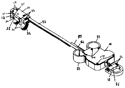
2o FIG. 1 is a perspective view of one embodiment of a finger-guided
suture device according to the present invention including an external
actuation
device;
FIG. 2 is a perspective view of a second embodiment of a finger-guided
suture device according to the present invention including an external
actuation
2s device;
FIG. 3 depicts the assembly of a surgical needle and a drive wheel
according to the embodiment of the finger-guided suture device depicted in
Figure 1;
FIG. 4 is an exploded view of the finger-guided suture device according
3o to the embodiment depicted in Figure 1;
CA 02410365 2002-11-25
WO 01/89360 PCT/ILO1/00485
11
FIG. 5 depicts a cartridge, with cover removed, of the finger-guided
suture device according to the embodiment depicted in Figure l;
FIG. 6 shows the assembly of the cartridge to a thimble like element
according to the embodiment of the finger-guided suture device depicted in
s Figure 1;
FIG. 7 shows how a surgical suture may be engaged by a surgical needle
according to the embodiment of the finger-guided suture device depicted in
Figure 1;
FIG. 8 is a perspective view of the finger-guided suture device
to according to the embodiment depicted in Figure 1 in which engagement of a
suture by the needle is depicted ;
FIG. 9 is an underside perspective view of the finger-guided suture
device according to the embodiment depicted in Figure 1;
FIG. 10 is a top view of a finger-guided suture device according to the
is embodiment depicted in Figure l;
FIG. 11 is perspective view of the finger-guided suture device according
to the embodiment depicted in Figure 2 in which the assembly of the surgical
needle is shown;
FIG. 12 is a an exploded view of a drive arm of the finger-guided suture
2o device according to the embodiment depicted in Figure 2;
FIG. 13 is an exploded view detailing assembly of pulleys of a finger-
guided suture device according to the embodiment depicted in Figure 2;
FIG. 14 is an exploded view of a finger-guided suture device according
to the embodiment depicted in Figure 2 showing assembly of the drive arm and
2s drive wheel;
FIG. 15 depicts the assembly of the upper and lower portions of the
finger-guided suture device according to the embodiment depicted in Figure 2;
FIG. 16 is an exploded view of a cartridge of the finger-guided suture
device according to the embodiment depicted in Figure 2;
CA 02410365 2002-11-25
WO 01/89360 PCT/ILO1/00485
12
FIG. 17 depicts the assembly of the cartridge of the type depicted in
Figure 16 into the finger-guided suture device according to the embodiment
depicted in Figure 2;
FIG. 18 is a perspective view of the finger-guided suture device
s according to the embodiment depicted in Figure 2 in which engagement of the
suture by the needle is depicted;
FIG. 19 is a cutaway view of the finger-guided suture device according
to the embodiment depicted in Figure 2 in which engagement of the suture by
the needle is depicted;
to FIG. 20 is an exploded view of a portion of the finger-guided suture
device according to the embodiment depicted in Figure 2 in which the relative
placements of the drive arm, needle and drive wheel are illustrated;
FIG. 21 is a perspective view of a handle of one embodiment of an
external actuation device according to the present invention as pictured in
is Figures 1 and 2;
FIG. 22 is an exploded view of a portion of the drive mechanism of one
embodiment of an external actuation device according to the present invention
as pictured in Figures 1 and 2;
FIG. 23 is an exploded view of the drive housing of the drive
2o mechanism of one embodiment of an external actuation device according to
the
present invention as pictured in Figures 1 and 2, showing the assembly of a
handle and a first drive wheel therein;
FIG. 24 is an exploded view of the drive housing of the drive
mechanism of one embodiment of an external actuation device according to the
2s present invention as pictured in Figures 1 and 2, showing the assembly of a
lockable ratchet and a ratchet locking arm therein;
FIG. 25 depicts the assembly of the portion of the drive mechanism
shown in Figure 22 into the drive housing of Figures 23 and 24 ;
FIG. 26 depicts positioning of a cover on the drive housing of Figures
30 23 and 24 and 25;
CA 02410365 2002-11-25
WO 01/89360 PCT/ILO1/00485
13
FIG. 27 shows the covered drive housing of Figure 26 with the handle
of Figure 21 protruding;
FIG. 28 is a cutaway top view of the assembled drive housing of Figures
25, 26 and 27 showing engagement of the ratchet locking arm with the locking
s ratchet;
FIG. 29 is a cutaway top view of the assembled drive housing of Figures
25, 26 and 27 and 28 showing dis-engagement of the ratchet locking arm from
the locking ratchet.
FIG 30 is a cutaway top view of the assembled drive housing of Figures
to 25, 26 and 27 and 28 and 29 showing partial dis-engagement of the ratchet
locking arm from the locking ratchet.
FIGS. 31a-c show a hook, at least one arm, and an openable loop at a
distal end of the surgical needle.
FIGS. 32a-b are of adapters for use with a thimble like element
~ s according to the present invention.
FIG. 33 is a schematic representation of the vagina and the urethra after
a surgical procedure is completed using a device according to the present
invention. (v is vagina, s is suture, U is Urethra and CL is Cooper's
Ligament);
FIGS. 34a-b show a finger guided suture device according to the present
2o invention equipped with an optical head.
DESCRIPTION OF THE PREFERRED EMBODIMENTS
The present invention is of a finger-guided suture device which can be
used to place sutures, especially in body locations of limited minimal-
invasive
2s accessibility and further to surgical procedures employing the device.
Specifically, the present invention can be used to allow a surgeon while
tactilely sensing an intrabodily location to collect surgical suture via a
distal
portion of a surgical needle upon contact therewith and retain and guide the
surgical suture while suturing.
CA 02410365 2002-11-25
WO 01/89360 PCT/ILO1/00485
14
The principles and operation of a finger guided suture device according
to the present invention may be better understood with reference to the
drawings and accompanying descriptions.
Before explaining at least one embodiment of the invention in detail, it
s is to be understood that the invention is not limited in its application to
the
details of construction and the arrangement of the components set forth in the
following description or illustrated in the drawings. The invention is capable
of other embodiments or of being practiced or carried out in various ways.
Also, it is to be understood that the phraseology and terminology employed
to herein is for the purpose of description and should not be regarded as
limiting.
In the drawings two embodiments of a finger-guided suture device,
which is referred to herein below as device 20, are pictured (Figures 1 and
2).
The preferred embodiments of the device depicted in Figure 1 are further
detailed in Figures 3-10, while the preferred embodiments of the device
15 depicted in Figure 2 are further detailed in Figures 11-20.
Device 20 includes a thimble like element 22, a surgical needle 24, and
a mechanism 30 for driving needle 24 in order to form a suture. Mechanism 30
is divided into a first portion 54 (Figures 3 and 12) and a second remote
portion
56 in the two pictured embodiments of device 20. Remote portion 56 of drive
2o mechanism 30 is detailed in Figures 21-30. The two portions 54 and 56 of
mechanism 30 are connected by a pipe or tube 53 containing a cable 100
(Figures 4 and 15) which serves to drive needle 24.
Thimble-like element 22 (Figures 8, 9, 10, 17, 18, and 19) is adapted to
surround a portion of a surgeon's finger. Thimble-like element 22 is
2s constructed to expose the ventral tactile portions of the distal phalanx of
the
surgeon's finger, so as to enable the surgeon to tactile sense a body location
to
be sutured (Figures 4, 6, 8, 9, 15, 17 and 18). In the preferred embodiments
of
Figures 1 l, 15, 17, 18 and 19 thimble-like element 22 is constructed so as to
be
mounted over a dorsal side of the distal phalanx of the surgeon's finger,
thereby
3o exposing the entire ventral tactile portions of the distal phalanx. In the
CA 02410365 2002-11-25
WO 01/89360 PCT/ILO1/00485
preferred embodiment pictured in Figures 4, 8, and 9, thimble-like element 22
is constructed so as to fully surround the distal phalanx and expose the tip
of
the ventral tactile portion of the distal phalanx such that it can be mounted
over
a ventral side of the distal phalanx of the surgeon's finger and expose the
tip of
s the ventral tactile portion of the distal phalanx. Device 20 may further
include
an adapter 600 insertable between the thimble-like element 22 and the
surgeon's finger, so as to adapt the suture device to fingers of different
size
(Figures 32a-b).
Surgical needle 24 is an ejectable substantially semi-circular needle
to engaged within a housing 25 being formed within, or connected to, a wall
112
of thimble-like element 22. Wall 112 may be, for example a sidewall (Figure
1) or a front wall (Figure 2) of thimble like element 22. Needle 24 is
designed
for collecting a surgical suture 26 via a distal portion 28 of needle 24 upon
contact with suture 26 and for retaining and guiding suture 26 while suturing.
is Suture 26 is collected retained and guided by, for example, notch 44 of
needle
24. The function of notch 44 may be performed equally well by, for example, a
hook 46, at least one arm 48, or an openable loop 50 at distal end 28 of
needle
24 (Figures 31 a, b and c).
According to a preferred embodiment of the present invention surgical
2o needle 24 is ejectable in a direction generally perpendicular to a
longitudinal
axis of thimble like element 22 (Figures 11-20). _
According to another preferred embodiment of the present invention
surgical needle 24 travels along at least a portion of a circular path, the
path
being on a plane which substantially parallels a plane traversing the
surgeon's
2s finger from top to bottom (Figures 11-20).
According to yet another preferred embodiment of the present invention
surgical needle 24 travels along at least a portion of a circular path, the
path
being on a plane which substantially parallels a plane traversing the
surgeon's
finger from side to side (Figures 3-10).
CA 02410365 2002-11-25
WO 01/89360 PCT/ILO1/00485
16
According to still another preferred embodiment of the present
invention surgical needle 24 travels along at least a portion of a circular
path,
the path being on a plane which is substantially perpendicular to the
longitudinal axis of the surgeon's finger (Figures 3-10).
s Mechanism 30 serves for ejecting surgical needle 24 from housing 25
formed in thimble-like element 22 via an exit point 21 and thereafter
withdrawing surgical needle 24 into housing 25 of thimble-like element 22 via
an entry point 23, so as to place a suture. Distal portion 28 of needle 24
collects suture 26 after passing through entry point 23 by engaging a loop 110
to of suture 26 in a notch 44 formed at a distal end of needle 24. In the
pictured
embodiments of device 20, needle 24 is then withdrawn back through entry
point 23 and into exit point 21 placing a suture. Mechanism 30 may be, for
example, a belt actuated mechanism, a gear actuated mechanism or a combined
gear and belt actuated mechanism (as depicted in the drawings). More details
15 of the alternative preferred embodiments of mechanism 30 are further
described hereinbelow.
Thus, in the pictured preferred embodiments finger-guided suture device
20 further include surgical suture 26 formed with loop 110 (Figures 5 and 16)
for collection by surgical needle 24. Loop 110 is contained within a cartridge
20 32 which serves for holding surgical suture 26 and for presenting it for
collection via notch 44 formed at distal portion 28 of surgical needle 24.
Cartridge 32 (Figures 5 and 16) includes at least one mechanism designed and
constructed, so as to maintain a predetermined tension of surgical suture 26.
A
mechanism which is suitable for maintaining such a predetermined tension may
2s be, for example, at least one piece of flexible material 36 containing at
least
one hole 38 through which surgical suture 26 passes. A single piece of
flexible
material 36 containing two holes 38 (Figure 5) or a pair of pieces of flexible
material 36 each containing one hole 38 (Figure 16) can, for example, be
employed. Flexible material 36 may be, for example, silicon, latex, rubber,
3o fabric, or fabric with an eyelet. An eyelet may be constructed of material
CA 02410365 2002-11-25
WO 01/89360 PCT/ILO1/00485
17
including, but not limited to, silicon, latex, rubber or fabric. Friction on
suture
26 as it passes through cartridge 32 is reduced by rounding of corners 35
within
cartridge 32. Cartridge 32 is covered with a cover 29 and affixed to housing
25
of device 20 by bolts 114 which pass through bolt holes 113 (Figures 6 and
s 17). Although bolts are pictured in all figures, other connecting means,
including but not limited to, screws, rivets, nails, pins, glue, soldering,
heat
pressing and/or welding might be employed to assemble components of device
20 without substantially affecting its functions.
Drive mechanism 30 which serves for driving needle 24 includes a first
to portion 54 (Figures 3, 7, 12 and 14) engaged within housing 25. First
portion
54 is in contact with needle 24. Drive mechanism 30 also includes a second,
remote, portion 56 (Figures 1 and 2) extending out of the patient's body and
which is operable by a free hand of the surgeon so as to eject needle 24 from
thimble-like element 22. Pipe or tube 53 containing cable 100 operatively
is connects first portion 54 to second portion 56.
According to one pictured preferred embodiment (Figures 3 and 7) first
portion 54 of mechanism 30 includes a rotatable wheel 58 having an axle 60.
Axle 60 serves for engaging surgical needle 24 and imparting thereto a
rotational motion 62 in at least one direction. Axle 60 fits into axle seats
59
20 (Figure 4). Cable 100 is contained in pipe 53 which is seated in pipe seat
55.
Pulley 27 serves to reduce friction on cable 100. According to this preferred
embodiment, needle 24 includes a mechanism 64 for engaging rotatable wheel
58. Further according to this preferred embodiment, first portion 54 of
mechanism 30 also includes a locking piece 66 for insuring that surgical
needle
2s 24 and rotatable wheel 58 remain engaged.
According to an alternative pictured preferred embodiment (Figures 12
and 14) first portion 54 of mechanism 30 includes rotatable wheel 58 which
has mechanism 64 which serves for engaging an axle 71 of a drive arm 68 and
imparting a rotational motion, as indicated by 62, in at least one direction
3o thereto. Drive arm 68 is designed and constructed to be engageable by both
CA 02410365 2002-11-25
WO 01/89360 PCT/ILO1/00485
18
rotatable wheel 58 and needle 24. A needle engaging piece 69 fits into a
mechanism 65 which serves for engaging drive arm 68 of needle 24 and
imparts a rotational motion, as indicated at 62, of rotatable wheel 58 in at
least
one direction to surgical needle 24. A disk 70 ensures that needle 24, drive
s arm 68 and rotatable wheel 58 remain engaged. In this preferred embodiment
cable 100 passes over a pair of pulleys 27 mounted on a pair of axles 31 in
housing 25 (Figure 13). Needle 24 rotates about axle 60 (Figure 11) and has a
range of motion which is restricted by a stopping piece 67 (Figure 12). Again,
pipe 53 serves to contain cable 100.
to Remote portion 56 (Figures 21-30) of drive mechanism 30 includes a
hand operable actuator 72 (Figure 21) for operating drive mechanism 30.
Remote portion 56 also includes a drive housing 76 for containing at least a
portion 74 of drive mechanism 30, and at least a portion 74 (Figure 22) of
drive
mechanism 30. Drive mechanism 30 functions to impart a rotational motion in
1 s at least one direction to needle 24.
Hand operable actuator 72 of remote portion 56 of drive mechanism 30
includes a handle 78 for engaging at least one finger of the free hand of the
surgeon. Actuator 72 of remote portion 56 also includes an extending piece 80
containing a plurality of arcuate teeth 82. Extending piece 80 is movable
2o through drive housing 76 by means of pressure applied to handle 78 by at
least
one finger of the free hand of the surgeon. Actuator 72 of -remote portion 56
also includes a pressure sensitive spring 84 and a brake handle 86. Brake
handle 86 is operable in a first direction by pressure sensitive spring 84 and
in a
second direction by the at least one finger of the free hand of the surgeon.
2s Remote portion 56 of drive mechanism 30 includes plurality of arcuate
teeth 82 deployed in a linear arrangement along an extending piece 80 of
handle 78. Drive mechanism 30 further includes a first gear 92 with a first
circular arrangement of arcuate teeth 94. First circular arrangement of
arcuate
teeth 94 serves for engaging plurality of arcuate teeth 82 along extending
piece
30 80. Linear displacement of extending piece 80 is therefore translated into
CA 02410365 2002-11-25
WO 01/89360 PCT/ILO1/00485
19
rotational motion of first gear 92. Remote portion 56 of drive mechanism 30
further includes a second gear 96. Second gear 96 includes a second circular
arrangement of arcuate teeth 98 for engaging first circular arrangement of
arcuate teeth 94 of first gear 92. In the pictured embodiment, second circular
s arrangement of arcuate teeth 98 is actually two concentric circular
arrangements of arcuate teeth, although a single circular arrangement of
arcuate teeth might be employed without significantly affecting the
performance of device 20. A cover 95 covers second gear 96. Cable 100 is
fitted around at least a portion of second gear 96 and is fixed to gear 96 in
at
to least one point by a cable holding piece 97, placed in a holding piece well
99
and secured via bolts 114 which fit into bolt holes 113. Therefore, rotational
motion of first gear 92 causes rotational motion of second gear 96. Remote
portion 56 of drive mechanism 30 further includes at least a portion of cable
100 in contact with at least one point on second gear 96, such that rotational
1 s motion of second gear 96 is translated to linear motion of cable 100.
First gear
92 and second gear 96 are fitted on, and rotate about, axles 91 and 101,
respectively (Figure 23).
In the pictured preferred embodiments of device 20, remote portion 56
of drive mechanism 30 further includes a ratchet 102 for alternately engaging
2o and releasing at least one arcuate tooth 94 of the first gear 92. Remote
portion
56 of drive mechanism 30 further includes a ratchet control arm 104 for
alternately engaging and releasing ratchet 102. Remote portion 56 of drive
mechanism 30 further includes a brake handle 86 for alternately operating the
ratchet control ann. These components are operatively arranged so that when
2s brake handle 86 operates ratchet control arm 104, ratchet control arm 104
releases ratchet 102, ratchet 102 engages at least one arcuate tooth 94 of
first
gear 92 and preventing it from rotating. This means that when brake handle 86
does not operate ratchet control arm 104, ratchet control 104 arm engages
ratchet 102, ratchet 102 releases at least one arcuate tooth 94 of first gear
92
3o which is then free to rotate.
CA 02410365 2002-11-25
WO 01/89360 PCT/ILO1/00485
A typical sequence of events during use of device 20 includes placement
of thimble like element 22 onto a finger of a first hand of a surgeon and
insertion of the finger bearing device 20 into an intrabody location. After
tactile sensing, the surgeon aligns device 20 with a _ location for suture
s placement. At this time the surgeon places at least one finger of a second
hand
into handle 78 of actuator 72 while stabilizing actuator 72 with one or more
additional fingers placed in additional loops 83. Referring now to Figure 28,
the surgeon then begins to move handle 78 towards housing 73 so that arcuate
teeth 82 of extending piece 80 engage arcuate teeth 94 of first gear 92. First
gear rotates in a clockwise direction, thereby rotating second gear 96
(covered
by cover 95). This causes a linear displacement of cable 100 which is
translated to rotational motion 62 of rotatable wheel 58 (Figures 3 and 12).
This rotational motion causes semi circular needle 24 to be ejected from
housing 25 of thimble like element 22 via exit point 21. As handle 78
~s continues to move towards drive housing 73, needle 24 enters housing 25 via
entry point 23. At this point distal portion 28 of needle 24 passes through
loop
110 of suture 26 so that notch 44 is in proximity to suture 26. During this
process, first engagement point 77 of ratchet control arm 104 engages second
engagement point 79 of ratchet 102 so that third engagement point 81 of
2o ratchet 102 does not engage arcuate teeth 94 of first gear 92. When brake
85 of
brake handle 86 reaches activator 103 of ratchet control arm 104 and presses
upon it, ratchet control arm 104 overcomes the tension of control arm spring
107 so that first engagement point 77 releases second engagement point 79
(Figure 29). At this point ratchet spring 109 moves ratchet 102 so that third
2s engagement point 81 engages at least one tooth 94 of first gear 92 thereby
arresting it. This prevents further motion of second gear 96, cable 100,
rotatable wheel 58 and needle 24. At this point, further progress of needle 24
is also blocked by stopping piece 67 of disc 70 According to a preferred
embodiment of the present invention of device 20, the surgeon now releases
3o finger pressure on brake handle 86 allowing spring 84 to move brake 85 away
CA 02410365 2002-11-25
WO 01/89360 PCT/ILO1/00485
21
from activator 103 of ratchet control arm 104 and continues to move handle 78
away from housing 73. At this point (Figure 30), first and second engagement
points (77 and 79) are disengaged but third engagement point 81 is still
holding
at least one arcuate tooth of first gear 92. As handle 78 moves away from
housing 73, arcuate teeth 82 impart a counterclockwise rotational motion to
first gear 92. Ratchet spring 109 is now free to release third engagement
point
81 of ratchet 102 from first gear 92. Counterclockwise rotational motion of
first gear 92 imparts a clockwise rotational motion to second gear 96 which,
as
is mentioned above, is covered by cover 95. The clockwise rotational motion
of second gear 96 is translated to linear displacement of cable 100 in a
second
direction. This reverses the direction of rotational motion 62 of rotatable
wheel
58 causing withdrawal of needle 24. At this point notch 44 collects suture 26
as needle 24 is withdrawn through entry point 23 and into exit point 21,
thereby
placing a suture.
is As shown, for example, in Figures 34a-b, according to a preferred
embodiment of the present invention finger-guided suture device 20 further
includes at least one optical head 700 engaged by thimble-like element 22
thereof. Optical head 700 communicates with a monitor or any other display
for presenting the surgeon with details of the path to the body location to be
2o treated or the treated body location itself prior, during or after
treatment.
Optical head 700 can include a miniaturized camera and/or preferably a bundle
of optic-fibers to generate an image which is representable on a monitor or
any
other display. In addition, optical head 700 can include one or more optical
elements such as, but not limited to, lenses, prisms, reflectors and the like.
Of
2s particular interest is a fish-eye lens which can be used to provide a
larger field
of view for optical head 700.
According to a preferred embodiment of the present invention, optical
head 700 includes a lens for focusing imagery data onto a bundle of fiber
optics
which transmit the imagery data to a sensor, such as, but not limited to, a
CA 02410365 2002-11-25
WO 01/89360 PCT/ILO1/00485
22
camera which is remote and connectable to the device or instrument. This
feature is of importance in cases where the device is of a disposal type.
Finger-guided suture device 20 may further include a reporting
mechanism for reporting a situation such as a full ejection of the
substantially
s semi-circular surgical needle 24, a full withdrawal of the substantially
semi-
circular surgical needle 24, a degree of ejection of the substantially semi-
circular surgical needle 24 or a degree of withdrawal of the substantially
semi-
circular surgical needle 24. The reporting mechanism may be, for example,
optical head 700.
to The suture device described hereinabove enjoy several important
advantages over the designs described in the background section since it
provides: (i) complete control of the needle motion at any time, i.e., the
surgeon can retrieve the needle back to its housing at any time of the
procedure
without loosing the needle in the tissue; (ii) the possibility to use
different types
is of suture material with the same needle which is realized in this case
since the
suture is not attached to the needle, thus allowing to load different types of
suture into the cartridge and use the same needle; (iii) optimal security to
the
surgeon during the needle motion; (iv) optimal security to the patient since
the
depth of the needle bite is fixed in advanced and can not be change during the
2o needle motion; and (v) optimal suture placement since the size of the
surgical
bite is fixed and known in advanced, thus the suture material can be placed in
an accurate way.
The following sections relate to the use of the finger-guided suture
devices herein described in various surgical procedures. It is understood that
2s these procedures are provided as examples and are not to be taken as
limiting.
It will be appreciated by one ordinarily skilled in the art of surgery that
many
other procedures can be performed using the devices of the present invention.
More particularly, the following exemplary surgical procedures describe
surgical protocols in which a single finger of a surgeon is inserted
intrabodily
3o and is employed to tactile sense a body location to be treated. However, it
will
CA 02410365 2002-11-25
WO 01/89360 PCT/ILO1/00485
23
be appreciated that the devices of the present invention may find uses in
other
extra or intrabody surgical procedures.
While the suturing devices according to the invention will be described
and explained herein as being applied in a novel procedure for bladder-neck
s suspension used for treatment of urinary incontinence (genuine stress
urinary
incontinence - GSUI) in females, it is also suitable for application in, e.g.,
sacro-spinous ligament fixation, and for anchoring suture material, even in
conventional transabdominal pelvic surgery, where in obese patients exposure
is limited and the surgeon has to rely on palpation of pelvic structures.
1o The procedure is a surgical treatment of genuine stress urinary
incontinence (GSUI) in females, and aims at the correction of the suspension
of
the anatomical area defined as the "bladder neck", i.e., returning the bladder
neck to its former, normal position. Such procedures are known, the one
having the highest success rate being the Burch Colposuspension, in which the
t s pelvic fascia and vaginal wall lateral to the urethra is suspended to
Cooper's
ligament. While this procedure indeed appears to be the most promising, it
still
is a transabdominal method, requiring general anesthesia, an extensive
abdominal incision and hospitalization.
While the procedure facilitated by the present invention follows the
2o same anatomical principles as the above-mentioned Burch method, it is, in
contradistinction thereto, a transvaginal, rather than a transabdominal,
bilateral
suspension of the bladder neck to Cooper's ligament. It is this distinction
which turns the treatment, as a matter of fact, into an office, outpatient
procedure.
2s In cases of Rectal Prolapse, which is a known complication of Cystic
Fibrosis, the surgical correction can be performed by constriction of the anal
opening which might cause chronic defecation dysfunction or through an
abdominal approach. In the transabdominal procedure the upper part of the
rectum is anchored to the Sacral bone. Using any of the suturing devices of
the
3o present invention can render the anchoring procedure in the small and deep
CA 02410365 2002-11-25
WO 01/89360 PCT/ILO1/00485
24
pelvic area an easier and shorter process, avoiding the need of extensive
dissection to expose the correct anatomical target.
Another procedure that will benefit from the use of the suturing devices
of the present invention is in the case of treating Esophageal reflux in
children.
s The surgical correction is based on reconstruction of a one way valve
mechanism around the Esophagus. Passing a "Vessel loop", i.e., a thin rubber
band, around the Esophagus prevents the reflux. Any of the suturing devices
of the present invention can replace the need for dissection of the Esophagus
and makes it easy to pass the Vessel loop behind the esophagus in a short and
~o safe fashion.
Normal vaginal delivery exposes the female pelvic floor to muscle and
connective tissue trauma which in some cases results in pelvic floor
relaxation
and pelvic organ prolapse. Vaginal prolapse is a result of weakening of
connective tissue support to the vaginal vault apex. One of the most common
is surgical techniques used to correct vaginal prolapse includes tying the
upper
part of the vagina to a connective tissue condensation stretched from both
sides
of the sacrum. This anatomical structure is called The Sacrospinous Ligament,
and the procedure is called Sacrospinous Ligament Fixation. In order to
perform the procedure, the surgeon needs to open the posterior wall of the
2o vagina and enter to a space beside the rectum to reach the ligament. A
surgical
thread is anchored to the ligament and is thereafter tied to the vagina, thus
fixing the upper part of the vagina to the ligament. Since the location of the
ligament is deep in the pelvic hole, the surgeon needs to perform extensive
dissection to expose the ligament and place the suture material under direct
2s visualization using long instruments. However, palpation of the ligament is
easy and within reach of the surgeon's finger. Mounting any of the suturing
devices according to the present invention over the surgeon's finger thus
enables the surgeon to place the suture in the correct location, avoiding the
need for extensive dissection, reducing blood lose and shortening operation
3o time. Palpation of the correct location makes the procedure even safer by
CA 02410365 2002-11-25
WO 01/89360 PCT/ILO1/00485
reducing the risk of injury to pelvic blood vessels behind certain areas of
the
ligament.
Rupture of the rectum in large animals, especially horses and cows,
oftentimes happens during rectal examination when a peristaltic wave passes
s over the wrist of the examiner, or following insertion of a stallion's penis
into
the rectum. Usually a colostomy is done to bypass the rectum and then an
attempt is made to suture the tear in the rectum at a distance of 30 to 40 cm
from the anus. The suture is placed blindly by palpation of the tear and an
attempt is made to place a suture using a needle held by the finger of the
to operator. Any of the suturing devices according the present invention can
be
employed to assist suturing the tear.
Injury to the cervix after foaling is a known complication. This leads to
infertility because of loss of the fetus through the cervix 1 to 3 months
after
conception. The present treatment involves placement of sutures into the
is cervix after conception, so as to reduce the size of the opening. These
sutures
are inserted blindly by a needle held by the fingers. Any of the suturing
devices according to the present invention can be used instead.
In cases of rupture of the uterus at parturition, often the tear is large and
repair must be done by means of a laparotomy. However, a small tear can be
2o caused by a foot of the foal. Present treatment is effected by placing
sutures in
the uterus after parturition. These sutures close the small openings and
prevent
rupture of the uterus in the next pregnancy. Presently, these sutures are
placed
blindly by a needle held by the fingers. Any of the suturing devices according
to the present invention can be used instead.
Although the invention has been described in conjunction with specific
embodiments thereof, it is evident that many alternatives, modifications and
variations will be apparent to those skilled in the art. Accordingly, it is
intended to embrace all such alternatives, modifications and variations that
fall
3o within the spirit and broad scope of the appended claims. All publications,
CA 02410365 2002-11-25
WO 01/89360 PCT/ILO1/00485
26
patents and patent applications mentioned in this specification are herein
incorporated in their entirety by reference into the specification, to the
same
extent as if each individual publication, patent or patent application was
specifically and individually indicated to be incorporated herein by
reference.
s In addition, citation or identification of any reference in this application
shall
not be construed as an admission that such reference is available as prior art
to
the present invention.