Note: Descriptions are shown in the official language in which they were submitted.
CA 02410907 2002-11-29
WO 01/92857 PCT/USO1/16204
DUAL BEAM FTIR METHODS AND DEVICES FOR USE IN ANALYTE
DETECTION IN SAMPLES OF LOW TRANSMISSIVITY
INTRODUCTION
Field of the Invention
The field of this invention is analyte detection and quantitation.
Background of the Invention
Analyte detection in physiological samples of tissue or fluids, e.g. blood or
blood
derived products, is of ever increasing importance to today's society. Analyte
detection
to assays find use in a variety of applications, including clinical laboratory
testing, home
testing, etc., where the results of such testing play a prominent role in
diagnosis and
management in a variety of disease conditions. Analytes of interest include
alcohol,
formaldehyde, glucose, glutamic acid, glycerol, beta-hydroxybutyrate, L-
lactate, leucine,
malic acid, pyruvic acid, steroids, ascorbic acid, acetone and other ketone
bodies, folate,
15 ammonia, bilirubin, creatinine, hemoglobins, lipids, phenylalanine,
proteins (including
albumin andglobulins), triglycerides, urea, as well as pharmaceuticals and
drugs of abuse. As
such, analyte testing is of increasing importance to today's society.
While the concentration of blood analytes can be monitored in a variety of
different
ways, of increasing interest are non-invasive methods of monitoring the
concentration of
2o blood analytes. For example, because of its importance in the management of
diabetes, much
research and effort has gone into the development of non-invasive methods and
devices for
monitoring the concentration of blood glucose.
One type of non-invasive method for measuring blood glucose involves the use
of
near infra-red spectroscopy, in which light in the near infra-red wavelength
region is passed
25 through or reflected from a sample and the emitted signal is used to derive
the concentration
of analyte in the sample. A number of non-invasive devices fox monitoring
blood analytes,
including blood glucose, with near infra-red spectroscopy are known to those
of skill in the
art, including those disclosed in the references listed in the relevant
literature section, supra.
In order to measure the absorption of light by a sample in discrete wavelength
3o regions of the near infrared spectrum, a method of separating the
wavelength contributions is
needed. Such methods described in prior art include filter wheels, diffraction-
grating-based
spectrometers, acousto-optic tunable filters (AOTF) and Fourier transform
infrared (FTIR)
spectrometers. If the analyte of interest is strongly light-absorbing and
easily distinguishable
CA 02410907 2002-11-29
WO 01/92857 PCT/USO1/16204
spectroscopically, a filter wheel apparatus may provide enough discrete
wavelengths to
allow the analyte concentration to be determined. However, in cases, such as
glucose in
tissue, where the analyte of interest is a weakly absorbing component in a
complex mixture,
a large number (greater than 10 and more commonly greater than 100) of
discrete
wavelength regions must be separately analyzed in order to measure the analyte
concentration.
In such cases, a diffraction-grating-based, AOTF, or FTIR spectrometer can be
used
to resolve the spectrum into multiple wavelength regions. In addition to the
wavelength
resolution of the measurement technique, an important consideration for highly
scattering
to samples such as tissue and blood, is the optical throughput or flux through
the spectrometer.
In a diffraction-grating-based spectrometer with a single detector element,
the throughput of
the spectrometer is inversely proportional to the wavelength resolution. Thus,
if a large
number of wavelength regions are to be resolved, the amount of light reaching
the detector
will be small. Arrays of detectors may be used to increase the throughput of
the
spectrometer, but such arrays with high sensitivity to near infrared
wavelengths (1-2.5 ~,m)
tend to be expensive. Further, the calibration and drift of the different
detector elements in
the array becomes a source of inaccuracy in the analyte determination.
In AOTF spectrometers, the individual wavelength regions are separately
measured
by tuning the filter. Since the entire spectrum is not simultaneously
measured, changes in
2o the sample with respect to time can distort the measured spectrum. Further,
the necessity of
separately measuring the wavelength xegions results in a loss in optical
throughput compared
to techniques that measure the entire spectrum simultaneously.
FTIR spectrometers offer the advantage of high optical throughput combined
with
high wavelength resolution with the use of a single detector. As a result, for
low
transmissivity samples (highly scattering and/or strongly absorbing)
containing a complex
mixture of analytes, FTIR provides an advantage compared to filter-wheel,
AOTF, and
grating-based spectrometers. While near infra-red FTIR devices and methods
show great
promise in the field of non-invasive analyte detection, technical hurdles
remain to be
overcome if such devices are to become commercially viable products. Such
technical
hurdles include: problems with instrument drift, the need for ultra high
precision analog to
digital converters, and the like.
As such, there is a continued interest in the development of new devices and
methods
for near infra-red based analyte concentration detection.
CA 02410907 2002-11-29
WO 01/92857 PCT/USO1/16204
Relevant Literature
Dual Beam Fourier Transform Infrared (DB-FTIR) spectroscopy is described in
U.S.
Patent No. 4,999,010, as well as in: Beduhn & White, Applied Spectroscopy
(1986) 40: 628-
632; Kuehl & Griffiths, Anal. Chem. (March 1978) 50:418-422 and P. R. Grif~ths
and J. A.
de Haseth, FouluElt TRANSFORM INFRARED SPECTROSCOPY, Chemical Analysis, Vol.
83(1986) John Wiley and Sons, New York, pp 298-311. See also FTIR: FOURIER
TRANSFORM INFRARED: A CONSTANTLY EVOLVING TECHNOLOGY, Sean
Johnston, Ellis Horwood, New York, (1991), pp. 260-274]. Infrared spectroscopy
based
non-invasive blood analyte detection protocols are described in U.S. Patent
Nos.: 6,016,435;
6,002,953; 5,957,841; 5,945,676; 5,830,132; 5,574,283; 5,424,545; 5,237,178;
5,222,496;
5,204,532; and 4,882,492; the disclosures of which are herein incorporated by
reference; as
well as Klonoff, "Noninvasive blood glucose monitoring ," Diabetes Care
(March,
1997)20(3):433-7.
SUn~llVIARY OF THE INVENTION
Methods and devices are provided for determining the presence and/or
concentration
of at least one analyte in a sample of low transmissivity. In the subject
methods, a forward
beam and a backward beam are produced by or introduced into an interferometer
from at
least one infrared radiation source. The forward beam is passed into the
sample and then
2o collected to produce a sample beam while the backward beam is passed into a
reference and
then collected to provide a reference beam. The sample and reference beams are
recombined
either optically into a null beam which is detected at a single detector or
electronically nulled
after detection on two separate detectors. The presence, and often amount, of
at least one
analyte in the sample is then derived from the detected null beam. Also
provided are devices
for practicing the above methods. The subject methods and devices are suitable
for use in a
variety of different applications, including the detection of the presence,
and amount, of one
or more blood analytes in a physiological sample, such as blood, tissue or
derivatives
thereof
3o BRIEF DESCRIPTION OF THE FIGURES
Fig. 1 provides a human forearm diffuse reflectance spectrum (forward beam)
and
water transmission reference beam (backward beam) and their resulting null.
CA 02410907 2002-11-29
WO 01/92857 PCT/USO1/16204
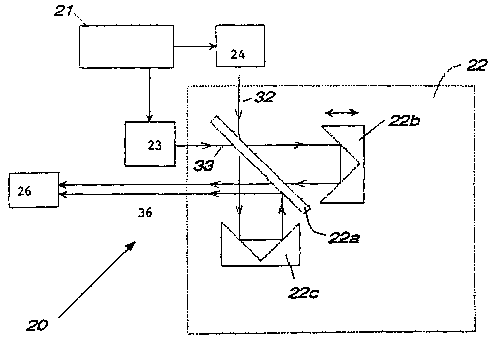
Fig. 2 provides a diagrammatic representation of a device according to the
subject
invention, of particular use for a sample that interacts with light with
strong scattering.
Fig. 3 provides a diagrammatic representation of a device according to the
subject
invention, of particular use for a sample that interacts with light with weak
scattering and
strong absorption.
Figs. 4A and 4B provide spectra of mufti-analyte aqueous solutions measured by
single beam FTIR (prior art method) and by dual beam FTIR (present invention),
respectively.
Fig. 5 provides a comparison of predicted and reference glucose concentration
in
to mufti-analyte aqueous solutions measured by single beam FTIR (prior art
method) and by
dual beam FTIR (present invention).
Fig. 6 provides of graphical representation of the standard error of
prediction of
glucose concentration vs. the number of factors derived from measurements of
mufti-analyte
aqueous solutions by single beam FTIR (prior art) and by dual beam FTIR
(present
15 invention) techniques.
Figs. 7A and 7B provide a graphical representation of glucose concentration
(predicted vs. reference) in mufti-analyte solutions measured over the course
of several
weeks by single beam FTIR (prior art) and by dual beam FTIR (present
invention)
techniques.
DESCRIPTION OF THE SPECIFIC EMBODIMENTS
Methods and devices are provided for determining the presence and/or
concentration
of at least one analyte in a sample of low transmissivity. In the subject
methods, a forward
beam and a backward beam are produced by or introduced into an interferometer
from at
least one infrared radiation source. The forward beam is passed through the
sample to
produce a sample beam while the backward beam is passed through a reference to
provide a
reference beam. The sample and reference beams are recombined either optically
into a null
beam which is detected at a single detector or electronically nulled after
detection on two
detectors. The presence, and often amount, of at least one analyte in the
sample is then
3o derived from the detected null signal. Also provided are devices for
practicing the above
methods. The subject methods and devices are suitable for use in a variety of
different
applications, including the detection of the presence, and amount, of one or
more blood
analytes in a physiological sample, such as blood, tissue or derivatives
thereof. In further
4
CA 02410907 2002-11-29
WO 01/92857 PCT/USO1/16204
describing the subject invention, the subject methods will be described first,
followed by a
review of a representative device of the subject method and a review of
various
representative applications in which the subject invention finds use.
. Before the subject invention is described fi~rther, it is to be understood
that the
invention is not limited to the particular embodiments of the invention
described below, as
variations of the particular embodiments may be made and still fall within the
scope of the
appended claims. It is also to be understood that the terminology employed is
for the purpose
of describing particular embodiments, and is not intended to be limiting.
Instead, the scope
of the present invention will be established by the appended claims.
In this specification and the appended claims, singular references include the
plural,
unless the context clearly dictates otherwise. Unless defined otherwise, all
technical and
scientific terms used herein have the same meaning as commonly understood to
one of
ordinary skill in the art to which this invention belongs.
METHODS
As summarized above, the subject invention provides a method for determining
the
presence, and often concentration, of at least one analyte in a sample having
low
2o transmissivity. Specifically, the subject invention provides a method for
determining the
presence, and even concentration, of an analyte in a sample using Fourier
Transform Infrared
(FTIR) spectroscopy. More specifically, the subject methods are 'dual beam
FTIR (DB-
FTII~) methods of determining the presence, and concentration, of at least one
analyte in a
sample of low transmissivity, e.g. glucose in a tissue sample.
In practicing the subject methods, the first step is to produce a forward beam
and a
backward beam from at least one infrared radiation source, where the forward
and backward
beams when combined, produce a cancellation (or null) in the a.c. signal and a
doubling of
the d.c. signal. The infrared radiation employed in the subject methods may be
obtained from
any convenient source of infrared radiation that is capable of providing
radiation in the
3o desired infrared wavelengths, where wavelengths of particular interest are
those ranging
from about 0.7 p.m to 3 p,m, usually from about 1.3 p,m to 2.4 p,m.
In one embodiment an interferometer is employed to produce the forward and
backward beams from an initial, single infrared radiation source. The forward
and backward
CA 02410907 2002-11-29
WO 01/92857 PCT/USO1/16204
beams are characterized in that, upon leaving or exiting the interferometer,
they are exact
complements of each other. As such, the backward beam is 180 ° out of
phase with respect
to the forward beam upon leaving the interferometer. The forward beam and the
reverse
beam produced by the interferometer are then passed into a sample and
reference,
respectively, to produce sample and reference beams.
In an alternative embodiment, two light sources are used to produce the
forward and
backward beams prior to entering the interferometer. The two light sources may
be derived
from a single light source by using a beam splitter or similar optical means.
The forward
and backward beams are then passed into a sample material and reference
material,
to respectively, to produce sample and reference beams. The sample and
reference beams are
then introduced into an interferometer.
In certain embodiments, the sample into which the forward beam is passed is a
low
transmissivity sample. By low transmissivity sample is meant that the sample
that is
characterized by high radiation losses, e.g. radiation losses that exceed
about 80%, usually at
least about 99% and more usually at least about 99.9%. The low transmissivity
samples that
may be analyzed according to the subject methods may be samples that are
highly absorbing,
highly scattering or both.
The subject methods may be used to analyze a variety of different samples. The
samples may be naturally occurring or synthetic compositions. Representative
samples that
2o may be analyzed according to the subject methods include: industrial
products, agricultural
products, environmental and waste products, and the like. Specific sample
materials of
interest include: solid and liquid drug formulations, fine chemicals,
plastics, polymers,
membranes especially those containing trace analytes of interest such as
enzymes, paints and
other chemical or physical coatings, liquid products such as petroleum oil and
its various
distillates including heating oil and gasoline, minerals, natural and
synthetic gemstones such
as diamond especially when in its powdered form, liquid manufacturing wastes,
natural and
synthetic fibers, wheat and other grains, milk and dairy products, eggs, meats
and other
foods, liquid and solid fertilizers, lake and other limnological sediments,
and histological
specimens. In many embodiments of the subject methods, the sample is a
physiological
3o sample. By physiological sample is meant a sample of material that is
contained, obtained or
derived from a living multicellular organism. In many embodiments, the sample
is a tissue
sample or derivative thereof. In yet other embodiments, the sample is a
physiological fluid
6
CA 02410907 2002-11-29
WO 01/92857 PCT/USO1/16204
sample, e.g. blood, or a derivative thereof. Depending on the particular
protocol employed,
the sample may be part of or separate from the multicellular organ from which
it is derived.
The reference may be any kind of material or composite thereof that provides
for a
reference beam that nulls at least a portion of, and in many embodiments
substantially all of,
s the non-sample components of the sample beam when the two beams are
combined, as
described infra. The nature of the reference material or cell may vary greatly
depending on
the nature of the sample, so long as the above parameters are met. In many
embodiments, the
reference will be an aqueous composition, where the composition may be pure
water, a water
solution or a water dispersion. In embodiments where the sample is tissue, the
reference may
1o contain pure water or water comprising one or more components that are
present in the tissue
sample, e.g. metabolites, proteins, lipids, nucleic acids, etc, as well as
other scattering
components that mimic the scattering qualities of tissue, e.g., an agents)
that emulates the
scattering properties of tissue. In many embodiments in which the sample is
tissue, the
reference comprises a solid material with water as a major component. Where
the reference
15 material is a fluid composition, it is generally present iri a suitable
containment means.
Suitable containment means include those fabricated from silicon, calcium
fluoride, infrasil,
crystal quartz and the like.
The reference material that is employed in the subject methods may be a fluid
contained in a cell having a variable pathlength or a constant pathlength.
Where the
2o reference cell has a static or constant pathlength, the pathlength of the
reference cell, i.e. the
distance that the backward beam traverses as it travels through the reference
cell, is generally
at least about 5 p,m, usually at least about 100 p.m and more usually at least
about 1 mm,
where the distance may be as long as 1 m or longer, but in many embodiments
does not
exceed about 1 cm and usually does not exceed about 2 mm. Where the reference
cell has a
25 variable pathlength, the length of the reference cell is generally
adjustable by as much as a
magnitude, and in certain embodiments is generally adjustable over a distance
of at least
about 1 cm, usually at least about 1 mm and more usually at least about 100
p,m. As such,
the pathlength may be varied by as much as an order of magnitude. However, in
many
embodiments the pathlength is varied, if at all, by a factor that generally
does not exceed
3o about 100%, usually does not exceed about 30% and more usually does not
exceed about
10%.
Alternatively, the reference material may be a solid scattering material. The
optical
scattering and absorption properties of the reference materials may be matched
to that of the
7
CA 02410907 2002-11-29
WO 01/92857 PCT/USO1/16204
sample. For samples, such as tissue, the reference material' may be a solid
with water as a
major component, such as gelatin. Another type of reference material may
consist of
multiple separate materials. For example, the reference beam may be generated
by
transmitting and reflecting the backward beam through a variety of materials.
In many embodiments, adjustments are made at this point to substantially
equalize
the energy of the two beams and therefore obtain an optimal null. By
substantially equalize
the energy of the two beams is meant that various parameters of the device
employed in the
subject methods are adjusted in order to obtain reference and sample beams
that vary in
energy by a magnitude of less than about 10%, usually less than about 5% and
more usually
less than about 2%. By "optimal null" is meant a null in which the nulling
ratio is at least
about 5:1, usually at least about 20:1 and more usually at least about 50:1,
where the nulling
ratio may be as high as 200:1 or higher, but typically does not exceed about
50:1. By nulling
ratio is meant: the modulated (a.c. component) of the energy present in the
forward beam
divided by the modulated (a.c. component) of the energy present in the
combined beams.
Adjustments that may be made to achieve the optimum nulling ratio include:
adjustments to
the reference cell pathlength and/or adjustments to the overlap of the sample
and reference
beams upon recombination or collimation into a single null beam, adjustments
to the
intensity of either the sample or reference beam using a variable attenuator
(two examples of
variable attenuators that are commonly known in the field: a circular gradient
metal-coated
2o attenuator, and a claw attenuator), and adjustments to the composition of
the reference
material (for example, if the reference cell contains multiple components, a
change in the
relative concentration of constituents in the reference cell). Where the
reference cell
pathlength is adjusted, it may be adjusted by as much as an order of
magnitude. However, in
many embodiments, the magnitude of the adjustment typically does not exceed
about lmm,
usually about 0.5 mm and more usually about 50 microns.
The next step in the subject methods is to detect the null beam(s). In one
embodiment, the reference and sample beams are combined at a point prior to
the detector
into a single beam in a manner sufficient to produce a null beam, where the
null beam is
characterized in that at least a portion of the non-analyte signal
contributions are absent, i.e.
3o they have been canceled out. In general, the beams are recombined using any
convenient
beam directing means, e.g. reflective means, beam splitter/collimators, fiber
optics, etc., into
a single null beam. Alternatively, the reference and sample beams may be
separately
detected and combined electronically. In the two-source embodiment of the
subject
CA 02410907 2002-11-29
WO 01/92857 PCT/USO1/16204
invention, the reference and sample beams axe injected into the forward and
backward ports
of the interferometer, followed by detection of the output beam(s).
Following detection of the beams) at the detector(s), the neat step is to
derive
information regarding the presence (and often amount) of the one or more
analytes of
s interest in the sample. In this derivation step, the detected A.C. signal(s)
is(are) generally
amplified while the D.C. component of the signal is rejected, the A.C.
component of the
signal is converted from an analog to digital signal using an AD converter,
and the resultant
digital signal is processed by the computer to provide information regarding
the presence
and concentration of analytes present in the sample.
to As an alternative to balancing the optical intensity of the two beams on a
single
detector, the forward and backward beams may be separately detected, and
electronically
balanced and combined. The electronic signals may combined by using a summing
amplifier. In this embodiment it is important that the spectral response of
the two detectors
be similar, if high null ratios are to be achieved. In yet another embodiment
of the present
1s invention, two light sources and two detectors may be used.
The above described methods may be practiced using any convenient device that
is
capable of providing the requisite forward and backward beams, holding the
sample and
references of interest, and recombining the reference and sample beams into a
null beam.
Representative devices which are suitable for use in practicing the subject
invention are now
2o described in greater detail below.
DEVICES
Devices of the subject invention that find use in practicing the subject
methods are
those that have at least the following components: (a) sources) of infrared
radiation; (b)
2s interferometer means for producing a forward and backward beam or
introducing forward
and backward beams into the interferometer; (c) a reference material; (d) a
sampling
apparatus or means, e.g. a holder, or other means depending on the nature of
the sample; (e)
means for producing a null signal from the reference and sample beams; and (f)
detector(s).
The device may fixrther include one or more additional components that find
use in
3o practicing the subject invention, such as an analog to digital converter
(ADC), and a digital
data processing or computing means, etc. These elements of the subject device
will now be
described in greater detail separately and in terms of Figures 2 and 3, which
schematically
depict representative devices according to the subject invention.
CA 02410907 2002-11-29
WO 01/92857 PCT/USO1/16204
In Figures 2 and 3, device 20 includes a source of infrared radiation 21. The
infrared
radiation source may be any convenient source, including a white light source,
a heated
filament, a metal carbide rod, etc., so long as it is capable of emitting
infrared light having
the wavelength spectrum of interest, i.e. light having a wavelength ranging
from about 0.7 to
50 microns.
Also present in device 20 is a Michelson interferometer 22, which, in the case
of the
device in Fig. 2, is capable of accepting a forward 32 and backward beam 33 of
light and in
the device in Fig. 3 is capable of converting an incident beam of light 31
from the infrared
radiation source 21 and converting it into a forward 32 and backward beam 33.
The
to Michelson interferometer typically includes a beam splitter 22a, a moving
mirror 22b and a
fixed mirror 22c, and optionally additional mirrors for directing the forward
beam into or out
of the interferometer. Also shown is optical tissue sampler 24, variable path
reference 23 and
detector 26.
Referring to the device in Fig. 3, the beam splitter 22a of the interFerometer
22
15 produces forward beam 32 and backward beam 33. Forward beam 32 is directed
out of the
interferometer in one direction while backward beam 33 is directed out of the
interferometer
along the path of incident light from the radiation source 21. The backward
beam is not
necessarily overlapping with the path of the incident light. For example, if
corner cube
optics are used in place of the fixed and moving mirrors in the
interferometer, the backward
2o beam path is offset from the path of the path of the incident light. In
this case the backward
beam can be collected without the need for a beam splitter. This arrangement
has the
advantage that no incident light is lost in the collection of the backward
beam and the total
amount of collected light compared to single beam methods, is doubled. A
commercial
interferometer that provides corner cube optics for the interferometer mirrors
and provides
25 easy access to the backward beam is the Bomem Model MB-100. Any convenient
interferometer may be employed, where suitable interferometers include: the
interferometer
found in the Perkin-Elmer 2000 FTIR. spectrometer, and the like.
Still referring to the device diagrammed in Fig. 3, a beam splitter 25 is
placed in the
radiation source incident beam which coincides with the backward beam as it
exits the
3o interferometer. The beam splitter is sui~cient to redirect a portion of the
backward beam out
of the incident light path so that at least a portion of the backward beam
exiting the
interferometer can be directed through a variable path length reference cell
23. Typically, the
beam splitter 25 is a 3% reflector, usually at least a 1% reflector, where the
beam splitter
CA 02410907 2002-11-29
WO 01/92857 PCT/USO1/16204
may reflect up to about 50% or higher, but generally does not exceed about
50%. Any
convenient beam splitter may be employed, such as uncoated CaF2, partially
metallized glass
or quartz, and the like. The redirected portion of the backward beam is then
directed, using
any convenient means such as reflectors, mirrors etc., to a reference
material.
The variable pathlength reference cell 23 is, in many embodiments, a variable
pathlength water cell, where the aqueous composition present in the reference
cell may or
may not include additional components, e.g. proteins, lipids, metabolites,
sugars, etc., as
desired. A representative example of a variable pathlength water cell that may
be present in
the subject device is a variable path length transmission water cell fitted
with calcium
1o fluoride windows. A reference beam 34 emerges from the variable pathlength
reference cell.
The backward beam and reference beam are directed through use of parabolic
reflectors 41 a
and 41b and mirror 41c.
Ideally the optical properties of the reference material will closely match
the optical
properties of the sample. For example, in the case where the sample is tissue,
the backward
15 beam may be directed into a highly scattering reference material from which
diffusely
reflected light is collected and used as the reference beam. In addition to
being highly
scattering, the reference material may contain absorption features that are
similar to water,
and may also contain other absorption features such as those due to collagen,
elastin and
lipids to further match the tissue properties. A gelatinous material
containing water,
2o collagen, and possibly other materials may serve as a suitable reference
material. For an
optimal match, the water and collagen content as well as other components of
the reference
material may be adjusted to match the particular tissue sample being examined.
An alternate method of matching the optical properties of the reference
material to
that of a complex sample such as tissue is to transmit andJor reflect the
backward beam
25 through multiple materials. For example, the backward beam could be
transmitted through
two variable path length cells, one containing water and another containing
lipid in water
followed by reflection and collection of the dill'usely reflected light from a
scattering
material. The path length of the water and lipid-containing transmission cells
could be
adjusted to match the optical properties of the sample.
3o The forward beam 32, after being directed by beam splitter 25c, is directed
by a
parabolic reflector 42 from the interferometer to the sample holder 24 which
contains the
sample to be analyzed. The sample holder may vary depending on the nature of
the sample
to be contained therein and the nature of the reference employed. Any
convenient sample
11
CA 02410907 2002-11-29
WO 01/92857 PCT/USO1/16204
holder configuration made out of any convenient material may be employed. In
many
embodiments, the sample holder is a tissue sample holder or means for
directing the forward
beam to a tissue sample. A sample beam 35 emerges from the sample and is
directed by
parabolic reflector 42b and mirror 42c.
d Fiber optic means are especially well suited to the delivery and collection
of light
from tissue and other scattering materials. The forward beam is typically
focussed onto a
single optical fiber or a bundle of fibers such that the focus of the input
beam is well
matched to the numerical aperture of the fiber or fibers. The fiber material
itself should be
substantially transparent in the optical region of interest. In order to
inject light efficiently,
to the fiber or bundle of fibers is then brought into close proximity or,
preferably, into direct
contact with the sample. The injected light is then collected with a separate
fiber or bundle
of fibers. The collection bundle is typically annular in arrangement, and
surrounds the input
fiber(s). Alternatively, the collection fiber or bundle may be centrally
disposed within an
annular ring of input fibers. The input and collection fibers may also be
arranged in a
random or an ordered grid. As an aide to increase optical throughput, the
input or output
fibers may be disposed at a non-normal angle with respect to the plane of the
sample. An
opaque shield may be placed between the input and output fibers and in contact
with the
sample to prevent light from passing directly from the input to output fibers
without first
passing through the sample.
2o As shown in Figure 3, the reference and sample beams, 34 and 35
respectively, are
then recombined at a second beamsplitter 25b, which may or may not be the same
type of
beam splitter as the first beam splitter 25. The beam splitter 25b is one that
is sufficient to
recombine the sample and reference beams to produce a null beam.
Alternatively, the reference and sample beams may be directly recombined on
the
surface of the detector without a beamsplitter. A convenient method for direct
recombination is to bring the reference and sample beams obtained with fiber
optic samplers
into close proximity or direct contact with the detector. As long as the
intensity of the
sample and reference beams is well matched, and the detector area is equal to
or larger than
the area illuminated by the sample and reference fibers, an excellent null can
be achieved.
3o In the device depicted in Figure 2, the forward and backward beams are
generated
prior to the interferometer using a single light source and a beam splitter.
As with the device
depicted in Figure 3, the forward and backward beams interact optically with
the sample and
reference materials, respectively, to generate sample and reference beams.
However, rather
12
CA 02410907 2002-11-29
WO 01/92857 PCT/USO1/16204
than being recombined after the interferometer as in the device depicted in
Fig. 3, the sample
and reference beams are now combined within the interferometer, by injecting
the two
beams into the two ports of a Michelson interferometer.
In the devices depicted in both Figs. 2 and 3, the emergent null beam 36 is
then
directed onto detector 26, optionally through a lens 26(a) which focuses the
null beam onto
the detector. The detector is a detector that is capable of converting the
incident null beam
into an analog signal. Any convenient detector may be employed, where suitable
detectors
include indium gallium arsenide (InGaAs), indium antimonide (InSb), germanium,
and the
like.
to The A.C. component of the detector-produced analog signal is then amplified
while
rejecting the D.C. component by an amplifier 27 whose gain is set to fill an
analog to digital
converter (ADC) 28 also present in the device. Any convenient amplifier may be
present in
the device, where representative amplifiers of interest include: the AD 797,
and the like. The
ADC may be any convenient ADC. Because of the nature of the device, the ADC
need not
be an ultra-high precision ADC. As such, the ADC need only be a 16-bit ADC.
The digital
output of the ADC is then processed by a data processing means 29, e.g. a
computing means,
which is capable of taking the digital signal and deriving the presence, and
often amount of,
analyte present in the sample.
A preferred method of processing the digital signal includes the following
steps:
(1) Optional Initial step: subtraction of the dual beam background
interferogram measured
with a background material in both the forward and backward beams from the
dual beam
sample interferogram measured with the sample in the forward beam and the
background material in the reference beam, resulting in a corrected dual beam
sample
interferogram.
(2) Fourier transformation of the dual beam sample interferogram (either
corrected as in step
1 or uncorrected), resulting in a transformation of the interferogram into a
dual beam
sample spectrum.
(3) Optional subsequent step contingent on optional initial step 1: Fourier
transformation of
the single beam sample interferogram measured with the sample in the forward
beam
3o and the backward beam blocked, resulting in a single beam sample spectrum.
(4) Computation of the logarithm of the dual beam sample spectrum, resulting
in a dual
beam sample pseudo-absorbance spectrum.
13
CA 02410907 2002-11-29
WO 01/92857 PCT/USO1/16204
(5) Optional subsequent step contingent on step 3 : Computation of the
logarithm of the
single beam sample spectrum followed by the subtraction of this spectrum from
the dual
beam sample pseudo-absorbance spectrum, resulting in a dual beam sample
absorbance
spectrum.
(6) Multiplication of the absorbance or pseudo-absorbance spectrum by a
scaling function,
resulting in a scaled absorbance spectrum.
(7) Subtraction of a mean spectrum from the scaled absorbance spectrum,
resulting in a
mean-centered scaled absorbance spectrum.
(8) Multiplication of each spectral point in the mean-centered scaled
absorbance spectrum
to by a regression coefficient.
(9) Summing the results of step 8 over all spectral points, resulting in a
prediction of the
analyte concentration in the sample.
The scaling function, mean spectrum, and regression coefficients are
determined
during a calibration phase. The calibration phase involves measurement of the
dual beam
15 FTIR spectra of samples whose analyte concentrations are known. The scaling
function,
mean spectrum, and regression coefficients are determined in a manner that
minimizes the
difference between the known analyte concentrations and the analyte
concentrations
predicted from the dual FTIR spectra. Techniques for accomplishing this are
well known in
the field and include partial least squares and principal component
regression. Both these
2o techniques are discussed in depth in the book "Multivariate Calibration" H.
Martens and T.
Naes, Wiley and Sons, New York (1989).
The above-described devices may be laboratory scale devices or miniaturized
for
field use, e.g. doctor's office, home use, etc.
25 ' UTILITY
The subject methods and devices find use in variety of different applications
in which
the detection of, and determination of the concentration of, one or more
analytes in a low
transmissive sample is desired. As such the subject methods and devices find
use in the
detection of analytes in a wide variety of different types of samples, such as
pollutants or
3o toxins in environmental samples, e.g. soil or water, toxins or pathogens in
agricultural and
food products; detection of impurities in industrial products, and the like.
One application of
particular interest is the use of the subject methods and devices to detect
the presence of one
14
CA 02410907 2002-11-29
WO 01/92857 PCT/USO1/16204
or more blood analytes in an in vivo or ex vivo physiological sample, e.g.
blood, tissue or a
derivative thereof.
A variety of different analytes may be detected using the subject methods,
where
representative analytes include: alcohol, formaldehyde, glucose, glutamic
acid, glycerol,
beta-hydroxybutyrate, L-lactate, leucine, malic acid, pyruvic acid, steroids,
ascorbic acid,
acetone and other ketone bodies, folate, ammonia, bilirubin, creatinine,
hemoglobins, lipids,
phenylalanine, proteins (including albumin and globulins), triglycerides,
urea, as well as
pharmaceuticals and drugs of abuse. While in principle the subject methods may
be used to
determine the presence, and often concentration, of an analyte in a variety of
different
1o physiological samples, such as urine, tears, saliva, and the like, they are
particularly suited
for use in determining the concentration of an analyte in blood or blood
fractions or tissue or
tissue fractions. One application of particular interest is the use of the
subject methods and
compositions to detect the presence of, and determine the amount of, glucose
in an in vivo or
ex vivo tissue sample.
Detection of the blood analytes according to the subject methods finds use in
a
variety of different medical applications, including disease diagnosis,
disease management,
and the like.
The following examples are offered by way of illustration and not by way of
limitation.
EXPERIl~~NTAL
I. Analyte detection in an weakly scattering aqueous sample
For a sample that is weakly scattering and strongly absorbing, such as an
aqueous
solution of analytes (eg. blood serum) interacting with light at near to mid
infrared
wavelengths, both the forward and backward beams may be employed in
transmission
mode. As an example, we compared the predictive capabilities of single beam
(prior art) and
dual beam FTTR for aqueous samples containing three analytes of physiological
relevance:
creatinine, glucose, and urea.
3o The instrument configuration used to perform the experiments is diagrammed
in Fig.
3. A commercial single beam FTIR spectrometer (Perkin Elmer Spectrum 2000) was
modified to function as a dual beam instrument. The instrument was kept open
to the
atmosphere (21 +/- 1 C, 40 +/- 5% RH). A 50% "polka dot" beam splitter (Oriel
CA 02410907 2002-11-29
WO 01/92857 PCT/USO1/16204
Instruments, model no. 38106) was used to separate the light source and
backward beams.
The forward beam was also reflected off of a 50% polka dot beam splitter to
equalize the
intensity of the two beams. Gold-coated parabolic reflectors focused the
forward and
backward beams into the sample and reference cells, respectively. The sample
and reference
cells had a path length of 0.5 mm, as defined by the spacing between their
quartz suprasil
windows. The temperature of the sample and reference cells was regulated at
22.0 C +/- 0.1
C. Gold-coated parabolic reflectors were then used to recollimate the forward
and backward
beams. The two beams were then combined using a 50% polka dot beam splitter,
and
focussed onto an InSb detector (7 mm diameter active area, cooled to 77 K)
using a silicon
to lens (2" diameter, approx. 25 mm focal length).
The D.C. component of the signal was removed and the A.C. component was
amplified to nearly fill the analog to digital (A!D) converter. The null ratio
for this set of
experiments was approximately 40:1. Therefore the amplification required to
fill the A/D
converter with the dual beam signal was approximately 40 times that of the
single beam
signal. The single beam and dual beam interferograms were interleaved, one
after the other
for each sample. The spectra were processed according to the procedures
(including the
optional steps) described already (see section: "DEVICES").
The samples consisted of 27 solutions containing creatinine, urea, and glucose
dissolved in water at three concentration levels (creatinine - 370, 650, and
930 mg/dL; urea -
2o 230, 585, and 940 mg/dL; glucose - 0, 250, and 500 mg/dL). The reference
cell contained
pure water. The complete set of 27 solutions was measured once per day on
three separate
days. The three measurement days spanned a period of approximately 7 weeks.
Samples
containing pure water were used as the background samples. Background samples
were
measured at the beginning and at the end of each set of 27 solutions. The 27
solutions were
. made up fresh and were measured in a different randomized order on each
experimental day.
Optical absorption by the three analytes is weak compared to that of water. As
a
result, the single beam spectra of the 27 samples are nearly indistinguishable
by eye. In
contrast, the dual beam spectra, from which, by virtue of the optical nulling
effect, the lamp
emission spectrum and water absorption effects have been largely removed, show
clear and
obvious spectral changes with changing analyte concentration. Figs. 4A and 4B
show
respectively the single beam and dual beam spectra in the 4000-5000 cm 1
region of three
samples for which the creatinine and urea concentrations are fixed at their
lowest levels
while the glucose concentration is varied between three levels. The region of
maximum
16
CA 02410907 2002-11-29
WO 01/92857 PCT/USO1/16204
spectral change with changing glucose concentration (4700 crri 1) corresponds
to a known
absorption band of glucose in water.
Partial Least Squares (PLS) was used to analyze the predictive content of the
NIR
spectra over the spectral range of 4000-8000 cm 1. Analyte predictions within
a given
experimental day were assessed by choosing a particular sample for prediction
and using the
remaining 26 samples for calibration. By rotating through all 27 samples in
this fashion the
"cross-validated" prediction performance was assessed. Predictions of glucose
concentration
for the spectra acquired in single beam and dual beam mode are compared in
Fig. 5. The
standard error of prediction (SEP) (i.e., the standard error of prediction is
the square root of
to theaverage squared difference between predicted and reference
concentration) of glucose
concentration from the dual beam and single beam spectra is 11.3 and 22.8
mg/dL,
respectively. In addition to the improved prediction performance compared to
single beam
FTIR, the dual beam FTIR calibration model was considerably simpler. This can
be seen in
a plot of SEP vs. number of factors in the PLS model (Fig.6). Only 5 factors
were used in
the best dual beam calibration model whereas at 13 factors the single beam
calibration model
has still not achieved a minimum SEP value.
Analyte predictions across multiple days were assessed by using the first
day's data
as a calibration set and predicting the two subsequent days. The results for
the single beam
and dual beam techniques at 12 and 4 factors, respectively, are summarized in
Figs. 7A to
7B. In summary, compared to single beam FTIR, the dual beam technique shows
better
predictive ability of analyte concentration in aqueous solution over both the
short (same day)
and long term (over 7 weeks).
II. Glucose detection in tissue
For a strongly scattering sample that contains a weakly absorbing analyte,
such as
glucose in mammalian tissue, the forward or sample beam may be employed in
reflectance
mode whereas the back or reference beam may be in transmission mode.
The instrumental configuration used to perform such a measurement is
diagrammed
in Fig. 2. A thin calcium fluoride plate may be used to separate the light
into forward and
so backward beams. Since most of the light will be lost in the highly
scattering tissue, 96% of
the total throughput of the interferometer is used for the forward beam with
the remaining
4% used for the back beam which is directed through the reference cell.
17
CA 02410907 2002-11-29
WO 01/92857 PCT/USO1/16204
The temperature of the reference cells should be regulated at the same
temperature as
the surface of the tissue being measured since the spectrum of water in the
near infra red
portion of the spectrum is strongly sensitive to temperature. An attenuator
may be used in
either or both beams to balance the energy at the detector. The forward beam
is focussed
with a calcium fluoride lens onto the input of a fiber optic bundle. The
bundle directs the
forward beam onto, for example, the volar forearm of the human subject being
measured.
Interleaved with the input fibers at the surface of the tissue are output
fibers which direct the
scattered and partially absorbed light from the tissue to the detector.
Interleaved at the
detector with the output fibers are reference (back beam) fibers which direct
the light that
to has passed through the reference cell also onto the detector. The detector
is chosen such that
its surface area is somewhat larger than the total area illuminated by the
interleaved output
fiber bundle. The sample and reference beams are thus combined directly at the
surface of
the detector to form a null.
The D.C. component of the signal is then electronically removed and the A.C.
component is electronically amplified to nearly fill the analog to digital
(AlD) converter.
The null ratio can easily approach approximately 20:1 even though the sample
beam consists
of scattered light from the tissue and the reference beam consists of light
that has been
transmitted substantially without any scatter through a reference cell. The
amplification
required to fill the A/D converter with the dual beam signal would be
approximately 20 fold
2o higher than that of the single beam signal. A calibration is generated by
measuring the null
spectra of subjects at random but known glucose levels in a analogous fashion
with the
solution spectra calibration described irtfi°a.
It is evident from the above results and discussion that the subject invention
provides
for an important breakthrough in the use of FTIR for detection of analytes.
Specifically, the
subject methods and devices overcome prior problems encountered with FTIR
determination
of glucose in tissue, such as problems with instrument drift, the requirement
for use of ultra-
high precision ADCs, etc. Importantly, the subject methods and devices are
capable of
providing highly accurate non-invasive measurements of blood analytes, e.g.
glucose. As
3o such, the subject invention represents a significant contribution to the
art.
All publications and patents cited in this specification are herein
incorporated by
reference as if each individual publication or patent were specifically and
individually
18
CA 02410907 2002-11-29
WO 01/92857 PCT/USO1/16204
indicated to be incorporated by reference. The citation of any publication is
for its disclosure
prior to the filing date and should not be construed as an admission that the
present invention
is not entitled to antedate such publication by virtue of prior invention.
Although the foregoing invention has been described in some detail by way of
illustration and example for purposes of clarity of understanding, it is
readily apparent to
those of ordinary skill in the art in light of the teachings of this invention
that certain changes
and modifications may be made thereto without departing from the spirit or
scope of the
appended claims.
to
19