Note: Descriptions are shown in the official language in which they were submitted.
CA 02411938 2008-01-17
SURGICAL ABLATION PROBE FOR FORMING A
CIRCUMFERENTIAL LESION
= TECI~TICAL FIELD
The field of the invention relates to a surgical device and method. More
particularly, it relates to a tissue ablation probe and method for ablating a
circumferential region of tissue at a location where a pulmonary vein extends
from
an atrium. The probe has particular utility during invasive or minimally
invasive
c.m-diac surgery.
EACKGROUND OF THE INVENTION,
Many local energy delivery devices and methods have been developed for
the treatment of various abnormal tissue conditions in the body, and
particularly for
treating abnormal tissue along body space walls which define various body
spaces in
the body. For example, various devices have been disclosed with the primary
purpose of treating or recanalizing atherosclerotic vessels with localized
energy
delivery . Several prior devices and methods combine energy delivery
assemblies in
combination with cardiovascular stent devices in order to locally deliver
energy to
tissue in order to maintain patency in diseased lumens such as blood vessels.
Endometriosis, another abnormal wall tissue condition wbich is associated with
the
endometrial cavity and is characterized by dangerously proliferative uterine
wall
tissue along the surface of the endometrial cavity, has also been treated by
local
energy delivery devices and methods.
Several other devices and methods have also been disclosed which use
catheter-based heat sources for the intended purpose of inducing thrombosis
and
controlling hemorrhaging within certain body lumens such as vessels. Detailed
examples of local energy delivery devices and related procedures such as those
of
the types descn'bed above are disclosed in the following references: U.S.
Patent Nos.
4,672,962 to Hershenson; U.S. Patent Nos. 4,676,258 to InoKuchi et al.; U.S.
Patent
No. 4,790,311 to Ruiz; 4,807,620 to Strnl et al.; U.S. Patent No. 4,998,933 to
Eggers
et al.; U.S. Patent No. 5,035,694 to Kasprzyk et al,; U.S. Patent No.
5,190,540 to
-1-
CA 02411938 2002-12-13
WO 01/95820 PCT/US01/18724
Lee; U.S. Patent No. 5,226,430 to Spears et al.; and U.S. Patent No. 5,292,321
to
Lee; U.S. Patent No. 5,449,380 to Chin; U.S. Patent No. 5,505,730 to Edwards;
U.S. Patent No. 5,558,672 to Edwards et al.; and U.S. Patent No. 5,562,720 to
Stern
et al.; U.S. Patent No. 4,449,528 to Auth et al.; U.S. Patent No. 4,522,205 to
Taylor
et al.; and U.S. Patent No. 4,662,368 to Hussein et al.; U.S. Patent No.
5,078,736 to
Behl; and U.S. Patent No. 5,178,618 to Kandarpa.
Other prior devices and methods electrically couple fluid to an ablation
element during local energy delivery for treatment of abnormal tissues. Some
such
devices couple the fluid to the ablation element for the primary purpose of
controlling the temperature of the element during the energy delivery. Other
such
devices couple the fluid more directly to the tissue-device interface either
as another
temperature control mechanism or in certain other known applications as a
carrier or
medium for the localized energy delivery. Detailed examples of ablation
devices
which use fluid to assist in electrically coupling electrodes to tissue are
disclosed in
the following references: U.S. Patent No. 5,348,554 to Imran et al.; U.S.
Patent No.
5,423,811 to Imran et al.; U.S. Patent No. 5,505,730 to Edwards; U.S. Patent
No.
5,545,161 to Imran et al.; U.S. Patent No. 5,558,672 to Edwards et al.; U.S.
Patent
No. 5,569,241 to Edwards; U.S. Patent No. 5,575,788 to Baker et al.; U.S.
Patent
No. 5,658,278 to Imran et al.; U.S. Patent No. 5,688,267 to Panescu et al.;
U.S.
Patent No. 5,697,927 to Imran et al.; U.S. Patent No. 5,722,403 to McGee et
al.;
U.S. Patent No. 5,769,846; and PCT Patent Application Publication No. WO
97/32525 to Pomeranz et al.; and PCT Patent Application Publication No. WO
98/02201 to Pomeranz et al.
Other prior devices and methods have been disclosed which use a probe as a
surgical device, thereby allowing the physician to directly apply an electrode
to
tissue. Detailed examples of surgical probes are disclosed in the following
references: U.S. Patent No. 6,023,638 to Swanson; U.S. Patent No. 4,841,979 to
Dow et al.; U.S. Patent No. 4,917,096 to Englehart et al.; and U.S. Patent No.
6,152,920 to Thompson et al.
Atrial Fibrillation
Cardiac arrhythmias, and atrial fibrillation in particular, persist as common
and dangerous medical ailments associated with abnormal cardiac chamber wall
tissue. In patients with cardiac arrhythmia, abnormal regions of cardiac
tissue do not
follow the synchronous beating cycle associated with normally conductive
tissue in
patients with sinus rhythm. Instead, the abnormal regions of cardiac tissue
-2-
CA 02411938 2002-12-13
WO 01/95820 PCT/US01/18724
aberrantly conduct electrical signals to adjacent tissue, thereby disrupting
the cardiac
cycle and causing an asynchronous cardiac rhythm. Such abnormal conductioin is
known to occur at various regions of the heart, such as, for example, in the
region of
the sino-atrial (SA) node, along the conduction pathways of the
atrioventricular
(AV) node and the Bundle of His, or in the cardiac muscle tissue forming the
walls
of the ventricular and atrial cardiac chambers.
Cardiac arrhythmias, including atrial arrhythmia, may be of a multiwavelet
reentrant type, characterized by multiple asynchronous loops of electrical
impulses
that are scattered about the atrial chamber and are often self propagating. In
the
alternative or in addition to the multiwavelet reentrant type, cardiac
arrhythmias may
also have a focal origin, such as when an isolated region of tissue in an
atrium fires
autonomously in a rapid, repetitive fashion. Cardiac arrhythmias, including
atrial
fibrillation, may be generally detected using the global technique of an
electrocardiogram (EKG). More sensitive procedures of mapping the specific
conduction along the cardiac chambers have also been disclosed, such as, for
example, in U.S. Patent No. 4,641,649 to Walinsky et al. and in PCT Patent
Application Publication No. WO 96/32897 to Desai.
A host of clinical conditions can result from the irregular cardiac function
and resulting hemodynamic abnormalities associated with atrial fibrillation,
including stroke, heart failure, and other thromboembolic events. In fact,
atrial
fibrillation is believed to be a significant cause of cerebral stroke, wherein
the
abnormal hemodynamics in the left atrium caused by the fibrillatory wall
motion
precipitate the formation of thrombus within the atrial chamber. A
thromboembolism is ultimately dislodged into the left ventricle which
thereafter
pumps the embolism into the cerebral circulation where a stroke results.
Accordingly, numerous procedures for treating atrial arrhythmias have been
developed, including pharmacological, surgical, and catheter ablation
procedures.
Several pharmacological approaches intended to remedy or otherwise treat
atrial arrhythmias have been disclosed, such as, for example, those approaches
disclosed in the following references: U.S. Patent No. 4,673,563 to Berne et
al.; U.S.
Patent No. 4,569,801 to Molloy et al.; and "Current Management of Arrhythmias"
(1991) by Hindricks, et al. Such pharmacological solutions, however, are not
generally believed to be entirely effective in many cases, and are even
believed in
some cases to result in proarrhythmia and long term inefficacy.
-3-
CA 02411938 2002-12-13
WO 01/95820 PCT/US01/18724
Several surgical approaches have also been developed with the intention of
treating atrial fibrillation. One particular example is known as the "maze
procedure," as is disclosed by Cox, J. L. et al. in "The surgical treatment of
atrial
fibrillation. I. Summary" Thoracic and Cardiovascular Surgery 101(3), pp. 402-
405
(1991); and also by Cox, JL in "The surgical treatment of atrial fibrillation.
IV.
Surgical Technique", Thoracic and Cardiovascular Surgery 101(4), pp. 584-592
(1991). In general, the "maze" procedure is designed to relieve atrial
arrhythmia by
restoring effective atrial systole and sinus node control through a prescribed
pattern
of incisions about the tissue wall. In the early clinical experiences
reported, the
"maze" procedure included surgical incisions in both the right and the left
atrial
chambers. However, more recent reports predict.that the surgical "maze"
procedure
may be substantially efficacious when performed only in the left atrium. See
Sueda
et al., "Simple Left Atrial Procedure for Chronic Atrial Fibrillation
Associated With
Mitral Valve Disease" (1996).
The "maze procedure" as performed in the left atrium generally includes
forming vertical incisions from the two superior pulmonary veins and
terminating in
the region of the mitral valve annulus, traversing the region of the inferior
pulmonary veins en route. An additional horizontal line also connects the
superior
ends of the two vertical incisions. Thus, the atrial wall region bordered by
the
pulmonary vein ostia is isolated from the other atrial tissue. In this
process, the
mechanical sectioning of atrial tissue eliminates the arrhythmogenic
conduction
from the boxed region of the pulmonary veins to the rest of the atrium by
creating
conduction blocks within the aberrant electrical conduction pathways. Other
variations or modifications of this specific pattern just described have also
been
disclosed, all sharing the primary purpose of isolating known or suspected
regions of
arrhythmogenic origin or propagation along the atrial wall.
While the "maze" procedure and its variations. as reported by Dr. Cox and
others have met some success in treating patients with atrial arrhythmia, its
highly
invasive methodology is believed to be prohibitive in most cases. However,
these
procedures have provided a guiding principle that electrically isolating
faulty cardiac
tissue may successfully prevent atrial arrhythmia, and particularly atrial
fibrillation
caused by arrhythmogenic conduction arising from the region of the puhnonary
veins. =
Less invasive catheter-based approaches to treat atrial fibrillation have been
disclosed which implement cardiac tissue ablation for terminating
arrhythmogenic
-4-
CA 02411938 2002-12-13
WO 01/95820 PCT/US01/18724
conduction in the atria. Examples of such catheter-based devices and treatment
methods have generally targeted atrial segmentation with ablation catheter
devices
and methods adapted to form linear or curvilinear lesions in the wall tissue
which
defines the atrial chambers. Some specifically disclosed approaches provide
specific
ablation elements which are linear over a defined length intended to engage
the
tissue for creating the linear lesion. Other disclosed approaches provide
shaped or
steerable guiding sheaths, or sheaths within sheaths, for the intended purpose
of
directing tip ablation catheters toward the posterior left atrial wall such
that
sequential ablations along the predetermined path of tissue may create the
desired
lesion. In addition, various energy delivery modalities have been disclosed
for
forming atrial wall lesions, and include the use of microwave, laser,
ultrasound,
thermal conduction, and more commonly, radio frequency energies to create
conduction blocks along the cardiac tissue wall.
Detailed examples of ablation device assemblies and methods for creating
lesions along an atrial wall are disclosed in the following U.S. Patent
references:
U.S. Patent No. 4,898,591 to Jang et al.; U.S. Patent No. 5,104,393 to Isner
et al.;
U.S. Patent No. 5,427,119; U.S. Patent No. 5,487,385 to Avitall; U.S. Patent
No.
5,497,119 to Swartz et al.; U.S. Patent No. 5,545,193 to Fleischman et al.;
U.S.
Patent No. 5,549,661 to Kordis et al.; U.S. Patent No. 5,575,810 to Swanson et
al.;
U.S. Patent No. 5,564,440 to Swartz et al.; U.S. Patent No. 5,592,609 to
Swanson et
al.; U.S. Patent No. 5,575,766 to Swartz et al.; U.S. Patent No. 5,582,609 to
Swanson; U.S. Patent No. 5,617,854 to Munsif; U.S. Patent No 5,687,723 to
Avitall;
U.S. Patent No. 5,702,438 to Avitall. Other examples of such ablation devices
and
methods are disclosed in the following PCT Patent Application Publication
Nos.:
WO 93/20767 to Stem et al.; WO 94/21165 to Kordis et al.; WO 96/10961 to
Fleischman et al.; WO 96/26675 to Klein et al.; and WO 97/37607 to Schaer.
Additional examples of such ablation devices and methods are disclosed in the
following published articles: "Physics and Engineering of Transcatheter Tissue
Ablation", Avitall et al., Journal of American College of Cardiology, Volume
22,
No. 3:921-932 (1993); and "Right and Left Atrial Radiofrequency Catheter
Therapy
of Paroxysmal Atrial Fibrillation," Haissaguerre, et al., Journal of
Cardiovascular
Electrophysiology 7(12), pp. 1132-1144 (1996).
In addition to the known assemblies summarized above, additional tissue
ablation device assemblies have been recently developed for the specific
purpose of
ensuring firm contact and consistent positioning of a linear ablation element
along a
-5-
CA 02411938 2008-01-17
Iength of tissue. This is aacomplished by anchoring the element at least at
one
predetermined location along that length, such as in order to form a"maze"-
type
lesion pattern in the left atrium. An example of such an assembly is disclosed
in
U.S. Patent No. 5,971,983 to Lesh, issued October 26, 1999.
The assembly includes an anchor at each of two ends of a
lineat ablation element in order to secure those ends to each of two
predetermined
locations along a left atrial wall, such as at two adjacent pulmonary veins,
so that
tissue may be ablated along the length of tissue extending therebetween.
In addition to attempting atriai wall segmentation with long linear lesions
for
treating atrial arrhythmia, other ablation devices and methods have also been
disclosed which are intended to use expandable members such as balloons to
ablate
cardiac tissue. Some such devices have been disclosed primarily for use in
ablating
tissue wall regions along the cardiac chambers. Other devices and methods have
been disclosed for treating abnomW conduction of the lett sided accessory
pathways, and in particular associated with "Wolff-Parlcinson-White" syndrome -
various such disclosures use a balloon for ablating from within a region of an
associated coronary sinus adjacent to the desired cardiac tissue to ablate.
Further
more detailed examples of devices and methods such as of the types just
described
are variously disclosed in the following published references: Frmm et al., in
"Feasibility of RF Powered Thermal Balloon Ablation of Atrioventricular Bypass
Tracts via the Coronary Sinus: In vivo Canine Studies," PACE, Vol. 18, p 1518-
1530
(1995) ; "Long-term effects of percutaneous laser balloon ablation from the
canine
coronary sinus", Schuger CD et al., Circulation (1992) 86:947-954; and
"Percutaneous laser balloon coagulation of accessory pathways", McMath LP et
al.,
Diagn Ther Cardiovasc Interven 1991; 1425:165-171.
Ar h,vthmias Oriainating from Foci in Pulmona;y Veins
As briefly discussed above, various modes of atrial fibrillation have been
observed to be focal in natnre, caused by the rapid and repetitive firing of
an isolated
center within cardiac muscle tissue associated with the atrium. Such foci may
act as
either a trigger of atrial fibrillatory paroxysmal or may even sustain the
fibrillation. Various disclosures have suggested that focal atrial arrhythmia
o.$ea originates from
at least one tissue region along one or more of tbe pulmonary veins of the
left
atrium, and even more particularly in the superior pulmonary veins.
Less invasive percutaneous catheter ablation techniques bave been disclosed
which use end-electrode catheter designs with the intention of ablating and
thereby
-6-
CA 02411938 2008-01-17
treating focal arthyth,mias in the pulmonary veins. These ablation procedures
are
typically characterized by the incremental application of eleetricai energy to
the
tissue to form focal lesions designed to terminate the arrhythmogenic
conduction.
One example of a focal ablation method intended to treat focal arrhythmia
originating from a pulmonary vein is disclosed by Haissaguerre, et al. in
"Right and
Left Atrial Radiofrequency Catheter Therapy of Paroxysmal Atrial Fibrillation"
in
Journal of Cardiovascular Electrophysiology 7(12), pp. 1132-1144 (1996).
Haissagueue, et al, discloses radio frequency catheter ablation of drug-
refrsaory
paroxysmal atrial fibrillation using linear atrial lesions complemented by
focal
ablation targeted at arrhythmogenic foci in a screened patient population. The
site of
the arrhythmogenic foci were generally located just inside the superior
pulmonary
vein, and the focal ablations were generally pe,rfornmed using a standard 4mm
tip
single ablation electrode.
Another focal ablation method of treating atrial arrhythmias is disclosed in
Jais ot aL, "A focal source of atrial fibrillation treated by discrete radio
frequency
ablation," Circulation 95:572-576 (1997). Jais et al. discloses treating
patients with
paroxysmal aahythmias originating from a focal source by ablating that source.
At
the site of aahythrnogenic tissue, in both right and left atria, several
pulses of a
diserete source of radio frequency energy were applied m order to elimmate the
fibrillatory process.
Othcr assemblies and methods have been disclosed addressing focal sources
of arrhythmia in pulmonary veins by ablating circumferential regions of tissue
either
along the puhnonary vein, at the ostium of the vein along the atrial wall, or
encircling the ostiunm and along the atrial wall. More detailed examples of
device
assermblies and methods for h=ting focal arrhythmia as just descn'bed are
disclosed
in PCT Patent Application Publication No. WO 99/02096 to Diederich et al., and
also in the following Patents and pending U.S. Patent Applications: USSN#
08/889,798 for "Circumferential Ablation Device Assembly" to Lesh et al.,
filed
July 8, 1997, now U.S. Patent No. 6,024,740, issued on February 15, 2000;
USSN#
08/889,835 for `Vevice and Method for Forming a Circumfereatial Conduction
Block in a Pulmonary Vein" to Lesh, filed July 8, 1997, now U.S. Patent No.
6,012,457, issued January 11, 2000; and USSN# 09/199,736 for "Circumferential
Ablation Device Assembly" to Dederich et al., filed February 3, 1998, now U.S.
Patent
No. 6,117,101, issued September 12, 2000.
-7-
CA 02411938 2008-01-17
Another specific device assembly and method which is intended to treat focal
atrial fibrillation by ablating a circumferential region of tissue between two
seals in
order to form a conduction block to isolate an arrhythmogenic focus within a
pulmonary vein is disclosed in U.S. Patent No. 5,938,660 and a related PCT
Patent
Application Publication No. WO 99/00064.
The device assemblies and methods disclosed heretofore for ablating a
circumferential region of tissue generally involve catheter-based therapies
wherein
an ablation elemcnt is mounted on the distal end of the catheter for placement
in a
pulmonary vein, such as in a percutaneous translumenal procedure. However, in
certain surgical approaches, such as trans-thoracic surgery, a physician can
access
the pulmonary vein in a more direct manner, such as through an atriotomy,
thereby
obviating the need for a catheter-based device. None of the disclosed
circumferential ablation devices provides a device assembly or method that can
be
used to directly place an ablation 'element in a pulmonary vein during trans-
thoracic
or minimally invasive cardiac surgical procedures.
Thus, a need exists for a rigid, handheld surgical probe for delivering
ablative energy at a location where a pulmonary vein extends from an atrial
wall. It
is desirable that such a surgical probe be adapted to precisely deliver
ablative energy
to form a circumferential lesion to treat atrial fibrillation.
Summarv of the Invention
The preferred embodiments of the present invention provide a ergonomically
designed, handheld surgical ablation probe that is substantially rigid and can
be used
to directly apply ablative energy to form a circumferential lesion in a
pulmonary
vein during trans-thoracic or minimally invasive surgery. The preferred
embodiments are provided with a deflectable tip for enhanced maneuverability
and
precise placement of the ablation element in a pulmonary vein. The prefeaed
embodiments also include an expandable member on the distal end for anchoring
the
ablation element to the surrounding tissue during ablation The surgical
ablation
probe is adapted for use with various types of ablation elements, such as, for
example, an ultrasonic transducer.
One aspect of the present invention involves a medical device system for
ablating a circumferential region of tissue in order to form a circumferential
-8-
CA 02411938 2002-12-13
WO 01/95820 PCT/US01/18724
conduction block at a location where a pulmonary vein extends from an atrium
in a
patient's heart. Such conduction block may be formed in order to, for example:
electrically isolate a focal source of arrhythmia in the pulmonary vein from
the rest
of the atrium; or connect linear lesions such that a pattern of conduction
blocks may
be formed to isolate a posterior region of the atrial wall from the rest of
the atrium.
In one mode, a tissue ablation probe of the present medical device system
ablates a substantial portion of a circumferential region of tissue at a
location in a
patient's body where a pulmonary vein extends from an atrium in a patient. The
ablation probe includes a handle attached to a proximal end portion of a
relatively
short shaft (i.e., short as compared to a percutaneous translumenal catheter).
An
ablation member is coupled to a distal end portion of the shaft. The ablation
member also comprises an expandable member coupled to the distal end portion
of
the shaft, wherein the expandable member is adjustable from a collapsed
position to
an expanded position. The expandable member is adapted to engage a substantial
portion of the circumferential region of tissue when in the expanded position.
The
ablation member also has an ablation element that is adapted to ablate at
least a
portion of the substantial portion of the circumferential region of tissue.
The ablation element employed in differing modes of the tissue ablation
probe can comprise a microwave ablation element, a cryogenic ablation element,
a
thermal ablation element, a light-emitting ablation element (e.g., laser), an
ultrasound transducer, or an electrical ablation element, such as an RF
ablation
element.
In one mode of the ablation apparatus, the expandable member is an
inflatable balloon. The expandable member can have an outer surface that is
adapted
to contact the substantial portion of the circumferential region of tissue
along an
ablative path when the expandable member is adjusted to the expanded position.
The ablation member may also include a sensor that is coupled to the
expandable member at a location at least when the expandable member is in the
expanded position. A conductor is coupled to the sensor in a manner that does
not
substantially affect the adjustment of the expandable member from the
collapsed
positioned to an expanded position. In a preferred form, the conductor also is
coupled to a coupler at the proximal end portion of the handle.
In a preferred mode, the ablation element preferably comprises an ultrasound
transducer adapted to emit a circumferential path of ultrasound ablative
energy. The
-9-
CA 02411938 2002-12-13
WO 01/95820 PCT/US01/18724
sensor may be positionable within the circumferential path when the expandable
member is in the expanded position.
In accordance with one method of using the ablation probe of the present
invention, during a trans-thoracic (open heart) or minimally invasive cardiac
procedure, e.g., for mitral valve replacement, a physician can place the
distal end of
the shaft, including the ablation member, at a location where a pulmonary vein
extends from an atrium. The expandable member is expanded to secure and/or
ablatively couple the ablation member to the location and the ablation element
is
energized to ablate at least a substantial portion of the circumferential
region of
tissue.
Also disclosed is a method for monitoring the ablation of a substantial
portion of a circumferential region of tissue at a location where a pulmonary
vein
extends from an atrium. The method involves positioning an ablation member,
which has an ablation element, along the location where the pulmonary vein
extends
from the atrium. The ablation element is activated to ablate the substantial
portion
of the circumferential region of tissue. This can be done simultaneously or
through a
sequential series of ablation steps (temporal and/or spatial). Temperature is
monitored along the substantial portion of the circumferential region of
tissue. The
ablation element is deactivated when the temperature along the substantial
portion of
the circumferential region of tissue has reached either a first predetermined
value or
a second predetermined valve for a predetermined period of time.
While various aspects and features of the present invention have particular
utility in the context of tissue ablation apparatuses and ablation processes,
such
aspects and features also can be practiced apart from such devices and
methods.
Various aspects, features and advantages of the present invention, in addition
to those described above, will also become apparent from the following
description
of preferred modes of the invention and from the appended description.
-10-
CA 02411938 2002-12-13
WO 01/95820 PCT/US01/18724
Brief Description of the Drawings
The advantages and features of the disclosed invention will readily ' be
appreciated by persons skilled in the art from the following detailed
description
when read in conjunction with the drawings listed below.
1 Figure 1 shows schematic, perspective views of various exemplary
circumferential conduction blocks formed in pulmonary vein wall tissue with a
circumferential ablation device assembly.
Figure 2 diagrammatically shows the sequential, general steps for treating
atrial arrhythmia.
Figure 3 diagrammatically shows the steps of forming a conduction block at
a location where a puhnonary vein extends from an atrium.
Figure 4 shows a perspective view of a circumferential ablation probe during
use in a left atrium subsequent to performing atrial access steps according to
the
method of Figure 3.
Figure 5 shows a similar perspective view of the circumferential ablation
device assembly shown in Figure 4, and further shows the circumferential
ablation
probe with an expandable member shown in a radially expanded condition during
use in ablating a circumferential region of tissue along a pulmonary vein
wall.
Figure 6 shows a similar perspective view of the left atrium that is shown in
Figures 4-5, although illustrating a cross-sectional view of a circumferential
lesion
after being formed by the circumferential probe ablation according to the
method of
Figure 3.
Figure 7 shows a perspective view of another circumferential ablation probe
variation during use in a left atrium according to the method of Figure 3
wherein the
ablation element is formed to also engage a circumferential path of tissue
along the
left posterior wall which surrounds the pulmonary vein ostium.
Figure 8 shows a perspective view of the circumferential ablation probe of
the Figure 7 variation during use in a left atrium according to the method of
Figure
3, showing the expandable member after advancing it into and engaging a
pulmonary vein ostium while in the radially expanded position.
Figures 9 shows the same perspective view of the left atrium shown in
Figures 7-8, although shown after forming a circumferential conduction block
according to the circumferential ablation procedure of Figure 3 wherein the
circumferential lesion extends onto the left posterior wall.
-11-
CA 02411938 2002-12-13
WO 01/95820 PCT/US01/18724
Figures 10 shows a perspective view of another circumferential ablation
probe during use in a left atrium wherein the ablation element is formed to
engage
only a circumferential path of tissue along the left posterior wall and does
not extend
into the pulmonary vein.
Figure 11 shows a resulting circumferential conduction block or lesion which
may be formed with the assembly and the method of use shown in Figure 10.
Figure 12 diagrammatically shows a method for using a circumferential
ablation device assembly to form a circumferential conduction block in a
pulmonary
vein in combination with a method for forming long linear lesions between
pulmonary vein ostia in a less-invasive "mazc"-type procedure.
Figure 13 shows a perspective view of a segmented left atrium after forming
several long linear lesions between adjacent pairs of pulmonary vein ostia
according
to the method of Figure 12.
Figure 14 shows a similar perspective view as that shown in Figure 13,
although showing a circumferential ablation device assembly during use in
forming
a circumferential lesion in a puhnonary vein which intersects with two linear
lesions
that extend into the pulmonary vein, according to the method of Figure 12.
Figure 15 shows a perspective view of a segmented left posterior atrial wall
with a lesion pattern which results from combining the formation of two linear
lesions according to Figure 12 with the formation of a circumferential
conduction
block according to the methods and devices shown in Figures 7-8.
Figure 1¾ shows a perspective view of a segmented left posterior atrial wall
with a lesion pattern which results from combining the formation of two linear
lesions according to Figure 12 with the formation of a circumferential
conduction
block according to the methods and devices shown in Figures 10-11.
Figure 17 shows a schematic perspective view of a left posterior atrial wall
with one complete lesion pattern in a variation of a less-invasive "maze"-type
procedure wherein circumferential conduction blocks are formed along
circumferential paths of tissue along a left posterior atrial wall such that
each
circumferential conduction block surrounds a pulmonary vein ostium, each pair
of
vertically adjacent circumferential conduction blocks intersects, and each
pair of
horizontally adjacent circumferential conduction blocks are connected with one
of
two linear lesions extending between the respective pair of horizontally
adjacent
pulmonary vein ostia.
-12-
CA 02411938 2002-12-13
WO 01/95820 PCT/US01/18724
Figure 18 diagrammatically shows a further method for using the
circumferential ablation device assembly of the present invention to form a
circumferential conduction block in a pulmonary vein wall, wherein signal
monitoring and "post-ablation" test elements are used to locate an
arrhythmogenic
origin along the pulmonary vein wall and to test the efficacy of a
circumferential
conduction block in the wall, respectively.
Figure 19 shows a circumferential ablation probe in accordance with a
preferred mode of the present invention having an inflatable balloon and an
ultrasonic transducer disposed on the distal end for forming a circumferential
lesion
to treat atrial fibrillation.
Figure 20 shows the circumferential ablation probe of Figure 19 wherein the
inflatable balloon is in a collapsed state.
Figure 21 shows the distal end portion of the circumferential ablation probe
of Figure 19 wherein the inflatable balloon is in an inflated state.
Figure 22 is a cross-sectional view taken along line 22-22 of the
circumferential ablation probe shown in Figure 20.
Figure 23 is a cross-sectional view taken along line 23-23 of the
circumferential ablation probe shown in Figure 20.
Figure 24 is a perspective view of a circumferential ablation probe having a
deflectable tip portion whereby the distal end is shown in various deflected
positions.
Figure 25 is a schematic view of the proximal end of a circumferential
ablation probe of Figure 19, showing proximal extensions of the various lumens
in
the multilumen probe shaft.
Figures 26A-B show perspective views of another circumferential ablation
member variation for use in a circumferential ablation device assembly for
pulmonary vein isolation, showing a circumferential ablation electrode
circumscribing the working length of an expandable member with a secondary
shape
along the longitudinal axis of the working length which is a modified step
shape, the
expandable member being shown in a radially collapsed position and also in a
radially expanded position, respectively.
Figures 26C-D show perspective views of two circumferential ablation
electrodes which form equatorial or otherwise circumferentially placed bands
that
circumscribe the working length of an expandable member and that have
serpentine
-13-
CA 02411938 2002-12-13
WO 01/95820 PCT/US01/18724
and sawtooth secondary shapes, respectively, relative to the longitudinal axis
of the
expandable member when adjusted to a radially expanded position.
Figures 26E-F show perspective views of another circumferential ablation
element which includes a plurality of individual ablation electrodes that are
spaced
circumferentially to form an equatorial band which circumscribes the working
length
of an expandable member either in an equatorial location or an otherwise
circumferential location that is bounded both proximally and distally by the
working
length, and which are adapted to form a continuous circumferential lesion
while the
working length is adjusted to a radially expanded position.
Figure 27A shows a cross-sectional view of another circumferential ablation
member for use in a circumferential ablation device assembly for pulmonary
vein
isolation, wherein the circumferential ablation element circumscribes an outer
surface of an expandable member substantially along its working length and is
insulated at both the proximal and the distal ends of the working length to
thereby
form an uninsulated equatorial band in a middle region of the working length
or
otherwise circumferential region of the working length which is bounded both
proximally and distally by end portions of the working length, wherein the
member
is adapted to ablate a circumferential path of tissue engaged by the
equatorial band.
Figure 27B shows a perspective view of another circumferential ablation
member which is adapted for use in a circumferential ablation device assembly
for
pulmonary vein isolation, wherein the expandable member is shown to be a cage
of
coordinating wires which are adapted to be adjusted' from a radially collapsed
position to a radially expanded position in order to engage electrode elements
on the
wires about a circumferential pattern of tissue to be ablated.
Figure 28 shows a cross-sectional view of another circumferential ablation
element which is adapted for use in a circumferential ablation device assembly
for
pulmonary vein isolation. A superelastic, looped electrode element is shown at
the
distal end of a pusher and is adapted to circumferentially engage puhnonary
vein
wall tissue to form a circumferential lesion as a conduction block that
circumscribes
the puhnonary vein lumen.
Figure 29A shows a longitudinal cross-sectional view of another
circumferential ablation probe, and shows the ablation element to include a
single
cylindrical ultrasound transducer which is positioned along an inner member
within
an expandable balloon which is further shown in a radially expanded condition.
-14-
CA 02411938 2002-12-13
WO 01/95820 PCT/US01/18724
Figure 29B shows a transverse cross-sectional view of the circumferential
ablation probe shown in Figure 29A taken along line 29B-29B.
Figure 29C shows a transverse cross-sectional view of the circumferential
ablation probe shown in Figure 29A taken along line 29C-29C.
Figure 29D shows a perspective view of the ultrasonic transducer of Figure
29A in isolation.
Figure 29E shows a modified version of the ultrasonic transducer of Figure
29D with individually driven sectors.
Figure 30A shows a perspective view of a circumferential ablation probe
similar to the probe shown in Figure 29A, and shows the distal end portion of
the
circumferential ablation probe during one mode of use in forming a
circumferential
conduction block in a pulmonary vein in the region of its ostium along a left
atrial
wall (shown in cross-section in shadow).
Figure 30B shows a similar perspective and cross-sectional shadow view of a
circumferential ablation probe and pulmonary vein ostium as that shown in
Figure
30A wherein the inflatable balloon has a tapered outer diameter for conforming
to
the shape of the ostium.
Figure 30C shows a similar view to that shown in Figures 30A-B, although
showing another circumferential ablation probe wherein the balloon has a
"pear"-
shaped outer diameter with a contoured surface along a taper which is adapted
to
seat in the ostium of a puhnonary vein.
Figure 30D shows a cross-sectional view of one circumferential conduction
block which may be formed by use of a circumferential ablation probe such as
that
shown in Figure 30C.
Figure 31A shows a cross-sectional view of the distal end portion of another
circumferential ablation probe, wherein an outer shield or filter is provided
along the
balloon's outer surface in order to form a predetermined shape for the
circumferential ablation element created by sonic transmissions from the inner
ultrasound transducer.
Figure 31B shows a similar view as that shown in Figure 31A, although
showing the distal end portion of another circumferential ablation probe which
includes a heat sink as an equatorial band within the circumferential path of
energy
emission from an inner ultrasound transducer.
-15-
CA 02411938 2002-12-13
WO 01/95820 PCT/US01/18724
Figure 32A is a perspective view of a suspended coaxial ultrasound
transducer wherein an outer layer is used to suspend the transducer over the
probe
such that a radial separation is maintained therebetween.
Figure 32B is a cross-sectional view taken along line 32B-32B through the
transducer of Figure 32A.
Further aspects, features and advantages of this invention will become
apparent from the detailed description of the modes of the invention which
follows.
Detailed Description of the Preferred Embodiment
As will be described with reference to the detailed embodiments below, the
invention is well adapted to treat patients with atrial arrhythmia by ablating
a
circumferential region of tissue at a location where a pulmonary vein extends
from
an atrium, such as (a) where cardiac tissue extends up from the vein; or (b)
along the
vein's ostium along the atrial wall; or (c) along the atrial wall and
surrounding the
vein's ostium. By ablating such a circumferential region of tissue, a
circumferential
conduction block is formed which either isolates the atrium from an
arrhythmogenic
focus upstream of the conduction block relative to the vein, or ablates the
focus.
For the purpose of further illustration, particular embodiments for pulmonary
vein isolation are shown and described by reference to Figures 1-18, with the
related
method of treatment broadly illustrated diagrammatically in the flow diagram
of
Figure 2. The details of the circumferential ablation probe of the present
invention
are described by reference to Figures 19-32B.
Definition of Terms
The following terms will have the following meanings throughout this
specification.
The terms "body space," including derivatives thereof, is herein intended to
mean any cavity or lumen within the body which is defined at least in part by
a
tissue wall. For example, the cardiac chambers, the uterus, the regions of the
gastrointestinal tract, and the arterial or venous vessels are all considered
illustrative
examples of body spaces within the intended meaning.
The term "body lumen," including derivatives thereof, is herein intended to
mean any body space which is circumscribed along a length by a tubular tissue
wall
and which terminates at each of two ends in at least one opening that
communicates
externally of the body space. For example, the large and small intestines, the
vas
deferens, the trachea, and the fallopian tubes are all illustrative examples
of lumens
within the intended meaning. Blood vessels are also herein considered lumens,
-16-
CA 02411938 2002-12-13
WO 01/95820 PCT/US01/18724
including regions of the vascular tree between their branch points. More
particularly, the pulmonary veins are lumens within the intended meaning,
including
the region of the pulmonary veins between the branched portions of their ostia
along
a left ventricle wall, although the wall tissue defining the ostia typically
presents
uniquely tapered lumenal shapes.
The terms "circumference" or "circumferential", including derivatives
thereof, as used herein include a continuous path or line which forms an outer
border
or perimeter that surrounds and thereby defines an enclosed region of space.
Such a
continuous path starts at one location along 'the outer border or perimeter,
and
translates along the outer border or perimeter until it is completed at the
original
starting location to enclose the defined region of space. The related term
"circumscribe," including derivatives thereof, as used herein includes a
surface to
enclose, surround, or encompass a defined region of space. Therefore, a
continuous
line which is traced around a region of space and which starts and ends at
substantially the same location "circumscribes" the region of space and has a
"circumference' which includes the distance the line travels as it translates
along the
path circumscribing the space.
Still further, a circumferential path or element may include one or more of
several shapes, and may be for example circular, oblong, ovular, elliptical,
or
otherwise planar enclosures. A circumferential path may also be three
dimensional,
such as for example two opposite-facing semi-circular paths in two different
parallel
or off-axis planes that are connected at their ends by line segments bridging
between
the planes.
A "circumferential conduction block" according to the present invention is
formed along a region of tissue that follows a circumferential path along the
pulmonary vein wall, circumscribing the pulmonary vein lumen and transecting
the
pulmonary vein relative to electrical conduction along its longitudinal axis.
The
transecting circumferential conduction block therefore isolates electrical
conduction
between opposite longitudinal portions of the pulmonary wall relative to the
conduction block and along the longitudinal axis.
For purpose of further illustration, Figures 1A-D therefore show various
circumferential paths A, B, C, and D, respectively, each translating along a
portion of
a pulmonary vein wall and circumscribing a defined region of space, shown at
a, b,
c, and d also respectively, each circumscribed region of space being a portion
of a
pulmonary vein lumen. For still further illustration of the three-dimensional
-17-
CA 02411938 2002-12-13
WO 01/95820 PCT/US01/18724
circumferential case shown in Figure 1D, Figure 1E shows an exploded
perspective
view of circumferential path D as it circumscribes multiplanar portions of the
pulmonary vein lumen shown at d', d", and d"', which together make up region d
as shown in Figure 1D.
The term "transect", including derivatives thereof, is also herein intended to
mean to divide or separate a region of space into isolated regions. Thus, each
of the
regions circumscribed by the circumferential paths shown in Figures 1A-D
transects
the respective puhnonary vein, including its lumen and its wall, to the extent
that the
respective pulmonary vein is divided into a first longitudinal region located
on one
side of the transecting region, shown, for example, at region "X' in Figure
1A, and a
second longitudinal region on the other side of the transecting plane, shown,
for
example, at region "Y" also in Figure 1A.
The terms "ablate" or "ablation," including derivatives thereof, are hereafter
intended to include the substantial altering of the mechanical, electrical,
chemical, or
other structural nature of tissue. In the context of ablation applications
shown and
described with reference to the variations of the illustrative device below,
"ablation"
is intended to include sufficient altering of tissue properties to
substantially block
conduction of electrical signals from or through the ablated cardiac tissue.
The term "element" within the context of "ablation element" is herein
intended to include a discrete element, such as an electrode, or a plurality
of discrete
elements, such as a plurality of spaced electrodes, which are positioned so as
to
collectively ablate a region of tissue.
Therefore, an "ablation element" according to the defined terms can include a
variety of specific structures adapted to ablate a defined region of tissue.
For
example, one suitable ablation element for use in the present invention may be
formed, according to the teachings of the embodiments below, from an "energy
emitting" type of structure which is adapted to emit energy sufficient to
ablate tissue
when coupled to and energized by an energy source. Suitable "energy emitting"
ablation elements for use in the present invention may therefore include, for
example: an electrode element adapted to couple to a direct current ("DC") or
alternating current ("AC") current source, such as a Radio Frequency ("RF")
current
source; an antenna element which is energized by a microwave energy source; a
heating element, such as a metallic element or other thermal conductor which
is
energized to emit heat such as by convective or conductive heat transfer, by
resistive
heating due to current flow, or by optical heating with light; a light
emitting element,
-18-
CA 02411938 2008-01-17
such as a fiber optic element which transmits light sufficient to ablate
tissue when
coupled to a light source; or an ultrasonic element such as an ultrasound
crystal
element which is adapted to emit ultrasonic sound waves sufficient to ablate
tissue
when coupled to a suitable excitation source.
In addition, other elements for altering the nature of tissue may be suitable
as
"ablation elements" under the present invention when,pdapted according to the
detailed deseription of the invention below. For example, a cryogenic ablation
(cryoblation) element adapted to sufficiently cool tissue to substantially
alter the
structure thereof may be suitable if adapted according to the teachings of the
current
invention. Furthernaore, a fluid delivery element, such as a discrete port or
a
plurality of ports which are fluidly coupled to a fluid delivery source, may
be
adapted to infuse an ablating fluid, such as a fluid containing alcohol, into
the tissue
adjacent to the port or ports to substantially alter the nature of that issue.
Formation of a Circumferential Conduction Block
In the context of the illustrative application of use, caflieter based cardiac
arrhythmia therapies generally involve introducing an ablation catheter into a
cardiac
clmnber, such as in a parcutaneous translumenal procedure, wherein an ablation
element on the catheter's distal end portion is positioned at or adjacent to
the aberrant
conductive tissue. The ablation elernent is used to ablate the targeted tissue
thereby
creating a lesion. A further description of such procedure is descn'bed in
U.S. Patent
No. 6,024,740, issued February 15, 2000.
The present invention is aimed at an ablation device with many of the same
characteristics of our previously patented catheter-based systems, however,
the present
invention is designed for direct plaaemeut at the location of pulmonary vein
teiminus
during open heart or mimmally invasive cardiac surgical procedures.
Returning to the inventive method as shown in Figure 2, a patient diagnosed
with atrial arrhythmia according to diagnosing step (1) is treated with a
circumferential conduction block according to treatment step (2). In one
aspect, a
patient diagnosed according to diagnosis step (1) with multiple wavelet
arrhythmia
- 30 originating fitim multiple regions along the atrial wall may also be
treated in part by
foiming the eircnmferential conduction block according to treatment step (2),
' although as an adjunct to forming long linear regions of conduction block
between
adjacent pulmonary vein ostia in a less-invasive "maze"-type catheter ablation
procedure. More detail regarding this particulaz aspect of the inventive
method is
provided below with reference to Figures 12-17.
-19-
CA 02411938 2002-12-13
WO 01/95820 PCT/US01/18724
In another aspect of the method of Figure 2, a patient diagnosed with focal
arrhythmia originating from an arrhythmogenic origin or focus in a pulmonary
vein
is treated according to this method when the circumferential conduction block
is
formed along a circumferential. path of wall tissue that either includes the
arrhythmogenic origin or is between the origin and the left atrium. In the
former
case, the arrhythmogenic tissue at the origin is destroyed by the conduction
block as
it is formed through that focus. In the latter case, the arrhythmogenic focus
may still
conduct abnormally, although such aberrant conduction is prevented from
entering
and affecting the atrial wall tissue due to the intervening circumferential
conduction
block.
In still a further aspect of the method shown in Figure 2, the circumferential
conduction block may be formed in one of several ways according to treatment
step
(2). In one example not shown, the circumferential conduction block may be
formed
by a surgical incision or other method to mechanically transect the pulmonary
vein,
followed by suturing the transected vein back together. As the circumferential
injury
is naturally repaired, such as through a physiologic scarring response common
to the
"maze" procedure, electrical conduction will generally not be restored across
the
injury site. In another example not shown, a circumferential conduction block
of
one or more pulmonary veins may be performed in an epicardial ablation
procedure,
wherein an ablation element is either placed around the target puhnonary vein
or is
translated circumferentially around it while being energized to ablate the
adjacent
tissue in an "outside-in" approach. This alternative method may be performed
during an open chest-type procedure, or may be done using other known
epicardial
access techniques.
Figure 3 diagrammatically shows the sequential steps of a method for using a
circumferential ablation probe assembly to form a circumferential conduction
block
in a pulmonary vein. The circumferential ablation method according to Figure 3
includes: positioning a circumferential ablation element at an ablation region
along
the pulmonary vein according to a series of detailed steps shown collectively
in
Figure 3 as positioning step (3); and thereafter ablating a continuous
circumferential
region of tissue in the pulmonary vein wall at the ablation region according
to
ablation step (4). Subsequent to gaining pulmonary vein access, positioning
step (3)
of Figure 3 next includes positioning a circumferential ablation element at an
ablation region of the pulmonary vein where the circumferential conduction
block is
to be desirably formed.
-20-
CA 02411938 2002-12-13
WO 01/95820 PCT/US01/18724
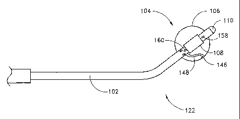
Figure 4 shows a circumferential ablation probe 100 during use in
performing positioning step (3) just described with reference to Figure 3. The
circumferential ablation probe 100 generally comprises a shaft 102, an
atraumatic tip
110, and a circumferential ablation member 104. The circumferential ablation
member 104 includes an expandable member 106 and an ablation element 108. The
ablation element 108 includes a circumferential band (shown in hatched) on the
outer surface of the expandable member that ablatively couples to the
surrounding
tissue to form a circumferential lesion.
More specifically, Figure 4 shows the circumferential ablation probe 100
subsequent to advancing the distal end portion into the inner atrium according
to
step (3) of Figure 3, and also subsequent to advancement and positioning of
the
circumferential ablation member 104 within a pulmonary vein, also according to
step (3) of Figure 3. Figure 4 also schematically illustrates the proximal end
of the
circumferential ablation probe 100 including an expansion actuator 154, an
ablation
actuator 156, and a ground patch 195.
Figure 4 shows the circumferential ablation probe 100 with the expandable
member 106 in a radially collapsed position adapted for delivery into the
puhnonary
vein according to positioning step (3) of Figure 3. However, the expandable
member 106 is adjustable to a radially expanded position when actuated by the
expansion actuator 154, as shown in Figure 5. The expansion actuator 154 may
include, but is not limited to, a pressurizable fluid source. According to the
expanded state shown in Figure 5, the expandable member 106 includes a working
length L relative to the longitudinal axis of the elongate catheter body which
has a
larger expanded outer diameter OD than when in the radially collapsed
position.
Furthermore, the expanded outer diameter OD is sufficient to circumferentially
engage the ablation region of the pulmonary vein. Therefore, the terms
"working
length" are herein intended to mean the length of an expandable member which,
when in a radially expanded position, has an expanded outer diameter that is:
(a)
greater than the outer diameter of the expandable membe'r when in a radially
collapsed position; and (b) sufficient to engage a body space wall or adjacent
ablation region surrounding the expandable member, at least on two opposing
internal sides of the body space wall or adjacent ablation region, with
sufficient
surface area to anchor the expandable member.
The circumferential ablation element 108 includes a circumferential band on
the outer surface of the expandable member 106 which is coupled to an ablation
-21-
CA 02411938 2002-12-13
WO 01/95820 PCT/US01/18724
actuator 156 at a proximal end portion of the probe shaft (shown schematically
in
Figure 4). The ablation element 108 is actuated by ablation actuator 156 to
ablatively couple to the surrounding circumferential path of tissue in the
puhnonary
vein wall, thereby forming a circumferential lesion that circumscribes the
puhnonary
vein lumen and transects the electrical conductivity of the pulmonary vein to
block
conduction in a direction along its longitudinal axis.
Figure 6 shows pulmonary vein 52 after removing the circumferential
ablation device assembly subsequent to forming a circumferential lesion 44
around
the ablation region of the pulmonary vein wall 48 according to the use of the
circumferential ablation probe assembly 100 shown in stepwise fashion in
Figures 3-
5. The circumferential lesion 44 is shown located along the pulmonary vein
adjacent
to the pulmonary vein ostium 54, and is shown to also be "transmural," which
is
herein intended to mean extending completely through the wall, from one side
to the
other. Also, the circumferential lesion 44 is shown in Figure 6 as a
"continuous"
lesion, which is herein intended to mean without gaps around the pulmonary
vein
wall circumference, thereby circumscribing the pulmonary vein lumen.
However, it is believed that a circumferential ablation probe with a
circumferential ablation element may leave some tissue, either transmurally or
along
the circumference of the lesion, which is not actually ablated, but which is
not
substantial enough to allow for the passage of conductive signals. Therefore,
the
terms "transmural" and "continuous" as just defined are intended to have
functional
limitations, wherein some tissue in the ablation region may be unablated but
there
are no functional gaps which allow for symptomatically arrhythmogenic signals
to
conduct through the conduction block and into the atrium from the pulmonary
vein.
Moreover, it is believed that the functionally transmural and continuous
lesion qualities just described are characteristic of a completed
circumferential
conduction block in the pulmonary vein. Such a circumferential conduction
block
thereby transects the vein, isolating conduction between the portion of the
vein on
one longitudinal side of the lesion and the portion on the other side.
Therefore, any
foci of originating arrhyt,hmogenic conduction which is opposite the
conduction
block from the atrium is prevented by the conduction block from conducting
down
into the atrium and atrial arrhythmic affects are therefore nullified.
Figures 7-8 illustrate another variation of a circumferential ablation member
204 that includes a radially compliant expandable member 206 and an ablation
element 208 adapted to ablatively couple to a larger region of tissue. Figure
7
-22-
CA 02411938 2002-12-13
WO 01/95820 PCT/US01/18724
illustrates the expandable member 206 after being adjusted to a radially
expanded
position while located in the left atrium. Figure 8 further shows the
expandable
member 206 after being advanced into the pulmonary vein 52 until at least a
portion
of the expanded working length L of the ablation element 208, which includes a
circumferential band, engages the puhnonary vein ostium (shown as 54 in Figure
7).
Figure 9 illustrates a portion of the circumferential lesion 44' that provides
a
circumferential conduction block in the region of the puhnonary vein ostium 54
subsequent to actuating the circumferential ablation element.
In the embodiment described in Figures 7-8, the expandable member 206 is
formed to also engage a circumferential path of tissue along the left
posterior atrial
wall that surrounds the ostium 54. Moreover, the ablation element 208 of the
circumferential ablation member 204 is also thereby adapted to engage that
atrial
wall tissue. Therefore, the circumferential lesion 44' formed according to the
method shown in part in Figure 9, and just described in sequential steps by
reference
to Figures 7-8, includes ablating a circumferential path of atrial wall tissue
which
surrounds the ostium 54. Accordingly, the entire pulmonary vein 52, including
the
ostium 54, is thereby electrically isolated from at least a substantial
portion of the
left atrial wall. The circumferential lesion 44' also isolates the other of
the
pulmonary vein ostia, as would be apparent to one of ordinary skill according
to the
sequential method steps shown in Figures 7-8 and by further reference to the
resulting circumferential lesion 44' shown in Figure 9.
Figure 10 shows yet another variation of a circumferential ablation member
308 and use thereof for electrically isolating a pulmonary vein and ostium
from a
substantial portion of the left posterior atrial wall. However, unlike the
embodiment
previously shown and described by reference to Figures 7-8, the Figure 10
embodiment isolates the pulmonary vein without also ablating tissue along the
lumen or lining of the puhnonary vein or ostium. This is apparent by reference
to
the resulting circumferential conduction block 44" shown in Figure 11.
In more detail, Figure 10 shows a similar device assembly as that shown in
Figures 7-8, except that ablation element 308 is adapted to ablatively couple
with
only a circumferential path of tissue along the left posterior atrial wall
which
surrounds the puhnonary vein ostium. In one aspect of this embodiment, the
compliant nature of the expandable member 306 may be self-conforming to the
region of the ostium such that the ablation element 308 is placed against this
atrial
wall tissue merely by way of conformability. Figure 11 illustrates a
circumferential
-23-
CA 02411938 2002-12-13
WO 01/95820 PCT/US01/18724
lesion 44" formed by the device assembly discussed with reference to Figure
10. As
shown, the circumferential lesion 44" is located along the posterior wall and
does
not extend into or around the ostium 54.
In another aspect of this embodiment, a "pear"-shaped expandable member
or balloon that includes a contoured taper may be suitable for use according
to the
Figure 10 embodiment. Such a pear shape may be preformed into the expandable
member or balloon, or the member may be adapted to form this shape by way of
controlled compliance as it expands, such as for example by the use of
composite
structures within the balloon construction. In any case, according to the
"pear"-
shaped variation, the circumferential band of the ablation member is
preferably
placed along the surface of the contoured taper which is adapted to face the
left
posterior atrial wall during use according to the method illustrated by Figure
10. It
is further contemplated that the ablation element may be further extended or
alternatively positioned along other portions of the taper.
The method of forming a circumferential conduction block along a
circumferential path of tissue along a left posterior atrial wall and which
surrounds a
puhnonary vein ostium without ablating the tissue of the vein or ostium should
not
be limited to the particular device embodiments just illustrated by reference
to
Figure 10. Other device variations may be acceptable substitute for use
according to
this method. In one particular example which is believed to be suitable, a
"looped"
ablation member such as the embodiment illustrated below by reference to
Figure 28
may be adapted to form a "looped" ablation element within the left atrium and
then
be advanced against the left posterior atrial wall such that the loop engages
the
circumferential path of tissue along the atrial wall and which surrounds a
vein
ostium. Thereafter, the looped ablation element may be actuated to ablate the
engaged tissue, such as for further illustration like a branding iron forming
the
predetennined pattern around the puhnonary vein ostium. In addition, other
device
or method variations may also be suitable substitutes according to one of
ordinary
skill.
Combining Circumferential Lesions with LongLinear Lesions
Figures 12-17 collectively illustrate a circumferential ablation device
assembly and method as used to form a circumferential lesion in combination
with
the formation of long linear lesions in a less-invasive "maze"-type procedure,
as
described above for the treatment of multiwavelet reentrant type fibrillation
along
the left atrial wall. As described in part by the flow diagram of Figure 12,
the
-24-
CA 02411938 2008-01-17
physician may use a li.uear ablation element to form linear conduction blocks
between the pulmonary vein ostia, wherein the circumferential ablation probe
ofthe
present invention is used to connect the linear lesions by forming
circumferential
ablation lesions around the pulmonary vein ostia.
More specifically, Figure 12 diagrammatically shows a summary of steps for
performing a`~aze"-type procedure by forming circumferential conduction blocks
tb.at intersect with long linear conduction blocks formed between the
pulmonary
veins. As disclosed in U.S. Patent No. 5,971,983 to Lesh entitled "Tissue
Ablation
Device and Method of Use".
' a box-like conduction block surrounding an arrhythmogenic atrial wall
region bounded by the pulmonary veins may be created by forming long linear
lesions between anchors in all pairs of adjacent puhnonary vein ostia. This
procedure is summarized in steps (5) and (6) of Figure 12. However, it is
further
believed that, in some particular applications, such linear lesions may be
made
sufficiently narrow with respect to the surface area of the pulmonary vein
ostia that
they may not intersect, thereby leaving gaps between them which may present
proarrhythmic pathways for abnormal conduction into and from the box. This is
iIlustrated in Figure 13 by the gaps between lesion 56 and 58 and also between
lesions 58 and 60. Therefore, by forming a circumferential conduction block
according to step (7) of Figure 12, and as shown by use of ablation element
208 in
Figare 14, the linear lesions are thereby bridged and the gaps, are closed.
Figure 15
illushates a lesion pattern formed by steps (5)-(7) of Figure 12. With the
addition of
circumferential lesion 44', there are no gaps between the linear lesions and
therefore
there are no proarhyttunic pathways for abnomzal conduction into and out of
the
box.
Moreover, the method shown schemaxically in Figure 12 and also in various
detail by reference to Figures 13-15 provides a specific sequence of steps for
the
purpose of illustration. According to this illust=ative sequence, the linear
lesions are
formed first and then are connected thereafter with the circumferential
conduction
bloek. However, a ciicumferential conduction block may be formed prior to the
foimation of the linear lesions or conduction blocks, or in any other
combination or
sub-combination of sequential steps, so long as the resulting combination of
lesions
allows for the circumferential block to intersect with and conaect with the
linear
lesions. In addition, the circumferential conduction block which conuects the
lineaz
lesions may also melude a circumferential path of tissue wlnch surrounds and
-25-
CA 02411938 2002-12-13
WO 01/95820 PCT/US01/18724
electrically isolates the pulmonary vein ostium from the rest of the left
posterior
atrial wall.
In addition to the particular embodiments just shown and described by
reference to Figures 12-15, other methods are also contemplated for combining
circumferential and linear conduction blocks device assemblies and uses in
order to
perform a less-invasive "maze"-type procedure. In a further example shown in
Figure 16, another lesion pattern is formed by combining the pair of linear
lesions of
Figure 13 with a circumferential conduction block 44". While the resulting
lesion
patterns of Figures 15 and 16 differ slightly as regards the particular
geometry and
position of the circumferential conduction block formed, the two variations
are also
similar in that the circumferential conduction block includes a
circumferential path
of atrial wall tissue. When such circumferential conduction blocks are formed
between adjacent pulmonary vein ostia, shorter linear lesions are therefore
sufficient
to bridge the circumferential lesions during the overall "maze"-type
procedure.
To this end, according to one contemplated less-invasive "maze"-type
procedure (not shown) wherein multiple circumferential conduction blocks are
formed in atrial wall tissue such that each pulmonary vein ostium is
surrounded by
and is electrically isolated with one circumferential conduction block. A
series of
four linear lesions may be formed between the various pairs of adjacent ostia
and
with just sufficient length to intersect with and bridge the corresponding
adjacent
circumferential blocks. A box-like conduction block is thereby formed by the
four
circumferential conduction blocks and the four bridging linear lesions. A
fifth linear
lesion may be also formed between at least a portion of the box-like
conduction
block and another predetermined location, such as for example the mitral value
annulus.
Figure 17 schematically illustrates yet a further variation for forming
circumferential conduction blocks along atrial wall tissue around the
pulmonary vein
ostia during a less invasive "maze"-type procedure. According to this further
variation, the circumferential conduction block patterns formed around each of
two
adjacent superior and inferior pulmonary vein ostia are shown in Figure 17 to
intersect, thereby alleviating the need for a linear lesion in order to form a
conduction block between the ostia. Furthermore, the distances between the
inferior
and superior ostia, both on the right and left side of the posterior atrial
wall, are
believed to be significantly shorter than the distances between the two
adjacent
superior or inferior ostia. Therefore, Figure 17 only shows the overlapping
-26-
CA 02411938 2002-12-13
WO 01/95820 PCT/US01/18724
circumferential conduction blocks as just described to be positioned
vertically
between the inferior-superior pairs of adjacent ostia, and further shows
linear lesions
which are used to connect the right and left sided ostia of the superior and
inferior
pairs. In some instances these linear lesions will not be required to cure,
treat or
prevent a particular atrial arrhythmia condition. However, other combinations
of
these patterns are further contemplated, such as for example using only
overlapping
circumferential conduction blocks between all adjacent pairs of ostia in order
to
form the entire "maze"-type left atrial pattem.
Monitoring; Electrical Simals During Surgical Procedure
Figure 18 diagranunatically shows a further method for using a
circumferential ablation device assembly wherein electrical signals along the
pulmonary vein are monitored with a sensing element before and after ablation
according to steps (8) and (9), respectively. Signals within the pulmonary
vein are
monitored prior to forming a conduction block, as indicated in step (8) in
Figure 18,
in order to confirm that the pulmonary vein chosen contains an arrhythmogenic
origin for atrial arrhythmia. Failure to confirm an arrhythmogenic origin in
the
pulmonary vein, particularly in the case of a patient diagnosed with focal
arrhythmia, may dictate the need to monitor signals in another pulmonary vein
in
order to direct treatment to the proper location in the heart. In addition,
monitoring
the pre-ablation signals may be used to indicate the location of the
arrhythmogenic
origin of the atrial arrhythmia, which helps determine the best location to
form the
conduction block. As such, the conduction block may be positioned to include
and
therefore ablate the actual focal origin of the arrhythmia, or may be
positioned
between the focus and the atrium in order to block aberrant conduction from
the
focal origin and into the atrial wall.
In addition or in the alternative to monitoring electrical conduction signals
in
the pulmonary vein prior to ablation, electrical signals along the pulmonary
vein
wall may also be monitored by the sensing element subsequent to
circumferential
ablation, according to step (9) of the method of Figure 18. This monitoring
method
aids in testing the efficacy of the ablation in forming a complete conduction
block
against arrhythmogenic conduction. Arrhythmogenic firing from the identified
focus will not be observed during signal monitoring along the pulmonary vein
wall
when taken below a continuous circumferential and transmural lesion formation,
and
thus would characterize a successful circumferential conduction block. In
contrast,
observation of such arrhythmogenic signals between the lesion and the atrial
wall
-27-
CA 02411938 2002-12-13
WO 01/95820 PCT/US01/18724
characterizes a functionally incomplete or discontinuous circumference (gaps)
or
depth (transmurality) which would potentially identify the need for a
subsequent
follow-up procedure, such as a second circumferential lesioning procedure in
the
ablation region.
A test electrode may also be used in a "post ablation" signal monitoring
method according to step (10) of Figure 18. In one particular embodiment not
shown, the test electrode is positioned on the distal end portion of a probe
shaft and
is electrically coupled to a current source for firing a test signal into the
tissue
surrounding the test electrode when it is placed distally or "upstream" of the
circumferential lesion in an attempt to simulate a focal arrhythmia. This test
signal
generally challenges the robustness of the circumferential lesion in
preventing atrial
arrhythmia from any such future physiologically generated aberrant activity
along
the suspect vein.
Further to the signal monitoring and test stimulus methods just described,
such methods may be performed with a separate electrode or electrode pair
located
on the probe distal end portion adjacent to the region of the circumferential
ablation
element, or may be performed using one or more electrodes which form the
circumferential ablation element itself, as will be further developed below.
Surgical Ablation Probe for Forming, a Circumferential Lesion
Circumferential ablation devices disclosed in the past are translumenal
catheter-based devices that are inserted into a peripheral vein (such as the
femoral
vein) and are advanced through a guide catheter into the right atrium and then
across
the septum into the left atrium. However, during certain open-heart or
minimally
invasive cardiac surgeries, the left atrium may be accessible through an
opening in a
patient's chest, thereby obviating the need for a catheter-based apparatus.
When the
physician can access the left atrium through an opening in a patient's chest,
a
relatively short and rigid surgical ablation probe is better suited for
placing an
ablation element in a selected pulmonary vein for the creation of a
circumferential
lesion.
With general reference to Figure 19, a preferred mode of the surgical ablation
probe in accordance with the present invention will now be described. The
surgical
ablation probe 100 comprises a handle 120 that includes multiple lumens and is
ergonomically designed to fit comfortably in the physician's hand. The handle
120
preferably is made of a molded or machined plastic. A suitable handle design
would
be similar to known handles used on the following types of devices: a laser
device to
-28-
CA 02411938 2008-01-17
perform trans-myocardial revascularization (TMR); a hand-held RF ablation
probe;
and a hand-held cryo-ablation probe. In the case of a deflectable tip version
of'the
probe, several exemplifying types of control handles are described in U.S.
Patent
Nos. 6,024,739 and 5,465,716:
The handle 120 supports a multi-lumen probe shaft 102 that is relatively
short in length and is generally rigid. The majority of the shaft 102 is
significently
less flexible than the catheter-based ablation devices described in U.S.
Patent No.
6,024,740. The distal end 122 prefcrably includes an atraumatic distal tip 110
made
of a soft thermoplastic. The shaft 102 preferably has a diameter ranging from
about
7 to about 12 F. However, this range of diameters merely exemplifies suitable
diameters for use in the described surgical procedure, and other diameter
probes of
course are also within the scope of the invention.
The snrgical ablation probe 100 also preferably, includes sensor leads 148, a
power cable 150, preferably a coaxial cable, and actuation means 154 for
deploying
the expandable member 106. These components extend from the handle 120,
through the lumens in the probe shaft 102, to the con-esponding components of
the
probe disposed on the distal end 122. The proximal ends of the cables and
lumens
connect to corresponding connectors 153, 155 that extend from the proximal end
of
the probe handle 120. The probe shaft 102 desirably includes a plarality of
lumens
(examples of which are illustrated in Figimo.s 22-23). Various wires and
electrical
leads are routed to the dista] end portion 122 through at least some of these
lumens. In
a prefen ed device, these lumen$ generally run the length of the shaft 102;
however, for
some applications, the lumens can be shorter.
The shafft 102 of the smgical ablation probe 100 is preferably formed with a
distal port 158 located distal to the ablation member 104 and a proximal port
160
located proximal of the ablation member 104. The distal port 158 allows the
clinician to introduce fluids into the patient, take fluid samples from the
patient, and
take fluid pressare reading on the distal side of the ablation member 104.
Similarly,
the proximal port 160 allows the clinician to introduce fluids into the
patient, take
fluid samples from the patient, and take fluid pressure reading on the
proximal side
of the ablation member 104. Thesc ports and lumens are patticulsrly useful
when
pressure or X-ray positioning techniques are employed, as explained below;
however, the probe assembly 100 need not include such ports and lumens, such
as
when only an A-mode or Doppler position monitoring system is used with the
probe
35- assembly.
-29-
CA 02411938 2002-12-13
WO 01/95820 PCT/US01/18724
The probe is primarily designed for use during trans-thoracic (open heart) or
minimally invasive cardiac surgery and can be used to form a conduction block
during the same surgery in which another procedure is being performed, e.g.,
when
repairing or replacing a mitral valve. For example, the physician may insert
the
probe through a chest access device, e.g., a trocar, or through an incision
during
open chest surgery. As illustrated in Figure 5, the probe shaft 102 is
introduced into
an atrium through an atriotomy 10 in the left atrial appendage, and is then
placed at a
location where a pulmonary vein extends from the posterior atrial wall.
Referring
again to Figure 19, the expandable member 106 is then expanded from its
collapsed
to its expanded state, (e.g., by inflation of a balloon by injection of
inflation fluid).
Once the expandable member 106 is positioned, in some cases engaged with,
along
the circumferential region of tissue, the physician actuates the ablation
element 108
to ablate the region of tissue. During open chest surgery, the short, rigid,
and
adjustable nature of the probe shaft 102 and handle make placement and
ablation
more efficient and precise than with a translumenal catheter-based apparatus.
It should be noted that' an open-heart procedure would require
cardiopulmonary bypass wherein the patient's blood is diverted and oxygenated
by
an extracorporeal device. In this case, the atrium and pulmonary vein region
will be
devoid of blood, flushed clear by saline. This would allow a relatively clear
field of
view in which to operate. It may be desirable for the entire atrium to be
flooded
with saline, such that the balloon is engulfed in fluid while inflated. This
may help
avoid a "dry interface" between the balloon and the tissue to be ablated; such
a "dry
interface" could be an impediment to ultrasound energy conduction into the
tissue
because ultrasound energy is substantially reflected by even a thin layer of
air.
Minimally invasive surgical procedures can be performed in similar fashion,
i.e. with cardio-pulmonary bypass. On the other hand, minimally invasive
cardiac
procedures are often done on a "beating heart", in which case blood flows
normally
through the cardio-pulmonary system. In this case, the probe would be used in
an
environment identical to that of the percutaneous translumenal catheter (i.e.
with
normal pulmonary vein/atrial blood flow). In such cases, it may be necessary
to
have a separate visualization system employed to aid placement of the ablation
probe, such as an endoscopic camera, intracardiac ultrasound probe, or
fluoroscopic
x-ray machine. It may also be desirable to include a perfusion lumen (not
shown)
that extends between ports located proximal and distal to the expandable
member.
Passive perfusion during expansion of the expandable member is believed to
-30-
CA 02411938 2002-12-13
WO 01/95820 PCT/US01/18724
minimize stasis and allow the target pulmonary vein to continue in its atrial
filling
function during the atrial arrythmia treatement procedure. Without this
perfusion
feature, the expandable member blocks the flow of blood from the vein to the
atrium
and may result in undesirable thrombogenesis in the pulmonary vein distal to
the
expandable member.
Figure 21 illustrates an enlarged view of the distal end 122 of the probe
shaft
102. The distal end 122 comprises a circumferential ablation member 104 having
an
expandable member 106 and an ablation element 108 (shown here as an ultrasound
transducer). The expandable member 106 may be provided with one or more
sensors 146 (e.g., temperature sensors or electrodes) on the exterior portion
thereof
for providing feedback to the physician during the ablation procedure.
The distal end 122 of the shaft 102 is preferably shaped to facilitate
placement initially into the atrium and then into a pulmonary vein ostium. For
example, the distal end may be angled to an angle of about 45 . The shaft 102
may
have a length ranging from about 20 to about 60 cm. These lengths and angles,
however, only exemplify one preferred form of the ablation probe 100 that has
been
found useful for a specific surgical procedure, and other lengths and shapes
of the
probe are also intended to be within the scope of this invention. In one
variation, the
shaft 102 may be shaped, by the physician manipulating the shaft, so that the
shaft
takes on a desired shape. In another variation, the probe may have a
deflectable
distal end which can be deflected by manipulation of a pull-wire system, as
described in further detail below.
It is also understood that the probe shaft 102 can be configured for following
different access paths into the atrium, such as, for example, but without
limitation,
via a retrograde procedure through the mitral valve, transeptally from a right
atriotomy, or through a left atriotomy. The probe shaft 102 may be made of
Pebax or
any other materials that provide adjustable shape, flexibility, and
maneuverability of
the probe. Other materials include for example, stainless steel, Nitinol,
thermoplastic braided material, polyamide braided tubing, etc.
Figure 20 shows the circumferential ablation probe 100 with the expandable
member 106 in a radially collapsed condition. In this configuration, the
ablation
probe 100 is adapted for delivery into the pulmonary vein according to
positioning
step (3) of Figure 3. The expandable member 106 is adjustable to a radially
expanded position when actuated by an expansion actuator 154. In a preferred
embodiment, the expansion actuator 154 comprises a pressurizeable fluid
source.
-31-
CA 02411938 2002-12-13
WO 01/95820 PCT/US01/18724
With reference to Figure 22, there is shown a cross-sectional view of the
probe shaft 102 taken along line 22-22 of the circumferential ablation probe
shown
in Figure 20. An outer extrusion 180 formed of a thin-walled, resilient tubing
defining the outer surface of the probe shaft 102. The outer extrusion 180 may
be
formed of any of the biocompatible resilient plastics typically used in
catheters, with
polyimide and polyurethane available under the trade name PEBAX (from Atochem
of Glen Rock, N.J.) being preferred materials.
Disposed within the outer extrusion 180, and radially outside of the inner
probe surface 182 is an intermediate layer 184, which is adapted to transmit
torque
along the probe shaft 102 so that a physician can turn the probe distal end
portion
152 by suitable manipulation of the handle 120. A preferred torque-
transmitting
material for the intermediate layer 184 is a metal braid formed of interleaved
lengths
of stainless steel encapsulated within the resilient plastic outer extrusion
180.
As illustrated in Figure 22, several lumens may be disposed within the probe
shaft 102, including, for example, a deflecting wire lumen 198, a coaxial
cable
lumen 192, a perfusion lumen 196, and a thermocouple leads lumen 194. When an
inflatable balloon is used as an expandable member, the probe shaft 102 also
includes an inflation lumen 190. The inflation lumen 190 preferably has an
inner
diameter of about 0.020 inch in order to allow for rapid deflation times,
although this
may vary based upon the viscosity of inflation medium used, length of the
lumen
190, and other dynamic factors relating to fluid flow and pressure.
With reference to Figure 23, there is shown a cross-sectional view of the
probe shaft 102 taken along line 23-23 of the circumferential ablation probe
shown
in Figure 20. The same lumens are present within the shaft 102 as described
with
reference to the proximal region shown in Figure 22, however, the
intermediate,
torque-transmitting braid (184 in Figure 22) is not present within the outer
extrusion
(180 in Figure 22). Moreover, the outer extrusion itself is thinner, to reduce
the
stiffness of the distal end portion 152. Thus, for illustrative purposes,
because of the
relative thinness of the shaft wall 185 in the distal region, the wall (inner
and outer
surfaces) is labeled using a single reference numeral 185.
Referring now to Figure 24, a preferred embodiment of the surgical ablation
probe 100 further comprises a deflectable tip design to independently select a
desired pulmonary vein and direct the transducer assembly toward the desired
location. Further to the deflectable variation, a deflecting pull wire is
incorporated
into the probe shaft 102. The pull wire is attached to the atraumatic tip 110
of the
-32-
CA 02411938 2002-12-13
WO 01/95820 PCT/US01/18724
shaft 102, slidably engaged within a pull-wire lumen (198 in Figure 22 and 23)
in the
shaft 102, and attached to a deflection mechanism within the handle 120. The
pull
wire is adapted to deflect the distal probe tip by applying tension along
varied
stiffness transitions along the probe's length. Still further to this pull
wire variation,
acceptable pull wires may have a diameter within the range from about 0.008
inch to
about 0.020 inch, and may further include a taper, such as, for example, a
tapered
outer diameter from about 0.020 inch to about 0.008 inch.
Still referring to Figure 24, deflection of the distal end 122 of the ablation
probe 100 is preferably produced by manipulation of a thumb slide 141 located
on
the handle 120. When the thumb slide 141 is moved from position A to position
A'
(drawn in phantom), the distal end portion 122 of probe shaft 102 is deflected
from
position B (zero deflection) to position B'. Likewise, when the thumb slide
141 is
moved from position A to position A" (drawn in phantom), the distal end
portion
122 of the shaft 102 is deflected from position B (zero deflection) to
position B".
Although, a variety of deflection handles are known in the art, they generally
operate
like the BIOSENSE/WEBSTER handles by placing tension on the proximal end of a
pull wire which is slidably engaged within the probe shaft and fixed to the
distal end
portion.
Figure 25 illustrates an expanded schematic view of one preferred
embodiment of the proximal end portion of the probe shaft. It is understood,
however, that any other extensions and modifications within the skill of those
in the
art are encompassed within the scope of the present disclosure. Here,
surrounding
the proximal end of the probe shaft 102 is a shrink-wrap layer 118, formed
from 1/8
inch plastic shrink-wrap, such as for example PET. The inflation lumen is
preferably extended about 16.5 cm using a hypotube 116, preferably of
.042"/.035".
The coaxial cable 110 extends about 16 cm proximally from the proximal end
portion 126. A .008" PTFE-coated mandrel was used for the deflecting pull wire
114, which is shown slidably engaged in a.026/.013" Teflon tube 112. The
Teflon
tubing 112 extends only about 1 cm past the proximal end portion and the pull
wire
114 extends about 4 cm beyond the proximal end portion, where it connects to
the
handle (not shown).
Referring again to Figure 19, the circumferential ablation member 104 of the
probe 100 will be discussed in further detail. The expandable member 106 of
ablation probe 100 preferably comprises a compliant elastomeric balloon or a
non-
compliant balloon made from silicone, latex, rubber, and polyvinylchloride,
with an
-33-
CA 02411938 2002-12-13
WO 01/95820 PCT/US01/18724
expandable diameter of between about 10 and about 40 mm. The probe shaft 102
includes an inflation lumen that communicates with the interior of the
balloon, and
the handle includes a balloon inflation/deflation port. The port is coupled to
a
source of pressurized inflation medium in a known manner to inflate the
balloon.
The ablation element 108 is disposed on the distal tip 122 and cooperates
with the expandable member 106 such that the ablation element 108 is held in a
generally fixed position relative to the target circumferential region of
tissue. In the
preferred embodiment, the ablation element 108 is an ultrasound transducer
adapted
to emit ultrasonic sound waves sufficient to ablate a circumferential region
of tissue
when coupled to a suitable excitation source. It is believed that driving the
ultrasonic
transducer at 20 acoustical watts at an operating frequency within the range
of 7 - 10
megahertz will form a sufficiently sized circumferntial lesion about the
pulmonary
vein ostium in a relatively short period of time (e.g., 1 to 2 minutes or
less).
The ablation element can be located outside or inside the expandable
member, or can be located at least partially outside the expandable member.
The
ablation element, in some forms, also includes a portion of the expandable
member.
The preferred embodiment illustrated in Figure 19 shows the ultrasonic
transducer
108 located within the expandable member 106. Electrical leads extend through
lumens in the probe shaft 102 and connect to one or more electrical connectors
that
extend from the probe handle.
It is also contemplated that the control level of energy can be delivered,
then
tested for lesion formation with a test stimulus in the puhnonary vein, either
from an
electrode provided at the tip area of the probe or on a separate device.
Therefore, the
procedure may involve ablation at a first energy level in time, then check for
the
effective conductive block provided by the resulting lesion, and then
subsequent
ablations and testing until a complete conductive block is formed.
In addition to the particular embodiment just described, the ultrasonic
ablation element and expandable member located at the distal end 122 of the
probe
shaft 102 can take a variety of other configurations. With regard to the
inflatable
balloon 106 shown in Figure 19, a central region is generally coaxially
disposed over
the probe shaft and is bordered at its end neck regions by proximal and distal
adaptions. The proximal adaption is sealed over the elongate body proximally
of the
distal inflation and the electrical lead ports and the distal adaption is
sealed over the
probe shaft proximal of the distal tip. According to this arrangement, a fluid
tight
interior chamber is formed within the inflatable balloon 106. This interior
chamber
-34-
CA 02411938 2002-12-13
WO 01/95820 PCT/US01/18724
is fluidly coupled to a pressurizeable fluid source via the inflation lumen
within the
probe shaft. In addition to the inflation lumen, the electrical lead lumen
also
communicates with the interior chamber of expandable balloon so that the
ultrasound transducer, which is positioned within that chamber and over the
shaft,
may be electrically coupled to the ultrasound drive source or actuator.
The inflatable balloon 106 may be constructed from a variety of known
materials, although the balloon preferably is adapted to conform to the
contour of a
pulmonary vein ostium. For this purpose, the balloon material can be of the
highly
compliant variety or of a predefined shape. Because the probe is not
restricted in
profile as is a percutaneous translumenal catheter, the balloon can have a
significantly
larger collapsed profile than the shaft diameter. This allows greater latitude
in the
possible balloon configurations and expanded diameter, including non-compliant
balloons, complex shaped balloons, balloons with dramatic surface features
such as
bumps or ridges, and non-balloon expandable members. These features may allow
treatment of veins with large diameter or difficult shape that are not
conducive to the
limitations of a percutaneous translumenal catheter design.
The designs for an expandable member and circumferential ablation element
for use in a circumferential ablation device assembly have been described
generically with reference to the embodiments shown in the previous figures.
Examples of more specific expandable member and ablation element embodiments
which are adapted for use in such ablation device assemblies are further
provided as
follows.
Notwithstanding their somewhat schematic detail, the circumferential
ablation members shown in the previous figures illustrate one particular
embodiment
wherein a circumferential electrode element circumscribes an outer surface of
an
expandable member. The expandable member of the embodiments shown may take
one of several different forms, although the expandable member is generally
herein
shown as an inflatable balloon that is coupled to an expansion actuator 154
and
wherein the expansion actuator 154 comprises a pressurizeable fluid source.
The
balloon is preferably made of a polymeric material and forms a fluid chamber
which
communicates with a fluid passageway (not shown in the figures) that extends
proximally along the elongate probe body and terminates proximally in a
proximal
fluid port that is adapted to couple to the pressurizeable fluid source.
In one expandable balloon variation, the balloon is constructed of relatively
inelastic plastics (e.g., polymers or monomers) such as a polyethylene ("PE";
-35-
CA 02411938 2002-12-13
WO 01/95820 PCT/US01/18724
preferably linear low density or high density or blends thereo fl, polyolefin
copolymer ("POC"), polyethylene terepthalate ("PET"), polyimide, or a nylon
material. In this construction, the balloon has a low radial yield or
compliance over
a working range of pressures and may be folded into a predetermined
configuration
when deflated in order to facilitate introduction of the balloon into the
desired
ablation location. In this variation, one balloon size may not suitably engage
all
pulmonary vein walls for performing the circumferential ablation methods
herein
described on all needy patients. Therefore, it is further contemplated that a
kit of
multiple ablation probes, with each balloon working length having a unique
predetermined expanded diameter, may be provided from which a treating
physician
may chose a particular device to meet a particular patient's pulmonary vein
anatomy.
In an alternative expandable balloon variation, the balloon is constructed of
a
relatively compliant, elastomeric material, such as, for example (but not
limited to),
a silicone, latex, polyurethane, or mylar elastomer. In this construction, the
balloon
takes the form of a tubular member in the deflated, non-expanded state. When
the
elastic tubular balloon is pressurized with fluid such as in the previous,
relatively
non-compliant example, the material forming the wall of the tubular member
elastically deforms and stretches radially to a predetermined diameter for a
given
inflation pressure. It is further contemplated that the compliant balloon may
be
constructed as a composite, such as, for example, a latex or silicone balloon
skin
which includes fibers, such as metal, Kevlar, or nylon fibers, which are
embedded
into the skin. Such fibers, when provided in a predetermined pattern such as a
mesh
or braid, may provide a controlled compliance along a preferred axis,
preferably
limiting longitudinal compliance of the expandable member while allowing for
radial compliance.
It is believed that, among other features, the relatively compliant variation
may provide a wide range of working diameters, which may allow for a wide
variety
of patients, or of vessels within a single patient, to be treated with just
one or a few
devices. Furthermore, this range of diameters is achievable over a relatively
low
range of pressures, which is believed to diminish a potentially traumatic
vessel
response that may otherwise be presented concomitant with higher pressure
inflations, particularly when the inflated balloon is oversized to the vessel.
In
addition, the low-pressure inflation feature of this variation is suitable
because the
functional requirement of the expandable balloon is merely to engage the
ablation
-36-
CA 02411938 2002-12-13
WO 01/95820 PCT/US01/18724
element against a circumferential path along the inner lining of the pulmonary
vein
wall.
Moreover, a circumferential ablation member is adapted to conform to the
geometry of the pulmonary vein ostium, at least in part by providing
substantial
compliance to the expandable member, as was shown and described previously by
reference to Figures 10-11. Further to this conformability to pulmonary vein
ostium
as provided in the specific design of Figure 10, the working length L of
expandable
member 306 is also shown to include a taper which has a distally reducing
outer
diameter from a proximal end to a distal end. In either a compliant or the non-
compliant balloon, such a distally reducing tapered geometry adapts the
circumferential ablation element to conform to the funneling geometry of the
puhn.onary veins in the region of their ostia in order to facilitate the
formation of a
circumferential conduction block there.
The circumferential ablation probe of the present invention preferably
comprises an ultrasound ablation element for ablating the surrounding tissue.
However, the circumferential ablation probe may be used with a wide variety of
ablation elements. For example, in another preferred embodiment, the outer
surface
of the expandable member includes one or more electrode bands adapted to
ablatively couple to the surrounding tissue to form circumferential lesions.
The
electrode bands are energized by an ablation actuator that generally includes
a radio-
frequency ("RF") current source (not shown) coupled to both the RF electrode
element and also a ground patch 195 which is in skin contact with the patient
to
complete an RF circuit. In addition, the ablation actuator preferably includes
a
monitoring circuit (not shown) and a control circuit (not shown) which
together use
either the electrical parameters of the RF circuit or tissue parameters such
as
temperature in a feedback control loop to drive current through the electrode
element
during ablation. Also, where a plurality of ablation elements or electrodes in
one
ablation element are used, a switching means may be used to multiplex the RF
current source between the various elements or electrodes.
Figures 26A-D show various patterns of electrically conductive,
circumferential electrode bands used as electrode ablation elements, each
circumscribing an outer surface of the working length of an expandable member.
Figures 26A-B show circumferential ablation member 550 to include a continuous
circumferential electrode band 552 that circumscribes an outer surface of an
expandable member 570. Figure 26B more specifically shows expandable member
-37-
CA 02411938 2002-12-13
WO 01/95820 PCT/US01/18724
570 as a balloon which is fluidly coupled to a pressurizeable fluid source),
and
further shows electrode band (circumferential ablation element) 552
electrically
coupled via electrically conductive lead 554 to ablation actuator 156. In
addition, a
plurality of apertures 572 is shown in the balloon skin wall of expandable
member
570 adjacent to electrode band 552. The purpose of these apertures 572 is to
provide
a positive flow of fluid such as saline or ringers lactate fluid into the
tissue
surrounding the electrode band 552. Such fluid flow is believed to reduce the
temperature rise in the tissue surrounding the electrode element during RF
ablation.
The shapes shown collectively in Figures 26A-D allow for a continuous
electrode band to circumscribe an expandable member's working length over a
range
of expanded diameters, a feature which is believed to be particularly useful
with a
relatively compliant balloon as the expandable member. In the particular
embodiments of Figures 26A-D, this feature is provided primarily by a
secondary
shape given to the electrode band relative to the longitudinal axis of the
working
length of the expandable member. Electrode band 552 is thus shown in Figures
26A-B to take the specific secondary shape of a modified step curve. Other
shapes
than a modified step curve are also suitable, such as the serpentine or
sawtooth
secondary shapes shown respectively in Figures 26C-D. Other shapes in addition
to
those shown in Figures 26A-D and which meet the defined functional
requirements
are further contemplated.
In addition, the electrode band provided by the circumferential ablation
elements shown in Figures 26C-D has a functional band width w relative to the
longitudinal axis of the working length which is only required to be
sufficiently wide
to form a complete conduction block against conduction along the walls of the
pulmonary vein in directions parallel to the longitudinal axis. In contrast,
the
working length L of the respective expandable element is adapted to securely
anchor
the distal end portion in place such that the ablation element is firmly
positioned at a
selected region of the pulmonary vein for ablation. Accordingly, the band
width w is
relatively narrow compared to the working length L of the expandable element,
and
the electrode band may thus form a relatively narrow equatorial band which has
a
band width that is less than two-thirds or even one-half of the working length
of the
expandable element. Additionally, it is to be noted here and elsewhere
throughout
the specification, that a narrow band may be placed at locations other than
the
equator of the expandable element, preferably as long as the band is bordered
on
both sides by a portion of the working length L.
-38-
CA 02411938 2002-12-13
WO 01/95820 PCT/US01/18724
In another aspect of the narrow equatorial band variation for the
circumferential ablation element, the circumferential lesion formed may also
*be
relatively narrow when compared to its own circumference, and may be less than
two-thirds or even one-half its own circumference on the expandable element
when
expanded. In one arrangement which is believed to be suitable for ablating
circumferential lesions in the pulrimonary veins as conduction blocks, the
band width
w is less than 1 cm with a circumference on the working length when expanded
that
is greater than 1.5 cm.
Figures 26E-F show a further variation of a circumferential ablation element
which is adapted to maintain a continuous circumferential lesion pattern over
a range
of expanded diameters and which includes electrode elements that form a
relatively
narrow equatorial band around the working length of an expandable balloon
member. In this variation, a plurality of individual electrode/ablation
elements 562
are included in the circumferential ablation element and are positioned in
spaced
arrangement along an equatorial band which circumscribes an outer surface of
the
expandable member's working length L.
The size and spacing between these individual electrode elements 562, when
the balloon is expanded, is adapted to form a substantially continuous
circumferential lesion in pulmonary vein wall tissue when in intimal contact
adjacent thereto, and is further adapted to form such a lesion over a range of
band
diameters as the working length is adjusted between a variety of radially
expanded
positions. Each individual electrode element 562 has two opposite ends 563,
564,
respectively, along a long axis LA and also has a short axis SA, and is
positioned
such that the long axis LA is at an acute angle relative to the longitudinal
axis LA of
the elongate probe body and expandable member 560. At least one of the ends
563,
564 along the long axis LA overlaps with an end of another adjacent individual
electrode element, such that there is a region of overlap along their
circumferential
aspect, i.e., there is a region of overlap along the circumferential
coordinates. The
terms "region of overlap along their circumferential coordinate" are herein
intended
to mean that the two adjacent ends each are positioned along the working
length
with a circumferential and also a longitudinal coordinate, wherein they share
a
common circumferential coordinate. In this arrangement, the circumferential
compliance along the working length, which accompanies radial expansion of the
expandable member also, moves the individual electrode elements apart along
the
circumferential axis. However, the spaced, overlapping arrangement described
-39-
CA 02411938 2002-12-13
WO 01/95820 PCT/US01/18724
allows the individual ablation elements to maintain a certain degree of their
circumferential overlap, or at least remain close enough together, such that a
continuous lesion may be formed without gaps between the elements.
The construction for suitable circumferential electrode elements in the RF
variations herein described, such as the various electrode embodiments
described
with reference to Figures 26A-F, may comprise a metallic material deposited on
the
outer surface of the working length using conventional techniques, such as by
plasma depositing, sputter coating, chemical vapor deposition, other known
techniques which are equivalent for this purpose, or otherwise affixing a
metallic
shaped member onto the outer surface of the expandable member such as through
known adhesive bonding techniques. Other RF electrode arrangements are also
considered, so long as they form a circumferential conduction block as
previously
described. For example, a balloon skin may itself be metallized, such as by
mixing
conductive metal, including but not limited to gold, platinum, or silver, with
a
plastic (e.g., polymer) to fonn a compounded, conductive matrix as the balloon
skin.
Still further to the RF electrode embodiments, another circumferential
ablation member variation (not shown) may also include an expandable member,
such as an inflatable balloon, that includes a porous skin that is adapted to
allow
fluid, such as hypertonic saline solution, to pass from an internal chamber
defined by
the skin and outwardly into surrounding tissues. Such a porous skin may be
constructed according to several different methods, such as by forming holes
in an
otherwise contiguous plastic (e.g., polymeric) material, including
mechanically
drilling or using laser energy, or the porous skin may simply be an inherently
porous
membrane. In any case, by electrically coupling the fluid within the porous
balloon
skin to an RF current source (preferably monopolar), the porous region of the
expandable member serves as an RF electrode wherein RF current flows outwardly
through the pores via the conductive fluid. In addition, it is further
contemplated
that a porous outer skin may be provided externally of another, separate
expandable
member, such as a separate expandable balloon, wherein the conductive flui& is
contained in a region between the porous outer skin and the expandable member
contained therein. Various other "fluid electrode" designs than those
specifically
herein described may also be suitable according to one of ordinary skill upon
review
of this disclosure.
In the alternative, or in addition to the RF electrode variations just
described,
the circumferential ablation element may also include other ablative energy
sources
-40-
CA 02411938 2002-12-13
WO 01/95820 PCT/US01/18724
or sinks, and particularly may include a thermal conductor that circumscribes
the
outer circumference of the working length of an expandable member. Examples of
suitable thermal conductor arrangements include a metallic element which may,
for
example, be constructed as previously described for the more detailed RF
embodiments above. However, in the thermal conductor embodiment such a
metallic element would be generally either resistively heated in a closed loop
circuit
internal to the probe, or conductively heated by a heat source coupled to the
thermal
conductor. In the latter case of conductive heating of the thermal conductor
with a
heat source, the expandable member may be, for example, a plastic (e.g.,
polymeric)
balloon skin which is inflated with a fluid that is heated either by a
resistive coil or
by bipolar RF current. In any case, it is believed that a thermal conductor on
the
outer surface of the expandable member is suitable when it is adapted to heat
tissue
adjacent thereto to a temperature between 40 and 80 Celsius.
Further to the thermal conduction variation for the circumferential ablation
element, a perfusion balloon embodiment may be particularly useful in such a
design. It is believed that ablation through increased temperatures, as
provided by
example above may also enhance coagulation of blood in the pulmonary vein
adjacent to the expandable member, which blood would otherwise remain stagnant
without such a perfusion feature.
One further circumferential ablation element design which is believed to be
highly useful in performing the ablation methods herein described is shown in
Figure 27A to include a circumferential ablation member 600 with two
insulators
602, 604 that encapsulate the proximal and distal ends, respectively, of the
working
length L of an expandable member 610. In the particular embodiment shown, the
insulators 602, 604 are thermal insulators, such as a thermal insulator
comprising a
Teflon material. Expandable member 610 is an inflatable balloon which has a
balloon skin 612 that is thermally conductive to surrounding tissue when
inflated
with a heated fluid which may contain a radiopaque agent, saline fluid,
ringers
lactate, combinations thereof, other known biocompatible fluids having
acceptable
heat transfer properties for these purposes, further to the thermal conductor
embodiments previously described. By providing these spaced insulators, a
circumferential ablation element is formed as an equatorial band 603 of
uninsulated
balloon skin is located between the opposite insulators. In this
configuration, the
circumferential ablation element is able to conduct heat externally of the
balloon
skin much more efficiently at the uninsulated equatorial band 603 than at the
-41-
CA 02411938 2002-12-13
WO 01/95820 PCT/US01/18724
insulated portions, and thereby is adapted to ablate only a circumferential
region of
tissue in a pulmonary vein wall which is adjacent to the equatorial band. It
is fiiirthher
noted that this embodiment is not limited to an "equatorial" placement of the
ablation element. Rather, a circumferential band may be formed anywhere along
the
working length of the expandable member and circumscribing the longitudinal
axis
of the expandable member as previously described.
Figure 27A further shows use of a radiopaque marker 620 to identify the
location of the equatorial band 603 in order to facilitate placement of that
band at a
selected ablation region of a pulmonary vein via X-ray visualization.
Radiopaque
marker 620 is opaque under X-ray, and may be constructed, for example, of a
radiopaque metal such as gold, platinum, or tungsten, or may comprise a
radiopaque
plastic (e.g., polymer) such as a metal loaded polymer. Such a radiopaque
marker
may also be combined with the other embodiments herein shown and described. To
note, when the circumferential ablation member which forms an equatorial band
includes a metallic electrode element, such electrode may itself be radiopaque
and
may not require use of a separate marker as just described.
The thermal insulator embodiment just described by reference to Figure 27A
is illustrative of a broader embodiment, wherein a circumferential ablation
member
has an ablating surface along the entire working length of an expandable
member,
but is shielded from releasing ablative energy into surrounding tissues except
for
along an unshielded or uninsulated equatorial band. As such, the insulator
embodiment contemplates other ablation elements, such as the RF embodiments
previously described above, which are provided along the entire working length
of
an expandable member and which are insulated at their ends to selectively
ablate
tissue only about an uninsulated equatorial band.
In a further example using the insulator embodiment in combination with a
circumferential RF electrode embodiment, a metallized balloon which includes a
conductive balloon skin may have an electrical insulator, such as a plastic
(e.g.,
polymeric) coating, at each end of the working length and thereby selectively
ablate
tissue with electricity flowing through the uninsulated equatorial band. In
this and
other insulator embodiments, it is further contemplated that the insulators
described
may be only partial and still provide the equatorial band result. For
instance, in the
conductive RF electrode balloon case, a partial electrical insulator will
allow a
substantial component of current to flow through the uninsulated portion due
to a
"shorting" response to the lower resistance in that region.
-42-
CA 02411938 2002-12-13
WO 01/95820 PCT/US01/18724
In still a fiirther example of an insulator combined with a RF ablation
electrode, a porous membrane comprises the entire balloon skin of an
expandable
member. By insulating the proximal and distal end portions of the working
length of
the expandable member, only the pores in the unexposed equatorial band region
are
allowed to effuse the electrolyte which carries an ablative RF current.
Further to the expandable member design for use in a circumferential
ablation member as herein described, other expandable members than a balloon
are
also considered suitable. For example, in one expandable cage embodiment shown
in Figure 27B, cage 650 comprises coordinating wires 651 and is expandable to
engage a desired ablation region in a puhnonary vein.
The radial expansion of cage 650 is accomplished as follows. Sheath 652 is
secured around the wires proximally of cage 650. However, core 653, which may
be
a metallic mandrel such as stainless steel, extends through sheath 652 and
distally
within cage 650 wherein it terminates in a distal tip 656. Wires 651 are
secured to
distal tip 656, for example, by soldering, welding, adhesive bonding, heat
shrinking
a plastic (e.g., polymeric) member over the wires, or any combination of these
methods. Core 653 is slidable within sheath 652, and may, for example, be
housed
within a tubular lumen (not shown) within sheath 652, the wires being housed
between a coaxial space between the tubular lumen and sheath 652. By moving
the
sheath 652 relative to core 653 and distal tip 656, the cage 650 is
collapsible along
its longitudinal axis in order to force an outward radial bias to wires 651 in
an
organized fashion to formed a working length of cage 650 which is expanded
(not
shown).
Further to the particular expandable cage embodiment shown in Figure 27B,
a plurality of ablation electrodes 655 is shown, each being positioned on one
of
wires 651 and being similarly located along the longitudinal axis of the cage
650.
The radial bias given to wires 651 during expansion, together with the
location of
the ablation electrodes 655, serves to position the plurality of ablation
electrodes/elements 655 along a circumferential, equatorial band along the
expanded
working length of cage 650. The wires forming a cage according to this
embodiment may also have another predetermined shape when in the radially
expanded position. For example, a taper similar to that shown for expandable
member 106 in Figure 19 may be formed by expanding cage 650, wherein the
ablation element formed by ablation electrodes 655 may be positioned between
the
proximal end and the distal end of the taper.
-43-
CA 02411938 2002-12-13
WO 01/95820 PCT/US01/18724
Further to the construction of the embodiment shown in Figure 27B, wires
651 are preferably metal, and may comprise stainless steel or a superelastic
metal
alloy, such as an alloy of nickel and titanium, or a combination of both.
Regarding
the case of nickel and titanium construction for wires 655, a separate
electrical
conductor may be required in order to actuate ablation electrodes 655 to
efficiently
emit ablative current into surrounding tissues. In the case where wires 651
are
constructed of stainless steel, they may also serve as electrical conductors
for
ablation electrodes 655. Further to the stainless steel design, the wires 651
may be
coated with an electrical insulator to isolate the electrical flow into
surrounding
tissues at the site of the ablation electrodes 655. Moreover, the ablation
electrodes
655 in the stainless steel wire variation may be formed simply by removing
electrical insulation in an isolated region to allow for current to flow into
tissue only
from that exposed region.
In a further cage embodiment (not shown) to that shown in Figure 27B, a
circumferential strip of electrodes may also be secured to the cage 650 such
that the
strip circumscribes the cage at a predetermined location along the cage's
longitudinal axis. By expanding cage 650 as previously described, the strip of
electrodes are adapted to take a circumferential shape according to the shape
of the
expanded cage 650. Such an electrode strip is preferably flexible, such that
it may
be easily reconfigured when the cage is adjusted between the radially
collapsed and
expanded positions and such that the strip may be easily advanced and
withdrawn
with the cage within the delivery sheath. Furthermore, the electrode strip may
be a
continuous circumferential electrode such as a conductive spring coil, or may
be a
flexible strip which includes several separate electrodes along its
circumferential
length. In the latter case, the flexible strip may electrically couple all of
the
electrodes to a conductive lead that interfaces with a drive circuit, or each
electrode
may be separately coupled to one or more such conductive leads.
Another circumferential ablation element adapted for use in a circumferential
conduction block assembly of the type herein described is shown in Figure 28,
wherein circumferential ablation member 700 includes a looped member 710
attached, preferably by heat shrinking, to a distal end of a pusher 730.
Looped
member 710 and pusher 730 are slidably engaged within delivery sheath 750 such
that looped member 710 is in a first collapsed position when positioned and
radially
confined within delivery sheath 750, and expands to a second expanded position
when advanced distally from delivery sheath 750.
-44-
CA 02411938 2002-12-13
WO 01/95820 PCT/US01/18724
Looped member 710 is shown in more detail in Figure 28 to include a core
712 which is constructed of a superelastic metal alloy such as a nickel-
titanium alloy
and which has a looped portion with shape memory in the looped configuration.
This looped configuration is shown in Figure 28 to be in a plane which is off-
axis,
preferably perpendicular, to the longitudinal axis of the pusher 730. This off-
axis
orientation of the loop is adapted to engage a circumferential path of tissue
along a
pulmonary vein wall which circumscribes the pulmonary vein lumen when the
looped member 710 is delivered from the delivery sheath 750 when the delivery
sheath is positioned within the vein lumen parallel to its longitudinal axis.
An
ablation electrode 714 is also shown in Figure 28 as a metallic coil which is
wrapped
around core 712 in its looped portion.
Pusher 730 is further shown in Figure 28 to include a tubular pusher member
732 which is heat shrunk over two ends 712' of core 712 which extend
proximally
of looped member 710 through pusher 730 in the particular variation shown.
While
in this embodiment core 712 extends through the pusher in order to provide
stiffiiess
to the composite design for the pusher, it is further contemplated that the
superelastic
metal of the core may be replaced or augmented in the pusher region with
another
different mandrel or pusher core (not shown), such as a stiffer stainless
steel
mandrel. Also shown within pusher 730 is an electrically conductive lead 735
which
is coupled to the ablation electrode 714 and which is also adapted in a
proximal
region of the pusher (not shown) to couple to an ablation actuator 156 such as
an RF
current source (shown schematically).
Figures 29A-31B show various specific embodiments of a broader
circumferential ablation device assembly which utilizes an ultrasonic energy
source
to ablate tissue. The present circumferential ablation device has particular
utility in
connection with forming a circumferential lesion within or about a pulmonary
vein
ostium or within the vein itself in order to form a circumferential conductive
block.
This application of the present ablation device, however, is merely exemplary,
and it
is understood that those skilled in the art can readily adapt the present
ablation
device for applications in other body spaces.
As common to each of the following embodiments, a source of acoustic
energy is provided for a delivery device that also includes an anchoring
mechanism.
In one mode, the anchoring mechanism comprises an expandable member that also
positions the acoustic energy source within the body; however, other anchoring
and
positioning devices may also be used, such as, for example, a basket
mechanism. In
-45-
CA 02411938 2002-12-13
WO 01/95820 PCT/US01/18724
a more specific form, the acoustic energy source is located within the
expandable
member and the expandable member is adapted to engage a circumferential path
of
tissue either about or along a pulmonary vein in the region of its ostium
along a left
atrial wall. The acoustic energy source in turn is acoustically coupled to the
wall of
the expandable member and thus to the circumferential region of tissue engaged
by
the expandable member wall by emitting a circumferential and longitudinally
collimated ultrasound signal when actuated by an acoustic energy driver. The
use of
acoustic energy, and particularly ultrasonic energy, offers the advantage of
simultaneously applying a dose of energy sufficient to ablate a relatively
large
surface area within or near the heart to a desired heating depth without
exposing the
heart to a large amount of current. For example, a collimated ultrasonic
transducer
can form a lesion, which has about a 1.5 mm width, about a 2.5 mm diameter
lumen,
such as a pulmonary vein and of a sufficient depth to form an effective
conductive
block. It is believed that an effective conductive block can be formed by
producing a
lesion within the tissue that is transmural or substantially transmural.
Depending upon
the patient as well as the location within the pulmonary vein ostium, the
lesion may
have a depth of 1 millimeter to 10 millimeters. It has been observed that the
collimated ultrasonic transducer can be powered to provide a lesion having
these
parameters so as to form an effective conductive block between the pulmonary
vein
and the posterior wall of the left atrium.
With specific reference now to the embodiment illustrated in Figures 29A
through 29D, a circumferential ablation device assembly 800 includes a shaft
802
with proximal and distal end portions 810,812, an expandable balloon 820
located
along the distal end portion 812 of elongate body 802, and a circumferential
ultrasound transducer 830 which forms a circumferential ablation member which
is
acoustically coupled to the expandable balloon 820. In more detail, Figures
29A-C
variously show shaft 802 to include inflation lumen 806 and electrical lead
lumen
808.
Each lumen extends between a proximal port (not shown) and a respective
distal port, which distal ports are shown as distal inflation port 807 for
inflation
lumen 806, and distal lead port 809 for electrical lead lumen 808. Although
the
inflation and electrical lead lumens are generally arranged in a side-by-side
relationship, the elongate body 802 can be constructed with one or more of
these
lumens arranged in a coaxial relationship, or in any of a wide variety of
configurations that will be readily apparent to one of ordinary skill in the
art.
-46-
CA 02411938 2002-12-13
WO 01/95820 PCT/US01/18724
In addition, the shaft 802 is also shown in Figures 29A and 29C to include an
inner member 803 which extends distally beyond distal inflation and lead ports
807,
809, through an interior chamber formed by the expandable balloon 820, and
distally
beyond expandable balloon 820 where the shaft terminates in a distal tip. The
inner
member 803 provides a support member for the cylindrical ultrasound transducer
830 and for the distal neck of the expansion balloon, as described in more
detail
below.
In addition to providing the requisite lumens and support members for the
ultrasound transducer assembly, the shaft 802 of the present embodiment must
also
be adapted to be introduced into the left atrium such that the distal end
portion with
balloon and transducer may be placed within the pulmonary vein ostium.
Therefore,
the distal end portion 812 is preferably flexible. In one further more
detailed
construction which is believed to be suitable, the proximal end portion is
adapted to
be at least 30% more stiff than the distal end portion. According to this
relationship,
the proximal end portion may be suitably adapted to provide push transmission
to
the distal end portion while the distal end portion is suitably adapted to
track through
bending anatomy during in vivo delivery of the distal end portion of the
device into
the desired ablation region.
The body may also comprise a "pullwire" lumen and associated fixed
pullwire which is adapted to deflect the probe tip by applying tension along
varied
stiffness transitions along the probe's length. Still further to this pullwire
variation,
acceptable pullwires may have a diameter within the range from about 0.008
inch to
about 0.020 inch, and may further include a taper, such as, for example, a
tapered
outer diameter from about 0.020 inch to about 0.008 inch.
More specifically regarding expandable balloon 820 as shown in varied detail
between Figures 29A and 29C, a central region 822 is generally coaxially
disposed
over the inner member 803 and is bordered at its end neck regions by proximal
and
distal adaptions 824, 826. The proximal adaption 824 is sealed over shaft 802
proximally of the distal inflation and the electrical lead ports 807,809, and
the distal
adaption 826 is sealed over inner member 803. According to this arrangement, a
fluid
tight interior chamber is formed within expandable balloon 820. This interior
chamber
is fluidly coupled to a pressurizeable fluid source (not shown) via inflation
lumen 806.
In addition to the inflation lumen 806, electrical lead lumen 808 also
communicates
with the interior chamber of expandable balloon 820 so that the ultrasound
transducer
830, which is positioned within that chamber and over the inner member 803,
may be
-47-
CA 02411938 2002-12-13
WO 01/95820 PCT/US01/18724
electrically coupled to an ultrasound drive source or actuator, as will be
provided in
more detail below.
The expandable balloon 820 may be constructed from a variety of known
materials, although the balloon 820 preferably is adapted to conform to the
contour of
a pulmonary vein ostium. For this purpose, the balloon material can be of the
highly
compliant variety, such that the material elongates upon application of
pressure and
takes on the shape of the body lumen or space when fully inflated. Suitable
balloon
materials include elastomers, such as, for example, but without limitation,
Silicone,
latex, or low durometer polyurethane (for example, a durometer of about 80A)..
In addition or in the alternative to constructing the balloon of highly
compliant
material, the balloon 820 can be formed to have a predefined fully inflated
shape (i.e.,
be preshaped) to generally match the anatomic shape of the body lumen in which
the
balloon is inflated. For instance, as described below in greater detail, the
balloon can
have a distally tapering shape to generally match the shape of a pulmonary
vein
ostium, and/or can include a bulbous proximal end to generally match a
transition
region of the atrium posterior wall adjacent to the pulmonary vein ostium. In
this
manner, the desired seating within the irregular geometry of a pulmonary vein
or vein
ostium can be achieved with both compliant and non-compliant balloon
variations.
Notwithstanding the alternatives which may be acceptable as just described,
the balloon 820 is preferably constructed to exhibit at least 300% expansion
at
3 atmospheres of pressure, and more preferably to exhibit at least 400%
expansion at
that pressure. The term "expansion" is herein intended to mean the balloon
outer
diameter after pressurization divided by the balloon inner diameter before
pressurization, wherein the balloon inner diameter before pressurization is
taken after
the balloon is substantially filled with fluid in a taught configuration. In
other words,
"expansion" is herein intended to relate to change in the diameter that is
attributable to
the material compliance in a stress strain relationship. In one more detailed
construction which is believed to be suitable for use in most conduction block
procedures in the region of the puhnonary veins, the balloon is adapted to
expand
under a normal range of pressure such that its outer diameter may be adjusted
from a
radially collapsed position of about 5 millimeters to a radially expanded
position of
about 2.5 centimeters (or approximately 500% expansion ratio).
The ablation member, which is illustrated in Figures 30A-D, takes the form
of annular ultrasonic transducer 830. In the illustrated embodiment, the
annular
ultrasonic transducer 830 has a unitary cylindrical shape with a hollow
interior (i.e.,
-48-
CA 02411938 2002-12-13
WO 01/95820 PCT/US01/18724
is tubular shaped); however, the transducer applicator 830 can have a
generally
annular shape and be formed of a plurality of segments. For instance, the
transducer
applicator 830 can be formed by a plurality of tube sectors that together form
an
annular shape. The tube sectors can also be of sufficient arc lengths so as
when
joined together, the sectors assembly forms a` clover-leaf' shape. This shape
is
believed to provide overlap in heated regions between adjacent elements. The
generally annular shape can also be formed by a plurality of planar transducer
segments which are arranged in a polygon shape (e.g., hexagon). In addition,
although in the illustrated embodiment the ultrasonic transducer comprises a
single
transducer element, the transducer applicator can be formed of a multi-element
array, as described in greater detail below.
Cylindrical ultrasound transducer 830 includes a tubular wall 831 which
includes three concentric tubular layers. The central layer 832 is a tubular
shaped
member of a piezoceramic or piezoelectric crystalline material. The transducer
preferably is made of type PZT-4, PZT-5 or PZT-8, quartz or Lithium-Niobate
type
piezoceramic material to ensure high power output capabilities. These types of
transducer materials are commercially available from Stavely Sensors, Inc. of
East
Hartford, Connecticut, or from Valpey-Fischer Corp. of Hopkinton,
Massachusetts.
The outer and inner tubular members 833, 834 enclose central layer 832
within their coaxial space and are constructed of an electrically conductive
material.
In the illustrated embodiment, these transducer electrodes 833, 834 comprise a
metallic coating, and more preferably a coating of nickel, copper, silver,
gold,
platinum, or alloys of these metals.
One more detailed construction for a cylindrical ultrasound transducer for
use in the present application is as follows. The length of the transducer 830
or
transducer assembly (e.g., multi-element array of transducer elements)
desirably is
selected for a given clinical application. In connection with forming
circumferential
condition blocks in cardiac or pulmonary vein wall tissue, the transducer
length can
fall within the range of approximately 2 mm up to greater than 10 mm, and
preferably equals about 5 mm to 10 mm. A transducer accordingly sized is
believed
to form a lesion of a width sufficient to ensure the integrity of the formed
conductive
block without undue tissue ablation. For other applications, however, the
length can
be significantly longer.
Likewise, the transducer outer diameter desirably is selected to account for
delivery through a particular access path (e.g., percutaneously and
transeptally), for
-49-
CA 02411938 2002-12-13
WO 01/95820 PCT/US01/18724
proper placement and location within a particular body space, and for
achieving a
desired ablation effect. In the given application within or proximate of the
pulmonary vein ostium, the transducer 830 preferably has an outer diameter
within
the range of about 1.8 mm to greater than 2.5 mm. It has been observed that a
transducer with an outer diameter of about 2 mm generates acoustic power
levels
approaching 20-50 Watts per centimeter radiator or greater within myocardial
or
vascular tissue, which is believed to be sufficient for ablation of tissue
engaged by
the outer balloon for up to about 2 cm outer diameter of the balloon. For
applications in other body spaces, the transducer applicator 830 may have an
outer
diameter within the range of about 1mm to greater than 3-4 mm (e.g., as large
as 1 to
2 cm for applications in some body spaces).
The central layer 832 of the transducer 830 has a thickness selected to
produce a desired operating frequency. The operating frequency will vary of
course
depending upon clinical needs, such as the tolerable outer diameter of the
ablation
and the depth of heating, as well as upon the size of the transducer as
limited by the
delivery path and the size of the target site. As described in greater detail
below, the
transducer 830 in the illustrated application preferably operates within the
range of
about 5 MHz to about 30 MHz, and more preferably within the range of about 7
MHz to about 10 MHz. Thus, for example, the transducer can have a thickness of
approximately 0.3 mm for an operating frequency of about 7 MHz (i.e., a
thickness
generally equal to i/a the wavelength associated with the desired operating
frequency).
The transducer 830 is vibrated across the wall thickness and to radiate
collimated acoustic energy in the radial direction. For this purpose, as best
seen in
Figures 30A and 30D, the distal ends of electrical leads 836, 837 are
electrically
coupled to outer and inner tubular members or electrodes 833, 834,
respectively, of
the transducer 830, such as, for example, by soldering the leads to the
metallic
coatings or by resistance welding. In the illustrated embodiment, the
electrical leads
are 4-8 mil (0.004 to 0.008 inch diameter) silver wire or the like.
The proximal ends of these leads are adapted to couple to an ultrasonic driver
or actuator 840, which is schematically illustrated in Figure 29D. Figures 29A-
D
further show leads 836, 837 as separate wires within electrical lead lumen
808, in
which configuration the leads must be well insulated when in close contact.
Other
configurations for leads 836, 837 are therefore contemplated. For example, a
coaxial
cable may provide one cable for both leads which is well insulated as to
inductance
-50-
CA 02411938 2002-12-13
WO 01/95820 PCT/US01/18724
interference. Or, the leads may be communicated toward the distal end portion
812
of the elongate body through different lumens which are separated by the probe
body.
The transducer also can be sectored by scoring or notching the outer
transducer electrode 833 and part of the central layer 832 along lines
parallel to the
longitudinal axis L of the transducer 830, as illustrated in Figure 29E. A
separate
electrical lead connects to each sector in order to couple the sector to a
dedicated
power control that individually excites the corresponding transducer sector.
By
controlling the driving power and operating frequency to each individual
sector, the
ultrasonic driver 840 can enhance the uniformity of the ultrasonic beam around
the
transducer 830, as well as can vary the degree of heating (i.e., lesion
control) in the
angular dimension.
The ultrasound transducer just described is combined with the overall device
assembly according to the present embodiment as follows. In assembly, the
transducer 830 desirably is "air-backed" to produce more energy and to enhance
energy distribution uniformity, as known in the art. In other words, the inner
member 803 does not contact an appreciable amount of the inner surface of
transducer inner tubular member 834. This is because the piezoelectric crystal
which forms central layer 832 of ultrasound transducer 830 is adapted to
radially
contract and expand (or radially "vibrate") when an alternating current is
applied
from a current source and across the outer and inner tubular electrodes 833,
834) of
the crystal via the electrical leads 836, 837. This controlled vibration emits
the
ultrasonic energy which is adapted to ablate tissue and form a circumferential
conduction block according to the present embodiment. Therefore, it is
believed that
appreciable levels of contact along the surface of the crystal may provide a
dampening effect which would diminish the vibration of the crystal and thus
limit
the efficiency of ultrasound transmission.
For this purpose, the transducer 830 seats coaxial about the inner member
803 and is supported about the inner member 803 in a manner providing a gap
between the inner member 803 and the transducer inner tubular member 834. That
is, the inner tubular member 834 forms an interior bore 835 which loosely
receives
the inner member 803. Any of a variety of structures can be used to support
the
transducer 830 about the inner member 803. For instance, spacers or splines
can be
used to coaxially position the transducer 830 about the inner member 803 while
leaving a generally annular space between these components. In the
alternative,
-51-
CA 02411938 2008-01-17
other conventional and known approaches to support the transducer can also be
used.
For instance, 0-rings that circumscribe the inner member 803 and lie between
the
inner member 803 and the transducer 830 can support the transducer 830 in a
manner similar to that illustrated in U.S. Patent No. 5,606,974; 5,620,479;
and
5,606,974,
In a farther mode, the probe shaft 802 can also include additional lumens
which lie either side by side to or coaxial, which terminate at ports located
within the
space between the inner member 803 and the transducer 830. A cooling medium
can
circulate through space defined by the stand-off 838 between the inner member
803
and the transducer 830 via these additional lumens. By way of example, carbon
dioxide gas, circulated at a rate of 5 liters per minute, can be used as a
suitable
cooling medium to maintain the transducer at a lower operating temperature. It
is
believed that such thermal cooling would allow more acoustic power to transmit
to
the targeted tissue without degradation of the tcansducer material.
The transducer 830 desirably is electrically and mechanically isolated from
the interior of the balloon 820. Again, any of a variety of coatings, sheaths,
sealants,
tubings and the like may be suitable for this purpose, such as those descdbed
in U.S.
Patent Nos. 5,620,479 and 5,606,974. In the iIlustrated embodiment, as best
illustrated in Figure 30C, a conventional, flexible, acoustically compatible,
and
medical grade epoxy 842 is applied over the transducer 830. The epoxy 842 may
be,
for example, Epotek 301, Epotek 310, which is available commercially from
Epoxy
Technology, or Tracon FDA-8. In addition, a conventional sealant, such as, for
example, General Electric Silicone H gasket glue and sealant, desirably is
applied at
the proximal and distal ends of the transduccr 830 around the exposed portions
of
the inner member 803, wires 836, 837 and stand off 838 to seal the space
between
the transduce,r 830 and the inner member 803 at these locations.
An ultra thin-walled polyester heat shrink tubing 844 or the like then seals
the epoxy coated transducer. Alternatively, the epoxy covered transducer 830,
inner
member 803 and stand-off 838 can be instead inserted into a tiglrt thin wall
rubber or
plastic tubing made from a material such as Teflon , polyethylene,
polyarethane,
silastic or the like. The tubing desirably has a thiolmess of 0.0005 to 0.003
inches.
When assembling the ablation device assembly, additional epoxy is injected ' =
into the tubing afkr the tubing is placed over the epoxy coated transducer
830. As
the tube sbrinks, excess epoxy flows out and a tbin layer of epoxy remains
between
the trausducer and the heat shrink tubing 844. These layers 842, 844 protect
the
-52-
CA 02411938 2002-12-13
WO 01/95820 PCT/US01/18724
transducer surface, help acoustically match the transducer 830 to the load,
makes the
ablation device more robust, and ensures air-tight integrity of the air
backing.
Although not illustrated in Figure 29A in order to simplify the drawing, the
tubing 844 extends beyond the ends of transducer 830 and surrounds a portion
of the
inner member 803 on either side of the transducer 830. A filler (not shown)
can
also be used to support the ends of the tubing 844. Suitable fillers include
flexible
materials such as, for example, but without limitation, epoxy, Teflon tape
and the
like.
The ultrasonic actuator 840 generates alternating current to power the
transducer 830. The ultrasonic actuator 840 drives the transducer 830 at
frequencies
within the range of about 5 to about 50 MHz, and preferably for the
illustrated
application within the range of about 7 MHz to about 10 MHz. In addition, the
ultrasonic driver can modulate the driving frequencies and/or vary power in
order to
smooth or unify the produced collimated ultrasonic beam. For instance, the
function
generator of the ultrasonic actuator 840 can drive the transducer at
frequencies
within the range of 6.8 MHz and 7.2 MHz by continuously or discretely sweeping
between these frequencies.
The ultrasound transducer 830 of the present embodiment sonically couples
with the outer skin of the balloon 820 in a manner which forms a
circumferential
conduction block in a pulmonary vein as follows. Initially, the ultrasound
transducer
is believed to emit its energy in a circumferential pattern which is highly
collimated
along the transducer's length relative to its longitudinal axis L (see Figure
30D).
The circumferential band therefore maintains its width and circumferential
pattern
over an appreciable range of diameters away from the source at the
transducer.. -
Also, the balloon is preferably inflated with fluid which is relatively
ultrasonically
transparent, such as, for example, degassed water. Therefore, by actuating the
transducer 830 while the balloon 820 is inflated, the circumferential band of
energy
is allowed to translate through the inflation fluid and ultimately sonically
couple
with a circumferential band of balloon skin which circumscribes the balloon
820.
Moreover, the circumferential band of balloon skin material may also be
further
engaged along a circumferential path of tissue which circumscribes the
balloon, such
as, for example, if the balloon is inflated within and engages a pulmonary
vein wall,
ostium, or region of atrial wall. Accordingly, where the balloon is
constructed of a
relatively ultrasonically transparent material, the circumferential band of
ultrasound
-53-
CA 02411938 2002-12-13
WO 01/95820 PCT/US01/18724
energy is allowed to pass through the balloon skin and into the engaged
circumferential path of tissue such that the circumferential path of tissue is
ablated.
Further to the transducer-balloon relationship just described, the energy is
coupled to the tissue largely via the inflation fluid and balloon skin. It is
believed
that, for in vivo uses, the efficiency of energy coupling to the tissue, and
therefore
ablation efficiency, may significantly diminish in circumstances where there
is poor
contact and conforming interface between the balloon skin and the tissue.
Accordingly, it is contemplated that several different balloon types may be
provided
for ablating different tissue structures so that a particular shape may be
chosen for a
particular region of tissue to be ablated.
In one particular balloon-transducer combination shown in Figure 29A and
also in Figure 31A, the ultrasound transducer preferably has a length such
that the
ultrasonically coupled band of the balloon skin, having a similar length d
according
to the collimated ultrasound signal, is shorter than the working length D of
the
balloon. According to this aspect of the relationship, the transducer is
adapted as a
circumferential ablation member which is coupled to the balloon to form an
ablation
element along a circumferential band of the balloon, therefore forming a
circumferential ablation element band which circumscribes the balloon.
Preferably,
the transducer has a length which is less than two-thirds the working length
of the
balloon, and more preferably is less than one-half the working length of the
balloon.
By sizing the ultrasonic transducer length d smaller than the working length D
of the
balloon 820 - and hence shorter than a longitudinal length of the engagement
area
between the balloon 820 and the wall of the body space (e.g., pulmonary vein
ostium) - and by generally centering the transducer 830 within the balloon's
working length D, the transducer 830 operates in a field isolated from the
blood
pool. A generally equatorial position of the transducer 830 relative to the
ends of the
balloon's working length also assists in the isolation of the transducer 830
from the
blood pool. It is believed that the transducer placement according to this
arrangement may be preventative of thrombus formation which might otherwise
occur
at a lesion sight, particularly in the left atrium.
The ultrasound transducer described in various levels of detail above has been
observed to provide a suitable degree of radiopacity for locating the energy
source at a
desired location for ablating the conductive block. However, it is further
contemplated
that the probe shaft 802 may include an additional radiopaque marker or
markers
(not shown) to identify the location of the ultrasonic transducer 830 in order
to
-54-
CA 02411938 2002-12-13
WO 01/95820 PCT/US01/18724
facilitate placement of the transducer at a selected ablation region 'of a
pulmonary
vein via X-ray visualization. The radiopaque marker is opaque under X-ray, and
can
be constructed, for example, of a radiopaque metal such as gold, platinum, or
tungsten, or can comprise a radiopaque plastic (e.g., polymer) such as a metal
loaded
polymer. The radiopaque marker is positioned coaxially over an inner tubular
member 803.
The present circumferential ablation device is introduced into a pulmonary
vein of the left atrium in a manner similar to that described previously. Once
properly positioned within the pulmonary vein or vein ostium, the pressurized
fluid
source inflates the balloon 820 to engage the lumenal surface of the pulmonary
vein
ostium. Once properly positioned, the ultrasonic driver 840 is energized to
drive the
transducer 830. It is believed that by driving the ultrasonic transducer 830
at 20
acoustical watts at an operating frequency of 7 megahertz, that a sufficiently
sized
lesion can be formed circumferentially about the pulmonary vein ostium in a
relatively
short period of time (e.g., 1 to 2 minutes or less). It is also contemplated
that the
control level of energy can be delivered, then tested for lesion formation
with a test
stimulus in the pulmonary vein, either from an electrode provided at the tip
area of the
ultrasonic probe or on a separate device. Therefore, the procedure may involve
ablation at a first energy level in time, then check for the effective
conductive block
provided by the resulting lesion, and then subsequent ablations and testing
until a
complete conductive block is formed. In the alternative, the circumferential
ablation
device may also include feedback control, for example, if thermocouples are
provided
at the circumferential element formed along the balloon outer surface.
Monitoring
temperature at this location provides indicia for the progression of the
lesion. This
feedback feature may be used in addition to or in the alternative to the multi-
step
procedure described above.
Figures 30A-C show various alternative designs for the purpose of
illustrating the relationship between the ultrasound transducer and balloon of
the
assemblies just described above. More specifically, Figure 30A shows the
balloon
820 having "straight" configuration with a working length L and a relatively
constant diameter X between proximal and distal tapers 824, 826. As is shown
in
Figure 30A, this variation is believed to be particularly well adapted for use
in
forming a circumferential conduction block along a circumferential path of
tissue
which circumscribes and transects a pulmonary vein wall. However, unless the
balloon is constructed of a material having a high degree of compliance and
-55-
CA 02411938 2002-12-13
WO 01/95820 PCT/US01/18724
conformability, this shape may provide for gaps in contact between the desired
circumferential band of tissue and the circumferential band of the balloon
skin along
the working length of the balloon 820.
The balloon 820 in Figure 30A is also concentrically positioned relative to
the longitudinal axis of the probe shaft 802. It is understood, however, that
the
balloon can be asymmetrically positioned on the elongate body, and that the
ablation
device can include more than one balloon.
Figure 30B shows another circumferential ablation device assembly for
pulmonary vein isolation, although this assembly includes a balloon 820 which
has a
tapered outer diameter from a proximal outer diameter X2 to a smaller distal
outer
diameter X,. (Like reference numerals have been used in each of these
embodiments
in order to identify generally common elements between the embodiments.)
According to this mode, this tapered shape is believed to conform well to
other
tapering regions of space, and may also be particularly beneficial for use in
engaging
and ablating circumferential paths of tissue along a pulmonary vein ostium.
Figure 30C further shows a similar shape for the balloon as that just
illustrated by reference to Figure 30B, except that the Figure 30C embodiment
further includes a balloon 820 and includes a bulbous proximal end 846. In the
illustrated embodiment, the proximate bulbous end 846 of the central region
822
gives the balloon 820 a"pear '-shape. More specifically, a contoured surface
848 is
positioned along the tapered working length L and between proximal shoulder
824
and the smaller distal shoulder 826 of balloon 820. As is suggested by view of
Figure 30C, this pear shaped embodiment is believed to be beneficial for
forming the
circumferential conduction block along a circumferential path of atrial wall
tissue
which surrounds and perhaps includes the pulmonary vein ostium. For example,
the
device shown in Figure 30C is believed to be suited to form a similar lesion
to that
shown at circumferential lesion 850 in Figure 30D. Circumferential lesion 850
electrically isolates the respective pulmonary vein 852 from a substantial
portion of
the left atrial wall. The device shown in Figure 30C is also believed to be
suited to
form an elongate lesion which extends along a substantial portion of the
pulmonary
vein ostium 854, e.g., between the proximal edge of the illustrated lesion 850
and the
dashed line 856 which schematically marks a distal edge of such an exemplary
elongate lesion 850.
As mentioned above, the transducer 830 can be formed of an array of
multiple transducer elements that are arranged in series and coaxial. The
transducer
-56-
CA 02411938 2002-12-13
WO 01/95820 PCT/US01/18724
can also be formed to have a plurality of longitudinal sectors. These modes of
the
transducer have particular utility in connection with the tapering balloon
designs
illustrated in Figures 30B and 30C. In these cases, because of the differing
distances
along the length of the transducer between the transducer and the targeted
tissue, it is
believed that a non-uniform heating depth could occur if the transducer were
driven
at a constant power. In order to uniformly heat the targeted tissue along the
length
of the transducer assembly, more power may therefore be required at the
proximal
end than at the distal end because power falls off as 1/radius from a source
(i.e., from
the transducer) in water. Moreover, if the transducer 830 is operating in an
attenuating fluid, then the desired power level may need to account for the
attenuation caused by the fluid. The region of smaller balloon diameter near
the
distal end thus requires less transducer power output than the region of
larger
balloon diameter near the proximal end. Further to this premise, in a more
specific
embodiment transducer elements or sectors, which are individually powered, can
be
provided and produce a tapering ultrasound power deposition. That is, the
proximal
transducer element or sector can be driven at a higher power level than the
distal
transducer element or sector so as to enhance the uniformity of heating when
the
transducer lies skewed relative to the target site.
The circumferential ablation device 800 can also include additional
mechanisms to control the depth of heating. For instance, the probe shaft 802
can
include an additional lumen which is arranged on the body so as to circulate
the
inflation fluid through a closed system, such as a heat exchanger. A heat
exchanger
can remove heat from the inflation fluid and the flow rate through the closed
system
can be controlled to regulate the temperature of the inflation fluid. The
cooled
inflation fluid within the balloon 820 can thus act as a heat sink to conduct
away
some of the heat from the targeted tissue and maintain the tissue below a
desired
temperature (e.g., 90 decrees C), and thereby increase the depth of heating.
That is,
by maintaining the temperature of the tissue at the balloon/tissue interface
below a
desired temperature, more power can be deposited in the tissue for greater
penetration. Conversely, the fluid can be allowed to warm. This use of this
feature
and the temperature of the inflation fluid can be varied from procedure to
procedure,
as well as during a particular procedure, in order to tailor the degree of
ablation to a
given application or patient.
The depth of heating can also be controlled by selecting the inflation
material
to have certain absorption characteristics. For example, by selecting an
inflation
-57-
CA 02411938 2002-12-13
WO 01/95820 PCT/US01/18724
material with higher absorption than water, less energy will reach the balloon
wall,
thereby limiting thermal penetration into the tissue. It is believed that the
following
fluids may be suitable for this application: vegetable oil, silicone oil and
the like.
Uniform heating can also be enhanced by rotating the transducer within the
balloon. For this purpose, the transducer 830 may be mounted on a torquable
member which is movably engaged within a lumen that is formed by the probe
shaft
802.
In general as to the variations embodied by those figures, the circumferential
ultrasound energy signal is modified at the balloon coupling level such that a
third
order of control is provided for the tissue lesion pattern (the first order of
control is
the transducer properties affecting signal emission, such as length, width,
shape of
the transducer crystal; the second order of control for tissue lesion pattern
is the
balloon shape, per above by reference to Figures 30A-C).
Another aspect of the balloon-transducer relationship of the present
embodiment is illustrated by reference to Figures 31A-B. In general, as to the
variations embodied by those Figures, the circumferential ultrasound energy
signal
is modified at the balloon coupling level such that a third order of control
is
provided for the tissue lesion pattern (the first order of control is the
transducer
properties affecting signal emission, such as length, width, shape of the
transducer
crystal; the second order of control for tissue lesion pattern is the balloon
shape, per
above by reference to Figures 30A-C).
This third order of control for the tissue lesion pattern can be understood
more particularly with reference to Figure 3 1A, which shows balloon 820 to
include
a shield or filter 860. The filter 860 has a predetermined pattern along the
balloon
surface adapted to shield tissue from the ultrasound signal, for example, by
either
absorbing or reflecting the ultrasound signal. In the particular variation
shown in
Figure 31A, the filter 860 is patterned so that the energy band which is
passed
through the balloon wall is substantially more narrow than the band that emits
from
the transducer 830 internally of the balloon 820. The filter 860 can be
constructed,
for example, by coating the balloon 820 with an ultrasonically reflective
material,
such as with a metal, or with an ultrasonically absorbent material, such as
with a
polyurethane elastomer. Or, the filter can be formed by varying the balloon's
wall
thickness such that a circumferential band 862, which is narrow in the
longitudinal
direction as compared to the length of the balloon, is also thinner (in a
radial
direction) than the surrounding regions, thereby preferentially allowing
signals to
-58-
CA 02411938 2002-12-13
WO 01/95820 PCT/US01/18724
pass through the band 862. The thicker walls of the balloon 820 on either side
of the
band 862 inhibit propagation of the ultrasonic energy through the balloon skin
at
these locations.
That is, the disclosed modes of suspension maintain an air gap between the
transducer and the probe shaft. As mentioned above, air backirig of a
cylindrical
acoustic transducer is desirable to ensure maximum radially outward
propagation of
the ultrasound waves. While the transducer is damped whenever it is in contact
with
any sort of mounting means between the back or inner side of the transducer
and the
probe shaft, even highly elastomeric ones, the disclosed designs of these
Figures are
constructed to minimize such damping. In addition, the air space desirably is
sealed
to prevent fluid infiltration, be it blood or water. These features are common
to the
following construction variations.
In each of the variations disclosed below, the transducer is constructed for
use in applications involving forming a circumferential lesion at a base of or
in a
puhnonary vein to treat atrial fibrillation as described above. In this
application, the
transducer preferably is driven in a range of about 6 to about 12 MHz. The
transducer for this purpose can have a thickness in the range of about 0.009
(0.23
mm) to about 0.013 inches (0.33 mm). For example, a preferred transducer in
accordance with the suspended coaxial transducer embodiment may have an inner
diameter of 0.070 inch (1.8 mm) and an outer diameter of 0.096 inch (2.4 mm);
thus,
having a thickness of 0.013 inch (0.3 mm).
VWhile the probe assemblies and associated methods of manufacture disclosed
for constracting a suspended, generally coaxial ultrasonic transducer have
applications in connection with forming circumferential lesions to treat
atrial
fibrillation as described above, those skilled in the art will readily
recognize that the
present constructions and methods of manufacture can be used for constructing
ultrasonic elements for the delivery into and the ablation of other body
spaces in the
treatment of other medical conditions, as well as in connection with other
applications outside the medical field. For instance, the ultrasound ablation
device
described above and the variations thereof described below may be used for
joining
adjacent linear lesions in a less-invasive "maze"-type procedure, or be used
within
the coronary sinus to ablate the atrioventricular (AV) node to treat Wolff-
Parkinson-
White syndrome and any other accessory conductive pathway abnormality. In this
latter application, it may be desirably to ablate only a portion of the
circumference of
the coronary sinus. In addition, these types of ablation devices can be
mounted onto
-59-
CA 02411938 2002-12-13
WO 01/95820 PCT/US01/18724
a pre-shaped probe shaft that has a curvature that generally matches a natural
curvature of the coronary sinus about the exterior of the heart. Such pre-
shaped
probe may self-orient within the coronary sinus to position the active
ultrasonic
transducer toward the inner side of the coronary sinus (i.e., toward the
interior of the
heart) so as to direct transmission toward the AV node. A probe constructed
with
the ultrasonic transducer mounting assemblies disclosed herein can also be
designed
without an anchoring balloon for use on an end of a probe for the treatment of
ventricular tachycardia.
For various reasons, the "narrow pass filter" device may be particularly well
suited for use in forming circumferential conduction blocks in left atrial
wall and
pulmonary vein tissues according to the present invention. It is believed that
the
efficiency of ultrasound transmission from a piezoelectric transducer is
limited by
the length of the transducer, which limitations are fiuther believed to be a
function of
the wavelength of the emitted signal. Thus, for some applications a transducer
may
be required to be longer than the length which is desired for the lesion to be
formed.
Many procedures intending to form conduction blocks in the left atrium or
pulmonary veins, such as, for example, less-invasive "maze"-type procedures,
require only enough lesion width to create a functional electrical block and
to
electrically isolate a tissue region. In addition, limiting the amount of
damage
formed along an atrial wall, even in a controlled ablation procedure, pervades
as a
general concern. However, a transducer that is necessary to form that block,
or
which may be desirable for other reasons, may require a length which is much
longer
and may create lesions which are much wider than is functionally required for
the
block. A "narrow pass" filter along the balloon provides one solution to such
competing interests.
Another variation of the balloon-transducer relationship in an ultrasound
ablation assembly according to the present invention has placement of an
ultrasonically absorbent band along balloon and directly in the central region
of the
emitted energy signal from transducer. According to this variation, the
ultrasonically absorbent band is adapted to heat to a significant temperature
rise
when sonically coupled to the transducer via the ultrasound signal. It is
believed
that some ablation methods may benefit from combining ultrasound/thermal
conduction modes of ablation in a targeted circumferential band of tissue. In
another
aspect of this variation, ultrasonically absorbent band may operate as an
energy sink
as an aid to control the extent of ablation to a less traumatic and invasive
level than
-60-
CA 02411938 2002-12-13
WO 01/95820 PCT/US01/18724
would be reached by allowing the raw ultrasound energy to couple directly to
the
tissue. In other words, by heating the absorbent band the signal is diminished
to a
level that might have a more controlled depth of tissue ablation. Further to
this
aspect, absorbent band may therefore also have a width which is more
commensurate with the length of the transducer.
It is further contemplated that, where outer shields, absorbent bands, or
sinks
are placed over and around the ultrasound transducer, use of the transducer as
a
position monitoring sensor, as described herein according to various devices,
may be
affected. For example, the ultrasonic shield or sink may produce a pronounced
signal reflecting the distance of the expanded balloon from the transducer,
which
signal may mask or otherwise affect the ability to sense the signal that
represents the
desired anatomical information radially disposed from the ablation region
along the
balloon. Therefore, signal processing or other means to recognize distinctive
characteristics of the desired anatomical signal may be required to decipher
between
the anatomical ultrasound data and sensed ultrasound data from the shield(s)
or
sink(s).
The ultrasonic transducer preferably has an annular shape so as to emit
ultrasonic energy around the entire circumference of the balloon. The present
circumferential ablation device, however, can emit a collimated beam of
ultrasonic
energy in a specific angular exposure. For instance, the transducer can be
configured to have only a single active sector (e.g., 180 exposure). The
transducer
can also have a planar shape. By rotating the elongate body, the transducer
can be
swept through 360 in order to form a circumferential ablation. For this
purpose, the
transducer may be mounted on a torquible member, in the manner described
above.
Another type of ultrasonic transducer, which can be mounted to a torquible
member within the balloon, can be constructed as follows. The transducer is
formed
by curvilinear section and is mounted on the torquible member with its concave
surface facing in a radially outward direction. The torquible member desirably
is
formed with recess that substantially matches a portion of the concave surface
of the
transducer. The torquible member also includes longitudinal ridges on the
edges of
the recess that support the transducer above the probe shaft such that an air
gap is
formed between the transducer and the torquible member. In this manner, the
transducer is "air-backed." This spaced is sealed and closed in the manner
described
above.
-61-
CA 02411938 2002-12-13
WO 01/95820 PCT/US01/18724
The inverted transducer section produces a highly directional beam pattern.
By sweeping the transducer through 360 of rotation, as described above, a
circumferential lesion can be formed while using less power than would be
required
with a planar or tubular transducer. This rotation is achieved by rotating the
torquible member, which rotates within a lumen of the probe shaft.
It is to be further understood that the various modes of the ultrasound-
balloon
devices just described may be used according to several different particular
methods
such as those methods otherwise set forth throughout this disclosure. For
example,
any of the ultrasound transducer devices may be used to form a conduction
block in
order to prevent or treat focal arrhythmia arising from a specific pulmonary
vein, or
may alternatively or additionally be used for joining adjacent linear lesions
in a less-
invasive "maze"-type procedure.
A circular array of ultrasonic transducers having the inner electrode may be
used as a common electrode and the cylindrical piezoelectric material as a
common
element. The single outer electrode, however, is separated by four
longitudinal
grooves into four electrodes disposed about the outer surface of the
piezoelectric
material. The four electrodes correspond to the array of four sensors, each
electrode
corresponding to one sensor.
When an AC voltage is impressed between the inner electrode and a selected
one of the four electrodes, the piezoelectric material vibrates in the region
between the
inner electrode and the selected electrode. For example, an AC voltage
impressed
between the inner electrode and the electrode will cause the region between
the
electrode and the electrode to vibrate. However, the piezoelectric material is
a single
piece of material, so a vibration between the inner electrode and the
electrode will also
cause some vibration in the regions between the electrodes. The vibration in
the
regions between the electrodes will generate a voltage. Thus, the sensors
produced by
the electrodes are not completely independent of one another and there will be
some
coupling between the sensors.
The coupling between the sensors produced by the electrodes can be reduced
by extending the longitudinal grooves between the electrodes into the single
piece of
piezoelectric material to provide a zoned piezoelectric material. The grooves
in the
piezoelectric material will tend to physically separate the piezoelectric
material into
four zones. Each zone will have less mass than the single piece of
piezoelectric
material, and thus each of the four zones will typically provide a faster
right-down time
than the single piezoelectric material.
-62-
CA 02411938 2002-12-13
WO 01/95820 PCT/US01/18724
The coupling between the sensors produced by the electrodes can be further
reduced by extending the longitudinal grooves all the way through the
piezoelectric
material, thereby producing four separate pieces of piezoelectric material.
The electrodes can be driven separately thereby providing four separate
transducers. The electrodes can also be driven in unison to provide a single
transducer.
Various forms of ablation elements may be suitable for use in an overall
ablation assembly as contemplated within the present invention.
In one example, the band includes one or more conductive electrodes. In one
device, the band includes a porous skin that is adapted to allow fluid, such
as
hypertonic saline solution, to pass from an internal chamber defined by the
probe
and outwardly to contact the tissues of the ostium. Such a porous skin can be
constructed according to several different methods, such as by forming holes
in an
otherwise contiguous polymeric material, including mechanically drilling or
using
laser energy, or the porous skin may simply be an inherently porous
construction,
such as a porous fluoropolymer, e.g. polytetrafluoroethylene (PTFE),
cellulose,
polyurethane, or other porous material, blend, or construction. In any case,
by
electrically coupling the fluid within the porous balloon skin to an RF
current source
(preferably monopolar), the porous band serves as an electrode wherein RF
current
flows outwardly through the pores via the conductive fluid. In addition, it is
further
contemplated that a porous outer skin may be provided externally of another,
separate expandable member, such as a separate expandable balloon, wherein the
conductive fluid is contained in a region between the porous outer skin and
the
expandable member contained therein. Various other "fluid electrode" designs
than
those specifically herein described may also be suitable according to one of
ordinary
skill upon review of this disclosure.
In the alternative, or in addition to the RF electrode variations just
described,
the circumferential ablation element may also include other ablative energy
sources
or sinks, and particularly may include a thermal conductor that circumscribes
the
outer circumference of the working length of an expandable member. Examples of
suitable thermal conductor arrangements include a metallic element, which can,
for
example, be constructed as previously described for the more detailed RF
devices
above. In one device, the thermal conductor, such a metallic element, can be
generally either resistively heated in a closed loop circuit internal to the
probe, or
conductively heated by a heat source coupled to the thermal conductor. In the
latter
case of conductive heating of the thermal conductor with a heat source, the
-63-
CA 02411938 2002-12-13
WO 01/95820 PCT/US01/18724
expandable member may be for example a polymeric balloon skin which is
inflated
with a fluid that is heated either by a resistive coil or by bipolar RF
current. In any
case, it is believed that a thermal conductor on the outer surface of the
expandable
member is suitable when it is adapted to heat tissue adjacent thereto to a
temperature
between 40 and 80 C.
As noted above, the probe assembly can include one or more temperature
sensors (e.g., thennocouples) to (1) determine the position of the ablation
member
and/or (2) monitor tissue ablation. Thus, such temperature sensors can be used
in
conjunction with all of the position monitoring systems described above.
The probe assembly can also include one or more electrodes arranged to
make contact with venous and/or cardiac tissue adjacent the targeted region of
tissue.
Such electrodes desirably are arranged for electrical mapping purposes as well
as to
check the integrity of the conductive block after ablation of the region of
tissue. For
instance, in one mode, an electrode is mounted distal of the ablation element
and is
used to invoke an arrythemogenic condition in venous/cardiac tissue distal of
the
formed lesion. This electrode can be used by itself or in combination with one
or
more electrodes that are positioned proximally of this distal-most electrode.
One or more of these proximal electrodes can be used to map the responsive
electro-physicological response to determine whether the response transcends
the
formed lesion (i.e., the produced conductive block). In one variation, the
probe
includes only one distal electrode and a proximal electrode positioned on
opposite
sides of the ablation element. In another variation, the probe includes an
array of
electrodes positioned along a length of the probe. When the expandable member
lies
in a collapsed position, the distal portion of the delivery member can be
manipulated
to position the array of electrodes against the tissue and across the formed
lesion. In
this manner, the integrity of the formed conduction block being formed can be
monitored and checked.
Both temperature sensors and electrodes desirably are arranged along at least
a portion of the length of the expandable member (e.g., the inflatable
balloon). The
following provides a description of several ways to attach such sensors and
electrodes to or use such sensors and electrodes with an expandable member.
The temperature sensor devices herein described are believed to be
particularly well suited for use with highly elastomeric balloons, wherein
such
designs are at least in part intended to account for and accommodate high
amounts
-64-
CA 02411938 2002-12-13
WO 01/95820 PCT/US01/18724
of elongation at the balloon/sensor interface. More particular examples of
such
highly compliant or elastomeric balloons are described elsewhere in this
disclosure.
Notwithstanding the highly beneficial aspects of such assemblies, the
embodiments may also be combined with other non-compliant balloon varieties,
or
may be further coupled to other ablation members not incorporating balloons,
such
as for example those using expandable cages, wherein the outer perimeter of
such
cage may be interchangeably substituted with the balloon skin in the devices
described. In other more isolated instances, the temperature monitoring sensor
assemblies herein disclosed may be combined with certain circumferential
ablation
members without reliance on any particular circumferential ablation member
design,
such as in the event of deployable thermocouple splines that may be positioned
in a
circumferential pattern in order to monitor ablation in a manner that is
relatively
independent of the ablation member features.
Suitable shapes for the thermocouple include, but are not limited to, a loop,
an oval loop, a` T ' configuration, an "S" configuration, a hook configuration
or a
spherical ball configuration. Such shapes are desirable both for anchoring the
thermocouple to the balloon and for sensing the temperature of tissue outside
the
balloon. That is, in each of the above shapes a portion of the thermocouple
lies
generally normal to, or at least skewed relative to, the axis of the
thermocouple wire
to enhance the coupling between the thermocouple and the adhesive that bonds
it to
the balloon wall, as described below. These shapes also provide more surface
area
for the thermocouple without lengthening the thermocouple. These
thermocouples,
with more exposed area than a straight thermocouple, are believed to have
better
accuracy and response time.
The thermocouple is attached to an inside wall of the balloon by a fastener.
In one variation, the fastener is a bead of adhesive that is compatible with
the
material used for manufacturing the balloon. Suitable adhesives include, but
are not
limited to, epoxies, cyanoacetate adhesives, silicone adhesives, flexible
adhesives,
etc. In alternate embodiments, the fastener is a tape that is bonded to the
balloon, a
bead of material that is molded or heat-bonded to the balloon.
The thermocouple wire preferably has sufficient flexibility so that it does
not
seriously impede the expansion of the balloon. Additionally, according to one
highly beneficial aspect of the embodiment, the thermocouple wire is provided
with
a looped or single-turn spring shape so that the wire expands with the
balloon, and
-65-
CA 02411938 2002-12-13
WO 01/95820 PCT/US01/18724
again does not seriously impede the expansion of the balloon, as well as not
pull on
the embedded thermocouple when the balloon is expanded.
Thermocouple wires may be cut to the desired length and then soldered
where the temperature monitoring is to be made - such solder removes
insulation
between the individual strands of the bifilar and electrically couples the
leads in a
manner that is sensitive to changes in temperature. Notwithstanding the
benefits
provided by such thermocouples in the present embodiments, other well-known
temperature sensors may be suitable substitutes for the thermocouples
described
herein without departing from the scope of the invention.
The attachment points are typically located in high-stress areas. In one
embodiment, the wall of the balloon may be reinforced near attachment points.
More specifically, a reinforcement wherein the wall surface of the balloon is
thickened on an inner side near the attachment point. Thickening the inner
surface
wall provides increased strength while still maintaining a smooth outer
surface of the
balloon, thus allowing the balloon to be easily manipulated inside the body of
the
patient.
Where a thermocouple is positioned within the path of ablative coupling
between an ablation element within the balloon and the balloon/tissue
interface,
there may be false temperature readings for that thennocouple due to a
response of
the thermocouple itself to the ablation energy (e.g. ultrasonic heating of the
thermocouple within an ultrasonic ablation energy path may heat the
thermocouple
to a greater temperature than its surroundings). In this case, providing
multiple
thermocouples at different locations and comparing their operating parameters
(e.g.
response times, etc.) may provide useful information to allow certain such
variables
to be filtered and thereby calculate an accurate temperature at the
thermocouple
location.
An ablation system can be provided with electrodes to be used for mapping
the conductivity of the pulmonary vein and to ascertain the effectiveness of
the
ablation. A distal electrode is distal to an ablated region of the tissue and
the
proximal electrode is proximal to the ablated region. According to this
orientation,
the distal and proximal electrodes are positioned to allow the monitoring of
an action
potential across the ablation zone where the thermocouple is located, thereby
enabling a user to confirm formation of a conduction block either during or
after
performing an ablation procedure with the assembly.
-66-
CA 02411938 2002-12-13
WO 01/95820 PCT/US01/18724
Referring again to Figure 19, the ablation probe 100 also desirably includes
feedback control. For instance, the expandable member 106 can include one or
more
thermal sensors 146 (e.g., thermocouples, thermistors, etc.) that are provided
to
either the outer side or the inside of the expandable member 106. Monitoring
temperature at this location provides indicia for the progression of the
lesion. If the
temperature sensors are located inside the expandable member 106, the feedback
control may also need to account for any temperature gradient that occurs
through the
wall of the expandable member. If the sensors are placed on the exterior of
the
expandable member, they may also be used to record electrogram signals by
reconnecting the signal leads to different input port of a signal-processing
unit. Such
signals can be useful in.mapping the target tissue both before and affter
ablation.
The thermocouples and/or electrodes desirably are blended into the expandable
member in order to present a smooth profile. Transition regions, which are
formed by
either adhesive or melted polymer tubing, "smooth out" the surface of the
expandable
member as the surface steps up from the outer surface of the expandable member
to
the thermocouple surface. Various constructions to integrate the thermocouples
and/or
electrodes into the expandable member,. as well as various approaches to using
then;nocouples and electrodes with an expandable member, are described in
detail
below.
The ablation probe assembly of the present invention is designed for treatment
of the more common forms of atrial fibrillation, resulting from perpetually
wandering
reentrant wavelets. Such arrhythmias are generally not amenable to localized
ablation
techniques, because the excitation waves may circumnavigate a focal lesion.
Thus, the
probe assembly uses the ablation element to form a substantially
circumferential
lesion, or lesions, to segment the atrial tissue so as to block conduction of
the reentrant
wave fronts.
Delivery of energy (e.g., thermal, RF, ultrasonic, electrical, etc.) to the
tissue of
the pulmonary vein ostium is commenced once the ablation element is positioned
at
the desired location and anchored there by expansion of the expandable member.
Good coupling of the energy produced by the ablation element with the tissue
facilitates creation of a continuous lesion. Energy from the ablation control
system is
typically delivered to the ablation element via electrical conductor leads.
The ablation
control system includes a current source for supplying current to the ablation
element, a monitoring circuit, and a control circuit. The current source is
coupled to
the ablation element via a lead set (and to a ground patch in some modes). The
-67-
CA 02411938 2002-12-13
WO 01/95820 PCT/US01/18724
monitor circuit desirably communicates with one or more sensors (e.g.,
temperature
and/or current sensors) which monitor the operation of the ablation element.
The
control circuit is connected to the monitoring circuit and to the current
source in
order to adjust the output level of the current driving the ablation element
based
upon the sensed condition (e.g., upon the relationship between the monitored
temperature and a predetermined temperature set point).
In some modes of the present deflectable ablation probe, a position monitoring
system may be employed to facilitate positioning of the ablation member. The
position monitoring system includes a sensor control system and a display. The
sensor
control system communicates with one or more sensor elements located in, or
near the
expandable member. In one variation, the ablation element and sensor element
are
combined in a single element that provides both sensing and ablation
capabilities. In
other variations, separate elements are used for the ablation element and the
sensor
element(s).
An ultrasonic position monitoring system uses a single, circumferentially
symmetric ultrasonic transducer. The sensor can be the ultrasonic ablation
element, or
a separate ultrasonic transducer in addition to an ultrasonic ablation
element. The
transducer is positioned in a pulmonary vein, and the transducer is operably
connected
to a sensor control system. In one devioe, the sensor control system is a
Panametrics
Model 5073PR. The sensor control system includes a transmitter, a receiver,
and a
diplexer. An output from the transmitter is provided to a transmitter port
(port 1) of
the diplexer. An output from a receiver port (port 3) of the diplexer is
provided to an
input of the receiver. A transducer port (port 2) of the diplexer is provided
through a
connector to the transducer. An output from the receiver is provided to the
display.
A diplexer is commonly used in radar and sonar systems to isolate the
transmitter output from the receiver input. Energy provided to the transmitter
port of
the diplexer (port 1) is provided to the transducer port (port 2) of the
diplexer, but not
to the receiver port of the diplexer (port 3). Energy provided from the
transducer to the
transducer port of the diplexer (port 2) is provided to the receiver port
(port 3) of the
diplexer, but not to the transmitter port (port 3) of the diplexer.
The diplexer can be a circulator or an electronically controlled switch
controlled by a timing generator. The timing generator sets the switch to
connect the
transmitter to the transducer for a first time period. The timing generator
then sets the
switch to connect the receiver to the transducer for a second time period. By
switching
-68-
CA 02411938 2002-12-13
WO 01/95820 PCT/US01/18724
the transducer between the transmitter and the receiver, the diplexer
effectively
"timeshares" the transducer between the transmitter and the receiver.
The transmitter generates a signal that drives the transducer. When the
diplexer connects the transmitter to the transducer, the drive signal from the
transmitter
causes the transducer to emit an ultrasonic sound wave. The ultrasonic sound
wave
propagates through the interior of the expandable member, through the wall of
the
expandable member, and reflects off of the inner wall of the ostium. The
reflected
ultrasonic energy returns to the transducer and causes the transducer to
generate an
echo signal. The echo signal is provided through the diplexer to the receiver.
The
receiver amplifies and processes the echo signal to produce a display signal.
The
display signal is provided to the display.
The transducer transmits a radiated wave. For a cylindrically symmetric
transducer, the radiated wave will approximate a cylindrical wave that expands
away
from the transducer. When the cylindrical wave reaches the ostium, the wave
will be
reflected in a substantially cylindrically symmetric fashion to produce a
reflected wave
that is similar to a cylindrical wave as well. The reflected wave propagates
back to the
transducer.
Reflections will occur when the ultrasonic sound wave propagating in a
medium strikes a transition (or interface) in the acoustic properties of the
medium.
Any interface between materials having different acoustic properties will
cause a
portion of the wave to be reflected.
The transmitted pulse causes the transducer to vibrate (in a manner very
similar
to a bell) during the ring-down period thereby producing the ring-down signal.
The
echo pulse is caused by ultrasonic energy that is reflected from the ostium
back to the
transducer. During the ring-down period it is difficult to see signals caused
by
reflections (such as the signal) because the signals produced by reflections
are typically
relatively small in amplitude and are easily masked by the relatively large
amplitude
portions of the ring-down signal. Thus, it is difficult to detect reflections
from targets
that are so close to the transducer that their reflections return during the
ring-down
period. This can limit the minimum useful range of the transducer.
The ring-down time of the transducer can be reduced by configuring the
transmitter to provide a shaped transmit pulse. The shaped transniit pulse
drives the
transducer in a manner that reduces the amplitude of the ringing and shortens
the ring-
down period. Since the ring-down period is shorter, the shaped transmit pulse
allows
the transducer to be used to detect targets at a shorter distance.
-69-
CA 02411938 2002-12-13
WO 01/95820 PCT/US01/18724
In a device where the transducer is also used as the ablation element, the
transmitter provides two power modes, a low-power mode used for position
measurements, and a high-power mode used for ablation. When ablation is
desired,
the diplexer stops switching between the receiver and the transmitter, and
stays locked
on the transmitter while the transmitter operates in the high-power mode.
Ultrasonic ablation requires that the transducer produce an ultrasonic wave
having relatively higher power. Higher power typically requires a transducer
having a
relatively large physical size. Larger transducers often have longer ring-down
times.
While the use of a shaped transmitter pulse will reduce ring-down times, for
some
transducers even the use of a shaped transmit pulse does not shorten the ring-
down
time sufficiently to allow the ablation element to be used for position
sensirig.
Moreover, in some devices, the ablation element is not an ultrasonic
transducer, and
thus may be unsuitable for use as a position sensor. Thus, in some devices, it
is
desirable to add one or more ultrasonic transducers to be used for position
sensing.
One more detailed construction for a cylindrical ultrasound transducer for
use in the present application is as follows. The length of the transducer or
transducer assembly (e.g., multi-element array of transducer elements)
desirably is
selected for a given clinical application. In connection with forming
circumferential
condition blocks in cardiac or pulmonary vein wall tissue, the transducer
length can
fall within the range of approximately 2 mm up to greater than 10 mm, and
preferably equals about 5 mm to 10 mm. A transducer accordingly sized is
believed
to form a lesion of a width sufficient to ensure the integrity of the formed
conductive
block without undue tissue ablation. For other applications, however, the
length can
be significantly longer.
Likewise, the transducer outer diameter desirably is selected to account for
delivery through a particular access path (e.g., percutaneously and
transeptally), for
proper placement and location within a particular body space, and for
achieving a
desired ablation effect. The positioning of the transducer within an
inflatable
member, e.g., a balloon, may be desirable for facilitating the positioning of
the
transducer within a pulmonary vein or puhnonary vein ostium at a suitable
distance
for delivering a circumferential lesion. The transducer preferably has an
outer
diameter within the range of about 1.8 mm to greater than 2.5 mm. It has been
observed that a transducer with an outer diameter of about 2 mm generates
acoustic
power levels approaching 20 Watts per centimeter radiator or greater within
myocardial or vascular tissue, which is believed to be sufficient for ablation
of tissue
-70-
CA 02411938 2002-12-13
WO 01/95820 PCT/US01/18724
engaged by an outer balloon for up to about 2 cm outer diameter of the
balloon. For
applications in other body spaces, the transducer applicator may have an outer
diameter within the range of about 1mm to greater than 3-4 mm (e.g., as large
as 1 to
2 cm for applications in some body spaces).
For this purpose, the transducer seats coaxial about the inner member and is
supported about the inner member in a manner providing a gap between the inner
member and the transducer inner tubular member. That is, the inner tubular
member
forms an interior bore that loosely receives the inner member. Any of a
variety of
structures can be used to support the transducer about the inner member. For
instance, spacers or splines can be used to coaxially position the transducer
about the
inner member while leaving a generally annular space between these components.
In the alternative, other conventional and lmown approaches to support the
transducer can also be used. For instance, 0-rings that circumscribe the inner
member and lie between the inner member and the transducer can support the
transducer. More detailed examples of the alternative transducer support
structures
just described are disclosed in U.S. Patent No. 5,620,479 to Diederich, issued
April 15,
1997, and entitled "Method and Apparatus for Thermal Therapy of Tumors," and
U.S.
Patent No. 5,606,974 to Castellano, issued March 4, 1997, and entitled
"Catheter
Having Ultrasonic Device."
In one embodiment, suspending the transducer from an external protective
layer resolves problems associated with maintaining a minimally damped
internal
mounting scheme. With reference to Figures 32A and 32B, the external layer
coupled to the transducer with a coupling adhesive is described below.
The transducer 904 is generally coaxially disposed over the tracking member
900; however, it is understood that the transducer 904 can be asymmetrically
positioned relative to an axis of the guide member tracking member 900
provided an
air gap exists between the transducer inner surface and the tracking member
900.
An air space 906 exists between the transducer 904 and the tracking member
900,
thereby providing an air-backing to maximize the outward radiation of the
ultrasonic
energy, as described above.
The transducer 904 is held suspended over the tracking member 900 by the
cooperative arrangement of an outer cover 910, for example, a shrink-wrap
polymeric material (e.g., PET), and end plugs 912 bonded to a length of the
tracking
member 900 proximal and distal to the transducer 904. In the embodiment
illustrated in Figures 32A and B, the end plugs 912 are formed of adhesive and
lie
-71-
CA 02411938 2002-12-13
WO 01/95820 PCT/US01/18724
under the cover 910, and a layer of adhesive 908 covers the transducer 904 and
couples the transducer 904 to an inner surface of the outer cover 910.
The proper air gap may be ensured during setting of the adhesive end plugs
912 by inserting three or more beading mandrels between the tracking member
and
the transducer. These mandrels would preferably be evenly distributed radially
about the tracking member 900 and would run axially along the length of the
transducer 910. The beading mandrels can be sized so as to create a desired
air gap
(e.g., 0.005 inches (0.13 mm)). Since the mandrels must be removed, it is
preferred
that the beading mandrels be made out of a material to which the epoxy
adhesive
will not stick, such as for example, metal or silicone, and extend beyond one
end of
the transducer 904 during the assembly process.
Figure 32B is a cross-sectional view through the transducer along line B-B of
Figure 32A. The thickness of the adhesive layer can be in the range of about
.0005
(0.013 mm) to about.001 inches (0.025 mm). The cover can have a thickness in
the
range of about 0.001 to about 0.003 inches.
In addition, the present embodiment may also include an external cover layer
surrounding the ablation member. The material may be a thermoset elastomer,
such
as urethane or silicon rubber. Alternatively, the material could be a
thermoplastic
polymer, such as polyurethane, PET, or any other polymeric thermoplastic. The
material could also be an adhesive.
In an alternative embodiment, the transducer may be suspended by mounting
flanges which extend from either end of the transducer. The mounting flanges
may
be formed in a variety of configurations. An end cap made of suitable plastic
or
elastomer may also receive the mounting flange.
The embodiments described herein are particularly useful in assemblies
adapted for ablating a circumferential region of tissue where a pulmonary vein
extends from a left atrium in the treatment of atrial fibrillation, as noted
above. The
circumferential pulmonary vein ablation aspect of the invention is therefore
suited
for combination or aggregation with, or where appropriate in substitution for,
the
various features and embodiments disclosed in the following patents and co-
pending
U.S. Patent Applications that also address circumferential ablation at a
location
where a puhnonaiy vein extends from an atrium: USSN# 08/889,798 for
"Circumferential Ablation Device Assembly" to Lesh et al., filed July 8, 1997,
now
U.S. Patent No. 6,024,740, issued on February 15, 2000; USSN# 08/889,835 for
"Device and Method for Forming a Circumferential Conduction Block in a
-72-
CA 02411938 2008-01-17
Pulmonary Vein" to Lesh, filed July 8, 1007, now U.S. Patent No. 6,012,457,
issued
January 11, 2000; and USSN# 09/199,736 for "Circumferential Ablation Device
Assembly" to Diederich et al., filed February 3, 1998, now U.S. Patent No.
6,117,101,
issued September 12, 2000.
It is further contemplated that the embod'unents and variations tbereof shown
and descnlbed herein may be combined, assembled together, or where appropriate
= substituted for, the various features and embodiments which are disclosed in
the
following patents: U.S. Patent No. 6,547,788, filed on March 2, 2000 for
"MEDICAL
DEVICE WITH SENSOR COOPERATING WITH EXPANDABLE MEMBER"; U.S.
Patent No. 6,500,174, filed on November 5, 1999 for "CIRCUMFERENTIAL
ABLATION DEVICE ASSEMBLY AND METHODS OF USE AND
MANUFACTURE PROVIDING AN ABLATIVE CIRCUMFERENTIAL BAND
ALONG AN EXPANDABLE MEMBER"; U.S. Patent No. 6,575,989 for "BALLOON
ANCHOR WIRE", filed May 11, 2000; U.S. Patent No. 6,652,515, filed on Nov. 5,
1999 for "TISSUE ABLATION DEVICE ASSEMBLY AND METHOD FOR
ELECTRICALLY ISOLATING A PULMONARY VEIN OSTIUM FROM A
POSTERIOR LEFT ATRIAL WALL", U.S. Patent No. 6,607,507, filed on Nov. 5,
1999 for "APPARATUS AND METHOD INCORPORATING AN ULTRASOUND
TRANSDUCER ONTO A DELIVERY MEMBER"; and U.S. Patent No. 6,514,249,
filed on March 2, 2000 for "POSITIONING SYSTEM AND METHOD OF
ORIENTING AN ABLATION ELEMENT WITHIN A PULMONARY VEIN."
In additaon, such a circumferential ablation device assembly may be used in
combination with other linear ablation assemblies and methods, as noted above,
and
various related components or steps of such assemblies or methods,
respectively, in
order to form a circumfcrmtial conduction block adjunctively to the formation
of
long linear lesions, such as in a less-invasive `tnaze"-type pmocedure.
Examples of
such assemblies and methods related to linear lesion foimation and which aie
contemplatcd in combination with tba. presently disclosed ranbodimcnts are
shown
and described in the following additional patents and U.S. Pateat
Applications: U.S.
Patent No. 5,971,983, issued on October 26, 1999, entitled `fiISSUE ABLATION
= 30 -73-
CA 02411938 2008-01-17
DEVICE AND METHOD OF USE" filed by Lesh on May 9, 1997; and US Patent No.
6,522,930 for "IRRIGATED ABLATION DEVICE ASSEMBLY", to Schaer et al.,
filed May 6, 1998.
While a number of variations of the invention have been shown and
descn-bed in detail, othez modifications and methods of use contemplated
within the
scope of this invention will be readily apparent to those of skill in the art
based upon
this disclosure. It is contemplated that various combinations or sub-
combinations of
the specific embodiments may be made and still fall within the scope of the
invention. Moreover, all assemblies descrn'bed are believed useful when
modified to
treat other tissues in the body, in particular other regions of the heart,
such as the
coronary sinus and surrounding areas. Further, the disclosed assemblies may be
useful in treating other conditions, wherein aberrant electrical conduction
may be
implicated, such as for example, heart flutter. Indeed, other conditions
wherein
probe-based, directed tissae ablation may be indicated, such as for example,
in the
ablation of fallopian tube cysts. Accordingly, it should be understood that
various
applications, modifications and substitutions may be made of equivalents
without
departing from the spirit of the invention.
-74-