Note: Descriptions are shown in the official language in which they were submitted.
CA 02413308 2008-01-18
60895-1633
- 1 -
SYSTEMS AND METHODS
FOR TREATING VERTEBRAL BODIES
FIELD OF THE INVENTION
The invention generally relates to the
treatment of bone conditions in humans and other animals.
BACKGROUND OF THE INVENTION
The deployment of expandable structures,
generically called "balloons," into cancellous bone is
known. For example, U.S. Patents 4,969,888 and 5,108,404
disclose apparatus and methods using expandable
structures in cancellous bone for the fixation of
fractures or other osteoporotic and non-osteoporotic
conditions of human and animal bones.
SUMMARY OF THE INVENTION
The invention provides systems and methods for
treating bone.
According to one aspect of the invention, the
systems and methods treat at least two vertebral bodies
in a spinal column. The systems and methods make use of
first and second tool assemblies operable to treat an
interior region of, respectively, a first vertebral body
and a second vertebral body in the spinal column. The
systems and methods provide directions for operating the
first and second tool assemblies to treat the first and
second vertebral bodies, at least for a portion of time,
CA 02413308 2008-01-18
60895-1633
- 2 -
concurrently.
According to another aspect of the invention, the
systems and methods employ a device for compacting
cancellous bone. The device comprises a wall adapted to be
inserted into bone and undergo expansion in cancellous bone
to compact cancellous bone. The systems and methods include
a cortical bone plugging material inserted into the bone
either before or after expansion of the device.
According to another aspect of the invention,
there is provided a system for treating a bone comprising a
device comprising a wall adapted to be inserted into the
bone and undergo expansion in a cancellous bone to compact
the cancellous bone, and a cortical bone plugging material
inserted into the bone either before or after expansion of
the device.
According to another aspect of the invention, the
systems and methods include an instrument introducer
defining an access passage into cancellous bone through
cortical bone. The systems and methods also include an
instrument including a distal body portion having a
dimension sized for advancement through the access passage
to penetrate cancellous bone. In one embodiment, the
instrument includes a proximal stop having a dimension
greater than the access passage and having a location to
prevent penetration of the distal body portion beyond a
selected depth in cancellous bone. In another embodiment,
the distal body region includes a blunt terminus to
tactilely indicate contact with cortical bone without
breaching the cortical bone.
CA 02413308 2008-01-18
60895-1633
- 2a -
According to another aspect of the invention,
the systems and methods use an instrument introducer
defining an access passage into cancellous bone through
cortical bone. A gripping device rests on an exterior
skin surface and engages the instrument introducer to
maintain the instrument introducer in a desired
orientation.
According to another aspect of the invention,
the systems and methods include a device adapted to be
inserted into bone in a collapsed condition and
thereafter expanded to form a cavity in cancellous bone.
The systems and methods employ a fluid transport passage
to convey fluid from a source into the cavity to resist
CA 02413308 2002-12-19
WO 01/97721 PCT/US01/19700
- 3 -
formation of a vacuum inside the cavity as the device is
returned to the collapsed condition and withdrawn from
bone.
According to another aspect of the invention,
the systems and methods include a device adapted to be
inserted into bone and undergo expansion in cancellous
bone. A transport passage conveys an expansion medium
into the device. The expansion medium includes an amount
of material to enable visualization of the expansion.
The systems and methods include an exchanger assembly
communicating with the transport passage and operating to
reduce the amount of material present in the expansion
medium within the device.
Another aspect of the invention provides
systems and methods for forming an opening in cortical
bone. In one embodiment, the systems and methods employ
a support body including a flexible shaft portion. A
cortical bone cutting element is carried on the flexible
shaft portion. The element operates to form an opening
in cortical body in response to application of force. In
another embodiment, a cortical bone cutting element is
carried on a support body to form an opening into the
bone. An expandable structure also carried on the
support body and adapted to be inserted through the
opening and expanded to form a cavity in cancellous bone.
Features and advantages of the various aspects
of the invention are set forth in the following
Description and Drawings, as well as in the appended
Claims.
BRIEF DESCRIPTION OF THE DRAWINGS
Fig. 1 is a lateral view of a human spinal
column;
Fig. 2 is a representative coronal view, with
portions broken away and in section, of a human vertebral
body, taken generally along line 2-2 in Fig. 1;
CA 02413308 2002-12-19
WO 01/97721 PCT/US01/19700
- 4 -
Fig. 3 is a lateral view, with portions broken
away and in section, of several vertebral bodies, which
are part of the spinal column shown in Fig. 1;
Fig. 4 is a plan view of a tool which carries
at its distal end an expandable structure, which, in use,
compresses cancellous bone, the structure being shown in
a collapsed condition;
Fig. 5 is enlarged side view of the expandable
structure carried by the tool shown in Fig. 4;
Fig. 6 is a coronal view of the vertebral body
shown in Fig. 2, with a single tool shown in Figs. 4 and
5 deployed through a lateral access in a collapsed
condition;
Fig. 7 is a coronal view of the vertebral body
and tool shown in Fig. 6, with the tool in an expanded
condition to compress cancellous bone and form a cavity;
Fig. 8 is a coronal view of the vertebral body
shown in Figs. 6 and 7, with the tool removed after
formation of the cavity;
Fig. 9A is a coronal view of the vertebral body
shown in Figs. 8, with the cavity filled with a material
that strengthens the vertebral body;
Fig. 9B depicts an alternate method of filling
a cavity within a vertebral body;
Fig. 9C depicts the vertebral body of Fig. 9B,
wherein the cavity is approximately half-filled with
material;
Fig. 9D depicts the vertebral body of Fig. 9B,
wherein the cavity is substantially filled with material;
Fig. 10 is a coronal view of the vertebral body
shown in Fig. 2, with two tools shown in Figs. 4 and 5
deployed through bilateral accesses and in an expanded
condition to compress cancellous bone and form adjoining,
generally symmetric cavities;
Fig. 11 is a coronal view of the vertebral body
CA 02413308 2002-12-19
WO 01/97721 PCT/US01/19700
- 5 -
shown in Fig. 10, with the tools removed after formation
of the generally symmetric cavities and the cavities
filled with a material that strengthens the vertebral
body;
Fig. 12 is a coronal view of the vertebral body
shown in Fig. 10, with the tools removed after formation
of generally asymmetric cavities;
Fig. 13 is a anterior sectional view of three
adjacent vertebral bodies, with six tools shown in Figs.
4 and 5 deployed in collapsed conditions through two
lateral accesses in each vertebral body;
Figs. 14A to 14D are schematic anterior views
of one of the vertebral bodies shown in Fig. 13, showing
the alternating, step wise application of pressure to the
expandable structures to compress cancellous bone and
form adjacent cavities;
Figs. 15A and 15B are schematic anterior views
of one of the vertebral bodies shown in Figs. 14A to 14D,
depicting the alternating sequence of filling the
adjacent cavities with a material to strength the
vertebral body;
Figs. 16A to 161 are coronal views of a
vertebral body as shown in Figs. 14A to 14D and 15A and
15B, showing tools deployed to create a lateral access to
compress cancellous bone in a vertebral body to form an
interior cavity, which is filled with a material to
strengthen the vertebral body;
Fig. 17 is an exploded side section view of a reduced
diameter obturator instrument with associated centering
sleeve, which can be deployed to create access in a
vertebral body, particularly through a pedicle;
Fig. 18A is a side section view of a drill bit
instrument that can be deployed to create access to a
vertebral body, the drill bit instrument having a
flexible shaft and deployed through a cannula instrument
CA 02413308 2002-12-19
WO 01/97721 PCT/US01/19700
- 6 -
having a deflected end;
Fig. 18B is a side view of a drill bit
instrument that can be deployed to create access to a
vertebral body, the drill bit instrument having a
flexible shaft and deployed over a guide wire having a
deflected end;
Fig. 18C is a side view of a drill bit
instrument that can be deployed to create access to a
vertebral body, the drill bit instrument having a
flexible shaft and including steering wires to deflect
its distal end;
Fig. 19 is a coronal view of a vertebral body
showing the deployment of a spinal needle tool in a
manner that creates a breach in an anterior cortical wall
of the vertebral body;
Fig. 20A is an enlarged side view of a drill
bit instrument having a mechanical stop to prevent breach
of an anterior cortical wall of the vertebral body;
Fig. 20B is an enlarged side view of a cortical
wall probe that can be deployed to gauge the interior
dimensions of a vertebral body without breaching an
anterior cortical wall of the vertebral body;
Fig. 21 is coronal view of a vertebral body
with an expandable structure deployed and expanded,
showing the introduction of a liquid to prevent formation
of a vacuum upon the subsequent deflation and removal of
the structure;
Fig. 22A is a side view of a tool to introduce
material into a cavity formed in cancellous bone, with a
nozzle having a stepped profile to reduce overall fluid
resistance;
Fig. 22B is a side view of a tool to introduce
material into a cavity formed in cancellous bone, with a
nozzle having a tapered profile to reduce overall fluid
resistance;
CA 02413308 2002-12-19
WO 01/97721 PCT/US01/19700
- 7 -
Fig. 22C is a side view of a tool to introduce
material into a cavity formed in cancellous bone, with a
nozzle having a reduced interior profile to reduce
overall fluid resistance;
Fig. 23 are top views of kits which hold, prior
to use, the various instruments and tools usable to
create multiple access paths in a single vertebral body,
to compact cancellous bone and form a cavity to be filled
with a material, as generally shown in Figs. 16A to 161;
Figs. 24A to 24C are coronal views of a
vertebral body, showing a small expandable body deployed
through a needle to create a small cavity, and the
injection of a filling material under pressure through
the needle to fill and enlarge the cavity to strengthen
the vertebral body;
Fig. 25 is an enlarged side section view of an
expandable body carried at the end of a catheter tube,
which further includes an integrated drill bit
instrument;
Fig. 26A is a perspective view of one
embodiment of a locking device for a cannula instrument;
Fig. 26B is a perspective view of another
embodiment of a locking device for a cannula instrument;
Fig. 27 is a perspective view of a composite
tool that includes a trocar and a cannula instrument;
Fig. 28 is a perspective view of the composite
instrument shown in Fig. 27, with the trocar separated
from the cannula instrument;
Fig. 29A is a perspective view of a hand
engaging the composite handle of the tool shown in Fig.
27;
Fig. 29B is a perspective view of a hand
engaging the handle of the cannula instrument when
separated from the trocar;
Fig. 30 is a top view showing deployment of the
CA 02413308 2008-01-18
60895-1633
- 8 -
composite instrument shown in Fig. 27 in a vertebral
body, by using the composite handle to apply an axial
and/or torsional force;
Fig. 31 is a top view of the vertebral body,
showing deployment of a drill bit through a cannula
instrument, which forms a part of the composite tool
shown in Fig. 27; and
Fig. 32 depicts an exchange chamber for
replacing and/or diluting the radiopaque medium within a
structure with a partially-radiopaque or radiopaque-free
medium;
Fig. _33 is an exploded perspective view of a
cannula and material introducing device, which embodies
features of the invention. .
The invention may be embodied in several forms
without departing from its spirit or essential
characteristics. The scope of the invention is defined in
the appended claims, rather than in the specific
description preceding them. All embodiments that fall
within the meaning and range of equivalency of the claims
are therefore intended to be embraced by the claims.
DETAILED DESCRIPTION OF THE PREFERRED EMBODIMENTS
This Specification describes new systems and
methods to treat bones using expandable bodies. The use
of expandable bodies to treat bones is generally
disclosed in United States Patent Numbers 4,969,888 and
5,108,404. Improvements in this regard are disclosed in
United States Patent Number 5,827,289, filed June 5, 1996.
The new systems and methods will be described
with regard to the treatment of vertebral bodies. It
CA 02413308 2002-12-19
WO 01/97721 PCT/US01/19700
- 9 -
should be appreciated, however, the systems and methods
so described are not limited in their application to
vertebrae. The systems and methods are applicable to the
treatment of diverse bone types, including, but not
limited to, such bones as the radius, the humerus, the
femur, the tibia, or the calcanus.
1. Vertebral Bodies
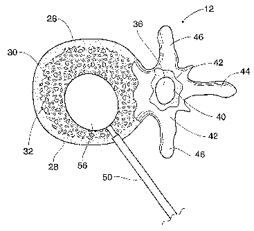
As Fig. 1 shows, the spinal column 10 comprises a
number of uniquely shaped bones, called the vertebrae 12,
a sacrum 14, and a coccyx 16(also called the tail bone).
The number of vertebrae 12 that make up the spinal column
10 depends upon the species of animal. In a human (which
Fig. 1 shows), there are twenty-four vertebrae 12,
comprising seven cervical vertebrae 18, twelve thoracic
vertebrae 20, and five lumbar vertebrae 22.
When viewed from the side, as Fig. 1 shows, the
spinal column 10 forms an S-shaped curve. The curve
serves to support the head, which is heavy. In four-
footed animals, the curve of the spine is simpler.
As Figs. 1 to 3 show, each vertebra 12 includes a
vertebral body 26, which extends on the anterior (i.e.,
front or chest) side of the vertebra 12. As Figs. 1 to
3 show, the vertebral body 26 is in the shape of an oval
disk. As Figs. 2 and 3 show, the vertebral body 26
includes an exterior formed from compact cortical bone
28. The cortical bone 28 encloses an interior volume 30
of reticulated cancellous, or spongy, bone 32(also called
medullary bone or trabecular bone). A"cushion," called
an intervertebral disk 34, is located between the
vertebral bodies 26.
An opening, called the vertebral foramen 36, is
located on the posterior (i.e., back) side of each
vertebra 12. The spinal ganglion 39 pass through the
foramen 36. The spinal cord 38 passes through the spinal
canal 37.
CA 02413308 2002-12-19
WO 01/97721 PCT/US01/19700
- 10 -
The vertebral arch 40 surrounds the spinal canal
37. The pedicle 42 of the vertebral arch 40 adjoins the
vertebral body 26. The spinous process 44 extends from
the posterior of the vertebral arch 40, as do the left
and right transverse processes 46.
II. Treatment of Vertebral Bodies
A. Lateral Access
Access to a vertebral body can be accomplished from
many different directions, depending upon the targeted
location within the vertebral body, the intervening
anatomy, and the desired complexity of the procedure.
For example, access can also be obtained through a
pedicle 42 (transpedicular), outside of a pedicle
(extrapedicular), along either side of the vertebral body
(posterolateral), laterally or anteriorly. In addition,
such approaches can be used with a closed, minimally
invasive procedure or with an open procedure.
Fig. 4 shows a tool 48 for preventing or treating
compression fracture or collapse of a vertebral body
using an expandable body.
The tool 48 includes a catheter tube 50 having a
proximal and a distal end, respectively 52 and 54. The
distal end 54 carries a structure 56 having an expandable
exterior wall 58. Fig. 4 shows the structure 56 with the
wall 58 in a collapsed geometry. Fig. 5 shows the
structure 56 in an expanded geometry.
The collapsed geometry permits insertion of the
structure 56 into the interior volume 30 of a targeted
vertebral body 26, as Fig. 6 shows. The structure 56 can
be introduced into the interior volume 30 in various
ways. Fig. 6 shows the insertion of the structure 56
through a single lateral access, which extends through a
lateral side of the vertebral body 12.
Lateral access is indicated, for example, if a
compression fracture has collapsed the vertebral body 26
CA 02413308 2002-12-19
WO 01/97721 PCT/US01/19700
- 11 -
below the plane of the pedicle 42, or for other reasons
based upon the preference of the physician. Lateral
access can be performed either with a closed, minimally
invasive procedure or with an open procedure. Of course,
depending upon the intervening anatomy, well known in the
art, lateral access may not be the optimal access path
for treatment of vertebrae at all levels of the spine.
The catheter tube 50 includes an interior lumen 80
(see Fig. 4).'The lumen 80 is coupled at the proximal end
of the catheter tube 50 to a pressurized source of fluid,
e.g., saline. A syringe containing the fluid can comprise
the pressure source. The lumen 80 conveys the fluid into
the structure 56 under pressure. As a result, the wall
58 expands, as Figs. 5 and 7 show.
The fluid is preferably rendered radiopaque, to
facilitate visualization as it enters the structure 56.
For example, Renograffin' can be used for this purpose.
Because the fluid is radiopacque, expansion of the
structure 56 can be monitored fluoroscopically or under
CT visualization. Using real time MRI, the structure 56
may be filled with sterile water, saline solution, or
sugar solution, free of a radiopaque material. If
desired, other types of visualization could be used, with
the tool 48 carrying compatible reference markers.
Alternatively, the structure could incorporate a
radiopaque material within the material of the structure,
or the structure could be painted or "dusted" with a
radiopaque material.
Expansion of the wall 58 enlarges the structure 56,
desirably compacting cancellous bone 32 within the
interior volume 30 (see Fig. 7) and/or causing desired
displacement of cortical bone. The compaction of
cancellous bone 32 forms a cavity 60 in the interior
volume 30 of the vertebral body 26 (see Fig. 8). As will
be described later, a filling material 62 can be safely
CA 02413308 2002-12-19
WO 01/97721 PCT/US01/19700
- 12 -
and easily introduced into the cavity 60 which the
compacted cancellous bone 32 forms. In one embodiment,
expansion of the structure 56 desirably forms a region of
compacted cancellous bone which substantially surrounds
the cavity 60. This region desirably comprises a barrier
which limits leakage of the filling material 62 outside
the vertebral body 26. In an alternate embodiment, the
expansion of the structure 56 desirably presses
cancellous bone 32 into small fractures which may be
present in cortical bone, thereby reducing the
possibility of the filling material 62 exiting through
the cortical wall. In another alternative embodiment, the
expansion of the structure 56 desirably flattens veins in
the vertebral body that pass through the cortical wall
(e.g., the basivertebral vein), resulting in less
opportunity for filling material 62 to extravazate
outside the vertebral body through the venous structure
in the cortical wall. Alternatively, expansion of the
structure 56 will compress less dense and/or weaker
regions of the cancellous bone, which desirably increases
the average density and/or overall strength of the
remaining cancellous bone.
The compaction of cancellous bone by the structure
56 can also exert interior force upon cortical bone.
Alternatively, the structure 56 can directly contact the
cortical bone, such that expansion and/or manipulation of
the structure will cause displacement of the cortical
bone. Expansion of the structure 56 within the vertebral
body 26 thereby makes it possible to elevate or push
broken and compressed bone back to or near its original
prefracture position.
The structure 56 is preferably left inflated within
the vertebral body 26 for an appropriate waiting period,
for example, three to five minutes, to allow some
coagulation inside the vertebral body 26 to occur. After
CA 02413308 2002-12-19
WO 01/97721 PCT/US01/19700
- 13 -
the appropriate waiting period, the physician collapses
and removes the structure 56. As Fig. 8 shows, upon
removal of the structure 56, the formed cavity 60 remains
in the interior volume 30.
As Figs. 9B, 9C, and 9D show, the physician next
introduces a filling material 62 into the formed cavity
60 using an appropriate nozzle 114 (as will be described
in greater detail later). The filling material 62 (which
Fig. 9A shows after its introduction into the cavity 60)
can comprise a material that resists torsional, tensile,
shear and/or compressive forces within the cavity 60,
thereby providing renewed interior structural support for
the cortical bone 28. For example, the material 62 can
comprise a flowable material, such as bone cement,
allograft tissue, autograft tissue, or hydroxyapatite,
synthetic bone substitute, which is introduced into the
cavity 60 and which, in time, sets to a generally
hardened condition. The material 62 can also comprise a
compression-resistant material, such as rubber,
polyurethane, cyanoacrylate, or silicone rubber, which is
inserted into the cavity 60. The material 62 can also
comprise a semi-solid slurry material (e.g., a bone
slurry in a saline base), which is either contained
within a porous fabric structure located in the cavity 60
or injected directly into the cavity 60, to resist
compressive forces within the cavity 60. Alternatively,
the material 62 could comprise stents, reinforcing bar
(Re-Bar) or other types of internal support structures,
which desirably resist compressive, tensile, torsional
and/or shear forces acting on the bone and/or filler
material.
The filling material 62 may also comprise a
medication, or a combination of medication and a
compression-resistant material, as described above.
Alternatively, the filling material 62 can comprise
CA 02413308 2002-12-19
WO 01/97721 PCT/US01/19700
- 14 -
a bone filling material which does not withstand
compressive, tensile, torsional and/or shear forces
within the cavity. For example, where the patient is not
expected to experience significant forces within the
spine immediately after surgery, such as where the
patient is confined to bed rest or wears a brace, the
filling material 62 need not be able to immediately bear
load. Rather, the filling material 62 could provide a
scaffold for bone growth, or could comprise a material
which facilitates or accelerates bone growth, allowing
the bone to heal over a period of time. As another
alternative, the filling material could comprise a
resorbable or partially-resorbable source of organic or
inorganic material for treatment of various bone or
non-bone-related disorders including, but not limited to,
osteoporosis, cancer, degenerative disk disease, heart
disease, acquired immune deficiency syndrome (AIDS) or
diabetes. In this way, the cavity and/or filler material
could comprise a source of material for treatment of
disorders located outside the treated bone.
In an alternative embodiment, following expansion,
the expandable structure 56 can be left in the cavity 60.
In this arrangement, flowable filling material 62 is
conveyed into the structure 56, which serves to contain
the material 62. The structure 56, filled with the
material 62, serves to provide the renewed interior
structural support function for the cortical bone 28.
In this embodiment, the structure 56 can be made
from an inert, durable, non-degradable plastic material,
e.g., polyethylene and other polymers. Alternatively,
the structure 56 can be made from an inert, bio-
absorbable material, which degrades over time for
absorption or removal by the body.
In this embodiment, the filling material 62 itself
can serve as the expansion medium for the structure 56,
CA 02413308 2002-12-19
WO 01/97721 PCT/US01/19700
- 15 -
to compact cancellous bone and form the cavity 60, to
thereby perform both compaction and interior support
functions. Alternatively, the structure 56 can be first
expanded with another medium to compact cancellous bone
and form the cavity 60, and the filling material 62 can
be subsequently introduced after the expansion medium is
removed from structure 56 to provide the interior support
function. As another alternative, the filling material
could comprise a two-part material including, but not
limited to, settable polymers or calcium alginate. If
desired, one part of the filling material could be
utilized as the expansion medium, and the second part
added after the desired cavity size is achieved.
The structure 56 can also be made from a permeable,
semi-permeable, or porous material, which allows the
transfer of medication contained in the filling material
62 into contact with cancellous bone through the wall of
the structure 56. If desired, the material can comprise
a membrane that allows osmotic and/or particulate
transfer through the material, or the material can
comprise a material that allows the medication to absorb
into and/or diffuse through the material. Alternatively,
medication can be transported through a porous wall
material by creating a pressure differential across the
wall of the structure 56.
As another alternative, fluids, cells and/or other
materials from the patient's body can pass and/or be
drawn through the material into the structure for various
purposes including, but not limited to, fluid/cellular
analysis, bony ingrowth, bone marrow harvesting, and/or
gene therapy (including gene replacement therapy).
B. Bilateral Access
As Figs. 10 and 11 show, an enlarged cavity 64,
occupying substantially all of the interior volume, can
be created_ by the deployment of multiple expandable
CA 02413308 2002-12-19
WO 01/97721 PCT/US01/19700
- 16 -
structures 56A and 56B through two lateral separate
accesses PLA1 and PLA2, made in opposite lateral sides of
a vertebral body 26. In Fig. 10, the expandable
structures 56A and 56B are carried by separate tools 48A
and 48B at the distal ends of catheter tubes 50A and 50B,
which are separate and not joined together.
Expansion of the multiple expandable structures 56A
and 56B forms two cavity portions 64A and 64B (shown in
Fig. 11). The cavity portions 64A and 64B are
transversely spaced within the cancellous bone 32. The
transversely spaced cavity portions 64A and 64B
preferably adjoin to form the single combined cavity 64
(shown in Fig. 11), into which a filling material is
injected.
Alternatively (not shown), the transversely spaced
cavity portions 64A and 64B can remain separated by a
region of cancellous bone. The filling material is still
injected into each cavity portion 64A and 64B.
Fig. 10 shows the structures 56A and 56B to possess
generally the same volume and geometry, when
substantially expanded. This arrangement provides a
symmetric arrangement for compacting cancellous bone 32.
A generally symmetric, enlarged cavity 64 (shown in Fig.
11) results.
Alternatively, the structures 56A and 56B may
possess different volumes and/or geometries when
substantially expanded, thereby presenting an asymmetric
arrangement for compacting cancellous bone 32. A
generally asymmetric cavity 66 (see, e.g., Fig. 12)
results.
The selection of size and shape of the structures
56A and 56B, whether symmetric or asymmetric, depends
upon the size and shape of the targeted cortical bone 28
and adjacent internal anatomic structures, or by the size
and shape of the cavity 64 or 66 desired to be formed in
CA 02413308 2002-12-19
WO 01/97721 PCT/US01/19700
- 17 -
the cancellous bone 32. It can be appreciated that the
deployment of multiple expandable structures 56A and 56B
makes it possible to form cavities 64 or 66 having
diverse and complex geometries within bones of all types.
It has been discovered that compression fracture or
collapse of one vertebral body can occur in combination
with compression fracture or collapse of an adjacent
vertebral body or bodies. For example, the failure of one
vertebral body may alter loading of adjacent vertebral
bodies, or can cause unequal loading of adjacent
vertebral bodies, resulting in failure of one of more of
the adjacent bodies as well. Because the factors which
weaken and/or cause fracture of one vertebral body will
often weaken and/or affect other vertebral bodies within
the spinal column, these adjacent vertebral bodies are
- susceptible to fracture and/or collapse. In a similar
manner, the treatment of a compression fracture of a
single vertebral body may alter the loading of the
adjacent vertebral bodies, possibly resulting in failure
of one of more of the adjacent bodies. The treatment of
two or more vertebral bodies during a single procedure
may therefore be indicated.
Fig. 13 shows a procedure treating three adjacent
vertebral bodies 26A, 26B, and 26C, each with bilateral
accesses. As shown, the multiple bilateral procedure
entails the deployment of six expandable structures 56(1)
to 56(6), two in each vertebral body 26A, 26B, and 26C.
As Fig. 13 shows, expandable structures 56(1) and 56(2)
are bilaterally deployed in vertebral body 26A;
expandable structures 56(3) and 56(4) are bilaterally
deployed in vertebral body 26B; and expandable structures
56(5) and 56(6) are bilaterally deployed in vertebral
body 26C.
The volume of a given cavity 64 formed in
cancellous bone using multiple expandable structures
CA 02413308 2002-12-19
WO 01/97721 PCT/US01/19700
- 18 -
(e.g., using a bilateral or other type of access) can be
optimized by alternating the expansion of the multiple
expandable structures deployed. For example, in the
illustrated embodiment, in each vertebral body, one of
the expandable structures 56(1) is first expanded,
followed by the expansion of the other expandable body
56(2).
When pressure is first applied to expand a given
structure 56(1) to 56(6), cancellous bone will begin to
compact and/or cortical bone will begin to displace. A
period of time follows in which the pressure within the
structure 56(1) to 56 (6) typically decays, as the
cancellous bone relaxes, further compacts and/or cortical
bone is further displaced. Pressure decay in one
structure also typically occurs as the other expandable
structure within the vertebral body is expanded. When
pressure is again restored to the structure 56(1) to
56(6), further cancellous bone compaction and/or cortical
bone displacement generally results. A further decay in
pressure in the structure 56(1) to 56(6) will then
typically follow. A decay of pressure will generally
follow the application of pressure, until the cancellous
bone is compacted a desired amount and/or cortical bone
is displaced to a desired position.
Optimal cavity formation therefore occurs when each
expandable structure 56 (1) to 56 (6) is allowed to
expand in a sequential, step wise fashion. By allowing
the pressure in each structure to decay before
introducing additional pressure, the peak internal
pressure experienced within each structure can be
reduced, thereby reducing the potential for failure of
the structure. Figs. 14A to 14D more particularly
demonstrate this step wise sequence of applying pressure
to a given pair of expandable structures, e.g., 56(1) and
56(2), when deployed bilaterally in a vertebral body 26A.
CA 02413308 2002-12-19
WO 01/97721 PCT/US01/19700
- 19 -
It should be appreciated, that the step wise application
of pressure can also be used when a single expandable
body is deployed, or when one or more expandable
structures are deployed in other than in a lateral
fashion, e.g., using a transpedicular, extrapedicular, or
anterior access.
It should also be understood that expandable
structures incorporating non-compliant materials could be
used in similar manners to accomplish various objectives
of the present invention. For example, where the
expandable structures comprise non-compliant materials,
such structures could be expanded within the cancellous
bone in the previously described manner to compress
cancellous bone, create a cavity and/or displace cortical
bone. Depending upon the density and strength of the
cancellous and/or cortical bone, the described
application of additional pressure to the structures
could cause a similar cycle of volumetric growth and
pressure decay. Upon reaching maximum capacity and/or
shape of the structures, the introduction of additional
pressure would typically result in little volumetric
expansion of the structures.
In Fig. 14A, the expandable structures 56(1) and
56(2) have been individually deployed in separate lateral
accesses in vertebral body 26A. The expandable structures
56(3)/56(4) and 56(5)/56(6) are likewise individually
deployed in separate lateral accesses in vertebral bodies
26B and 26C, respectively, as Fig. 13 shows.
Representative instruments for achieving these lateral
accesses will be described later.
Once the expandable structures 56(1) to 56(6) are
deployed, the physician successively applies pressure
successively to one expandable structure, e.g., 56(1),
56(3), and 56(5), in each vertebral body 26A, 2GB, and
26C. Fig. 14A shows the init'ial application of pressure
CA 02413308 2002-12-19
WO 01/97721 PCT/US01/19700
- 20 -
to structure 56(1). Alternatively, the physician can
deploy expandable structures in a single vertebral body,
expand those structures as described herein, and tlien
deploy and expand expandable structures within another
vertebral body. As another alternative, the physician
can deploy the expandable structures in a single
vertebral body, expand those structures as described
herein, fill the cavities within that vertebral body, and
then deploy and expand expandable structures within
another vertebral body.
The pressure in the structures 56 (1) , 56(3), and
56(5) will, over time decay, as the cancellous bone in
each vertebral body 26A, 26B, and 26C relaxes, further
compresses and/or cortical bone displaces in the presence
of the expanded structure 56(1), 56(3), and 56(5),
respectively. As pressure decays in the structures 56(1),
56(3), and 56(5), the physician proceeds to successively
apply pressure to the other expandable structures 56(2),
56(4), and 56(6) in the same vertebral bodies 26A, 26B,
and 26C, respectively. Fig. 14B shows the application of
pressure to structure 56(2), as the pressure in structure
56(1) decays.
The pressure in each structure 56(2), 56(4), and
56(6) will likewise decay over time, as the cancellous
bone in each vertebral body 26A, 26B, and 26C is
compressed in the presence of the expanded structure
56(2), 56(4), and 56(6), respectively. As pressure
decays in the structures 56(2), 56(4), and 56(6), the
physician proceeds to successively apply additional
pressure to the other expandable structures 56(1), 56(3),
and 56(5) in the vertebral bodies 26A, 26B, and 26C,
respectively. The introduction of additional pressure in
these structures 26(1), 26(3), and 26(5) further enlarges
the volume of the cavity portions formed as a result of
the first application of pressure. Fig. 14C shows the
CA 02413308 2002-12-19
WO 01/97721 PCT/US01/19700
- 21 -
introduction of additional pressure to structure 56(1) as
pressure decays in structure 56(2).
Pressure, once applied, will typically continue to
decay in each structure 56(1)/56(2), 56(3)/56(4), and
56(5)/56(6), as the cancellous bone relaxes, continues to
compact and/or cortical bone is displaced. As pressure is
successively applied and allowed to decay, the volumes of
the cavity portions also successively enlarge, until
desired cavity volumes have been achieved in the
vertebral bodies 26A, 26B, and 26C and/or desired
displacement of cortical bone has been achieved.
This deliberate, alternating, step wise application
of pressure, in succession first to the structures
26(1)/26(3)/26(5) and then in succession to the
structures 26(2)/26(4)/26(6) in the three vertebral
bodies 26A/B/C continues until a desired endpoint for
each of the vertebral bodies 26A, 26B, and 26C is
reached. In one embodiment, the desired cavity volume is
achieved when cancellous bone is uniformly, tightly
compacted against surrounding cortical bone. In an
alternative embodiment, desired cavity volume is achieved
when a significant pressure decay no longer occurs after
the introduction of additional pressure, such as where
substantially all of the cancellous bone has been
compressed and/or the cortical bone does not displace
further.
It should be understood that compaction of
cancellous bone may be non uniform due to varying
factors, including local variations in bone density. In
addition, it should be understood that desired
displacement of cortical bone can be accomplished in a
similar manner, either alone or in combination with
compaction of cancellous bone. By utilizing multiple
structures to displace the cortical bone, a maximum
amount of force can be applied to the cortical bone over
CA 02413308 2002-12-19
WO 01/97721 PCT/US01/19700
- 22 -
a larger surface area, thereby maximizing the potential
for displacement of the cortical bone while minimizing
damage to the cortical bone from contact with the
structure(s) and/or cancellous bone.
Once the desired volume for each cavity 64 and/or
desired displacement of cortical bone in each vertebral
body 26A, 26B, and 26C is reached, the physician begins
the task of conveying a selected filling material 62 into
each formed cavity 64. It should be appreciated that the
cavities 64 can be filled with filling material 62
essentially in any order, and it is not necessary that
all expandable structures be expanded to form all the
cavities 64 before the filling material is conveyed into
a given cavity.
In one embodiment, the filling material is conveyed
in alternating steps into the cavity portions 64A and 64B
of each vertebral body 26A, 26B, and 26C. In this
technique, the cavity volumes 64A formed by the
expandable structures 56(1), 56(3), and 56(5) are filled
in succession before the cavity volumes 64B formed by the
expandable structures 56(2), 56(4), and 56(6) are filled
in succession.
Figs. 15A and 15B show this embodiment of a filling
sequence for the vertebral body 26A. The vertebral bodies
26B and 26C are filled in like manner. In the vertebral
body 26A, the expandable structure 56(1) is deflated and
removed. The filling material 62 is then conveyed into
the corresponding cavity portion 64A. Next, in the
vertebral body 26B, the expandable structure 56(3) is
deflated and removed, and the filling material 62
conveyed into the corresponding cavity portion 64A. Next,
in the vertebral body 26C, the expandable structure 56(5)
is deflated and removed, and the filling material 62
conveyed into the corresponding cavity portion 64A. The
expandable structures 56(2), 56(4), and 56(6) are left
CA 02413308 2002-12-19
WO 01/97721 PCT/US01/19700
- 23 -
inflated within the respective vertebral bodies 26A, 26B,
and 26C during this portion of the filling process.
The physician waits for the filling material 62
conveyed into the vertebral bodies 26A, 26B, and 26C to
harden. Then, as Fig. l5B shows for the vertebral body
26A, the expandable structure 56(2) is deflated and
removed. The filling material 62 conveyed into the
corresponding cavity portion 64B. Next, in the vertebral
body 26B, the expandable structure 56(4) is deflated and
removed, and the filling material 62 conveyed into the
corresponding cavity portion 64B. Last, in the vertebral
body 26C, the expandable structure 56(6) is deflated and
removed, and the filling material 62 conveyed into the
corresponding cavity portion 64B. The above sequence
allows a single batch of the filling material 62 to be
mixed and expeditiously dispensed to fill multiple
cavities 64.
In one alternative embodiment, the filling material
is conveyed in alternating steps into the cavity portions
of each respective vertebral body prior to filling the
next vertebral body. In this technique, the expandable
structure 56(1) is removed from the vertebral body, and
filling material is conveyed into the corresponding
cavity portion 64A. The expandable structure 56(2) is
then removed from the vertebral body, and filling
material is conveyed into the corresponding cavity
portion 64B. If desired, the filling material can be
allowed to harden to some degree before the expandable
structure 56(2) is removed from the vertebral body. The
process is then repeated for each remaining vertebral
body to be treated. In this embodiment, the vertebral
body is desirably substantially supported by the filling
material and/or an expandable structure during the
filling process, which reduces and/or eliminates the
opportunity for the cavity to collapse and/or cortical
CA 02413308 2002-12-19
WO 01/97721 PCT/US01/19700
- 24 -
bone to displace in an undesired direction during the
filling operation.
III. Instruments for Establishing Bilateral Access
During a typical bilateral procedure, a patient
lies on an operating table. The patient can lie face down
on the table, or on either side, or at an oblique angle,
depending upon the physician's preference.
A. Establishing Multiple Accesses
1. Use of Hand Held Instruments
For each access (see Fig. 16A), the physician
introduces a spinal needle assembly 70 into soft tissue
ST in the patient's back. Under radiologic or CT
monitoring, the physician advances the spinal needle
assembly 70 through soft tissue down to and into the
targeted vertebral body 26. The physician can also employ
stereotactic instrumentation to guide advancement of 'the
spinal needle assembly 70 and subsequent tools during the
procedure. In this arrangement, the reference probe for
stereotactic guidance can be inserted through soft tissue
and implanted on the surface of the targeted vertebral
body. The entire procedure can also be monitored using
tools and tags made of non-ferrous materials, e.g.,
plastic or fiber composites, such as those disclosed in
U.S. Patents 5,782,764 and 5,744,958, which are each
incorporated herein by reference, which would be suitable
for use in a computer enhanced, whole-room MRI
environment.
The physician will typically administer a local
anesthetic, for example, lidocaine, through the assembly
70. In some cases, the physician may prefer other forms
of anesthesia.
The physician directs the spinal needle assembly 70
to penetrate the cortical bone 28 and the cancellous bone
32 through the side of the vertebral body 26. Preferably
the depth of penetration is about 60% to 95% of the
CA 02413308 2002-12-19
WO 01/97721 PCT/US01/19700
- 25 -
vertebral body 26.
The physician holds the stylus 72 and withdraws the
stylet 74 of the spinal needle assembly 70. As Fig. 16B
shows, the physician then slides a guide pin instrument
76 through the stylus 72 and into the cancellous bone 32.
The physician now removes the stylus 72, leaving the
guide pin instrument 76 deployed within the cancellous
bone 32.
The physician next slides an obturator instrument
78 over the guide pin instrument 76, distal end first, as
Fig. 16C shows. The physician can couple the obturator
instrument 78 to a handle 80, which facilitates
manipulation of the instrument 78.
The physician makes a small incision in the
patient's back. The physician twists the handle 80 while
applying longitudinal force to the handle 80. In
response, the obturator instrument 78 rotates and
penetrates soft tissue through the incision. The
physician may also gently tap the handle 80, or otherwise
apply appropriate additional longitudinal force to the
handle 80, to advance the obturator instrument 78 through
the soft tissue along the guide pin instrument 76 down to
the cortical bone entry site. The physician can also tap
the handle 80 with an appropriate striking tool to
advance the obturator instrument 78 into a side of the
vertebral body 26 to secure its position.
The obturator instrument 78 shown in Fig. 16C has
an outside diameter that is generally well suited for
establishing a lateral access. However, if access is
desired through the more narrow region of the vertebral
body 26, e.g., a pedicle 42 (called transpedicular
access), the outside diameter of the obturator instrument
78 can be reduced (as Fig. 17 shows) . The reduced
diameter of the obturator instrument 78 in Fig. 17
mediates against damage or breakage of the pedicle 42.
CA 02413308 2002-12-19
WO 01/97721 PCT/US01/19700
- 26 -
The reduced diameter obturator instrument 78 shown in
Fig. 17 includes a pointed tip 82 to help secure its
position against cortical bone 28. It should be
understood that the disclosed methods and devices are
well suited for use in conjunction with other approach
paths, such as pedicular, extra-pedicular, posterolateral
and anterior approaches, with varying results.
The physician then proceeds to slide the handle 80
off the obturator instrument 78 and to slide a cannula
instrument 84 over the guide pin instrument 76 and,
further, over the obturator instrument 78. If desired,
the physician can also couple the handle 80 to the
cannula instrument 84, to apply appropriate twisting and
longitudinal forces to rotate and advance the cannula
instrument 84 through soft tissue ST over the obturator
instrument 78. When the cannula instrument 84 contacts
cortical bone 28, the physician can appropriately tap the
handle 80 with a striking tool to advance the end surface
into the side of the vertebral body 26 to secure its
position.
When a reduced diameter obturator 78 is used, as
shown in Fig. 17, the cannula instrument 84 can carry a
removable inner sleeve 86 (as Fig. 17 also shows) to
center the cannula instrument 84 about the reduced
diameter obturator instrument 78 during passage of the
cannula instrument 84 to the treatment site.
The physician now withdraws the obturator
instrument 78, sliding it off the guide pin instrument
76, leaving the guide pin instrument 76 and the cannula
instrument 84 in place. When a reduced diameter
obturator instrument 78 is used, the physician can remove
the inner centering sleeve 86.
As Fig. 16D shows, the physician slides a drill bit
instrument 88 over the guide pin instrument 76, distal
end first, through the cannula instrument 84, until
CA 02413308 2002-12-19
WO 01/97721 PCT/US01/19700
- 27 -
contact between the machined or cutting edge 90 of the
drill bit instrument 88 and cortical bone 28 occurs. The
physician then couples the drill bit instrument 88 to the
handle 80 .
Guided by X-ray (or another external visualizing
system), the physician applies appropriate twisting and
longitudinal forces to the handle 80, to rotate and
advance the machined edge 90 of the drill bit instrument
88 to open a lateral passage PLA through the cortical
bone 28 and into the cancellous bone 32. The drilled
passage PLA preferably extends no more than 95% across
the vertebral body 26.
As Fig. 18A shows, the drill bit instrument 88 can
include a flexible shaft portion 92 to aid in its
manipulation. The flexible shaft portion 92 allows the
cutting edge 90 of the instrument 88 to flex relative to
the axis of the instrument. As Fig 18A also shows, the
cannula instrument 84 can, if desired, include a
deflector element 94 on its distal extremity, to flex the '
flexible shaft portion 92 and guide the cutting edge 90
along a desired drill axis. Desirably, in such a
flexible embodiment the drill bit instrument 88 is made
of a flexible plastic material, e.g., polyurethane, or a
flexible metal material encapsulated in or surrounding a
plastic material, to possess sufficient torsional
rigidity to transmit rotating cutting force to bone.
Alternatively, as Fig. 18B shows, the drill bit
instrument 88 can include an interior lumen 180 to
accommodate passage of a guide wire 182. In this
arrangement, the flexible shaft portion 92 conforms to
the path presented by the guide wire 182. The guide wire
182, for example, can be pre-bent, to alter the path of
the cutting edge 90 after it enters the vertebral body.
Alternatively, the guide wire can be made of memory wire,
shape memory alloys (including nickel-titanium, copper or
CA 02413308 2008-01-18
60895-1633
- 28 -
iron based alloys, to name a few}, or comprise a self-
steering guiding catheter.
Still alternatively, as Fig. 18C shows, the drill
bit instrument 88 itself can carry interior steering
wires 184. The steering wires 184 are operated by the
physician using an external actuator 186, to deflect the
flexible shaft portion 92, and with it the cutting edge,
without aid of a guide wire and/or cannula instrument 84.
Further details regarding the formation of cavities
within cancellous bone, which are not symmetric with
relation to the axis of a vertebral body, can be found in
United States Patent 5,972,018, entitled "Expandable
Asymmetric Structures for Deployment in Interior Body
Regions"=
Once the passage PLA in cancellous bone 32 has been
formed, the physician removes the drill bit instrument 88
and the guide pin instrument 76, leaving only the cannula
instrument 84 in place, as Fig. 16E shows. The passage
PLA made by the drill bit instrument 88 remains.
Subcutaneous lateral access to the cancellous bone 32 has
been accomplished.
The physician repeats the above described sequence
of steps, as necessary, to form each access desired. In
Fig. 13, six accesses are made.
2. Using Composite Hand Held Instruments
Other forms of hand held instruments may be used to
provide access.
For example, Figs. 27 and 28 show a.composite
instrument 310 that can be used for this purpose. The
composite instrument 310 includes a trocar instrument 320
and a cannula instrument 340. The composite instrument
310 also includes a composite handle 312 comprising a
first handle 322 and a second handle 342. The composite
handle 312 aids a physician in manipulating the composite
instrument 310. Still, as Figs. 29A and 29B show, a
CA 02413308 2002-12-19
WO 01/97721 PCT/US01/19700
- 29 -
physician can also desirably use the first handle 322 to
independently manipulate the trocar instrument 320 or the
second handle 342 to independently manipulate the cannula
instrument 340 during use.
The trocar instrument 320 comprises a trocar 330
having a distal end that is tapered to present a
penetrating surface 334. In use, the penetrating surface
334 is intended to penetrate soft tissue and/or bone in
response to pushing and/or twisting forces applied by the
physician at the first handle 322, or the composite
handle 312.
The cannula instrument 340 performs the function of
the cannula instrument 84 previously described, but also
includes the handle 342, which mates with the handle 322
to form the composite handle 312. In this embodiment,
the cannula instrument 84 is desirably somewhat larger in
diameter than and not as long as the trocar 330. The
cannula instrument 84 includes an interior lumen 344 that
is sized to accept the trocar 330. The size of the
interior lumen 344 desirably allows the cannula
instrument 84 to slide and/or rotate relative to the
trocar 330, and vice versa. The distal end 354 of the
cannula instrument 84 presents an end surface 360 that
desirably presents a low-profile surface, which can
penetrate soft tissue surrounding the trocar 330 in
response to pushing and/or twisting forces applied at the
composite handle 312 or the second handle 342.
In use, as shown in Fig. 30, the physician directs
the composite instrument 310 such that the trocar 330 and
the cannula instrument 84 penetrate the cortical bone and
the cancellous bone of the targeted vertebra. If
desired, the physician can twist the composite handle 312
while applying longitudinal force to the handle 312. In
response, the penetrating end surface 334 of the trocar
330, and the end surface of the cannula instrument 84
CA 02413308 2002-12-19
WO 01/97721 PCT/US01/19700
- 30 -
rotate and penetrate soft tissue and/or bone.
If penetration through the cortical bone and into
the cancellous bone is not achievable by manual
advancement of the composite instrument 310, a physician
can continue penetration by gently striking a striking
plate 314 on the composite handle 312 with a blunt
instrument such as a surgical hammer (not shown), or
otherwise applying appropriate additional longitudinal
force to the composite handle 312, to advance the distal
end 334 of the trocar 330 and the end surface of the
cannula instrument 84.
If desired, the physician can utilize a spinal
needle assembly 70, as already described, to initially
access the vertebral body. In this arrangement, the
composite instrument 310 is later guided through soft
tissue and into the targeted vertebra body along the
stylet 74, which (in this arrangement) passes through an
interior lumen in the trocar 330 (not shown). Once the
trocar 330 has sufficiently penetrated cortical bone, the
physician can withdraw the stylet 74, thereby arriving at
the step in the procedure shown in Fig. 30.
After penetrating the cortical bone, the physician
may continue advancing the composite instrument 310
through the cancellous bone of the vertebral body to form
the passage through the cancellous bone, as already
described. The trocar 330 may then be withdrawn from the
cannula instrument 84. The cannula instrument 84 remains
to provide access to the passage formed in the interior
of the vertebral body, in the manner previously
described.
Alternatively, after penetrating the cortical bone,
the physician may choose to withdraw the trocar 330 from
the cannula 50 and form the passage in the cancellous
bone using a drill bit instrument 88, as Fig. 31 shows.
In such a case, the physician removes the trocar 330 and,
CA 02413308 2002-12-19
WO 01/97721 PCT/US01/19700
- 31 -
in its place, advances the drill bit instrument 88
through the cannula instrument 84, as Fig. 31 shows.
With the removal of the drill bit instrument 88,
access to the cancellous bone has been accomplished.
3. Breach Prevention and Plugging
To create access into the vertebral body in the
manners shown in Figs. 16A to 16D, the physician
typically advances a stylet 74 of the spinal needle
assembly 70 and also the cutting edge of the drill bit
instrument 88 a significant distance into the cancellous
bone 32, as Figs. 16B and 16D show, toward cortical bone
28 on the anterior wall of the vertebral body 26. The
density of the cancellous bone 32 desirably offers
resistance to the passage of these instruments, to
thereby provide tactile feed back to the physician, which
aids in guiding their deployment. Still, the density of
cancellous bone 32 is not uniform and can change
abruptly. Even with the utmost of care and skill, it is
possible that the stylet 74 or the cutting edge 90 can
slide into and poke through cortical bone 28 in the
anterior wall of the vertebral body 26. This can create
a hole or breach B in the anterior cortical wall 28 of
the vertebral body 26, as Fig. 19 shows.
To aid in the advancement of the cutting edge 90
through cancellous bone 32 (see Fig. 20A), the drill bit
instrument 88 may include a mechanical stop 96. In use,
the mechanical stop 96 abuts against the proximal end of
the cannula instrument 84. The abutment stops further
advancement of the drill bit instrument 88 into the
interior of the vertebral body 26.
The location of the mechanical stop 96 may be
adjustable, to provide variable lengths of advancement,
depending upon the size of the vertebral body 26 or other
bone volume targeted for treatment.
Alternatively, or in combination, the drill bit
CA 02413308 2002-12-19
WO 01/97721 PCT/US01/19700
- 32 -
instrument 88 may include markings 98 located along its
length at increments from its terminus. The markings 98
register with the exposed proximal edge of the cannula
instrument 84 (see Fig. 20A), to allow the physician to
remotely gauge the position of the instrument in the
vertebral body 26.
To aid the advancement of the stylet 74, the trocar
330, or the drill bit instrument 88 within the vertebral
body, without breach of the anterior cortical wall, the
physician can also make use of a cortical wall probe 140,
as shown in Fig. 20B. The cortical wall probe 140
comprises a generally rigid stylet body 142 having a
blunt distal tip 144, which desirably cannot easily
pierce the anterior cortical wall of the vertebral body.
In the illustrated embodiment, the blunt distal tip 144
comprises a rounded ball shape.
The cortical wall probe 140 can be deployed through
the formed access opening before any significant
penetration of cancellous bone occurs. For example, after
the access opening is formed using the spinal needle
assembly 70, but before the stylus 72 and stylet 74 are
advanced a significant distance into cancellous bone, the
stylet 74 can be withdrawn and, instead, the cortical
wall probe 140 advanced through the stylus 72. The
physician advances the cortical wall probe 140 through
cancellous bone, until the physician tactilely senses
contact between the blunt distal tip 144 and the anterior
cortical wall. Desirably, the probe 140 is radiopaque, so
that its advancement through cancellous bone and its
contact with the anterior cortical wall within the
vertebral body can be visualized, e.g., either by x-ray
or real time fluoroscopy or MRI. Using the cortical wall
probe 140, the physician can gauge the distance between
the access opening into the vertebral body and the
anterior cortical wall, in a manner that avoids
CA 02413308 2002-12-19
WO 01/97721 PCT/US01/19700
- 33 -
penetration of the anterior cortical wall.
The cortical wall probe 140 can carry length
markings 146 on its proximal region, which, when contact
with the anterior cortical wall occurs and/or is
imminent, indicate the distance a subsequent instrument
can be advanced down the stylus 72 (or cannula instrument
84) before contacting the anterior cortical wall. The
information obtained from the cortical wall probe 140 can
also be used to set the mechanical stop 96 (previously
described), to physically prevent advancement of the
trocar 330 or drill bit instrument 88 before contact with
the anterior cortical wall occurs.
In the event of a breach or suspected breach of the
anterior cortical wall of the vertebral body, the
physician can alternatively utilize the cortical wall
probe 140 to safely and easily determine the existence
and/or extent of a wall breach. Because the distal tip
144 of the probe is blunt, the tip 144 desirably will not
easily pass through an intact anterior cortical wall,
which allows the physician to "tap" the tool along the
inner surface of the anterior cortical wall while
searching for breaches. Where a wall breach has occurred,
and the tool could pass through the breach, the blunt tip
144 of the tool desirably will not pierce or damage soft
tissues, such as the aorta or major veins, located
forward of the cortical wall. If desired, the blunt tip
144 can alternatively be formed of a soft, deformable
material such as rubber or plastic.
If a breach B occurs, a suitable material may be
placed into the breach B to plug it. For example, a
demineralized bone matrix material, such as GRAFTON""
material, may be used. The material can be placed, e.g.,
on the distal end of the obturator instrument 78 or
trocar 330. The instrument 78 is deployed carrying the
plugging material to the exterior side wall where the
CA 02413308 2002-12-19
WO 01/97721 PCT/US01/19700
= 34 -
breach B occurs. The instrument 78 deposits the plugging
material in the breach B, to thereby close it from the
outside of the vertebral body 26.
The physician can take steps to counteract
undetermined cortical wall breaches, either as may
possibly preexist before cavity formation or which may
possibly exist after cavity formation. Even if a breach
is not known to exist, the physician can nevertheless
elect to insert a suitable plug material (e.g., GRAFTONT"
bone matrix material, or CollagraftTM sheet material, or
a mesh-type material) into the vertebral body, either
before or after the structure 56 is expanded. The
presence of a plug material guards against the
possibility of leaks, whether they exist or not.
Furthermore, if inserted before the structure 56 is
expanded, the presence of the plug material in the
vertebral body can serve to make the distribution of the
expansion force of the structure 56 more uniform. The
presence of the plug material within the vertebral body
as the structure 56 expands can also protect against
protrusion of the expanding structure 56 through any
preexisting breach in the cortical wall as well as any
breaches created during expansion of the structure 56, or
can otherwise protect weakened cortical walls during
expansion of the structure 56.
4. Cannula Locking Device
Referring to Fig. 26A, a cannula locking device 190
can be used to aid in stabilizing the cannula instrument
84 while accessing a vertebral body. The locking device
190 can be variously constructed.
In the embodiment shown in Fig. 26A, the locking
device 190 includes a generally planar base 192. In use,
the base 192 rests upon a skin surface surrounding the
targeted incision site. If desired, the base 192 can
incorporate an adhesive (not shown) to secure the base to
CA 02413308 2002-12-19
WO 01/97721 PCT/US01/19700
- 35 -
the patient's skin or to other material located at or
near the surgical site.
An instrument grip 194 is supported on the base
192. The instrument grip 194 includes a channel 218 which
slidingly receives the cannula instrument 84, which, in
this embodiment, is intended to be placed into the grip
194 distal end first. A ring 220, threaded to the grip
194, can be provided to tighten the channel 218 about the
cannula instrument 84, to thereby prevent axial movement
of the cannula instrument 84 within the channel 218.
The grip 194 also includes a tenon 196, which fits
within a mortise 198 on the base 192. The mortise 198
and tenon 196 together form a joint 200. The grip 194
pivots 360-degrees in transverse and/or orbital paths
within the joint 200.
The mortise 198 is bounded by a collet 210, about
which a retaining ring 202 is threadably engaged.
Twisting the ring 202 in one direction (e.g., clockwise)
closes the collet 210 about the tenon 196, locking the
position of the grip 194 relative to the base 192.
Twisting the ring 202 in an opposite direction opens the
collet 210 about the tenon 196, freeing the grip 194 for
pivotal movement relative to the base 192.
To use the device 190, the physician manipulates
the cannula instrument 84 held in the grip 194 into a
desired axial and angular orientation. The physician
thereafter locks the grip 194 (tightening the rings 202
and 220) to hold the cannula instrument 84 in the desired
axial and angular orientation. The physician can
manipulate and lock the cannula instrument 84 in any
desired order, either before or after passage of the
instrument 84 through the skin, and/or before or after
passage of the instrument 84 through cortical bone, or
combinations thereof. Markings 204 on the grip 194 and
base 192 allow the physician to gauge movement of the
CA 02413308 2002-12-19
WO 01/97721 PCT/US01/19700
- 36 -
grip 194 relative to the base 192 or another reference
point.
The locking device 190 is preferably made from a
material that is not highly radiopaque, e.g.,
polyurethane or polycarbonate. The device 190 will
therefore not obstruct fluoroscopic or x-ray
visualization of the cannula instrument 84 during use.
When locked, the device 190 prevents unintended
movement of the cannula instrument 84 along the skin
surface. The likelihood that the cannula instrument 84
will be bent or its position inadvertently shifted during
use is thereby mitigated. The device 190 also allows the
physician to remove his/her hands from the instrument 84,
e.g., to allow clear fluoroscopy or x-ray visualization.
The device 190 obviates the need for other types of
clamps that are radiopaque or are otherwise not well
suited to the task.
As Fig. 26B shows, in an alternative embodiment,
the retaining ring 202 can be loosened to a point that
opens the collet 210 enough to free the grip 194 from the
base 192. In this arrangement, the grip 194 comprises
members 206 and 208 that can be split apart when
separated from the confines of the collet 210. The
cannula instrument 84 can be captured between the spit-
apart members 206 and 208 as they are fitted back
together, obviating the need to load the cannula
instrument 84 distal end first in the grip 194.
When fitted together, the tenon 196 can be returned
to the mortise 198. The retaining ring 202 can be
tightened sufficiently to close the collet 210 about the
tenon 196, forming the joint 200. Further tightening of
the retaining ring 202 about the mortise 198 closes the
joint 200 (as before described), locking the grip 194 a
desired orientation relative to the base 192. Subsequent
loosening of the retaining ring 202 permits separation of
CA 02413308 2002-12-19
WO 01/97721 PCT/US01/19700
- 37 -
the grip 194 from the base 192, so that the members 206
and 208 can be split apart to free the cannula instrument
84. In one embodiment, the grip 194 can contact the
cannula directly, such that the cannula is substantially
"locked" in position when the grip 194 is compressed
against the cannula. In an alternate embodiment, an 0-
ring (not shown) can be located within the grip 194, such
that compression of the grip causes the 0-ring to push
against the cannula, desirably substantially "locking"
the cannula in position within the grip 194.
B. Forming the Cavities
Once the accesses PLA have been formed, the
physician advances individual catheter tubes 50 through
the cannula instrument 84 and passage of each access,
into the interior volume of the associated vertebral body
26A, 26B, and 26C. Fig. 16F shows this deployment in
vertebral body 26A.
The expandable structures 56(1) to 56(6) are then
expanded in the alternating, step wise'fashion as already
described. The compression forms the interior cavity 64
in each vertebral body 26A, 26B, and 26C.
As Figs. 4 and 5 show, the expandable structure 56
can carry at least one radiopaque marker 102, to enable
remote visualization of its position within the vertebral
body 26. In the illustrated embodiment, the expandable
structure 56 carries a radiopaque marker 102 on both its
distal and proximal end.
As before described, when fluoroscopic or CT
visualization is used to monitor expansion of the
structure 56, the fluid used to cause expansion of the
structure 56 is preferably rendered radiopaque (e.g.,
using Renograffin'." material). The visualization
instrument (e.g., a C-arm fluoroscope) is typically
positioned on the operating table to view laterally along
one side of the spinal column. The presence of radiopaque
CA 02413308 2002-12-19
WO 01/97721 PCT/US01/19700
- 38 -
expansion medium in a expanded structure 56 in the
vertebral body 26 can block effective visualization
elsewhere in the vertebral body, e.g., where cavity
formation using another structure 56 or where
vertebroplasty or another form of treatment is intended
to occur.
Visualization can be facilitated under these
circumstances by removal or dilution of the radiopaque
medium within the structure 56 after the structure is
expanded to create a cavity.
In one embodiment (see Fig. 32), an exchange
chamber 400 is provided, which is divided into two
compartments 402 and 404 by a piston 414 that is movable
by pressure upon a plunger 420. Dual lumens 406 and 408
communicate with the interior of the structure 56. The
lumen 406 communicates with the source 422 of radiopaque
medium 410 to convey the medium 410 into the structure 56
to cause expansion and cavity formation in the first
instance. The lumen 406 also communicates with the
compartment 402 on one side of the piston 414.
The other compartment 404 of the chamber 400
contains a replacement expansion medium 412. The
replacement medium 412 is free of a radiopaque material
or, if desired, can contain a partially-radiopaque
material. The lumen 408 communicates with this
compartment 404.
After expansion of the structure 56 with the
radiopaque medium 410, movement of the piston 414 will
draw the radiopaque medium 410 from the structure 56
(through lumen 402). Simultaneously, the piston 414 will
displace the radiopaque-free medium 412 into the
structure 56 (through lumen 404) . Piston movement
exchanges the radiopaque medium 410 with the radiopaque-
free medium 412, without collapsing the structure 56.
In an alternative embodiment, an ion exchange
CA 02413308 2002-12-19
WO 01/97721 PCT/US01/19700
- 39 -
material for the radiopaque material in the medium 410
(e.g., iodine) can be introduced into the radiopaque
medium 410 contained within the structure 56. The ion
exchange material selectively binds the radiopaque
material, diluting the radiopaque qualities of the medium
410. The radiopaque medium 410 can be circulated through
an ionic exchange chamber outside the structure 56, or
the ion exchange material can be introduced into the
structure 56 through an interior lumen within the
structure 56 itself.
Alternatively, a material that causes precipitation
of radiopaque material can be introduced into the
radiopaque medium 410 within the structure 56 (e.g.,
through an interior lumen). The precipitation selectively
causes the radiopaque material to settle downward within
the structure 56, out of the lateral visualization path,
thereby diluting the radiopaque qualities of the medium
410.
As Fig. 5 shows, the expandable structure 56 can
also include an interior tube 104. The interior tube 104
contains an interior lumen 106 that passes through the
expandable structure 56.
The interior lumen 106 can be used to convey a
flowable material or liquid, e.g. saline or sterile
water, to flush materials free of the distal region of
the structure 56, when in use. The interior lumen 106 can
also be used to aspirate liquid material from the
interior of the vertebral body 26 as the procedure is
performed. The interior lumen 106 can also be used to
introduce a thrombogenic material, e.g., a clotting
agent, into contact with cancellous bone 32 during the
procedure. The expandable structure 56 itself can be also
dipped into thrombin prior to its introduction into the
vertebral body 26 to facilitate in situ coagulation.
The interior lumen 106 can also be sized to receive
CA 02413308 2002-12-19
WO 01/97721 PCT/US01/19700
- 40 -
a stiffening member or stylet 108 (see Fig. 5) The
stylet 108 keeps the structure 56 in a desired distally
straightened condition during its passage through the
cannula instrument 84. Once the structure 56 is located
in the desired location within cancellous bone, the
physician can remove the stylet 108, and thereby open the
interior lumen 106 for conveyance of liquids to and from
cancellous bone, as just described.
The stylet 108 can also have a preformed memory, to
normally bend its distal region. The memory is overcome
to straighten the stylet 108 when passed through the
cannula instrument 84. However, as the structure 56 and
stylet 108 advance free of the cannula instrument 84,
passing into cancellous bone 32, the preformed memory
bends the stylet 108. The bent stylet 108 shifts the
axis of the structure relative to the axis of the access
path PLA. The prebent stylet 108, positioned within the
interior of the structure 56, aids in altering the
geometry of the structure 56 to achieve a desired
orientation when deployed for use.
If the stylet 108 is comprised of a shape memory
alloy, such as nickel-titanium(Nitinol), copper or iron
based alloys, the distal end of the stylet 108 can be set
to a prebent "parent shape," and then subsequently bent
to a substantially straight shape for introduction into
the vertebral body. When the stylet 108 is in its desired
position, and bending of the distal end is desired, heat
can be applied to the proximal end of the stylet 108,
which desirably will cause the distal end of the stylet
108 to assume its parent shape in a known manner.
Alternatively, the stylet 108 can be comprised of a shape
memory allowing material having a transition temperature
at or below human body temperature. Such a stylet 108 can
be cooled prior to and/or during introduction into the
human body, and once in the proper position, the cooling
CA 02413308 2002-12-19
WO 01/97721 PCT/US01/19700
- 41 -
source can be removed, and the patient's body heat will
cause the stylet 108 to assume its pre-bent parent shape.
If desired, the stylet can be initially positioned within
the vertebral body, with the distal end deflecting within
the cancellous bone, or the distal end can be deflected
during insertion into the vertebral body.
As Fig. 25 shows, the catheter tube 50 can itself
carry a drill bit element 170. The drill bit element 170'
may be variously constructed. As shown in Fig. 25, the
drill bit element 170 comprises a metal cutting cap
bonded or otherwise mounted on the distal end of the
interior catheter tube 104, beyond the expandable
structure 56. In this arrangement, the stylet 108 can
include a keyed distal end 172, which mates within an
internal key way 174 in the drill bit element 170. The
stylet 108 thereby serves to stiffen the distal end of
the catheter tube 104, so that torsional and compressive
loads can be applied to the drill bit element 170.
Alternatively, the interior structure of the catheter
tube 104 can be otherwise reinforced to transmit
torsional and compressive load forces to the drill bit
element 170. Using the drill bit element 170, the
physician can open an access opening in the cortical
bone, without use of the separate drill bit instrument
88.
1. Desired Physical and Mechanical
Properties for the Expandable Structure
The material from which the structure 56 is made
should possess various physical and mechanical properties
to optimize its functional capabilities to compact
cancellous bone. Important properties are the ability to
expand its volume; the ability to deform in a desired way
when expanding and assume a desired shape inside bone;
and the ability to withstand abrasion, tearing, and
puncture when in contact with cancellous bone.
CA 02413308 2002-12-19
WO 01/97721 PCT/US01/19700
- 42 -
2. Expansion Property
A first desired property for the structure material
is the ability to expand or otherwise increase its volume
without failure. This property enables the structure 56
to be deployed in a collapsed, low profile condition
subcutaneously, e.g., through a cannula, into the
targeted bone region. This property also enables the
expansion of the structure 56 inside the targeted bone
region to press against and compress surrounding
cancellous bone, or move cortical bone to a prefracture
or other desired condition, or both.
The desired expansion property for the structure
material can be characterized in one way by ultimate
elongation properties, which indicate the degree of
expansion that the material can accommodate prior to
failure. Sufficient ultimate elongation permits the
structure 56 to compact cortical bone, as well as lift
contiguous cortical bone, if necessary, prior to wall
failure. Desirably, the structure 56 will comprise
material able to undergo an ultimate elongation of at
least 50%, prior to wall failure, when expanded outside
of bone. More desirably, the structure will comprise
material able to undergo an ultimate elongation of at
least 150%, prior to wall failure, when expanded outside
of bone. Most desirably, the structure will comprise
material able to undergo an ultimate elongation of at
least 300%, prior to wall failure, when expanded outside
of bone.
Alternatively, the structure 56 can comprise one or
more non-compliant or partially compliant materials
having substantially lower ultimate elongation
properties, including, but not limited to, kevlar,
aluminum, nylon, polyethylene, polyethylene-terephthalate
(PET) or mylar. Such a structure would desirably be
initially formed to a desired shape and volume, and then
CA 02413308 2008-01-18
60895-1633
- 43 -
contracted to a collapsed, lower profile condition for
introduction through a cannula into the targeted bone
region. The structure could then be expanded to the
desired shape and volume to press against and compress
surrounding cancellous bone and/or move cortical bone to
a prefracture or desired condition, or both. As another
alternative, the structure could comprise a combination
of non-compliant, partially compliant and/or compliant
materials.
3. Shape Property
A second desired property for the material of the
structure 56, either alone or in combination with the
other described properties, is the ability to predictably
deform during expansion, so that the structure 56
consistently achieves a desired shape inside bone.
The shape of the structure 56, when expanded in
bone, is desirably selected by thephysician, taking into
account the morphology and geometry of the site to be
treated. The shape of the cancellous bone to be
compressed and/or cortical bone to be displaced, and the
local structures that could be harmed if bone were moved
inappropriately, are generally understood by medical
professionals using textbooks of human skeletal anatomy
along with their knowledge of the site and its disease or
injury, and also taking into account the teachings of U.S.
Patent No. 6,235,043, filed January 23, 1997, and entitled
"Improved Inflatable Device for Use in Surgical Protocol
Relating to Fixation of Bone". The physician is also
desirably able to select the desired expanded shape inside
bone based upon prior analysis of the morphology of the
targeted bone using, for example, plain film x-ray,
fluoroscopic x-ray, or MRI or CT scanning.
Where compression of cancellous bone and/or cavity
CA 02413308 2002-12-19
WO 01/97721 PCT/US01/19700
- 44. -
creation is desired, the expanded shape inside bone is
selected to optimize the formation of a cavity that, when
filled with a selected material, provides support across
the region of the bone being treated. The selected
expanded shape is made by evaluation of the predicted
deformation that will occur with increased volume due to
the shape and physiology of the targeted bone region.
Where displacement of cortical bone is desired, the
expanded shape can be chosen to maximize the amount of
force the structure exerts on cortical bone, maximize
force distribution over the largest possible surface area
of the cortical bone and/or maximize displacement of the
cortical bone in one or more desired directions.
Alternatively, the structure can be designed to impart a
maximum force on a specific area of the cortical bone so
as to cause desired fracture and/or maximum displacement
of specific cortical bone regions.
To aid in selecting a suitable size for the
expandable structure 56, the trocar 330 of the composite
instrument 310 (see Figs. 27 and 28) can carry an array
of grooves or like markings 380, which can be viewed
under fluoroscopic visualization. The markings 380 allow
the physician to estimate the distance across the
vertebral body, thereby making it possible to estimate
the desired size of the expandable structure 56. Because
the cannula instrument 84 is a relatively thin-walled
structure, and the trocar 330 is a relatively thicker
solid structure, the physician is able to visualize the
markings 380 by fluoroscopy, even when the markings 380
are inside the cannula instrument 84.
In some instances, it is desirable, when creating
a cavity, to also move or displace the cortical bone to
achieve the desired therapeutic result. Such movement is
not per se harmful, as that term is used in this
Specification, because it is indicated to achieve the
CA 02413308 2002-12-19
WO 01/97721 PCT/US01/19700
- 45 -
desired therapeutic result. By definition, harm results
when expansion of the structure 56 results in a worsening
of the overall condition of the bone and surrounding
anatomic structures, for example, by injury to
surrounding tissue or causing a permanent adverse change
in bone biomechanics.
If desired, the structure 56 can be used to
generate sufficient force to fracture cortical bone and
position the fractured cortical bone in a new orientation
and/or into a more desired position. Where the bone has
fractured and/or compressed in the past, and subsequently
healed, the present methods and devices can be utilized
to safely reposition the cortical bone to a more desired
position. For example, where a vertebral compression
fracture has healed in a depressed and/or fractured
position, the disclosed devices and methods can be
utilized to re-fracture and reposition the fractured bone
to a more desirable position and/or orientation. By
generating sufficient force to fracture the bone from the
interior, through expansion of an expandable body, only
a single access portal through the cortical bone need be
f ormed .
If desired, the structure could alternatively be
used in conjunction with various devices, including but
not limited to lasers, drills, chisels or sonic
generators (e.g. lithotripers), these devices being used
to selectively weaken and/or fracture cortical bone along
desired lines and/or in a desired manner. Once the
targeted cortical bone is sufficiently weakened, the
structure 56 can be used to fracture the bone and/or
reposition the cortical bone to a new orientation and/or
into a more desired position.
In a similar manner, the structure 56 can be used
to fracture and reposition a portion of the cortical
bone, such as where the bone has grown and/or healed in
CA 02413308 2002-12-19
WO 01/97721 PCT/US01/19700
- 46 -
a deformed condition. For example, in a patient having
severe scoliosis (e.g., osteopathic scoliosis), the
vertebral column may be laterally curved due to bone
deformation. The present methods and devices can be
utilized to safely fracture and/or reposition the
cortical bone to a more desired position. If desired,
sections of the bone can be scored, weakened and/or pre-
fractured by various devices including, but not limited
to, sharp knives, saws, awls, drills, lasers and/or
lithotripters, creating desired lines along which the
bone will tend to fracture. The depressed sections of
the vertebral body can desirably be elevated and
reinforced, thereby reducing the lateral curve of the
vertebral column and preventing further lateral
deformation of the spine. By fracturing and/or
displacing only a portion of the cortical bone, the
present methods and devices minimize unnecessary
muscular-skeletal trauma while permitting treatment of
the disease.
As one general consideration, in cases where the
bone disease causing fracture (or the risk of fracture)
is the loss of cancellous bone mass (as in osteoporosis),
the selection of the expanded shape of the structure 56
inside bone should take into account the cancellous bone
volume which should be compacted to achieve the desired
therapeutic result. An exemplary range is about 30% to
90% of the cancellous bone volume, but the range can vary
depending upon the targeted bone region. Generally
speaking, compacting less of the cancellous bone volume
leaves more uncompacted, diseased cancellous bone at the
treatment site.
Another general guideline for the selection of the
expanded shape of the structure 56 inside bone is the
amount that the targeted fractured bone region has been
displaced or depressed. The expansion of the structure
CA 02413308 2002-12-19
WO 01/97721 PCT/US01/19700
- 47 -
56 inside a bone can elevate or push the fractured
cortical wall back to or near its anatomic position
occupied before fracture occurred. Where the structure 56
directly contacts the depressed cortical bone, and
elevates the cortical bone through direct contact with
the expanding structure, compaction of cancellous bone
may not be necessary or desired.
For practical reasons, it is desired that the
expanded shape of the structure 56 inside bone, when in
contact with cancellous bone, substantially conforms to
the shape of the structure 56 outside bone, when in an
open air environment. This allows the physician to select
in an open air environment a structure having an expanded
shape desired to meet the targeted therapeutic result,
with the confidence that the expanded shape inside bone
will be similar in important respects.
In some instances, it may not be necessary or
desired for the structure to predictably deform and/or
assume a desired shape during expansion inside bone.
Rather, it may be preferred that the structure expand in
a substantially uncontrolled manner, rather than being
constrained in its expansion. For example, where
compaction of weaker sections of the cancellous bone is
desired, it may be preferred that the structure initially
expand towards weaker areas within the bone. In such
cases, the structure can be formed without the
previously-described shape and/or size, and the expanded
shape and/or size of the structure can be predominantly
determined by the morphology and geometry of the treated
bone.
An optimal degree of shaping can be achieved by
material selection and by special manufacturing
techniques, e.g., thermoforming or blow molding, as will
be described in greater detail later.
4. Toughness Property
CA 02413308 2002-12-19
WO 01/97721 PCT/US01/19700
- 48 -
A third desired property for the structure 56,
either alone or in combination with one or more of the
other described properties, is the ability to resist
surface abrasion, tearing, and puncture when in contact
with cancellous bone. This property can be characterized
in various ways.
One way of measuring a material's resistance to
abrasion, tearing and/or puncture is by a Taber Abrasion
test. A Taber Abrasion test evaluates the resistance of
a material to abrasive wear. Typically, a lower Taber
Abrasion value indicates a greater resistance to
abrasion. Desirably, one embodiment of an expandable
structure will comprise material having a Taber Abrasion
value under these conditions of less than approximately
200 mg loss. More desirably, the structure will comprise
material having a Taber Abrasion value under these
conditions of less than approximately 145 mg loss. Most
desirably, the structure will comprise material having a
Taber Abrasion value under these conditions of less than
approximately 90 mg loss. Of course, materials having a
Taber Abrasion value of greater than or equal to 200 mg
loss may be utilized to accomplish some or all of the
objectives of the present invention.
Another way of measuring a material's resistance to
abrasion, tearing and/or puncture is by Elmendorf Tear
Strength. Typically, a higher Tear Strength indicates a
greater resistance to tearing. Desirably, an alternative
embodiment of an expandable structure will comprise
material having a Tear Strength under these conditions of
at least approximately 150 lb-ft/in. More desirably, the
structure will comprise material having a Tear Strength
under these conditions of at least approximately 220 lb-
ft/in. Most desirably, the structure will comprise
material having a Tear Strength under these conditions of
at least approximately 280 lb-ft/in. Of course, materials
CA 02413308 2002-12-19
WO 01/97721 PCT/US01/19700
- 49 -
having a Tear Strength of less than or equal to 150 lb-
ft/in may be utilized to accomplish some or all of the
objectives of the present invention.
Another way of measuring a material's resistance to
abrasion, tearing and/or puncture is by Shore Hardness.
Typically, a lower Shore Hardness number on a given
scale indicates a greater degree of elasticity,
flexibility and ductility. Desirably, another alternative
embodiment of an expandable structure will comprise
material having a Shore Hardness under these conditions
of less than approximately 75D. More desirably, the
structure will comprise material having a Shore Hardness
under these conditions of less than approximately 65D.
Most desirably, the structure will comprise material
having a Shore Hardness under these conditions of less
than approximately 100A. Of course, materials having a
Shore Hardness of greater than or equal to 75D may be
utilized to accomplish some or all of the objectives of
the present invention.
It should also be noted that another alternative
embodiment of a expandable structure incorporating a
plurality of materials, such as layered materials and/or
composites, may possess significant resistance to surface
abrasion, tearing and puncture. For example, a layered
expandable structure incorporating an inner body formed
of material having a Taber Abrasion value of greater than
200 mg loss and an outer body having a shore hardness of
greater than 75D might possess significant resistance to
surface abrasion, tearing and puncture. Similarly, other
combinations of materials could possess the desired
toughness to accomplish the desired goal of compressing
cancellous bone and/or moving cortical bone prior to
material failure.
5. Creating a Pre-Formed Structure
The expansion and shape properties just described
CA 02413308 2002-12-19
WO 01/97721 PCT/US01/19700
- 50 -
can be enhanced and further optimized for compacting
cancellous bone by selecting an elastomer material, which
also possess the capability of being preformed (i.e., to
acquire a desired shape by exposure, e.g., to heat and
pressure), e.g., through the use of conventional
thermoforming or blow molding techniques. Candidate
materials that meet this criteria include polyurethane,
silicone, thermoplastic rubber, nylon, and thermoplastic
elastomer materials.
In the illustrated embodiment, polyurethane
material is used. This material is commercially
available in pellet form. The pellets can be processed
and extruded in a tubular shape. The structure 56 can be
formed by exposing a cut length of the tubular extrusion
to heat and then enclosing the heated tube within a mold
while positive interior pressure is applied to the tube
length 60, such as in a conventional balloon forming
machine.
6. Saline Infusion
In the treatment of crush, compression, or
depression fracture, the expandable structure 56 serves
to move cortical bone 28 back to its original or proper
anatomic condition. This result can'be achieved as the
cavity is formed, by expansion of the structure 56 within
cancellous bone 32 to physically move surrounding
compressed or depressed cortical bone 28. Alternatively,
as previously described, the cortical bone can be
displaced through direct contact with the expanding
structure.
It has been discovered that a localized vacuum
condition may be created within the cavity 64 in response
to the deflation and removal of the structure 56. The
vacuum may abruptly move surrounding cortical bone 28,
causing pain. The movement of bone after formation of the
cavity 64 can also take back some of the distance the
CA 02413308 2002-12-19
WO 01/97721 PCT/US01/19700
- 51 -
cortical bone 28 has been displaced as a result of
expanding the structure 56 to form the cavity 64 in the
first place. In a vertebral body 26, the vacuum can
prevent full restoration of the vertebral body height.
As Fig. 21 shows, to prevent formation of the
vacuum, a flowable material or sterile liquid 110, e.g.,
saline or radiopaque contrast medium, can be introduced
into the cavity 64 before and during deflation of the
structure 56 and its removal from the cavity 64. The
volume of liquid 110 introduced into the cavity 64 at
this time is not critical, except that it should be
sufficient to prevent the formation of a significant
vacuum. For example, a volume of saline equal to or
greater than the volume of the cavity will typically
prevent significant vacuum formation. Alternatively,
volumes of saline less than the volume of the cavity can
also prevent significant vacuum formation to varying
degrees.
The liquid 110 can be introduced through the
interior lumen 106 passing through the structure 56, as
previously described. Alternatively, a small exterior
tube can be carried along the catheter tube 50 or
inserted separately through the cannula instrument 84 to
convey vacuum-preventing liquid 110 into the cavity 64.
Alternatively, air can be used to prevent vacuum
formation. Once pressure used to expand the structure 56
is released, air can pass through the interior lumen 106
to replace the volume occupied by the collapsing
structure 56. If the rate of air flow through the
interior lumen 106 under ambient pressure is not
sufficient to replace the volume as it is formed, the air
flow rate can be augmented by use of a pump.
C. Filling the Cavities
Upon formation of the cavities 64 (see Fig. 16G),
the physician fills a syringe 112 with the desired volume
CA 02413308 2002-12-19
WO 01/97721 PCT/US01/19700
- 52 -
of filling material 62, a batch of which has been
previously prepared. When using an expandable structure
56 having a preformed configuration, the cavity volume
created is known. The physician thereby knows the
desired volume of material 62 to place in the syringe 112
for each cavity portion 64A and 64B formed in the
vertebral body 26.
The physician attaches a nozzle 114 to the filled
syringe 112. The physician then proceeds to deflate and
remove the expandable structures 56(1) to 56(6) through
the associated cannula instrument 84, in the sequential
fashion already described, and to fill the associated
cavity portion 64A/64B with the material 62.
To fill a given cavity portion 64A/64B (see Fig.
16H), the physician inserts the nozzle 114 through the
associated cannula instrument a selected distance into
the cavity portion, guided, e.g., by exterior markings
116 or by real-time fluoroscope or x-ray or MRI
visualization. The physician operates the syringe 112 to
cause the material 62 to flow through and out of the
nozzle 114 and into the cavity portion. As Fig. 16H
shows, the nozzle 114 may posses a uniform interior
diameter, sized to present a distal end dimension that
facilitates insertion into the vertebral body. To reduce
the overall flow resistance, however, the nozzle 114 can
possess an interior diameter (e.g., see Fig. 22A) that
steps down from a larger diameter at its proximal region
118 to a smaller diameter near its distal end 120. This
reduces the average interior diameter of the nozzle 114
to thereby reduce the overall flow resistance. Reduced
flow resistance permits more viscous material to be
conveyed into the vertebral body. The more viscous
material is desirable, because it has less tendency to
exude from the bone. compared to less viscous materials.
In addition to the embodiment shown in Fig. 22A,
CA 02413308 2002-12-19
WO 01/97721 PCT/US01/19700
- 53 -
various other constructions are possible to create a
reduced diameter nozzle or tool for introducing material
into bone. For example, as shown in Fig. 22B, a tool 160
can possess an interior lumen 162 that gradually tapers
from a larger interior diameter to a smaller interior
diameter. Or, as shown in Fig. 22C, a tool 164 can
possess an interior lumen 166 that steps from a larger to
a smaller interior diameter. An associated cannula
instrument 168 (see Fig. 22C) may also include a reduced
diameter passage, which is downsized to accommodate the
reduced diameter tool and to present less flow resistance
to filling material conveyed through the cannula
instrument.
The reduced diameter tool may also be used in
association with a vertebroplasty procedure, which
injects cement under pressure into a vertebral body,
without prior formation of a cavity, as will be described
later.
The filling material 62 may contain a predetermined
amount of a radiopaque material, e.g., barium or
tungsten, sufficient to enable visualization of the flow
of material 62 into the cavity portion. The amount of
radiopaque material (by weight) is desirably at least
10%, more desirably at least 20%, and most desirably at
least 30%. The physician can thereby visualize the cavity
filling process.
As material 62 fills the cavity portion, the
physician withdraws the nozzle 114 from the cavity
portion and into the cannula instrument 84. The cannula
instrument 84 channels the material flow toward the
cavity portion. The material flows in a stream into the
cavity portion.
As Fig. 16H shows, a gasket 122 may be provided
about the cannula instrument 84 to seal about the access
passage PLA. The gasket 122 serves to prevent leakage of
CA 02413308 2002-12-19
WO 01/97721 PCT/US01/19700
- 54 -
the material about the cannula instrument 84.
The physician operates the syringe 112 to expel the
material 62 through the nozzle 114, first into the cavity
portion and then into the cannula instrument 84.
Typically, at the end of the syringe injection process,
material 62 should extend from the cavity and occupy
about 40% to 50% of the cannula instrument 84.
Alternatively, the physician can utilize the syringe 112
to fill the lumen of the nozzle 114 and/or cannula
instrument 84 with material 62, and then utilize a
tamping instrument 124 to expel the material from the
lumen into the vertebral body.
When a desired volume of material 62 is expelled
from the syringe 112, the physician withdraws the nozzle
114 from the cannula instrument 84. The physician may
first rotate the syringe 112 and nozzle 114, to break
loose the material 62 in the nozzle 114 from the ejected
bolus of material 62 occupying the cannula instrument 84.
As Fig. 161 shows, the physician next advances a
tamping instrument 124 through the cannula instrument 84.
The distal end of the tamping instrument 124 contacts the
residual volume of material 62 in the cannula instrument
84. Advancement of the tamping instrument 124 displaces
progressively more of the residual material 62 from the
cannula instrument 84, forcing it into the cavity
portion. The flow of material 62 into the cavity portion,
propelled by the advancement of the tamping instrument
124 in the cannula instrument 84, serves to uniformly
distribute and compact the material 62 inside the cavity
portion, into other cavities and/or openings within the
bone, and into fracture lines, without the application of
extremely high pressure.
The use of the syringe 112, nozzle 114, and the
tamping instrument 124 allows the physician to exert
precise control when filling the cavity portion with
CA 02413308 2002-12-19
WO 01/97721 PCT/US01/19700
- 55 -
material 62. The physician can immediately adjust the
volume and rate of delivery according to the particular
local physiological conditions encountered. The
application of low pressure, which is uniformly applied
by the syringe 112 and the tamping instrument 124, allows
the physician to respond to fill volume and flow
resistance conditions in a virtually instantaneous
fashion. The chance of overfilling and leakage of
material 62 outside the cavity portion is significantly
reduced.
Moreover, the tamping instrument 124 will desirably
permit highly-controlled injection of material 62 under
higher injection pressures as well. For example, Fig. 32
depicts a material injection instrument 500 comprising a
reduced diameter nozzle 180 and a stylet l'82. The stylet
182 is desirably sized to pass through the reduced
diameter nozzle 180. In turn, the nozzle 180 is desirably
sized to pass through the cannula instrument 184. For
material strength, the nozzle 180 can be formed from a
substantially rigid metal material, e.g., stainless steel
or a high strength plastic.
The stylet 182 includes a handle 192, which rests
on the proximal connector 186 of the nozzle when the
stylet 182 is fully inserted into the nozzle 180. When
the handle is rested, the distal ends of the stylet 182
and nozzle 180 align. The presence of the stylet 182
inside the nozzle 180 desirably closes the interior bore.
In use, the nozzle 180 can be coupled to the
syringe 104 and inserted through the cannula instrument
184 into a material-receiving cavity (not shown) formed
within a bone. Material 62 in the syringe 104 is injected
into the nozzle 180 where it desirably passes into the
bone. When a sufficient amount of material 62 is injected
into the bone and/or nozzle 180, the syringe 104 may be
removed from the nozzle 180.
CA 02413308 2002-12-19
WO 01/97721 PCT/US01/19700
- 56 -
The stylet 182 can then be inserted into the nozzle
180, and advanced through the nozzle, desirably
pressurizing the material 62 and pushing it out of the
nozzle 180.
The nozzle 180 and stylet 182 can be used in a
similar manner as a combination ram 183 to push the
filler material 62 through the cannula instrument 184
into the bone. For example, where filler material 62 is
within the cannula instrument 184, the insertion of the
ram 183 into the cannula 184 will desirably displace the
material 62, forcing the material 62 from the distal end
of the cannula 184 into the bone. As the ram 183 advances
through the cannula 184, it will desirably displace the
filler material 62 in the cannula 184. The ram 183,
therefore, acts as a positive displacement "piston" or
"pump," which permits the physician to accurately gauge
the precise amount of filler material 62 that is injected
into the bone.
If the filler material is very viscous, this
material will typically strongly resist being pumped
through a delivery system. Generally, the greater
distance the filler material must travel through the
system, the greater the pressure losses will be from such
factors as viscosity of the material and frictional
losses with the walls. In order to account for these
losses, existing delivery systems typically highly
pressurize the filler material, often to many thousands
of pounds of pressure. Not only does this require
stronger pumps and reinforced fittings for the delivery
system, but such systems often cannot dispense filler
material in very precise amounts. Moreover, if the
filler material hardens over time, the system must
produce even greater pressures to overcome the increased
flow resistance of the material.
The disclosed systems and methods obviate and/or
CA 02413308 2002-12-19
WO 01/97721 PCT/US01/19700
- 57 -
reduce the need for complex, high pressure injection
systems for delivery of filler materials. Because the
disclosed ram 183 travels subcutaneously through the
cannula 184, and displaces filler material 62 out the
distal end of the cannula 184, the amount of filler
material being pushed by the ram 183 (and the total
amount of filler material 62 within the cannula 184)
progressively decreases as filler material is injected
into the bone. This desirably results in an overall
decrease in resistance to movement of the ram during
injection. Moreover, because the amount of material
being "pushed" by the ram 183 decreases, an increase in
the flow resistance of the curing filler material does
not necessarily require an increase in injection
pressure. In addition, because the ram 183 travels
within the cannula 184, and can travel percutaneously to
the injection site, the filler material need only be
"pumped" a short length before it exits the cannula and
enters the bone, further reducing the need for extremely
high pressures. If injection of additional filler
material is required, the ram can be withdrawn from the
cannula, additional filler material can be introduced
into the cannula, and the process repeated. Thus, the
present arrangement facilitates injection of even
extremely viscous materials under well controlled
conditions. Moreover, by utilizing varying diameters of
cannulas, nozzles and stylets in this manner, a wide
range of pressures can be generated in the filler
material 62. If desired, the disclosed devices could
similarly be used to inject filler material through a
spinal needle assembly directly into bone, in a
vertebroplasty-like procedure, or can be used to fill a
cavity created within the bone.
If desired, after the physician has filled the
cavity with material 62, the physician may choose to
CA 02413308 2002-12-19
WO 01/97721 PCT/US01/19700
- 58 -
continue injecting additional material 62 into the
vertebral body. Depending upon the local conditions
within the bone, this additional material may merely
increase the volume of the cavity (by further compacting
cancellous bone), or may travel into the compressed
and/or uncompressed cancellous bone surrounding the
cavity, which may serve to further compress cancellous
bone and/or further enhance the compressive strength of
the vertebral body.
When the physician is satisfied that the material
62 has been amply distributed inside the cavity portion,
the physician withdraws the tamping instrument 124 from
the cannula instrument 84. The physician preferably first
twists the tamping instrument 124 to cleanly break
contact with the material 62.
Once all cavity portions have been filled and
tamped in the above described manner, the cannula
instruments 84 can be withdrawn and the incision sites
sutured closed. The bilateral bone treatment procedure is
concluded.
Eventually the material 62, if cement, will harden
to a rigid state within the cavities 64. The capability
of the vertebral bodies 26A, 26B, and 26C to withstand
loads has thereby been improved.
Figures 9B through 9D depict an alternate method of
filling a cavity 60 formed within a vertebral body. In
this embodiment, a cannula instrument 84 has been
advanced through a pedicle 42 of the vertebral body by,
providing access to a cavity 60 formed therein. A nozzle
180 is advanced into the vertebral body, with the distal
tip of the nozzle 180 desirably positioned near the
anterior side of the cavity 60. Filler material 62 is
slowly injected through the nozzle 180 into the cavity
60. As injection of filler material 62 continues, the
nozzle 180 is withdrawn towards the center of the cavity
CA 02413308 2002-12-19
WO 01/97721 PCT/US01/19700
- 59 -
60. See Fig. 9c. Desirably, as the nozzle 180 is
withdrawn, the distal tip of the nozzle 180 will remain
substantially in contact with the growing bolus of filler
material 62. Once the nozzle 180 is positioned near the
center of the cavity 60, additional filler material 62 is
injected through the nozzle 180 to substantially fill the
cavity 60. The nozzle is then removed from the cavity
60.
If desired, the nozzle can be attached to a syringe
104 (see Fig. 33) containing filler material. In one
embodiment, the syringe 104 will contain an amount of
filler material equal to the volume of the cavity 60
formed within the vertebral body, with the nozzle
containing an additional 1.5 cc of filler material. In
this embodiment, the cavity 60 will initially be filled
with filler material expelled from the syringe 104. Once
exhausted, the syringe 104 can be removed from the nozzle
180, a stylet 182 inserted into the nozzle 180, and the
remaining filler material within the nozzle 180 pushed by
the stylet 182 into the vertebral body. Desirably, the
additional filler material from the nozzle 180 will
extravazate into the cancellous bone, compress additional
cancellous bone and/or slightly increase the size of the
cavity 60.
The disclosed method desirably ensures that the
cavity is completely filled with filler material.
Because the patient is often positioned front side
(anterior side) down during the disclosed procedures, the
anterior section of the cavity is often the lowest point
of the cavity. By initially filling the anterior section
of the cavity with filler material, and then filling
towards the posterior side of the cavity, fluids and/or
suspended solids within the cavity are desirably
displaced by the filler material and directed towards the
posterior section of the cavity, where they can exit out
CA 02413308 2002-12-19
WO 01/97721 PCT/US01/19700
- 60 -
the cannula. In this manner, "trapping" of fluids within
the cavity and/or filler material is avoided and a
complete and adequate fill of the vertebral body is
ensured.
If desired, the filler material can be allowed to
harden and/or cure before injection into the vertebral
body. For example, in one embodiment, the filler
material comprises bone cement, which is allowed to cure
to a glue or putty-like state before being injected into
the cavity. In this embodiment, the cement would
desirably have a consistency similar to toothpaste as the
cement begins to extrude from the nozzle.
The selected material 62 can also be an autograft
or allograft bone graft tissue collected in conventional
ways, e.g., in paste form m(see Dick, "Use of the
Acetabular Reamer to Harvest Autogenic Bone Graft
Material: A Simple Method for Producing Bone Paste,"
Archives of Orthopaedic and Traumatic Surgery (1986),
105: 235-238), or in pellet form (see Bhan et al,
"Percutaneous Bone Grafting for Nonunion and Delayed
Union of Fractures of the Tibial Shaft," International
Orthopaedics (SICOT) (1993) 17: 310-312). Alternatively,
the bone graft tissue can be obtained using a Bone Graft
Harvester, which is commercially available from
SpineTech. Using a funnel, the paste or pellet graft
tissue material is loaded into the cannula instrument 84
30. The tamping instrument 124 is then advanced into the
cannula instrument 84 in the manner previously described,
to displace the paste or pellet graft tissue material out
of the cannula instrument 84 and into the cavity portion.
The selected material 62 can also comprise a
granular bone material harvested from coral, e.g.,
ProOsteon'" calcium carbonate granules, available from
Interpore. The granules are loaded into the cannula
instrument 84 using a funnel and advanced into the cavity
CA 02413308 2002-12-19
WO 01/97721 PCT/US01/19700
- 61 -
using the tamping instrument 124.
The selected material 62 can also comprise
demineralized bone matrix suspended in glycerol (e.g.,
Grafton'M allograft material available from Osteotech), or
SRS." calcium phosphate cement available from Norian.
These viscous materials, like the bone cement previously
described, can be loaded into the syringe 112 and
injected into the cavity using the nozzle 114, which is
inserted through the cannula instrument 84 into the
cavity portion. The tamping instrument 124 is used to
displace residual material from the cannula instrument 84
into the cavity portion, as before described.
The selected material 62 can also be in sheet form,
e.g. Collagraft' material made from calcium carbonate
powder and collagen from bovine bone. The sheet can be
rolled into a tube and loaded by hand into the cannula
instrument 84. The tamping instrument 124 is then
advanced through the cannula instrument 84, to push and
compact the material in the cavity portion.
The various instruments just described to carry out
a bilateral procedure can be arranged in one or more
prepackage kits 126, 128, and 130, as Fig.'23 shows. For
example, a first kit 126 can package an access instrument
group for achieving bilateral access into a single
vertebral body (comprising, e.g., at least one spinal
needle instrument 70, at least one guide wire instrument
76, at least one obturator instrument 78, two cannula
instruments 84, and at least one drill bit instrument
88) . Alternatively, the first kit 126 can contain at
least one trocar 330 and two cannula instruments 84,
which together form two composite instruments 310.
A second kit 128 can package a cavity forming
instrument group for the bilateral access (comprising,
e.g., two cavity forming tools 48). A third kit 130 can
package a material introduction instrument group for the
CA 02413308 2002-12-19
WO 01/97721 PCT/US01/19700
- 62 -
bilateral access (comprising, e.g., at least one syringe
112, at least one nozzle 114, and at least one tamping
instrument 124). Alternatively, the kit 130 can comprise
a material introduction instrument group comprising a
syringe 112, a cannula 84 and a tamping instrument 124
sized to fit within the cannula. The kits 126, 128, and
130 also preferably include directions for using the
contents of the kits to carry out a desired bilateral
procedure, as above described.
A fourth kit 132 can also be included, to include
the ingredients for the filling material 62, which, as
before explained, may contain a predetermined amount of
a radiopaque material, e.g., barium or tungsten,
sufficient to enable visualization of the flow of
material 62 into the cavity portion. The kit 132 also
preferably include directions for mixing the material 62
to carry out a desired bilateral procedure, as above
described.
Of course, it should be understood that the
individual instruments could be kitted and/or sold
individually, with instructions on their use. if
desired, individual instrument kits could be combined to
form procedure kits tailored to individual procedures
and/or physician preference. For example, a general
instrument kit for performing a single level procedure
could comprise a guide wire instrument 76, an obturator
instrument 78, a cannula instrument 84, and a drill bit
instrument 88.
D. Alternative Cavity Formation and Filling
Techniques
A cavity, filled with a compression-resistant
material, can be created within a vertebral body in
alternative ways.
For example (see Fig. 24A), a small diameter
expandable body 150 can be introduced into a vertebral
CA 02413308 2002-12-19
WO 01/97721 PCT/US01/19700
- 63 -
body through the stylus 72 of a spinal needle assembly
70, or another needle sized approximately 8 to 11 gauge.
Expanding the small structure 150 compacts cancellous
bone to form a desired cement flowpath within the
vertebral body and/or a barrier region 152 substantially
surrounding the structure 150. Alternatively, a
mechanical tamp, reamer, or single or multiple hole
puncher can be used to create a desired cement flowpath
within the vertebral body and/or compact cancellous bone
to form a small barrier region 152. The desired cement
flowpath and/or compacted cancellous bone surrounding the
barrier region 152 will reduce and/or prevent
extravazation of the flowable material injected outside
the vertebral body.
A flowable filling material 154, e.g., bone cement,
can be pumped under high pressure by a pump 156 through
the needle 72 into the desired cement flowpath and/or
barrier region 152 (see Fig. 24B) in a volume that
exceeds the volume of the flowpath/barrier region 152.
The filling material 154 pushes under pressure against
the compacted cancellous bone, enlarging the volume of
the flowpath/barrier region 152 as material 154 fills the
flowpath/region 152 (see Fig. 24C). The interior pressure
exerted by the filling material can also serve to move
recently fractured cortical bone back toward its pre-
fracture position. The flowable material is allowed to
set to a hardened condition, as previously explained.
A multiple level procedure can be performed using
different treatment techniques on different vertebral
body levels. For example, if a given vertebral body layer
has developed cracks and experienced compression
fracture, the cavity-forming and bone lifting techniques
previously described can be advantageously used. The
cavity forming and bone lifting technique can comprise
use of one or more expandable larger bodies 56 followed
CA 02413308 2002-12-19
WO 01/97721 PCT/US01/19700
- 64 -
by low pressure introduction of filling material (as
shown in Figs. 10 to 12), or use of one or more smaller
expandable bodies 150 followed by high pressure
introduction of filling material (as shown in Figs. 24A
to 24C), or use of a combination thereof (e.g., in a
bilateral procedure).
It may be indicated to treat another vertebral body
utilizing vertebroplasty techniques, which have as their
objectives the strengthening of the vertebral body and
the reduction of pain. For example, where the end plates
of the vertebral body have depressed to a point where an
expandable structure cannot be safely inserted and/or
expanded within the vertebral body, bone cement can be
injected under pressure through a needle directly into
the cancellous bone of the vertebral body (without cavity
formation). The bone cement penetrates cancellous bone.
To reduce flow resistance to the cement, the needle can
possess an increasing interior diameter, as shown in
Figs. 22A, 22B, or 22C. The reduced flow resistance
makes possible the use of more viscous cement, to thereby
reduce the possibility that the cement will exude from
the vertebral body.
Different treatment techniques can also be used in
different regions of the same vertebral body. For
example, any of the above described cavity-forming and
bone lifting techniques can be applied in one region of
a vertebral body, while conventional vertebroplasty can
be applied to another region of the same vertebral body.
Such a procedure would be especially well suited for
treatment of scoliosis, as previously discussed herein.
Alternatively, the various disclosed techniques can be
utilized in separate vertebral bodies within the same
spinal column.
The features of the invention are set forth in the
following claims.