Note: Descriptions are shown in the official language in which they were submitted.
CA 02413338 2002-12-23
WO 02/00244 PCT/USO1/41110
-1-
Protein Mixtures for Wound Healing
Description
Background Art
The invention relates to use of protein mixtures, comprising a variety of
growth
factors, for use in the treatment of wounds.
Wound healing is a complex process involving several cell types and growth
factors
for an effective closure. The normal wound healing process can be broadly
classified into
three stages namely the inflammatory, proliferative and maturation phases. The
inflammatory phase lasts 0-2 days and involves an orderly recruitment of cells
to the wound
area. This is followed by the 2-6 day proliferative phase, in which
fibroblasts,
keratinocytes and other cells in the wound bed begin to actively proliferate
to close the
wound. The maturation phase follows the proliferative phase, peaking at 21
days, by which
time the wound is completely healed by restructuring the initial scar tissue.
A problematic wound does not follow the normal timetable for the healing
process
as described above. A problematic wound could fail to follow the normal
healing process
for any number of reasons, including nutrition, vascular status, metabolic
factors, age,
immune status, drug therapy, neurologic status and psychologic status, among
others.
Several local factors also play an important role in wound healing, including
the presence of
necrotic tissue in the area, infection, foreign body presence, degree of
desiccation, presence
of edema, pressure, friction, shear maceration and dermatitis.
It has been shown from wound fluid composition studies that growth factors
play an
important role in all three phases of wound healing. The cell types that are
recruited to the
wound area secrete growth factors that assist in and promote the wound healing
process.
Platelets, for example, are the first cell type to be recruited at the wound
site, and initiate
the wound healing process by secreting growth factors (i.e., platelet derived
growth factors,
or PDGF) which are chemotactic for other cell types. By so doing, the
platelets assist in the
recruitment and proliferation of additional cell types that promote synthesis
of new tissue.
In addition to the above mentioned functional properties, growth factors also
have the
ability to regulate protein synthesis within the cell and control
intracellular signaling thus
allowing cells to communicate with one another.
Since wound healing is a complex process, which involves formation of
connective
tissue, and new blood vessels to nourish the site, it is evident that several
growth factors
CA 02413338 2002-12-23
WO 02/00244 PCT/USO1/41110
-2-
come into play. In chronic wounds there is an increase in collagenase activity
and higher
levels of inflammatory cytokines. Additionally, there is an absence of growth
factors in the
wound fluid, which causes the cells to be mitotically incompetent. All of
these factors
cause impaired wound healing. Some of these factors have been studied in the
preclinical
animal models as well as in the clinic. Most growth factor studies involving
the wound
healing process involve tests in the 20-25 day range, which appears to
adequately model the
normal wound healing process. However, it is now realized that to get 100 %
closure of
problematic wounds, longer study periods such as long as 6 months or more
would be
advantageous.
The only FDA approved growth factor for wound healing use in the clinic is
platelet
derived growth factor (PDGF) marketed by Ortho-McNeil Pharmaceutical as
REGRANEX(r). REGRANEX(r) contains becaplermin, a recombinant human platelet-
derived growth factor (rhPDGF-BB) for topical administration. Becaplermin is
produced by
recombinant DNA technology by insertion of the gene for the B chain of
platelet derived
growth factor (PDGF) into yeast. Becaplermin has a molecular weight of
approximately 25
KD and is a homodimer composed of two identical polypeptide chains that are
bound
together by disulfide bonds. REGRANEX(r) is a non-sterile, low bioburden,
preserved,
sodium carboxymethylcellulose-based (CMC) topical gel, containing the active
ingredient
becaplermin and the inactive ingredients sodium chloride, sodium acetate
trihydrate, glacial
acetic acid, water for injection, and methylparaben, propylparaben, and m-
cresol as
preservatives and 1-lysine hydrochloride as a stabilizer.
Studies of various growth factors in the wound healing process have been
conducted. Some of the findings from these studies are summarized below:
1) PDGF-BB (the growth factor in REGRANEX(r)) is a chemoattractant for
neutrophils, monocytes, and fibroblasts. In wound healing applications it has
been shown to
increase extracellular matrix deposition and enhance proliferation of
fibroblasts. PDFG is
not an angiogen, however. Thus, additional growth factors will be required for
the healthy
maintenance of neodermis.
2) Fibroblast Growth Factor (FGF) increases capillary density and
proliferation of
fibroblasts. A topical application in gel form was tested and it was shown
that there was no
systemic absorption of the protein ( < 1 % of the dose detected).
3) Transforming growth factor 13-2 (TGF 13-2) is a growth factor that enhances
proliferation of several cell types both in vitro and in vivo and has been
tested in venous
ulcer healing and in diabetic foot ulcer trials. In a two-arm clinical study a
40 % reduction
CA 02413338 2002-12-23
WO 02/00244 PCT/USO1/41110
-3-
of wound size compared to the control wound was observed in 6 weeks when used
at 0.5
~,g/cm2. However, in a 3 arm clinical study when 2.5 ~g/cm2 was tested for
comparison
against standard XEROFORM(tm) dressing, the results were not very encouraging.
4) Epidermal growth Factor (EGF) produced by platelets and macrophages is a
mitogen for epithelial cells. This growth factor was first tested in burn
patients and the
initial results were promising. However, when tested in volunteers there was
no difference
between growth factor treatments and placebo. This could be due to the fact
that EGF is
not good for migration of keratinocytes, but is a good mitotic agent.
5) Keratinocyte Growth Factor-2 (KGF-2) was tested for its ability to increase
. ephithelialization. By day 6 the interstices were closed. KGF-2 promotes re-
epithelialization in young and old animals suggesting indirect mechanisms for
neo-
granulation tissue formation. Xia Y.D., et al., J. Pathol. (1999) 188: 431-
438. There is
increased resistance to mechanical stress of healed wounds; hence KGF-2 may be
useful for
the treatment of surgical wounds. Jiminez, P.A. & Rampy, M.A., (1999) J. Surg.
Res. 81:
238-242.
6) Connective tissue growth factor (CTGF) is a secreted, mitogenic,
chemotactic
and cell matrix inducing factor encoded by an immediate early growth
responsive gene.
Involvement of CTGF in human atherosclerosis and fibrotic disorders suggests a
role in
tissue regeneration like wound repair, but also in aberrant deposition of
extracellular
matrix. In fact, anti-CTFG antibodies have been used to block the fibrotic
cascade.
Studies on the kinetics of action of various growth factors demonstrated that
some
growth factors such as granulocyte-monocyte colony stimulating factor (GMCSF)
and
bovine FGF acted sequentially. It was hypothesized that a combination of
growth factors
would be better than a single growth factor treatment. However, in animal
models, a
combination of these two factors actually slowed the regenerative process and
healing never
achieved 100% . Hence, sequential delivery of these factors was attempted:
GMCSF was
administered first followed by FGF delivery 25 days later. In a single study,
no
improvement over control could be demonstrated.
In yet another study combining TGF-B, bFGF (basic FGF) and CTGF it was found
that TGF-131, TGF-132 or TGF-133 caused skin fibrosis after 3 days of
continuous injection
but the change was transient and disappeared after 7 days of continuous
injection. In
contrast, irreversible fibrosis was observed upon simultaneous injection of
TGF-13 and
bFGF or TGF-13 and CTGF, or TGF-13 injection for the first 3 days followed by
bFGF or
CA 02413338 2002-12-23
WO 02/00244 PCT/USO1/41110
-4-
CTGF injection for the next 4 days. These observations suggest that TGF-131
induces skin
fibrosis and bFGF or CTGF maintains it in various skin fibrotic disorders.
Another way of obtaining growth factor mixtures considered the use of platelet
releasate, which contains a collection of growth factors released from
platelets derived from
blood. The advantages of this material are that it is autologous or
homologous, and is
readily available and presumably contains the required factors in the proper
ratio. To date,
although some improvement in the healing process was observed initially, by 24
weeks
there was no difference between growth factor and placebo treatments.
It is thus apparent that although several polypeptide growth factors have
shown
significant biological activity in pre-clinical wound repair models, the only
growth factor
that has proven to be effective in the clinic is the human recombinant PDGF-
BB. This may
be due to poor delivery, drug instability or the inability of a single factor
to orchestrate the
complex process of wound healing. An effective treatment should address issues
such as
angiogenesis, efficient collagen deposition and proper epithelialization to
close the wound.
Summary of Invention
The invention comprises compositions and methods for improving the wound
healing process in living animals, including human subjects. In preferred
embodiments, the
invention comprises a mixture of growth factors, which improve the wound
healing process.
In this context, the terms "excluding," "exclusion," or "excluded" refers to
the removal of
substantially all of an indicated component, to the extent that such component
can be
removed from a mixture with inmmunoaffinity chromatography or otherwise not
included in
the mixture. The term "pharmaceutically acceptable carrier" is used herein in
the ordinary
sense of the term and includes all known carriers including water.
"BP" is a protein cocktail derived from bone as described in U.S. Patent Nos.
5,290,763, 5,371,191, and 5,563,124 (each of which is hereby incorporated by
reference
herein in its entirety). In brief, the cocktail is prepared by guanidine
hydrochloride protein
extraction of demineralized bone particles. The extract solution is filtered,
and subjected to
a two step ultrafiltration process. In the first ultrafiltration step an
ultrafiltration membrane
having a nominal molecular weight cut off (MWCO) of 100 kD is employed. The
retentate
is discarded and the filtrate is subjected to a second ultrafiltration step
using an
ultrafiltration membrane having a nominal MWCO of about 10 kD. The retentate
is then
subjected to diafiltration to substitute urea for guanidine. The protein-
containing urea
solution is then subjected to sequential ion exchange chromatography, first
anion exchange
chromatography followed by cation exchange chromatography. The osteoinductive
proteins
CA 02413338 2002-12-23
WO 02/00244 PCT/USO1/41110
-5-
produced by the above process are then subjected to HPLC with a preparative
VYDAC(tm)
column at and eluted with shallow increasing gradient of acetonitrile. One
minute fractions
of the HPLC column eluate are pooled to make the BP cocktail (fraction number
can vary
slightly with solvent composition, resin size, volume of production lot,
etc.). One
embodiment of the BP cocktail is characterized as shown in Figures 1-6.
Absolute and
relative amounts of the growth factors present in the BP cocktail can be
varied by collecting
different fractions of the HPLC eluate. In a particularly preferred
embodiment, fractions.
29-34 are pooled. It is also contemplated that certain proteins may be
excluded from the BP
mixture without affecting wound healing activity.
BP was originally discovered as a mixture of proteins known to have osteogenic
activity. However, it contains a plurality of growth factors and is strongly
angiogenic. In
particular, BP contains a number of bone morphogenetic proteins (BMPs),
including BMP-
2, BMP-3, BMP-4, BMP-5, BMP-6, and BMP-7, as well as TGF-f31, TGF-132, and TGF-
133. FGF-1 is also present in the mixture. The presence of each of the
foregoing proteins
was detected using immunoblot techniques, as depicted Figure 14. When BP was
tested in
an animal model to determine if it would be effective in aiding wound closure,
it was
surprisingly discovered that BP promotes wound healing, even though it is a
markedly
different process than osteogenesis.
The protein compositions of the invention can be advantageously combined with
traditional wound dressings including primary and secondary dressings, wet-to-
dry
dressings, absorbent dressings, nonadherent dressings, semipermeable
dressings,
transparent dressings, hydrocolloid dressings, hydrogels, foam dressings,
alginate
dressings, surgical tapes and the like as is appropriate for the type of wound
being treated.
Compositions according to the present invention may also be combined with a
variety of other active ingredients, such as aloe vera, arginine, glutamine,
zinc, copper,
vitamin C, B vitamins and other nutritional supplements, antibiotics,
antiseptics,
antifungals, deodorizers, and the like. Embodiments of the invention can also
be combined
with a variety of anti-inflammatory agents that inhibit the action of
proinflammatory
cytokines such as interleukin-1, interleukin-6 and tumor necrosis factor-
alpha. Many such
inhibitors are well known, such as IL-lRa, soluble TGF-13 receptor,
cortocosteroids, and it
is believed that more will be discovered in the future.
In one embodiment, the invention is a composition for the treatment of wounds
comprising the proteins BMP-3 and TGF-132 in a pharmaceutically acceptable
carrier. As
shown in Figure 18, BMP-3 is the growth factor present in the highest
concentration in the
CA 02413338 2002-12-23
WO 02/00244 PCT/USO1/41110
-6-
BP mixture. TGF-132 is believed to play an important role in wound healing
because it
promotes the proliferation of several cell types, which is important, for
example, in the
proliferative stage of the wound healing process. As already noted, TGF-f32
alone has been
the subject of study as a wound healing agent. Without limitation as to
specific
S mechanisms, it is believed that these two growth factors may be significant
in the wound
healing activity displayed by BP.
In another embodiment, compositions of the present invention comprise BMP-3,
TGF-132, and one or more of BMP-2, BMP-4, BMP-5, BMP-6, and BMP-7 in a
pharmaceutically acceptable carrier. BMP-6 is known to induce a cascade of
events leading
to the expression of both BMP-2 and BMP-4, both of which are known to have
osteogenic
activity. BMP-2 has also been implicated in the regulation of kidney tissue
regeneration.
BMP-7 (also known as OP-1) is currently undergoing preclinical testing as a
wound-healing
agent.
In still another embodiment, compositions of the present invention comprise
BMP
3, TGF-f32, one or more of BMP-2, BMP-4, BMP-5, BMP-6, and BMP-7, and one or
more
of FGF-1, TGF-I31, and TGF-133. FGF-1 is known to be an angiogenic growth
factor,
although its activity is not as pronounced as FGF-2, which has not been
detected in BP.
TGF-131 and TGF-133 are both known to enhance cell proliferation.
The presence of a number of proteins, which are believed to have no growth
factor
activity has been detected in BP. Accordingly, these proteins, including
histone proteins,
ribosomal proteins, or both, may be excluded from compositions of the present
invention.
Alternatively, the composition may comprise the BP mixture isolated as
described in U.S.
Patent Nos. 5,290,763, 5,371,191, and 5,563,124 as shown in Figures 2 and 3
(lanes inside
the box pooled to make BP). Histones and ribosomes may be excluded from the BP
by, for
example, antibody binding or other techniques known in the art. Additionally,
the
composition of matter may contain one or more of the listed active components
supplied as
a recombinantly produced protein. Preferably, the components are isolated from
a natural
source and are at least partially phosphorylated and glycosylated.
In another embodiment, the above compositions are used in wound healing
applications together with a pharmaceutically acceptable carrier. The
pharmaceutically
acceptable carrier includes dressings such as hydrocolloid dressings,
hydrogels, foam
dressings, and alginate dressings. Additional active ingredients may include
arginine,
glutamine, zinc, copper, vitamin C, vitamin B1, vitamin B2, vitamin B3,
vitamin B6,
vitamin B 12, and folate or growth factors such as epidermal growth factor,
platelet derived
CA 02413338 2002-12-23
WO 02/00244 PCT/USO1/41110
_7_
growth factor, insulin-like growth factor, keratinocyte growth factor,
vascular endothelial
growth factor, transforming growth factor alpha, nerve growth factor,
connective tissue
growth factor and granulocyte-monocyte colony stimulating factor. Inflammation
inhibitor,
such as interleukin-1 inhibitor, interleukin-6 inhibitor and tumor necrosis
factor-alpha
inhibitor may also be added to the composition. Of course, pain relief agents,
disinfectants,
antibiotics and other active ingredients suitable for particular wound
applications may also
be added thereto.
Brief Description of Drawings
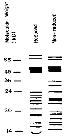
Figure 1 illustrates an SDS-PAGE of a protein mixture according to the present
invention, both in reduced and nonreduced forms.
Figure 2 is an SDS-PAGE gel of HPLC fractions 27-36 of a protein mixture
according to an embodiment of the present invention.
Figure 3 is an SDS-PAGE gel with identified bands indicated according to the
legend of Figure 4.
Figure 4 is an SDS-PAGE gel of a protein mixture according to an embodiment of
the present invention with identified bands indicated, as provided in the
legend.
Figure 5 is two-dimensional (2-D) SDS-PAGE gel of a protein mixture according
to
an embodiment of the present invention with internal standards indicated by
arrows.
Figure 6 is a 2-D SDS-PAGE gel of a protein mixture according to an embodiment
of the present invention with circled proteins identified as in the legend.
Figures 7A-O are mass spectrometer results for tryptic fragments from one-
dimensional (1-D) gels of a protein mixture according to an embodiment of the
present
invention.
Figure 8 is a 2-D gel Western blot of a protein mixture according to an
embodiment
of the present invention labeled with anti-phosphotyrosine antibody.
Figures 9A-D are 2-D gel Western blots of a protein mixture according to an
embodiment of the present invention, labeled with indicated antibodies. Figure
9A indicates
the presence of BMP-3 and BMP-2. Figure 9B indicates the presence of BMP-3 and
BMP-
7. Figure 9C indicates the presence of BMP-7 and BMP-2, and Figure 9D
indicates the
presence of BMP-3 and TGF-131.
Figure 10 is a PAS (periodic acid schiff) stained SDS-PAGE gel of HPLC
fractions
of a protein mixture according to an embodiment of the present invention.
Figure llis an anti-BMP-7 stained SDS-PAGE gel of a PNGase F treated protein
mixture according to an embodiment of the present invention.
CA 02413338 2002-12-23
WO 02/00244 PCT/USO1/41110
_g_
Figure 12 is an anti-BMP-2 stained SDS-PAGE gel of a PNGase F treated protein
mixture according to an embodiment of the present invention.
Figures 13A-B are bar charts showing explant mass of glycosylated components
in a
protein mixture according to an embodiment of the present invention (Figure
13A) and ALP
score (Figure 13B) of the same components.
Figure 14 is a chart showing antibody listing and reactivity.
Figures 15A-B together comprise a chart showing tryptic fragment sequencing
data
for components of a protein mixture according to an embodiment of the present
invention.
Figures 16A-F together comprise a chart showing tryptic fragment mass
spectrometry data for components of a protein mixture according to an
embodiment of the
present invention.
Figures 17A-B are an SDS-gel (Figure 17B) and a scanning densitometer scan
(Figure 17A) of the same gel for a protein mixture according to an embodiment
of the
present invention.
Figure 18 is a chart illustrating the relative mass, from scanning
densitometer
quantification, of protein components in a protein mixture according to an
embodiment of
the present invention.
Figures 19A-D together comprise a chart showing mass spectrometry data of
various protein fragments from 2D gels of a protein mixture according to an
embodiment of
the present invention.
Detailed Description of the Invention
EXAMPLE 1. BP IN SINGLE DOSE APPLICATION TO NUDE MICE
A single dose application of BP to full thickness wounds in nude mice covered
with
human meshed split thickness skin grafts has been found to heal the wound
completely and
faster than wounds not receiving the growth factor mixture. Although the
specific manner
in which the growth factors in BP affect the wound healing process is not
fully understood,
it is hypothesized that the synergistic action of the multiple growth factors
present in BP
helps the wounds recover better than those in control animals that have
received the carrier
alone.
Full thickness wounds were created in nude mice such that the wound area
comprised about 20% of the total body surface. BP was prepared as in U.S.
Patent Nos.
5,290,763, 5,371,191, and 5,563,124, and applied to the wound in a povidone
carrier. The
wound was then covered with human meshed split thickness skin grafts. The
control group
of animals received only the povidone carrier. The graft sites were dressed
and closed with
CA 02413338 2002-12-23
WO 02/00244 PCT/USO1/41110
-9-
band-aids to keep the dressing securely in place. The first dressing changes
were carried
out on day 5 post operative and every third day thereafter. The basic protocol
is also
described in "Clinical and Experimental Approaches to Dermal and Epidermal
Repair:
Normal and Chronic Wounds," pp. 429-442 (1991) Wiley-Liss, Inc. and Cooper
M.L., et
al., The Effects of Epidermal Growth factor and basic Fibroblast Growth factor
on
Epithelialization of Meshed Skin Graft Interstices, Prog. Clin. Biol. Res.
(1991) 365: 429-
42. Such protocols are known to persons of skill in the art.
The results were strongly encouraging. Single application of two
concentrations
(either 100 ~.g/wound site or 200 ~.g/wound site) of growth factor were
tested. There was
no difference either in the rate or degree of wound healing between the two
groups.
However, there was a marked difference between the group of animals that
received the
growth factor treatment and the control animals that did not receive the
growth factor. By
day 11 POD (post operative day), a 95 % wound closure was observed in the
animals that
received the growth factor whereas the control animals showed only a 74 %
closure. By day
14 POD all growth factor treated animals had a 100 % closure while the control
animals had
only a 85 % closure as of day 20 POD.
The thickness of the epithelial layer in BP treated wounds was significantly
higher
in BP treated animals compared to the control animals, as shown in Table 1.
The data
represents the thickness of neodermis in mm measured on day 11 for the BP
treated animals
and day 16 for the control animals such that measurements are made at
equivalent extents of
healing. Histological analysis revealed that the wounds were closed by the
human cells
from the grafted material and there was collagen deposition in the closed
wounds as
revealed by involucrin and collagen type 1 immuno histological staining (data
not shown).
The capillary density in the wound bed following BP treatment was also
significantly higher
at the time of wound closure compared to untreated controls, as shown in Table
1. Further,
in the animals treated with the lower BP dosage, there was a significant
increase in the
smooth muscle cell (SMC) count in the BP treated wounds as compared to the
controls, as
also seen in Table 1.
CA 02413338 2002-12-23
WO 02/00244 PCT/USO1/41110
-10-
Table 1. Wound Thickness, Capillary Count and SMC Count for BP and Control
Treated
Wounds.
Treatment
100 ~,g BP (n=5) 200 ~,g BP (n=5) Control (n=10)
Epithelial Thickness1.60 0.12 (P 1.55 0.09 (P < 1.1 0.25
(mm) < 0.001) 0.001)
Capillary/Field 37 6 (P < 0.01) 35 7 (P < 0.01) 25 5.9
SMC counts/Field 53 3.5 (P < 0.001)46.8 4.4 (P < 46 5.8
0.05)
In summary, a single dose application of BP was effective in reducing the
healing
time of full thickness wound in nude mice grafted with human meshed split
thickness skin.
Additionally, the thickness of the neodermis and the density of the
capillaries in the treated
wounds were significantly higher compared to the control group of animals. In
contrast,
bFGF, also an angiogenic growth factor, was shown to have a deleterious effect
on
epithelialization when tested in a similar animal model. (Cooper, M.L. et al.,
1991; Clinical
and experimental approaches to dermal and epidermal repair: normal and chronic
wounds,
pp 429-442; Weilly-Liss, Inc.).
EXAMPLE 2. BP IN HYDROGEL
A small number of animals (n=3) were treated with BP solubilized in a hydrogel
(carboxy-methyl cellulose) in the same animal model as described above. In
this study, it
was observed that the wounds (n=2) treated with BP in the hydrogel showed
initiation of
epithelialization as early as 5 days post operation compared to the wounds
treated with BP
solubilized in 1 % povidone, which showed initiation of epithelialization only
at 8 days post
operation (data not shown). In both instances, the control animals that
received the carrier
alone did not show initiation of epithelialization until POD 8. Detailed
histology is being
carried out on the tissue samples to determine the thickness of the neodermis
and the degree
of angiogenesis in the wounds treated with the hydrogel formulation. However,
wound
closure data is presented in Table 2, below.
CA 02413338 2002-12-23
WO 02/00244 PCT/USO1/41110
-11-
Table 2. Percent Wound Closure for BP and Control Treated Wounds.
~.:a~~ .
.~ ~: ~, ~ .~=..__
.~~ -
~ Percent=Wound~Closu
' e
%
.~ E, ~~
.
~_. .
~ z
~ _.-
x.
m m
P D: ~.P. - _~3,. _
~- ~.,_ ~..puu :-g D~ ~ ~ .
L . O .5 ~. ~:0 4
. a #.. OD 1.1 POD 1
.
~: -
: .
. ,....
. .._ _ _ ~ : W ~-
. . . ~
_F _ __
*Control (no BP) 1 0 50 70 70
Control (hydrogel, 2 25 70 70 100
no BP, no
salts)
BP & hydrogel, no 3 0 70 90 100
salts
BP & hydrogel, no 4 25 80 90 90
salts
BP & hydrogel, salts 5 0 80 90 100
(some
precipitate formed,
probably
due to buffering salts)
* The control animal had very thin and fragile skin at the time of biopsy
compared to the
animals, which received BP.
In summary, the results were very promising although preliminary, showing
quicker
wound closure in BP treated than control animals. Thus, more extensive
experiments were
undertaken to confirm the results, as described below.
EXAMPLE 3. COMPARATIVE STUDY BETWEEN REGRENEX(r) AND BP
REGRANEX(r) (PDGF-BB), the only approved growth factor product in the market
for treating diabetic foot ulcers, showed complete healing in 50% of the
patient population
compared to the 35 % placebo gel treatment that demonstrated complete healing
after repeat
application for about 20 weeks in diabetic patients (see REGRANEX(r) U.S. full
prescribing information - package. insert). Hence, a comparison of REGRANEX(r)
(tm)
versus BP was undertaken in a study similar to that described above. The
results are
presented in Tables 3 and 4.
CA 02413338 2002-12-23
WO 02/00244 PCT/USO1/41110
-12-
Table 3. BP, Hydrogel (HG) and Regranex~ Treated Wounds and Percent Wound
Closure
(%), Epithelial Thickness (mm) and Degree of Angiogenesis (# Estimated
Capillaries per
20x Field).
Percent: E ~ ~ An
~iae P io
Wound 'Thickg.
ClosurE ~ ~ (#
. =~ ~ ;, est
-~"" -
-- ~ .,~ .,~aa
~ (gym) j
?~ ~~ ~= cap/hpf
~ ~ 2~~x
~- ~
,"~~-
~ _
~~ _..._'~
~~
z -
:~
v~ ~ r .
al ~ ~T .eatment-~-PODSC?D =OD.~ OD.~ OD.,~.PO
~m ~: ~.~ . . P~~ -_
#~ -~ ~ --; ~ ~ w ~ z14- ~~.14_~-n.D
~ ... ~~8._..,~_ _
~ .. ..~~.Grou~ Wl ~....14~m~
:. __d l
5 ._
1 BP 10 25 85 100 17.5 28
2 BP 10
3 BP 15
4 BP 10
BP 10 30 85 80 7.5 16
6 BP 10
7 BP 10 10
8 BP 10 30 85 100 11.5 26
9 BP 30 50 85 100 16 21
BP 30 50 85 100 12 20
11 BP 20 45 85 100 18 18
12 BP 10 15 85 90 6 20
13 BP 10 20 95 100 5.5 23
14 BP 15 25 90 100 10 32
BP 5 50 90 95 14 25
n 15 11 10 10 10 10
mean 13.67 31.8287.00 96.50 11.80 22.9
SD 7.43 14.713.50 6.69 4.58 4.88
SEM 0.54 0.46 0.04 0.07 0.39
16 HG 15 35 75 55 12.5 28
17 HG 10 60 70 95 10.5 5
18 HG 5 25 60 95 9 34
19 HG 10 30 70 90 17.5 8
HG 20 40 80 95 17.5 20
21 HG 10 10 80 95 13 15
22 HG 30 80 70 90 10
23 HG 10 80 80 90 20 10
24 HG 15 40 70 90 18 15
HG 20 35 70 90 10.5 16
26 HG 10 10 70 90 12.5 20
27 HG 10 35 70 90 8 32
28 HG 10 55
29 HG 5 40
HG 15 40 70
n 15 15 13 12 12 11
mean 13.00 41.0071.92 88.75 13.25 18.455
CA 02413338 2002-12-23
WO 02/00244 PCT/USO1/41110
-13-
SD 6.49 20.72 5.60 10.90 4.01 9.55
SEM 0.50 0.51 0.08 0.12 0.30
31 Regranex 20 30 55 75 16
32 Regranex 15 80 13
33 Regranex 20 80 100 100 8.5 4
34 Regranex 15 50 90 100 10
35 Regranex 40 75 6
36 Regranex 15 70 90 100 7.5 10
37 Regranex 15 70 90 18
38 Regranex 10 80
39 Regranex 40 80
40 Regranex 15 50 80 90 15 13
41 Regranex 15 10
42 Regranex 5 50 100 100 16 21
43 Regranex 40 70 100 100 22.5 10
44 Regranex 5 40 80 100 16.5 6
45 Regranex
n 14 14 9 8 9 9
mean 19.2959.64 87.22 95.63 14.44 10.375
SD 12.0721.88 14.39 9.04 4.88 5.4
SEM ~63 0.37 0.16 0.09 0.34
~ ~ I
The percent closure results can be summarized as follows:
Table 4. Summary
POD's BP:(mean)HG ,.(mean)REG;.(mean)~,_;,;
~
wound closure (%) 0 0.00 0.00 0.00
5 13.67 13.00 19.29
8 31.82 41.33 59.64
11 87.00 71.92 87.22
14 96.25 89.17 95.63
epithelial thickness14 11.8 13.25 14.44
(mm)
angiogenesis (#/filed)14 22.9 18.45 10.38
Thus, the BP treatment is as good as REGRENEX(tm) in closing wounds although
slightly slower healing rates are initially observed. BP treatment also shows
slightly less
thickening of the epithelium and shows considerably improved angiogenesis in
the wound
area.
CA 02413338 2002-12-23
WO 02/00244 PCT/USO1/41110
-14-
EXAMPLE 4. FUTURE APPLICATIONS
Because BP has shown promise as a wound healing agent, it will next be tested
in
applications where wound healing is known to be deficient. Experiments similar
to those
described above will be performed with diabetic animals to test the healing of
full and
partial thickness wounds. The response of venous stasis ulcers and diabetic
ulcers to BP
will also be tested.
In preliminary experiments, Male Sprague Dawley rats weighing greater than 325
g
were rendered diabetic by treatment with streptozotocin and the hyperglycemia
was
confirmed by glucometry. Four full thickness incisional wounds were introduced
on the
dorsal surface of each animal perpendicular to the longitudinal axis. The
wounds were
closed with silk sutures and the growth factor or the placebo applied into the
wound gap or
on top of the incision after closure. The application was done at two time
points: 1) on day
0, which is on the day of introducing the wound (surgery) and a second
application 2) on
day 3 following the introduction of the wound. The incisional strength was
measured on
day 7 and day 10 after surgery. The data is given in Table 5 and is very
encouraging that
the BP treatment will be particularly useful in treating a variety of diabetic
ulcers, or other
wounds characterized by delayed and/or poor healing.
Table 5. Tensile Strength of Wounds in Diabetic Rats
Tensile~Strength=(kg/mm)
~ seW
: Control . <BP
: - -
~ ~ ~-
~ -_ :
_..... ._. 4 . .
~ ~. ~ .
~ ..... .
... 3.6 1 4.2 .7
Day 7
DaylO 5.2.7 9.1.8
EXAMPLE 5: FURTHER CHARACTERIZATION OF BP
The BP has been partially characterized as follows: high performance liquid
chromatography ("HPLC") fractions have been denatured, reduced with DTT, and
separated by sodium dodecyl sulfate polyacrylamide gel electrophoresis (SDS-
PAGE). One
minute HPLC fractions from 27 to 36 minutes are shown in Figure 2. Size
standards (ST)
of 14, 21, 31, 45, 68 and 97 kDa were obtained as Low Range size standards
from
BIORAD(tm) and are shown at either end of the coomassie blue stained gel. In
the usual
protocol, HPLC fractions 29 through 34 are pooled to produce BP (see boxes,
Figures 2
and 3), as shown in a similarly prepared SDS-PAGE gel in Figure 17B.
The various components of the BP were characterized by mass spectrometry and
amino acid sequencing of tryptic fragments where there were sufficient levels
of protein for
CA 02413338 2002-12-23
WO 02/00244 PCT/USO1/41110
-15-
analysis. The major bands in the 1D gel (as numerically identified in Figure
3) were
excised, eluted, subjected to tryptic digestion and the fragments were HPLC
purified and
sequenced. The sequence data was compared against known sequences, and the
best
matches are shown in Figures 15A-B. These identifications are somewhat
tentative in that
only portions of the entire proteins have been sequenced and, in some cases,
there is
variation between the human and bovine analogs for a given protein.
The same tryptic protein fragments were analyzed by mass spectrometry and the
mass spectrograms are shown in Figures 7A-O. The tabulated results and
homologies are
shown in Figures 16A-F which provides identification information for the bands
identified
in Figures 3-4. As above, assignment of spot identity may be tentative based
on species
differences and post-translational modifications. A summary of all protein
identifications
from ID gels is shown in Figure 4.
The identified protein components of BP, as described in Figures 15A-B, 16A-F
and 19A-D, were quantified as shown in Figures 17A and 17B. Figure 17B is a
stained
SDS-PAGE gel of BP and Figure 17A represents a scanning densitometer trace of
the same
gel. The identified proteins were labeled and quantified by measuring the area
under the
curve. These results are presented in Figure 18 as a percentage of the total
peak area.
Thus, there are 11 major bands in the BP SDS-PAGE gel representing about 60 %
of the protein in BP. The identified proteins fall roughly into three
categories: the ribosomal
proteins, the histones and growth factors, including bone morphogenic factors
(BMPs). It is
expected that the ribosomal proteins and histone proteins may be removed from
the BP
without loss of activity, since these proteins are known to have no growth
factor activity.
Upon this separation, the specific activity is expected to increase
correspondingly.
Experiments are planned to confirm the hypothesis that the histone and
ribosomal
proteins may be removed from the BP with no resulting loss, or even an
increase, in
specific activity. Histones will be removed from the BP cocktail by
immunoaffinity
chromatography using either specific histone protein antibodies or a pan-
histone antibody.
The histone depleted BP (BP-H) will be tested as described above for wound
healing and/or
osteogenic activity. Similarly, the known ribosomal proteins will be stripped
and the
remaining mixture (BP-R) tested.
An SDS-PAGE gel of BP was also analyzed by Western immunoblot with a series
of antibodies, as listed in Figure 14. Visualization of antibody reactivity
was by horse
radish peroxidase conjugated to a second antibody and using a chemiluminescent
substrate.
Further, TGF-131 was quantified using commercially pure TGF-131 as a standard
and was
CA 02413338 2002-12-23
WO 02/00244 PCT/USO1/41110
-16-
determined to represent less than 1 % of the BP protein. The antibody analysis
indicated
that each of the proteins listed in Figure 14 is present in BP.
The BP was further characterized by 2-D gel electrophoresis, as shown in
Figures 5-6. The proteins are separated in horizontal direction according to
charge (pI) and
in the vertical direction by size as described in two-dimensional
electrophoresis adapted for
resolution of basic proteins was performed according to the method of
O'Farrell et al.
(O'Farrell, P.Z., Goodman, H.M. and O'Farrell, P.H., Cell, 12: 1133-1142,
1977) by the
Kendrick Laboratory (Madison, WI). Two-dimensional gel electrophoresis
techniques are
known to those of skill in the art. Nonequilibrium pH gradient electrophoresis
("NEPHGE") using 1.5 % pH 3.5-10, and 0.25 % pH 9-11 ampholines (Amersham
Pharmacia Biotech, Piscataway, NJ) was carried out at 200 V for 12 hrs.
Purified
tropomyosin (lower spot, 33,000 KDa, pI 5.2), and purified lysozyme (14,000
KDa, pI
10.5 - 11) (Merck Index) were added to the samples as internal pI markers and
are marked
with arrows.
After equilibration for 10 min in buffer "0" (10% glycerol, 50 mM
dithiothreitol,
2.3 % SDS and 0.0625 M tris, pH 6.8) the tube gel was sealed to the top of a
stacking gel
which is on top of a 12.5 % acrylamide slab gel (0.75 mm thick). SDS slab gel
electrophoresis was carried out for about 4 hrs at 12.5 mA/gel.
After slab gel electrophoresis two of the gels were coomassie blue stained and
the
other two v~ere transferred to transfer buffer (12.5 mM Tris, pH 8.8, 86 mM
Glycine, 10%
MeoH) transblotted onto PVDF paper overnight at 200 mA and approximately 100
volts/two gels. The following proteins (Sigma Chemical Co. , St. Louis, MO)
were added
as molecular weight standards to the agarose which sealed the tube gel to the
slab gel:
myosin (220,000 KDa), phosphorylase A (94,000 KDa), catalase (60,000 KDa),
actin
(43,000 KDa), carbonic anhydrase (29,000 KDa) and lysozyme (14,000 KDa).
Figure 5
shows the stained 2-D gel with size standards indicated on the left.
Tropomyosin (left
arrow) and lysozyme (right arrow) are also indicated.
The same gel is shown in Figure 6 with several identified proteins indicated
by
numbered circles. The proteins were identified by mass spectrometry and amino
acid
sequencing of tryptic peptides, as described above. The identity of each of
the labeled
circles is provided in the legend of Figure 6 and the data identifying the
various protein
spots is presented in Figures 19A-D.
Because several of the proteins migrated at more than one size (e.g., BMP-3
migrating as 6
bands) investigations were undertaken to investigate the extent of post-
translation
CA 02413338 2002-12-23
WO 02/00244 PCT/USO1/41110
-17-
modification of the BP components. Phosphorylation was measured by anti-
phosphotyrosine immunoblot and by phosphatase studies. Figure 8 shows a 2-D
gel,
electroblotted onto filter paper and probed with a phosphotyrosine mouse
monoclonal
antibody by SIGMA (~ A-5964). Several proteins were thus shown to be
phosphorylated at
one or more tyrosine residues.
Similar 2-D electroblots were probed with BP component specific antibodies, as
shown in Figures 9A-D. The filters were probed with BMP-2, BMP-3 (Fig. 9A),
BMP-3,
BMP-7 (Fig. 9B), BMP-7, BMP-2 (Fig. 9C), and BMP-3 and TGF-131 (Fig. 9D). Each
'
shows the characteristic, single-size band migrating at varying pI, as is
typical of a protein
existing in various phosphorylation states.
For the phosphatase studies, BP in 10 mM HCl was incubated overnight at
37° C
with 0.4 units of acid phosphatase (AcP). Treated and untreated samples were
added to
lyophilized discs of type I collagen and evaluated side by side in the
subcutaneous implant
rat bioassay, as previously described in U.S. Patent Nos. 5,290,763, 5,563,124
and
5,371,191. Briefly, 10 (g of BP in solution was added to lyophilized collagen
discs and the
discs implanted subcutaneously in the chest of a rat. The discs were then
recovered from
the rat at 2 weeks for the alkaline phosphotase ("ALP" - a marker for bone and
cartilage
producing cells) assay or at 3 weeks for histological analysis. For ALP
analysis of the
samples, the explants were homogenized and levels of ALP activity measured
using a
commercial kit. For histology, thin sections of the explant were cut with a
microtome, and
the sections stained and analyzed for bone and cartilage formation.
Both native- and phosphatase-treated BP samples were assayed for morphogenic
activity by mass of the subcutaneous implant (explant mass) and ALP score. The
results
showed that AcP treatment reduced the explant mass and ALP score from 100 % to
about
60 % . Thus, phosphorylation is important for BP activity.
The BP was also analyzed for glycosylation. Figure 10 shows an SDS-PAGE gel
stained with periodic acid schiff (PAS) - a non-specific carbohydrate stain,
indicating that
several of the BP components are glycosylated (starred protein identified as
BMP-3).
Figures 11-12 show immunodetection of two specific proteins (BMP-7, Fig. 11
and BMP-2,
Fig. 12) treated with increasing levels of PNGase F (Peptide-N-Glycosidase F).
Both BMP-
2 and BMP-7 show some degree of glycoslyation in BP, but appear to have some
level of
protein resistant to PNGase F as well (plus signs indicate increasing levels
of enzyme).
Functional activity of PNGase F and sialadase treated samples were assayed by
explant
CA 02413338 2002-12-23
WO 02/00244 PCT/USO1/41110
-18-
mass and by ALP score, as shown in Figure 13A and 13B which shows that
glycosylation is
required for full activity.
In summary, BMPs 2, 3 and 7 are modified by phosphorylation and glycosylation.
These post-translation modifications affect protein morphogenic activity, 33 %
and 50
respectively, and care must be taken in preparing BP not to degrade these
functional
derivatives.