Note: Descriptions are shown in the official language in which they were submitted.
CA 02414896 2008-09-15
64371-541
- 1-
IMPLANTABLE PROSTHESIS
Field of the Invention
The present invention relates to an inZplantable prosthesis and, more
particularly, to a
prosthesis for use in soft tissue repair and reconstruction.
Discussion of Related Art
Various prosthetic materials have been proposed to repair and reinforce
anatomical
lo defects, such as tissue and muscle wall hernias. For example, an inguinal
hernia is commonly
repaired using a sheet of biocompatible fabric, such as a knitted
polypropylene mesh (BARD
MESH). The fabric is typically sutured, stapled or otherwise provisionally
anchored in place
over, under or within the defect. Tissue integration with the fabric, such as
by tissue
ingrowth into and/or along the fabric, eventually completes the repair.
1s The attachment of the fabric to host tissue adjacent the defect can result
in tension at
the anchoring locations between the fabric and the tissue when abdominal
pressure at the
repair site is applied to the fabric, such as when an individual stands or
strains, coughs,
sneezes and the like. Tension at the anchoring locations may lead to
postoperative pain for
the patient, a recurrence of the defect, or the formation of a new defect. For
example, a
2o recurrence of the original defect or the creation of a new defect may
result from failure of the
suture line or other fastener, or tearing of the tissue due to tension at one
or more of the
anchoring locations. Tension on the suture line at the anchoring locations may
also lead to
ischemia of the tissue, resulting in enlargement of the suture holes and an
eventual defect.
Scar formation (scarification) associated with the repair of tissue and wall
defects may
25 cause tissue shrinkage at the repair site, thereby contracting the repair
fabric that has been
integrated with tissue. Contraction of the fabric may cause patient discomfort
as the fabric
strains against the host tissue at the anchoring locations. Contraction of the
fabric may cause
failure of the fasteners and/or tearing of the tissue, in either case
potentially leading to a
recurrence of the defect, due to the fabric being pulled away from the tissue
or muscle at the
3o anchoring locations.
CA 02414896 2008-09-15
64371-541
- 2 -
Summary of the Invention
According to one broad aspect of the present
invention, there is provided an implantable prosthesis for
repairing a tissue or muscle wall defect, the implantable
prosthesis comprising: a patch of prosthetic repair fabric
including a body portion to cover at least a portion of the
tissue or muscle wall defect and an anchoring portion to
secure the patch to host tissue or muscle, the patch further
including a preformed region to reduce the incidence of the
detrimental effect of tension at the anchoring portion when
the patch is secured to the tissue or muscle and a force is
applied at the tissue or muscle wall defect and/or to
compensate for contraction of the patch relative to the
tissue or muscle wall defect due to tissue shrinkage during
scarification, the preformed region having a three-
dimensional shape and including a preformed dome having an
open end that is configured to be disposed over the tissue
or muscle wall defect.
Embodiments of the present invention provide an
implantable prosthesis and a method of repairing an
anatomical defect, such as a tissue or muscle wall defect.
The prosthesis is configured to reduce the likelihood that
an applied force, such as due to intraabdominal pressure or
tissue shrinkage, at the repair site can lead to detrimental
effects associated with tension at the anchoring locations
of the prosthesis and host tissue and/or contraction of the
prosthesis. The prosthesis may reduce postoperative pain,
and reduce the likelihood of either a recurrence of the
defect or the creation of a new defect associated with
tension and/or prosthetic contraction.
CA 02414896 2008-09-15
64371-541
- 2a -
The prosthesis is particularly indicated for use in a repair in which tension
andlor
contraction are of potential concern. For techniques that employ fasteners,
such as sutures,
staples, adhesives and the like, to secure the prosthesis to the host tissue,
the prosthesis niay
limit tension at the anchoring locations between the prosthesis and the host
tissue. For -
fastener-less techniques, use of the prosthesis may be beneficial by limiting
tension at the
locations where tissue integrates with the prosthesis and/or by accommodating
prosthetic
contraction associated with shrinkage of tissue that has grown into and/or
adhered to the
prostliesis. The locations where fasteners join the prosthesis to host tissue
and/or where
tissue integrates with the prosthesis to secure the implant in place may all
be referred to as
1o "anchoriiig locations".
The prosthesis may include a patch formed of a biologically conipatible,
flexible
implantable repair fabric suitable for reinforcing tissue and closing tissue
or muscle wall
defects. The repair fabric may include a body portion for covering at least a
portion of the
tissue or muscle wall defect and an anchoring portion for securing, with or
without fasteners,
ls the fabric to host tissue, including tissue, muscle or the like, adjacent
the defect. The repair
fabric may have a plurality of interstices that are constructed and arranged
to allow tissue
ingrowth. The prosthesis may also include a plug that is enzployed in
combination with the
patch and is configured to be placed within the defect.
In one embodiment of the invention, the repair fabric may include a preformed
region
20 configured to reduce the prospects that an applied force at the repair site
will ca~,~se
detrimental tension at the anchoring location and/or to compensate for
contraction of the
prosthesis that may occur as a result of tissue sluiiikage at the repair site
during scarification.
CA 02414896 2003-01-08
WO 02/07648 PCT/US01/22515
- 3-
According to one aspect of the invention, the preformed region has a
predetermined
ainount of laxity that is greater than an amount of laxity at an anchoring
portion of the fabric
to reduce the prospects that an applied force at the repair site will cause
detrimental tension at
the anchoring location. According to another aspect of the invention, the
preformed region
has a predetermined amount of compensation to compensate for contraction of
the prosthesis.
According to a further aspect of the invention, the preformed region is
configured to both
reduce detrimental tension at the anchoring portion and to compensate for
prosthetic
contraction.
In another embodiment of the invention, the implantable prosthesis may include
a
body portion having a preformed three-dimensional shape and a cavity with an
open end that
is configured to be disposed over the defect.
In a further embodiment of the invention, the implantable prosthesis may
include a
plurality of preformed indicia on the anchoring portion that correspond to
predetermined
anchoring locations between the prosthesis and the tissue or muscle.
The prosthesis is particularly suited for repairing a tissue or inuscle wall
hernia
located in one or more of the inguinal region, the inguinofemoral region and
the femoral
region: The prosthesis may include a medial section and a lateral section that
are configured
to be positioned adjacent the medial conler and the lateral end of the
inguinal canal,
respectively, when the prosthesis is placed in the inguinal canal. The
prosthesis may include
an extension that is configured to extend into the femoral region and cover
the femoral ring.
Other objects and features of the present invention will become apparent from
the
following detailed description when taken in connection with the accompanying
drawings. It
is to be understood that the drawings are designed for the purpose of
illustration only and are
not intended as a definition of the limits of the invention.
Brief Description of the Drawings
The foregoing and other objects and advantages of the invention will be
appreciated
more fully from the following drawings, wherein like reference characters
designate like
features, in which:
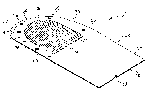
FIG. 1 is a top perspective view of an implantable prosthesis in accordance
with one
illustrative embodiment of the present invention;
FIG. 2 is a top plan view of the implantable prosthesis of FIG. 1;
CA 02414896 2003-01-08
WO 02/07648 PCT/US01/22515
- 4-
FIG. 3 is a side view of the implantable prosthesis of FIG. 1;
FIG. 4 is a front view of the implantable prosthesis of FIG. 1;
FIG. 5 is a cross-sectional view of the implantable prostliesis of FIG. 1
taken along
section line 5-5 of FIG. 3;
FIG. 6 is a top plan view of an implantable prosthesis in accordance with
another
illustrative embodiment of the present invention;
FIG. 7 is a top plan view of an iinplantable prosthesis in accordance with a
further
illustrative embodiment of the present invention;
FIG. 8 is a top plan view of an implantable prostliesis in accordance with
another
illustrative embodiment of the present invention;
FIG. 9 is a cross-sectional view of the implantable prosthesis of FIG. 8 taken
along
section line 9-9 of FIG. 8 and rotated counter-clockwise;
FIG. 10 is a top plan view of an implantable prosthesis in accordance with a
further
illustrative embodiment of the present invention; and
FIG. 11 is an exploded perspective view of an implantable prosthesis in
accordance
with another illustrative embodiment of the present invention.
Description of Illustrative Embodiments
FIGS. 1-5 illustrate one embodiment of an implantable prosthesis for repairing
an
anatomical defect, such as a soft tissue or muscle wall defect. The
prostliesis is configured
to reduce the likelihood that an applied force at the repair site, such as due
to intraabdominal
pressure or tissue shrinkage, can lead to detrimental effects associated with
tension at the
anchoring locations between the prosthesis and host tissue and/or contraction
of the
prosthesis. In this regard, the prosthesis may be configured to limit the
amount of tension at
the anchoring locations caused by the application of a force or pressure to
the prosthesis
and/or contraction of the prosthesis. Alternatively, the prosthesis may be
configured to
compensate for contraction of the prosthesis due to tissue shrinlcage at the
repair site. The
prosthesis may be configured to both limit tension at the anchoring locations
and
compensate for tissue shrinkage. The prosthesis may facilitate a reduction in
postoperative
discomfort, a recurrence of the defect, or the creation of a new defect
associated with
tension and/or prosthetic contraction.
CA 02414896 2003-01-08
WO 02/07648 PCT/US01/22515
- 5-
The prosthesis 20 includes a patch of prosthetic repair fabric'22 formed of a
biologically coinpatible, flexible material. The patch 22 may include a body
portion 24 for
covering at least a portion of the tissue or muscle wall defect and an
anchoring portion 26
for securing the fabric to host tissue, which may include tissue, muscle or
the like, adjacent
the defect. The patch may be combined with a plug of repair fabric that is
configured to be
placed within the defect.
In some repair techniques, a fastener, such as sutures, staples, adhesives and
the lilce,
may be employed to secure the anchoring portion 26 to the tissue or muscle. In
a fastener-
less repair technique, pressure, friction, tissue ingrowth and the like acting
on the anchoring
portion 26 may be used to secure the fabric to the tissue or muscle. The
repair fabric may
integrate with liost tissue over time, and may include a plurality of
interstices or openings
which allow sufficient tissue ingrowth. The prosthetic fabric may,
alternatively, be
relatively non-porous, so as not to be susceptible to tissue infiltration.
Abdominal pressure or other forces applied at the repair site may lead to one
or more
of various detrimental effects associated with tension at the anchoring
locations. Such
detrimental effects may be alleviated by providing the implantable prostliesis
with a
preformed region having a predetermined amount of laxity that limits the
amount of tension.
In this regard, laxity refers to one or more of loose, slaclc, stretchable and
like characteristics
that accommodate forces at the repair site in a manner that limits tension.
In one illustrative embodiment, the body portion 24 is configured with a
region of
laxity. As shown, the body portion 24 may have a preformed three-dimensional
shape, such
as a dome, that is configured to overlie and cover the defect. The body
portion may be
configured to be larger than the defect so that the body portion extends
across and covers the
entire defect. One or more anchoring portions 26 extend from the body portion
24 for
securing the fabric to the tissue or muscle adjacent the defect at one or more
anchoring
locations. The anchoring portions 26 may be disposed partially or completely
about the
body portion 24, and may be planar, as illustrated, or configured to the shape
of the
anchoring locations at the repair site. Further, the anchoring portions may be
conformable
to the anatomy of interest.
The dome is configured to provide an amount of laxity that is greater than the
laxity
of the anchoring portion. The dome includes a surface that is placed over and
in spaced
relation to the defect when the prosthesis is implanted in the repair site.
The dome has an
CA 02414896 2003-01-08
WO 02/07648 PCT/US01/22515
- 6-
open end that may be disposed over and exposed to the defect so that the
defective tissue or
muscle may extend into the dome, such as due to intraabdominal pressure,
without exerting
an excessive amount of force on the prosthesis that may lead to detrimental
tension. In this
regard, the dome may be configured to allow the defective tissue or muscle to
fill the dome
cavity and contact the dome surface with less force than were the body portion
not initially
spaced from the defect. This arrangement accommodates an increase in pressure
or other
forces in the repair region with minimal, if any, tension being applied to the
tissue or muscle
at the anchoring locations.
As illustrated, the dome may be configured witli a generally spherical or
curved
shape. It is to be appreciated, however, that the dome may be configured to
have any shape
that is suitable to receive the tissue or muscle therein.
Although illustrated as part of the body portion 24, it is contemplated that
the region
of laxity may be provided on any portion of the repair fabric 22 that is
suitable to limit
tension at the anchoring locations between the prosthesis and tissue. For
example, and
without limitation, the region of laxity also may be located in the anchoring
portion 26, or
between the body portion 24 and the anclioring portion. The region of laxity
may also be
comprised of two or more regions located in various portions of the patch 22
to provide
laxity in selected regions of the prosthesis.
It is also to be appreciated that the region of laxity may be formed in any
manner
suitable to reduce the detrimental effects of tension at the anchoring
locations. For example,
the region may employ a pleated or other configuration that allows the region
to expand
under an applied force or pressure so as to alleviate or limit tension between
the fabric and
the tissue or muscle at the anchoring locations. The region may also employ a
material
having different size filaments and/or a knit or weave pattern that allows the
region to
expand or otherwise absorb an increase in pressure in the repair region. The
region of laxity
may be formed with a material that is different than the material forming
other portions of
the prosthesis.
When tissue integration with a prosthesis occurs during the repair process,
tissue
shrinkage at the repair site may lead to one or more of various detrimental
effects associated
with contraction of the prosthesis. Such detrimental effects may be alleviated
by providing
the implantable prosthesis with a preformed region of compensation that
compensates for
CA 02414896 2003-01-08
WO 02/07648 PCT/US01/22515
- 7-
the tissue shrinkage by accommodating an anticipated amount of fabric
contraction in the
repair region.
In one illustrative embodiment, the body portion 24 includes a region of
compensation that provides additional material to compensate for fabric
contraction that
may occur due to tissue shrinkage, so that the body portion continues to cover
the defect
after scarification. This arrangement may reduce the incidence of a recurrence
to the extent
that the prosthesis does not tend to pull away and separate from the tissue or
muscle to
which it is secured when the integrated tissue shrinks.
The amount of contraction generally depends on the extent of tissue
integration with
the prosthesis. For example, as the amount of tissue integration increases,
the amount of
fabric contraction also tends to increase. In this regard, the pore size of
the fabric may
influence the amount of tissue integration, and therefore the amount of
prosthetic
contraction. As the pore size increases, the fabric may experience a greater
amount of tissue
growth into the pores, which may lead to a larger amount of fabric contraction
during tissue
shrinkage.
In the embodiment shown in FIGS. 1-5, the body portion 24 may have a preformed
three-dimensional shape, such as a dome, which is configured to overlie the
defect and to
provide an additional amount of material, as compared to a flat body portion,
that
compensates for fabric contraction. During tissue shrinkage, the dome
contracts to alleviate
detrimental effects on the prosthesis. In one embodiment, the dome is
configured with an
amount of fabric that is capable of accommodating a range of fabric
contraction from
approximately 15% to approximately 25%. It is to be appreciated, however, that
the
particular amount of material provided by the dome may be adjusted to
accommodate any
anticipated amount of contraction.
It is contemplated that the region of compensation may be provided on any
portion
of the patch 22 that is suitable to compensate for contraction of the
prosthesis. For example,
the region of compensation may be located adjacent the body portion 24. The
region of
coinpensation may also be comprised of two or more regions located in various
portions of
the patch to provide compensation for fabric contraction in selected regions
of the
prosthesis.
It is also to be appreciated that the region of compensation may be formed in
any
manner suitable to accommodate prosthetic contraction. For example, siinilar
to the region
CA 02414896 2003-01-08
WO 02/07648 PCT/US01/22515
- 8-
of laxity described above, the region of compensation may employ a pleated or
other
configuration that provides sufficient material to allow the region to
accommodate fabric
contraction so as to alleviate patient discomfort or separation of the fabric
from the tissue or
muscle. In this regard, the pleats may be configured to allow fabric expansion
due to
increased pressure or other forces applied at the repair region which, if
retained, may
compensate for prosthetic contraction The region of compensation may also
employ a
material having different size filainents and/or a knit or weave pattern that
allows the region
to accommodate tissue shrinkage during scarification. The region of
compensation may be
formed from a material that is different than the material forming other
portions of the
prosthesis.
As one of ordinary skill would readily appreciate, the=prosthesis may be
configured
both to relieve tension at the anchoring locations, associated with pressure
or other variables
at the repair site, and to accommodate fabric contraction associated with
tissue shrinkage at
the repair region. In the embodiment illustrated in FIGS. 1-5, the body
portion 24 is
configured to include the region of laxity and the region of compensation. It
is to be
understood, however, that the prosthesis 20 may include a region of laxity
that is separate
from the region of compensation. Further, the region of laxity may have a
construction that
differs from the region of compensation in terms of one or more of shape,
size, material and
the like.
The prosthesis 20 may be particularly suited to the repair of inguinal
hernias. In the
illustrative embodiment shown in FIGS. 1-5, the patch 22 includes a medial
section 28 and a
lateral section 30 that are configured to be positioned adjacent the medial
corner and the
lateral end of the inguinal canal, respectively. The medial section 28 of the
prosthesis may
include a generally rounded medial edge 32 that is configured to extend beyond
the medial
corner of the inguinal canal for anchoring the prosthesis. The body portion 24
may be
configured with a rounded medial segment 34. As illustrated, the medial
segment 34 may
be spaced inwardly from the medial edge 32 of the prosthesis to provide the
patch with an
anchoring portion 26 between the medial edge 32 and the medial segment 34. It
is to be
appreciated, however, that the medial segment 34 of the body portion may
extend to the
peripheral edge, including the medial edge 32, of the patch. The body portion
may also
include a lateral segment 36 that is located proximate to the center of the
prostllesis so that it
is positioned adjacent the internal ring. As shown, the body portion may have
a generally
CA 02414896 2003-01-08
WO 02/07648 PCT/US01/22515
- 9-
D-shaped periphery, although any shape may be employed to suit a particular
prosthetic
application.
As illustrated in an embodiment shown in FIG. 6, the prosthesis 20 may have a
slit
3 8 extending inwardly from the lateral edge 40 of the patch to create a pair
of tails 42, 44
that are separated to receive the spermatic cord proximate the lateral segment
36 of the body
portion. The tails 42, 44 may be crossed around the cord and stitched or
otherwise secured
to each other to reinforce the internal ring. To aid a surgeon in this regard,
it may be
desirable to provide the prosthesis witlz a loclcing feature that may be
employed to secure the
tails to each other.
In one embodiment, the locking feature includes one or more pairs of fasteners
46,
48 that are disposed on the tails 42, 44 and configured to mate with each
other in a
male/female-type arrangement when the tails are crossed about the cord. As
shown in FIG.
6, a series of fasteners 46, 48 may be located along a portion of the length
and on opposite
sides of the slit 38. The fasteners 46, 48 may extend along nonparallel lines
47, 49 that are
angled relative to the slit 38 so that the fasteners become aligned with each
other when the
tails are crossed about the cord. The fasteners may include snaps, zipper-type
strips, and
like arrangements that may be integrally preformed in the fabric. In another
embodiment,
the locking feature may include a suture that is attached to one of the tails
and is to be
passed through at least the opposite tail to secure the tails together when
they are crossed
about the cord. Of course, it is to be understood that any suitable locking
feature apparent to
one of skill in the art may be employed to secure the tails about the cord.
The slit 38 may be preformed with the prosthesis 20 or formed by the surgeon
during
the repair procedure. To facilitate formation of the slit 38, the prosthesis
may include an
indicator that is configured to identify the location and/or length of the
slit. In one
embodiment, the patch includes a notch 50 along the lateral edge 40 that
identifies a
preferred location for the slit. It is to be appreciated, however, that any
suitable indicator
may be employed to aid the surgeon in locating and forming the slit. For
example, the
indicator may include a series of holes, contrasting color stitches and the
like that run along
a portion of the prosthesis and correspond to the location, length and/or
orientation of the
slit. The indicator may be marked on the prosthesis using a suitable ink or
dye.
In some instances, a surgeon may find it desirable to repair or reinforce the
femoral
region by itself or in conjunction with an inguinal hernia repair. It is,
therefore,
CA 02414896 2003-01-08
WO 02/07648 PCT/US01/22515
- 10-
contemplated that the prosthesis 20 may be configured to reinforce the
inguinofemoral
region or femoral region of an individual.
In one illustrative embodiment shown in FIG. 7, the prosthesis 20 includes an
extension 52 that is configured to extend downwardly into the space of Retzius
to Cooper's
ligament and cover the femoral ring upon implantation. As shown, the extension
52 may be
configured with a triangular shape that conforms generally to the femoral
region. It is to be
appreciated, however, that the prosthesis may employ an extension having any
desirable
shape. The extension is foldable along a fold line 54 so that it may be
oriented
approximately 90 relative to the remainder of the prosthesis to accommodate
the
configuration of the inguinofemoral region when implanted. It is contemplated
that the
prosthesis may be preformed with the extension 52 oriented at a desired angle
to facilitate
implantation of the prostllesis in the inguinofemoral region.
In the illustrative embodiment, the extension 52 is integrally formed with the
prosthesis from a single sheet of fabric. Alternatively, the extension may be
formed as a
separate piece of the prosthesis that is attached to the patch 22. It is to be
appreciated,
however, that the prosthesis 20 may be formed in any suitable manner.
As indicated above, the prosthesis may include a region of laxity and/or a
region of
coinpensation that employ any configuration suitable to provide a desired
amount of laxity
and compensation, respectively. FIGS. 8-10 illustrate various other
embodiments for a
prosthesis that includes a region of laxity and/or a region of compensation.
It is to be
appreciated that these embodiments are exemplary and other suitable prosthetic
configurations may be implemented for such repairs.
FIGS. 8-9 illustrate an embodiment of a prosthesis 20 that includes a body
portion 24
preformed with a plurality of pleats 56 to provide a region of laxity. As
illustrated, the
pleats 56 may be arranged in an accordion-like fashion to provide additional
material across
the width of the patch 22 that are configured to expand to accommodate
increased pressure
or other forces applied at the repair region. When expanded, the additional
material
provided by the pleats may also compensate for tissue shrinkage at the repair
region.
FIG. 10 illustrates another embodiment of a prosthesis that includes a body
portion
24 preformed with a plurality of pleats 58 that are arranged in an umbrella-
like, radial
fashion. This arrangement of pleats 58 may also provide a region of laxity and
a region of
compensation.
CA 02414896 2003-01-08
WO 02/07648 PCT/US01/22515
- 11-
It is to be appreciated that the various body portion configurations
illustrated in
FIGS. 1-10 are not intended to be exhaustive.
The body portion 24 may be integrally formed with the anchoring portion 26
from a
single sheet of fabric. The body portion 24 may be molded or otllerwise
preformed in the
fabric to have any shape and size suitable for providing a desired amount of
laxity and/or
compensation. It is to be appreciated, however, that the prosthesis 20 may be
formed in any
suitable manner.
It is contemplated that the body portion 24 may be preformed as a separate
piece of
the prosthesis that is attached to the patch fabric. A prosthesis formed in
this manner may
facilitate the use of various shapes, materials and the like for the body
portion.
In one illustrative embodiment shown in FIG. 11, a preformed body portion 24
is
positioned in a hole 60 in a piece of fabric with the outer periphery 62 of
the body portion
24 being attached to the inner periphery 64 of the hole 60 employing any
suitable fastening
technique. For example, the preformed body portion 24 may be stitched or
bonded to the
fabric using stitches or an adhesive dispensed about the periphery of the body
portion.
Alternatively, the body portion 24 may be laminated or heat fused to the
fabric by a
combination of heat and pressure. The junction between the body portion and
the fabric
may also be configured to enhance the laxity and/or compensation
characteristics of the
prosthesis by being more elastic than one or both of the body portion 24 and
the remaining
patch fabric.
In some instances, it may be desirable to provide one or more indicia on the
prosthesis that aid a surgeon in attaching the prosthesis at preferred
anchoring locations,
such as by suturing, stapling and the like, to the tissue or muscle adjacent
the defect. In one
illustrative shown in FIGS. 1-5, a plurality of preformed indicia 66 are
provided on the
anchoring portion 26 about several sides of the body portion 24. The indicia
66 include a
monofilament thread of contrasting color relative to the repair fabric to
indicate the desired
anchoring location. The number and location of the indicia 66 may be chosen
based on the
particular type of repair and/or surgical technique beirig employed to make
the repair. It is
to be appreciated that any suitable indicia 66 may be provided on the
prosthesis. For
example, the indicia may include bumps, dimples, holes and the like, or may be
marlced on
the prosthesis witli a contrasting color inlc or dye.
CA 02414896 2008-09-15
64371-541
- 12-
In one embodiment, the prosthesis 20 is formed of a sheet of knitted
polypropylene
TM
monofilament mesh fabric such as BARD MESH available from C.R. Bard, Inc. When
implanted, the polypropylene mesh promotes rapid tissue ingrowth into and
around the mesh
structure. Alternatively, other surgical materials which are suitable for
tissue reinforcement
TM TM
and defect closure may be utilized including PROLENF,, SOFT TISSUE PATCH
TM TM
(microporous ePTFE), SURGIPRO, TRELEX, ATRIUIVI M
and MERSELENE: Absorbable
materials, including polyglactin (VICRYL), polyglycolic acid (DEXOw') and
collagen, may
be suitable for applications involving temporary repair of tissue or wall
defects. It also is
contemplated that the mesh fabric may be formed from multifilament yams and
that any
suitable method, such as weaving, molding and the like, may be employed to
form the
prostlletic mesh material.
In some instances, it may be desirable to employ a repair fabric that does not
promote tissue ingrowth. For such an application, the repair fabric may be
formed from a
TM
sheet of expanded polytetrafluoroethylene (ePTFE), such as PRECLUDE
Pericardial
TM TM
Membrane, PRECLUDE Peritoneal Membrane and PRECLUDE" Dura Substitute membrane
available from W.L. Gore & Associates, Inc., having a_pore size (submicronal)
that
discourages tissue ingrowth. A representative and non-limiting sampling of
other suitable
TM
non-porous materials includes silicone elastomer, such as SILASTIC Rx Medical
Grade
Sheeting (Platinum Cured) distributed by Dow Corning Corporation, TEFL
ONTmesh, and
TM
microporous polypropylene sheeting (CELGARD) and film.
It is to be appreciated that any suitable materials may be used for the repair
fabric as
would be apparent to one of skill in the art.
In an exemplary embodiment particularly suited for the repair of an inguinal
hernia,
TM
the patch 22 includes an approximately 0.025 to 0.030 inch thick sheet of BARD
MESIA
knitted from polypropylene monofilament with a diameter of approximately 0.006
inches.
The patch 22 has a length along the longitudinal axis of approximately 5.97
inches and a
width between the side edges of approximately 2.36 inches. The body portion 24
includes a
dome having a height of approximately 0.256 inches. In anotlier embodiment,
the dome
height may be approximately 0.334 inches. The body portion 24 has a length of
approximately 2.36 inches and a width of approximately 1.86 inches. The medial
segment
34 of the body portion has a radius of approximately 0.93 inches. These
dimensions
represent a prosthesis that may be trinuned as necessary by a surgeon to
conform to the
CA 02414896 2003-01-08
WO 02/07648 PCT/US01/22515
- 13-
particular size asld shape of the inguinal canal. It should be understood,
however, that these
dimensions are merely exemplary and that any suitable sizes and shapes may be
employed
for the prosthesis.
In one embodiment, the body portion 24 is formed in the sheet of mesh fabric
using a
die having male and female components that are configured with the desired
shape for the
dome. The fabric is sandwiched between the die components and exposed to a
temperature
of approximately 270 F for approximately 2 hours. It is to be appreciated,
however, that
any suitable manufacturing process may be employed to fabricate the
prosthesis.
The present invention provides an implantable prosthesis that may have one or
more
of the following advantages. The prosthesis 20 may reduce the likelillood that
an applied
force and/or tissue shrinlcage at the repair site can lead to detrimental
effects associated with
either tension at the anchoring locations between the prosthesis and host
tissue, contraction
of the prosthesis, or both tension and contraction. The prosthesis may also
reduce
postoperative pain associated with tension and/or contraction that could
interfere with the
individual's ability to undertake daily activities and/or lengthen the
recovery period from the
repair of a defect. The prosthesis may be sutured, stapled and the like to the
tissue or
muscle and/or anchored in place by tissue integration.
The prosthesis 20 may be pliable to allow a surgeon to manipulate the shape of
the
implant to conform to the anatomical site of interest and to be secured
thereto. The shape
and size of the implant may vary according to the surgical application as
would be apparent
to one of skill in the art. In this regard, it is contemplated that the patch
22 may be
preshaped or, instead, selectively shaped by the surgeon during the surgical
procedure.
The prosthesis 20 of the present invention is particularly indicated for
repair of
inguinal hernias. One representative repair technique includes the
Lichtenstein "tension-
free" repair technique in which a piece of flat mesh fabric is sutured in the
inguinal canal to
cover and reinforce the hernia in a manner that limits tension at the
anchoring locations
between the mesh and the surrounding tissue. The prosthesis 20 of the present
invention
facilitates this technique by providing the surgeon with a repair fabric that
includes a
preformed region of laxity and/or compensation that enhances the surgeon's
ability to
achieve a tension-free repair.
CA 02414896 2003-01-08
WO 02/07648 PCT/US01/22515
- 14-
It should be understood that the foregoing description of the invention is
intended
merely to be illustrative thereof and that other embodiments, modifications,
and equivalents
of the invention are within the scope of the invention recited in the claims
appended hereto.
What is claimed is: