Note: Descriptions are shown in the official language in which they were submitted.
CA 02415328 2003-O1-07
WO 02/006834 PCT/USO1/22821
COLLECTIONS OF BINDING PROTEINS AND TAGS AND USES THEREOF FOR
NESTED SORTING AND HIGH THROUGHPUT SCREENING
RELATED APPLICATIONS
For U.S. purposes benefit of priority under 35 U.S.C. ~ 119(e) is claimed
to U.S, provisional application Serial No. 60/21 9,183, filed July 19, 2000,
to
Dana Ault-Riche entitled "COLLECTIONS OF ANTIBODIES FOR NESTED
SORTING AND HIGH THROUGHPUT SCREENING". For international purposes
priority is claimed to.U.S. provisional application Serial No. 60/219,183.
Where
permitted, the subject matter of U.S. provisional application Serial No.
60/219,183 is incorporated in its entirety by reference thereto.
FIELD OF INVENTION
The present invention relates to collections of binding proteins, called
capture agents herein, and methods of use thereof far functional surveys of
large
diversity libraries, including gene libraries. The methods and collection
technology integrate robotic micro-well high throughput screening and array
and
related techniques.
BACKGROUND OF THE INVENTION
Genomics and proteomics
The Human Genome Project has generated an avalanche of genomic
lt'ICVpGISP
data. Unraveling this data will ir-re~easee the understanding of biology and
ultimately will lead to the development of a new generation of drugs. The
availability of gene sequence information is changing the way biomedical
research is conducted and the rate of discovery. Having the sequence of a
genome, however, does not reveal what the genes do nor how the encoded
0 .1
proteins function, how cells and tissues develop, nor give insights .i~n the
etiology
and cure of diseases. Before the fruits of the information obtained by
sequencing a genome can be realized, encoded proteins and their functions must
be identified.
-I-her a. t-~ ~ ~t
Hence, ~f~e, emergence of proteomics in which the challenge is to unravel
the plethora of information that has been obtained by virtue of sequencing of
the
human genome and other genomes. The focus is assigning functions to genes
that have been identified by sequence. It is, however, a simpler task to
identify
RECTIFIED SHEET (RUI-~ ~~~
1~~~~
CA 02415328 2003-O1-07
WO 02/006834 PCT/USO1/22821
-2-
a gene by sequencing it than it is to discover a function of the gene or the
encoded protein. Various approaches, including biochemical, genetic and
informatics approaches, to identifying proteins encoded by genes have been
pursued in the attempt to do this. Informatics approaches attempt to define
gene functions based on computer searches that compare gene sequences with
the sequences of genes that encode proteins with known or purportedly known
functions. Because of the discontinuity between gene sequence and function,
these approaches have had limited success. Defining gene functions remains
dependent on traditional approaches of genetics and biochemistry. The genetic
approach is based on disrupting a genes function and then observing the
effects
of that disruption; the biochemical approach is based on correlating
biochemical
changes with function. To make any headway, high throughput analyses are
required.
For genomics, high throughput arrays relying upon hybridization reactions
have been employed as a means to identify genes. Proteomics does not as yet
have suitable high throughput methodologies. For example, DNA microarrays
have been used to determine the amount of messenger RNA (mRNA) for
thousands of genes in a given sample. Genes in the DNA are transcribed into
mRNA as intermediate molecules before being translated into proteins. The
mRNA from two samples are labeled separately by polymerase chain reaction
(PCR) amplification with two different dyes, mixed, and then bathed over the
array. The PCR products specifically bind to the spots in the array containing
nucleic acid that includes complementary sequences of nucleotides. The ratio
of
dyes, defines the relative amounts of mRNA in the two samples. Computer
algorithms are then used to evaluate and interpret the data. Because proteins
are central in cellular regulation and because there is a Pack of direct
correlation
between mRNA expression and protein expression, this DNA microarray analysis
is inherently limited. The activity of a protein can be modulated by subtle
changes in its structure, often as a result of interactions with other
proteins or
metabolites. Additionally, proteins have differing half-lives and are
compartmentalized within the cell. As a result, information about the protein
CA 02415328 2003-O1-07
WO 02/006834 PCT/USO1/22821
-3-
status of a cell, or its "proteome", in combination with mRNA expression is
difficult to obtain.
Protein analysis technologies are based on a combination of protein
separation and detection. In two-dimensional (2-D) gel systems, proteins are
separated by charge in one dimension and by size in the other. Following
separation, proteins are identified by excision from the gel and analysis by
mass
spectrometry. Although 2-D gel methods can simultaneously analyze over 1,000
proteins, these methods are limited by large sample requirements, poor
resolution, low sensitivity, inconsistencies in the results and low
throughput.
Protein evolution methods, such as gene shuffling and random saturation
mutagenesis by error-prone PCR, link mutation with selection to "evolve"
desired
traits in proteins thereby providing, for example, a means for creating
catalysts
for use in industrial processes, for generating new research reagents, and
improving the performance of recombinant antibodies. The amount of structural
variation possible is enormous. For example, the number of possible
combinations for a relatively small protein containing 100 amino acids is
20'0.
Additional diversity is provided by including synthetic, or "unnatural", amino
acids. The protein evolution methods can create collections of genes
containing
trillions of protein variants. Among these trillions are proteins having
desirable
characteristics. The key to exploiting these diversity-generating methods is
the
ability to then find the desired "needle" in these very large "haystacks."
This
has been attempted using selection methodologies, such as the acquisition of
antibiotic resistance, binding to an immobilized capture molecule, and the
acquisition of fluorescence followed by particle sorting. Depending on the
trait
to be evolved, selection schemes are not always possible. Individual testing
using high throughput robotic systems are alternatives to selection systems,
but
these systems become impractical for surveys of greater than half a million
clones. None of these methods permits exploitation of the full potential of
these
diversity-creating methods.
It is apparent that there is a need to identify new methods to sample large
diverse collections of proteins and to identify proteins and functions
thereof.
Therefore, it is an object herein to provide methods and products for
identifying
CA 02415328 2003-O1-07
WO 02/006834 PCT/USO1/22821
-4-
desired proteins among large diverse collections of proteins. It is also an
object
herein to provide products for performing such methods.
SUMMARY OF THE INVENTION
Provided herein are methods and products for screening and identifying
molecules, particularly proteins and nucleic acids, from among large
collections.
In particular, collections of capture agents (i.e., receptors, such as
antibodies or
other receptors) that specifically bind to identifiable protein binding
partners,
designated polypeptide tags herein, in which each capture agent has been
selected or designed to bind with high selectivity and specificity to a pre-
t~r~~Y ~ a ~c~ect
selected polypeptide tag, such as an epitope or ligand or portion thereon t he
~ LB y1~'1 ~'~ Lr (U Ie.
collections, which contain i~r~d~ewtifrabl~e capture agents, such as
antibodies, are
provided in any suitable format, including liquid phase and solid phase
formats,
as long as the capture agents, such as antibodies are identifiable
(addressable).
Addressable arrays of the capture agents are exemplified herein. The methods
herein exemplified with respect to arrays can be practiced with any other
format,
including capture agents, such as antibodies, Linked to RF tags, detectable
beads, bar coated beads and other such formats. The collections serve as
devices to sort, and ultimately, identify, proteins and genes and other
molecules
of interest.
The pre-selected polypeptide tags, such as epitope tags, are linked to the
molecules, such as proteins, to be sorted. Such linkage can be effected by any
means, and is conveniently effected using an amplification scheme or ligation
with amplification that incorporates nucleic acids encoding the tags into
nucleic
acids that encode the proteins to be screened.
Methods of sorting using the protein-tag-labeled collections are provided
herein. Hence, provided herein are methods for identification of proteins with
desired properties from large, diverse collections of proteins by sorting.
Critical
to the methods and the addressable collections of binding proteins (capture
agents) provided herein is the selection of capture agents, such as
antibodies,
that bind to a set of pre-selected polypeptide tags of known sequence. The
polypeptide tags include a sufficient number of amino acids to specifically
binding to the capture agent, such as an antibody. The collections of capture
I~EC'TIFIED SHEE1' (RU~.E.~1)
IEAIEP
CA 02415328 2003-O1-07
WO 02/006834 PCT/USO1/22821
-5-
agents, such as antibodies, contain at least about 10, more least about 30,
50,
100, 200, 250, and more, such as at least about 500, 1000, or more, different
capture agents, such as antibodies, which bind to different members of the set
of palypeptide tags. Methods for producing collections of the capture agents,
such as antibodies, are provided herein.
c~,,~n
The addressable capture agent, such as~antibody, collections provide a
means to sort molecules tagged with the sequence of amino acids of the
polypeptide that specifically reacts with the capture agent. The sorting
relies on
the highly specific interaction between capture agents, such as antibodies, in
the
collection and the polypeptide tags, such as epitope tags, that are introduced
into collections of molecules to be sorted.
In one embodiment the addressable capture agents, such as antibodies,
are provided as an array, which contains a plurality of capture agents, that
are
provided on discrete addressable loci on a solid phase. Each address on the
array contains capture agents, such as antibodies, that bind to a specific pre-
selected tag. Generally all capture agents, such as antibodies, at each locus
are
identical or substantially identical, but it is only necessary for each agent
to have
specific high binding affinity (ka us generally at least about 10-' to 10-9),
to
selectively bind to a molecule, generally a protein, that bears the
predesigned or
preselected polypeptide tag.
In practice proteins tagged with the polypeptide tags are bathed over an
array of capture agents or reacted with the collection of capture agents
linked to
identifiable supports, such as beads, under suitable binding conditions. By
virtue
of the binding specificity of the preselected tags for particular capture
agents,
the proteins are sorted according their preselected tag. The identity of the
tag
-ar~d~ is then known, since it reacts with a particular capture agent Whose
identity
is known by virtue of its position in the array~or its identifier, such as its
linkage
to an optically coded, including as color coded or bar coded, or an
eiectronically-
tagged, such as a microwave or radio frequency (RF)-tagged, particle.
In one embodiment, the antibodies are provided in a solid phase format,
more preferably organized as an addressable array in which each locus can be
identified. Bar codes or other symbologies or indicia of identity may also be
RECTIFIED SHEEP (RUI-E.~'~)
IEAIEP
CA 02415328 2003-O1-07
WO 02/006834 PCT/USO1/22821
-6-
included on the solid phase arrays to aid in orientation or positioning of the
antibodies. A plurality of such arrays can be included on a single matrix
support.
in one embodiment, the arrays are arranged and are of a size that matches, for
example a 96-well, 384-well, 1536-wel( or higher density format. In another
embodiment, for example, 24 such arrays, with 30 fio 1000 antibody loci, such
as 30, 100, 200, 250, 500, 750, 1000 or other convenient number, each are in
such arrangement. In one embodiment, for example, 96 or more arrays, with 30
to 1000 antibody loci, such as 30, 100, 200, 250, 500, 750, 1000 or other
convenient number, each are in such arrangement.
In another embodiment, the solid supports canstitute coded particles
(beads), such as microspheres that can be handled in liquid phase and then
layered into a two dimensional array. The particles, such as microspheres, are
encoded~y optically, such as by color or bar coded, chemically coded,
electronically coded or coded using any suitable code that permits
identification
of the bead and capture agent bound thereto. The capture agent is coated on or
otherwise linked to the support.
The collections of capture agents, such as antibodies, are tools that can
be used in a variety of processes, including, but not limited to, rapid
identification of antibodies for therapeutics, diagnostics, research reagents,
proteomics affinity matrices; enzyme engineering to identify improved
catalysts,
for antibody affinity maturation, for small molecule capture proteins and
sequence-specific DNA binding prateins; for protein interaction mapping; and
for
development and identification of high affinity T cell receptors (see,
e.g.,Shusta
et al. (2000) Directed evolution of a stable scaffold for T cell receptor
engineering, Nature Biotechnology 78:754-759).
The pofypeptide, such as epitope, tags can be introduced into molecules
by any suitable methods, including chemical linkage. They can be introduced
into proteins by a variety of methods. These include, for example,
introduction
into nucleic acid encoding the proteins by amplification with primers that
encode
the tags or by ligation of the oligonucleotides, optionally followed by an
amplification, or by cloning into sets of plasmids encoding the tags. For
example, the polypeptide, such as epitope, tags are introduced into proteins
by
~iECTIF.IED SHEET .(H~~-~ gig
,ISA~E~'
CA 02415328 2003-O1-07
WO 02/006834 PCT/USO1/22821
_7_
amplification, typically PCR, from cDNA libraries using primers that are
designed
to introduce the tags into the resulting amplified nucleic acid. A plurality
of such
tags are ultimately introduced into the nucleic acid, to permit sorting upon
translation of the nucleic acids and to provide sepuences for selective
amplification of nucleic acids encoding desired proteins.
The polypeptide tags include a sequence of amino acids (designated "E"
herein and for purposes herein genericaily called epitopes, but including
sequence of amino acids to which any capture agent binds?, to which the
capture agents, such as antibodies, are designed or selected to bind. The E
portion (as noted generally referred to herein as an epitope, but not limited
to
sequences of amino acids that bind to antibodies) of the tag includes a
sufficient
number of amino acids to selectively bind to a capture agent. It also, in
certain
embodiments, includes a sequence referred to herein as a divider (D), which
includes one or more amino acids, typically, at least three amino acids, and
generally includes 4 to 6 amino acids. The epitope and divider sequences can
include more amino acids and additional regions, as needed, for amplification
of
DNA encoding such tags or for other purposes. As noted below, the polypeptide
tag may also include a region designated "G."
Methods using the capture agent (also referred to herein as a receptor)
collections, such as antibody collections, for sorting molecules labeled with
the
binding pair, such as an epitope, tags are provided. The methods include the
steps of creating a master tagged library by adding nucleic acids encoding the
(3 sv~~l~'~y tdtc
tags; dividing a portion of the master library into N reactions;
.a.pafa.l.i~g~each
reaction with the nucleic acid encoding the divider sequences and translating
to
produce N translated reactions mixtures; reacting each of the reactions
mixtures
with one collection of the antibodies, using for example conditions used for
western blotting; identifying the proteins of interest by a suitable screen,
thereby
identifying the particular polypeptide tag on the protein by virtue of the
capture
agent which the protein of interest binds.
SO The first sort is designed to reduce diversity by a significant factor.
Standard screening methods may then be employed to screen the new
~r1
sublibrary. If a further reduction .NS diversity is desired a second sort can
be
6~~ ~~
CA 02415328 2003-O1-07
WO 02/006834 PCT/USO1/22821
_g_
performed. By appropriate selection of the number of antibodies f or other
receptors), the number of D's and pools and the number of collections in the
fiirst
screen, the optional second screen can be designed so that the resulting
collection should contain only a single protein or only a small number of
proteins.
A second sort starting from the nucleic acid reaction mixture re~athat
contains the nucleic acid from which the protein of interest was translated
can
be performed ~~f-err-rr~~d. In this step, a new set of the polypeptide tags is
added to the nucleic acid by amplification or ligation followed by
amplification.
Prior to or simultaneously with this, the nucleic acid encoding the prior
polypeptide tag, such as epitope tag, is removed either by cleavage, such as
with a restriction enzyme or by amplification with a primer that destroys part
or
and
all of the epitope-encoding nucleic acid. The new tags are added,~resulting
nucleic acids are translated and are reacted with a single addressable
collection
of antibodies. The proteins sort according to their polypeptide tag, and a
screen
is run to identify the protein of interest. At this point, the diversity of
the
molecules at the addressable locus ofi the antibody collection should be 1 f
or on
the order of 1 to 10). The nucleic acids that contain the protein of interest
are
then amplified with a tag that amplifies nucleic acid molecules that contain
nucleic acids encoding the identified polypeptide tag, to thereby produce
nucleic
acid encoding a protein of interest. The primer for amplification,
particularly in
i'o m!-? vn P) c ~~><~
methods in which a second or additional sorting steps are c-ontexnplate; can
include all or only a sufficient portion of the tag to serve as a primer to
thereby
~p5y.~tN3-;w
remove at least part of the "F" portion of the pQFye~-tWe tag from the encoded
protein.
For a particular sorting step (step i), there are Mi polypeptide tags,
designated E~.- Em, which are equal to the number of different capture agents,
such as antibodies in the collection, and N' divider regions, where N is the
number of samples that are amplified by each individual divider region, and
"i",
which is at least 1, refers to the sorting step. At each sorting step, the
number
of tags and divider regions may be different. Hence there are N divider
regions,
designated D~ - D~. N is also the number of replicate arrays or collections
used
~E~TIFIE~ SHEEP ~t~UL~' 9~~
I~AIEP
CA 02415328 2003-O1-07
WO 02/006834 PCT/USO1/22821
-g-
in the fiirst step in the sorting process. The fiirst step in the process
reduces the
diversity by a particular amount depending upon the initial diversity and M
and
N.
In exemplified embodiments, the master libraries are complementary DNA
(cDNA) libraries~and the polypeptide tags are encoded by primers yr
oligonucleotides that are introduced into the cDNA molecules in the library.
In
the first step in these methods, a master collection of nucleic acids, which
each
include, generally at one end, such as at the 3'-end or 5'- end of the nucleic
acid
molecule, nucleic acid encoding a preselected polypeptide containing an
epitope
(i.e., specific sequence of amino acids required for specific binding to the
capture agent), is prepared. Samples from the master collection are divided
into
N pools, such as 50, 100, 200, 250 (or conveniently 96 or a multiple (96, 96 x
1, 96 x 2 . . , n, wherein n is 1 to as many pools as needed, such as 10, 20,
30, 40, 50, 60, 70, 80, 90, 100, 150, 200, 300, 500, 10', where r is 2 or
more, thereof). In each pool one of the n divider sequences (D~) is used to
amplify all nucleic acids that include that particular D.
Each amplified pool is translated and the proteins contained therein are
Ca pkure
contacted with one of the os~e agent collections, such as antibody
collections,
in which the tag for which each capture agent is specific and is known, such
as
by virtue of its position in an addressable two or three-dimensional array or
its
linkage to an identifiable particulate support. After contacting, capture
agent-
protein complexes are identified using standard methods, such as an assay
specific for the proteins) of interest, or by addition of other suitable
reagents.
Colorimetric, luminescent, fluorescent and other such assays are among the
screening assays contemplated. By identifying the capture agent, i.e.,
antibody,
to which the protein of interest binds and the pool containing such capture
agent, the original D~ pool is known as well as the epitope in the pool and
diversity is reduced by n x m. A set of primers containing a portion of the
epitope, designated FA, and including al! of the E's, is used to amplify the
Dm
pool. This specifiically amplifies only members of the pool that include the
identified E tag, destroys the epitope in the translated protein and
introduces a
new set vfi polypeptide tags encoding nucleic acid molecules into the pool,
which
E~~~~'t~4Si7 S~IE~T (~tUL~ ~1~
~S~~P
CA 02415328 2003-O1-07
WO 02/006834 PCT/USO1/22821
-10-
is then translated and contacted with a single collection ofi antibodies; the
collection is screened to identify complexes. A~mplification of the nucleic
acid
CP ~1~c;, V~ r n~
encoding the identified E tag with a primer eor-rtai~n ~B, where FB is al( or
a
portion of the epitope, followed by translation results in a sample containing
the
proteins) of interest.
If further reduction in diversity is desired, additional sorting steps may be
employed using M; and N, tags, where "i" refers to the sorting step number and
signifies that M and N may be different at each step. Each M and N can be
selected to achieve the desired reduction in diversity. The diversity of the
library
= Div, is the number of different genes or proteins in a Iibrary~~N; is the
number
of divider sequences (each divider sequence is designated D"'used in a
particular
sorting step, wherein n is from 2 up to N, typically at least about 10 to N; x
M;,
is the number of polypeptide tags, M; is the number of different capture
agents,
such as antibodies and/or other receptors or portions thereof, in a
collection, and
each polypeptide tag is designated Em, where m is 2 to M;, preferably at feast
about 10 to M, and i is from 1 to Q, and Q is the number of sorting steps with
the antibody collection. In particular, the diversity of the library (Div),
Div = (N;
x Mi)(N;+~ x M;+,) . . . (NQ x MQ) where i, the sorting step is 1 to Q. fl N,
N; . . .
NQ are the same number at each step, and M, M; . . . MQ are the same number at
each step, the DIV= (N x M)n. If the goal is to reduce diversity to a desired
level, such as 1, then Div/(N; x M,)(N,_~ x M;_~1 . . . (NQ x M~) = the
desired level
of diversity, and M and N at each sort should be selected accordingly.
Hence, for example, ifi there are 106 proteins in a library, if there~h~r-e-
are
100 different antibodies in each collection (M), and 100 replicate antibody
26 collections are used (N), and there are two (Q = 2) sorting steps, then for
a
library with a diversity of 106 (Div), the number of reactions into which the
initial
master collection is divided, will be 100. Generally the number of sorts is
one or
two. It can be more, but the last step is designed so that at this step
substantially all of the molecules at a locus are the same. Alternatively,
there
may be fewer sorting steps, typically one, which substantially reduce the
diversity. Other screening methods can be used in place of further sorting
steps
~n.J~r~;.~'
to identify proteins corresponding to library members of in~e~rst. in this
example,
RI:CTIFIE~ SHSST (R~~-~.9'~~
ISAIEP
CA 02415328 2003-O1-07
WO 02/006834 PCT/USO1/22821
-1 1-
after the first sort, the diversity is reduced such that a protein
corresponding to
library member of interest is present at about 1 in 100; diversity (DIV) has
been
reduced by a factor of 104. Rather than perform a second sort, other screening
methodologies can be used to identify the desired one amongst 100.
Methods for selecting and preparing the capture agent, such as antibody,
members of the collections are also provided. Methods for designing
polypeptide
tags and for preparing antibodies that specifically bind to the tags are
provided.
Methods for preparing primers and sets of primers are also provided.
Oligonucleotides and sets thereof for introducing the tags for performing
the sorting processes are also provided. Sets of oligonucleotides, which are
single-stranded for embodiments in which they are used as primers or double-
stranded (or partially double-stranded) for embodiments in which they are
introduced by ligation for preparation of tagged proteins are also provided.
Methods for designing the primers are also provided.
Combinations of an array or set of beads (i.e., particulate supports) linked
or coated with capture agents, such as anti-tag antibodies, and the
polypeptide
tags to which the capture agents specifically bind or a set of expression
vectors
encoding the polypeptide tags are provided. The vectors optionally contain a
multiple cloning site for insertion of a cDNA library of interest. The
combinations
may further include enzymes and buffers that are necessary for the subcloning,
and competent cells for transformation of the library and oligonucleotide
primers
to use for recovery of the sublibrary of interest. Also provided are
combinations
containing two or more of the array or set of beads coated with or linked to
the
capture agents, such as anti-tag antibodies, a set of oligonucleotides
encoding
the polypeptide tags, any common regions necessary for appending to a cDNA
library of interest, and optionally any enzymes and buffers that are used in
the
ligation, ligase chain reaction (LCR), polymerise chain reaction (PCR), andYor
recombination necessary for appending the panel of tags to the cDNA in a
library. The combinations may further include a system for in vitro
transcription
and translation of the protein products of the tagged cDNA, and optionally
oligonucleotide primers to use for recovery of the sublibrary of interest.
CA 02415328 2003-O1-07
WO 02/006834 PCT/USO1/22821
Kits containing these combinations suitably packaged for use in a laboratory
and
optionally containing instructions for use are also provided.
In one embodiment, combinations of the collections of capture agents,
such as antibodies and oligonucleotides that encode polypeptide epitopes to
which the capture agents selectively bind are provided. Kits containing the
oligonucleotides and capture agents, such as antibodies, and optionally
containing instructions andlor additional reagents are provided. The
S'~c I ~ c~tl
combinations include a collection of capture agents, antibodies, that sped-
~iea~ly
bind to a set of preselected epitopes, and a set of oligonucleotides that
encode
each of the epitopes. The oligonucleotides are single-stranded, double-
stranded
s in ~ s l-va~nc~e~
or include double-stranded and single-stranded portions, such as ~i.n.-raided
overhangs created by restriction endonuclease cleavage.
DESCRIPTION OF THE DRAWINGS
FIGURE 1 illustrates the concept of nested sorting.
FIGURE 2 also illustrates nested sorting; this sort is identical to the sort
S~,l h bra ri zs
illustrated in Fig 1 except that the F2 and F3 subl.ibra~ys have been arranged
into
arrays.
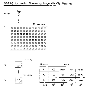
FIGURE 3 illustrates the use antibody arrays as a tool for nested sorts of
high diversity gene libraries.
FIGURE 4 illustrates application of the mefihods provided herein for
searching libraries of mutated genes.
FIGURE 5 illustrates a method for constructing recombinant antibody
libraries.
FIGURE 6 depicts one method for incorporating polypeptide (epitope) tags
into recombinant antibodies using primer addition.
~ I ~-~rnc, vve~
FIGURE 7 depicts an alten-ativ~e scheme using linker addition.
FIGURE 8 depicts application of the methods herein for searching
recombinant antibody libraries.
FIGURE 9 schematically depicts elements of the primers provided herein
and the sets of primers required.
FIGURES 10 and 11 depict alternative methods for constructing the ED
and EDC primers; in FIGURE 10 ofigonucieotides are chemically synthesized 3'
to
RECTIFIED SHEEP (RUI-E.~'~)
IEAIEP
CA 02415328 2003-O1-07
WO 02/006834 PCT/USO1/22821
-13-
5' on a solid support; in the method in FIGURE 1 1, the oligonucleotides self-
assemble based upon over4apping hybridization.
FIGURE 12 depicts a high throughput screen for discovering
immunoglobulin (1g) produced from hybridoma cells for use in the arrays.
FIGURES 13 (13A and 13B) depict exemplary primers (see SEQ ID Nos.
12-73) for amplification of antibody chains for preparation of recombinant
human
antibodies (see Table 33, pages 87-88 in McCafferty et al. (1996) Antibody
engineering: A practical Approach, Oxford University Press, Oxford, see also,
Marks et al. (1992) BiolTechnology 90:779-783; and Kay et al. (1996) Phage
Display of Peptides and Proteins: A Laboratory Manual, Academic Press, San
Diego).
FIGURES 14 (A-D) depict use of the methods herein for antibody
engineering.
FIGURE 15 depicts use of the methods herein for identification of
antibodies with modified specificity (or any protein with modified
specificity).
FIGURE 16 depicts use of the methods herein for simultaneous antibody
searches.
FIGURE 17 depicts use of the methods herein in enzyme engineering
protocols
FIGURE 18 depicts use of the methods herein in protein interaction
mapping protocols.
FIGURE 19 depicts the rate of and increase in the number of tags when
multiple polypeptide tags are used for sorting.
For clarity of disclosure, and not by way of limitation, the detailed
description is divided into the subsections that follow.
DETAILED DESCRIPTION
A. DfcFtNITlONS
Unless defined otherwise, all technical and scientific terms used herein
have the same meaning as is commonly understood by one of skill in the art to
r,(e S~ ~, y~ c ~~s
which this invention belongs. In the event there are different
de#.ir~t.t~.tions for
terms herein, the definitions in this section control. Where permitted, all
patents, applications, published applications and other publications and
RECTIFIED SHEET (RU'~.E ~'~~
ISAJEP
CA 02415328 2003-O1-07
WO 02/006834 PCT/USO1/22821
-14-
sequences from GenBank and other databases referred to throughout in the
disclosure herein are incorporated by reference in their entirety.
As used herein, nested sorting refers to the process of decreasing
diversity using the addressable collections of antibodies provided herein.
As used herein, an addressable collection of anti-tag capture agents (also
referred to herein as an addressable collection of capture agents) protein
agents
(i.e., receptors), such as antibodies, that specifically bind to pre-selected
polypeptide tags that contain epitopes (sequences of amino acids, such as
epitopes in antigens) in which each member of the collection is labeled and/or
is
positionally located to permit identification of the capture agent, such as
the
antibody, and tag. The addressable collection is typically an array or other
codable collection in which each locus contains receptors, such as antibodies,
of
a single specificity and is identifiable. The collection can be in the liquid
phase if
other discrete identifiers, such as chemical, electronic, colored, fluorescent
or
other tags are included. Capture agents, include antibodies and other anti-tag
receptors. Any protein that specifically binds to a pre-determined sequence of
amino acids, such as an epitope, is contemplated for use as a capture agent.
As used herein, polypeptide tags, herein to generically refer to the tags
include a sequence of amino acids, that specifically binds to a capture agent.
As used herein, an epitope tag refers to a sequence of amino acids that
includes the sequence of amino acids, herein referred to as epitope, to which
an
anti-tag capture agent, such as an antibody specifically binds, For
polypeptide
and epitope tags, the specific sequence of amino acids to which each binds is
referred to herein generically as an epitope. ~ Any -any sequence of amino
acids
that binds to a receptor therefor is contemplated. For purposes herein the
sequence of amino acids of the tag, such as epitope portion of the epitope
tag,
um a ~
that specifically binds to the capture agent is designated "E", and each
u.~a'~u.ie
epitope is an Em. Depending upon the context "Em" can also refer to the
sequences of nucleic acids encoding the amino acids constituting the epitope.
The polypeptide tag, such as epitope tag, may also include amino acids that
are
encoded by the divider region. In particular, the epitope tag is encoded by
the
oligonucleotides provided herein, which are used to introduce the tag. When
RECTIFIED SHEET (RULE 9~~
ISA/EP
CA 02415328 2003-O1-07
WO 02/006834 PCT/USO1/22821
-15-
refierence is made to an epitope tag (i.e. binding pair for a particular
receptor or
portion thereof) with respect to a nucleic acid, it is nucleic acid encoding
the tag
to which reference is made. Far simplicity each polypeptide ~aGg~ is referred
to as
Em; when nucleic acids are being described,the Em is nucleic acid and refers
to
the sequence of nucleic acids that encode the epitope; when the translated
proteins are described Em refers to amino acids (the actual epitope). The
number
of E's corresponds to the number of antibodies in an addressable collection.
"m" is typically at least 10, more preferably 30 or more, more preferably 50
or
100 or more, and can be as high as desired and as is practical. Most
preferably
"m" is about a 1000 or more.
As used herein, D" refers to each divider sequence. As described herein
in certain embodiments in which division is effected by other methods D~ is
optional. As with each Em the D" is either nucleic acid or amino acids
depending
upon the context. Each Dn is a divider sequence that is encoded by'an nucleic
a c t'i't
~aied that serves as a priming site to amplify a subset of nucleic acids. The
yG: i-foiti'7S
resulting amplified subset of nucleic acids co.nains all of the collection of
Em
sequences and the D~ sequences used as a priming site for the amplification.
As described herein, the nucleic acids include a portion, preferably at the
end,
that encodes each EmD~. Generally the encoding nucleic acid is 5'- Em-D~ -3'
on
the nucleic acid molecules in the library). D is an optional unique sequence
of
Sv;htihtc..tW z~
nucleotides for specific amplification to create the sublfbr~arys. For large
libraries,
the original library can be divided into sublibraries and then the tag-
encoding
Se-~ ~, fvn ra~.r
seuqences added, rather than adding the tag-encoding sequences to the master
library, The size of D is a function of the library to be sorted, since the
larger
Y 1 _:Ct:.C j
the library the longer the sequence neeeded to specify a unique sequence in
the
clep~rtontj .
library. Generally D, dependentng upon the application, should be at Least 14
to
16 nucleic acid bases Song and it may or may not encoded a sequence of amino
i ~ h.
acids, since its function in the method is to serve as a priming site for
I~CrFR
amplification,~~ D is 2 to n, where n is 0 or is any desired number and is
generally
10 to 10,000, 10 to 1000, 50 to 500, and about 100 to 250. The number of
D can be as high as 106 or higher. The divider sequences D are used to amplify
each ofi the "n" samples from the tagged master library, and generally is
equal to
RE~'CIFIED SHEET (Rl~~l~ ~"9)
ISAJEP
CA 02415328 2003-O1-07
WO 02/006834 PCT/USO1/22821
-16-
the number of antibody collections, such as arrays, used in the initial sort.
The
more collections (divisions) in the initial screen, the lower diversity per
i~ Y~e Y S ~ ~-
addressable locus. The initial division number is selected based upon the
diveri~y ~
of the library and the number of capture agents. The more E's, the fewer D's
are needed, and vice versa, for a library having a particular diversity (Div).
As
used herein, diversity (Div) refers to the number of different molecules in a
library, such as a nucleic acid library. Diversity is distinct from the total
number
of molecules in any library, which is greater. The greater the diversity, the
lower
the number of actual duplicates there are. Ideally the (number of different
molecules)/(total molecules) is approximately 1. If the number of molecules
that
are randomly tagged to create the master IibraryX is less than the initial
diversity,
then statistically each of the molecules in the master library should be
different.
As used herein, an array refers to a collection of elements, such as
antibodies, containing three or more 'members. An addressable array is one in
which the members of the array are identifiable, typically by position on a
solid
phase support or by virtue of an identifiable or detectable label, such as by
color,
fluorescence, electronic signal (i.e. RF, microwave or other frequency that
does
s.n hrnc H n ;~
not substantially alter the intexat-ion of the molecules of interest), bar
code or
other symbology, chemical or other such label. Hence, in general the members
of the array are immobilized to discrete identifiable loci on the surface of a
solid
phase or directly or indirectly linked to or otherwise associated with the
identifiable label, such as affixed to a microsphere or other particulate
support
(herein referred to as beads) and suspended in solution ar spread out on a
surface.
As used herein, a support (also referred to as a matrix support, a matrix,
an insoluble support or solid support) refers to any solid or semisolid or
insoluble
support to which a molecule of interest, typically a biological molecule,
organic
molecule or biospecific ligand is linked or contacted, Such materials include
any
materials that are used as affinity matrices or supports for chemical and
biological molecule syntheses and analyses, such as, but are not limited to:
polystyrene, polycarbonate, polypropylene, nylon, glass, dextran, chitin,
sand,
pumice, agarose, polysaccharides, dendrimers, buckyballs, polyacrytamide,
RECTIFIED SHEE'~ (~'~~-~~~
ISRIEP
CA 02415328 2003-O1-07
WO 02/006834 PCT/USO1/22821
-17-
silicon, rubber, and other materials used as supports for solid phase
syntheses,
affinity separations and purificatians, hybridization reactions, immunoassays
and
other such applications. The matrix herein may be particulate or may be a be
in
the form of a continuous surface, such as a microtiter dish or well, a glass
slide,
a silicon chip, a nitrocellulose sheet, nylon mesh, or other such materials.
When
particulate, typically the particles have at least one dimension in the 5-10
mm
range or smaller. Such particles, referred collectively herein as "beads", are
often, but not necessarily, spherical. Such reference, however, does not
constrain the geometry of the matrix, which may be any shape, including
random shapes, needles, fibers, and elongated. Roughly spherical "beads",
particularly microspheres that can be used in the liquid phase, are also
contemplated. The "beads" may include additional components, such as
magnetic or paramagnetic particles (see, e.g." Dyna beads (Dynal, Oslo,
Norway)) for separation using magnets, as Long as the additional components do
not interfere with the methods and analyses herein.
As used herein, matrix or support particles refers to matrix materials that
are in the form of discrete particles. The particles have any shape and
dimensions, but typically have at least one dimension that is 100 mm or less,
50
mm or less, 10 mm or less, 1 mm or less, 100 ,um or less, 50 ,um or less and
typically have a size that is 100 mm3 or less, 50 mm3 or less, 10 mm~ or less,
and 1 mm3 or less, 100,um3 or less and may be~'order of cubic microns. Such
particles are collectively called "beads."
As used herein, a capture agent, which is used interchangeably with a
receptor, refiers to a molecule that has an affinity for a given ligand or a
with a
defined sequence of amino acids. Capture agents may be naturally-occurring or
synthetic molecules, and include any molecule, including nucleic acids, small
organics, proteins and complexes that specifically bind to specific sequences
of
amino acids. Capture agents are receptors may also be referred to in the art
as
the
anti-ligands. As used herein, tlaee terms, capture agent, receptor and anti-
ligand
are interchangeable. Capture agents can be used in their unaltered state or as
aggregates with other species. They may be attached or in physical contact
with, covalently or noncovalently, a binding member, either directly or
indirectly
RECTIRIED S~iEE'~ (~U~E ~1~
I~~°EP
CA 02415328 2003-O1-07
WO 02/006834 PCT/USO1/22821
-18-
via a specific binding substance or linker. Examples of capture agents,
include,
but are not limited to: antibodies, cell membrane receptors surface receptors
and internalizing receptors, monoclonal antibodies and antisera reactive or
isolated components thereof with specific antigenic determinants (such as on
viruses, cells, or other materials), drugs, polynucleotides, nucleic acids,
peptides,
cofactors, lectins, sugars, polysaccharides, cells, cellular membranes, and
organelles,
Examples of capture agents, include but are not restricted to:
a) enzymes and other catalytic polypeptides, including, but are not limited
to, portions thereof to which substrates specifically bind, enzymes modified
to
retain binding activity lack catalytic activity;
b) antibodies and portions thereof that specifically bind to antigens or
sequences of amino acids;
c) nucleic acids;
d) cell surface receptors, opiate receptors and hormone receptors and
other receptors that specifically bind to ligands, such as hormones. For the
collections herein, the other binding partner, referred to herein as a
polypeptide
tag for each refers the substrate, antigenic sequence, nucleic acid binding
protein, receptor ligand, or binding portion thereof.
As noted, contemplated herein, are pairs of molecules, generally proteins
that specifically bind to each other. One member of the pair is a polypeptide
I~hY~,-y
that is used as a tag and encoded by nucleic acids linked to the li~ar~r; the
other
member is anything that specifically binds thereto. The collections of capture
agents, include receptors, such as antibodies or enzymes or portions thereof
and
mixtures thereof that specifically bind to a known or knowable defined
sequence
of amino acids that is typically at least about 3 to 10 amino acids in length.
As used herein, antibody refers to an immuoglobulin, whether natural or
fnca ac ail
partially or wholly synthetically produed, including any derivative thereof
that
retains the specific binding ability of the antibody. Hence antibody includes
any
protein having a binding domain that is homologous or substantially homologous
to an immunoglobulin binding domain. For purposes herein, antibody includes
antibody fragments, such as Fob fragments, which are composed of a light chain
RECT1~1E~ S~iEET (~UL~ ~'~ ~
ISAJEP
CA 02415328 2003-O1-07
WO 02/006834 PCT/USO1/22821
-19~
and the variable region of a heavy chainoAntibodies include members of any
immunoglobulin class, including IgG, IgM, IgA, 1gD and IgE. Also contemplated
herein are receptors that specifically binding to a sequence of amino acids.
Hence for purposes herein, any set of pairs of binding members, referred
to generically herein as a capture agentJpolypeptide tag, can be used instead
of
antibodies and epitopes per se. The methods herein rely on the capture
~~I ~ raP ~ ~ d ~° o, r~
agentlpolyp~~ptdie tag, such as er~F antibodylepitope tag, for their specific
interactions, any such combination of receptors/ligands (epitope tag) can be
used. Furthermore, for purposes herein, the the capture agents, such as
antibadies employed, can be binding portions thereof.
As used herein, antibody fragment refers to any derivative of an antibody
1z tl~
that is less than full length, retaining at least a portion of the full-.lela
antibody's
specific binding ability. Examples of antibody fragments include, but are not
limited to, Fab, Fab', F(ab)~, single-chain Fvs (seFv), Fv, dsFv diabody and
Fd
fragments. The fragment can include multiple chains linked together, such as
by
disulfide bridges. An antibody fragment generally contains at least about 50
amino acids and typically at least 200 amino acids.
As used herein, an Fv antibody fragment is composed of one variable
heavy domain (VH) and one variable light (V~) domain linked by noncovalent
interactions,
As used herein, a dsFv refers to an Fv with an engineered intermolecular
5-ha~~l~ zes
disulfide bond, which s~ta-bl+(i~s the VH-V~ pair.
As used herein, an F(ab)2 fragment is an antibody fragment that results
from digestion of an immunoglobulin with pepsin at pH 4.0-4.5; it may be
recombinantly produced.
As used herein, an Fab fragment is an antibody fragment that results from
digestion of an immunoglobulin with papain; it may be recombinantly produced.
As used herein, scFvs refer to antibody fragments that contain a variable
light chain (V~) and variable heavy chain (V,.~) covalently connected by a
polypeptide linker in any order. The linker is of a length such that the two
variable domains are bridged without substantial interference. Exemplary
linkers
I~ECTII~IED SKEET (RULE 91)
ISAfEP
CA 02415328 2003-O1-07
WO 02/006834 PCT/USO1/22821
-20-
are (Gly-Ser)~ residues with some Glu or Lys residues dispersed throughout to
increase solubility.
As used herein, diabodies are dimeric scFv; diabodies typically have
shorter peptide linkers than scFvs, and they preferentially dimerize.
As used herein, humanized antibodies refer to antibodies that are
modified to include "human" sequences of amino acids so that administration to
a human does not provoke an immune response. Methods for preparation of
such antibodies are known. For example, the hybridoma that expresses the
monoclonal antibody is altered by recombinant DNA techniques to express an
antibody in which the amino acid composition of the non-variable regions is
based on human antibodies. Computer programs have been designed to identify
such regions.
As used herein, macromolecule refers to any molecule having a molecular
weight from the hundreds up to the millions. Macromolecules include peptides,
proteins, nucleotides, nucleic acids, and other such molecules that are
generally
synthesized by biological organisms, but can be prepared synthetically or
using
recombinant molecular biology methods.
As used herein, the term "biopolymer" is used to mean a biological
molecule, including macromolecules, composed of two or more monomeric
subunits, or derivatives thereof, which are linked by a bond or a
macromolecule.
A biopolymer can be, for example, a polynucleotide, a polypeptide, a
carbohydrate, or a lipid, or derivatives or combinations thereof, for example,
a
nucleic acid molecule containing a peptide nucleic acid portion or a
glycoprotein,
respectively. Biopolymer include, but are not limited to, nucleic acid,
proteins,
polysaccharides, lipids and other macromolecules. Nucleic acids include DNA,
RNA, and fragments thereof. Nucleic acids may be derived from genomic DNA,
RNA, mitochondria) nucleic acid, chloroplast nucleic acid and other organelles
with separate genetic material.
As used herein, a biomolecule is any compound found in nature, or
derivatives thereof. Biomolecules include but are not limited to:
oligonucleotides,
oligonucleosides, proteins, peptides, amino acids, peptide nucleic acids
(PNAs),
oligosaccharides and monosaccharides.
CA 02415328 2003-O1-07
WO 02/006834 PCT/USO1/22821
-21-
As used herein, the term "nucleic acid" refers to single-stranded and/or
double-stranded polynucleotides such as deoxyribonucleic acid (DNA), and
ribonucleic acid (RNA) as well as analogs or derivatives of either RNA or DNA.
Also included in the term "nucleic acid" are analogs of nucleic acids such as
peptide nucleic acid (PNA), phosphorothioate DNA, and other such analogs and
derivatives or combinations thereof.
As used herein, the term "polynucleotide" refers to an oligomer or
polymer containing at least two linked nucleotides or nucleotide derivatives,
including a deoxyribonucleic acid (DNA), a ribonucleic acid (RNA), and a DNA
or
RNA derivative containing, for example, a nucleotide analog or a "backbone"
bond other than a phosphodiester bond, for example, a phosphotriester bond, a
phosphoramidate bond, a phophorothioate bond, a thioester bond, or a peptide
bond (peptide nucleic acid). The term "oligonucleotide" also is used herein
essentially synonymously with "polynucleotide," although those in the art
recognize that oiigonucleotides, for example, PCR primers, generally are less
than about fifty to one hundred nucleotides in length.
Nucleotide analogs contained in a polynucleotide can be, for example,
mass modified nucleotides, which allows for mass differentiation of
polynucleotides; nucleotides containing a detectable label such as a
fluorescent,
radioactive, luminescent or chemiluminescent label, which allows for detection
of
a polynucleotide; or nucleotides containing a reactive group such as biotin or
a
thiol group, which facilitates immobilization of a polynucleotide to a solid
support. A polynucleotide also can contain one or more backbone bonds that
are selectively cleavable, for example, chemically, enzymatically or
photolytically. For example, a polynucleotide can include one or more
deoxyribonucleotides, followed by one or more ribonucleotides, which can be
followed by one or more deoxyribonucleotides, such a sequence being cleavable
at the ribonucleotide sequence by base hydrolysis. A polynucleotide also can
contain one or more bonds that are relatively resistant to cleavage, for
example,
a chimeric oligonucleotide primer, which can include nucleotides linked by
peptide nucleic acid bonds and at least one nucleotide at the 3' end, which is
linked by a phosphodiester bond or other suitable bond, and is capable of
being
CA 02415328 2003-O1-07
WO 02/006834 PCT/USO1/22821
-22-
extended by a polymerase. Peptide nucleic acid sequences can be prepared
using weal known methods (see, for example, Weiler et al., Nucleic acids Res.
25:2792-2799 (1997)).
As used herein, oligonucleotides refer to polymers that include DNA,
f;UGI('.IC Git~.GtIJt 5
RNA, x~u.l.e.ic acid anolo~s, such as PNA, and combinations thereof. For
purposes
herein, primers and probes are single-stranded oligonucleotides.
As used herein, production by recombinant means by using recombinant
DNA methods means the use of the well known methods of molecular biology
for expressing proteins encoded by cloned DNA.
As used herein, substantially identical to a product means sufficiently
similar so that the property of interest is sufficiently unchanged so that the
substantially identical product can be used in place of the product.
As used herein, equivalent, when referring to two sequences of nucleic
acids, means that the two sequences in question encode the same sequence of
amino acids or equivalent proteins. When "equivalent" is used in referring to
two proteins or peptides, it means that the two proteins or peptides have
substantially the same amino acid sequence with only conservative amino acid
substitutions (see, e.g., Table 1, above) that do not substantially alter the
activity or function of the protein or peptide. When "equivalent" refers to a
property, the property does not need to be present to the same extent but the
activities are preferably substantially the same. "Complementary," when
referring to two nucleotide sequences, means that the two sequences of
nucleotides are capable of hybridizing, preferably with less than 25%, more
preferably with less than 15%, even more preferably with less than 5%, most
preferably with no mismatches between opposed nucleotides. Generally to be
considered complementary herein the two molecules hybridize under conditions
of high stringency.
As used herein, to hybridize under conditions of a specified stringency is
used to describe the stability of hybrids formed between two single-stranded
DNA fragments and refers to the conditions of ionic strength and temperature
at
which such hybrids are washed, following annealing under conditions of
stringency less than or equal to that of the washing step. Typically high,
f~EC'~"1~~~!~ ~~~~'~ ~~~~~.,~~'~~
CA 02415328 2003-O1-07
WO 02/006834 PCT/USO1/22821
-23-
medium and low stringency encompass the following conditions or equivalent
conditions thereto:
1 ) high stringency: 0.1 x SSPE or SSC, 0.1 °to SDS, 65°C
2) medium stringency: 0.2 x SSPE or SSC, 0.1 % SDS, 50°C
3) fow stringency: 1.0 x SSPE or SSC, 0.1 % SDS, 50°C.
Equivalent conditions refer to conditions that select for substantially the
same
percentage of mismatch in the resulting hybrids. Additions of ingredients,
such
as formamide, Ficoll, and Denhardt's solution affect parameters such as the
temperature under which the hybridization should be conducted and the rate of
the reaction. Thus, hybridization in 5 X SSC, in 20% formamide at 42° C
is
substantially the same as the conditions recited above hybridization under
conditions of low stringency. The recipes for SSPE, SSC and Denhardt's and the
preparation of deionized formamide are described, for example, in Sambrook et
a/. 11989) Molecular Cloning, A Laboratory Manual, Cold Spring Harbor
Laboratory Press, Chapter 8; see, Sambrook et al., vol. 3, p. B.13, see, also,
numerous catalogs that describe commonly used laboratory solutions). It is
understood that equivalent stringencies may be achieved using alternative
buffers, salts and temperatures.
The term "substantially" identical or homologous or similar varies with the
context as understood by those skilled in the relevant art and generally means
at
least 70%, preferably means at least 80%, more preferably at least 90%, and
most preferably at least 95% identity.
As used herein, a composition refers to any mixture. It may be a
solution, a suspension, liquid, powder, a paste, aqueous, non-aqueous or any
combination thereof.
As used herein, a combination refers to any association between among
two or more items. The combination can be two or more separate items, such as
two compositions or two collections, can be a mixture thereof, such as a
single
mixture of the two or more items, or any variation thereof.
As used herein, fluid refers to any composition that can flow. Fluids thus
encompass compositions that are in the form of semi-solids, pastes, solutions,
aqueous mixtures, gels, lotions, creams and other such compositions.
CA 02415328 2003-O1-07
WO 02/006834 PCT/USO1/22821
-24-
As used herein, suitable conservative substitutions of amino acids are
known to those of skill in this art and may be made generally without altering
the biological activity of the resulting molecule. Those of skill in this art
recognize that, in general, single amino acid substitutions in non-essential
regions of a polypeptide do not substantially alter biological activity (see,
e.g.,
Watson et al. Molecular Biology of the Gene, 4th Edition, 1987, The
8~~)avn irr
Be~c-~n/Cummings Pub. co., p.224~,
Such substitutions are preferably made in accordance with those set forth
in TABLE 1 as follows:
TABLE 1
Original residue ~ Conservative substitution
Ala (A) Gty; Ser
Arg (R) Lys
Asn (N) Gln; His
Cys (C) Ser
Gtn (Q) Asn
Glu (E) Asp
Gly (G) AIa; Pro
His (H) Asn; Gln
Ile (I) Leu; Val
Leu (L) Ile; Val
Lys (K) Arg; Gln; Glu
Met (M) Leu; Tyr; tle
Phe (F) Met; Leu; Tyr
Ser (Si Thr
Thr (T) Ser
Trp (W) Tyr
Tyr (Y) Trp; Phe
Val (V) Ile; Leu
Other substitutions are
also permissible and
may be determined empirically
or in
accord with known conservative substitutions.
As used herein, the amino acids, which occur in the various amino acid
sequences appearing herein, are identified according to their well-known,
three-
letter or one-letter abbreviations. The nucleotides, which occur in the
various
DNA fragments, are designated with the standard single-letter designations
used
routinely in the art.
RECTIFIED SHEEP (RUI-E.~'~)
IEAIEP
CA 02415328 2003-O1-07
WO 02/006834 PCT/USO1/22821
-25-
As used herein, the abbreviations for any protective groups, amino acids
and other compounds, are, unless indicated otherwise, in accord with their
common usage, recognized abbreviations, or the IUPAC-IUB Commission on
Biochemical Nomenclature (see, (1972) Biochem. ~ x:1726).
The methods and collections herein are described and exemplified with
particular reference to antibody capture agents, and polypeptide tags that
include epitopes to which the antibodies bind, but is it to be understood that
the
methods herein can foe practiced with any capture agent and any polypeptide
tag
therefor. !t also to be understood that combinations of collections of any
capture agents and polypeptide tag therefor are contemplated for use in any of
the embodiments described herein. It is also to be understood that reference
to
v 255r~i 19~e.
array is intended to encompass any ~l~resabde collection, whether it is in the
form of a physical array or labeled collection, such as capture agents bound
to
colored beads.
B. Design and Preparation of f3ligonucleotides/Primers
Sorting large diversity libraries onto arrays and amplifying specific pools
containing clones with the desired properties is dependent on the ability to
uniquely tag a library with specific polypeptide tags. Oligonucleotide sets
are
chemically synthesized, randomly combined by overlapping sequences, and
ligated together to produce a template for enzymatic synthesis of the
collection
of primers or linkers,
The oligonucleotides are either single-stranded or double-stranded
depending upon the manner in which they are to be incorporated into the master
library. For example, they can be incorporated, for example by ligation of the
double stranded version, such as through a convenient restriction site,
followed
by amplification with a common region, or they can be incorporated by PCR
amplification, in which case the oligonucleotides are single-stranded.
1. Primers
Provided herein are sets of nucleic acid molecules that are primers or
double-stranded oligonucleotides, which are double-stranded versions of the
nrimarc anr~ r:nmbina~ions ~f sefis of nrimars anrl/nr rirnihiP-ctranrlprl
RECTIFIED SHEEP (RUI-E.~'~)
IEAIEP
CA 02415328 2003-O1-07
WO 02/006834 PCT/USO1/22821
-26-
use in the various steps of th8 methods provided herein and/or depends upon
the
embodiment employed. The primers, which are employed in some of the
embodiments of the methods far tagging molecules, are central to the practice
of
such methods. The primers contain oligonucleotides, which include the formulae
as depicted in Figure 9. The primers and double-stranded oligonucleotides may
include restriction sites) and for targeted amplifications, as exemplified
below
for example for antibody libraries, of sufficient portions of genes of
interest.
These primers may be forward or reverse primers, where the forward primer is
that used for the first round in a PCR a~~~t~.
The primers, described below and depicted in the figure, are provided as sets.
Also provided are combinations of one or more of each set. The primers are
central to the methods provided herein.
2. Preparation of the oligonucleotides/primers
Any suitable method for constructing double-stranded or single-stranded
oligonucleotides may be employed. Methods that can be adapted for preparing
large numbers of such oligomers are particularly of interest. Two methods are
depicted in Figures 10 and 7 1 and are discussed below.
Fig 9 illustrates the physical elements for construction of a tagged library
and use of the addressable anti-tag antibody collections for identification of
genes (proteins) of interest. Four oligonucleotidelprimer sets are provided in
addition to the addressable collections, which for exemplification purposes
are
provided as arrays, an imaging system or reader to analyze the arrays and,
optionally software to manage the information collected by the reader. In the
embodiment depicted, the primer sets include EmD"C, where C is a portion in
common amongst all of the oligonucleotides and can serve as a region for
amplification of all tagged nucleic acids with differing E andlor D sequences
(e.g., Dj thru D"; E~ thru Em); DC, with differing D sequences (D~ thru Dn),
and an
cn~v;tcv
gppttara.a.l C, for common region, FAEC, with differing FA sequences (e.g.,
FAQ
thru FAn); and FBC, with differing F8 sequences (e.g., FB, thru FB"?. Each FA
includes a portion of each epitope and can serve as a primer to amplify
nucleic
ae:irl~ that PnrnrlP a rnrrPSnnndina E_, but the resuitina amnlifiPrl nllrlpir
ari~tc
RECTIFIED SHEEP (RUI-E.~'~)
IEAIEP
CA 02415328 2003-O1-07
WO 02/006834 PCT/USO1/22821
-27-
does not include the Em epitope. FB~ is similar to FA~, except that it can
include
En, if it is desired to retain the epitope.
Fig 10 and Fig 11 outline two different methods for constructing the ED,
and EDC, FA and FB oligonucleotides/primers for antibody screening as an
example. For example, synthesis of the VLFOR primer, which combines n , such
as a 1,000, different E sequences with m, such as 1,000 different D sequences
and approximately 13 different JkaPPa For sequences. This makes a total of
(1,000)(1,000)(13) = 13,000,000 different oligonucleotides. By randomly
combining the different sequence regions in progressive synthesis steps, this
large diverse collection of primers can be prepared.
The first method (Fig 10) uses a solid-phase synthesis strategy. The
second method (Fig 11 ) uses the ability of DNA molecules to self-assemble
based on overlapping complementary sequences. Solid-phase synthesis has the
advantage that the immobilized product molecules can be easily purified from
substrate molecules between reactions, allowing for greater control of the
reaction conditions. The self assembly method has the advantage of requiring
much less work.
Fig 10 Oligonucleotides are chemically synthesized 3' to 5' from a solid
support. In contrast, DNA is enzymatically synthesized 5' to 3'. To create the
2O VLFOR primer, the C and D sequences are chemically synthesized using
standard
methods from a solid support. In order to couple the oligonucleotide to a
solid-
phase for further synthesis, a strong nucleophile is incorporated by addition
of an
aminolink prior to cleavage of the oligonucleotide from its substrate. The
aminolink introduces a primary amine to the 5' end of the oligonucleotide. The
amine group on the aminolink can then be coupled to a solid support, such as
paramagnetic beads, by reaction with amine reactive groups on the beads, such
as tosyl, N hydroxysuccinimide or hydrazine groups. The resulting
oligonucleotides are covalently coupled to the beads with the C and D
sequences
in the proper 5' to 3' orientation.
A mixture of E sequences are added to the oligonucleotide by use of a
DNA "patch" and the resulting nick is sealed with DNA ligase. Unincorporated
substrate DNA is purified from the extended product and a mixture of JkaPPa
ro'
CA 02415328 2003-O1-07
WO 02/006834 PCT/USO1/22821
-28-
sequences are added to the primer. Although the completed V~FOR primer can be
released from the bead, the beads do not interfere with the ability of
oligonucleotides to prime cDNA synthesis.
The method illustrated in Fig 11 relies on the oligonucleotides to self-
assemble based on overlapping hybridization. A double stranded DNA molecule
is first created from oligonucleotides encoding the + and - strands of the
molecule. These oligonucleotides are combined and allowed to hybridize to
produce a nicked double-stranded DNA molecule and the nicks on the molecule
are sealed by the addition of DNA ligase. The sealed molecules are used as
templates for enzymatic synthesis of a new DNA molecule. DNA synthesis is
primed using an ofigonucleotide with a group on its 5' end to allow coupling
to a
solid support, such as biotin or the aminolink chemistry described above.
Incorporation of the reactive group during enzymatic synthesis enables
purification of a single stranded molecule after the synthesis is complete.
Although the completed V~FOR primer can be released from the bead, the beads
do not interfere with the ability of oligonucleotides to prime cDNA synthesis.
C. Nested Sorting using addresable anti-tag receptor collections
Prior methods for identifying and selecting proteins of interest are
hampered by selection biases that are created during successive rounds of
enrichment. As provided herein, selection biases can be avoided with the use
of
identification methods based on sorting rather than selection.
These method herein rely upon the use of collections of capture agents, such
as
a plurality of substantially identical, preferably replicate, collections of
agents,
such as antibodies, that specifically bind to preselected selected sequences
of
amino acids lgenerally at least about 5 to 10, typically at least 7 or 8 amino
acids, such as epitopes), that are linked to proteins in a target library or
encoded
by a target nucleic acid library. Combinations of the capture agents and
polypeptide tags that contain the sequence of amino acids to which the capture
agent or a binding portion thereof specifically binds are provided. The tags
may
be linked to members of a nucleic acid library or other library of molecules
to be
sorted.
CA 02415328 2003-O1-07
WO 02/006834 PCT/USO1/22821
-29-
1. Overview
The addressable anti-tag capture agent collections, such as an positionally
,-
addressable array, contains a collection different capture a~~.t-ras, such as
antibodies that bind to pre-selected and/or pre-designed polypeptide tags,
such
as epitope tags, with high affinity and specificity. A typical collection
contains
at feast about 30, more prefereably 100, more preferably 500, most preferably
at (east 1000 capture agents, such as antibodies, that are addressable, such
as
by occupying a unique locus on an array or by virtue of being bound to bar-
coded support, color-coded, or RF-tag labeled support or other such
addressable
format. Each locus or address contains a single type of capture agent, such as
antibody, that binds to a single specific tag. Tagged proteins are contacted
with
the collection of receptors, such as antibodies in an array, under conditions
suitable for complexation with the receptor, such as an antibody, via the
epitope
tag. As a result, proteins are sorted according to the tag each possesses.
These addressable anti-tag antibody collections have a variety of
applications including, but not limited to, rapid identification of
antibodies; far
therapeutics, diagnostics, reagents, and proteomics affinity matrices; in
enzyme
engineering applications such as, but not limited to, gene shuffling
methodologies; for identification of improved catalysts, for antibody affinity
maturation; for identification of small molecule capture proteins, sequence-
specific DNA binding proteins, for single chain T-cell receptor binding
proteins,
and for high affinity molecules that recognize MHC; and for protein
interaction
mapping. Exemplary protocols are depicted in Figures 1-4, 12, 14A-D and 15-
18.
2. Sorting Methods
Methods of using the receptor, such as antibody, collections for sorting
molecules labeled with the epitope tags are provided. The methods include the
steps of creating a master tagged library by adding nucleic acids encoding the
tags; dividing a portion of the master library into N reactions; amplifying
each
reaction with the nucleic acid encoding the divider sequences and translating
to
produce N translated reactions mixtures; reacting each of the reactions
mixtures
with one collection of the capture agents, such as antibodies; identifying the
RECTIFIE~ SHEET (R~~-E 9~)
ISAIEP
CA 02415328 2003-O1-07
WO 02/006834 PCT/USO1/22821
-30-
proteins of interest by a suitable screen, thereby identifying the particular
ED tag
on the protein by virtue of the capture agent to which the tag on the protein
of
interest binds.
The first sorting step substantially reduces diversity. If desired further
sorts are performed or the resulting library is sreened by any method known to
those of skill in the art. The optional second sort, which is started from the
nucleic acid reaction mixture that contains the nucleic acid from which the
protein of interest was translated, is performed. In this step, a new set of
the
epitope tags is added to the nucleic acid by amplification or ligation
followed by
amplification. Prior to, or simulataneously with this, the nucleic acid
encoding
the prior epitope tag is removed either by cleavage, such as with a
restriction
enzyme or by amplification with a primer that destroys part or all of the
epitope-
encoding nucleic acid. The new tags are added, resulting nucleic acids are
translated and are reacted with a single addressable collection of antibodies.
The proteins sort according to their polypeptide tag, and a screen is run to
identify the protein of interest At this point, the diversity of the molecules
at
the addressable locus of the antibody collection should be 1 (or on the order
of 1
to 100, typically 1 to 10). The nucleic acids that contain the protein of
interest
are then amplified with a tag that amplifies nucleic acid molecules that
contain
nucleic acids encoding the identified epitope tag, to thereby produce nucleic
acid
encoding a protein of interest. The primer for amplificiation includes all or
only
a sufficient portion of the tag to serve as a primer to thereby removing the
epitope from the encoded protein. Hence the methods, provided herein permit
sorting (i.e., reduction of diversity) of diverse collections. A sort that
involves
one step will substantially reduce diversity. The use of an optional sorting
steps
generally reduces diversity of less than 10, generally one.
Dividing the master library
As noted above, the first step in the sorting processes herein includes
dividing the master library into N sublibraries. As described above, the"D"
sequence and tags can be introduced into the master library, which is then
subdivided using the different D's for amplification into "N" sublibraries.
CA 02415328 2003-O1-07
WO 02/006834 PCT/USO1/22821
_g1_
As noted above, the inclusion of "D" is optional; division can be effected
by physically dividing the master library into sublibraries, and then
introducing
the "E" tag-encoding or "EC" tag-encoding sequences into the sublibraries.
This
is generally done when the initial library is very large so that the resulting
sublibraries are large to ensure a uniform distribution of tags.
3. Creating the masfier library for sorting
fn this step, tags that encode each of the epitopes linked to each of the
~~~',YHI'~
divider sequences are incorporated into the master li~~a~, which is typically
a
cDNA library. Any way known to those of skill in the art to add and
incorporate
a double stranded DNA fragment into nucleic acid may be used. In particular,
at a.
variety of ways are contemplated herein. These include (1 ) using PCR
amplification to incorporate them (exemplified herein); (2) ligating them
directly
or via linkers (see below), the ligated product, if needed, can be amplified,
and
other methods described herein (see below) and that can be readily devised by
those of skill in the art in light of the description herein.
In the initial tagging step, when adding the ~, ED or ~DC set of
oligonucleotides on the constituent members of the nucleic acid library, the
goal
is to get an even distribution of all Em and all Dn and to have them on only
one of
each type of molecule. The tags must be randomly distributed among the
different molecules. As long as the number of molecules is large compared to
the number of tags (so that on the average only about one of each type of
molecule in the collection gets each tag), the tags are evenly distributed.
Hence
it is preferable to have the total number of molecules in the collection in
substantial excess compared to the number of tags. Such excess is at least
700-fold, more preferably 1000-fold. The exact ratios, if necessary, can be
determined empirically. In practice there should be no more molecules in the
reaction than the diversity. On the average each different molecule should
have
a different tag and only one of each different molecule should be tagged.
To practice the methods, a library of epitope-labeled molecules is
prepared by randomly introducing the tags into an unlabeled library so that
each
tag is randomly distributed amongst the molecules. Experiments have
RECTIFIED ShIEET (I~~~E 9'I~
ISA/EP
CA 02415328 2003-O1-07
WO 02/006834 PCT/USO1/22821
-32-
demonstrated that the tags can be introduced randomly and equally into a cDNA
library.
The master library is divided into pools, identified as D, - D", reacted with
n number of addressable collections of antibodies, each collection containing
antibodies with m different epitope specificities. Each collection, such as an
~~td ~l,n
array, is associated with one of the pools, such as by an optical code, io~r-
te~td'm~g
a bar code a notation or a symbol or a colored code, an electronic tag or
other
identifier, such as color or a identifiable chemical tag, on the collection or
other
such identifier. The reaction is performed under conditions whereby the
epitopes
bind to the antibod'ies specific therefor, and the resulting complexes of
Q~1~2'7(.''
antibodies and e.p~pe-tag-labeled molecules are screened using an assay that
specifically identifies molecules that have a desired property. The particular
collection (s) of antibodies and antibodies with a particular tag that
includes
~~l~v~+,f ~ i ;~~
molecules with the desired property are identified, thereby also id~en~fty+ng
the
particular D~ pool and epitope tag on the molecule, thereby reducing the
diversity
of the collection by n x m.
4. Methods for epitope tag incorporation
Any method known to one of skill in the art to link a nucleic acid
molecule encoding a polypeptide to another nucleic acid or to link polypeptide
to
another molecule is contemplated. For exemplification, a variety of such
methods are described. As noted, they are described with particular reference
to
antibody capture agents, and polypeptide tags that include epitopes to which
the
antibodies bind, but is it to be understood that the methods herein can be
practiced with any capture agent and polypeptide tag therefor.
a. Ligation to create circular plasmid
vector for infiroductio~ o~tags
As noted above, in addition to use of a~~~i~attan protocols for
introducing the primers into the library members, the primers may be
introduced
by direct ligation, such as by introduction into plasmid vectors that contain
the
nucleic acid that encode the tags and other desired sequences. Subcloning of a
cDNA into double stranded plasmid vectors is well known to those skilled in
the
art. One method involves digesting purified double stranded plasmid with a
site-
RECTIFIED St~EET (RUSE 9'~ ~
ISAJEP
CA 02415328 2003-O1-07
WO 02/006834 PCT/USO1/22821
-33-
specific restriction endonuclease to create 5' or 3' overhangs also known as
sticky ends. The double stranded cDNA is digested with the same restriction
endonuclease to generate complementary sticky ends. Alternately, blunt ends in
both vector DNA and cDNA are created and used for ligation. The digested
cDNA and plasmid DNA is mixed with a DNA ligase in an appropriate buffer
(commonly, T4 DNA ligase and buffer obtained from New England Biolabs are
used) and incubated at 16°C to allow ligation to proceed. A portion of
the
ligation reaction is transformed into E. coli that has been rendered competent
for
uptake of DNA by a variety of methods (electroporation, or heat shock of
chemically competent cells are two common methods). Aliquots of the
transformation mix are plated onto semi-solid media containing the antibiotic
appropriate for the plasmid used. Only those bacteria receiving a circular
plasmid gives rise to a colony on this selective media. Creation of a library
of
unique members is performed in a similar manner, however the cDNA being
inserted into the vector is a mixture of different cDNA clones. These
different
cDNA clones are created via a wide variety of methods known to those skilled
in
the art.
For directional cloning of cDNA clones, which is desirable for the creation
of a library used for expression of proteins from the cDNA library, two
different
restriction endonucleases which generate different sticky ends are used for
digestion of the plasmid. The cDNA library members are created such that they
contain these two restriction endonuclease recognition sites at opposite ends
of
the cDNA. Alternately, different restriction endonucleases that generate
complementary overhangs are used (for example digestion of the plasmid with
NgoMIV and the cDNA with BspEl both leave a 5'CCGG overhang and are thus
compatible for ligation). Furthermore, directional insertion of the cDNA into
the
plasmid vector brings the cDNA under the control of regulatory sequences
contained in the vector. Regulatory sequences can include promoter,
transcriptional initiation and termination sites, translational initiation and
termination sequences, or RNA stabilization sequences. If desired, insertion
of
the cDNA also places the cDNA in the same translational reading frame with
sequences coding for additional protein elements including those used for the
CA 02415328 2003-O1-07
WO 02/006834 PCT/USO1/22821
-34-
purification of the expressed protein, those used for detection of the protein
with
affinity reagents, those used to direct the protein to subcellular
compartments,
those that signal the post-translational processing of the protein.
For example, the pBAD/glll vector (Invitrogen, Carlsbad CA) contains an
arabinose inducible promoter (araBAD), a ribosome binding sequence, an ATG
initiation codon, the signal sequence from the M13 filamentous phage gene 111
protein, a myc epitope tag, a polyhistidine region, the rrnB transcriptional
terminator, as well as the araC and beta-lactamase open reading frames, and
the
ColE1 origin of replication. Cloning sites useful for insertion of cDNA clones
are
designed and/or chosen such that the inserted cDNA clones are not internally
digested with the enzymes used and such that the cDNA is in the same reading
frame as the desired coding regions contained in the vector. It is common to
use Sfil and Notl sites for insertion of single chain antibodies (scFv) into
expression vectors. Therefore, to modify the pBAD/glll vector for expression
of
scFvs, oligonucleotides PDK-28 (SEC2 ID No. 6) and PDK-29 (SEQ ID no. 7) are
hybridized and inserted into Ncol and Hindlll digested pBAD/glll DNA. The
resultant vector permits insertion of scFvs (created with standard methods
such
as the "Mouse scFv Module" from Amersham-Pharmacia) in the same reading
frame as the gene III leader sequence and the epitope tag.
For use herein, a library of expressed proteins is subdivided using a
plurality of epitope tags and the antibodies that recognize them. To create
the
library for expressing proteins with a plurality of epitope tags, slight
modifications of the subcloning techniques described above are used. A
plurality
of cDNA clones are inserted into a mixture of different plasmid vectors
(instead
of a single type of plasmid vector) such that the resulting library contains
cDNA
clones tagged with the different epitope tags, and each epitope tag is
represented equally. Multiple plasmid vectors are created such that they
differ in
the epitope tag that is translated in fusion with the inserted cDNA member.
For
example, if there are 1000 epitope tag sequences, 1000 different vectors are
constructed; if there are 250 epitope tag sequences, 250 different vectors are
constructed. Those skilled in the art understand that there are a variety of
methods for construction of these vectors. For illustration the myc epitope
CA 02415328 2003-O1-07
WO 02/006834 PCT/USO1/22821
-3 5-
encoding region of the pBAD/glll plasmid is removed by digestion with Xbal and
Sall restriction enzymes, and the large 4.1 kb fragment is isolated. The
hybridization of oligonucleotides PDK-32 (SEQ ID No. 8) and PDK-33 (SEQ ID
No. 9) creates overhangs compatible with Xbal and Sall, such that the product
is
inserted directionally, and encodes the epitope for the HA1 1 antibody (see
table
below). Insertion of the hybridization product of PDK-34 (SEQ ID No. 10) and
PDK-35 (SEQ ID No. 1 1 ) results in a vector with the FLAG M2 epitope (see
table below) in frame with the inserted cDNA.
oligo numberoligo Sequence 5' to 3' SEQ ID
name
PDK-028 SfiINotIForcatggcggcccagccggcctaatgagcggccgca6
PDK-029 SfiINotIRevagcttgcggccgctcattaggccggctgggccgc7
PDK-032 HAFor ctagaatatccgtatgatgtgccggattatgcgaatagcgccg8
PDK-033 HARev tcgacggcgctattcgcataatccggcacatcatacggataaa9
PDK-034 M2For ctagaagattataaagatgacgacgataaaaatagcgccg10
PDK-035 M2Rev tcgacggcgctatttttatcgtcgtcatctttataatcaa11
Antibody Epitope name Sequence
9E10 myc EQKLISEEDL
HA.11, HA.7, or 12CA5HA YPYDVPDYA
M1, M2, M5 FLAG DYKDDDDK
Each of these vectors still shares the Sfil and Notl restriction
endonuclease sites to allow subcloning of cDNA clones into the vectors.
Similarly, additional oligonucleotides can be designed to encode a wide
variety of
epitope tags that can be inserted in the same position to create a collection
of
different vectors.
Plasmid DNA corresponding to the vectors containing different epitope
tags is prepared using methods known to those in the art (Qiagen columns, CsCI
density gradient purification, etc). Purified double stranded DNA from each of
the piasmids is quantified by OD260 or other methods and then is combined in
equivalent amounts prior to digestion with the two restriction enzymes, and
treatment with calf intestinal phosphatase (CIP, New England Biolabs). The
cDNA clones of interest are also digested with the same restriction enzymes.
CA 02415328 2003-O1-07
WO 02/006834 PCT/USO1/22821
-3 6-
Digested plasmid DNA and cDNA clones are separated on agarose gels to
remove unwanted sticky ends and purified from agarose slices using standard
methods (Qiagen gel purification kit, GeneClean kit, etc). The cDNA clones and
the mixture of plasmids are reacted in 1 x ligase buffer at a 3:1 molar ratio
(insert
to vector) with T4 DNA ligase (New England Biolabs). Typically, a ligation
reaction contains about 10 ng/,ul plasmid DNA and 0.5 units/,ul of T4 DNA
ligase
in a suitable buffer, and is incubated at 16°C for 12 to 16 hours. The
reaction
is diluted 8-10 fold with sterile water, and aliquots are transformed by
electroporation into TOP10F' (electrocompetant E. coli cells from Invitrogen).
Liquid medium such as SOC (see, Sambrook et al. (1989) Molecular Cloning: A
Laboratory Manual, 2nd Edition, Cold Spring Harbor Laboratory Press; SOC is
2% (w/v) tryptone, 0.5% (w/v) yeast extract, 8.5 mM NaCI, 2.5 mM KCI, 10
mM MgCl2 and 20 mM glucose at pH 7) is added, and cells are allowed to
recover for 1 hour at 37°C. An aliquot of the transformation mixture is
plated
on LB-agar plates containing 100Ng/ml ampicillin. Plates are incubated
at.37°C
for 12 to 16 hours, and then individual clones are analyzed. This analysis
indicates that each of the epitope tags present in the initial mixture is
represented equally in the final library.
For example, a series of plasmid vectors containing the EDC sequences is
created such that each vector in the series .contains a single combination of
EDC
sequences. For example, if there are 1000 E sequences in combination with
1000 D sequences and a single C sequence, there are 1 O6 (1000 x 1000 x 1 )
possible combinations and therefore 106 vectors are created. Each of these
vectors shares restriction endonuclease sites to allow subcloning (preferably
directional) of cDNA clones into the vectors. Purified plasmid DNA from all
106
vectors is mixed and then digested with the restriction endonucleases.
Alternatively, DNA representing each vector is digested and then mixed to
create
the pool of recipient vectors. Double stranded cDNA representing the library
of
interest is also digested with restriction endonucleases to create ends that
are
compatible for ligation to the ends created by vector digestion. This is
accomplished by using the same enzymes for vector and cDNA digestion or by
using those that generate complementary overhangs (for example NgoMIV and
CA 02415328 2003-O1-07
WO 02/006834 PCT/USO1/22821
-3 7-
BspEl both leave a 5'CCGG overhang and are thus compatible for ligation).
Alternately, blunt ends in both vector DNA and cDNA are created and used for
ligation. Digested cDNA clones and digested vector DNAs are ligated using a
DNA ligase such as T4 DNA ligase, E. coli DNA ligase, Taq DNA ligase or other
comparable enzyme in an appropriate reaction buffer. The resultant DNA is
transformed into bacteria, yeast, or used directly as template for in vitro
transcription of RNA. The design of the vectors is such that insertion of the
cDNA at the restriction endonuclease sites places the cDNA under control of
promoter sequences to allow expression of the cDNA. Additionally fihe cDNA
are in the same reading frame as the E sequence such that upon protein
expression from this vector, a fusion protein containing the cDNA-encoded
polypeptide fused to the epitope tag is produced. The E sequence is positioned
in the vector such that the encoded epitope tag is fused to either the N or
the C
terminus of the resultant protein. (#.or restriction enzyme digestion, DNA
ligation,
and transformation, see, e.g., see, Sambrook et al. (1989) Molecular Cloning:
A
Ga~boratory Manual, 2nd Edition, Cold Spring Harbor Laboratory press, Chapter
1 ).
b. Ligation of sequences resutting in linear tagged
cDNA
Following creation of the cDNA library, sequences are appended to cDNA
clones via ligation. Linear, double stranded DNA containing each of the EDC
sequence combinations is created via various methods (synthesis, digestion out
of plasmid containing the sequences, assembly of shorter oligonucleotides,
etc.).
These linear dsDNAs containing the different EDC sequences, are mixed such
that each individual is equally represented in the mixture. This mixture is
combined with the double stranded cDNA library and ligated using a nucleic
acid
ligase in an appropriate buffer. This is preferably a DNA ligase, but an RNA
ligase is used if the EDC tags are composed of RNA or are RNAlDNA hybrid
molecules and the library is also in the form of an RNA or RNA/DNA hybrid. In
one embodiment, the EDC sequence is blunt-ended on both ends yet only one
end is phosphorylated such that ligation occurs in a directional manner (with
respect to the EDC sequences and the E sequence .are brought into the same
RECTIFIED SHEET (RULE 91)
IS6~/'EP
CA 02415328 2003-O1-07
WO 02/006834 PCT/USO1/22821
-38-
reading frame as the cDNA (at either the N or C terminus of the resulting
protein). In another embodiment, the EDC sequence is blunt-ended at one end
and has an overhang on the other end such that ligation occurs in a
directional
manner (see, Sambrook et al. (1989) Molecular Cloning: A Laboratory Manual,
2nd Edition, Cold Spring Harbor Laboratory Press Chapter 8). The EDC
sequences can be continuously double stranded, or partially double stranded
with a single stranded central portion.
In another embodiment, the cDNA library is created to contain a
restriction endonuclease site and the same restriction site is included in the
EDC
sequences such that upon digestion of each with the appropriate enzyme,
compatible ends are created. The digested library is ligated to a mixture of
digested EDC sequences using a DNA ligase in an appropriate buffer. In another
embodiment, the cDNA library is created to contain a restriction endonuclease
site and the EDC sequences are designed to contain a restriction site that
leaves
an overhang compatible to the overhang generated on the cDNA. Upon ligation
of these two compatible sites, a sequence is generated that is not susceptible
to
cleavage with either of the enzymes used to generate the overhangs. In this
case, the products of the ligation reaction are digested with the enzymes used
to
generate the overhangs. Alternately, the ligation reaction occurs in the
presence
of the enzymes used to generate the overhangs (Biotechniques 1999
Aug;27(2):328-30, 332-4, Biotechniques 1992 Jan;12(1):28, 30).
This method reduces and/or eliminates the ligation of cDNA to cDNA or
EDC sequence to EDC sequence, and thus enrich for the cDNA-EDC product.
Pairs of enzymes capable of generating such compatible overhangs include
Agel/Xmal, Ascl/Mlul, BspEl/NgoMIV, Ncol/Pcii and others (New England Biolabs
2000-2001 catalog p184 and 218 for partial list). The EDC sequences and the
cDNA are designed such that they are in the same reading frame following
ligation. Therefore, upon protein expression from this construct, a fusion
protein
containing the cDNA-encoded polypeptide fused to the epitope tag is produced.
The E sequence is positioned in the final construct such that the encoded
epitope tag is fused to either the N or the C terminus of the resultant
protein.
CA 02415328 2003-O1-07
WO 02/006834 PCT/USO1/22821
-39-
In another embodiment, the cDNA, the EDC sequence or both are created
such that they contain a region with RNA hybridized to DNA. The RNA can be
removed by digestion with the appropriate RNAse (including type 2 RNAse H)
such that a single stranded DNA overhang results. This overhang can be ligated
to compatible overhangs generated either by the above method or by restriction
endonuclease digestion. Additionally, overhangs and flanking sequence are
designed in such a way that if an EDC sequence is ligated to another EDC
sequence, the resulting sequence is susceptible to digestion with a particular
restriction enzyme. Likewise, if a cDNA is ligated to another cDNA, the
resulting
sequence is susceptible to cleavage by another restriction enzyme. Ligation
reactions occur in the presence of those restriction enzymes, or are
subsequently
treated with those enzymes to reduce the incidence of cDNA-cDNA or EDC-EDC
ligation events (see enzymes pairs and references above ). The EDC sequences
and the cDNA are designed such that they are in the same reading frame
following ligation. Therefore, upon protein expression from this construct, a
fusion protein containing the cDNA-encoded polypeptide fused to the epitope
tag
is produced. The E sequence is positioned in the final construct such that the
encoded epitope tag is fused to either the N or the C terminus of the
resultant
protein. In another embodiment, PCR is used to generate the cDNA and the
various EDC sequences using PCR primers that contain regions of RNA sequence
that cannot be copied by certain thermostable DNA polymerases. Therefore
RNA overhangs remain that can be ligated to complementary overhangs
generated by the same method or by restriction enzyme digestion. RNA or DNA
overhang cloning is described by Coljee et al (Nat Biotechnol 2000
Ju1;18(7):789-91).
In another embodiment, an EDC sequence is brought into close apposition
to a cDNA sequence by hybridization to a splint oligonucleotide that is
complementary to the 3' region of the cDNA and also the 5' region of the EDC
sequence (Landegen et al., Science 241 :487, 1988). Joining of the cDNA and
EDC is accomplished by a nucleic acid ligase under appropriate reaction
conditions. In another embodiment, the splint oligonucleotide is complementary
to the 5' region of the cDNA and the 3' region of the EDC sequence. In both
CA 02415328 2003-O1-07
WO 02/006834 PCT/USO1/22821
-40-
cases, the different members of the cDNA library share a common sequence (at
the 3' or 5' end), and the different EDC sequences also share a common
sequence (at the 5' or 3' end), such that a single splint oligonucleotide
sequence
can hybridize to any member of the cDNA library and also to any individual of
the series of EDC sequences. In each of these embodiments, the splint
oligonucleotide, the cDNA and the EDC sequences can be single or double
stranded DNA, or combinations of DNA and RNA. Mixtures of cDNA, EDC
sequences and splint oligonucleotides are denatured at elevated temperatures
to
eliminate secondary structure and existing hybridization. The reaction is then
cooled to allow hybridization to occur. In cases where the splint
oligonucleotide
is present in molar excess, a hybridization product containing the three
desired
components (cDNA, EDC and splint oligonucleotide) is obtained. A nucleic acid
ligase is added and the reaction is incubated under appropriate conditions.
In another embodiment, the splint oligonucleotide, cDNA library and EDC
sequences are designed as in the above example. The ligase chain reaction
(see,
e.g., LCR, F. Barany (1991 ) The Ligase Chain Reaction in a PCR World, PCR
Methods and Appiieations, vol. 1 pp. 5-16; see, also, U.S. Patent No.
5,494,810) is then performed using multiple cycles of denaturation,
hybridization, and ligation with a thermostable ligase. For geometric
amplification of cDNA-EDC product, double stranded cDNA and double stranded
EDC sequences are needed.
c. Primer extension and PCR for tag incorporation
In another embodiment, the EDC sequences are appended to the cDNA
clones during the creation of the cDNA library. In this case, the EDC sequence
is designed such that it can hybridize to a desired population of mRNA. This
EDC serves as a primer and the RNA serves as a template for synthesis of DNA
using reverse transcriptase (AMV-RT, M-MuLV-RT or other enzyme that
synthesizes DNA complementary to RNA as template). The newly synthesized
cDNA is complementary to the RNA and has an EDC sequence at the 5'end.
Second strand synthesis using a DNA polymerase results in double stranded
DNA with the EDC at the end corresponding to the 3' end of the RNA. In this
embodiment, all members in the series of EDC sequences share a common 3'
CA 02415328 2003-O1-07
WO 02/006834 PCT/USO1/22821
-41-
end for hybridization to the RNA (e.g., in the case of a library of similar
members
of a gene family). Alternately, EDC sequences have a sequence of random
nucleotides at the 3' end for random priming of RNA (Molecular cloning: a
laboratory manual 2"d edition, Sambrook et al, Chapter 8).
In another embodiment, the polymerase chain reaction (PCR) is used to
append EDC sequences to cDNA clones. A cDNA library is created in such a
way that all members share a common sequence at the 3' end (e.g. prime first
strand cDNA synthesis with an oligonucleotide containing this common
sequence, or ligation of linker sequences to double stranded cDNA clones).
Additionally, each member of the cDNA library share a different common
sequence ("C") at the 5' end. Each unique member in the series of EDC
sequences have a common 3' end that is complementary to one of the common
regions in the cDNA. This mixture of EDC sequences serve as one of the
amplification primers in a polymerase chain reaction. An oligonucleotide
complementary to the common region at the opposite end of the cDNA serve as
the second amplification primer. The cDNA library is mixed with the series of
EDC amplification primers, the second primer and a thermostable polymerase
(Taq, Vent, Pfu, etc) in the appropriate buffer conditions and multiple cycles
of
denaturation, hybridization, and DNA polymerization are executed.
Alternatively,
the cDNA library is subdivided after the addition of the common sequences, and
aliquots are combined with individual EDC sequences, the second primer and a
thermostable polymerase (Taq, Vent, Pfu, etc) in the appropriate buffer
conditions and multiple cycles of denaturation, hybridization, and DNA
polymerization are executed.
d. Insertion by Gene Shuffling
In another embodiment, EDC sequences are appended to cDNA clones via
"DNA shuffling" or molecular breeding (see, e.g., Gene 1995 Oct 16;164(1 ):49-
53; Proc Natl Acad Sci U S A. 1994 Oct 25;91 (22):10747-51; U.S. Patent No.
6,1 17,679). Each member in the series of EDC sequences have a common 3'
end that is complementary to one of the common regions in the cDNA library
members. During creation, or mutagenesis of the cDNA library, EDC sequences
CA 02415328 2003-O1-07
WO 02/006834 PCT/USO1/22821
-42-
are included in the PCR reaction to allow the EDC sequences to be assembled
along with the fragments of the cDNA clones.
e. Recombination strategies
Recombination strategies can also be used for introduction of tags into
cDNA clones. For example, triple-helix induced recombination is used to append
EDC sequences to cDNA clones. A cDNA library is created in such a way that
all members share a common sequence at one end. The series of EDC
sequences is designed to include a region with considerable homology to the
common sequence in the cDNA library. The EDC sequences and the cDNA
library are combined in a cell free recombination system (J Biol Chem 2001 May
25;276(21 ):18018-23) with a third homologous oligonucleotide and
recombination is allowed to occur.
In another embodiment, site-specific recombination is used to append
EDC sequences to cDNA clones. Site specific recombination systems include
IoxP/cre (U.S. Patent No. 6,171,861; U.S. Patent No. 6,143,557; ), FLP/FRT
(Broach et al. Cell 29:227-234 (1982)), the Lambda integrase with attB and
attP
sites (U.S. Patent No. 5,888,732), and a multitude of others. The series of
EDC
sequences as well as the members of the cDNA library are designed to include a
common sequence recognized by the recombinase protein (e.g. IoxP sites). The
EDC sequences and the cDNA library are combined in a cell free recombination
system (Protein Expr Purif 2001 Jun;22(1 ):135-40) including the site specific
recombinase (e.g. cre recombinase) under appropriate conditions to allow
recombination to take place. Alternately, the recombination events take place
inside cells such as bacteria, fungus, or higher eukaryotic cells expressing
the
desired recombinase (see U.S. Patent Nos. 5,916,804, 6,174,708 and
6,140,129 as example).
In another embodiment, homologous recombination in cells is used to
append EDC sequences to cDNA clones. E. coli (Nat Genet 1998
Oct;20(2):123-8), yeast (Biotechniques 2001 Mar;30(3):520-3), and mammalian
cells (Cold Spring Harb Symp Quant Biol. 1984;49:191-7) are used for
recombination of DNA segments. The EDC sequences are designed to contain
both 5' and 3' regions with homology to two separate regions in a plasmid
CA 02415328 2003-O1-07
WO 02/006834 PCT/USO1/22821
-43-
vector containing the cDNA. The lengths of homologous regions are dependent
on the cell type being used. The cDNA and the EDC sequences are co-
transformed into the cells and homologous recombination is carried out by
recombination/repair enzymes expressed in the cell (see, e.g., U.S. Patent No.
6,238
f. Incorporation by transposases
In another embodiment, transposases are used to transfer EDC sequences
to cDNA clones. Integration of transposons can be random or highly specific.
Transposons such as Tn7 is highly site-specific and is used to move segments
of
DNA (Lucklow et al., J. Virol. 67:4566-4579 (1993). The EDC sequences are
contained between inverted repeat sequences (specific to the transposase
used).
The members of the cDNA library (or the plasmid vectors they are in) contain
the
target sequence recognized by the transposase (e.g attTn7). In vitro or in
vivo
transposition reactions insert the EDC sequences into this site.
g. Incorporation by splicing
In another embodiment, EDC sequences flanked by RNA splice acceptor
and donor sequences are inserted into the genome of various cell lines in such
a
way as to incorporate them into the mRNA being transcribed and translated (See
U.S. Patent No. 6,096,717 and U.S. Patent No. 5,948,677). Proteins isolated
from these organisms, or cell lines therefore contain the epitope tags and are
amenable to separation by our collection of antibodies.
In another embodiment, EDC sequences are appended to library members
via trans-splicing of RNA. The RNA form of EDC sequences, and preceded by
RNA splice acceptor sequences, or followed by splice donor sequences are
expressed in cells that then receive the library of cDNA clones. Trans-
splicing of
RNA (Nat Biotechnol 1999 Mar;17(3):246-52, and U.S. Patent No. 6,013,487)
append the EDC sequence to the library member.
4. First Sorting step
For sorting in embodiments in which the proteins are encoded by a
nucleic acid library, the proteins are produced from the nucleic acids that
contain
the pre-selected tags. At least one up to a series of sorting steps are
performed. In the first step, a first tag is introduced into the nucleic acid
by
direct linkage or by primer incorporation of oligonucleotides that encode the
CA 02415328 2003-O1-07
WO 02/006834 PCT/USO1/22821
-44-
epitope Em and divider regions D" to create a master library. Each nucleic
acid
molecule includes a region at one end that encodes one of the m epitopes and
one of the n dividers.
In the next step, each of n samples is amplified with a primer that
comprises D" to produce n sets of amplified nucleic acid samples, where each
sample contains amplified sequences that contain primarily a single D" and all
of
the E's (E~ - Em). An aliquot or portion of all of each of the n samples is
translated to produce n translated samples. Proteins from each of the "n"
translated reactions are contacted with one of the capture agent, such as
antibody, collections, where each of the capture agents in the collection
specifically reacts with an Em; and each of the capture agents, such as
antibodies, can be identified and produces capture-agent-protein complexes via
specific binding of the capture agents to the polypeptide tags.
The resulting complexes are screened, preferably using a chromogenic,
luminescent or fluorgenic reporter to identify those that have bound to a
protein
of interest, thereby identifying the Em and D" that is linked to a protein of
interest.
5. The second sorting step
If the diversity of the proteins to be sorted is such that multiple possible
proteins are identified after the initial sort, additional sorting steps may
be
employed. Alternatively, routine or other screening methods may be used to
identify proteins of interest from the identified proteins. If the diversity
at this
stage is relatively low (1 to about 5000 or so, for example), the sample that
contains the identified D~ can be screened using rautine or standard screening
procedures, or subjected to a second sorting step to further reduce the
diversity.
Thus, if the diversity after the first sort is fairly high (such as about 100
more, or 500 or more or 103 or more, or, depending upon the application and
desired result, whatever the skilled artisan deems too high to screen by other
methods), additional sorting steps are performed.
For these additional steps, the nucleic acid in the sample that contains
t,;",;~,a~f,r.~i
the identified D" is a.rnplfied with a set of primers that each contains a
portion
RECTIFIED SHEET (R~~E 91)
ISA/EP
CA 02415328 2003-O1-07
WO 02/006834 PCT/USO1/22821
-45-
(designated FAP) of each epitope-encoding tag (each designated EP) sufficient
to
~nsuf~~~ca,~.4
amplify the linked nucleic acid, but it~su~it~t to reintroduce Ep, where each
primer includes or is of a sequence of nucleotides of formula HO-FA-Ep, where
p
is an integer of 7 to m. This amplification introduces a different one of the
epitope-encoding sequences into the nucleic acid to produce a collection of
cDNA clones (a sublibrary of the original) that again contains all of the
epitopes
distributed among the sublibrary members.
In this second sorting step, if amplification is used to introduce the new
~~ t n i ~n~ j ze.
set of tags, concatamer formation can be rrrirrrr~e~ by using a low
concentration of the FA primers followed by an excess of primers encoding the
common region, which region is introduced by the FA primer. After the FA
primer is used, the common primers out compete the FA primers for
incorporation, since the C region will then be incorporated into the template
nucleic acid molecule.
Alternatively, as noted above, the new set of epitope-encoding sequences
can be ligated via linkers to-to the template. To do this the template can be
cut
with a unique restriction enzyme and the linkers ligated. This can get rid of
the
existing epitope encoding nucleic acid and replace it with a new set of
epitopes.
Ligation can be followed by amplification with the common region. Other
methods may also be used.
In creating the sublibrary for the second sorting step, as with the master
library, it is necessary to use conditions that ensure that on the average
each
different molecule has a different tag and one of each kind is tagged. In this
round, one tag, on the average, should attach to each of the different
molecules.
In this round, however, the diversity is much lower, since the first sorting
step
~~ n
achieves an m x n reduction in diversity. A-n~u- of the methods described
above
to attach and distribute polypeptide tag-encoding sequences among the
sublibrary members can be used.
Selecting the appropriate stoichiometry assures that a different tag gets
on each different member in the library. The number of epitope-encoding
molecules should be small relative the number of molecules in the sublibrary,
thereby ensuring an even distribution thereof among the population of
different
RECTIFIED SHEET (RIf~E 91''~
ISAJEP
CA 02415328 2003-O1-07
WO 02/006834 PCT/USO1/22821
-4 6-
molecules, such that the probability that any particular tag ends up on any
particular library member is small. As with the first sorting step and
preparation
of the master library, preferable ratios and concentrations can be empirically
determined by varying them and testing.
The nucleic acids in the resulting sublibrary are translated and the
translated proteins contacted, such as under western blotting conditions, with
one collection of capture agents (or a plurality of replicas thereof), such as
antibodies, to form capture agent-protein complexes. The proteins in the
complexes are screened to identify the capture agent, such as antibody or
receptor, locus (or loci) that binds to the epitope linked to the protein of
interest,
thereb identif in the "E", the ~ ~~~ ~e
y y g ~ .o~e sequence associated with the protein of
~n l-~t~z.5 .-
~~er~. Nucleic acid molecules in the sublibrary that contain the identified
"E",
a~ 1I )'~ e<(
epitope sequence, designated Eq, are specifically a~p~~fed, with primers that
include the formula 5' FBS 3' (or 5'CFBS3'), where each FB is sufficient to
amplify the linked nucleic acid using an Em portion of the epitope sequence
and
includes all or a portion of the Em. This specifically amplifies the nucleic
acid
molecule of interest.
In summary, the diversity (Div) equals the total number of different
molecules in a library (i.e., 10$), N = number of divisions D~-D~, which is
the
number of different collections of capture agents, such as 102; M = number of
different epitope tags (and capture agents) E,-Em, such as 103. To start the
method, a master tagged library is prepared, and divided N times. Portions of
the N samples are translated and spotted onto N arrays each containing M
capture agents (sort 1 ). At this stage M x N =105. For the second sort, "M"
new epitopes, such as 103 are used, the nucleic acid is translated and sorted
S 6'.C~
onto one array of 103 capture agents, s~xas antibodies, thereby achieving a
10$ reduction in diversity. As a result, each locus (or member of a collection
if
provided linked to particulate identifiable supports) in the array has a
single type
(G'. ~~'U Y~2
of protein as well as a single c~~pt-err-ea agents. The number of sorting
steps can
be any desired number, but is typically one or two. If a higher number of
sorts
are performed, then the sensitivity of the detection assay at the first sort
should
be very high, since, as a result of the diversity, the concentration of the
protein
RECTIFIES SHI~~~' (~~j~.~ 91)
ISA/EP
CA 02415328 2003-O1-07
WO 02/006834 PCT/USO1/22821
-47-
of interest will be low. As noted above, M and N may be different each sorting
step.
The process of nested sorting, which is applicable to sorting a variety of
collections of molecules, particularly collections of proteins, DNA, small
molecules and other collections is exemplified in Figures 1-18. The
concept of nested sorting is illustrated in Fig 1. In this example, a master
collection containing 74,088 different item~_s,' such as cDNA, is searched by
jb'.!?ll~~rc;i'lilt Si,i~l;~,/1~o?t'~lel
randomly dividing the collection into 42 .sublabraxys (F1 str~bl~Ebr-ar-y~s).
After
3t;.bL~;~G,ri2S
identifying which of the 42 F1 s.ubli-br-ar-ys contains the item of interest,
such as
by binding or reaction with a probe or by a protein-protein specific
interaction,
SLt~?~1~~~YwYI;~3 Sut~If~'o-'aYIC-~
that group is further divided randomly into 42 new su~bh~b~arys (F2 su~arys)
and again the sublibrary containing the item of interest is identified. A
final
division of the F2 sublibrary containing the item of interest produces 42 new
groups, each containing only one item. The item of interest can be uniquely
identified based on its sorting lineage.
!n the example shown, the item of interest was identified in the fifth F1
sublibrary, the thirty first F2 sublibrary, and the sixteenth F3 sublibrary.
Of the
74,088 items in the master collection, only one has the sort lineage
F15/F23,/F31~.
The sort illustrated in Fig 2 is identical to the sort illustrated in Fig 1
except that the F2 and F3 sublibraries have been arranged into arrays. This
figure also illustrates that as the sort proceeds, the diversity of items
within each
sublibrary decreases; the exemplified master collection contains 74,088 items,
the 42 F1 sublibraries contain 1,764 items each, the 42 F2 sublibraries
contain
42 items, and the 42 F3 sublibraries contain only a single item. The first two
figures illustrate a theoretical search based on nested sorting.
Fig 3 illustrates the use of capture agent arrays, such as antibody arrays,
as a tool far nested sorts of high diversity gene libraries. A master gene
library
SLth 1 ib v' l-; ~W
is first randomly divided into a number of s~+bl+br-ar-ys by separate
amplification,
such as PCR, reactions. The amplification reactions use sets of unique
sequences of nucleotides that encode preselected epitopes and incorporate
these
sequences into the genes by appropriate design of primers to specifically
amplify
RECTIFIED SHEET (RULE J'6~
ISA/EP
CA 02415328 2003-O1-07
WO 02/006834 PCT/USO1/22821
-48-
Svbl~hr~:m zs sul~Ir~~rar.r ru
different su.bla.br-ar-ys of genes from the master template pool (F1 -sublibr-
ar-ys).
These amplification reactions are performed, for example, in 96-well (or 384-
well
or higher density) PCR plates with a compatible thermocycler.
The amplified genes in each well are translated into their protein products
and samples from each are then applied to separate capture agent collections,
such as arrays (i.e., proteins from each well in the 96-well plate are applied
to
one of 96 capture agent arrays). The proteins by binding to capture agents,
such as antibodies, in the array, sort into defined locations on the array
that
recognize the known unique amino acid sequences (the epitopes) that have been
added to the proteins using the primers. After sorting, addresses on the array
that contain the protein of interest are identified and nucleic acids from the
sublibrary from which those proteins with the epitope encoding sequences that
bind to the spot in the array are amplified, such as by PCR.
During this second amplification step, new sets of known epitopes are
incorporated into the nucleic acid, so that they rnay be further sorted using
additional capture agent arrays (F3).
The table in Fig 3 illustrates how the number of initial divisions by PCR
and the number of capture agents the array can be combined to search gene
libraries containing, for example, from a million (106) to over a billion
(109)
different genes. For example, an initial gene library can be divided into 100
F1
sublibraries by amplification and then further divided using two arrays with
capture agents recognizing 100 different epitopes. If the initial gene library
contained 106 different genes, the F3 addresses in the sublibraries contain a
single type of gene (106/100/100/ 100 = 1 ). An initial gene library divided
into
7 ,000 F1 sublibraries by PCR amplification and then further divided using two
arrays With capture agents recognizing 1,000 different epitopes to create the
F2
s vbl ~hrrnr°r
and F3 s-erb~i~bra~r-ys can be used to search 109 different genes
(109/1,000/1,00011,000 = 1 ).
Sub I ~b~re;vi ~,s
Dividing the gene libraries into su~bl~ibr-arys is based on the ability of a
PCR
amplification reaction to specifically amplify DNA sequences using pairs of
primers. Although both primers need to hybridize to sequences on either end of
the template DNA, a subset of template sequences can be amplified using a
RECTI~IE~ SHEET (RII~.E 9'i~
I SA/EP
CA 02415328 2003-O1-07
WO 02/006834 PCT/USO1/22821
-49-
primer pair in which one of the primers is common to all of the template
sequences and the other primer is specific for the gene sequence of interest.
For
example, specific genes are often amplified from cDNA libraries using one
primer
that is specific for the gene of interest and another that hybridizes to the
oligo(dA) tai) common to all of the cDNA molecules.
6, Use of multiple tags in a single fusion protein
The system provided herein uses epitope tags to subdivide protein
libraries, such as libraries of scFvs. For example, with 1000 tags and a
library of
1 O9 scFvs, there is 1 Os scFvs for each tag. To identify a single library
member,
such as an scFv of interest, either a large number of individual scFvs (106),
are
screened or more than one subdivision is employed. Using a larger number of
tags a library can be reduced to small number of proteins in fewer steps.
Using a combinatorial approach, a small set of capture agent-tag pairs can
be used effectively as a much larger set. By incorporating multiple tags into
a
protein, such as a single scFv fusion protein, better use of fewer tags can be
made. For comparison, if there are 300 capture-agent tag pairs, and a library
of
109 members, with a single tag appended to each member, the 300 tags divide
the 109 members such that each type of tag is attached to 3.3 x 10⁶ members.
With three tags incorporated into each member in a combinatorial fashion such
that 1 /3 of the tags are used at each of three sites, there is a total of 100
x 100
x 100 (or 106) combinations. Using these 106 tag combinations the 109
members are divided into 1000 members per tag. Therefore in a single step with
a limited number of tags, the library is effectively subdivided.
In its simplest embodiment, consider an example of x tags at site X, y
tags at site Y, and z tags at site Z. If these tags are used individually,
then there
are x + y + z combinations. If these tags are used in combination then there
fls~~;mtn
are (x)(y)(z) combinations. A-ssermi~~ that the number of tags at each site
(x, y
and z) is one third the total (n), then for the case of individual use,
C=(n/3)x3=n or there are as many total combinations (C) as there are tags;
whereas for combinatorial use, there are C = (n/3)3. As the number of
individual
tags at each site increases, the number of combinatorial tags increases at a
much higher rate (See Figure 19). With a greater number of effective tags, the
'~ECT1~IED SHEET (RULE 9'~)
ISAIE~'
CA 02415328 2003-O1-07
WO 02/006834 PCT/USO1/22821
-5 0-
number of members of the library per tag decreases. Fewer members per tag in
the initial library results in either fewer sequential rounds of screening or
lower
numbers of clones that to be assessed with high throughput screening.
Whether using a single tag or multiple tags in combination, the procedure
is substantially the same. The protein from the expressed library is
subdivided
by virtue of the epitope tag binding to a capture agent, such as an antibody,
against that tag. in the example presented above (using three tags in
combination), each library member binds to three different anti-tag capture
agents. Each combinatorial tag has its own set of addresses on an array
instead
of a single address. For example, if there are a total of 300 tags with 1-100
in
site X, 101-200 in site Y and 201-300 in site Z, a exemplary combinatorial tag
has the address X27-Y132-2289. Other combinatorial tags also use the X27
anti-tag capture agents, such as capture agents, or the Y132 or 2289 capture
agents, but no other combination uses all three. If an antigen binds to a
library
1 5 member tethered to the three capture agents to which each tag binds, the
combinatorial tag is now (mown and the library member can be recovered from
the original library.
Recovery of a specific library pool with a combinatorial fag is done in
substantially the way a library pool with a single tag is recovered. As
described
herein, one way to recover subpopulations from in the library is to use the
~~ 0 w~z.
polymerase chain reaction. For exemplification, a.s-su~r~i~ng that all three
tags are
at the C-terminus of an expressed protein such that the X tag is the most
~uc.~ o,S
proximal to the library member, sucl~a~s an scFv, followed by the Y tag and
then
the Z tag. The order of DNA segments on the coding strand of cDNA is:
5' Common>scFv>X>Y>Z 3'
A particular sub-population can be recovered by sequential rounds of PCR
amplification starting with a common primer and a primer corresponding to the
2289 tag. The product from this reaction is used in the next reaction using
the
common primer and the Y132 tag primer. The product from this reaction is used
in a subsequent reaction with the common primer and the X27 primer. After
U.aY~tY
three sequential rounds of amplification, the products all correspond to
li~bar~
members, such as scFvs, that were originally tagged with the X27-Y132-2289
R'~CTIFIRD SH~E'~ (I~~IL~ ~1)
I~~J~P
CA 02415328 2003-O1-07
WO 02/006834 PCT/USO1/22821
-51-
combination. ,Those skilled in the art understand that, as long as the
library has multiple nested common sequences, multiple different common
primers are used in the different rounds. Those skilled in the art also
understand
that the multiple tags can be at opposite ends of the encoding DNA and
therefore the expressed protein. It is also understood that the expressed
epitope
tags can be linear, constrained by disulfide bonds, constrained by a scaffold
structure, expressed in loops of a fusion protein, contiguous or separated by
flexible or inflexible linker sequences.
One embodiment uses, for example, a single scaffold fusion protein
containing multiple sites with inserted epitope tags. This spatially separates
the
epitopes and allows them all to be recognized without interference with one
another. The following ~c~llu~w~i~g criteria are considered in selecting a
protein
scaffold: 1 ) known crystal structure to more easily identify surface exposed
amino acids with high propensity for antigenicity, 2) free N and C-termini for
fusion to the cDNA library of interest, 3) high levels of production and
solubility
in various protein expression systems (especially the E.coli periplasm), 4)
capacity for in vifiro transcription/translation, 5) absence of disulfide
bonds, 6)
wild-type protein is monomeric, 7) has capacity to increase solubility or
function
of scFvs. Using the crystal structure, positions are chosen for insertion of
epitope tag libraries. These sites should be spatially separated epitopes that
are
relatively linear in nature (e.g. one side of an alpha helix, a turn between
beta
strands or a loop between helices).
D. Preparation of Antibodies
1. Antibodies and collections of addressable anti-tag antibodies
The methods herein, rely upon the ability of the capture agents, such as
antibodies, to specifically bind to the polypeptide tags, which are linked to
libraries (or collections) of molecules, particularly proteins. The
specificity of
each antibody (or other receptor in the collection) for a particular tag is
known or
can be readily ascertained, such as by arraying the antibodies so that all of
the
antibodies at a locus in the array are specific for a particular epitope tag.
Alternatively, each antibody can be identified, such as by linkage to
optically encoded tags, including colored beads or bar coded beads or
supports,
~~~~-nr~~ ~H~~~ ~~~r~.~ g~ )
n~~~
CA 02415328 2003-O1-07
WO 02/006834 PCT/USO1/22821
-5 z-
or linked to electronic tags, such as by providing microreactors with
electronic
tags or bar coded supports (see, e.g., U.S. Patent No. 6,025,129; U.S. Patent
No. 6,017,496; U.S. Patent No. 5,972,639; U,S. Patent No. 5,961,923; U.S.
Patent No. 5,925,562; U.S. Patent No. 5,874,214; U.S. Patent No. 5,751,629;
U.S. Patent No. 5,741,462), or chemical tags (see, U.S. Patent No. 5,432,018;
U.S. Patent No. 5,547,839) or colored tags or other' such addressing methods
that can be used in place of physically addressable arrays. For example, each
antibody type can be bound to a support matrix associated with a color-coded
tag (i.e. a colored sortable bead) or with an electronic tag, such as an radio-
frequency tag (RF), such as 1ROR1 M1CROKANS~ and MICROTUBES~
microreactors (see, U.S. Patent No. 6,025,129; U.S. Patent No. 6,017,496;
U.S. Patent No. 5,972,639; U.S. Patent No. 5,961,923; U.S. Patent No.
5,925,562; U.S. Patent No. 5,874,214; U.S. Patent No. 5,751,829; U.S. Patent
No. 5,741,462; International PCT application No. W098131732; International
1 5 PCT application No. W098/15825; and, see, also U.S. Patent No. 6,087,186
).
For the methods and collections provided herein, the antibodies of each type
can
be bound to the MICROKAN or MICROTUBE microreactor support matrix and the
associate RF tag, bar code, color, colored bead or other identifier-to-serves
to
identify the receptors, such as antibodies, and hence the epitope tag to which
the receptor, such as an antibody, binds.
For exemplary purposes herein, reference is made to antibodies and tags
that encode epitopes to which the antibody specifically binds. It is
understood
that any pair of molecules that specifically bind are contemplated; for
purposes
herein the molecules, such as antibodies, are designated receptors, and the
molecules, such as ligands, that bind thereto are epitopes. The epitopes are
typically short sequences of amino acids that specifically bind to the
receptor,
such as an antibody or specific binding fragment thereof.
Also, for exemplary purposes herein, reference is made to positional
arrays. It is understood, however, that such other identifying methods can be
readily adapted for use with the methods herein. It is only necessary that the
identity (L e., epitope-tag specificity) of the receptor, such as an antibody,
is
known. The resulting collections of addressable receptors (i.e., antibodies),
~EGTI~IED Sl~EE1' (~~~.~ 91)
l~Jd~l.Ep
CA 02415328 2003-O1-07
WO 02/006834 PCT/USO1/22821
-5 3-
v p-h r.~, I l '~
whether in a two-dimensional or three-dimensional array, or linked to
op~t~iEial~i~y
encoded beads or colored supports or RF tags or other format, can be employed
in the methods herein.
By reacting a collection of antibodies with libraries of polypeptide tag-
labeled molecules, and then performing screening assays to identify the
members of the collection of the antibodies to which epitope-labeled molecules
of a desired property have bound, a reduction in the diversity of the library
of
molecules is achieved. Each collection of antibodies serves as a sorting
device
for effecting this reduction in diversity. Repeating the process a plurality
of
times can effect a rapid and substantial reduction in diversity.
2. Preparation of the capture agents
The quality of the sorts is dependent on the quality of the collection of
capture agents, such as antibodies, that make up the sorting array. In
addition to
requirements on binding affinity and specificity, the epitopes bound by the
capture agents (antibodies) in the array determine the E, FA and FB sequences
used as priming sites for the-tt~e amplification reactions (PCRs). Fig 12
outlines a
high throughput screen for discovering immunoglobulin (1g) produced from
hybridoma cells for use in generating antibodies for use in the collections.
Hybridoma cells are created either from non-immunized mice or mice
immunized with a protein expressing a library of random disulfide-constrained
heP-!-a vneonL
h~pt-r~.er-is epitopes or~other random peptide libraries. Stable hybridoma
cells are
initially screened for high Ig production and epitope binding. 1g production
is
measured in culture supernatants by ELISA assay using a goat anti-mouse IgG
antibody. Epitope binding is also measured by ELISA assay in which the mixture
of haptens (epitope tagged proteins) used for immunization are immobilized to
the ELISA plated and bound igG from the culture supernatants is measured using
a goat anti-mouse IgG antibody. Both assays are done in 96-well formats or
other suitable formats. For example, approximately 10,000 hybridomas are
selected from these screens.
Next, the Ig are separately purified using 96-well or higher density
purification plates containing filters with immobilized Ig-binding proteins
(proteins
A, G or L). The quantity of purified Ig is measured using a standard protein
RECTIFIED SHEEP (RUI-E.~'~)
IEAIEP
CA 02415328 2003-O1-07
WO 02/006834 PCT/USO1/22821
-5 4-
assay formatted for 96-well or higher density plates. Low microgram quantities
of Ig from each culture are expected using this purification method.
The purified Ig are spotted separately onto a nitrocellulose filter using a
standard pin-style arraying system. The purified Ig are also combined to
produce
a mixture with equal quantities of each Ig. The mixed Ig are bound to
paramagnetic beads which are used as a solid-phase support to pan a library of
~ a l~~~t ~ n~~v~t~
bacteriophage expressing the random disulfide-constrained .~eptm.e.ri.c
epitopes.
The batch panning enriches the phage display library for phage expressing
epitopes to the purified Ig. This enrichment dramatically reduces the
diversity in
the phage library.
The enriched phage display library is then bound to the array of purified !g
and stringently washed. !g-binding phage are detected by staining with an anti-
yh~~rn'~ luwov~.r5c~~~!'
phage antibody-HRP conjugate to produce a ciaena.ia.um.m~e~~ent signal
detectable with a charge coupled device (CCD)-based imaging system. Spots in
the array producing the strongest signals are cut out and the phage eluted and
propagated. Epitopes expressed by the recovered phage are identified by DNA
sequencing and further evaluated for affinity and specificity. This method
generates a collection of high-affinity, high-specificity antibodies that
recognize
the cognate epitopes. Continued screening produces larger collections of
antibodies of improved quality.
3. Preparation of anti-tag capture agent arrays
Each spot contains a multiplicity of capture agents, such as antibodies
with a single specificity. Each spot is of a size suitable for detection.
Spots on
the order of 1 to 300 microns, typically 1 to 100, 1 to 50, ands 1 to 10
microns,
CTVl2r"
depending upon the size of the array, target molecules and ot-la.err
parameters.
Generally the spots are 50 to 300 microns. In preparing the arrays, a
sufficient
GI ~ dpc H o .-,
amount is delivered to the surface to functionally cover it for det~ec-tiot~
of
proteins having the desired properties. Generally the volume of antibody-
containing mixture delivered for preparation of the arrays is a nanoliter
volume
(1 up to about 99 nanoliters) and is generally about a nanoliter or less,
typically
between about 50 and about 200 picoliters. This is very roughly about 10
million to 100,000 molecules per spot, where each spot has capture agents,
RECTIFIED SHEEP (RUI-E.~'~)
IEAIEP
CA 02415328 2003-O1-07
WO 02/006834 PCT/USO1/22821
-5 5-
such as antibodies, that recognize a single epitope. For example, if there are
10
million molecules and 1000 different ones in the protein mixture reacting with
the locus, there are 10~ of each type of molecule per spot. The size of the
array
and each spot should be such that positive reactions in the screening step can
l~ t~al~ l~y
be imaged, preferably by imaging the entire array or a-pa~~a+~y therof, such
as
24, 96, or more arrays, at the same time.
A support (see below for exemplary supports), such as KODAK paper
plus gelatin or other suitable matrix can be used, and then ink jet and
stamping
~ (~~-~a ~u tv;s
technology or other suitable dispensing' methods and a.ppax~rs, are used to
reproducibly print the arrays. The arrays are printed with, for example, a
piezo
or inkjet printer or other such nanoliter or smaller volume dispensing device.
For
example, arrays with 1000 spots can be printed. A plurality of replicate
arrays,
such as 24 or 48, 96 or more can be placed on a sheet the size of a
conventional 96 well plate.
Among the embodiments contemplated herein, are sheets of arrays each
with replicates of the antibody array. These are prepared using, for example,
a
piezo or inkjet dispensing system. A large number, for example, 1000 can be
printed at a fiime using, for example a print head with 1000 different holes
(like a
stamp with 500 ~M holes). It can be fabricated from, for example, molded
plastic with many holes, such as 1000 holes each filled with 1000 different
capture agents, such as antibodies. Each hole can be linked to reservoirs that
are linked to conduits of decreasing size, which ultimately dispense the
capture
agents, such as antibodies into the print head. Each array on the sheet can be
;~~~;any
spa~ciavly separated, andlor separated by a physical barrier, such as a
plastic
ridge, or a chemical barrier, such a hydrophobic barrier (i.e., hydrogels
separated
by hydrophobic barriers). The sheets with the arrays can be conveniently the
pl~~~~l~(y
size of a 96 well plate or higher density. Each array contains a pl~al~y of
addressable anti-tag antibodies specific for the pre-selected set of epitope
tags.
For example, 33 x 33 arrays contain roughly 1000 antibodies, each spot on each
ctn~4a~:1~2s
array containing ar~tbodi~es that specifically bind to a single pre-selected
epitope.
A plurality of arrays separated by barriers can be employed.
RECTIFIED SHEET (RI~~.E g'1 )
ISI4IE1'
CA 02415328 2003-O1-07
WO 02/006834 PCT/USO1/22821
-5 6-
For dispensing the antibodies onto the surface, the goal is functional
surface coverage, such that a screened desired protein is detectable. To
',~n~~n~I
achieve this, for example, about 1 to 2 r~s~ml from the starting collection
are
used and about 500 picoliters per antibody are deposited per spot on the
array.
The exact amounts) can be empirically determined and depend upon several
':,: yts 1+w ~~-v
variables, such as the surface and the .se~s'#s~'ity of the detection methods.
The
antibodies are preferably covalently linked, such as by sulfhydryl linkages to
amides on the surface. ,Other exemplary dispensing and immobilizing
systems include, but are not limited to, for example, systems available from
Genometrix, which has a system for printing on glass; from Illumina, which
employs the tips of fiber optic cables as supports; from Texas Instruments,
which has chip surface plasmon resonance (i.e., protein derivatized gold);
injet
systems, such as those from Microfab Technologies, Plano TX; Incyte, Palo
Alto,
CA, Protogene, Mountain View, CA, Packard BioSciences, Meriden CT, and
other such systems for dispensing and immobilizing proteins to suitable
support
surfaces. Other systems such as blunt and quill pins, solenoid and piezo
nanoliter dispensers and others are also contemplated.
4. Preparation of other collections
The capture agents are linked to beads or other particulate supports that
are identifiable. For example, the capture agents are linked to optically
encoded
microspheres, such as those available from Luminex, Austin Tx, the contain
fluorescent dyes encapsulated therein. The microsphere, which encapsulate
dyes, are prepared from any suitable material (see, e.g., International PCT
application Nos. WO 01 /13119 and WO 99/19515; see description below),
s-oyr2nt
including str-ye~e-ethylene-butylene-styrene block copolymers, homopolymers,
gelatin, polystyrene, polycarbonate, polyethylene, p~.o.l~p~pyy.~ brae,
resins, glass,
and any other suitable support (matrix material), and are of a size of a about
a
nanometer to about 10 millimeters in diameter. By virtue of the combination
of,
for example two different dyes at ten different concentrations, a plurality
''~llAG1 ~S~l'il C,l.
microspheres (100 in this instance), each identifiable by a unique
fluo~resence,
are produced.
REC'Tll=IED SHEET (R1~1_E 91)
I~AJEP
CA 02415328 2003-O1-07
WO 02/006834 PCT/USO1/22821
_g7_
Alternatively, combinations of chromophores or colored dyes or other
colored $u~s~#~a~iies-es are encapsulated to produce a variety of different
colors
encapsulated in microspheres or other particles, which are then used as
supports
for the capture agents, such as antibodies. Each capture agent, such as an
antibody, is linked to a particular colored bead, and, is thereby
identifiable. After
producing the beads with linked capture agents, such as antibodies, reaction
with the epitope-tagged molecules can be performed in liquid phase. The beads
that react with the epitopes are identified, and as a result of the color of
the
bead the particular epitope and is then known. The sublibrary from which the
linked molecule is derived is then identified.
E. Supports for immobilizing antibodies
Supports for immobilizing the antibodies are any of the insoluble materials
known for immobilization of ligands and other molecules, used in many chemical
syntheses and separations, such as in affinity chromatography, in the
1 5 immobilization of biologically active materials, and during chemical
syntheses of
biomolecules, including proteins, amino acids and other organic molecules and
polymers. Suitable supports include any material, including biocompatible
polymers, that can act as a support matrix for attachment of the antibody
mater-
ial. The support material is selected so that it does not interfere with the
chemistry or biological screening reaction. ~e
~ILlCi2 '-~etG~.
Supports that are also contemplated for use herein include f-I~opl~or-e-
containing or -impregnated supports, such as microplates and beads
(commercially available, for example, from Amersham, Arlington Heights, IL;
plastic scintillation beads from Nuclear Technology, Inc., San Carlos, CA and
Packard, Meriden, CT, and colored bead-based supports (fluorescent particles
encapsulated in microspheres) from Luminex Corporation, Austin, TX (see,
International PCT application No. W0/01 14589, which is based on U.S.
application Serial No. 09/147,710; see International PCT application No.
W0/0113119, which is U.S. application Serial No. 09/022,537). The
rnicrospheres from Luminex, for example, are internally color-coded by virtue
of
the encapsulation of fluorescent particles and can be provided as a liquid
array.
The capture agents, such as antibodies (epitopesj are linked directly or
indirectly
CA 02415328 2003-O1-07
WO 02/006834 PCT/USO1/22821
-5 8-
by any suitable method and linkage or interaction to the surface of the bead
and
bound proteins can be identified by virtue of the color of the bead to which
they
are linked. Detection can be effected by any means, and can be combined with
chromogenic or fluorescent detectors or reporters that result in a detectable
change in the color of the mierosphere (bead) by virtue of the colored
reaction
and color of the bead. For the bead-based arrays, the anti-tag capture agents
are attached to the color-coded beads in separate reactions. The code of the
bead identifies the capture agent, such as antibody, attached to it. The beads
s~~~~k~.~t,;~ ~i )-
can then be mixed and swbseuequent binding steps performed in solution. They
can then be arrayed, for example, by packing them into a microfabricated flow
chamber, with a transparent lid, that permits only a single layer of beads to
form
resulting in a two-dimensional array. The beads on which a protein is bound
identified, thereby identifying the capture agent and the tag. The beads are
imaged, for example, with a CCD camera to identify beads that have reacted.
icc~'ie.c~°',=
The codes of the such beads are identified, thereby identifying the captuer
agent,
which in turn identifies the polypeptide tag and, ultimately, the protein of
interest.
The support may also be a relatively inert polymer, which can be grafted
by ionizing radiation to permit attachment of a coating of polystyrene or
other
such polymer that can be derivatized and used as a support. Radiation grafting
of monomers allows a diversity of surface characteristics to be generated on
supports (see, e.g., Maeji et al. (1994) Reactive Polymers 22:203-212; and
Berg et al. (1989) J. Am. Chem. Soc. 7 7 7:8024-8026). For example, radiolytic
~'>-.v.:v,o lPlz-Y~.s
grafting of monomers, such as vinyl momom~er~s, or mixtures of monomers, to
polymers, such as polyethylene and polypropylene, produce composites that
have a wide variety of surface characteristics. These methods have been used
to graft polymers to insoluble supports for synthesis of peptides and other
molecules,
The supports are typically insoluble substrates that are solid, porous,
deformable, or hard, and have any required structure and geometry, including,
but not limited to: beads, pellets, disks, capillaries, hollow fibers,
needles, solid
fibers, random shapes, thin films and membranes, and most preferably, form
solid surfaces with addressable loci. The supports may also include an inert
RECTIFIED SHEET (RULE 91)
I~~VEP
CA 02415328 2003-O1-07
WO 02/006834 PCT/USO1/22821
-5 9-
strip, such as a teflon strip or other material to which the capture agents
antibodies and other molecules do not adhere, to aid in handling the supports,
and may include an identifying symbology. .
The preparation of and use of such supports are well known to those of
skill in this art; there are many such materials and preparations thereof
known.
For example, naturally-occurring materials, such as agarose and cellulose, may
be isolated from their respective sources, and processed according to known
protocols, and synthetic materials may be prepared in accord with known
protocols. These materials include, but are not limited to, inorganics,
natural
polymers, and synthetic polymers, including, but are not limited to:
cellulose,
cellulose derivatives, acrylic resins, glass, silica gels, polystyrene,
gelatin,
polyvinyl pyrrolidone, co-polymers of vinyl and acrylamide, polystyrene cross-
linked with divinylbenzene or the like (see, Merrifield (1964) Biochemistry
3:1385-1390), polyacrylamides, latex gels, polystyrene, dextran, polyacryl-
amides, rubber, silicon, plastics, nitrocellulose, celluloses, natural
sponges, and
many others. Selection of the supports is governed, at least in part, by their
physical and chemical properties, such as solubility, functional groups,
mechanical stability, surface area swelling propensity, hydrophobic or
hydrophilic
properties and intended use.
1. Natural support materials
Naturally-occurring supports include, but are not limited to agarose, other
polysaccharides, collagen, celluloses and derivatives thereof, glass, silica,
and
alumina. Methods for isolation, modification and treatment to render them
suitable for use as supports is well known to those of skill in this art (see,
e.g.,
Hermanson et al. (1992) immobilized Affinity Ligand Technigues, Academic
Press, Inc., San Diego). Gels, such as agarose, can be readily adapted for use
herein. Natural polymers such as polypeptides, proteins and carbohydrates;
metalloids, such as silicon and germanium, that have semiconductive
properties,
may also be adapted for use herein. Also, metals such as platinum, gold,
nickel,
copper, zinc, tin, palladium, silver may be adapted for use herein. Other
supports of interest include oxides of the metal and metalloids such as Pt-
PtO,
Si-SiO, Au-AuO, Ti02, Cu-CuO, and the like. Also compound semiconductors,
CA 02415328 2003-O1-07
WO 02/006834 PCT/USO1/22821
-60-
such as lithium niobate, gallium arsenide and indium-phosphide, and nickel-
coated mica surfaces, as used in preparation of molecules for observation in
an
atomic force microscope (see, e.g., III et al. (1993) Biophys J. 64:919) may
be
used as supports. Methods for preparation of such matrix materials are well
known.
For example, U.S. Patent No. 4,175,183 describes a water insoluble
hydroxyalkylated cross-linked regenerated cellulose and a method for its
preparation. A method of preparing the product using near stoichiometric
proportions of reagents is described. Use of the product directly in gel
chromatography and as an intermediate in the preparation of ion exchangers is
also described.
2. Synthetic supports
There are innumerable synthetic supports and methods for their
preparation known to those of skill in this art. Synthetic supports typically
produced by polymerization of functional matrices, or copolymerization from
two
or more monomers from a synthetic monomer and naturally occurring matrix
monomer or polymer, such as agarose.
Synthetic matrices include, but are not limited to: acrylamides, dextran-
derivatives and dextran co-polymers, agarose-polyacrylamide blends, other
polymers and co-polymers with various functional groups, methacrylate
derivatives and co-polymers, polystyrene and polystyrene copolymers (see,
e.g.,
Merrifield (1964) Biochemistry 3:1385-1390; Berg et al. (1990) in Innovation
Perspect. Solid Phase Synth. Col%ct. Pap., Int.
Symp., fist, Epton, Roger (Ed); pp. 453-459; Berg et al. (1989) in Pept.,
Proe.
Eur. Pept. Symp., 20th, Jung, G. et al. (Eds), pp. 196-198; Berg et al. (1989)
J.
Am. Chem. Soc. 7 7 7:8024-8026; Kent et al. (1979) Isr. J. Chem. 77:243-247;
Kent et al. (1978) J, Org. Chem. 43:2845-2852; Mitchell et al. (1976)
Tetrahedron Lett. 42:3795-3798; U.S. Patent No. 4,507,230; U.S. Patent No.
4,006,117; and U.S. Patent No. 5,389,449). Methods for preparation of such
support matrices are well-known to those of skill in this art.
Synthetic support matrices include those made from polymers and co-
polymers such as polyvinylalcohols, acrylates and acrylic acids such as poly-
ethylene-co-acrylic acid, polyethylene-co-methacrylic acid, polyethylene-co-
CA 02415328 2003-O1-07
WO 02/006834 PCT/USO1/22821
-61-
ethylacrylate, polyethylene-co-methyl acrylate, polypropylene-co-acrylic acid,
polypropylene-co-methyl-acrylic acid, polypropylene-co-ethyfacrylate,
polypropylene-co-methyl acrylate, polyethylene-co-vinyl acetate, poly-
propylene-co-vinyl acetate, and those containing acid anhydride groups such as
polyethylene-co-maieic anhydride, polypropylene-co-malefic anhydride and the
like. Liposomes have also been used as solid supports for affinity
purifications
(Powell et al. (1989) Biotechnoi. Bioeng. 33:173).
For example, U.S. Patent No. 5,403,750, describes the preparation of
polyurethane-based polymers. U.S. Pat. No. 4,241,537 describes a plant
growth medium containing a hydrophilic polyurethane gel composition prepared
Pc'r ~orm,2 c(
from chain-extended palyols; random copolymerization can be peer-med with up
to 50% propylene oxide units so that the prepolymer is a liquid at room
temperature. U.S. Pat. No. 3,939,123 describes lightly crosslinked
polyurethane
polymers of isocyanate terminated prepolymers containing poly(ethyleneoxy)
glycols with up to 35% of a poly(propyleneoxy) glycol or a poly(butyleneoxy)
glycol. In producing these polymers, an organic polyamine is used as a
crosslinking agent. Other supports and preparation thereof are described in
U.S.
Patent Nos. 4,177,038, 4,175,183, 4,439,585, 4,485,227, 4,569,981,
5,092,992, 5,334,640, 5,328,603.
U.S. Patent No. 4,162,355 describes a polymer suitable for use in
affinity chromatography, which is a polymer of an aminimide and a vinyl
compound having at least one pendant halo-methyl group. An amine ligand,
which affords sites for binding in affinity chromatography is coupled to the
polymer by reaction with a portion of the pendant halo-methyl groups and the
remainder of the pendant halo-methyl groups are reacted with an amine
containing a pendant hydrophilic group. A method of coating a substrate with
this polymer is also described. An exemplary aminimide is 1,1-dimethyl-1-
(2-hydroxyoctyi)amine methacrylimide and vinyl compound is a chloromethyl
styrene.
S'U ~ ho i'-f -S
U.S. Patent No. 4,171,412 describes specific sup~p~ar~s based on
hydrophilic polymeric gels, preferably of a macroporous character, which carry
covalently bonded D-amino acids or peptides that contain D-amino acid units.
RECTIFIED SHEET (R~J~,~ ~'i ~
IS~EP
CA 02415328 2003-O1-07
WO 02/006834 PCT/USO1/22821
-62-
The basic support is prepared by copolymerization of hydroxyalkyl esters or
hydroxyalkylamides of acrylic and methacrylic acid with crosslinking acrylate
or
methacrylate comonomers are modified by the reaction with diamines,
aminoacids or dicarboxylic acids and the resulting carboxyterminal or
aminoterminal groups are condensed with D-analogs of aminoacids or peptides.
The peptide containing D-aminoacids also can be synthesized stepwise on the
surface of the carrier.
U.S. Patent No. 4,178,439 describes a cationic ion exchanger and a
method for preparation thereof. U.S. Patent No. 4,180,524 describes chemical
syntheses on a silica support. '
Immobilized Artificial Membranes (IAMs; see, e.g., U.S. Patent Nos.
4,931,498 and 4,927,879) may also be used. IAMs mimic cell membrane
environments and may be used to bind molecules that preferentially associate
with cell membranes (see, e.g., Pidgeon et al: (1990) Enzyme Microb. Technol.
72:149).
Among the supports contemplated herein are those described in
International PCT application Nos WO 00/04389, WO 00/04382 and
WO 00/04390; KODAK film supports coated with a matrix material; see also,
U.S. Patent Nos. 5,744,305 and 5,556,752 for other supports of interest. Also
of interest are colored "beads", such as those from L_uminex (Austin, TX).
3. Immobilization and activation
Numerous methods have been developed for the immobilization of
proteins and other biomolecules onto solid or liquid supports (see, e.g.,
Mosbach
(1976) Methods in Enzymology 44; Weetall (1975) Immobilized Enzymes,
Antigens, Antibodies, and Peptides; and Kennedy et al. (1983) Solid Phase
Biochemistry, Analytical and Synthetic Aspects, Scouten, ed., pp. 253-391;
see,
generally, Affinity Technigues. Enzyme Purifica'fion: Part B. Methods in
Enzymology, Vol. 34, ed. W. B. Jakoby, M. Wilchek, Acad. Press, N.Y. (1974);
Immobilized Biochemicals and Affinity Chromatography, Advances in
Experimental Medicine and Biology, vol. 42, ed. R. Dunlap, Plenum Press, N.Y.
( 1974)).
CA 02415328 2003-O1-07
WO 02/006834 PCT/USO1/22821
-63-
Among the most commonly used methods are absorption and adsorption
or covalent binding to the support, either directly or via a linker, Such as
the
numerous disulfide linkages, thioether bonds, hindered disulfide bonds, and
covalent bonds between free reactive groups, such as amine and thiol groups,
known to those of skill in art (see, e.g., the PIERCE CATALOG,
ImmunoTechnology Catalog & Handbook, 1992-1993, which describes the
preparation of and use of such reagents and provides a commercial source for
such reagents; and Wong (1993) Chemistry of Protein Conjugation and Cross
Linking, CRC Press; see, also DeWitt et al. (1993) Proc. Nat/. Acad. Sci.
U.S.A.
90:6909; Zuckermann et al. ( 1992) J. Am. Chem. Soc. 7 74:10646; Kurth et al.
(1994) J. Am. Chem. Soc. 7 76:2661; Ellman et al. (1994) Proc. Nat/. Acad.
Sci.
U.S.A. 97:4708; Sucholeiki (1994) Tetrahedron Lttrs. 35:7307; and Su-Sun
Wang ( 1976) J. Org. Chem. 4 7:3258; Padwa et al. ( 1971 ) J. Org. Chem.
47:3550 and Vedejs et al. (1984) J. Org. Chem. 49:575, which describe photo-
sensitive linkers).
To effect immobilization, a solution of the protein or other biomolecule is
contacted with a support material such as alumina, carbon, an ion-exchange
resin, cellulose, glass or a ceramic. Fluorocarbon polymers have been used as
supports to which biomolecules have been attached by adsorption (see, U.S.
Patent No. 3,843,443; Published International PCT Application WO/86 03840)
A large variety of methods are known for attaching biological molecules,
including proteins and nucleic acids, molecules to solid supports (see. e.g.,
U.S.
Patent No. 5451683). For example, U.S. Pat. No. 4,681,870 describes a
method for introducing free amino or carboxyl groups onto a silica support.
These groups may subsequently be covalently linked to other groups, such as a
protein or other anti-ligand, in the presence of a carbodiimide.
Alternatively, a
silica matrix may be activated by treatment with a cyanogen halide under
alkaline conditions. The anti-ligand is covalently attached to the surface
upon
addition to the activated surface. Another method involves modification of a
polymer surface through the successive application of multiple layers of
biotin,
avidin and extenders (see, e.g., U.S. Patent No. 4,282,287); other methods
involve photoactivation in which a polypeptide chain is attached to a solid
CA 02415328 2003-O1-07
WO 02/006834 PCT/USO1/22821
-64-
substrate by incorporating a light-sensitive unnatural amino acid group into
the
polypeptide chain and exposing the product to low-energy ultraviolet light
(see,
e.g., U.S. Patent No. 4,762,881 ). Oligonucleotides have also been attached
using photochemically active reagents, such as a psoralen compound, and a
coupling agent, which attaches the photoreagent to the substrate (see, e.g.,
U.S. Patent No. 4,542,102 and U.S. Patent No. 4,562,157). Photoactivation of
the photoreagent binds a nucleic acid molecule to the substrate to give a
surface-bound probe.
Covalent binding of the protein or other biomolecule or organic molecule
or biological particle to chemically activated solid matrix supports such as
glass,
synthetic polymers, and cross-linked polysaccharides is a more frequently used
immobilization technique. The molecule or biological particle may be directly
linked to the matrix support or linked via a linker, such as a metal (see,
e.g., U.S.
Patent No. 4,179,402; and Smith et al. (1992) Methods: A Companion to
Methods in Enz. 4:73-78). An example of this method is the cyanogen bromide
activation of polysaccharide supports, such as agarose. The use of
perfluorocarbon polymer-based supports for enzyme immobilization and affinity
chromatography is described in U.S. Pat. No. 4,885,250). In this method the
biomolecule is first modified by reaction with a perfluoroalkylating agent
such as
perfluorooctylpropylisocyanate described in U.S. Pat. No. 4,954,444. Then, the
modified protein is adsorbed onto the fluorocarbon support to effect
immobilization.
The activation and use of supports are well known and may be effected
by any such known methods (see, e.g., Hermanson et al. (1992) Immobilized
Affinity Ligand Technigues, Academic Press, Inc., San Diego). For example, the
coupling of the amino acids may be accomplished by techniques familiar to
those
in the art and provided, for example, in Stewart and Young, 1984, Solid Phase
Synthesis, Second Edition, Pierce Chemical Co., Rockford.
Molecules may also be attached to supports through kinetically inert
metal ion linkages, such as Co(III), using, for example, native metal binding
sites
on the molecules, such as IgG binding sequences, or genetically modified
proteins that bind metal ions (see, e.g., Smith et al. (1992) Methods: A
CA 02415328 2003-O1-07
WO 02/006834 PCT/USO1/22821
-65-
Companion to Methods in Enzymology 4, 73 (1992); III et al. (1993) Biophys J.
64:919; Loetscher et al. (1992) J. Chromatography 595:1 13-199; U.S. Patent
No. 5,443,816; Hale (1995) Analytical Biochem. 237:46-49).
Other suitable methods for linking molecules and biological particles to
solid supports are well known to those of skill in this art (see, e.g., U.S.
Patent
No. 5,416,193). These linkers include linkers that are suitable for chemically
linking molecules, such as proteins and nucleic acid, to supports include, but
are
not limited to, disulfide bonds, thioether bonds, hindered disulfide bonds,
and
covalent bonds between free reactive groups, such as amine and thiol groups.
These bonds can be produced using heterobifunctional reagents to produce,
reactive thiol groups on one or both of the moieties and then reacting the
thiol
groups on one moiety with reactive thiol groups or amine groups to which
reactive maleimido groups or thiol groups can be attached on the other. Other
tinkers include, acid cleavable linkers, such as bismaleimideothoxy propane,
acid
labile-transferrin conjugates and adipic acid diihydrazide, that would be
cleaved
in more acidic intracellular compartments; cross linkers that are cleaved upon
exposure to UV or visible light and linkers, such as the various domains, such
as
CH1, CH2, and CH3, from the constant region of human IgG, (see, Batra et al.
(1993) Molecular lmmunol. 30:379-386).
Presently preferred linkages are direct linkages effected by adsorbing the
molecule or biological particle to the surface of the support. Other preferred
linkages are photocleavable linkages that can be activated by exposure to
light
(see, e.g., Baldwin et al. (1995) J. Am. Chem. Soc. 7 77:5588; Goldmacher et
a/. (1992) Bioconj. Chem. 3:104-107, which linkers are herein incorporated by
reference). The photocleavable linker is selected such that the cleaving
wavelength that does not damage linked moieties. Photocleavable linkers are
linkers that are cleaved upon exposure to light (see, e.g., Hazum et al. (1981
) in
Pept., Proc. Eur. Pept. Symp., 76th, Brunfeldt, K (Ed), pp. 105-1 10, which
describes the use of a nitrobenzyl group as a photocleavable protective group
for
cysteine; Yen et al. (1989) Makromol. Chem 790:69-82, which describes water
soluble photocleavable copolymers, including hydroxypropylmethacrylamide
copolymer, glycine copolymer, fluorescein copolymer and methylrhodamine
CA 02415328 2003-O1-07
WO 02/006834 PCT/USO1/22821
-6 6-
copolymer; Goldmacher et al. (1992) Bioconj. Chem. 3:104-107, which des-
cribes a cross-Pinker and reagent that undergoes photolytic degradation upon
exposure to near UV light (350 nm); and Senter et al. (1955) Photochem.
Photobiol 42:231-237, which describes nitrobenzyloxycarbonyl chloride cross
linking reagents that produce photocleavable linkages). Other linkers include
fluoride labile linkers (see, e.g., Rodolph et al. (1995) J. Am. Chem. Soc.
i T 7:5712), and acid labile linkers (see, e.g., Kick et al. (1995) J. Med.
Chem.
38:1427)). The selected linker depends upon the particular application and, if
needed, may be empirically selected.
F. Use of the methods for identificafiion of proteins of desired properties
from a library
1. Arraying capture agents
The capture agent molecules to which the epitope tags specifically bind
are linked to supports, such as identifiable beads, such as microsheres, or
solid
surfaces. Linkage can be effected through any suitable bond, such as ionic,
Cr r 4V~ u,s
covalent, physical, van ~e r~aa~ai~s bonds. It can be effected directly or via
a
suitable tinker. For exemplary purposes arraying on surfaces is described.
Purified antibodies (1 ,u! at a concentration of 1-2 mg/ml in a buffer of 0.1
M PBS (phospahte buffered saline, pH 7.4) on glycerol ( 1-20% vol/vol), are
spotted onto a membranes (such as; UItraBind membrane, Pall Gelman; FAST
nitrocellulose coated slides, Schleicher & Schuell), chemically deactivated
glass
slides, superaldehyde slides (Telechem), polylysine coated glass, activated
glass,
or specific thin films and self-assembled monolayers International PCT
application Nos WO 00/04389, WO 00!04382 and WO 00/04390)x~using an
automated arraying tool (such as systems available from, for example,
Microsys;
PixSys NQ; Cartesian Technologies; BioChip Arrayer; Packard Instrument
Company; Total Array System; BioRobotics; Affymetrix 417 Arrayer; Affymetrix,
and others). The spots are allowed to air dry for a suitable period of time, 1-
2
minutes or more, typically 30 min to 1 ~~~~. Two membrane attachments are
described. The UItraBind membrane (Pal) Gelman) contains active aldehyde
groups that react with primary amines to form a covalent linkage between the
membrane and the capture agent, such as an antibody. Unreacted aldehydes are
~t~CTI~IED ~H~~T (RULE J'~~
ISp/EP
CA 02415328 2003-O1-07
WO 02/006834 PCT/USO1/22821
-67-
blocked by incubation with suitable blocking solution, such as a solution of
50
mM PBS, pH 7.4, 2 % bovine serum albumin (BSA) or with BBSA-T (a protein-
containing solution such as Blocker BSA'"" (Pierce) diluted to 1 x in
phosphate-
buffered saline (PBS) with Tween-20 (polyoxyethylenesorbitan monolaurate;
Sigma) added to a final concentration of 0.05% (vol:vol)) far a suitable time,
such as about 30 minutes. The filter can be rinsed with PBS.
Capture agents, such as antibodies, also can be deposited onto
membranes, such as, for example, nitrocellulose paper (Schliecher& Schuell)
with, for example, an inject printer (i.e:, Canon model BJC 8200, color inject
printer), modified for this use and connected to a computer, such as a
personal
computer (PC). Such modifications, include, removal of the color ink
cartridges
from the print head and replacement with, for example, 1 milliliter pipette
tips,
which are hand-cut to fit in a sealed manner over the #~e inkpad reservoir
wells
in the print head. Antibody solutions are pipetted into the pipette tips
reservoirs
iv~!c;~d
that are seated on the i.r~pa~d reservoirs.
Printed images, using the modified printer, are generated, with, for
example, Microsoft PowerPoint. The images are then printed onto nitrocellulose
paper, which is cut to fit and then taped over the center of a sheet of
printing
paper. The set of papers is then fed into the printer immediately prior to
printer.
Purified capture agents, such as antibodies can also be spotted onto
FAST nitrocellulose coated slides, (Schleicher-& Schuell). Nitrocellulose
binds
c~ d sp,r i-i o,~
proteins by noncovalent ad~f~-tin. Nitrocellulose binds approximately 100 ,ug
per cm2. After binding of the capture agents, such as antibodies, remaining
binding sites are blocked by incubation with a solution of 50 mM PBS, pH 7.4,
2
% bovine serum albumin (BSA) or BBSA-T for a suitable time, such as for 30
minutes.
Direct binding of antibodies to the nitrocellulose results in non-oriented
binding. The percentage of active immobilized antibody molecules can be
increased by binding to nitrocellulose that has been coated with an antibody
capture protein (such as protein A, protein G or anti-IgG monoclonal
antibody).
~°.vt lauvn~f
The antibody capture proteins a-rebe~d to the nitrocellulose before
application
of the library proteins, such as tagged antibodies, with an arrayer.
Biotinylated
RECTIFIED SHEEP (RUI-E.~'~)
IEAIEP
CA 02415328 2003-O1-07
WO 02/006834 PCT/USO1/22821
_~8_
antibodies can also be printed onto surfaces coated with avidin or
strepavidin.
The size and spacing of the spots can be adjusted depending on the filter used
and the sensitivity of the assay. Typical spots are about 300-500,um in
diameter
with 500-800 ~m pitch.
Antibodies can also be printed onto activated glass substrates. Prior to
printing the glass is cleaned ultrasonically in succession with a 1:1O
dilution of
detergent in warm tap water for 5 minutes in Aquasonic Cleaning Solution
(VWR), multiple rinses in distilled water and 100% methanol (HPLC grade)
followed by drying in a class 100 oven at 45° C. Clean glass is
chemically
functionalized by immersion in a solution of 3-aminopropyltriethoxysilane
(APTS)
(5% vol/vol in absolute ethanol) for 10 minutes. The glass is then rinsed in
95%
ethanol, allowed to air dry, and then heated to 80° C in a vacuum oven
for 2
hours to cure. The surface can then be further modified to bind primary amines
or free sulfhydryl groups in the antibody or avidin or strepavidin linked to
the
antibody with biotin. To create an amine-reactive surface, the functionalized
glass is treated with a solution of Bis[sulfosuccinimidyl]suberate (BS3)(5
mg/mt in
PBS, pH 7.4) for 20 minutes at room temperature. The N hydroxysuccinimide
(NHS)-activated glass surface is rinsed with distilled water and placed in a
37° C
dust-free class 100 oven for 15 minutes to dry. Antibodies can be directly
attached to this surface or the surface can be coated with a protein such as
protein A that binds the antibodies, protein G or anti-IgG monoclonal antibody
or
avidin/strepavidin, to bind biotinylated proteins. To create a sulfhydryl-
reactive
surface, the functionalized glass is treated with a solution of
sulfosuccinimidyl 4-
fN maleimidomethyl]-cyclohexane-1-carboxylate (Sulfo-SMCC) for 20 minutes at
Z5 roam temperature. The maleimide-activated glass surface is rinsed with
distilled
water and placed in a 3-~-CC dust-free class 100 oven for 15 minutes to dry.
To create a biotinylated surface, the functionalized glass is treated with a
solution of EZ-link Sulfo-NHS-LC-Biotin (Pierce) for 20 minutes at room
temperature. The biotinylated glass surface is rinsed with disfiilled water
and
placed in a 37° C dust-free class 100 oven for 15 minutes to dry. The
same
immobilization strategies described above also can be used in self-assembled
monolayers formed on top of inorganic thin films.
RECTIFIED SHEEP (RUI-E.~'~)
IEAIEP
CA 02415328 2003-O1-07
WO 02/006834 PCT/USO1/22821
-69-
2. Exemplary use for identification of a genes from a library of
mutated genes
Fig 4 illustrates the use of the methods herein to search a library of
mutated genes. Mutation of specific gene regions by a variety of methods is
often used to improve the properties of proteins encoded by the mutated genes,
such as mutated genes produces by error-prone PCR or gene shuffling
mutagenesis techniques to improve the binding affinity of a recombinant
antibody. This technique coupled with selection by surface display has been
used to improve the binding affinities of antibodies by several orders of .
magnitude. Mutation has also been used to improve the catalytic properties of
enzymes. The methods herein provide means to screen and identify mutated
genes encoding proteins having desired properties.
Initially a set of oligonucleotides containing various functional domains
are added to the 3' ends of a gene to be mutated by incorporation of a primer
f~UCLep des
that contains sequences of r~~c-Ieo~es that hybridize to the gene and also
additions! sets of sequences, designated E for "Epitopes" D for "Divider", and
C for "Common"). The E D C sequences constitute sets of sequences, each
defined by the functions in the nucleic acid. As noted, the E sequences encode
the epitopes specifically recognized by antibodies in the collection. They are
incorporated in-frame with the coding sequences of the gene to be mutated and
are expressed as a fusion with the parent protein. The D sequences are unique
sequence sets downstream from the epitopes. They serve as specific priming
sites to "Divide" the master group. They can be non-coding sequences and do
not necessarily end up being part of the expressed mutated proteins. The C
sequence is a sequence "Common" to all of the genes and provides a means for
simultaneous PCR amplification of all the gene templates. As noted previously,
in certain embodiments the D andlor C sequences are optional. Importantly, the
E and D sequences are randomly distributed among the resulting DNA molecules.
For example, 100 E sequences and 100 D sequences combine to create 10,000
(100 x 100 = 10,000) uniquely tagged cDNA molecules. Likewise, 1,000 E
sequences and 1,000 D sequences combine to create 1,000,000 ( 1,000 x
1,000 = 1,000,000) uniquely tagged cDNA molecules.
RECTI~tED SHEET (~~~E ~~ ~'
ISA/EP
CA 02415328 2003-O1-07
WO 02/006834 PCT/USO1/22821
-70-
Before, or after the E C and D sequences have been added to the ends of
the molecule to be mutated, defined regions within the gene are mutated by a
variety of standard methods. The mutation procedure should not produce
mutations in the E D C sequences. After the mutagenesis has been completed,
the mutated DNA is added as template to a first set of PCR reactions to create
the F1 sublibrary. In addition to the template DNA, D C primer sets are
separately added such that each PCR contains a primer complementary to a
different D sequence. For example, in Fig 4 the second PCR tube is identical
to
the rest of the tubes except it contains a D C primer containing only one of
the
100 D sequences (D2). In this illustration, tube 50 is identical to the rest
of the
F1 reaction tubes except it contains a different one of the 100 D sequences
(D5o). The resulting PCR amplification products contain all of the 100
different E
sequences randomly distributed among the genes but only containing one of the
100 D~sequences. In the illustration, PCR tube 50 produces a sublibrary DNA
molecules (F15o) that all have the same D5o sequences, the same C sequence but
different E sequences randomly distributed among the molecules (EDSO C).
The generated F1 DNA molecules are expressed in vitro using a
transcription-translation extract. Appropriate regulatory DNA sequences,
including promoters, ribosome binding sites and other such regulatory
sequences
known to those of skill in the art, for efficient in vitro transcription and
translation are incorporated into the DNA fragments during the tagging
process.
As illustrated in Fig 4, expression of the F15o DNA molecules produces a
collection of proteins containing the various epitope tags. Proteins produced
in
bacteria or in other in vivo systems also can be used.
The resulting expressed proteins are incubated with the antibody
collection, such as in an array format under conditions that permit binding
between the epitopes and the antibody(ies) specifically selected to bind to
each
of the epitopes. This results in specific binding of proteins to antibodies.
If the
antibodies are arranged in an array, this results in the distribution of the
tagged
proteins to locations on the array containing immobilized antibodies that bind
the
proteins cognate epitopes. After binding, the array is washed, probed, and
analyzed by any method known to those of skill in the art, such as by
enzymatic
CA 02415328 2003-O1-07
WO 02/006834 PCT/USO1/22821
-71-
labeling, such as with luciferase. For example, analysis can be effected by
photon collection using detectors, such as a photomultiplier tube, a
photodiode
array or preferably charge coupled device (CCD)-based imaging detector to
detect emitted light. Photons can be produced by local enzymatic
chemifuminescent, particularly bioluminescent reactions. Photon collection is
preferred, since it advantageously is relatively inexpensive, very sensitive
and
the sensitivity can be amplified by increased collection times.
As an example, if the search is used to identify mutations to the
luciferase enzyme that confer increased activity, the array is washed, bathed
in
substrate and then analyzed for increased luciferase activity as measured by
increased photon output. The "brightest spot" in the array has bound the
enzyme with the most favorable mutations.
As another example, if the search is used to identify increased affinity of
an antibody for its antigen, the array is washed then incubated with tagged
antigen. The tag on the antigen is used to bind to a secondary detection
reagent
such as strepavidin conjugated HRP if the antigen is tagged with biotin, or an
antibody-HRP complex, if the tag is a defined epitope. Again, the "brightest
spot" contains the mutant antibody with the greatest affinity, having bound
the
greatest amount of antigen.
Knowing the location of the "brightest spot" and epitope binding
specificity of the antibodies in that spot, identifies the E sequence
associated
with the mutant gene of interest. At this point in the sort, the template for
the
gene of interest (as illustrated in Fig 4) is known to be in the F15o
sublibrary and
contain the E23 sequence (F15o/F2a3).
Genes containing the E23 sequence can be amplified using template DNA
from the F15o sublibrary and PCR primers with sequences corresponding to the
E23 sequence (FA~3 E C). Like the D C set of primers used to initially divide
the
master library, the FA E C set of primers are used to amplify templates
containing specific E sequences and at the same time re-distribute E sequences
among the amplified genes. The FA E C primer is composed of 3 functional
regions. The FA region contains sequences corresponding to an upstream
fragment (Fragment A) of the E sequence present in the template. The FA
CA 02415328 2003-O1-07
WO 02/006834 PCT/USO1/22821
_~2_
region contains any amount of the E sequence that confers hybridization
specificity, but that, upon translation, does not confer the epitope binding
specificity. As before, the E region encodes epitope sequences and the C
region
encodes a common sequence for amplification. The FA and E sequences are in-
s frame with the coding region of the gene. The resulting amplified genes
represent an F2 sublibrary (F223).
The amplified genes from the F2 sublibrary are expressed in vitro,
incubated with the antibody array, re-probed and analyzed. As before, "bright
spots" in this array identifies the E sequence associated with the mutant gene
of
interest. At this point in the sort, the ene of interest (as illusfirated in
Fig 4) is
5v ~ I y~~ ef
known to be in the F15o and F223 subtibr-a~r-ys and contains the E45 sequence
(Fl6o/F223/F345). This information identifies a specific gene that can be
amplified
using a primer specific for the E45 sequence (FB45 C). The FB C primer is
composed of two functional regions. The FB region contains sequences
corresponding to a downstream fragment (Fragment B) of the E sequence
present in the template. FB can contain all or part of E; C is optional. FB
contains any part, up to and including all of the E encoding sequence, to
confer
hybridization specificity. As before, the C region encodes a common sequence
for amplification. The resulting amplified genes represent an F3 sublibrary
(F345).
G. identification of recombinant antibodies
Another application of the technology is its use for the identification of
recombinant antibodies. Antibodies with desired properties are sorted out of
large pools of recombinant antibody genes. An overview of a standard method
for constructing recombinant antibody libraries is illustrated in Fig 5. The
initial
steps involve cloning recombinant antibody genes from mRNA isolated from
spleenocytes or peripheral blood lymphocytes (PBLs). Functional antibody
fragments can be created by genetic cloning and recombination of the variable
heavy (VH) chain and variable light (V~) chain genes. The VH and V~ chain
genes
are cloned by first reverse transcribing mRNA isolated from spleen cells or
PBLs
into cDNA. Specific amplification of the VH and V~ chain genes is accomplished
with sets of PCR primers that correspond to consensus sequences flanking these
genes. The VH and V~ chain genes are joined with a linker DNA sequence. A
REGTII~IED SHEET.(I~UL.E 9~)
ISA/EP
CA 02415328 2003-O1-07
WO 02/006834 PCT/USO1/22821
-7 3-
typical linker sequence for a single-chain antibody fragment (scFv) encodes
the
amino acid sequence (GIy~Ser)3. After the VH -linker-V~ genes have been
assembled and amplified by PCR, the products can be transcribed and translated
directly or cloned into an expression plasmid and then expressed either in
vivo or
in vitro to produce functional recombinant antibody fragments.
The method of recombinant antibody library construction care be adapted
for use with the sorting methods herein. This is accomplished by incorporating
the E D C sequences into the V~ chain genes before assembly with the V,.,
chain
and linker sequences. After the recombinant antibody library has been tagged
Subly,~~~yes
with the E D C sequences, it is sorted by division into the F1 s~u-bl+br-ar~rs-
followed by screening with the arrays as described above.
Two different methods are illustrated for incorporating the E D C
sequences into the amplified V~ chain genes. In the first method, the E D C
sequences are part of the first-strand cDNA synthesis primer and get
incorporated during cDNA synthesis (Fig 6) in the second method the E D C
sequences are incorporated after cDNA synthesis (Fig 7) by the addition of
double-stranded DNA linker molecules.
Fig 6 illustrates how E D C sequences are put onto the V~ chain genes by
primer incorporation. The VH chain genes are cloned using standard methods.
The mRNA isolated from spleen cells or PBLs is converted to cDNA using a
universal oligo dT primer or IG gene-specific primers. The VH genes are then
specifically amplified using a set of. primers that are complementary to
consensus sequences that flank these genes. The VHBacK primer also contains
promoter sequences that are required for in vitro transcription and
translation of
the assembled genex andlor allows subcloning into plasmid vectors for in vivo
expression in cells, such as, but are not limited to, bacterial, yeast, insect
and
mammalian cells.
The V~ gene is cloned using a set of reverse transcription primers (V~FOR)
that contain sets of sequences that are complementary to downstream
consensus sequences flanking the V, genes (.Jkappa fo~) and the E D C
sequences.
The E D C sequences are located 5' to the Jkappa for sequences in the V~FOR
primer. The second strand of the cDNA is primed using an oligonucleotide
RECTI~IE~ SHEE'~' (RULE 91~
15~/E~
CA 02415328 2003-O1-07
WO 02/006834 PCT/USO1/22821
-74-
~VLBACK~ containing complementary sequences to the upstream consensus region
of the V~ gene lVkappa be~~). After the second strand cDNA synthesis the
V~genes
are amplified with a combination of the V~BACK and V~FOR-C primers. The V~FOR-
C
primer consists of sequences complementary to the C region of the E D C
sequence.
After amplification of the VH and V, genes the fragments are digested
with a restriction enzyme to produce overlapping ends with the linker. The VH
linker-V~ fragments are sealed with DNA ligase and then amplified using the
UHBACK and VLFOR-C primers.
In the second method, illustrated in Fig 7, the VH genes are amplified as
described above. This method differs from the first in that the V~ gene first-
strand synthesis is primed with an oligonucleotide containing a unique
restriction
site 5' to the ..kappa la, sequences. This restriction site is incorporated
into the 3'-
end of the resulting cDNA such that a unique cohesive end can be produced by
restriction enzyme digestion. The linkers are mixed with the cut cDNA, sealed
with ligase and then amplified with a combination of the VHeACK and V~FOR-C
primers.
Fig 8 outlines a method for searching a recombinant antibody library. The
VH and VL genes are cloned as described above and the E D C sequences are
added to the 3'-end of the antibody genes to create the master library. The F1
sv; In a~i.ts
~a~~ys a're created using the D C set of PCR primers. The illustration depicts
S ~~bl~ ~~V~~~'1 Z5
100 F1 se~bl+br-a~r-ys, shows D C primers for F12, F15o and F199, and shows
the
amplified product from the Fl SO reaction.
Transcription and translation of the FlSO sublibrary genes produces a
variety of recombinant capture agents, such as antibodies, that can be
randomly
grouped according to the epitopes (E sequences) they contain. The expressed
proteins are bathed over the array and allowed to sort onto spots in the array
S<~ Fvt;
that contain antibodies that bind their specific epitope tags. After the sc-F-
v~s~s
from sublibrary F15o are bound to the array, labeled antigen is bathed over
the
array. The label on the antigen can be a chemical tag, such as biotin, used to
bind a secondary detection reagent such as strepavidin conjugated HRP, or the
antigen can be epitope tagged and detection achieved with an anti-epitope
CA 02415328 2003-O1-07
WO 02/006834 PCT/USO1/22821
-75-
antibody-HRP complex. Afiter binding, the array is washed, probed, and
analyzed. Analysis is typically by photon collection using a CCD-based imaging
detector and photons are typically produced by local enzymatic
chemiluminescent reactions. Again, the "brightest spot" contains the
recombinant antibody with the greatest affinity having bound the greatest
amount of antigen.
Knowing the location of the "brightest spot" and epitope binding
specificity of the antibodies in that spot, identifies the E sequence
associated
with the recombinant antibody gene of interest. At this paint in the sort, the
template for the gene of interest (as illustrated in Fig 8) is known to be in
the
F15o sublibrary and contain the E23 sequence.
Genes containing the E23 sequence can be amplified using template DNA
from the F15o sublibrary and PCR primers with sequences corresponding to the
E23 sequence (FA23 E C). Like the D C set of primers used to initially divide
the
master library, the FA E C set of primers are used to amplify templates
containing specific E sequences and at the same time re-distribute E sequences
among the amplified genes. The FA23 E C primer is used to amplify template
DNA from the F15o sublibrary. The resulting amplified genes represent an F2
sublibrary, F223. The initial lineage for the antibody of interest is
F15oIF223~
The amplified genes from the F2 sublibrary are expressed in vitro or in in
~ivo systems, incubated with the antibody array, re-probed and analyzed. As
previously, "bright spots" in this array identifies the E sequence associated
with
the recombinant antibody gene of interest. At this point in the sort, the gene
of
SU~ I gray )~.1
interest (as illustrated in Fig 8) is known to be in the F15o and F223
s~tbl.rbr-ar-ys
and contains the E45 sequence (F15o~F223~F345J. This information identifies a
specific gene that can be amplified using, a primer specific for the E45
sequence
(FB45 C). The resulting amplified genes represent an F3 sublibrary (F34577)
that
contains a single type of recombinant antibody.
RECTIFIED SHEET ~R~LE ~'1~
ISAIEP
CA 02415328 2003-O1-07
WO 02/006834 PCT/USO1/22821
-7 6-
H. Detection of bound antigens)
Bound ~~a<~'e~'ttc~.fe-tagged molecules can be detected by any suitable
method known to those of skill in the art and is a function of the target
molecules, Exemplary detection methods include the use of chemiluminescence
and bioluminescence generating reagents, such as horse radish peroxidase (HRP)
a~~~ ,hq-ypS<~
systems and luciferin/luciferase systems, alkaline p~~o-s.~Ia~ase (AP),
labeled
antibodies, f(uorophores and isotopes. These can be detected using film,
photon
collection, scanning lasers, waveguides, ellipsometry, CCDs and other imaging
means.
As .noted, uses of the addressable anti-tag capture agent collections
include, but are not limited to: searching a recombinant antibody scFv library
to
me1 u~~'m.
identify scFV i~re~t~de~, but is not limited to, finding single antigen or
multiple
antigens; searchirig mutation libraries, including tagging mutant libraries;
mutation by error prone PCR; mutation by gene shuffling for searching for
small
~ 5 molecule binders, searching for increased antibody affinity, searching for
enhanced enzymatic properties (AP, HRP, Luciferase, GFP); searching for
sepuence-specific DNA binding proteins; searching a cDNA library for protein-
protein interactions; and any other such application.
EXAMPLES
The following examples are included for illustrative purposes only and are
not intended to limit the scope of the invention.
EXAMPLE 1
Preparation of Anti-tag Antibody collections
A. Generating a collection of antibody - tag pairs
A collection of antibodies that bind peptide tags is used to sort molecules
finked to the tags. The collection of antibodies that specifically bind to the
polypeptide tags can be generated by a variety of methods. Two examples are
described below.
REC-~~F~~~ ~~~~-~ (~uL~ ~~~
i~~~~
CA 02415328 2003-O1-07
WO 02/006834 PCT/USO1/22821
-77-
1. Hybridoma Screening
)n the first example, high affinity and high specificity antibodies for the
array are identified by screening a randomly selected collection of individual
hybridoma cells against a phage display library expressing a random collection
of
peptide epitopes. The hybridoma cells are created by fusion of spleenocytes
isolated from a naive (non-immunized) mouse with myeloma cells. After a stable
culture is generated, approximately 10-30,000 individual cell clones
(monoclonals) are isolated and grown separately in 96-well plates. The culture
supernatants from this collection are screened by ELISA with an anti-IgG
antibody to identify cultures secreting significant amounts of antibody.
Cultures
with low antibody production are discontinued. Antibodies from this monoclonal
collection are separately affinity purified from culture supernatants using
high
throughput 96-well purification methods and the amounts purified and
quantified.
1''~ I?ov-ic,
The purified antibodies are arrayed by r~bi~ic spotting onto a filter and are
also separately mixed then bound to paramagnetic beads to create a substrate
for panning high affinity epitopes from a filamentous M13 bacteriophage
library
displaying random cysteine-constrained heptameric amino acrd sequences. The
phage library is enriched for phage displaying high affinity epitopes by
mixing the
phage library with the antibody-coated beads and washing away loosely-bound
phage from the beads ("panning"). Several rounds of panning leads to a highly
enriched library containing phage that tightly bind to the monoclonal
antibodies
present in the collection. To separate and identify high affinity phage-
antibody
pairs, the enriched phage library is incubated with the filter containing the
arrayed antibodies under high stringency binding conditions. Phage bound to
antibodies on the filter are identified by staining with HRP-conjugated anti-
phage
antibodies and a chemiluminescent substrate to produce a luminescent signal.
The signal is quantified using a high resolution CCD camera imaging device.
High
affinity binding phage are recovered from the filter and propagated. Several
independent phage clones recovered from each spot are sequenced to identify
consensus high-affinity epitopes for the corresponding antibodies.
~~CTi 1FIS~ SHSET (RU~~ ~'~)
~S~/EP
CA 02415328 2003-O1-07
WO 02/006834 PCT/USO1/22821
_78_
a. Making hybridomas
Hybridoma cells are prepared by well known methods known to those of
skill in the art (see, e.g., Harlow et al, (1988) Antibodies: A Laboratory
Manual,
Cold Spring Harbor Laboratory, Cold Spring Harbor). Hybridoma cells are
created
by the fusion of mouse spleenocytes and mouse myeloma cells. For the fusion,
antibody-producing cells isolated from the spleen of a non-immunized mouse are
mixed with the myeloma cells and fused. Alternatively, the hybridoma cells are
created from spleenocytes isolated from a mouse previously immunized with a
recombinant protein (e.g. dihydrofolate reductase, DHFR) containing a mixture
of
different epitope tags and conjugated to a carrier (i.e. Keyhole limpet
hemocyanin, KLH). The epitope tags are random cysteine-constrained peptides
expressed as part of a genetic fusion to the DHFR gene. The random peptides
are encoded by a DNA insert assembled from synthetic degenerate
oligonucleotides and cloned into the gene III protein (gill) of the
filamentous
bacteriophage M13. DNA encoding the peptide library is available commercially
(Ph.D.-C7CT"" Disulfide Constrained Peptide Library Kit, New England Biolabs).
The Ph.D.-C7CT"~ library contains approximately 3.7 x 10⁹ different peptides
After fusion, cells are diluted into selective media and plated into
multiwell tissue culture dishes. A healthy, rapidly dividing culture of mouse
myeloma cells are diluted into 20 ml of medium containing 20% fetal bovine
serum (FBS) and 2 x OPI. Medium is typically Dulbecco's modified Eagle's (DME)
or RPMI 1640 medium. Ingredients of mediums are well known (see, e.g.,
Harlow et al. (1988) Antibodies: A Laboratory Manual, Cold Spring Harbor
Laboratory, Cold Spring Harbor). Antibody producing cells are prepared by
aseptic removal of a spleen from a mouse and disruption of the spleen into
cells
and removal of the larger tissue by washing with 2 x OPI medium. A typical
mouse spleen contains approXimately 5 x 10' to 2 x 1 O$ lymphocytes. As the
hybridomas being prepared are not enriched by immunization to any antigen,
spleens from more than one mouse can be used and the cells mixed. Equal
numbers of spleen cells and myeloma cells are pelleted by centrifugation (400
x
g for 5 min) and the pellets separately resuspended 5 ml of medium without
serum and then combined. Polyethylene glycol (PEG) is added to 0.84% from a
CA 02415328 2003-O1-07
WO 02/006834 PCT/USO1/22821
_79-
43% solution. The cells are gently resuspended in the PEG-containing medium
and then repelleted by centrifugation at 400 x g for 5 minutes, washed by
resuspension in 5 ml of medium containing 20% FBS, repelleted and washed a
second time in medium supplemented with 20% FBS, 1 x OPI, and 1 x AH (AH
is a selection medium; 1 x AH contains 5.8 NM azaserine and 0.1 mM
hypoxanthine). Cells are incubated at 37 °C in a COa incubator. Clones
should be
visible by microscopy after 4 days.
b. Isolating hybridoma cells
Stable hybridomas are selected by growth for several days in poor
medium. The medium is then replaced with fresh medium and single hybridomas
are isolated by limited dilution cloning. Because hybridoma cells have a very
low
plating efficiency, single cell cloning is done in the presence of feeder
cells or
conditioned medium. Freshly isolated spleen cells can be used as feeder cells
as
they do not grow in normal tissue culture conditions and are lost during
expansion of the hybridoma cells. In this procedure a spleen is aspectically
removed from a mouse and disrupted. Released cells are washed repeatedly in
medium containing 10% FBS. A spleen typically produces 100 ml of 106 cells
per ml. The feeder cells are plated in 96-well plates, 50 girl per well, and
grown
for 24 ~. Healthy hybridoma cells are diluted in medium containing 20% FBS,
2 x OPI to a concentration of 20 cells per ml. Cells should be as free of
clumps
as possible. Add 50,u1 of the diluted hybridoma cells to the feeder cells,
final
volume is 100 ,u1. Clones begin to appear in 4 days. Alternatively single
cells can
be isolated by single-cell picking by individually pipetting single cells and
then
depositing in wells containing feeder cells. Single cells can also be obtained
by
growth in soft agar. Once healthy, stable cultures are achieved the cells are
maintained by growth in DME (or RPMI 1640) medium supplemented with 10%
FBS. Stable cells can be stored in liquid nitrogen by slow freezing in medium
containing a cryoprotectant such as dimethylsulfoxide (DMSO). The amount of
antibody being produced by the cells is determined by measuring the amount of
antibody in the culture supernatants by the ELISA method.
~ECTIFII~~ S~~ET' (R~L~ 9'~)
ISr4/~p
CA 02415328 2003-O1-07
WO 02/006834 PCT/USO1/22821
-8 0-
2. Purification of antibodies from hybridoma culture supernatants
Purification of antibodies from the individual culture supernatants is
achieved by affinity binding. A number of affinity binding substrates are
available. The procedure described below is based on commercially available
substrates containing immobilized protein L (Pierce) and follows the
manufacturers suggested procedure. Briefly, dilute the culture supernatant 1:1
with Binding buffer (0.1 M phosphate, 0.15 M sodium chloride (NaCI), pH 7.2)
and apply up to 0.2 ml of the diluted sample to a Reacti-BindT"" Protein L
Coated
plate (Pierce) pre-equilibrated with Binding buffer. Wash the wells with 3 x
0.2
ml of binding buffer. Elute the bound antibodies with 2 x 0.1 ml of Elution
buffer
(0.1 M glycine, pH 2.8) and combine with 20,u1 of 1 M Tris, pH 7.5. Desalt the
purified antibodies using Sephadex G-25 gel filtration in combination with 96-
well filter plates (Nalge Nunc).
To create the phage panning substrates, antibodies separately purified as
described above can be combined. Alternatively, purified antibody mixtures can
be obtained by batch purification from pooled culture supernatants.
Purification
of antibodies from the pooled culture supernatants is also achieved by
affinity
binding. A number of affinity binding substrates are available. The procedure
described below is based on commercially available substrates containing
immobilized protein L (Pierce) and follows the manufacturers suggested
procedure. Briefly, dilute the culture supernatant 1:1 with Binding buffer and
apply up to 4 ml of the diluted sample to an Affinity Pack~'M Immobilized
Protein
L Column (Pierce) pre-equilibrated with Binding buffer. Wash the column with
20
ml of Binding buffer, or until the absorbance at 250 nm has returned to
background. Elute the bound antibodies with 6-10 ml of Elution buffer and
collect into 1 ml fractions containing 100 NI of 1 M Tris, pH 7.5. Monitor
release
of bound proteins by absorbance at 280 nm and pool appropriate fractions.
Desalt the purified antibodies using an Excellulose~' Desalting Column
(Pierce).
CA 02415328 2003-O1-07
WO 02/006834 PCT/USO1/22821
-81-
3. Arraying antibodies onto filters
The antibodies purified from individual hybridama cultures are spotted
onto a membrane (such as; UItraBind membrane, Pall Gelman; FAST
nitrocellulose coated slides, Schleicher & Schuell) 1 ,u1 at a concentration
of 1,cig-
1 mg/ml in a buffer of 0.1 M PBS (phospahte buffered saline), pH 7.4, using an
automated arraying tool (such as; PixSys NQ nanoliter dispensing workstation,
Cartesian Technologies; BioChip Arrayer; Packard Instrument Company; Total
Array System; BioRobotics; Affymetrix 417 Arrayer; Affymetrix). The spots are
allowed to air dry 1-2 minutes. The UItraBind membrane contains active
aldehyde groups that react with primary amines to form a covalent linkage
between the membrane and the antibody. Unreacted aldehydes are blocked by
incubation with a solution of 50 mM PBS, pH 7.4, 2 % bovine serum albumin
(BSA) for 30 minutes. The filter can be rinsed with 50 mM PBS and then air
dried completely.
4. Panning a phage display library on paramagnetic beads
A phage library containing random cysteine-constrained peptides
expressed as part of an N-terminal genetic fusion to the gene Ill protein (
11l) of
ae!re~i he
the filamentous bacteriophage M13 is constructed essentially as ~~~rrbe~(Kay
et
al. (1996) Phage Display of Peptides and Proteins: A Laboratory Manual,
Academic Press, San Diego). The random peptides are encoded by a DNA insert
assembled from synthetic degenerate oiigonucleotides and cloned into glli.
These
libraries are available commercially (Ph.D.-C7CT"" Disulfide Constrained
Peptide
Library Kit, New England Biolabs). The Ph.D.-C7CT"" library contains
approximately 3.7 x 10⁹ independent clones.
Combine 2 x 10" phage virions from the Ph.D.-C7CTM library with 300,ug
of the purified antibodies and 300 ng of the human IgG4 monoclonal antibody
specific for the Fc domain of mouse IgG (Dynal; this monoclonal does not bind
to
human antibodies) to a final volume of 0.2 ml with TBST (50 mM Tris-HCI (pH
7.4), 150 mM NaCI, 0.1 % Tween-20). The final concentration of antibody is
approximately 10 nM. Incubate at roam temperature for 20 minutes.
Combine the phage-antibody solution with Dynabeads Pan Mouse IgG
(Dynal). The beads are supplied as a suspension in PBS, pH 7.4, 0.1 % BSA,
RECTIPiED SHEET (i~ULE ~1)
ISAlEP
CA 02415328 2003-O1-07
WO 02/006834 PCT/USO1/22821
-82-
0.02% sodium azide. The beads are washed with TBS (50 mM Tris-HCI (pH
7.4), 150 mM NaCI ) several times prior to mixing with phage. The beads are
separated from the solution by application of a magnet (Magnetic Particle
Concentrator, Dynal). Add the phage-antibody solution to a concentration of
0.1
,ug/10' beads and incubate at 4°C for 30 minutes with gentle tilting
and rotation.
Inclusion of the human antibody prevents selection of phage that bind to the
human antibody immobilized on the Dynabeads. Additionally, inclusion of human
proteins from a lysed human cell as a blocker will prevent the selection of
phage
epitopes also present in human cells. The selected antibody-phage pairs should
Pry ec~_
not be competed with proteins naturally pesertt in the samples to be tested.
1n the next step of the method, remove the fluid using the magnet and
resuspend the beads in a Wash buffer of 1 ml of TBST. Repeat wash step 10
times. After the last wash step, elute the captured phage by suspending the
beads in 1 ml of 0.2 M glycine-HCI, pH 2.2, 1 mg/ml BSA and incubating for 10
minutes at room temperature before recovering the fluid. The pH of the
recovered fluid is immediately neutralized with the addition of 0.15 ml of 1 M
a ' ~GU~~-
Tris, pH 9.1 . A small aFiqu~at of the eluate is titered by infecting ER2738
Escherichia coli (E. colt' cells on LB-Tet plates.
Amplify the eluate by the addition of 20 ml of a mid-log culture of
ER2738 E, coil and continue to grow in LB-Tet for 4.5 hours. Separate phage
virions from E, coil cells by centrifugation at 10,000 rpm, 10 minutes, and
rrc; ~S~vfi va
transfer to fresh tube. Repeat, t-vansterm-g~the upper 80°l0 of the
supernatant to a
fresh tube. Concentrate the phage by the addition of 1l6 volume of PEG/NaCI
(20% w/v polyethylene glycol-8000, 2.5 M NaCI) followed by precipitation
overnight at 4°C. The phage are recovered by centrifugation at 10,000
rpm for
15 minutes and the pellet is resuspended in 1 ml of TBS. Re-precipitate the
phage in a microcentrifuge tube with PEG/NaCI and resuspend the pellet in 0.2
ml TBS, 0.02% sodium azide. Microcentrifuge for 1 minute to remove any
residual material. The supernatant is the amplified eluate. Titer the
amplified
eluate and repeat the panning as described above 3 times. With each round of
panning and amplification, the pool of phage becomes enriched for phage that
bind the antibodies. if the concentration of phage used as input is kept
constant,
RECTIFIED SHEEP (RUI-E.~'~)
IEAIEP
CA 02415328 2003-O1-07
WO 02/006834 PCT/USO1/22821
-83-
an increase in the number of phage recovered should occur. Phage can be stored
at 4°C or diluted 1:1 with sterile glycerol and stored at -20°C.
5. Staining the antibody array with phage
The filter containing arrayed antibodies prepared from individual culture
supernatants is probed with the enriched phage library. This method is similar
to
standard Western blotting or Dot blotting procedures. Briefly, the blocked
filter is
re-hydrated in TBST, pH 7.4, 0.1 % vlv Tween-20, 1 mg/ml BSA, and incubated
for 1 hour at 4°C. Phage are added to a concentration of 2 x 10" phage
/ ml
and incubated with the filter for 30 minutes at room temperature. The
hybridization solution is recovered and the filter is washed extensively with
Blocking solution (TBST, pH 7.4, 0.1 % v/v Tween-20, 1 mg/ml BSA and soluble
proteins from human cells). To the Blocking solution add HRP-conjugated anti-
M13 antibody (available commercially from, for, example, Amersham) diluted
1:100,000 to 1:500,000 in blocking buffer from a 1 mg/ml stock concentration
and incubate for 1 hour with gentle shaking. Wash the membrane at least 4 to 6
times with TBST. Completely wet the blot in SuperSignal West Femto Substrate
Working Solution (Pierce) for 5 minutes. The filter can be imaged by exposure
to
autoradiographic film (Kodak) or imaged using an imaging device such as a
phosphoimager (BioRad) or charged coupled device (CCD) camera
(Alphalnnotech; Kodak).
6. Recovery of phage from fitter and sequencing the epitapes
Phage can be recovered from the filter by cutting out the spots containing
phage identified from the imaging. Phage are eluted from the filter by
suspending
the filter piece in 0.5 ml of 0.2 M glycine-HCI, pH 2.2, 1 mg/ml BSA and
incubating for 10 minutes at room temperature before recovering the fluid. The
pH of the recovered fluid is immediately neutralized with the addition of
0.075
a I~r~to-I-.
ml of 1 M Tris, pH 9.1. A small alfc~:rat of the eluate is titered by
infecting
ER2738 E. co// cells on L.B-Tet plates. Isolated plaques (typically 10
plaques) are
picked for DNA isolation and sequenced to define a consensus epitope. Plaques
are amplified by inoculating 1 rnl cultures of ER2738 E, coli cells freshly
diluted
1:100 from a healthy mid-log culture, using a sterile pipet tip or toothpick
and
incubated at 37°C for 4 to 5 hours with shaking. Phage are recovered by
RECTIFIE~ SHEET (RULE 9~~..
ISAJEP
CA 02415328 2003-O1-07
WO 02/006834 PCT/USO1/22821
-84-
microcentrifugation for 30 seconds, and 0.5 ml of the supernatant transferred
to
a fresh tube and 0.2 ml of PEG/NaCI is added and allowed to stand at room
temperature after gentle mixing for 10 minutes. Pellet the phage by
centrifugation for 10 minutes at top speed in a microcentrifuge. Discard any
remaining supernatant and thoroughly suspend the pellet in 0.1 ml iodine
buffer
and 0.25 ml ethanol to precipitate single-stranded DNA. The DNA pellets are
washed in 70% ethanol and air-dried. DNA is sequenced by standard methods.
B. Selective infection
Selective infection technologies, such as phage display, are used to
identify interacting protein-peptide pairs. These systems take advantage of
the
requirement for protein-protein interactions to mediate the infection process
between a bacteria and an infecting virus (phage). The filamentous M13 phage
normally infects E,coli by first binding to the F pilus of the bacteria. The
virus
binds to the pilus at a distinct region of the F pilin protein encoded by the
traA
gene. This binding is mediated by the minor coat protein (protein 3) on the
tip of
the phage. The phage binding site on the F pilin protein (a 13 amino acid
sequence on the traA gene) can be engineered to create a large population of
bacteria expressing a random mixture of phage binding sites.
The phage coat protein (protein 3) can also be engineered to display a
library of diverse single chain antibody structures. Infection of the bacteria
and
internalization of the virus is therefore mediated by an appropriate antibody-
peptide epitope interaction. By placing appropriate antibiotic resistance
markers
on the bacteria and virus DNA, individual colonies can be selected that
contain
both genes for the antibody and its corresponding peptide epitope. The
recombinant antibody phage display library prepared from non-immunized mice
and the bacterial strains containing a random peptide sequence in the phage
binding site in the traA gene are commercially available (Biolnvent, Lund,
Sweden). Creation of a recombinant antibody library is described below.
CA 02415328 2003-O1-07
WO 02/006834 PCT/USO1/22821
_85_
C. Expression and purificafiion of antibodies
Purification of antibodies from hybridoma supernatants is achieved by
affinity binding. A number of affinifiy binding substrates are available. The
procedure described below is based on commercially available substrates
containing immobilized protein L (Pierce) and follows the manufacturers
suggested procedure. Briefly, dilute the culture supernatant 1:1 with Binding
buffer (0.1 M phosphate, 0.15 M sodium chloride (NaCI), pH 7.2) and apply up
to 4 ml of the diluted sample to an Affinity PackT"" Immobilized Protein L
Column
(Pierce) pre-equilibrated with Binding buffer. Wash the column with 20 ml of
Binding buffer, or until the absorbance at 250 nm has returned to background.
Elute the bound antibodies with 6-10 ml of Elution buffer (0.1 M glycine, pH
2.8)
and collect into 1 ml fractions containing 100,u1 of 1 M Tris, pH 7.5. Monitor
release of bound proteins by absorbance at 280 nm and pool appropriate
fractions. Desalt the purified anfiibodies using an ExcelluloseTM Desalting
Column (Pierce). The purification can be scaled as appropriate.
Alternafiively,
antibodies can be purified by affinity chromatography using protein A (or
protein
G) HiTrap columns (Amersham Pharmacia) and an FPLC chromatographic system
(Amersham Pharmacia). Following the manufacturers suggested protocols.
Recombinant antibodies are expressed and purified as described
(McCafferty et al. (1996) Antibody engineering: A praeticalApproach, Oxford
University Press, Oxford). Briefly, the gene encoding the recombinant antibody
is
cloned into an expression plasmid containing an induc~le promoter. The
()~ rlda~~.1-~
production of an active recombinant antibody is de~~en~ia~t on the formation
of a
number of intramolecular disulfide bonds. The environment of the bacterial
cytoplasm is reducing, thus preventing disulfide bond formation. One solution
to
this problem is to genetically fuse a secretion signal peptide onto the
antibody
which directs its transport to the non-reducing environment of the periplasm
(Hones et al. (1997) Proc. Nat/. Acad. Sci. U.S.A. 94:4937-4942).
Alternatively, the antibodies can be expressed as insoluble inclusion
bodies and then refolded in vitro under conditions that promote the formation
of
fihe disulfide bonds. Inoculate 0.5 liters of LB medium containing an
appropriate
3 ~ °G
antibiotic and shake for 10 hours at~3~o-~C. Use the starter culture to
inoculate
f~ECTIFIED SHEET (RULE 9~~
IS~4/EP
CA 02415328 2003-O1-07
WO 02/006834 PCT/USO1/22821
-86-
9.5 liters of production medium (3 g ammonium sulfate, 2.5 g potassium
phosphate, 30 g casein, 0.25 g magnesium sulfate, 0.1 mg calcium chloride, 10
ml M-63 salts concentrate, 0.2 ml MAZU 204 Antifoam (blazer Chemicals), 30 g
glucose, 0.1 mg biotin, 1 mg nicotinamide, appropriate antibiotic, per liter,
pH
7.4). Ferment using a Chemap (or like) fermenter at pH 7.2, aeration at 1:1
v/v
Air to medium per minute, 800 rpm agitation, 32° C. When the
absorbance at
600 nm reaches 18-20, raise temperature to 42° C for 1 hour then cool
to 10° C
for 10 minutes before harvesting cell paste by centrifugation at 7,000 x g for
minutes. Recovery is typically 200-300 g wet cell paste from a 10 liter
10 fermentation and should be kept frozen.
The recombinant antibody is solubilized from the thawed cell paste by
resuspension in 2.5 liters cell lysis buffer (50 mM Tris-HCI, pH 8.0, 1.0 mM
EDTA, 100 mM KCI, 0.1 mM phenylmethylsulfonyl fluoride; PMSF) and kept at
4° C. The resuspended cells are passed through a Manton-Gaulin cell
homogenizer 3 times and the insoluble antibodies recovered by centrifugation
at
24,300 x g for 30 minutes at 6° C. The pellet is resuspended in 1.2
liters of cell
lysis buffer and the homogenization and recovery is repeated as described
above
5 times. The washed pellet can be stored frozen. The recombinant antibody is
renatured by resolubilization in 6 ml denaturing buffer (6 M guanidine
hydrochloride, 50 mM Tris-HCI, pH 8.0, 10 mM calcium chloride, 50 mM
potasium chloride) per gram of cell pellet. The supernatant from a
centrifugation
at 24,300 x g for 45 minutes at 6° C is diluted to optical density of
25 at 280
nm with denturing buffer and slowly diluted into cold (4-10° C)
refolding buffer
(50 mM Tris-HCI, pH 8.0, 10 mM calcium chloride, 50 mM potassium chloride,
0.1 mM PMSF) until a 1 :10 dilution is achieved over a 2 hour period. The
solution is left to stand for at least 20 hours at 4° C before
filtering through a
0.45 um microporous membrane. The filtrate is then concentrated to about 500
ml before final purification using an HPLC.
The filtrate is dialyzed against HPLC buffer A (60 mM MOPS, 0.5 mM
calcium acetate, pH 6.5) until the conductivity matches that of HPLC buffer A.
The dialyzed sample (up to 60 mg) is loaded onto a 21.5 mm x 150 mm
polyaspartic acid PoIyCAT column, equilibrated with HPLC buffer A and eluted
CA 02415328 2003-O1-07
WO 02/006834 PCT/USO1/22821
-87_
from the column with a 50 minute linear gradient between HPLC buffers A and 8
(HPLC buffer B is 60 mM MOPS, 0.5 mM calcium acetate, pH 7.5). Remaining
protein is eluted with HPLC buffer C (60 mM MOPS, 100 mM calcium acetate,
pH 7.5). The collected fractions are analyzed by SDS-PAGE.
D. Exemplary array and use thereof for capture of proteins with epitope tags
and detection thereof
As also described in EXAMPLE 6, to demonstrate the functioning of the
methods herein, capture, antibodies, specific, far example, for various
peptide
epitopes, such as human influenza virus hemagglutinin (HA) protein epitope,
which has the amino acid sequence YPYDVPDYA, are used to tag, for example,
scFvs. For example, an scFv with antigen specificity far human fibroneetin
(HFN) is tagged with an HA epitope, thus generating a molecule (HA-HFN),
which is recognized by an antibody specific for the HA peptide and which has
antigen specificity of HFN.
After depositing the capture antibodies, including anti-HA tag capture
antibodies onto a membrane, such as a nitrocellulose membrane, they are dried
at ambient temperature and relative humidity for a suitable time period (e.g.,
10
minutes to 3r~r, which can be determined empirically). After drying, membranes
with deposited and dried anti-HA capture antibodies are blocked, if necessary,
with a protein-containing solution such as Blocker BSA'°" (Pierce)
diluted to 1 x in
phosphate-buffered saline (PBS) with Tween-20 (polyoxyethylenesorbitan
monolaurate; Sigma) added to a final concentration of 0.06% (vof:vol) to
eliminate background signal generated by non-specific protein binding to the
membrane. For subsequent description contained herein, blocking agent is
referred to as BBSA-T, and PBS with 0.05 % (vol:vol) Tween-20 is referred to
as
PBS-T. Blocking times can be varied from 30 mm to 3 ~h~r, for example. For all
subsequent incubations (except for washes) described below for this procedure,
incubation times are varied from about 20 min to 21i~. Likewise, incubation
temperatures can be varied from ambient temperature to about 37° C. In
all
instances, the precise conditions can be determined empirically.
After blocking the membranes containing the deposited anti-HA capture
antibodies, an incubation with peptide epitope-tagged scFvs can be performed.
R~GT~1=IED SIi~~T (SUI~~ 9'I
ISrtl,/Eh
CA 02415328 2003-O1-07
WO 02/006834 PCT/USO1/22821
_8g_
Purified scFvs (or bacterial culture supernatants: or various crude
subcellular
fractions obtained during purification of such scFvs from E. coli cultures
harboring plasmid constructs that direct the expression of such scFvs upon
induction, far example HA-HFN scFv, containing the HA peptide tag, can be
diluted to various concentrations (for example, between 0.1 and 100 ~rglml) in
BBSA-T. Membranes with deposited anti-peptide tag capture antibodies are then
incubated with this HA-HFN scFv antigen solution. Membranes with deposited
anti-HA capture antibodies and bound HA-HFN scFv antigen are then washed
one or more times (e.g., 3 times) with PBST, for suitable periods of time
(e.g., 3-
5 min per wash). at various temperatures.
Membranes with deposited anti-HA capture antibodies and bound HA-
oC i,,vys
HFN scFcv antigen is then washed a plurality's (typically 3 times) with PBS-T,
for
suitable times (typically 3 to 5 min per wash, for example), at various
temperature. Membranes with deposited,anti-HA capture antibodies and bound
i r~culrxt~er ~
HA-HFN scFv are then a~+ubated with, for purposes of demonstration,
H, ~-1~'ny ic,~ec~
-biot-yi~~la-ted human fibronectin (Bio-HFN), which is an antigen that will be
recognized by the capture HA-HFN scFv. Bio-HFN is serially diluted (e.g., from
1
to 10 ,~g/ml) in BBSA-T. The resulting membranes are washed a suitable
number of time (typically 3) with PBS-T for a suitable period of time
(typically 3
to 5 min per wash) at various temperatures, and are then incubated with
Neutravidin~HRPO (Pierce) serially diluted (e.g., 1 :1000 to 1:100,000 in BBSA-
T). The resulting membranes are washed as before, rinsed with PBS and
developed with Supersignaf° ELISA Femto Stable Peroxide Solution and
Supersignal'~ ELISA Femto Lumino Enhancer Solution (Pierce), and then imaged
26 using an imaging system, such as, for example, a Kodak Image Station 440CF
or
other such imaging system. A 1:1 mixture of peroxide solution:luminol is
prepared and a small volume is plated on the platen of the image station.
Membranes are then placed array-side down into the center of the platen,
thus placing the surface area of the antibody-containing portion of the
membrane
into the center of the imaging field of the camera lens. In this way the small
volume of developer, present on the platen, can then contact the entire
surface
area of the antibody-containing portion of the slide. The Image Station cover
is
RECTIFIE~ SHEET (~~LE 9~1 j~
IS~IEP
CA 02415328 2003-O1-07
WO 02/006834 PCT/USO1/22821
_89_
then closed for antibody array image capture. Camera focus (zoom) varies
depending on the size of the membrane being imaged. Exposure times can vary
depending on the signal strength (brightness) emanating from the developed
membrane. Camera f-stop settings are infinitely adjustable between 1 .2 and
16.
Archiving and analysis of array images can be performed, for example,
using the Kodak ID 3.5.2 software package. Regions of interest (ROIs) are
drawn
using the software to frame groups of capture antibodies (printed at known
locations on the arrays). Numerical ROI values, representing net, sum,
minimum,
maximum, and mean intensities, as well standard deviations and R01 pixel
areas,
for example, are automatically calculated by the software. These data then are
transformed, for example into Microsoft Excel, for statistical analyses.
EXAMPLE 2
Preparation of a tagged cDNA library and preparation of primers
The array of antibodies to tags is used as a sorting device. Proteins from
a cDNA library are bathed over the surface of the array and bind to spots
containing antibodies that specifically recognize and bind peptide epitopes
that
have been genetically fused to the library proteins. Key to this system is the
ability to randomly attach and evenly distribute a relatively small number of
tags
(approximately 1,000) onto a relatively large number of genes (approximately
106 to 109). To ensure that the tags are evenly distributed among the genes in
the library, the tags should be incorporated into the genes before
amplification
by PCR. A variety of methods are described herein to accomplish this task.
To create a cDNA library, message RNA (mRNA) is first isolated from
cells and then converted into DNA in two steps. In the first step, the enzyme
RNA-dependant DNA polymerase (reverse transcriptase; RTase) is used to
produce a RNA:DNA duplex molecule. The RNA strand is then replaced by a
newly synthesized DNA strand using DNA-dependant DNA polymerase (DNA
polymerase or a fragment of the polymerase such as the Klenow fragment). The
DNA:DNA duplex molecule is then be amplified by PCR.
One method relies on the use of a collection of primers for the first strand
cDNA synthesis that contain DNA sequences for the tags. In this case, the
primers are single stranded oligonucleotides and the tags are incorporated
before
CA 02415328 2003-O1-07
WO 02/006834 PCT/USO1/22821
-90-
the second strand cDNA synthesis. After the second strand cDNA synthesis the
resulting molecules are amplified by PCR. In another method, the DNA:DNA
duplex molecule is created using primers that incorporate a unique restriction
enzyme cut site at the 3'-end of the new molecule which is cut to leave a
defined nucleotide overhang. A collection of linker DNA molecules containing a
complementary overhang and DNA sequences for the tags is ligated onto the
DNA molecules of the cDNA library and then amplified by PCR. In the second
method, the linkers are double stranded molecules and the tags are
incorporated
after the second strand cDNA synthesis. Both methods depend on the generation
of a large diverse collection of molecules as either primers or linkers. The
preparation of these molecules is described below.
A. Method I: Primer extension
Library construction starts with the isolation of mRNA. Direct isolation of
mRNA is done by affinity purification using oligo dT cellulose. Kits
containing the
reagents for this method are commercially available from a number of suppliers
(Invitrogen, Stratagene, Clonetech, Ambion, Promega, Pharmacia) and is
isolated
according to manufacturers suggested methods. Additionally, mRNA purified
from a number of tissues can also be obtained directly from these suppliers.
The cDNA library construction is done essentially as described (Sambrook
et al. (1989) Molecular Cloning: A Laborat~ry Manual, 2nd Edition, Cold Spring
Harbor Laboratory Press). First strand synthesis is done by mixing the
following
at 4° C to 50 NI final volume; 10 Ng mRNA (poly(A)+RNA), 10 pug of
V~FOR
common primer mix (V~FOR common is described below), 50 mM Tris-HCI, pH
7.6, 70 mM potassium chloride, 10 mM magnesium chloride, dNTP mix (1 mM
each), 4 mM dithiothreitol, 25 units RNase inhibitor, 60 units murine reverse
transcriptase (Pharmacia). Incubate for 1 hour at 37° C. For the second
strand
synthesis a mixture of the following is directly added to the first strand
synthesis
solution to a final volume of 142 ~I; 5 mM magnesium chloride, 70 mM Tris-HCI,
pH 7.4, 10 mM ammonium sulfate, 1 unit RNAse H, 45 units E. eoli DNA
polymerase I, and allowed to incubate at room temperature for 15 minutes. To
this mix is added 5 ,u1 of 0.5 M EDTA, pH 8.0, to stop the reaction. The final
volume should be 150 ,u1. The newly synthesized cDNA is purified by extraction
CA 02415328 2003-O1-07
WO 02/006834 PCT/USO1/22821
-91-
with an equal volume of phenol:chloroform and the unincorporated dNTPs are
separated by chromatography through Sephadex G-50 equilibrated in TE buffer
(10 mM Tris-HCI, 1 mM EDTA), pH 7.6, containing 10 mM sodium chloride. The
eluted DNA is precipitated by the addition of 0.1 x volume 3 M sodium acetate
(pH 5.2) and 2 volumes of ethanol incubated at 25 C for at least 15 minutes
and
recovered by centrifugation at 12,OOOg for 15 minutes at 4C, washed with 70%
ethanol, air dried, then redissolved in 80 ~I of TE (pH 7.6).
An alternative method involves the generation of a cDNA library using
solid-phase synthesis (McPherson et al. (1995) PCR 2: A Practical
Approach. Oxford University Press, Oxford). In this method the primer used for
first strand cDNA synthesis is coupled to a solid support (such as
paramagnetic
beads, agarose, or polyacrylamide). The mRNA is captured by hybridization to
the immobilized oligonucleotide primer and reverse transcribed. Immobilization
of
the cDNA has the advantage of facilitating buffer and primer changes. Further,
cDNA immobilized to a solid phase increases the stability of the cDNA enabling
the same library to be amplified multiple times using different sets of
primers.
Generation of primers using solid-phase PCR is described herein; any method
for
generating such primers is contemplated.
B. Method II: Linker fusion
As with Method I, library construction starts with the isolation of mRNA.
Direct isolation of mRNA is done by affinity purification using oligo dT
cellulose.
kits containing the reagents for this method are commercially available from a
number of suppliers (Invitrogen, Stratagene, Clonetech, Ambion, Promega,
Pharmacia) and is isolated according to manufacturers suggested methods.
Additionally, mRNA purified from a number of tissues can also be obtained
directly from these suppliers.
The cDNA library construction is done essentially as described (Sambrook
et al. (1989) Molecular Cloning: A Laboratory Manual, 2nd Edition, Cold Spring
Harbor Laboratory Press). First strand synthesis is done by mixing the
following
at 4° C to 50,u1 final volume; 10,ug mRNA (poly(A)+RNA), 10,ug of 5'-
restriction
sequence-oligo(dT)~~_~$ primers, 50 mM Tris-HCI, pH 7.6, 70 mM potassium
chloride, 10 mM magnesium chloride, dNTP mix (1 mM each), 4 mM
CA 02415328 2003-O1-07
WO 02/006834 PCT/USO1/22821
-9 2-
dithiothreitol, 25 units RNase inhibitor, 60 units marine reverse
transcriptase
(Pharmacia). Incubate for. 1 haur at 37° C. For the second strand
synthesis, a
mixture of the following is directly added to the first strand synthesis
solution to
a final volume of 142 NI,~p 5 mM magnesium chloride, 70 mM Tris-HCI, pH 7.4,
r
10 mM ammonium sulfate, 1 unit RNAse H, 45 units E, coli DNA polymerise I, 1
U of the restriction enzyme recognizing the site on the 5'-end of the oligo
(dT)
primer and allowed to incubate at room temperature for 15 minutes. To this mix
is added 5 ,al of 0.5 M EDTA, pH 8.0, to stop the reaction. The final volume
should be 150 ,al. The newly synthesized cDNA is purified by extraction with
an
equal volume of phenol:chlorofarm and the unincorporated dNTPs are separated
by chromatography through Sephadex G-50 equilibrated in TE buffer (10 mM
Tris-HCI, 1 mM EDTA), pH 7.6, containing 10 mM sodium chloride. The eluted
DNA is precipitated by the addition of 0.1 x volume 3 M sodium acetate (pH
5.2)
and 2 volumes of ethanol incubated at 25''C for at least 15 minutes and
..~ ~~e
recovered by centrifugation at 12,OOOg for 15 minutes at 4C, washed with 70%
ethanol, air dried, then redissolved in 80 ,al of TE (pH 7.6) and the DNA
C! h ;ca; (er GY
concentration measured by absorbtion at 260 nm. The cDNA library is then
tagged by the addition of unique linkers to the restriction digested 3'-end of
the
cDNA molecules. Linkers are prepared as described below and ligated to the
purified cDNA in a reaction containing an equal number of cDNA and linker
molecules, 10 U T4 DNA ligase (100 U/,ul), 1 ,~l 10 mM ATP, 1 ~I Ligation
buffer
;LI r t1,..
(0.5 M Tris-HCI, pH 7.6, 100 mM MgCl2, 100 mM DTT, 500 ug BSA), and
water to 10 of final volume, and incubated for 4 hours at 16~'C. After
ligation the
cDNA is amplified using a linker specific primer. The PCR conditions are~,35
~I of
water, 5,u1 of Tap buffer (100 mM Tris-HCI, pH 8.3, 500 mM KCI, 15 mM
fUc~C! l
MgCl2~, and 0.01 °J° (w/v) gelatin), 1.5 NI 5 mM dNTP mix
(equimolar mixture of
dATP, dCTP, dGTP, dTTP with a concentration of 1.25 mM each dNTP), 2.5 ,al
of linker specific primers (10 pmol/,ul), 2.5 ~I Of VHBacK Primers (10
pmol/~1), 2.5
NI of cDNA and overlay 2 drops of mineral oil. Heat to 94° C and add 1
U of Taq
DNA polymerise. Amplify using 30 cycles of 94° C for 1 minute,
57° C for 1
minute, 72° C for 2 minutes. To the PCR reaction add 7.5M ammonium
acetate
to a final concentration of 2 M and precipitate the DNA by the addition of 1
RECTI~IEp SHEET (RULE ~~~
I~~'EP
CA 02415328 2003-O1-07
WO 02/006834 PCT/USO1/22821
-93-
volume of isopropanol and incubate at 25° C for 10 minutes. Pellet the
DNA by
centrifugation (13,000 rpm, 10 minutes) and dissolve the pellet in 100 NI of
0.3
M sodium acetate and reprecipitate by the addition of 2.5 volumes of ethanol.
Incubate at -20° C for 30 minutes. Pellet the DNA by centrifugation
(13,000
rpm, 10 minutes) and rinse the pellet with 70% ethanol. Dry the pellet in
vaeuo
for 10 minutes then redissolve the dried pellets in 10-100 ,u1 of TE buffer to
0.2-
1 .0 mg/ml. Determine the DNA concentration by absorbance at 260 nm.
EXAMPLE 3
Recombinant antibodies
Antibodies are highly valuable reagents with applications in therapeutics,
diagnostics and basic research. There is a need for new technologies that
enable the rapid identification of highly specific, high affinity antibodies.
The
most valuable antibodies are those that can be directly used in the treatment
of
disease. Therapeutic antibodies have become an accepted part of the
pharmaceutical landscape. Recombinant antibodies can be made from human
antibody genes to create antibodies that are less immunogenic than non-human
monoclonal antibodies. For example, Herceptin, a recombinant humanized
antibody that binds to the ectodomain of the plHS"ERZ/neu oncoprotein, is now
an
accepted and important therapy for the treatment of breast cancer.
Other examples of therapeutic antibodies include; OICT3 for the treatment
of kidney transplant rejection; Digibind for the treatment of digoxin
poisoning;
ReoPro for the treatment of angioplasty complications; Panorex for the
treatment
of colon cancer; Rituxan for the treatment of non-Hodgkin's lymphoma; Zenapax
for the treatment of acute kidney transplant rejection; Synagis for the
treatment
of infectious diseases in children; Simulect for the treatment of kidney
transplant
rejection; Remicade for the treatment of Crohn's disease. Current methods to
discover therapeutic antibodies are laborious and time intensive.
Antibodies have transformed the medical diagnostics industry. The
specificity of antibodies for their substrates has enabled their use in
clinical tests
for a wide variety of protein disease markers such as prostate specific
antigen,
small molecule metabolites and drugs. New antibody-based diagnostic tools aid
CA 02415328 2003-O1-07
WO 02/006834 PCT/USO1/22821
-94-
physicians in making better diagnostic assessments of disease stages and
prognostic predictions.
Antibodies are also powerful research reagents used to purify proteins, to
measure the amounts of specific proteins and other biomolecules in a sample,
to
identify and measure protein modifications, and to identify the location of
proteins in a cell. The current knowledge of the complex regulatory and
signaling systems in cells is largely due to the availability of research
antibodies.
As part of our bodies immune defense system, antibodies are designed to
specifically recognize and tightly bind other proteins (antigens). The body
has
evolved an elegant system of combinatorial gene shuffling to produce an
enormous diversity of antibody structures. Our bodies use a combination of
negative selection (apoptosis) and positive selection (clonal expansion) to
identify useful antibodies and eliminate billions of non-useful structures.
The
binding of the antibody for its antigen is further refined in a second phase
of
selection known as "affinity maturation". In this process further diversity is
created by fortuitous somatic mutations that are selected by clonal expansion
(i.e. cells expressing antibodies of higher affinity proliferate at faster
rates than
cells producing weaker antibodies). These processes can now be mimicked in a
test tube.
Antibodies are composed of four separate protein chains held strongly
together by chemical bridges; two longer "heavy" chains and two shorter
"light"
chains. The extreme range of antigen recognition by antibodies is accomplished
by the structural variation in the antigen recognition sites at the ends of
the
antibody molecules where the "heavy" and "light" chains come together (called
the "variable region"). The antibody producing cells of the immune system
randomly rearrange their DNA to produce a single combination of variable heavy
(VH) and variable light (V~) chain genes.
The process of antibody assembly can now be accomplished using
recombinant DNA technology. Consensus DNA sequences flanking the V,., and V~
chain genes can serve as priming regions that allow amplification of these
genes
by PCR from mRNA purified from populations of human cells and the amplified
genes can be randomly assembled in a test tube mimicking the natural process
CA 02415328 2003-O1-07
WO 02/006834 PCT/USO1/22821
-95-
of recombination. The assembled recombinant antibody genes form a collection,
or "library", that typically contains over a billion different combinations.
To identify the desired antibody clones in the library a variety of selection
schemes have been developed. Protein display technologies fink genotypes (the
genetic material or DNA) with phenotypes (the structural expression of the
genetic material or proteins). The ability to express proteins on the surfaces
of
viruses or cells can be coupled with affinity selection techniques. This
powerful
combination enables proteins with the highest affinities to be selected out of
large diverse populations, often containing over a billion different
structural
variations.
In filamentous bacteriophage display systems, antibody gene libraries are
expressed on the tips of bacteria viruses (phage) and those displaying high
affinity antibodies are selected by binding to immobilized antigens. Repeated
rounds of selection enriches for antibodies containing the desired properties.
However, phage display is limited by the DNA uptake ability of bacterial cells
and artificial selection biases.
In ribosome display, cloned antibody genes are transcribed into mRNA
and then translated in vitro such that the translated proteins remain attached
to
their cognate mRNAs through association with the ribosomes. The antibody-
ribosome-mRNA complexes are selected by affinity purification and amplified by
PCR. Repeated rounds of selection enriches for antibodies containing the
desired
properties. Another approach uses mRNA-protein fusions created by covalent
puromycin linkage of the mRNA to its transcribed protein and the resulting
hybrid
molecules are selected by affinity enrichment.
A. Tagging a recombinant antibody cDNA library
The following describes the method for tagging a recombinant antibody
cDNA library. The tagging primer, V~FOR, includes five different functional
units
('kappa fore Epitope, D, and Common)(Figures 10 and 1 1 ). The J~aPpa for
region
functions to specifically recognize and amplify consensus sequences located on
mRNA encoding the immunoglobulin genes. Natural immunoglobulin molecules
are made up of two identical heavy chains (H chains) and two identical light
chains (L chains). B-cells express H and L chain genes as separate mRNA
CA 02415328 2003-O1-07
WO 02/006834 PCT/USO1/22821
-96_
molecules. The H and L chain mRNAs are composed of functional regions;
variable regions and constant regions. The variable heavy chain region (VH) is
created by recombination of variable, diversity, and joining genes (referred
to as
VDJ recombination). The variable light chain region (V~) is created by
recombination of variable and joining genes (referred to as VJ recombination).
The joining genes precede the constant region genes of the light chain.
The Jkappa for sequences constitute a set of 25 different DNA sequences
that have been identified and used to amplify a large number of V~ genes.
These
sequences are commonly used in the creation of recombinant antibody libraries
and serve as primers to initiate amplification of the V~ genes by PCR.
The functional region "D" refer to sequences which are used to "divide"
the library by providing sequences for specific PCR amplification. They are
composed of a known sequences. An example is the sequence 5'-
GATC(A)(T)GATC(G)TC(C)GA(A)G-3' SECZ ID No. 1 in which the positions in
parenthesis vary. Oligonucleotides encoding the D sequences are designed to
provide a minimum of sequence identity among each other and among known
i-i~.
sequences in the database, to maximize specific amplification during t-1~ PCR.
Incorporating these sequences in the tags enables the library to be divided by
PCR amplification using primers that are specific far the various sequences.
For
example, if the library has been tagged with the above sequence, a primer
containing the sequence 5'-GATC(A)(T)GATC(G)TC(C)GA(A)G-3' SEC2 ID No. 2
specifically amplifies one group of tagged molecules; whereas a primer
containing the sequence 5'-GATC(G)(G)GATC(A)TC(A)GA(A)G-3' SEQ ID No. 3
amplifies a different group of tagged molecules.
The functional region "Epitope" contains sequences encoding the peptide
"epitopes" specifically recognized by the capture agents, such as antibodies,
in
the array. These sequences are joined to the JkaPpa fa~ sequences in-frame so
that
a functional peptide tag results. A termination sequence follows the epitope.
The functional region "common" (C) contains a non-variable sequence
that includes termination sequences for transcription and translation. As this
sequence is common to ali the tags, it can be used to amplify the entire
collection of molecules in the tagged cDNA library. The possible number of
RECTIFOED SHEE'~ (MULE ~~)
pS~1/Ep
CA 02415328 2003-O1-07
WO 02/006834 PCT/USO1/22821
_9'_
different sequences that can be used for creating the primer/linker collection
is
extremely large and can be readily deduced. ~. Solid phase PCR for generation
of primers and other methods
Solid phase PCR for generation of primers is exemplified for use in this
method. In this method, the upstream oligonucleotide is coupled to a solid
phase (such as paramagnetic beads, agarose, or polyacrylamide). Coupling is
achieved by first coupling an aminolink to the 5'-end of the oligonucleotide
prior
to cleavage of the oligonucleotide from the synthesizer support. The amino
link
can then be reacted with an activated solid phase containing NHS-, tosyl-, or
hydrazine reactive groups.
An alternative method involves using ( + ) strand and (-) strand
oiigonucleotides separately synthesized by micro-scale chemical DNA synthesis
for the 4 functions! regions. The oligonucleotides are designed to contain
overlapping regions such that when mixed in equal amounts, they combine by
hybridization to form a collection of "nicked" double-stranded DNA molecules.
The nicks are enzymatically sealed with DNA ligase. The sealed double stranded
molecules are used as a template for DNA synthesis using a biotinylated
oligonucleotide as the primer. To generate single-stranded molecules for
primers,
the biotinylated strand is purified by binding to strepavidin-coated
paramagnetic
beads. The non-biotinylated strand is separated after dbnaturation.
EXAMPLE 4
Construction of recombinant antibody libraries
A. Preparation of recombinant antibodies
Recombinant antibody libraries are prepared by methods known to those
I~ictc~ ~i-E<°~~ ~'~
of skill in the art (see, e.g., et al, (1996) Phage Display of Peptides and
Proteins: A Laboratory Manual, Academic Press, San Diego); McCafferty et al.
( 1996) Antibody engineering: A practical Approach, Oxford University Press,
Oxford). Functional antibody fragments can be created by genetic cloning and
recombination of the variable heavy (VH) chain and variable light (V~) chain
genes
from a mouse or human. The VH and V~ chain genes are cloned by reverse
transcribing poly(A)RNA isolated from spleen tissue and then using specific
primers to amplify the V,., and V~ chain genes by PCR. The VH and V~ chain
genes
RECTIFlEL? St-~EET (RILL ~'~y
IS~/EF'
CA 02415328 2003-O1-07
WO 02/006834 PCT/USO1/22821
_g8_
are joined by a linker region (a typical linker to produce a single-chain
antibody
fragment, scFv, includes DNA sequences encoding the amino acid sequence
(GIy4Ser)3). After the V,.i -linker-V~ genes have been assembled and amplified
by
PCR, the products are transcribed and translated directly or cloned into an
expression plasmid and then expressed either in viva or in vitro.
Library construction starts with the isolation of mRNA. Direct isolation of
mRNA is done by affinity purification using oligo dT cellulose. Kits
containing the
reagents far this method are commercially available from a number of suppliers
(Invitrogen, Stratagene, Clonetech, Ambion, Promega, Pharmacia) and is
isolated
according to manufacturers suggested methods. The mRNA purified from a
number of tissues can also be obtained directly from these suppliers. The
first
strand cDNA synthesis is essentially as described above.
Amplification of the VH and V~ chain genes is accomplished with sets of
PCR primers that correspond to consensus sequences flanking these genes
(McCafferty et al. (1996) Antibody engineering: A practical Approach, Oxford
University Press, Oxford). In a 0.5 ml microcentrifuge tube mix the
following~~35
p1 of water, 5,u1 of Taq buffer (100 mM Tris-HCI, pH 8.3, 500 mM KCI, 15 mM
N(~j tip
M~Gf~, and 0.01 % (w/v) gelatin), 1.5 ,u1 5 mM dNTP mix (equimolar mixture of
dATP, dCTP, dGTP, dTTP with a concentration of 1 .25 mM each dNTP), 2.5 p1
of FOR primers (10 pmol/pl), 2.5,u1 of BACK primers (10 pmol/,cil). The
mixture is
irradiated with UV light at 254 nm for 5 minutes. In a new 0.5 ml tube add
47.5
,~I of the irradiated mix to 2.5 ,u1 of cDNA and optionally overlay 2 drops of
mineral oil. Heat to 94° C and add 1 U of Taq DNA polymerase. Amplify
using
cycles of 94° C for 1 minute, 57°'C for 1 minute, 72° C
for 2 minutes. Isolate
25 and purify :the amplified DNA from the primers by electrophoresis in a low
melting temperature agarose gel. Estimate the quantities of purified VH and V~
chain DNA. For a mouse antibody library set up the following reactiorr~ s
approximately 50 ng each of VH and V~ chain DNA and linker DNA, 2.5 u1 of Taq
buffer, 2 p1 of 5 mM dIVTP mix, water up to 25 Nl, and 1 U of Taq DNA
30 polymerase, (1 U/,ul). Amplify using 20 cycles of 94° C for 1.5
minute, 65° C for 3
minutes.
I~EC1°~FIE~ SNEE'~ (RULE 91)
rs~~~
CA 02415328 2003-O1-07
WO 02/006834 PCT/USO1/22821
_99_
To the reaction add 25 ,u1 of the following mixture; 2.5 NI of Taq buffer, 2
p1 of 5 mM dNTP, 5 ~ul of VHBACK primers (10 pmol/pl), 5 p1 of VLFOR primers
110 pmol/NI), water and 1 U of Taq DNA polymerise. Amplify using 30 cycles of
94° C for 1 minute, 50° C for 1 minute, 72° C for 2
minutes and a final
extension step at 72° C for 10 minutes. Isolate and purify the
amplified DNA
from the primers by electrophoresis in a low melting temperature agarose gel.
A
further amplification is done using primers that incorporate DNA sequences
required for efficient transcription and translation of the gene or
appropriate
restriction sites for cloning into an expression plasmid. The amplification is
essentially as described above. After amplification the DNA is purified and
transcribed/translated or digested with a restriction enzyme and cloned.
B. Expression and purification of recombinant antibodies
For in vitro transcription/translation with E. coli S30 systems (McPherson
et al. 11995) PCR 2: A Practical Approach, Oxford University Press, Oxford;
Mattheakis etai. (1994) Proc. Natl. Acid. Sci. U.S.A. 97; 9022-9026) amplify
with an upstream primer containing T7 RNA polymerise initiation sites and an
optimally positioned Shine-Dalgarno sequence (AGGA) such as:
5'-
gaattctaatacgactcactataGGGTTAACTTTAAGAAGGAGATATACATATGATGGTC
CAGCT(G/T)CTCGAGTC-3' (SEQ ID NO. 4, non-transcribed sequences in
lowercase). PCR products used for in vitro transcription/translation are
purified
as follows. To the PCR reaction add 7.5M ammonium acetate to a final
concentration of 2 M and precipitate the DNA by the addition of 1 volume of
isopropanol and incubate at 25° C for 10 minutes, Pellet the DNA by
centrifugation (13,000 rpm, 10 minutes) and dissolve the pellet in 100,u1 of
0.3
M sodium acetate and reprecipitate by the addition of 2.5 volumes of ethanol.
Incubate at -20° C for 30 minutes. Pellet the DNA by centrifugation
(13,000
rpm, 10 minutes) and rinse the pellet with 70% ethanol. Dry the pellet in
vacuo
for 10 minutes then redissolve the dried pellets in 10-100 ,u1 of TE buffer to
0.2-
1.0 mg/ml. Determine the DNA concentration by absorbance at 260 nm.
Coupled transcription/translation is carried out with the following reaction.
To a
0.5 ml tube on ice add 20 p1 of Premix (87.5 mM Tris-acetate, pH 8.0, 476 mM
CA 02415328 2003-O1-07
WO 02/006834 PCT/USO1/22821
-100-
potassium glutamate, 75 mM ammonium acetate, 5 mM DTT, 20 mM
magnesium acetate, 1.25 mM each of 20 amino acids, 5 mM ATP, 1.25 mM
each of CTP, TTP, GTP, 50 mM phosphvenolpyruvate{trisodium salt), 2.5 mg/ml
E. coli tRNA, 87.5 mglml polyethylene glycol (8000 MW), 50 ~glml folinic acid,
2.5 mM cAMP), purified PCR product (approximately 1 ~g in TE), 40 U phage
RNA polymerise (40 U/ul), water to give final volume of 35 ,u1. Add 15 ~I of
S30, mix gently and incubate at 37° C far 60 minutes. Terminate
reaction by
cooling back down to 0° C.
For in vitro transcription/translation with rabbit reticulocyte lysates
(Makeyev et al. (1999} FEES Letters 444:177-180) the assembled VH -linker-V~
gene fragments are amplified in a fresh PCR mixture containing 250 nM of each
T7VH and VLFOR primers and amplified for 25 cycles of 94° C for 7
minute,
64° C for 1 minute, 72° C for 1.5 minutes. The upstream primer,
T7VH has the
sequence:
5'-taatacgactcactataGGGAAGCTTGGCCACCATGGTCCAGCT(G/T)CTCGAGTC-
3' (SEQ ID No. 5), which includes a T7 RNA polymerise promoter (lower case)
and an optimally positioned ATG start codon.
Alternatively, the recombinant antibodies may be expressed in vivo in a
variety of expression systems, such as, but are not limited to: bacterial,
yeast,
insect and mammalian systems and cells. Expression in E. coil is described
above.
EXAMPLE 5
Creation and production of seFvs
G cCess ~ °v~
The HFN7.1 hybridoma (HFN7.1 deposited under ATCC aeessf~ no.
CRL-1606) and 10F7MN hybridomas (10F7MN deposited under ATCC ac-~sio+i
no. HB-8162} are obtained from American Tissue type collection. The IgG
produced by HFN7.1 recognizes human fibronectin, while the lgG produced by
10F7MN recognizes human glycophorin-MN. Cells are expanded by growth in
culture (Covance, Richmond CA) and provided as a frozen pellet. Messenger
RNA is prepared using the mRNA direct kit (Qiagen) according to the
manufacturer's instructions. 500ng of purified mRNA is diluted to 25ng/,ul in
sterile RNAse free Hz0 and denatured at 65 °C for 10 minutes, then
cooled on
RECTIFIED SHEET (RU~~ ~'~~
ISAJEP
CA 02415328 2003-O1-07
WO 02/006834 PCT/USO1/22821
-101-
ice for 5 minutes. First strand cDNA is created using the reagents and methods
described in the "Mouse scFv Module" (Amersham Pharmacia).
This kit is also used essentially as described for creation of single chain
fragment-variable antigen binding molecules (see, e.g., U.S. Patent No.
4,946,778, which describes construction of scFvs described). Briefly, the
variable regions of the immunoglobulin heavy and light chain genes are
amplified
during 30 cycles with Pfu Turbo polymerase (Stratagene, 94°C, 1:00;
55°C,
1:00; 72°C, 1:00), the products are separated on a 2% agarose gel and
DNA is
purified from agarose slices by phenol/chloroform extraction and
precipitation.
Following quantification of heavy and light chain fragments, they are
assembled
with a linker (provided by Amersham-Pharmacia in the Mouse scFv Module) by 7
cycles of amplification (94°C, 1 :00; 63°C, 4:00). Primers are
added and 30
additional cycles (94°C, 1:00; 55°C, 1:00; 72°C, 1:00)
are performed to
append the Sfil and Notl restriction enzyme sites to the scFv.
The pBAD/glll vector (Invitrogen) is modified for expression of scFvs by
alteration of the multiple cloning sites to make it compatible with the Sfil
and
Notl sites used for most scFv construction protocols. The oligonucleotides PDK-
28 and PDK-29 are hybridized and inserted into Ncol and Hindlll digested
pBAD/glll DNA by ligation with T4 DNA ligase. The resultant vector (pBADmyc)
permits insertion of scFvs in the same reading frame as the gene III leader
sequence and the epitope tag. Other features of the pBAD/glll vector include
an
arabinose inducible promoter (araBAD) for tightly controlled expression, a
ribosome binding sequence, an ATG initiation codon, the signal sequence from
the M13 filamentous phage gene III protein for expression of the scFv in the
periplasm of E. coli, a myc epitope tag for recognition by the 9E10 monoclonal
antibody, a polyhistidine region for purification on metal chelating columns,
the
rrnB transcriptional terminator, as well as the araC and beta-lactamase open
reading frames, and the ColE1 origin of replication.
Additional vectors are created to contain the HA epitope (pBADHA, for
recognition of fusion proteins with the HA1 1, 12CA5 or HA7 monoclonal
antibodies) or FLAG epitope (pBADM2, for recognition of fusion proteins with
the FLAG-M2 antibody) in place of the myc epitope.
CA 02415328 2003-O1-07
WO 02/006834 PCT/USO1/22821
-102-
The scFvs derived from the hybridomas and the pBADmyc expression
vector are digested sequentially with Sfil and Notl and separated on agarose
gels. DNA fragments are purified from gel slices and ligated using T4 DNA
ligase. Following transformation into E. coli, and overnight growth on
ampicillin
containing LB-agar plates, individual colonies are inoculated into 2 x YT
medium
(YT medium is 0.5% yeast extract, 0.5% NaCI, 0.8% bacto-tryptone) with 100
,ug/ml ampicillin and shaken at 250rpm overnight at 37°C. Cultures are
diluted 2
fold into 2xYT containing 0.2% arabinose and shaken at 250 rpm for an
additional 4 hours at 30°C. Cultures are then screened for reactivity
to antigen
in a standard ELISA.
Briefly, 96-well polystyrene plates are coated overnight with 10~g/ml
antigen (Sigma) in 0.1 M NaHC03, pH 8.6 at 4°C. Plates are rinsed twice
with
50mM Tris, 150mM NaCI, 0.05 % Tween-20, pH 7.4 (TBST), and then blocked
with 3% non-fat dry milk in TBST (3%NFM-TBST) for 1 hour at 37°C.
Plates
are rinsed 4x with TBST and 40,u1 of unclarified culture is added to wells
containing 10N1 10%NFM in 5x PBS: Following incubation at 37°C for 1
hour,
plates are washed 4x with TBST. The 9E10 monoclonal (Covance) recognizing
the myc epitope tag is diluted to 0.5~g/ml in 3%NFM-TBST and incubated in
wells for 1 hour at 37 °C. Plates are washed 4x with TBST and incubated
with
horseradish peroxidase conjugated goat-anti-mouse IgG (Jackson
Immunoresearch, 1:2500 in 3%NFM-TBST) for 1 hour at 37°C. After 4
additional washes with TBST, the wells are developed with o-phenylene diamine
substrate (Sigma, 0.4mg/ml in 0.05 Citrate phosphate buffer pH 5.0) and
stopped with 3N HCI. Plates are read in a microplate reader at 492nm. Cultures
eliciting a reading above 0.5 OD units are scored positive and retested for
lack of
reactivity to a panel of additional antigens. Those clones that lack
reactivity to
other antigens, and repeat reactivity to the specific antigen are grown, DNA
is
prepared and the scFv is subcloned by standard methods into the pBADHA and
pBADM2 vectors.
For large scale preparation of purified scFv, osmotic shock fluid from an
induced culture is reacted with a metal chelate to capture the polyhistidine
tagged scFv. Briefly, a single colony representing the desired clone is
inoculated
CA 02415328 2003-O1-07
WO 02/006834 PCT/USO1/22821
-103-
~~ W I 45U ~-~sy~
into 400~r1~s of 2xYT containing 100,ug/ml ampicillin and shaken at 25~Or-pm
'6 UC inn ~
overnight at 37°C. The culture is diluted to 8~OOr~=tts of 2xYT
containing 0.1
arabinose and 100,ug/ml ampicillin. This culture is now shaken at 250rpm for 4
hours at 30°C to allow expression of the scFv. Bacteria are pelleted at
3000x g
at 4°C for 15 minutes, and resuspended in 20% sucrose, 20mM Tris-HCI,
~t~1- $.o
2.5mM EDTA, pf~f~8:0 at 5.0 OD Units (absorbance at 600nrn). Cells are
incubated on ice for 20 minutes and then pelleted at 3000xg for 10 minutes at
4°C. The supernatant is removed and saved. Following resuspension in
20mM
Eli y.~
Tris-HCI, 2.5mM EDTA, p~ at 5.0 OD units, cells are incubated on ice for 10
minutes and then pelleted at 3000xg for 10 minutes at 4°C. The
supernatant
from this step is combined with the previous supernatant and NaCI, imidazole,
i~~Cl x.
and MgCI~ are added to final concentrations of 1 M, 1 OmM, and 1 OmM
respectively. Nickel-nitriloacetic acid agarose beads (Ni-NTA, Qiagen) are
stirred
with the combined supernatants overnight at 4°C. The beads are
collected with
centrifugation at 3000xg for 10 minutes at 4°C, and resuspended in 50mM
NaH2P04, 20mM imidazole, 300mM NaCI, pH 8.0 and loaded into a column.
After allowing the resin to pack and this wash buffer to flow through, the
scFv is
eluted with successive 0.5m1 fractions of 50mM NaH2P04, 250mM Imidazole,
300mM NaCI, 50mM EDTA, pH 8Ø Fractions are analyzed by SDS-PAGE and
staining with GeICode Blue (Pierce-Endogen) and those containing sufficient
quantities of scFv are pooled and dialyzed vs PBS overnight at 4°C.
Purified
scFv is quantified using a modified Lowry assay (Pierce-Endogen) according to
the manufacturer's instructions and stored in PBS+20% glycerol at -80°C
until
use.
EXAMPLE 6
Preparation of Arrays and use thereof for capturing antibodies
Sandwich assay ELISA kits
Enzyme-linked immunosorbent assay (ELISA) CytoSetsT"' kits, available for
the detection of human cytokines, were used to generate "sandwich assays" for
certain experiments. The "sandwich" is composed of a bound capture antibody,
a purified cytokine antigen, a detector antibody, and streptavidin~HRPO. These
kits, obtained from BioSource, allowed for the detection of the following
human
RECTIFIED SHEEP (RUI-E.~'~)
IEAIEP
CA 02415328 2003-O1-07
WO 02/006834 PCT/USO1/22821
-104-
cytokines: human tumor necrosis factor alpha (Hu TNF-a; catalog # CHC1754,
lot # 001901 ) and human interleukin 6 (Hu IL-6; catalog # CHC1264, lot #
0029011.
Anfii-tag capture antibodies
For microarray analyses of s_ cFv function and specificity, capture
~'1~ t~°1 G. c ~ tc ~~ ~ :~1 ~ f~
antibodies specific for ~e.ra:~a~l!gglut-i~ni~n (HA.11, specific for the
influenza virus
hemagglutinin epitope YPYDVPDYA; Covance catalog # MMS-101 P, lot #
139027002) and Myc (9E10, specific for the EQKLlSEEDL amino acid region of
the Myc oncoprotein; Covance catalog # MMS-150P, lot # 139048002) were
used. A negative control mouse IgG antibody (FLOPC-21; Sigma catalog #
M3645) was also included in these assays.
Preparation of CytoSetsTM capture antibodies for printing with either
a modified inkjet printer or a pin-style microarray printer
Prior to printing CytoSetsT'" antibodies using a modified inkjet printer or a
pin-style microarray printer (see below), capture antibodies from these kits
were
diluted in glycerol (Sigma catalog # G-6297, lot # 20K0214) to 1-2 mg/ml, in a
final glycerol concentration of 1 % or 10%. Typically these mixtures were made
in bulk and stored in microcentrifuge tubes at 4°C.
Preparation of anti-peptide tag capture antibodies for printing with a pin-
style microarray printer
Capture antibodies specific for peptide tags present on certain scFvs were
prepared by serial two-fold dilution. Capture antibody stocks (1 mg/ml) were
diluted into a final concentration of 20% glycerol to yield typical fins!
capture
antibody concentrations of from 800 to 6 ig/ml. Capture antibody dilutions
were
prepared in bulk and stored in microcentrifuge tubes at 4°C and loaded
into 96-
well microtiter plates (VWR catalog # 62406-241 ) immediately prior to
printing.
Alternatively, capture antibody dilutions were made directly in a 96-well
microtiter plate immediately prior to printing.
Capture antibody printing using a modified inkjet printer
CytoSetsT"" capture antibodies were printed with an inkjet printer (Canon
model BJC 8200 color inkjet) modified for this application. The six color ink
cartridges were first removed from the print head. One-milliliter pipette tips
were
then cut to fit, in a sealed fashion, over the inkpad reservoir wells in the
print
RECTIFIE~ SHEET (R!ULE 9'~~
ISI~/EP
CA 02415328 2003-O1-07
WO 02/006834 PCT/USO1/22821
-105-
head. Various concentrations of capture antibodies, in glycerol, were then
pipetted into the pipette tips which were seated on the inkpad reservoirs
(typically the pad for the black ink reservoir was used).
For generation of printed images using the modified printer, Microsoft
PowerPoint was used to create various on-screen images in black-and-white.
The images were then printed onto nitrocellulose paper (Schleicher and Schuell
(S&S) Protran BA85, pore size 0.45,um, VWR catalog # 10402588, lot #
CF0628-1 ) which was cut to fit and taped over the center of an 8.5 x 1 1 in
piece of printer paper. This two-paper set was hand fed into the printer
immediately prior to printing. After printing of the image, the antibodies
were
dried at ambient temperature for 30 min. The nitrocellulose was then removed
from the printer paper, and processed as described below (see Basic protocol
for
antibody and antigen incubations: FAST slides and nitrocellulose filters
printed
with CytoSets'h' capture antibodies).
Capture antibody printing using a pin-style microarray printer
Capture antibody dilutions were printed onto nitrocellulose slides
(Schleicher and Schuell FASTTM slides; VWR catalog # 10484182, lot #
EMDZ018) using a pin-printer-style microarrayer (MicroSys 5100; Cartesian
Technologies; TeleChem Arraylt~" Chipmaker 2 microspotting pins, catalog #
CMP2). Printing was performed using the manufacturer's printing software
program (Cartesian Technologies' AxSys version 1, 7, 0, 79) and a single pin
ifor some experiments), or four pins (for some experiments). Typical print
program parameters were as follows: source well dwell time 3 sec; touch-off 16
times; microspots printed at 0.5 mm pitch; pins down speed to slide (start at
10
mm/sec, top at 20 mm/sec, acceleration at 1000 mm/sec2); slide dwell time 5
millisec; wash cycle (2 moves + 5 mm in rinse tank; vacuum dry 5 sec);
vacuum dry 5 sec at end. Microarray patterns were pre-programmed (in-house)
to suit a particular microarray configuration. In many cases, replicate arrays
were printed onto a single slide, allowing subsequent analyses of multiple
analyte parameters (as one example) to be performed on a single printed slide.
This in turn maximized the amount of experimental data generated from such
slides. Microtiter plates (96-well for most experiments, 384-well for some
CA 02415328 2003-O1-07
WO 02/006834 PCT/USO1/22821
-106-
experiments) containing capture antibody dilutions were loaded into the
microarray printer for printing onto the slides. Based on the reported print
volume (post-touch-off, see above) of 1 nl/microspot for the Chipmaker 2 pins,
the capture antibody concentrations contained in the printed microspots
typically
ranged from 800 to 6 pg/microspot.
Printing was performed at 50-55% relative humidity (RH) as
recommended by the microarray printer manufacturer. RN was maintained at
50-55% via a portable humidifier built into the microarray printer. Average
printing times ranged from 5-15 min; print times were dependent on the
particular microarray that was printed. When printing was completed, slides
were removed from the printer and dried at ambient temperature and RH for 30
min.
Blocking Agent, PBS, and PBS-T
Following capture antibody printing, blocking of slides was done with
Blocker BSA~" (10% or 10X stock; Pierce catalog # 37525) diluted to in
phosphate-buffered saline (PBS) (BupH~" modified Dulbecco's PBS packs; Pierce
catalog # 28374). Tween-20 (polyoxyethylene-sorbitan monolaurate; Sigma
catalog # P-7949) was then added to a final concentration of 0.05% (vol:vol).
The resulting blocker is hereafter referred to as BBSA-T, while the resulting
PBS
with 0.05% (vol:vol) Tween-20 is referred to as PBS-T.
Incubation chamber assemblies for FAST slides
For isolation of individual microarrays of capture antibodies on a single
FAST slide, slotted aluminum blocks were machined to match the dimensions of
the FAST" slides. Silicone isolator gaskets (Grace BioLabs; VWR catalog #s
1048501 1 and 10485012) were hand-cut to fit the dimensions of the slotted
aluminum blocks. A "sandwich" consisting of a printed slide, gasket, and
aluminum block was then assembled and held together with 0.75 in binder clips.
The minimum and maximum volumes for one such isolation chamber, isolating
one antibody microarray, were 50-200 NI.
CA 02415328 2003-O1-07
WO 02/006834 PCT/USO1/22821
-107-
Basic protocol for antibody and antigen incubations: FAST slides
and nitrocellulose filters printed with CytoSets~' capture antibodies
After printing CytoSetsT"" capture antibodies onto FAST slides or
nitrocellulose filters, these support media were allowed to dry as described.
Slides and filters were then blocked with BBSA-T, for 30 min to 1-~~', at
ambient
temperature (filters) or 37°C (slides). Ali incubations were done on an
orbital
table (ambient temperature incubations) or in a shaking incubator (37°C
incubations).
Purified, recombinant cytokine antigen (contained in each kit) was then
diluted to various concentrations (typically between 1-10 ng/ml) in BBSA-T.
Slides or filters, containing CytoSets~" capture antibodies, were then
incubated
with this antigen solution at ambient temperature (filters) or 37°C
(slides). Slides
and filters were then washed three times with PBS-T, 3-5 min per wash, at
ambient temperature. These slides and filters, containing capture antibody
with
bound antigen, were then incubated with detector antibody (contained in each
kit) diluted 1:2500 in BBSA-T for 1-hhr, at ambient temperature (filters) or
37°C
(slides). Slides and filters were then washed with PBS-T as described above.
These slides and filters, containing capture antibody, bound antigen, and
bound detector antibody, were then incubated with streptavidin~HRPO
(contained in each kit) diluted 1 :2500 in BBSA-T for 1 ~~~, at ambient
temperature
(filters) or 37°C (slides). Slides and filters Were then washed with
PBS-T as
described above. The slides and filters were then developed and imaged as
described below.
Basic protocol for antibody and antigen incubations: FAST slides printed
with anti-peptide tag capture antibodies
After printing anti-peptide tag capture antibodies onto FAST slides, the
slides were allowed to dry as described. Slides Were then blocked with BBSA-T,
for 30 min to 1 ~i i~, at 37°C in a shaking incubator (37°C
incubations).
Purified scFvs, containing peptide tags, were then diluted to various
concentrations (typically between 0.1 and 100 ig/ml) in BBSA-T. Slides
containing anti-peptide tag capture antibodies were then incubated with this
RECTI~IE~ SHEET (RUL.E 91 ~
IS~4/EP
CA 02415328 2003-O1-07
WO 02/006834 PCT/USO1/22821
-1 O8-
antigen solution for 1 .fir- at 37°C. Slides were then washed three
times with
PBS-T, 3-5 min per wash, at ambient temperature.
Slides containing anti-peptide tag capture antibodies and bound scFvs
were then incubated with biotinylated human fibronectin or biotinylated human
glycophorin (as antigens) diluted to various concentrations (typically 1-10
ig/ml)
in BBSA-T, for 1 -~h.r at 37°C. Slides were then washed with PBS-T as
described
above.
Slides containing anti-peptide tag capture antibodies, bound scFvs, and
bound biotinylated antigens were then incubated with Neutravidin~HRPO diluted
1:1000 or 1:100,000 in BBSA-T, for 1 .~~ at 37°C. Slides were then
washed
with PBS-T as described above. These slides were then developed and imaged
as described below.
Developing and imaging of FAST'"' slides and nitrocellulose filters
containing antibody microarrays
After washing in PBS-T, slides containing anti-peptide tag antibodies,
bound scFvs, antigens, and Neutravidin~HRPO, or nitrocellulose filters
containing CytoSets~" antibodies, bound cytokine antigens, detector antibody,
and streptavidin~HRPO, were rinsed with PBS, then developed with
SupersignaITM EL1SA Femto Stable Peroxide Solution and Supersignal~" EL1SA
Femto Luminol Enhancer Solution (Pierce catalog # 37075) following the
manufacturer's recommendations.
FASTT"" slides and fitters were imaged using the Kodak Image Station
440CF. A 1:1 mixture of peroxide solution:luminol was prepared, and a small
volume of this mixture was placed onto the platen of the image station. Slides
were then placed individually (microarray-side down) into the center of the
platen, thus placing the surface area of the nitrocellulose-containing portion
of
the slide (containing the microarrays) into the center of the imaging field of
the
camera lens. In this way the small volume of developer, present on the platen,
then contacted the entire surface area of the nitrocellulose-containing
portion of
the slide. Nitrocellulose filters were treated in the same manner, using
somewhat
larger developer volumes on the platen. The Image Station cover was then
closed and microarray images were captured. Camera focus (zoom) was set to
RECTIFIE~ SHEET (RI~~,E 9'~~
~EA.IEF
CA 02415328 2003-O1-07
WO 02/006834 PCT/USO1/22821
-109-
75mm (maximum; for FASTTM slides ) or 25mm for filters. Exposure times ranged
from 30 sec to 5 min. Camera f-stop settings ranged from 1.2 to 8 (Image
Station f-stop settings are infinitely adjustable between 1 .2 and 16).
Archiving and analysis of microarray images
Archiving and analysis of microarray images is done using the Kodak 1 D
3.5.2 software package. Regions of interest (ROIs) were drawn to frame groups
of capture antibodies (printed at known locations on the microarrays),
typically in
groups of four (two-by-two) or 64 (eight-by-eight) microspots. Numerical ROI
values, representing net, sum, minimum, maximum, and mean intensities, as
well standard deviations and ROI pixel areas, were automatically calculated by
the software. These data were then transformed into Microsoft Excef for
statistical analyses.
Results
Two microarray-type patterns of human tumor necrosis factor a (TNF-a)
capture antibody (from CytoSets~' kit) were printed onto nitrocellulose with a
modified inkjet printer using Microsoft PowerPoint. TNF-a capture antibody was
diluted to 1.25 ng/ml in 1 % glycerol for printing. After drying, the filter
was
blocked with BBSA-T. The microarrays were then probed with purified
recombinant human TNF-a (5.65 ng/ml) as antigen. The filter was then washed
with PBS-T. Detector antibody and streptavidin~HRPO were then used for
detection of bound antigen. After washing in PBS-T, the microarrays were
developed using chemiluminescence and imaged on a Kodak Image Station
440CF. High resolution images were gerature with feature sizes below 50,um.
A single microarray of human interleukin-6 (IL-6) capture antibody (from
CytoSets~" kit) was printed onto a FASTTM slide with a pin-style microarray
printer
(4-pin print pattern) programmed to print the pattern depicted in the figure.
IL-6
capture antibody was diluted to 0.5 mg/ml in 10% glycerol. One nanoliter
microspots of capture antibody were printed which contained 500 pg/microspot.
After drying, the slide was blocked with BBSA-T. The microarray was then
probed with purified recombinant human IL-6 (5 ng/ml) as antigen. The slide
was
then washed with PBS-T. Detector antibody and streptavidin~HRPO were then
used for detection of bound antigen. After washing in PBS-T, the microarrays
CA 02415328 2003-O1-07
WO 02/006834 PCT/USO1/22821
-1 10-
were developed using chemiluminescence and imaged on a Kodak Image Station
440CF. The method produced bright images with array feature sizes
corresponding to 300 Nm spots. In additional experiments, dilution of capture
antibody or antigen gave increased or reduced signals corresponding to a
direct
relationship between the amount of antigen bound and the signal produced.
Microarrays (8-by-8 microspots) of anti-peptide tag capture antibodies
(HA.11, specific for the influenza virus hemagglutinin epitope YPYDVPDYA;
9E10, specific for the EQKLISEEDL amino acid region of the Myc oncoprotein;
and FLOPC-21, a negative control antibody of unknown specificity) were printed
onto a FAST'T" slide with a pin-style microarray printer (4-pin print pattern)
programmed to print the pattern depicted in the figure. Capture antibodies
were
diluted to 0.5 mg/ml in 20% glycerol. One nanoliter microspots were printed
which contained serial two-fold dilutions of 500, 250, 125, and 62.5
pg/microspot. After drying, the filter was blocked with BBSA-T. The
microarrays were then successively probed with aliquots of culture supernatant
and periplasmic lysate harvested from an E. coli strain harboring the plasmid
construct which directs the expression of the HA-HFN scFv upon arabinose
induction. The slide was then washed with PBS-T. The microarrays were then
probed with biotinylated human fibronectin (3.3 ig/ml). After washing with PBS-
T, the microarrays were probed with excess Neutravidin~HRPO (1:1000). After
washing in PBS-T, the microarrays were developed using chemiluminescence and
imaged on a Kodak Image Station 440CF.
Microarrays of human interleukin-6 (IL-6) capture antibody (from
CytoSetsTM kit) were printed onto a FAST" slide, and 4 different surfaces,
with a
pin-style microarray printer (4-pin print pattern) programmed to print the
pattern
depicted in the figure. Human IL-6 capture antibody was diluted in 20%
glycerol
and printed to yield serial three-fold dilutions ranging from 300, 100, 33, 1
1,
3.6, 1, 0.3, and 0.1 pg/microspot. A negative control capture antibody,
specific
for human interferon-a (IFN- a) was also printed at 50 pg/microspot. After
drying, the slide was blocked with BBSA-T. The microarrays were then probed
with purified recombinant human IL-6 (5 ng/ml) as antigen. The slide was then
washed with PBS-T. Detector antibody and streptavidin~HRPO were then used
CA 02415328 2003-O1-07
WO 02/006834 PCT/USO1/22821
-111-
for detection of bound antigen. After washing in PBS-T, the microarrays were
developed using chemiluminescence and imaged on a Kodak Image Station
440CF. Signal was seen from spots containing 1 pg/spot and higher
concentrations.
Since modifications will be apparent to those of skill in this art, it is
intended that this invention be limited only by the scope of the appended
claims.
CA 02415328 2003-O1-07
WO 02/006834 PCT/USO1/22821
-1 of 14-
SEQUENCE LISTING
<110> POINTILLESTE
<120> COLLECTIONS OF BINDING PROTEINS AND TAGS
AND USES THEREOF FOR NESTED SORTING AND HIGH THROUGHPUT
SCREENING
<130> 25885-1751
<140> Not Yet Assigned
<l41> 2001-07-18
<150> 60/219,183
<151> 2000-07-19
<160> 73
<170> FastSEQ for Windows Version 4.0
<210> 1
<211> 18
<212> DNA
<213> Artificial Sequence
<220>
<223> Primer
<221> variation
<222> 5,6,11,14,17
<223> N is any
<400> 1
gatcnngatc ntcngang 18
<210> 2
<211> 18
<212> DNA
<213> Artificial Sequence
<220>
<223> Primer
<221> variation
<222> 5,6,11,14,17
<223> N is any
<400> 2
gatcnngatc ntcngang 18
<210> 3
<211> 18
<212> DNA
<213> Artificial Sequence
<220>
<223> Primer
<221> variation
<222> 5,6,11,14,17
<223> N is any
CA 02415328 2003-O1-07
WO 02/006834 PCT/USO1/22821
-2 of 14-
<400> 3
gatcnngatc ntcngang 18
<210> 4
<211> 74
<212> DNA
<213> Artificial Sequence
<220>
<223> Primer
<221> variation
<222> 66
<223> N is G or T
<221> misc feature
<222> 39-42
<223> Shine-Dalgarno sequence (ALGA)
<400> 4
gaattctaat acgactcact atagggttaa ctttaagaag gagatataca tatgatggtc 60
cagctnctcg agtc 74
<210> 5
<211> 53
<212> DNA
<213> Artificial Sequence
<220>
<223> Primer
<221> variation
<222> 45
<223> N is G or T
<221> misC_feature
<222> (1) . . (17)
<223> T7 RNA polymerase promotor
<221> misc_feature
<222> 34-36
<223> Start codon
<400> 5
taatacgact cactataggg aagcttggcc accatggtcc agctnctcga gtc 53
<210> 6
<211> 34
<212> DNA
<213> Artificial Sequence
<220>
<223> Oligonucleotide: SfilNotIFor
<400> 6
catggcggcc cagccggcct aatgagcggc cgca 34
<210> 7
<211> 34
<212> DNA
<213> Artificial Sequence
<220>
CA 02415328 2003-O1-07
WO 02/006834 PCT/USO1/22821
-3 of 14-
<223> Oligonucleotide: SfilNotIRev
<400> 7
agcttgcggc cgctcattag gccggctgggccgc 34
<210> 8
<211> 43
<212> DNA
<213> Artificial Sequence
<220>
<223> Oligonucleotide: HAFor
<400> 8
ctagaatatc cgtatgatgt gccggattatgcgaatagcg ccg 43
<210> 9
<211> 43
<212> DNA
<213> Artificial Sequence
<220>
<223> Oligonucleotide: HARev
<400> 9
tcgacggcgc tattcgcata atccggcacatcatacggat aaa 43
<210> 10
<211> 40
<212> DNA
<213> Artificial Sequence
<220>
<223> Oligonucleotide: M2For
<400> 10
ctagaagatt ataaagatga cgacgataaaaatagcgccg 40
<210> 11
<211> 40
<212> DNA
<213> Artificial Sequence
<220>
<223> Oligonucleotide: M2Rev
<400> 11
tcgacggcgc tatttttatc gtcgtcatctttataatcaa 40
<210> 12
<211> 23
<212> DNA
<213> Artificial Sequence
<220>
<223> Primer: HuVHlaBACIt
<400> 12
caggtgcagc tggtgcagtc tgg 23
<210> 13
<211> 23
<212> DNA
CA 02415328 2003-O1-07
WO 02/006834 PCT/USO1/22821
-4 of 14-
<213> Artificial Sequence
<220>
<223> Primer:HuVH2aBACK
<400> 13
oagctcaact taagggagtc tgg 23
<210> 14
<211> 23
<212> DNA
<213> Artificial Sequence
<220>
<223> Primer:HuVH3aBACK
<400> 14
gaggtgcagc tggtggagtc tgg 23
<210> 15
<211> 23
<212> DNA
<213> Artificial Sequence
<220>
<223> Primer:HuVH4aBACK
<400> 15
caggtgcagc tgcaggagtc ggg 23
<2l0> 16
<211> 23
<212> DNA
<213> Artificial Sequence
<220>
<223> Primer:HuVH5aBACK
<400> 16
gaggtgcagc tgttgcagtc tgc 23
<210> 17
<211> 23
<212> DNA
<213> Artificial Sequence
<220>
<223> Primer:HuVH6aBACK
<400> 17
caggtacagc tgcagcagtc agg 23
<210> 18
<211> 24
<212> DNA
<213> Artificial Sequence
<220>
<223> Primer:HuJH1-2FOR
<400> 18
tgaggagacg gtgaccaggg tgcc 24
CA 02415328 2003-O1-07
WO 02/006834 PCT/USO1/22821
-5 of 14-
<210> 19
<211> 24
<212> DNA
<213> Artificial Sequence
<220>
<223> Primer: HuJH3FOR
<400> 19
tgaagagacg gtgaccattg tccc 24
<210> 20
<211> 24
<212> DNA
<213> Artificial Sequence
<220>
<223> Primer: HuJH4-5FOR
<400> 20
tgaggagacg gtgaccaggg ttcc 24
<210> 21
<211> 24
<212> DNA
<213> Artificial Sequence
<220>
<223> Primer: HuJH6FOR
<400> 21
tgaggagacg gtgaccgtgg tccc 24
<210> 22
<211> 23
<212> DNA
<213> Artificial Sequence
<220>
<223> Primer: HuVkappalaBACK
<400> 22
gacatccaga tgacccagtc tcc 23
<210> 23
<211> 23
<212> DNA
<213> Artificial Sequence
<220>
<223> Primer: HuVkappa2aBACK
<400> 23
gatgttgtga tgactcagtc tcc 23
<210> 24
<211> 23
<212> DNA
<213> Artificial Sequence
<220>
<223> Primer: HuVkappa3aBACK
CA 02415328 2003-O1-07
WO 02/006834 PCT/USO1/22821
-6 of 14-
<400> 24
gaaattgtgt tgacgcagtc tcc 23
<210> 25
<211> 23
<212> DNA
<213> Artificial Sequence
<220>
<223> Primer: HuVkappa4aBACK
<400> 25
gacatcgtga tgacccagtc tcc 23
<210> 26
<211> 23
<212> DNA
<213> Artificial Sequence
<220>
<223> Primer: HuVkappa5aBACK
<400> 26
gaaacgacac tcacgcagtc tcc 23
<210> 27
<211> 23
<212> DNA
<213> Artificial Sequence
<220>
<223> Primer: HuVkappa6aBACK
<400> 27
gaaattgtgc tgactcagtc tcc 23
<210> 28
<211> 23
<212> DNA
<213> Artificial Sequence
<220>
<223> Primer: HuVlambdalBACK
<400> 28
cagtctgtgt tgacgcagcc gcc 23
<210> 29
<211> 23
<212> DNA
<213> Artificial Sequence
<220>
<223> Primer: HuVlambda2BACK
<400> 29
cagtctgccc tgactcagcc tgc 23
<210> 30
<211> 23
<212> DNA
<213> Artificial Sequence
CA 02415328 2003-O1-07
WO 02/006834 PCT/USO1/22821
-7 of 14-
<220>
<223> Primer: HuVlambda3aBACK
<400> 30
tcctatgtgc tgactcagcc acc 23
<210> 31
<211> 23
<212> DNA
<213> Artificial Sequence
<220>
<223> Primer: HuVlambda3bBACK
<400> 31
tcttctgagc tgactcagga ccc 23
<210> 32
'<211> 23
<212> DNA
<213> Artificial Sequence
<220>
<223> Primer: HuVlambda4BACK
<400> 32
cacgttatac tgactcaacc gcc 23
<210> 33
<211> 23
<212> DNA
<213> Artificial Sequence
<220>
<223> Primer: HuVlambdaSBACK
<400> 33
caggctgtgc tcactcagcc gtc 23
<210> 34
<211> 23
<212> DNA
<213> Artificial Sequence
<220>
<223> Primer: HuVlambda6BACK
<400> 34
aattttatgc tgactcagcc cca 23
<210> 35
<211> 24
<212> DNA
<213> Artificial Sequence
<220>
<223> Primer: HuJKappalFOR
<400> 35
acgtttgatt tccaccttgg tccc 24
<210> 36
<211> 24
CA 02415328 2003-O1-07
WO 02/006834 PCT/USO1/22821
-8 of 14-
<212> DNA
<213> Artificial Sequence
<220>
<223> Primer: HuJKappa2FOR
<400> 36
acgtttgatc tccagcttgg tccc 24
<210> 37
<211> 24
<212> DNA
<213> Artificial Sequence
<220>
<223> Primer: HuJKappa3FOR
<400> 37
acgtttgata tccactttgg tccc 24
<210> 38
<211> 24
<212> DNA
<213> Artificial Sequence
<220>
<223> Primer: HuJKappa4FOR
<400> 38
acgtttgatc tccaccttgg tccc 24
<210> 39
<211> 24
<212> DNA
<213> Artificial Sequence
<220>
<223> Primer: HuJKappaSFOR
<400> 39
acgtttaatc tccagtcgtg tccc 24
<210> 40
<211> 24
<212> DNA
<213> Artificial Sequence
<220>
<223> Primer: HuJlambdalFOR
<400> 40
acctaggacg gtgaccttgg tccc 24
<210> 41
<211> 24
<212> DNA
<213> Artificial Sequence
<220>
<223> Primer: HuJlambda2-3FOR
<400> 41
acctaggacg gtcagcttgg tccc 24
CA 02415328 2003-O1-07
WO 02/006834 PCT/USO1/22821
-9 of 14-
<210> 42
<211> 24
<212> DNA
<213> Artificial Sequence
<220>
<223> Primer: HuJlambda4-5FOR
<400> 42
acctaaaacg gtgagctggg tccc 24
<210> 43
<211> 28
<212> DNA
<213> Artificial Sequence
<220>
<223> Primer: RHuJHI-2
<400> 43
gcaccctggt caccgtctcc tcaggtgg 28
<210> 44
<211> 28
<212> DNA
<213> Artificial Sequence
<220>
<223> Primer: RHuJH3
<400> 44
ggacaatggt caccgtctct tcaggtgg 28
<210> 45
<211> 28
<212 > DNA
<213> Artificial Sequence
<220>
<223> Primer: RHuJH3
<400> 45
gaaccctggt caccgtctcc tcaggtgg 28
<210> 46
<211> 28
<212> DNA
<213> Artificial Sequence
<220>
<223> Primer: RHuJH6
<400> 46
ggaccacggt caccgtctcc tcaggtgg 28
<210> 47
<211> 32
<212> DNA
<213> Artificial Sequence
<220>
<223> Primer: RHuVkappalaBACKFv
CA 02415328 2003-O1-07
WO 02/006834 PCT/USO1/22821
-10 of 14-
<400> 47
ggagactggg tcatctggat gtccgattcgcc 32
<210> 48
<211> 32
<212> DNA
<213> Artificial Sequence
<220>
<223> Primer: RHuVkappa2aBACKFv
<400> 48
ggagactgag tcatcacaac atccgatccgcc 32
<210> 49
<211> 32
<212> DNA
<213> Artificial Sequence
<220>
<223> Primer: RHuVkappa3aBACKFv
<400> 49
ggagactgcg tcaacacaat ttccgatccgCC 32
<210> 50
<211> 32
<212> DNA
<213> Artificial Sequence
<220>
<223> Primer: RHuVkappa4aBACKFv
<400> 50
ggagactggg tcatcacgat gtccgatccgcc 32
<210> 51
<211> 32
<212> DNA
<213> Artificial Sequence
<220>
<223> Primer: RHuVkappaSaBACKFv
<400> 51
ggagactgcg tgagtgtcgt ttccgatccgcc 32
<210> 52
<211> 32
<212> DNA
<213> Artificial Sequence
<220>
<223> Primer: RHuVkappa6aBACKFv
<400> 52
ggagactgag tcagcacaat ttccgatccgcc 32
<210> 53
<211> 42
<212> DNA
<213> Artificial Sequence
CA 02415328 2003-O1-07
WO 02/006834 PCT/USO1/22821
-11 of 14-
<220>
<223> Primer: RHuVlambdaBACKIFv
<400> 53
ggcggctgcg tcaacacaga ctgcgatccgccaccgccag ag 42
<210> 54
<211> 42
<212> DNA
<213> Artificial Sequence
<220>
<223> Primer: RHuVlambdaBACK2Fv
<400> 54
gcaggctgag tcagagcaga ctgcgatccgccaccgccag ag 42
<210> 55
<211> 42
<212> DNA
<213> Artificial Sequence
<220>
<223> Primer: RHuVlambdaBACK3aFv
<400> 55
ggtggctgag tcagcacata ggacgatccgccaccgccag ag 42
<210> 56
<211> 42
<212> DNA
<213> Artificial Sequence
<220>
<223> Primer: RHuVlambdaBACK3bFv
<400> 56
gggtcctgag tcagctcaga agacgatccgccaccgccag ag 42
<210> 57
<211> 42
<212> DNA
<213> Artificial Sequence
<220>
<223> Primer: RHuVlambdaBACK4Fv
<400> 57
ggcggttgag tcagtataac gtgcgatccgccaccgccag ag 42
<210> 58
<211> 42
<212> DNA
<213> Artificial Sequence
<220>
<223> Primer: RHuVlambdaBACKSFv
<400> 58
gacggctgag tcagcacaga ctgcgatccgccaccgccag ag 42
<210> 59
<211> 42
CA 02415328 2003-O1-07
WO 02/006834 PCT/USO1/22821
-12 of 14-
<212> DNA
<213> Artificial Sequence
<220>
<223> Primer: RHuvlambd.aBACK6Fv
<400> 59
tggggctgag tcagcataaa attcgatccgccaccgccag ag 42
<210> 60
<211> 56
<212> DNA
<213> Artificial Sequence
<220>
<223> Primer: HuVHIaBACKSfi
<400> .60
gtCCtCgCaa CtgCggCCCa gccggccatggcccaggtgc agctggtgca gtctgg56
<210> 61
<211> S6
<212> DNA
<213> Artificial Sequence
<220>
<223> Primer: HuVH2aBACKSfi,
<400> 61
gtCCtCgCaa CtgCggCCCa gccggccatggcccaggtca acttaaggga gtctgg56
<210> 62
<211> 56
<212> DNA
<213> Artifcial sequence
<220>
<223> Primer:HuVH3aBACKSfi
<400> 62
gtCCtCgCaa CtgCggCCCa gccggccatggccgaggtgc agctggtgga gtctgg56
<210> 63
<211> 56
<212> DNA
<213> Artificial Sequence
<220>
<223> Primer: HuVH4aBACKSfi
<400> 63
gtcctcgcaa ctgcggccca gccggccatggcccaggtgc agctgcagga gtcggg56
<210> 64
<211> 56
<212> DNA
<213> Artificial Sequence
<220>
<223> Primer: HuVH5aBACKSfi
<400> 64
gtcctcgcaa ctgcggccca gccggccatggcccaggtgc agctgttgca gtctgc56
CA 02415328 2003-O1-07
WO 02/006834 PCT/USO1/22821
-Z3 of 14-
<210> 65
<211> 56
<212> DNA
<213> Artifcial sequence
<220>
<223> Primer: HuVH6aBACKSfi
<400> 65
gtcctcgcaa ctgcggccca gccggccatg gcccaggtac agctgcagca gtcagg 56
<210> 66
<211> 48
<212> DNA
<213> Artificial Sequence
<220>
<223> Primer: HuJKappalFORNot
<400> 66
gagtcattct cgacttgcgg ccgcacgttt gatttccacc ttggtccc 48
<210> 67
<211> 48
<212> DNA
<213> Artificial Sequence
<220>
<223> Primer: HuJKappa2FORNot
<400> 67
gagtcattct cgacttgcgg ccgcacgttt gatctccagc ttggtccc 48
<210> 68
<211> 48
<212> DNA
<213> Artificial Sequence
<220>
<223> Primer: HuJKappa3FORNot
<400> 68
gagtcattct cgacttgcgg ccgcacgttt gatatccact ttggtccc 48
<210> 69
<211> 48
<212> DNA
<213> Artificial Sequence
<220>
<223> Primer: HuJKappa4FORNot
<400> 69
gagtcattct cgacttgcgg ccgcacgttt gatctccacc ttggtccc 48
<210> 70
<211> 48
<212> DNA
<213> Artificial Sequence
<220>
<223> Primer: HuJKappaSFORNot
CA 02415328 2003-O1-07
WO 02/006834 PCT/USO1/22821
-14 of 14-
<400> 70
gagtcattct cgacttgcgg ccgcacgttt aatctccagt cgtgtccc 48
<210> 71
<211> 48
<212> DNA
<213> Artificial Sequence
<220>
<223> Primer: HuJlambdalFORNot
<400> 71
gagtcattct cgacttgcgg ccgcacctag gacggtgacc ttggtccc 48
<210> 72
<211> 48
<212> DNA
<213> Artificial Sequence
<220>
<223> Primer: HuJlambda2-3FORNot
<400> 72
gagtcattct cgacttgcgg ccgcacctag gacggtcagc ttggtccc 48
<210> 73
<211> 48
<212> DNA
<213> Artificial Sequence
<220>
<223> Primer: HuJlambda4-5FORNot
<400> 73
gagtcattct cgacttgcgg ccgcacctaa aacggtgagc tgggtccc 48