Note: Descriptions are shown in the official language in which they were submitted.
CA 02415493 2003-01-07
WO 02/04655 PCT/1B01/01538
1
CATION MEDIATED TRIPLEX HYBRIDIZATION ASSAY
SPECIFICATION
BACKGROUND OF THE INVENTION
1. Field of Invention
The invention relates to nucleic acid triplexes, and
more particularly to methods of accurately assaying triplex
nucleic acid complexes employing fluorescent intensity
measurements.
2. Description of Related Art
Fluorescent dyes have been used to detect and
quantitate nucleic acids for decades. In their most basic
form, fluorescent intensity-based assays have typically
comprised contacting a target with a fluorophore-containing
probe, removing any unbound probe from bound probe, and
detecting fluorescence in the washed sample. Homogeneous
assays improve upon such basic assays, in that the former
do not require a washing step or the provision of a
non-liquid phase support.
For example, U.S. Patents Nos. 5,538,848 to Livak et
al. and 4,220,450 to Maggio disclose homogeneous
fluorescence-based assays of nucleotide sequences using
oligonucleotide probes in solution. However, these patents
require the use of a quenching agent in combination with a
reporting agent, so as to distinguish between the signals
generated by hybridized probes and unhybridized probes.
Livak et al. also requires the use of enzymes in its
disclosed method. Quenching agents and enzymes add
complexity and expense to the methods.
U.S. Patent No. 5,332,659 to Kidwell discloses a
method for detecting nucleotide sequences in solution using
probes comprising at least two fluorophore moieties. The
fluorophores must be selected to electronically interact
with each other when close enough to vary the wavelength
CA 02415493 2003-01-07
WO 02/04655 PCT/1B01/01538
2
dependence of their spectra. Unhybridized probes are much
more flexible than probes hybridized to the target
sequence, and consequently the two fluorophore moieties on
each probe are more likely to be close to each other when
the probe is unhybridized than when the probe is
hybridized. Thus, a change in emission wavelength
correlated with free probe can be monitored as an
indication of the amount of free probe in the sample.
U.S. Patent No. 5,846,729 to Wu et al. also discloses
homogeneous fluorescence-based assays for detecting nucleic
acid.
In addition to the aforementioned developments which
detect fluorescent intensity, some have touted the
advantages of fluorescent polarization assays. However,
there are significant drawbacks to polarization-based
assays. The degree of change in polarization as a function
of binding can be unpredictable, and interpretation of data
to conform inconsistent data to theoretical expectations
can require more effort than is desirable in an analytical
method, particularly when the method is to be automated.
There are as well constraints arising from the molecular
weight of the molecules whose motion is being evaluated in
a fluorescent polarization assay.
Conventional assays for nucleic acids have generally
been based on a duplex hybridization model, wherein a
single-stranded probe specifically binds to a complementary
single-stranded target sequence. Triplex hybridization of
nucleic acids has been previously identified in the art;
however, hybridization among three strands was largely
believed to be confined to very limited species of nucleic
acids (e.g., polypurine or polypyrimidine sequences). See,
e.g., Floris et al., "Effect of cations on
purine-purine-pyrimidine triple helix formation in
mixed-valence salt solutions," 260 Eur. J. Biochem. 801-809
(1999). Moreover, such triplex formation or hybridization
CA 02415493 2007-05-14
3
was based on Hoogsteen binding between limited varieties
of adjacent nucleobases, rather than Watson-Crick base
pairing. See, e.g., Floris et al. and U.S. Patent No.
5,874,555 to Dervan et al.
Despite the foregoing developments, a need has
continued to exist in the art for additional simple,
highly sensitive, effective and rapid methods for
analyzing interaction between nucleic acids and/or
nucleic acid analogs.
SUMMARY OF THE INVENTION
The invention provides triplex complexes comprising
a single-stranded probe bound to a double-stranded
nucleic acid target, wherein the probe comprises a
heteropolymeric nucleic acid or a heteropolymeric nucleic
acid analog, and all base triplets of the complex are
members selected from the group consisting of A-T-A, T-A-
T, U-A-T, T-A-U, A-U-A, U-A-U, G-C-G and C-G-C.
Also provided is a method for assaying binding, said
method comprising:
providing a double-stranded nucleic acid comprising
a target sequence, wherein said target sequence
contains at least one purine base and at least
one pyrimidine base;
providing a probe comprising a nucleic acid sequence
or a nucleic acid analog sequence;
providing a cation;
adding said probe, said target sequence and said
cation to a medium to provide a test sample
containing a triplex complex comprising said
probe bound to said target sequence, wherein
all base triplets of said complex are members
selected from the group consisting of A-T-A,
CA 02415493 2003-01-07
WO 02/04655 PCT/1B01/01538
4
T-A-T, U-A-T, T-A-U, A-U-A, U-A-U, G-C-G and
C-G-C;
irradiating said test sample with exciting radiation
to cause test sample to emit fluorescent
radiation;
detecting an intensity of said fluorescent radiation,
wherein said intensity is correlated with a
binding affinity between said probe and said
target sequence; and
determining from said intensity an extent of matching
between said probe and said target sequence.
BRIEF DESCRIPTION OF THE DRAWINGS
The invention will be described in conjunction with
the following drawings in which like reference numerals
designate like elements and wherein:
Figs. 1A, 13, 2A, 2B, 2C, 3A, 33, 3C, 4A, 4B, 4C, 5A,
5B, 5C, 5D and 5E are composite graphs of fluorescent
intensity plotted as a function of wavelength for each
sample analyzed.
DETAILED DESCRIPTION OF PREFERRED EMBODIMENTS
The invention provides triplex complexes comprising a
single-stranded probe bound to a double-stranded nucleic
acid target, wherein the probe comprises a heteropolymeric
nucleic acid or a heteropolymeric nucleic acid analog, and
all base triplets of the complex are members selected from
the group consisting of A-T-A, T-A-T, U-A-T, T-A-U, A-U-A,
U-A-U, G-C-G and C-G-C.
Unlike certain Hoogsteen triplexes disclosed by the
prior art, the triplexes of the invention are stable at pH
values greater than 7.6. Moreover, the inventive triplexes
do not require the presence of homopyrimidine sequences or
homopurine sequences, as in certain prior art triplexes.
For example, the target sequence can contain 25% to 75%
purine bases and 75% to 25% pyrimidine bases in any order.
CA 02415493 2003-01-07
WO 02/04655 PCT/1B01/01538
Preferably the single-stranded nucleic acid or nucleic
acid analog of the triplex is 5 to 30 bases long and the
double-stranded nucleic acid target is 8 to 3.3 X 10⁹ base
pairs long.
5 Triplex formation according to the invention is
suitable for a variety of uses. For example, probes
covalently bound to a double-stranded nucleic acid cleaving
agent can be used to specifically cleave target sequences
of double-stranded nucleic acids. Probes covalently bound
to a chemotherapeutic agent can be used to specifically
treat target sequences of double-stranded nucleic acids.
In preferred embodiments, the invention provides a
rapid, sensitive, environmentally friendly, and safe method
for assaying binding between a double-stranded target and
a single-stranded probe, wherein the target comprises a
nucleic acid sequence or a nucleic acid analog sequence and
the probe comprises a nucleic acid sequence or a nucleic
acid analog sequence.
Unlike certain prior art assays, the invention not
only detects the presence of specific probe-target binding,
but also provides qualitative and quantitative information
regarding the nature of interaction between a probe and
target. Thus, the invention enables the practitioner to
distinguish among a perfect match, a one base pair
mismatch, a two base pair mismatch, a three base pair
mismatch, a one base pair deletion, a two base pair
deletion and a three base pair deletion arising between a
base sequence in the probe and in a strand of the double-
stranded target.
Embodiments of the invention comprise calibrating the
measured signal (e.g., fluorescent intensity) for a first
probe-target mixture against the same type of signal
exhibited by other probes combined with the same target,
wherein each of the other probes differs from the first
probe by at least one base.
CA 02415493 2003-01-07
WO 02/04655 PCT/1B01/01538
6
A calibration curve can be generated, wherein the
magnitude of the measured signal (e.g., fluorescent
intensity) is a function of the binding affinity between
the target and probe. As the binding affinity between the
target and a plurality of different probes varies with the
number of mismatched bases, the nature of the mismatch(es)
(A-G vs. A-C vs. T-G vs. T-C, etc.), the location of the
mismatch(es) within the triplex, etc., the assay of the
invention can be used to sequence the target.
In embodiments, the signal measured can be the
fluorescent intensity of a fluorophore included in the test
sample. In such embodiments, the binding affinity between
the probe and target can be directly or inversely
correlated with the intensity, depending on whether the
fluorophore signals hybridization through signal quenching
or signal amplification. Under selected conditions, the
fluorescent intensity generated by intercalating agents can
be directly correlated with probe-target binding affinity,
whereas the intensity of preferred embodiments employing a
non-intercalating fluorophore covalently bound to the probe
can be inversely correlated with probe-target binding
affinity. The fluorescent intensity decreases for non-
intercalating fluorophores as the extent of matching
between the probe and target increases, preferably over a
range inclusive of 0-2 mismatches and/or deletions, more
preferably over a range inclusive of 0-3 mismatches and/or
deletions.
The invention enables quantifying the binding affinity
between probe and target. Such information can be valuable
for a variety of uses, including designing antisense drugs
with optimized binding characteristics.
Unlike prior art methods, the assay of the invention
is preferably homogeneous. The assay can be conducted
without separating the probe-target complex from the free
probe and target prior to detecting the magnitude of the
CA 02415493 2007-05-14
7
measured signal. The assay does not require a gel
separation step, thereby allowing a great increase in
testing throughput. Quantitative analyses are simple and
accurate. Consequently the binding assay saves a lot of
time and expense, and can be easily automated. Furthermore,
it enables binding variables such as buffer, pH, ionic
concentration, temperature, incubation time, relative
concentrations of probe and target sequences, intercalator
concentration, length of target sequences, length of probe
sequences, and possible cofactor requirements to be rapidly
determined.
The assay can be conducted in, e.g., a solution within
a well, on an impermeable surface or on a biochip.
Moreover, the inventive assay is preferably conducted
without providing a signal quenching agent on the target or
on the probe.
Although the inventors have previously disclosed the
advantages of fluorescent intensity assays for hybridization
(see, e.g., U.S. Patent No. 6,294,333, issued September 25,
2001), assays according to the present invention
specifically detect triplexes of the probe and the double-
stranded target, thus obviating the need to denature the
target. While nucleic acid (and nucleic acid analog) probes
have been known to form triplexes with certain limited
classes of targets (see, e.g., Floris et al., supra, Dervan
et al., supra, Egholm et al., 365 Nature 566 (1993), and
Tomac et al., 118 J.Am.Chem.Soc. 5544 (1996)), it is
surprising that the inventors have been able to specifically
assay triplexes formed between single-stranded nucleic acid
(e.g., ssDNA and RNA) probes and double-stranded nucleic
acid (e.g., dsDNA) targets, wherein the interaction between
the probes and targets is based on Watson-Crick base pairing
(at least in the sense that A binds to T (or U, in the case
of RNA) and G binds to C), rather than the very limited
Hoogsteen
CA 02415493 2007-05-14
8
model of triplex hybridization of, e.g., Dervan et al.
The term "Watson-Crick triplex," which is employed
herein, is intended to crystallize these differences by
limiting the nature of base pairing between the single-
stranded probe and the double-stranded target to A-T-A,
T-A-T, U-A-T, T-A-U, A-U-A, U-A-U, G-C-G and/or C-G-C
(including C+-G-C, and/or any other ionized species of
base). These three-member groups are hereinafter denoted
Watson-Crick base triplets and the resulting structures
denoted Watson-Crick triplexes.
Suitable probes for use in the inventive assay
,include, e.g., ssDNA, RNA, PNA and other nucleic acid
analogs having uncharged or partially-charged backbones.
Probe sequences having any length from 8 to 20 bases are
preferred since this is the range within which the
smallest unique DNA sequences of prokaryotes and
eukaryotes are found. Probes of 12 to 18 bases. are
particularly preferred since this is the length of the
smallest unique sequences in the human genome. In
embodiments, probes of 5 to 30 bases are most preferred.
However, a plurality of shorter probes can be used to
detect a nucleotide sequence having a plurality of non-
unique target sequences therein, which combine to
uniquely identify the nucleotide sequence. The length of
the probe can be selected to match the length of the
target.
In parent U.S. Patent No. 6,403,313, the inventors
disclosed the surprising development that they were able
to specifically assay a wide-variety of triplexes formed
in a Watson-Crick base-pair dependent manner between
single-stranded nucleic acid (e.g., ssDNA, RNA, ssPNA and
other analogs of DNA or RNA) probes and double-stranded
nucleic acid (e.g., dsDNA) targets. The inventors
disclosed that triplex formation and/or stabilization is
enhanced by the presence of an intercalating agent in the
sample being tested.
CA 02415493 2003-01-07
WO 02/04655 PCT/1B01/01538
9
The instant disclosure expands upon the earlier one by
disclosing that Watson-Crick triplex formation and/or
stabilization is enhanced by the presence of cations in the
sample being tested. Suitable cations include, e.g.,
monovalent cations, such as Na+ (preferably at a
concentration of 50mM to 125mM), K+, and other alkali metal
ions; divalent cations, such as alkaline earth-metal ions
(e.g., Mg+2 and Ca+2) and divalent transition metal ions
(e.g., Mn+2 , Ni+2 , Cd+2 , Co+2 and Zn+2) ; and cations having a
positive charge of at least three, such as Co(NH3)6+3,
trivalent spermidine and tetravalent spermine. Mn+2 is
preferably provided at a concentration of 10mM to 30mM.
Mg+2 is preferably provided at a concentration of 15mM to
20mM. Ni+2 is preferably provided at a concentration of
about 20mM. In embodiments, Mg+2 and Mn+2 are provided in
combination at a concentration of 10mM each, 15mM each or
20mM each (i.e., 10-20 mM each).
The amount of cation added to the medium in which the
triplex forms depends on a number of factors, including the
nature of the cation, the concentration of probe, the
concentration of target, the presence of additional cations
and the base content of the probe and target. The
preferred cation concentrations and mixtures can routinely
be discovered experimentally.
The instant invention does not require the use of
radioactive probes, which are hazardous, tedious and
time-consuming to use, and need to be constantly
regenerated. Probes of the invention are preferably safe
to use and stable for years. Accordingly, probes can be
made or ordered in large quantities and stored.
In embodiments, the probe is labeled with a
multi-molecule signaling complex or a redox pair, or with
a label that elicits chemiluminescent or
electrochemiluminescent properties.
CA 02415493 2003-01-07
WO 02/04655 PCT/1B01/01538
It is preferred that the probe or target (preferably
the probe) have a fluorescent label covalently bound
thereto. The label is preferably a non-intercalating
fluorophore. In such embodiments, the fluorophore is
5 preferably bound to the probe at either end. Preferred
fluorescent markers include biotin, rhodamine and
fluorescein, and other markers that fluoresce when
irradiated with exciting energy.
The excitation wavelength is selected (by routine
10 experimentation and/or conventional knowledge) to
correspond to this excitation maximum for the fluorophore
being used, and is preferably 200 to 1000 nm. Fluorophores
are preferably selected to have an emission wavelength of
200 to 1000 nm. In preferred embodiments, an argon ion
laser is used to irradiate the fluorophore with light
having a wavelength in a range of 400 to 540 nm, and
fluorescent emission is detected in a range of 500 to 750
nm.
The assay of the invention can be performed over a
wide variety of temperatures, such as, e.g., from 5 to
85 C. Certain prior art assays require elevated
temperatures, adding cost and delay to the assay. On the
other hand, the invention can be conducted at room
temperature or below (e.g., at a temperature below 25 C).
The reliability of the invention is independent of
guanine and cytosine content in said target. Since G-C
base pairs form three hydrogen bonds, while A-T base pairs
form only two hydrogen bonds, target and probe sequences
with a higher G or C content are more stable, possessing
higher melting temperatures. Consequently, base pair
mismatches that increase the GC content of the hybridized
probe and target region above that present in perfectly
matched hybrids may offset the binding weakness associated
with a mismatched probe. Triplexes containing every
possible base pair mismatch between the probe and the
CA 02415493 2003-01-07
WO 02/04655 PCT/1B01/01538
11
target proved to be more unstable than perfectly matched
triplexes, always resulting in lower fluorescent
intensities than did perfectly complementary hybrids, when
an intercalating fluorophore was used.
The inventive assay is extremely sensitive, thereby
obviating the need to conduct PCR amplification of the
target. For example, it is possible to assay a test sample
having a volume of about 20 microliters, which contains
about 10 f emtomoles of target and about 10 femtomoles of
probe. Embodiments of the invention are sensitive enough
to assay targets at a concentration of 5 X 10⁻⁹ M,
preferably at a concentration of not more than 5 x 10⁻¹⁰ M.
Embodiments of the invention are sensitive enough to employ
probes at a concentration of 5 X 10⁻⁹ M, preferably at a
concentration of not more than 5 x 10⁻¹⁰ M. It should go
without saying that the foregoing values are not intended
to suggest that the method cannot detect higher
concentrations.
The medium in which triplexes form can be any
conventional medium known to. be suitable for preserving
nucleotides. See, e.g., Sambrook et al., "Molecular
Cloning: A Lab Manual," Vol. 2 (1989). For example, the
liquid medium can comprise nucleotides, water, buffers and
standard salt concentrations. When divalent cations are
used exclusively to promote triplex formation, chelators
such as EDTA or EGTA should not be included in the reaction
mixtures.
Specific binding between complementary bases occurs
under a wide variety of conditions having variations in
temperature, salt concentration, electrostatic strength,
and buffer composition. Examples of these conditions and
methods for applying them are known in the art.
Unlike many Hoogsteen-type triplexes, which are
unstable or non-existent at pH levels above about 7.6, the
Watson-Crick triplexes of the invention are stable over a
CA 02415493 2003-01-07
WO 02/04655 PCT/1B01/01538
12
wide range of pH levels, preferably from about pH 5 to
about pH 9.
It is preferred that triplexes be formed at a
temperature of about 5 C to about 25 C for about one hour
or less. Longer reaction times are not required, but
incubation for up to 24 hours in most cases did not
adversely affect the triplexes. The fast binding times of
Watson-Crick triplexes of the invention contrast with the
much longer binding times for Hoogsteen triplex-based
assays.
Although not required, it is possible to facilitate
triplex formation in solution by using certain reagents in
addition to cations. Preferred examples of these reagents
include single stranded binding proteins such as Rec A
protein, T4 gene 32 protein, E. coli single stranded
binding protein, major or minor nucleic acid groove binding
proteins, viologen and intercalating substances such as
ethidium bromide, actinomycin D, psoralen, and angelicin.
Such facilitating reagents may prove useful in extreme
operating conditions, for example, under abnormal pH levels
or extremely high temperatures.
The inventive assay can be used to, e.g., identify
accessible regions in folded nucleotide sequences, to
determine the number of mismatched base pairs in a
hybridization complex, and to map genomes.
The inventors may sometimes herein suggest that
Watson-Crick triplexes result from hybridization of the
probe to duplex target. While fluorophores tethered to the
probe produced quenched fluorescent emissions upon being
exposed to duplex targets containing a strand of
Watson-Crick complementary bases, which indicates the
occurrence of some kind of binding event, the inventors are
not sure that what occurs in the Watson-Crick triplex is
best described as hybridization in the sense traditionally
associated with Watson-Crick duplex formation. While the
CA 02415493 2003-01-07
WO 02/04655 PCT/1B01/01538
13
formation of a Watson-Crick triplex may sometimes be
referred to as a hybridization event herein, that is merely
for convenience and is not intended to limit the scope of
the invention with respect to how the formation of a
Watson-Crick triplex can be best characterized.
The invention will be illustrated in more detail with
reference to the following Examples, but it should be
understood that the present invention is not deemed to be
limited thereto.
CA 02415493 2003-01-07
WO 02/04655 PCT/1B01/01538
14
EXAMPLES
Example 1
Sense and antisense 50-mer ssDNA target sequences,
derived from exon 10 of the human cystic fibrosis gene
(Nature 380, 207 (1996)) were synthesized on a DNA
synthesizer (Expedite 8909, PerSeptive Biosystems) and
purified by HPLC. Equimolar amounts of complementary
oligonucleotides were denatured at 95 C for 10 min and
allowed to anneal gradually as the temperature cooled to
21 C over 1.5 hours. Double stranded DNA (dsDNA)
oligonucleotides were dissolved in ddH2O at a concentration
of 1 pmole/,ul.
Sequence for the sense strand of the wild-type target
DNA (SEQ ID NO:1): 5'-TGG CAC CAT TAA AGA AAA TAT
CAT CTT TGG TGT TTC CTA TGA TGA ATA TA-3'.
Sequence for the antisense strand of the wild-type
target DNA (SEQ ID NO:1): 5'-TAT ATT CAT CAT AGG AAA
CAC CAA AGA TGA TAT TTT CTT TAA TGG TGC CA-3'.
SEQ ID NO:2 was a 50-mer mutant dsDNA target sequence
identical to wild-type target DNA (SEQ ID NO:1) except for
a one base pair mutation (underlined) at amino acid
position 507 at which the wild-type sequence CAT was
changed to CGT.
Sequence for the sense strand of SEQ ID NO:2: 5'-TGG
CAC CAT TAA AGA AAA TAT CGT CTT TGG TGT TTC CTA
TGA TGA ATA TA-3'.
Sequence for the antisense strand of SEQ ID NO:2:
5'-TAT ATT CAT CAT AGG AAA CAC CAA AGA CGA TAT
TTT CTT TAA TGG TGC CA-3'.
SEQ ID NO:3 was a 47-mer mutant dsDNA target sequence
identical to wild-type target DNA (SEQ ID NO:1) except for
a consecutive three base pair deletion (indicated by an
ellipsis) at amino acid positions 507 and 508 at which the
wild-type sequence CTT is deleted.
CA 02415493 2003-01-07
WO 02/04655 PCT/1B01/01538
Sequence for the sense strand of SEQ ID NO:3: 5'-TGG
CAC CAT TAA AGA AAA TAT CAT . . . TGG TGT TTC
CTA TGA TGA ATA TA-3'.
Sequence for the antisense strand of SEQ ID NO:3:
5 5'-TAT ATT CAT CAT AGG AAA CAC CA . . . A TGA TAT
TTT CTT TAA TGG TGC CA-3'.
Probe No. 1 (SEQ ID NO:4), a 15-mer ssDNA probe with
an attached fluorescein moiety at the 5' position, was
designed to be completely complementary to a 15 nucleotide
10 segment of the sense strand of the 50-mer wild-type target
DNA (SEQ ID NO:1), overlapping amino acid positions 505 to
510 (Nature 380, 207 (1996)). Probe No. 1 was synthesized
on a DNA synthesizer, purified by HPLC, and dissolved in
ddH2O at a concentration of 1 pmole// 1.
15 Sequence for SEQ ID NO:4: 5'-Flu-CAC CAA AGA TGA
TAT-3'.
The hybridization reaction mixture (40yl) contained
the following: 0.4 pmoles of target dsDNA, 4 pmoles of
5'-fluorescein labeled ssDNA Probe No. 1, 10 mM Tris-HC1,
pH 7.5 and 0, 10, 25, 50, 75, 100, 125 or 150 mM NaCl. The
reaction mixtures were incubated at room temperature (21 C)
for 1 hour, without prior denaturation. Samples were
placed into a quartz cuvette, irradiated with an argon ion
laser beam having a wavelength of 488 nm and monitored for
fluorescent emission. The maximum fluorescent intensities
occurred at a wavelength of 525 nm, the emission wavelength
for fluorescein. The intensity of fluorescence was plotted
as a function of wavelength for each sample analyzed.
In the absence of NaCl or presence of 10 mM or 25 mM
NaCl, no hybridization between the dsDNA targets and the
ssDNA-F probe was detected, resulting in similar
fluorescent intensities observed when wild-type target SEQ
ID NO:1 or mutant target SEQ ID NO:2 were mixed with Probe
No. 1 (SEQ ID NO:4) or when Probe No. 1 was present alone
(data not shown).
CA 02415493 2003-01-07
WO 02/04655 PCT/1B01/01538
16
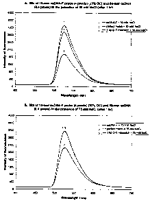
After a one-hour incubation at 21 C in the presence of
50mM NaCl, dsDNA:ssDNA-F triplexes consisting of perfectly
complementary sequences (SEQ ID NO:1 + Probe No. 1) formed
readily, resulting in a 49% decrease in fluorescent
intensity compared to that emitted by Probe No. 1 alone
(labeled ssDNA-F) (Fig. lA). In contrast, incompletely
complementary dsDNA:ssDNA-F triplexes containing a 1 bp G-T
mismatch (SEQ ID NO:2 + Probe No. 1) were less stable in
these reaction conditions, yielding only an 11% decrease in
fluorescent intensity compared to that exhibited by Probe
No. 1 alone.
Incubation for one hour in the presence of 75 mM NaCl
was slightly less conducive to triplex formation, resulting
in a 30% decrease in fluorescent intensity for the
perfectly matched dsDNA:ssDNA-F triplex (Fig. 1B). Minimal
formation of the 1 bp G-T mismatched dsDNA:ssDNA-F triplex
was observed, resulting in only a 0.4% decrease in
fluorescence.
The presence of 100 mM and 125 mM NaCl also
facilitated maximum triplex DNA formation between the
perfectly matched SEQ ID NO:1 target and Probe No. 1, and
less stable triplex DNA formation between the 1 bp G-T
mismatched SEQ ID NO:2 and Probe No. 1 hybrid (data not
shown). At 150 mM NaCl, no triplex DNA formation was
evident.
Therefore, the inclusion of monovalent cations such as
Na+ and K+ at specific concentrations, was sufficient to
allow detection of triplex formation between dsDNA targets
and ssDNA probes labeled with fluorescein in the absence of
prior denaturation. Moreover, the reaction occurred at
room temperature within just one hour of incubation at a
ratio of probe to target of 10 to 1, using natural dsDNA.
The dsDNA targets and ssDNA probe used in this example
contained a 33% GC content, and did not contain homopurine
or homopyrimidine stretches of DNA. Despite the presence
CA 02415493 2003-01-07
WO 02/04655 PCT/1B01/01538
17
of 6 pyrimidine bases interspersed within the 15 nucleotide
ssDNA probe, DNA triplexes formed easily. Significantly,
the hybridization assay of the invention was able to
discriminate between perfectly complementary DNA sequences
and those containing a single 1 bp mismatch using natural
DNA.
Example 2
To ensure that the hybridization assay, which used
5'-fluorescein labeled ssDNA probes and dsDNA targets in
the absence of prior denaturation, would apply to probe and
target DNAs possessing dramatically different percent GC
contents (and potentially different annealing preferences),
new 15-mer ssDNA-F probes and 50-mer dsDNA target sequences
were synthesized, purified and annealed as above. Both
ssDNA-F probes and dsDNA targets were dissolved in ddH2O at
a concentration of 1 pmole/,ul.
SEQ ID NO:5 was a 50-mer dsDNA target sequence
modified from SEQ ID NO:1, wherein the percent GC content
was changed from 30% to 52%.
Sequence for the sense strand of the wild-type target
DNA (SEQ ID NO:5): 5'-GAG CAC CAT GAC AGA CAC TGT
CAT CTC TGG TGT GTC CTA CGA TGA CTC TG-3'.
Sequence for the antisense strand of the wild-type
target DNA (SEQ ID NO:5): 5'-CAG AGT CAT CGT AGG ACA
CAC CAG AGA TGA CAG TGT CTG TCA TGG TGC TC-3'.
SEQ ID NO:6 was a 50-mer mutant dsDNA target sequence
identical to SEQ ID NO:5, except for a one base pair
mutation (underlined), at which the sequence CTC was
changed to CTT.
Sequence for the sense strand of mutant SEQ ID NO:6:
5'-GAG CAC CAT GAC AGA CAC TGT CAT CTT TGG TGT
GTC CTA CGA TGA CTC TG-3'.
Sequence for the antisense strand of mutant SEQ ID
NO:6: 5'-CAG AGT CAT CGT AGG ACA CAC CAA AGA TGA
CAG TGT CTG TCA TGG TGC TC-3'.
CA 02415493 2003-01-07
WO 02/04655 PCT/1B01/01538
18
SEQ ID NO:7 was a 50-mer mutant dsDNA target sequence
identical to SEQ ID NO:5, except for a one base pair
mutation (underlined), at which the sequence CAT was
changed to CGT.
Sequence for the sense strand of mutant SEQ ID NO:7:
5'-GAG CAC CAT GAC AGA CAC TGT CGT CTC TGG TGT
GTC CTA CGA TGA CTC TG-3'.
Sequence for the antisense strand of mutant SEQ ID
NO:7: 5'-CAG AGT CAT CGT AGG ACA CAC CAG AGA CGA
CAG TGT CTG TCA TGG TGC TC-3'.
SEQ ID NO:8 was a 50-mer mutant dsDNA target sequence
identical to SEQ ID NO:5, except for a one base pair
mutation (underlined), at which the sequence CAT was
changed to CTT.
Sequence for the sense strand of mutant SEQ ID NO:8:
5'-GAG CAC CAT GAC AGA CAC TGT CTT CTC TGG TGT
GTC CTA CGA TGA CTC TG-3'.
Sequence for the antisense strand of mutant SEQ ID
NO:8: 5'-CAG AGT CAT CGT AGG ACA CAC CAG AGA AGA
CAG TGT CTG TCA TGG TGC TC-3'.
SEQ ID NO:9 was a 50-mer mutant dsDNA target sequence
identical to SEQ ID NO:5, except for a one base pair
mutation (underlined), at which the sequence CTC was
changed to CCC.
Sequence for the sense strand of mutant SEQ ID NO:9:
5'-GAG CAC CAT GAC AGA CAC TGT CAT CCC TGG TGT
GTC CTA CGA TGA CTC TG-3'.
Sequence for the antisense strand of mutant SEQ ID
NO:9: 5'-CAG AGT CAT CGT AGG ACA CAC CAG GGA TGA
CAG TGT CTG TCA TGG TGC TC-3'.
SEQ ID NO:10 was a 50-mer mutant dsDNA target sequence
identical to SEQ ID NO:5, except for a one base pair
mutation (underlined), at which the sequence CTC was
changed to CGC.
CA 02415493 2003-01-07
WO 02/04655 PCT/1B01/01538
19
Sequence for the sense strand of mutant SEQ ID NO:10:
51-GAG CAC CAT GAC AGA CAC TGT CAT CGC TGG TGT
GTC CTA CGA TGA CTC TG-3'.
Sequence for the antisense strand of mutant SEQ ID
NO:10: 5'-CAG AGT CAT CGT AGG ACA CAC CAG CGA TGA
CAG TGT CTG TCA TGG TGC TC-3'.
SEQ ID NO:11 was a 50-mer mutant dsDNA target sequence
identical to SEQ ID NO:5, except for a consecutive two base
pair mutation (underlined), at which the sequence CAT was
changed to ACT.
Sequence for the sense strand of mutant SEQ ID NO:11:
5'-GAG CAC CAT GAC AGA CAC TGT ACT CTC TGG TGT
GTC CTA CGA TGA CTC TG-3'.
Sequence for the antisense strand of mutant SEQ ID
NO:11: 5'-CAG AGT CAT CGT AGG ACA CAC CAG AGA GTA
CAG TGT CTG TCA TGG TGC TC-3'.
SEQ ID NO:12 was a 50-mer dsDNA target sequence
modified from SEQ ID NO:1, wherein the percent GC content
was changed from 30% to 72%.
Sequence for the sense strand of the wild-type target
DNA (SEQ ID NO:12): 5'-GAG CAC CCT CCC AGG CAC GGT
CGT CCC TGG TGC GAC CTC CGA CGA GCG TG-3'.
Sequence for the antisense strand of the wild-type
target DNA (SEQ ID NO:12): 5'-CAC GCT CGT CGG AGG TCG
CAC CAG GGA CGA CCG TGC CTG GGA GGG TGC TC-31.
SEQ ID NO:13 was a 50-mer mutant dsDNA target sequence
identical to SEQ ID NO:12, except for a one base pair
mutation (underlined), at which the sequence CGT was
changed to CAT.
Sequence for the sense strand of mutant SEQ ID NO:13:
5'-GAG CAC CCT CCC AGG CAC GGT CAT CCC TGG TGC
GAC CTC CGA CGA GCG TG-3'.
Sequence for the antisense strand of mutant SEQ ID
NO:13: 5'-CAC GCT CGT CGG AGG TCG CAC CAG GGA TGA
CCG TGC CTG GGA GGG TGC TC-3'.
CA 02415493 2003-01-07
WO 02/04655 PCT/1B01/01538
Probe No. 2 (SEQ ID NO:14), a 15-mer ssDNA probe with
an attached fluorescein moiety at the 5' position, was
designed to be completely complementary to a 15 nucleotide
segment of the sense strand of the 50-mer wild-type target
5 DNA (SEQ ID NO:5).
Sequence for SEQ ID NO:14: 5'-Flu-CAC CAG AGA TGA
CAG-3'.
Probe No. 3 (SEQ ID NO: 15) was a 15 -mer 5' -fluorescein
labeled ssDNA probe designed to be completely complementary
10 to a 15 nucleotide segment of the sense strand of the
50-mer wild-type target DNA (SEQ ID NO:12).
Sequence for SEQ ID NO:15: 5'-Flu-CAC CAG GGA CGA
CCG-3'.
The triplex DNA hybridization assays performed in
15 Example 1 were facilitated by the addition of monovalent
cations in the reaction mixtures. The specificity of the
hybridization assay was further examined utilizing divalent
cations (instead of monovalent cations) to promote triplex
DNA formation with dsDNA targets and ssDNA-F probes
20 possessing various percent GC contents.
The hybridization reaction mixture (401u1) contained
the following: 0.4 pmoles of target dsDNA, 4 pmoles of
5'-fluorescein labeled ssDNA probe, 10 mM Tris-HC1, pH 7.5
and 5 mM to 30 mM MnC12 or 5 mM to 30 mM MgC12 or 5 mM to
30 mM NiCl2. The reaction mixtures were incubated at room
temperature (21 C) for 1 hour, without prior denaturation.
Samples were placed into a quartz cuvette, irradiated with
an argon ion laser beam having a wavelength of 488 nm and
monitored for fluorescent emission. The samples were saved
and allowed to incubate at room temperature overnight for
a total of 22 hours, at which time a second fluorescent
intensity measurement was taken following irradiation with
the argon ion laser beam. The intensity of fluorescence
was plotted as a function of wavelength for each sample
analyzed.
CA 02415493 2003-01-07
WO 02/04655 PCT/1B01/01538
21
When the ssDNA-F Probe No. 2 (with a 53% GC content)
was incubated with the 50-mer wild-type dsDNA target (SEQ
ID NO:5) and mutant dsDNA targets (SEQ ID NO:6 to SEQ ID
NO:11) in the presence of 10 mM MnCl2, dsDNA:ssDNA-F
triplexes were formed at room temperature under
non-denaturing conditions. While perfectly matched DNA
triplexes yielded the maximum decrease in fluorescent
intensity (a 43% decrease after a one-hour incubation) , the
less stable dsDNA:ssDNA-F triplexes containing a 1 bp T-G
mismatch (SEQ ID NO : 6 + Probe No. 2) produced a fluorescent
intensity that was 20% lower than that observed with Probe
No. 2 alone after a one-hour incubation (Fig. 2A).
dsDNA:ssDNA-F triplexes that resulted in a 1 bp G-T
mismatch (SEQ ID NO:7 + Probe No. 2), a 1 bp T-T mismatch
(SEQ ID NO:8 + Probe No. 2), a 1 bp C-A mismatch (SEQ ID
NO:9 + Probe No. 2) and a consecutive 2 bp A-G and C-T
mismatch (SEQ ID NO:11 + Probe No. 2) were all less stable
than the perfectly matched DNA triplex (SEQ ID NO:5 + Probe
No. 2) yielding fluorescent intensities in between that
observed for Probe No. 2 alone and that observed for the
perfectly matched DNA triplex (data not shown) . Except for
the 1 bp T-T mismatched DNA triplex, which was the least
stable (resulting in only a 5% decrease in fluorescent
intensity after 1 hour), all of the other mismatched DNA
triplexes generated very similar fluorescent intensities.
Only the dsDNA:ssDNA-F triplex that contained a 1 bp G-A
mismatch (SEQ ID NO:10 + Probe No. 2) yielded a fluorescent
intensity lower than that produced by the perfectly matched
DNA triplex (data not shown).
DNA triplex formation was more efficient after a
22-hour incubation in the presence of 10 mM MnCl2.
Nevertheless, a more prominent discrimination between DNA
triplexes containing perfectly matched sequences and DNA
triplexes containing base pair mismatched sequences was
observed. As illustrated in Fig. 2B, the dsDNA:ssDNA-F
CA 02415493 2003-01-07
WO 02/04655 PCT/1B01/01538
22
triplexes containing perfectly complementary sequences (SEQ
ID NO:5 + Probe No. 2) or a 1 bp T-G mismatch (SEQ ID NO:6
+ Probe No. 2) produced fluorescent intensities that were
92% and 66% lower, respectively, than the intensity
achieved by Probe No. 2 alone, following a 22-hour
incubation in the presence of 10 mM MnCl2. Similarly,
incubation in the presence of 30 mM MnC12 for 22 hours,
resulted in a 90% and a 57% reduction in fluorescent
intensity for perfectly matched DNA triplexes and 1 bp T-G
mismatched DNA triplexes, respectively (Fig. 2C).
The inclusion of 20 mM MgC12 or 20 mM MnC12 or 20 mM
NiCl2 also facilitated dsDNA:ssDNA triplex formation when
the ssDNA-F Probe No. 3 (possessing a 73% GC content) was
reacted with the corresponding 50-mer wild-type dsDNA
target (SEQ ID NO:12) and mutant dsDNA target (SEQ ID
NO:13) for one hour (data not shown). As expected, the
perfectly matched DNA triplexes generated the maximum
decreases in fluorescent intensity, while the less stable
1 bp A-C mismatched DNA triplexes (SEQ ID NO: 13 + Probe
No. 3) produced intermediate levels of fluorescence (data
not shown). The perfectly matched DNA triplexes formed
very efficiently in the presence of 10 mM MnC12 after a 22
hour incubation, yielding an 89% decrease in fluorescent
intensity. The 1 bp A-C mismatched DNA triplexes were
formed with equal efficiency in these reaction conditions,
generating a 90% decrease in fluorescence compared to that
observed with Probe No. 3 alone (data not shown).
Therefore, better discrimination was achieved between the
perfectly matched and 1 bp mismatched 73% GC DNA triplexes
following short incubation times of 1 hour in the presence
of 20 mM divalent cations.
Perfectly matched dsDNA:ssDNA-F triplexes (possessing
a 33% GC content) (SEQ ID NO:1 + Probe No. 1) formed
readily within 1 hour in the presence of 10 mM MnCl2,
resulting in a 57% decrease in fluorescent intensity
CA 02415493 2003-01-07
WO 02/04655 PCT/1B01/01538
23
compared to that emitted by Probe No. 1 alone (data not
shown). These reaction conditions were highly unfavorable
for DNA triplexes that contained a 1 bp G-T mismatch (SEQ
ID NO:2 + Probe No. 1), resulting in an increased
fluorescence compared to that observed by Probe No. 1 alone
(data not shown). Similar results were obtained following
a 22 hour incubation in the presence of 15 mM MgCl2.
Regardless of the percent GC content of the dsDNA
targets and ssDNA probes, the addition of divalent cations
such as Mn+2, Mg+2 or Ni+2 promoted DNA triplex formation
under non-denaturing conditions, to allow accurate
discrimination between perfectly complementary sequences
and those containing 1 bp mutations.
Example 3
The triplex DNA hybridization assays in Examples 1 and
2 were performed in the presence of one type of monovalent
or divalent cation. The next examples will demonstrate the
reliability of the assay of the invention to differentiate
between perfect matches and 1 bp mismatches in triplex DNA
when combinations of divalent cations are present in the
reaction mixtures.
The hybridization reaction mixture (40,21) contained
the following: 0.4 pmoles of target dsDNA, 4 pmoles of
5'-fluorescein labeled ssDNA probe, 10 mM Tris-HC1, pH 7.5
and 5 mM MgC12 and 5 mM MnCl2, or 10 mM MgC12 and 10 mM
MnC12, or 15 mM MgC12 and 15 mM MnC12 1 or 20 mM MgCl2 and 20
mM MnCl2. The reaction mixtures were incubated at room
temperature (21 C) for 1 hour, without prior denaturation.
Samples were placed into a quartz cuvette, irradiated with
an argon ion laser beam having a wavelength of 488 nm and
monitored for fluorescent emission. The samples were saved
and allowed to incubate at room temperature overnight for
a total of 22 hours, at which time a second fluorescent
intensity measurement was taken following irradiation with
the argon ion laser beam. The intensity of fluorescence
CA 02415493 2003-01-07
WO 02/04655 PCT/1B01/01538
24
was plotted as a function of wavelength for each sample
analyzed.
In all mixtures of dsDNA target and ssDNA-F probe, the
addition of 5 mM MgCl, and 5 mM MnC12 was insufficient to
allow detection of triplex DNA formation (data not shown).
When the ssDNA-F Probe No. 3 (with a 73% GC content) was
incubated for one hour with the 50-mer wild-type dsDNA
target (SEQ ID NO:12) in the presence of 10 mM MgC12 and 10
mM MnC12, or 15 mM MgC12 and 15 mM MnC121 perfectly
complementary dsDNA:ssDNA-F triplexes were formed with
equal efficiency, generating a 29% decrease in fluorescence
compared to that emitted by Probe No. 3 alone. Both
reaction conditions were highly unfavorable for DNA
triplexes that contained a 1 bp A-C mismatch (SEQ ID NO:13
+ Probe No. 3), resulting in a 14% increase in fluorescence
compared to that observed with Probe No. 3 alone. The
fluorescent spectra obtained after a one hour incubation in
the presence of 15 mM MgC12 and 15 mM MnC12 are shown in
Fig. 3A.
Incubation for 22 hours yielded more DNA triplex
formation. The dsDNA:ssDNA-F triplexes containing
perfectly matched sequences (SEQ ID NO:12 + Probe No. 3) or
a 1 bp A-C mismatch (SEQ ID NO:13 + Probe No. 3) produced
fluorescent intensities that were 62% and 21% lower,
respectively, than that achieved by Probe No. 3 alone,
following a 22 hour incubation in the presence of 10 mM
MgC12 and 10 mM MnCl2 (Fig. 3B). Very similar results were
obtained with the samples containing 15 mM MgC12 and 15 mM
MnC12 after 22 hours (data not shown).
Treatment with 20 mM MgC12 and 20 mM MnC12 for just one
hour, resulted in a 46% and a 3% reduction in fluorescence
for perfectly matched DNA triplexes and 1 bp A-C mismatched
DNA triplexes, respectively (Fig. 3C). In this case, no
benefit was achieved by further incubating the samples for
22 hours (data not shown).
CA 02415493 2003-01-07
WO 02/04655 PCT/1B01/01538
When dsDNA targets containing a 73% GC content are
tested in the hybridization assay of the invention, a
one-hour treatment with 20 MM MgC12 and 20 mM MnC12 provides
the maximum difference in stability and fluorescence
5 between perfectly complementary DNA triplexes and DNA
triplexes containing a 1 bp mismatch.
Example 4
When the ssDNA-F Probe No. 1 (with a 33% GC content)
was incubated with the wild-type dsDNA target (SEQ ID NO:1)
10 or mutant dsDNA targets (SEQ ID NO:2 and SEQ ID NO:3), in
the presence of 10 mM MgC12 and 10 mM MnC12, minimal DNA
triplex formation was observed (data not shown). However,
incubation in the presence of 15 mM MgC12 and 15 mM MnC12
for one hour facilitated perfectly matched DNA triplex
15 formation, as evidenced by the 49% decrease in fluorescent
intensity observed, compared to that obtained by Probe No.
1 (Fig. 4A). dsDNA:ssDNA-F triplexes that resulted in a 1
bp G-T mismatch (SEQ ID NO:2 + Probe No. 1) or a 3 bp
deletion (SEQ ID NO:3 + Probe No. 1) were very unstable in
20 the presence of 15 MM MgC12 and 15 mM MnC12, yielding a 2%
decrease in fluorescence and a 5% increase in fluorescence,
respectively, compared to that emitted by Probe No. 1 alone
(Fig. 4A).
Treatment with 20 MM MgC12 and 20 mM MnC12 for 1 hour,
25 resulted in a 68%, a 48% and a 6% reduction in fluorescence
for perfectly matched DNA triplexes, and for dsDNA:ssDNA-F
triplexes containing a 1 bp G-T mismatch or a 3 bp
deletion, respectively, compared to that observed with
Probe No. 1 alone (Fig. 4B). Optimum discrimination
between the 33% GC DNA triplexes containing wild-type
sequences or base pair mismatches was achieved when these
same samples were incubated for 22 hours. The perfectly
complementary DNA triplexes (SEQ ID NO:1, + Probe No. 1)
remained stable over the 22 hours, producing a 62% decrease
in fluorescent intensity, compared to that achieved by
CA 02415493 2003-01-07
WO 02/04655 PCT/1B01/01538
26
Probe No. 1 alone (Fig. 4C). By contrast, the
dsDNA:ssDNA-F triplexes containing a 1 bp G-T mismatch (SEQ
ID NO:2 + Probe No. 1) or a 3 bp deletion (SEQ ID NO:3 +
Probe No. 1) proved to be very unstable during the 22 hour
incubation, generating a 1% and a 13% increase in
fluorescence, respectively, compared to that emitted by
Probe No. 1 alone (Fig. 4C).
Example 5
Perfectly matched dsDNA:ssDNA-F triplexes (possessing
a 53% GC content) (SEQ ID NO:5 + Probe No. 2) formed
readily within one hour in the presence of 10 mM MgC12 and
10 mM MnC12, resulting in a 68% decrease in fluorescence
compared to that observed by Probe No. 2 alone (Fig. 5A).
The DNA triplexes that contained a 1 bp T-G mismatch (SEQ
ID NO:6 + Probe No. 2) were less stable, generating a 20%
decrease in fluorescent intensity compared to that achieved
by Probe No. 2 alone (Fig. 5A).
Incubation of the same samples for 22 hours produced
an even more dramatic difference in fluorescence achieved
by the perfectly matched or mismatched DNA triplexes. As
illustrated in Fig. 5B, the dsDNA:ssDNA-F triplexes
containing perfectly complementary sequences (SEQ ID NO:5
+ Probe No. 2) or a 1 bp T-G mismatch (SEQ ID NO:6 + Probe
No. 2) generated fluorescent intensities that were 92% and
33% lower, respectively, than that emitted by Probe No. 2
alone, in the presence of 10 mM MgCl2 and 10 mM MnC12.
In a similar experiment, while the perfectly matched
DNA triplex (SEQ ID NO:5 + Probe No. 2) yielded a 85%
decrease in fluorescence compared to that observed with
Probe No. 2 alone following a 22 hour incubation in the
presence of 10 mM MgC12 and 10 mM MnC12, the dsDNA:ssDNA-F
triplexes that resulted in a 1 bp G-T mismatch (SEQ ID NO:7
+ Probe No. 2), a 1 bp C-A mismatch (SEQ ID NO:9 + Probe
No. 2) and a consecutive 2 bp A-G and C-T mismatch (SEQ ID
NO:11 + Probe No. 2) produced a 43%, a 69% and a 32%
CA 02415493 2003-01-07
WO 02/04655 PCT/1B01/01538
27
reduction in fluorescence (Fig. 5C). Only the dsDNA:ssDNA-
F triplex that contained a 1 bp G-A mismatch (SEQ ID NO:10
+ Probe No. 2) yielded a fluorescent intensity slightly
lower than that produced by the perfectly matched DNA
triplex (data not shown).
Optimum discrimination between the 53% GC DNA
triplexes containing perfectly complementary sequences or
base pair mismatches was achieved following a one-hour
incubation in the presence of 15 mM MgCl2 and 15 mM MnCl2.
These reaction conditions greatly facilitated DNA triplex
formation between the perfectly matched DNA sequences (SEQ
ID NO:5 + Probe No. 2), resulting in a 74% reduction in
fluorescence compared to that achieved by Probe No. 2 alone
(Fig. 5D). By contrast, dsDNA:ssDNA-F triplexes that
contained a 1 bp T-G mismatch (SEQ ID NO:6 + Probe No. 2)
were much less stable in the presence of 15 mM MgC12 and 15
mM MnC12, yielding a 15% decrease in fluorescence compared
to that emitted by Probe No. 2 alone after a one-hour
incubation (Fig. 5D).
Similarly, DNA triplexes that resulted in a 1 bp G-T
mismatch (SEQ ID NO:7 + Probe No. 2), a 1 bp C-A mismatch
(SEQ ID NO:9 + Probe No. 2), a 1 bp G-A mismatch (SEQ ID
NO:10 + Probe No. 2) and a consecutive 2 bp A-G and C-T
mismatch (SEQ ID NO:11 + Probe No. 2) were all much less
stable than the perfectly matched DNA triplex (data not
shown). The 1 bp G-A mismatched DNA triplex that formed
relatively easily in the presence of 10 mM MnC12 or 10 mM
MgC12 and 10 mM MnCl21 was now disrupted in the presence of
15 mM MgC12 and 15 mM MnC12, producing only a 7% reduction
in fluorescence compared to that observed with Probe No. 2
alone (data not shown). When Probe No. 2 (containing a 53%
GC content) was reacted with the dsDNA target SEQ ID NO:3
(containing a 33% GC content), a 3% increase in
fluorescence was observed compared to that obtained by
Probe No. 2 alone (Fig. 5D), indicative of no DNA triplex
CA 02415493 2003-01-07
WO 02/04655 PCT/1B01/01538
28
formation. This result was expected considering this probe
and target combination would result in a 5 bp mismatch.
Treatment with 15 mM MgCl2 and 15 mM MnCl2 for 22
hours, generated a 76% and a 44% decrease in fluorescent
intensity for dsDNA:ssDNA-F triplexes containing perfectly
complementary sequences (SEQ ID NO:5 + Probe No. 2) and a
1 bp T-G mismatch (SEQ ID NO:6 + Probe No. 2),
respectively, compared to that obtained with Probe No. 2
alone (Fig. 5E).
Collectively, the above Examples demonstrated that the
addition of at least one type of cation to a hybridization
medium promotes DNA triplex formation between dsDNA targets
and fluorescently-labeled ssDNA probes, possessing
dramatically different percent GC contents, to allow
accurate and reliable discrimination between perfectly
complementary sequences and those containing various
mutations.
While the invention has been described in detail and
with reference to specific examples thereof, it will be
apparent to one skilled in the art that various changes and
modifications can be made therein without departing from
the spirit and scope thereof.
CA 02415493 2003-01-07
SEQUENCE LISTING
<110> Ingeneus Corporation
<120> CATION MEDIATED TRIPLEX HYBRIDIZATION ASSAY
<130> 08-896851CA
<140>
<141> 09-07-2001
<150> 09/613,263
<151> 10-07-2000
<160> 15
<170> Patentln Ver. 2.1
<210> 1
<211> 50
<212> DNA
<213> Artificial Sequence
<220>
<223> Description of Artificial Sequence: derived from
exon 10 of the human cystic fibrosis gene
<400> 1
tggcaccatt aaagaaaata tcatctttgg tgtttcctat gatgaatata 50
<210> 2
<211> 50
<212> DNA
<213> Artificial Sequence
<220>
<223> Description of Artificial Sequence: derived from
exon 10 of the human cystic fibrosis gene
<400> 2
tggcaccatt aaagaaaata tcgtctttgg tgtttcctat gatgaatata 50
<210> 3
<211> 47
<212> DNA
<213> Artificial Sequence
<220>
<223> Description of Artificial Sequence: derived from
exon 10 of the human cystic fibrosis gene
<400> 3
tggcaccatt aaagaaaata tcattggtgt ttcctatgat gaatata 47
1
CA 02415493 2003-01-07
<210> 4
<211> 15
<212> DNA
<213> Artificial Sequence
<220>
<223> Description of Artificial Sequence: ssDNA probe
<400> 4
caccaaagat gatat 15
<210> 5
<211> 50
<212> DNA
<213> Artificial Sequence
<220>
<223> Description of Artificial Sequence: derived from
exon 10 of the human cystic fibrosis gene
<400> 5
gagcaccatg acagacactg tcatctctgg tgtgtcctac gatgactctg 50
<210> 6
<211> 50
<212> DNA
<213> Artificial Sequence
<220>
<223> Description of Artificial Sequence: derived from
exon 10 of the human cystic fibrosis gene
<400> 6
gagcaccatg acagacactg tcatctttgg tgtgtcctac gatgactctg 50
<210> 7
<211> 50
<212> DNA
<213> Artificial Sequence
<220>
<223> Description of Artificial Sequence: derived from
exon 10 of the human cystic fibrosis gene
<400> 7
gagcaccatg acagacactg tcgtctctgg tgtgtcctac gatgactctg 50
<210> 8
<211> 50
<212> DNA
<213> Artificial Sequence
2
CA 02415493 2003-01-07
<220>
<223> Description of Artificial Sequence: derived from
exon 10 of the human cystic fibrosis gene
<400> 8
gagcaccatg acagacactg tcttctctgg tgtgtcctac gatgactctg 50
<210> 9
<211> 50
<212> DNA
<213> Artificial Sequence
<220>
<223> Description of Artificial Sequence: derived from
exon 10 of the human cystic fibrosis gene
<400> 9
gagcaccatg acagacactg tcatccctgg tgtgtcctac gatgactctg 50
<210> 10
<211> 50
<212> DNA
<213> Artificial Sequence
<220>
<223> Description of Artificial Sequence: derived from
exon 10 of the human cystic fibrosis gene
<400> 10
gagcaccatg acagacactg tcatcgctgg tgtgtcctac gatgactctg 50
<210> 11
<211> 50
<212> DNA
<213> Artificial Sequence
<220>
<223> Description of Artificial Sequence: derived from
exon 10 of the human cystic fibrosis gene
<400> 11
gagcaccatg acagacactg tactctctgg tgtgtcctac gatgactctg 50
<210> 12
<211> 50
<212> DNA
<213> Artificial Sequence
<220>
<223> Description of Artificial Sequence: derived from
exon 10 of the human cystic fibrosis gene
3
CA 02415493 2003-01-07
= <400> 12
gagcaccctc ccaggcacgg tcgtccctgg tgcgacctcc gacgagcgtg 50
<210> 13
<211> 50
<212> DNA
<213> Artificial Sequence
<220>
<223> Description of Artificial Sequence: derived from
exon 10 of the human cystic fibrosis gene
<400> 13
gagcaccctc ccaggcacgg tcatccctgg tgcgacctcc gacgagcgtg 50
<210> 14
<211> 15
<212> DNA
<213> Artificial Sequence
<220>
<223> Description of Artificial Sequence: ssDNA probe
<400> 14
caccagagat gacag 15
<210> 15
<211> 15
<212> DNA
<213> Artificial Sequence
<220>
<223> Description of Artificial Sequence: ssDNA probe
<400> 15
caccagggac gaccg 15
4