Note: Descriptions are shown in the official language in which they were submitted.
CA 02416828 2003-O1-22
WO 02/07854 PCT/USO1/22651
- 1 -
BLOOD COLLECTION SYSTEMS AND METHODS USING
A POROUS MEMBRANE ELEMENT
Field of the Invention:
The invention generally relates to blood col-
lection and processing systems and methods.
Backaround of the Invention:
Systems composed of multiple, interconnected
plastic bags have met widespread use and acceptance in
the collection, processing and storage of blood
components. Using these systems, whole blood is collected
and separated into its clinical components (typically red
blood cells, platelets, and plasma). The components are
individually stored and used to treat a multiplicity of
specific conditions and diseased states.
Before storing blood components for later
transfusion, it is believed to be desirable to minimize
the presence of impurities or other materials that may
cause undesired side effects in the recipient. For
example, because of possible reactions, it is generally
considered desirable to remove substantially all the
leukocytes from blood components before storage, or at
least before transfusion.
Filtration is conventionally used to accomplish
leuko-reduction. Systems and methods for reducing the
number of leukocytes by filtration in multiple blood bag
configurations are described, e.g., in Stewart U.S.
Patent 4,997,577, Stewart et al. U.S. Patent 5,128,048,
Johnson et al. U.S. Patent 5,180,504, and Bellotti et.
al. U.S. Patent 5,527,472.
3 0 Summary of the Invention
CA 02416828 2003-O1-22
WO 02/07854 PCT/USO1/22651
- 2 -
One aspect of the invention provides systems
and methods for removing leukocytes from blood using a
filter media having a main filter region comprising a
porous membrane structure extending between first and
second skin surfaces. The porous membrane structure is
formed by intersecting cells having a range of diameters.
The cells adjacent to the first skin surface have
diameters generally smaller than the diameters of the
cells adjacent to the second skin surface. The first
skin surface includes an open area defined by pores,
which are formed by the intersection of cells with the
first skin surface. The majority of the open area is
defined by pores having a diameter of between about 12 um
and 28 Vim.
In one embodiment, the main filter region
includes a polyethersulfone material.
In one embodiment, the filter media is enclosed
in a housing. The housing comprises first and second
flexible sheets made of a meltable material. A
peripheral seal joins the sheets directly to the filter
media to encapsulate the filter media between the first
and second sheets. The seal includes a commingled melted
matrix comprising material of the sheets and material of
the filter media.
Another aspect of the invention provides
systems and methods for removing leukocytes from blood
using a filter media having a main filter region. The
main filter region comprises a layered porous membrane
structure that includes several regions of larger pore
sizes alternating in the direction of flow with several
regions of smaller pore sizes, or vice versa. Blood
traversing the main filter region thereby passes in
succession through several alternating regions of
smaller, then larger, then smaller diameter pores, or
vice versa.
CA 02416828 2003-O1-22
WO 02/07854 PCT/USO1/22651
- 3 -
Other features and advantages of the invention
will become apparent upon review of the following de-
scription, drawings, and appended claims.
Brief Descrit~tion of the Drawinc,~s
Fig. 1 is a schematic view of a blood
collection and storage system that includes a filter that
embodies features of the invention and that removes
leukocytes from red blood cells;
Fig. ~ is an exploded perspective view of the
filter that forms a part of the system shown in Fig. 1;
Fig. 3 is an assembled perspective view of the
filter shown in Fig. 2;
Fig. 4 is a side section SEM (x900) view
showing a membrane that the filter shown in Fig. 3
incorporates as its main filter for removing leukocytes;
Fig. 5 is a plane SEM view (x400) of the
downstream skin surface of the membrane shown in Fig. 4;
and
Fig. 6 is a plane SEM view (xl.5k) of the
upstream skin surface of the membrane shown in Fig. 4;
Fig. 7 is a side section view of the pre-
assembled form of the filter shown in Fig. 3, located
between two spaced apart radio frequency energy die s
Fig. 8 is a side section view of the' pre
assembled form of the filter shown in Fig. 3, engaged by
the dies, which apply radio frequency energy to form a
unitary peripheral seal;
Fig. 9 is a schematic view of a blood
collection and storage system that includes two integral
filters that embody features of the invention, one to
remove leukocytes from red blood cells and the other to
remove leukocytes from platelet-rich plasmas and
Fig. 10 is a' schematic view of a blood
collection and storage system that includes a filter that
embodies features of the invention to remove leukocytes
CA 02416828 2003-O1-22
WO 02/07854 PCT/USO1/22651
- 4 -
from whole blood prior to centrifugal processing.
The invention is not limited to the details of
the construction and the arrangements of parts set forth
in the following description or shown in the drawings.
The invention can be practiced in other embodiments and
in various other ways. The terminology and phrases are
used for description and should not be regarded as
limiting.
Descriution of the Preferred Embodiments:
Fig. 1 shows a blood collection and storage
system 10 having an integral flexible filter 20. The
filter 20 can be incorporated into various types of blood
collection systems, and representative examples of such
systems will be described.
In Fig. 1, the system 10 provides red blood
cells for long term storage that are substantially free
of leukocytes. The system 10 also provides platelet
concentrate and the platelet-poor plasma for long term
storage. The blood collection and storage assembly 10,
once sterilized, constitutes a sterile, "closed" system,
as judged by the applicable standards in the United
States. The system 10 is a disposable, single use item.
As shown in Fig. 1, the system 10 includes a
primary bag 12 and three transfer bags or containers 14,
16, and 18. Like the flexible filter 20, the transfer
bags 14, 16, and 18 are integrally attached to the system
10.
In use, the system 10 is manipulated in
conventional ways. The primary bag 12 (which is also '
called a donor bag) receives whole blood from a donor
through integrally attached donor tube 22 that carries an
phlebotomy needle 24. A suitable anticoagulant A is
contained in the primary bag 12. The whole blood is
centrifugally separated by convention means inside the
primary bag 12 into red blood cells and platelet-rich
CA 02416828 2003-O1-22
WO 02/07854 PCT/USO1/22651
- 5 -
plasma. Leukocytes dwell in the interface between the
red blood cells and platelet-rich plasma.
The transfer bag 14 is intended to receive
platelet-rich plasma separated from the whole blood
collected in the primary bag 12. Attempts are made when
transferring the platelet-rich plasma out of the primary
bag 12 to keep as many leukocytes in the primary bag 12
as possible. The transfer of platelet-rich plasma into
the transfer bag 14 leaves the red blood cells and the
leukocytes behind in the primary bag 12.
The transfer bag 16 contains a suitable storage
solution S for red blood cells. One such solution is
disclosed in Grode et al U.S. Patent 4,267,269, which is
sold by Baxter Healthcare Corporation under the brand
name ADSOZ~ Solution. The storage solution S is
transferred into the primary bag 12 after transfer of the
platelet-rich plasma into the transfer bag 14.
The platelet-rich plasma is centrifugally
separated by conventional means in the transfer bag 14
into platelet concentrate and platelet-poor plasma. The
platelet-poor plasma is transferred into the transfer bag
16, which is now emptied of storage solution S. The
transfer bag 16 serves as the storage container for the
platelet-poor plasma. The transfer bag 14 serves as its
storage container for the platelet concentrate.
The storage solution S is mixed with the red
blood cells and leukocytes remaining in the primary bag
12. The mixture of storage solution S, red blood cells,
and leukocytes is transferred from the primary bag 12
through tubing 26. The tubing 26 carries in-line the
integral, flexible filter 20. The flexible filter 20
includes a filtration medium 28 contained within a
housing 30. The filtration medium is selected to remove
leukocytes from red blood cells.
The leukocyte-reduced red blood cells enter the
CA 02416828 2003-O1-22
WO 02/07854 PCT/USO1/22651
- 6 -
transfer bag 18. The transfer bag 18 serves as the
storage container for the leukocyte-reduced red blood
cells. Prior to storage, residual air in the transfer
bag 18 can be vented into the primary bag 12 through
tubing 60.
The bags and tubing associated with the
processing system 10 can all be made from conventional
approved medical grade plastic materials, such as
polyvinyl chloride plasticized with di-2-ethylhexyl-
phthalate (PVC-DEHP). The bags are formed using
conventional heat sealing technologies, e.g., radio
frequency (RF) heat sealing.
Alternatively, since the transfer bag 14 is
intended to store the platelet concentrate, it can be
made of polyolefin material (as disclosed in Gajewski et
al U.S. patent 4,140,162) or a polyvinyl chloride
material plasticized with tri-2-ethylhexyl trimellitate
(TEHTM). These materials, when compared to DEHP-
plasticized polyvinyl chloride materials, have greater
gas permeability that is beneficial for platelet storage.
The flexible filter 20, like the rest of the
system 10, is a disposable, single use item. Also, like
the rest of the system 10, the filter housing 30 is made
using conventional approved medical grade plastic
materials. Furthermore, like the rest of the system 10,
the filter housing 30 is formed using conventional radio
frequency heat sealing technology. The filter 20, being
flexible, facilitates handling and reduces the incidence
of damage to other components of the system 10 during
centrifugal processing.
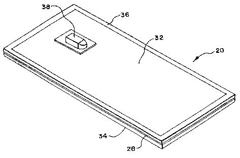
In the illustrated embodiment (see Figs. 2 and
3), the filter housing 30 comprising first and second
sheets 32 and 34 of medical grade plastic material, such
as polyvinyl chloride plasticized with di-2-ethylhexyl-
phthalate (PVC-DEHP). Other medical grade plastic
CA 02416828 2003-O1-22
WO 02/07854 PCT/USO1/22651
materials can be used that are not PVC and/or are DEHP-
free, provided that the material heats and flows when
exposed to radio frequency energy.
As Fig. 2 best shows, the filtration medium 28
comprises, in the blood flow direction, a prefilter
region PRF, a transfer filter region TRF, a main filter
region MF, and a postfilter region POF. The regions are
sandwiched between the sheets 32 and 34 and joined along
a continuous peripheral seal 36 (as Fig. 3 shows).
The prefilter region PRF and postfilter region
POF can be made of fibrous material, e.g., include melt
blown or spun bonded synthetic fibers (e.g., nylon or
polyester or polyethylene or polypropylene), semi-
synthetic fibers, regenerated fibers, or inorganic
fibers. The prefilter and postfilter regions PRF and POF
desirably have a pore size and fiber diameter not well
suited for leukocyte removal. Instead, the fibrous
material of the prefilter region PRF is sized to remove
gross clots and aggregations present in the blood. The
fibrous material of the postfilter region POF is sized to
provide a fluid manifold effect at the outlet of the
filter. In a representative embodiment, the material of
the prefilter region PRF has a pore size of between about
15 um to about 20 um, and the material of the postfilter
region POF~has a pore size of about 20 um.
The transfer region TR is made of fibrous
material (e. g., polyethylene) having a fiber diameter
less than the fiber diameter of the prefilter region PRF.
In a representative embodiment, the material of the
prefilter region PRF possesses an average fiber diameter
of about 12 um, and the material of the transfer filter
region TFR possesses a fiber diameter of about 4 um.
Preferably, the fibrous material of the transfer filter
region TFR is also coated with a polymer material
including polyalkylene oxide (PEO), such as disclosed in
CA 02416828 2003-O1-22
WO 02/07854 PCT/USO1/22651
_ g _
US Patent 6,045,701, which is incorporated herein by
reference.
Preferably, the fibrous material of the
transfer region TFR is arranged in more than a single
layer. In a preferred embodiment, a transfer filter
region TFR comprises four formed layers, each having an
individual thickness in the flow direction of about 0.4
mm.
The main filter region MF comprises a membrane
100 that removes leukocytes. With reference to Figs. 4 to
6, the membrane 100 of the main filter region MF can be
characterized as follows:
(i) as Fig. 4 shows, in side section, the
membrane 100 possess an interior porous structure formed
by intersecting cells 102 having a range of diameters,
with interior apertures 104 formed by intersections of
the cells 102,
(ii) as Fig. 4 also shows, the diameters of the
cells 102 can be grouped into two general regions: larger
diameter cells 102 adjacent to one skin surface 108
(which Fig. 6 shows in plane view) and smaller diameter
cells 102 adjacent to the other skin surface 106 (which
Fig. 5 shows in plane view). It is not believed
important as to whether the blood flow is from skin
surface 108 to 106, or vice versa,
(iii) as Fig. 5 shows, the cells 102 intersect
the skin surface 106, forming pores 110, and
(iv) the majority of the skin surface 106
occupied by the pores 110 (i.e., the total open area of
the skin surface 106 shown in Fig. 5) is formed by pores
110 having a diameter of between about 12 um to about 28
um.
Alternatively, the main filter region MF can
comprise alternating layers of isotropic membranes of
small and large pore size. The main filter region MF thus
CA 02416828 2003-O1-22
WO 02/07854 PCT/USO1/22651
- 9 -
comprises a layered porous membrane structure that
includes regions of larger pore sizes alternating in the
direction of flow with regions of smaller pore sizes, or
vice versa. Blood traversing the main filter region
thereby passes in succession through alternating regions
of smaller, then larger pores, or vice versa.
In a preferred embodiment, the membrane 100 is
made of a polyethersulfone (PES) material, which can be
obtained from Osmonics, Inc. (Minnetonka, Minnesota).
To achieve a 3 to 4 log reduction in the number
of leukocytes carried in unit of whole blood (typically
between 2 x 10⁹ to 6 x 10⁹) without plugging, the total
surface area of the membrane .100 forming the main filter
region MF should be between about 500 cm2 and about 1500
cmz .
In a preferred arrangement, PES membranes 100
are arranged in multiple individual layers, each
individual layer having the characteristics listed above,
which together forming the main filter region MF. Blood
traversing the multiple layers of the main filter region
MF thereby encounter alternating regions of large pore
size and then small pore size and then large pore size
and then small pore size, etc, or vice versa. This serial
transition between large and small pore size regions
along the flow path create successive changes in the flow
dynamics of the blood and are believed to enhance
leukocyte removal,
The assembly of the layered PES membranes in
the main filter region MF, in association with a
prefilter region PRF, a transfer filter region TFR, and
a postfilter region POF, as above described, provides a
filter 20 that allows the passage of upwards to 90% to
95% of platelets contained in a unit of whole blood,
while achieving a 3 to 4 log reduction in the number of
leukocytes. The filter 20 is therefore well suited for
CA 02416828 2003-O1-22
WO 02/07854 PCT/USO1/22651
- 10 -
inclusion in multiple blood bag systems in which whole
blood is filtered to remove leukocytes before
centrifugation, as will be described.
In forming the filter 20, a unitary, continuous
peripheral seal 36 (see Fig. 3) is formed by the
application of pressure and radio frequency heating in a
single process to the two sheets 32 and 34 and filtration
medium 28. The seal 36 joins the two sheets 32 and 34 to
each other, as well as joins the filtration medium 28 to
the two sheets 32 and 34. The seal 36 integrates the
material of the filtration medium 28 and the material of
the plastic sheets 32 and 34, for a reliable, robust,
leak-proof boundary. Since the seal 36 is unitary and
continuous, the possibility of blood shunting around the
periphery of the filtration medium 28 is eliminated.
The filter 20 also includes inlet and outlet
ports 38 and 40 (see Fig. 3). The ports 38 and 40
comprise tubes made of medical grade plastic material,
like PVC-DEHP. As Fig. 2 shows, the ports 38 and 40
comprise separately molded parts that are heat sealed by
radio frequency energy over a hole 40 formed in the
sheets 32 and 34 before the unitary peripheral seal 36 is
formed.
The filter 20 (see Fig. 7) is formed by
sandwiching layers of the prefilter region PRF, transfer
filter region TFR , main filter region MF, and postfilter
region POF between the first and second plastic sheets 32
and 34. The layered filter pre-assembly is placed between
a pair of opposed dies 50 and 52 (as Fig. 7 shows). The
opposed dies 50 and 52 are moved together (see Fig. 8),
to apply pressure to press the peripheral edge of the
pre-assembly 48 together. Preferably a stop 54 is
provided to accurately space the dies 50 and 52 apart
from each other.
As the dies 50 and 52 apply pressure about the
CA 02416828 2003-O1-22
WO 02/07854 PCT/USO1/22651
- 11 -
peripheral edge, RF energy is applied through the dies 50
and 52. The combination of RF energy and pressure softens
the plastic material of the sheets 32 and 34. The applied
pressure causes the heat softened material of the sheets
32, 34 to penetrate the interstices of the. filtration
medium 28, creating an interior matrix of sheet material
commingled with filtration medium material. Within the
matrix, the filtration medium melts, creating a composite
seal 36.
At its surface, along the sheets 32 and 34, the
seal 36 comprises mostly the material of the sheets 32
and 34. With increasing distance from the surface, the
seal 36 comprises a commingled melted matrix of the
material of the sheets 32 and 34 and the material of the
filtration medium 28. This is believed to occur because
the sheet material, which is electrically heated and
caused to flow by the applied radio frequency energy, is
further caused by the applied pressure to flow into and
penetrate the interstices of the medium 28. The heated
sheet material that flows under pressure into the
interstices of the medium 28 causes the medium 28 itself
to melt about it.
After a brief period of cooling, the seal 36
sets and the dies 50 and 52 are withdrawn. In a
representative embodiment, the dies 50 and 52 are
coupled to a 4 KW radio frequency energy generator.
Pressure of 60 PSI is applied, maintaining a die gap of
1.2 mm. A sealing time of about 5.5 seconds is realized,
followed by a cooling time of about 5 seconds.
A flexible filter can be integrated in
different ways into multiple blood bag systems. For
example (see Fig. 9) , a system 10' like that shown in
Fig. 1 can include a second integral flexible filter 20'
in-line between the primary bag 12 and the transfer bag
14. In this arrangement, the filtration medium 28' is
CA 02416828 2003-O1-22
WO 02/07854 PCT/USO1/22651
- 12 -
selected to remove leukocytes from platelet-poor plasma
prior to entering the transfer bag 14.
As another example, Fig. 10 shows a system 70
that includes a primary bag 72 and transfer bags 74, 76,
78. The primary bag 72 receives whole blood from a donor.
The whole blood is transferred from the primary bag 72
through tubing 80 into the transfer bag 74. The tubing
80 carries in-line an integral, flexible filter 82 of the
type previously described. The filtration medium 84 is
selected to remove leukocytes from the whole blood,
without also removing platelets or red blood cells. The
leukocyte-depleted whole blood is centrifugally processed
in the transfer bag 74 into red blood cells and platelet
rich plasma, both of which are in a leukocyte-depleted
condition.
The transfer bag 76 receives the leukocyte-
depleted platelet-rich plasma, leaving the leukocyte-
depleted red blood cells in the transfer bag 74 for
storage. The platelet-rich plasma is centrifugally
separated by conventional means in the transfer bag 76
into platelet concentrate and platelet-poor plasma. The
platelet-poor plasma is transferred into the transfer bag
78 for storage. This leaves the platelet concentrate in
the transfer bag 76, which serves as its storage
container.
The flexible filter that embodies the invention
avoids the handling and processing problems rigid filter
housings have presented in the past. Unlike a rigid
housing, the flexible housing 30 will not puncture
associated bags, which are also made of flexible plastic
materials. Unlike a rigid housing, the flexible housing
30 conforms and is compliant to stress and pressures
induced during use.
The close proximity of the flexible sheet 32
and the filtration medium 28 on the inlet side of the
CA 02416828 2003-O1-22
WO 02/07854 PCT/USO1/22651
- 13 -
filter 20 creates a capillary effect, which promotes
displacenment of air and automatic priming of the filter
30 under the fluid head pressure of gravity flow from a
source container. The fluid head pressure causes the
flexible sheet 32 to distend or expand after priming. It
thus creates a natural pressure manifold, which evenly
distributes the fluid across the inlet face of the
filtration medium 28. This assures that entrapped air is
vented and that the fluid flows through the filtration
medium 28 under uniform pressure and distribution.
As the fluid container empties, negative
pressure is created downstream of the filter 20. Because
the inlet and outlet sheets 32 and 34 of the housing 30
are flexible, they will collapse around the space
occupied by the filtration medium 28, minimizing the
amount of residual blood left in the filter 30 after use.
Fluid drains from the outlet side without the use of an
auxiliary air vent.
Furthermore, the flexible housing 30 will not
crack during heat sterilization. The flexible housing 30
also does not impede heat penetration during heat
sterilization processes. Instead, the housing 30
accommodates uniform heat penetration into the filtration
medium 28. The filter 20 can undergo sterilization at
the same time the entire system 10 is sterilized, making
a one-step sterilization process possible.
Various features of the invention are set forth
in the following claims.