Note: Descriptions are shown in the official language in which they were submitted.
CA 02418912 2003-02-07
WO 02/11724 PCT/USO1/41565
2-PYRIDINAMINE COMPOSITIONS AND RELATED METHODS
Cross Reference to Related Application
This application claims priority from United States provisional application
Serial No. 60/223,795, filed August 8, 2000, which is hereby incorporated by
reference.
Field of the Invention
The present invention relates to neuroprotective 2-pyridinamine compositions,
and methods of using same to prevent cell death after an ischemic event. The
instant
compositions have particular importance in preventing neuronal cell death and
its
resulting disorders.
Background of the Invention
Glutamate is the major fast excitatory neurotransmitter in the mammalian
central nervous system. It depolarizes neurons by opening three classes of
ligand-gated
ion channels: AMPA, kainate, and NMDA receptors. Transient increases in
synaptic
glutamate levels occur during normal excitatory transmission. However,
excessive
increases in synaptic glutamate levels are toxic to neurons, and trigger the
process of
neuronal cell death commonly referred to as glutamate excitotoxicity (Meldrum
and
Garthwaite 1990). Glutamate excitotoxicity contributes to ischemia-induced
brain
damage, epilepsy, and various chronic neurodegenerative diseases (Meldrum and
Garthwaite 1990).
Of the three classes of glutamate-gated channels, specific overactivation of
the
N-methyl-D-aspartate (NMDA) receptor is primarily responsible for triggering
excitotoxic neuron death in a variety of neuron types (Meldrum and Garthwaite
1990).
In animal stroke models, ischemia-induced brain damage can be largely
alleviated by
pretreatment with the specific NMDA receptor antagonist, MK-801 (Park et al.
1988).
In many types of neurons, glutamate exictotoxicity is thought to result
primarily from
excessive influx of calcium ions due to the high permeability of the NMDA
receptor
for calcium (Schneggenburger et al. 1993). High intracellular calcium levels
may lead
to overactivation of calcium-regulated enzymes such as nitric oxide synthase,
CA 02418912 2003-02-07
WO 02/11724 PCT/USO1/41565
phospholipases, proteases and kinases. Further, higgh intracellular calcium
levels may
mediate excitotoxicity.
Glutamate signaling through the NMDA receptor induces phosphorylation and
activation of mitogen-activated protein kinases (MAPK) in primary neuronal
cultures
(Bading and Greenberg 1991; Xia et al. 1995). Animal models of ischemic brain
injury
suggest that increased activity of MAPK family members may mediate neuronal
injury
(Alessandrini et al. 1999; Yang et al. 1997). Deletion of Jnk3, a member of
the JNK
family of MAP kinases which is predominantly expressed in brain, protects
hippocampal neurons from kainic acid-induced excitotoxicity i~z vivo, although
the role
of the NMDA receptor in this form of toxicity is not clear (Yang et al. 1998).
Specific
inhibition of the upstream activating kinases of ERKl/2, (p44/42) MAP kinase,
protects against neuronal damage due to focal cerebral ischemia (Alessandi-ini
et al.
1999). In cultured primary hippocampal neurons, inhibition of the ERKl/2
(p44/42
MAPK) signaling pathway protects against neuron death induced by removal of
kynurenate, a broad spectrum glutamate-receptor antagonist (Murray et al.
1998). Non-
receptor-mediated glutamate-induced oxidative toxicity is also blocked by
inhibition of
the ERKll2 signaling pathway (Stanciu et al. 2000). Collectively, these
reports clearly
indicate an important role for ERKl/2 MAPK signaling in glutamate-induced
neuronal
toxicity. However, it remains unclear as to what class of glutamate receptor
can trigger
the excitotoxic signaling cascade in which the ERKl/2 MAPK pathway is so
critically
involved.
Substantial evidence from the literature suggests that MEK (MAP Kinase or
ERK Kinase, a threonine-tyrosine kinase activator of ERKl and ERK2) inhibition
is an
effective neuroprotective strategy ih vivo (Alessandrini et al. 1999; Hu and
Wieloch
1994; Kindy 1993). These reports indicate that transient cerebral ischemia
induces p42
MAP kinase phosphorylation in rodent brain. A selective inhibitor of MEKl/2,
PD
098059, can block this induction in phosphorylation, and can reduce the extent
of
neuronal damage (Alessandrini et al. 1999). Primary neuronal culture
literature also
suggests that the MAP kinase pathway is relevant to excitotoxic damage in
vitro
(Bading and Greenberg 1991; Fiore et al. 1993; Kurino et al. 1995; Murray et
al. 1998;
Rosen et al. 1994; Xia et al. 1996). These reports indicate that glutamate
signaling
through its various ionotropic and/or metabotropic receptors results in p42/44
MAP
kinase activation. Increased p44/42 MAP kinase activation induces immediate
early
2
CA 02418912 2003-02-07
WO 02/11724 PCT/USO1/41565
gene transcription (Xia et al. 1996) and is implicated in seizure activity-
induced cell
death of cultured hippocampal neurons (hurray et al. 1998).
The signaling pathways that link the NMDA receptor to p42/44 MAP kinase
activation, or the downstream pathways which link p42/44 MAP kinase to delayed
neurotoxicity, are not well understood. The upstream activators of p42/44 MAP
kinases are MEKl and MEK2 (Anderson et al. 1990; Crews, Alessandrini, and
Erikson
1992; Zheng and Guan 1993). MEKl/2 are phosphorylated by the Raf family of
kinases (Jaiswal et al. 1994; Moodie et al. 1993), which are activated by the
Ras family
of small GTP-binding proteins (Papin et al. 1995).
One candidate intermediate molecule that may couple NMDA receptor
activation to the Ras/Raf/MEK/p42/44 MAPK signaling cascade is the calcium-
dependent tyrosine kinase PYKZ (Lev et al. 1995). Increased intracellular
calcium
levels can activate PYK2, which can in turn activate MAP kinase signaling.
A second candidate intermediate that may link ion channel activation to MAP
1 S kinase signaling is calmodulin kinase (CaM-K). Two types of CaM-Ks are
highly
expressed in neurons, CaM-KII and CaM-KIV (Sakagami and Kondo 1993; Sola,
Tusell, and Serratosa 1999). These protein kinases are activated upon binding
of
calcium and calinodulin, and they can regulate p38, JNK, and p42/44 MAP kinase
activity (Enslen et al. 1996).
A third candidate intermediate molecule may be nitric oxide (NO). In cortical
neurons, NMDA receptor coupling to NO production through PSD-95 is required
for
NMDA receptor-triggered neurotoxicity (Sattler et al. 1999). Increased NO
production can also increase p42144 MAP kinase activity (Larder et al. 1996).
Molecules that are downstream of p42/44 MAP kinase include transcription
factors such as CREB, Elk-l, c-Jun, and c-Fos (Vanhoutte et al. 1999). The
p42/44
MAP kinase pathway can also induce phosphorylation of cytoskeletal components
such
as neurofilaments (Li et al. 1999a), regulate synapsin I-actin interactions
(Jovanovic et
al. 1996), phosphorylate myelin basic protein (Ahn et al. 1991), and regulate
the
secretion of amyloid precursor protein (Desdouits-Magnen et al. 1998).
Therefore,
there are many potential mediators of neurotoxicity downstream of p42/44 MAP
kinase
activation.
A detailed understanding of the signaling pathways that are activated
downstream of glutamate receptor stimulation would be useful for determining
3
CA 02418912 2003-02-07
WO 02/11724 PCT/USO1/41565
efficient means of preventing hypoxialischemia-induced neuronal damage.
Numerous
attempts to study these pathways have been made toward this end. Lipton, U.S.
Patent
5,506,231, describes a method of reducing damage to CNS neurons in a patient
infected with human immunodeficiency virus by administration of a compound
that
antagonizes the NMDA receptor. This patent does not suggest neuroprotective
effects
by administration of a compound that modulates signal transduction pathway
components downstream of the NMDA receptor. Maiese, U.S. Patent 5,519,035,
describes Protein Kinase C inhibitors as neuroprotective from cerebral
ischemia
induced by nitric oxide administration. A model utilizing hippocampal neuronal
cultures is described. Mahanthappa, WO 99/00117, describes compounds,
including
H89, that mimic the Hedgehog effects on the Patched-mediated signals,
particularly
inhibitors of protein kinase A (PKA) as neuroprotective agents. Liu, WO
99/58982,
describes methods for identifying neuroprotective compounds that antagonize c-
Jun N-
termina Kinase (JNK) or mixed-lineage kinase (MLK) in neuronal cells,
particularly
HN33 hippocampal neuronal cells. Finally, Alessandrini, WO 99/34792, describes
a
mouse model of stroke in which focal cerebral ischemia is induced, and MEKl
inhibitors are administered to monitor neuroprotective effects.
Despite what is known about glutamate receptor-mediated excitotoxicity, much
remains to be learned about its mechanisms of action and compounds that can
selectively inhibit the neuronal cell death it causes.
Summar~of the Invention
This invention provides a pharmaceutical composition comprising a
pharmaceutically acceptable Garner and a compound having the formula
Rs
Rs
Rs R2
or a pharmaceutically acceptable salt thereof, wherein
(a) R, is H or a substituent bound at either the 5 or 6 ring position and
selected from the group consisting of alkyl, alkenyl, alkynyl, thienyl,
furanyl,
pyrrolyl, phenyl, pyrimidinyl, substituted pyrimidinyl, pyridinyl, substituted
4
CA 02418912 2003-02-07
WO 02/11724 PCT/USO1/41565
pyridinyl, phenyl alkenyl, substituted phenyl alkenyl, benzo[b]thien-2-yl, 2-
benzofuranyl and substituted phenyl,
said substituted phenyl having the formula
wherein (i) R6 is selected from the group consisting of H, OH, halogen,
alkylamino, dialkylamino, hydroxy-substituted dialkyl amino, lower alkyl,
acidic lower alkyl, alkoxy, halogen-substituted lower alkoxy, phenyl and
morpholinyl, and (ii) R~ represents between one and four substituents which
may be the same or different and are selected from the group consisting of H,
halogen, amino, alkyl, lower alkyl, halogen-substituted lower alkyl,
alkylamino,
dialkylamino, acidic lower alkoxy, alkoxy, halogen-substituted lower alkoxy,
allcoxy and phenylalkoxy, with the proviso that R6 and R~ may be fused to form
2-naphthyl or 1,3, benzodioxolyl;
(b) Each Ra is independently H or lower alkyl;
(c) Each R3 is independently selected from the group consisting of H, lower
alkyl, amino, alkylamino, dialkylamino and lower alkoxy;
(d) R4 is H, alkoxy or morpholinyl, with the proviso that R4 may be fused
with R3 to form 2,3-dihydro-1,4-benzodioxinyl or 9-alkyl 9H carbazolyl; and
(e) RS is H or lower alkyl.
This invention also provides a method for reducing ischemic death in a cell
population comprising contacting the cell with a prophylactically effective
amount of
the compound contained in the instant pharmaceutical composition.
This invention further provides a method for reducing neuronal cell death in
response to a traumatic event comprising contacting the neuronal cell with a
prophylactically effective amount of the compound contained in the instant
pharmaceutical composition prior to, during, or within a suitable time period
following
the traumatic event.
5
CA 02418912 2003-02-07
WO 02/11724 PCT/USO1/41565
This invention still further provides a method of reducing neuronal cell death
in
response to a traumatic event in a subject, comprising administering to the
subject a
prophylactically effective amount of the instant pharmaceutical composition
prior to,
during, or within a suitable time period following the traumatic event.
Finally, this invention provides an apparatus for administering to a subject
the
instant pharmaceutical composition comprising a container and the
pharmaceutical
composition therein, wherein the container has a device for delivering to the
subject a
prophylactic dose of the pharmaceutical composition.
Brief Description of the Figures
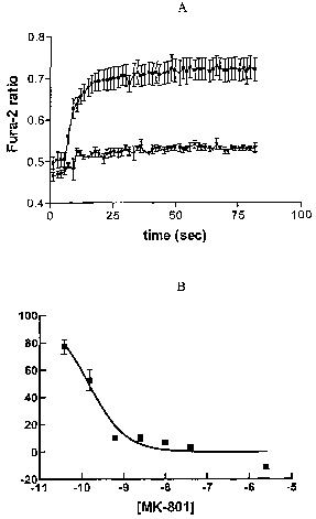
Figure 1. A: NMDA receptor-mediated functional intracellular calcium response.
Filled square symbols represent the control, filled triangle symbols represent
100~,M
MK-80-l; B: [3H]-MK-801 binding in differentiated P19 neurons.
Figure 2. A: P19 neuron viability experiment using Alamar Blue fluorescence
measurements. B: Glutamate dose-response of death with alamar blue readings.
Data
presented as % control. C: MK-801 dose-dependent block.
Figure 3. A: Compound A, a p38 inhibitor, pretreatment dose response. B:
U0126, a
MEKl/2 inhibitor, pretreatment dose response.
F~. A: U0126 does not block glutamate-induced calcium responses. B: U0126
does not block [3H]-MK-801 binding in P19 neurons.
Figure 5. A: U0126 post treatment time course of efficacy. B: Compound A post
treatment time course of efficacy.
Fi ug re 6. A: U0126 does not inhibit staurosporine-induced toxicity. Filled
square
symbols represent no compound; filled triangle symbols represent 10~,M U0126.
B:
U0126 does not block A23187-induced toxicity. C: U0126 does not affect basal
P19
neuron viability.
6
CA 02418912 2003-02-07
WO 02/11724 PCT/USO1/41565
Figure 7. 2-pyridinamine and 4-pyrimidinamine compounds (listed by compound
number) exhibit post-treatment delayed neuroprotection. Efficacy that is
achieved at 2
hours post-glutamate treatment is equivalent to what is achieved when P19
neurons are
pretreated with these compounds. This temporal profile matches that of the MEK
inhibitor, U0126. Open, dotted bars represent pre treatment % NP; filled bars
represent
%NP 2 hours post treatment.
Detailed Description of the Invention
This invention provides a pharmaceutical composition comprising a
pharmaceutically acceptable carrier and a compound having the formula
R Rs
! ~ N R2 , I R4
Rs
R5 R2
or a pharmaceutically acceptable salt thereof, wherein
(a) Rl is H or a substituent bound at either the 5 or 6 ring position and
selected from
the group consisting of alkyl, alkenyl, alkynyl, thienyl, furanyl, pyrrolyl,
phenyl, pyrimidinyl, substituted pyrimidinyl, pyridinyl, substituted
pyridinyl,
phenyl alkenyl, substituted phenyl alkenyl, benzo[b]thien-2-yl, 2-benzofuranyl
and substituted phenyl,
said substituted phenyl having the formula
R6
wherein (i) R6 is selected from the group consisting of H, OH, halogen,
alkylamino, dialkylamino, hydroxy-substituted dialkyl amino, lower alkyl,
acidic lower alkyl, alkoxy, halogen-substituted lower alkoxy, phenyl and
morpholinyl, and (ii) R~ represents between one and four substituents which
may be the same or different and are selected from the group consisting of H,
halogen, amino, alkyl, lower alkyl, halogen-substituted lower alkyl,
alkylamino,
dialkylamino, acidic lower alkoxy, alkoxy, halogen-substituted lower alkoxy,
7
CA 02418912 2003-02-07
WO 02/11724 PCT/USO1/41565
alkoxy and phenylalkoxy, with the proviso that R6 and R, may be fused to form
2-naphthyl or 1,3, benzodioxolyl;
(b) Each RZ is independently H or lower alkyl;
(c) Each R3 is independently selected from the group consisting of H, lower
alkyl,
amino, alkylamino, dialkylamino and lower alkoxy;
(d) R4 is H, alkoxy or morpholinyl, with the proviso that R4 may be fused with
R3 to
form 2,3-dihydro-1,4-benzodioxinyl or 9-alkyl 9H carbazolyl; and
(e) RS is H or lower alkyl:
In one embodiment of the instant pharmaceutical composition, R, is a
substituted phenyl at the 5 ring position, and each RZ is H. In another
embodiment, R4
is morpholinyl. In a fiuther embodiment, each R3 is a lower alkoxy and R4 is a
lower
alkoxy. In still a further embodiment, R, is at the 6 ring position, each RZ
is H, and
preferably, each R3 and R4 are lower allcoxy.
In the preferred embodiment of the instant pharmaceutical composition, the
compound contained therein is selected from the following group, whose
structures are
set forth in the Experimental Details:
5-(3-ethoxyphenyl)-N-(3,4,5-trimethoxyphenyl)-2-pyridinamine;
N-[4-(4-morpholinyl)phenyl]-5-(2-naphthyl)-2-pyridinamine;
5-benzo[b]thien-2-yl-N-[4-(4-morpholinyl)phenyl]-2-pyridinamine;
5-[3,5-bis(trifluoromethyl)phenyl]-N-[4-(4-morpholinyl)phenyl]-2-
pyridinamine;
5-[4-(4-morpholinyl)phenyl]-N-[4-(pentyloxy)phenyl]-2-pyridinamine;
5-[4-(dimethylamino)phenyl]-N-[4-(pentyloxy)phenyl]-2-pyridinamine;
5-[4-(dimethylamino)phenyl]-N-(4-methoxyphenyl)-2-pyridinamine;
5-(1,3-benzodioxol-5-yl)-N-[4-(pentyloxy)phenyl]-2-pyridinamine;
4-[6-[[4-(pentyloxy)phenyl]amino]-3-pyridinyl]-benzenepropanoic acid;
S-(2-methoxyphenyl)-N-[4-(pentyloxy)phenyl]-2-pyridinamine;
N-(2,3-dihydro-1,4-benzodioxin-6-yl)-5-[(E)-2-phenylethenyl]-2-pyridinamine;
N-[6-[3-(dimethylamino)phenyl]-2-pyridinyl]-9-ethyl-9H-carbazol-3-amine;
6-(3-ethoxyphenyl)-N-(3,4,5-trimethoxyphenyl)-2-pyridinamine;
8
CA 02418912 2003-02-07
WO 02/11724 PCT/USO1/41565
6-[3-(trifluoromethoxy)phenyl]-N-(3,4,5-trimethoxyphenyl)-2-pyridinamine; .
6-(1,3-benzodioxol-5-yl)-N-(3,4,5-trimethoxyphenyl)-2-pyridinamine;
6-phenyl-N-(3,4,5-trimethoxyphenyl)-2-pyridinamine;
6-(3,4-dimethoxyphenyl)-N-(3,4,5-trimethoxyphenyl)-2-pyridinamine;
6-(3,4-dimethylphenyl)-N-(3,4,5-trimethoxyphenyl)-2-pyridinamine;
N-(4,5-dimethoxy-2-methylphenyl)-6-(3,4-dimethylphenyl)-2-pyridinamine;
6-(2-naphthyl)-N-(3,4,5-trimethoxyphenyl)-2-pyridinamine;
6-(2-phenoxyphenyl)-N-(3,4,5-trimethoxyphenyl)-2-pyridinamine; and
6-[(E)-2-phenylethenyl]-N-(3,4,5-trimethoxyphenyl)-2-pyridinamine.
Unless specified otherwise, as used herein, the term "alkyl" refers to a
saturated
straight, branched or cyclic substituent consisting solely of carbon and H.
Lower alkyl
refers to an alkyl containing 1 to 4 carbon atoms. The term "alkenyl" refers
to an
unsaturated straight, branched or cyclic substituent consisting solely of
carbon and H
1 S that contains at least one double bond. The term "alkynyl" refers to an
unsaturated
straight, branched or cyclic substituent consisting solely of carbon and H
that contains
at least one triple bond.
The term "alkoxy" refers to O-alkyl where alkyl is as defined. The term
"alkylthio" refers to S-alkyl where alkyl is as defined. An acidic alkyl is a
carbon chain
with a terminal COOH group.
As used herein, the term "alkylamino" shall mean an allcyl substituted amine
group. Similarly, the term "dialkylamino" shall mean an amino group
substituted with
two independently selected alkyl groups. The term "hydroxy substituted
dialkylamino"
shall refer to a dialkylamino group wherein either or both of the alkyl groups
are
independently substituted with a hydroxy group, independently at any of the
carbon
atoms of the alkyl group(s).
The term "halo" means fluoro, chloro, bromo and iodo. The symbol "Ph" or
"PH" refers to phenyl.
When a particular group is "substituted" (e.g., aryl, heterocycloalkyl,
heteroaryl, and the like), that group may have one or more substituents,
preferably from
one to five substituents, more preferably from one to three substituents, most
preferably
from one to two substituents, independently selected from the list of
substituents.
9
CA 02418912 2003-02-07
WO 02/11724 PCT/USO1/41565
With reference to substituents, the term "independently" means that when more
than one of such substituents is possible, such substituents may be the same
or different
from each other.
For the purposes of this invention, the pyridine ring system shall have the
following numbering.
6
R1 S ~N 1
/ 2
3 NH
The compounds contained in the instant pharmaceutical compositions are
exemplified in the Experimental Details section below. These compounds are
commercially available from BioFocus PCC (UK) as part of a chemical library.
Alternatively, the compounds exemplified below can be prepared using known
methods.
As used herein, the phrase "pharmaceutically acceptable salt" means a salt of
the
free base which possesses the desired pharmacological activity of the free
base and
which is neither biologically nor otherwise undesirable. These salts may be
derived
from inorganic or organic acids. Examples of inorganic acids are hydrochloric
acid,
nitric acid, hydrobromic acid, sulfuric acid, and phosphoric acid. Examples of
organic
acids are acetic acid, propionic acid, glycolic acid, lactic acid, pyruvic
acid, malonic
acid, succinic acid, malic acid, malefic acid, fumaric acid, tartaric acid,
citric acid,
benzoic acid, cinnamic acid, mandelic acid, methanesulfonic acid,
ethanesulfonic acid,
p-toluenesulfonic acid, methyl sulfonic acid, salicyclic acid and the like.
The instant pharmaceutical composition can be prepared according to
conventional pharmaceutical techniques. The pharmaceutically acceptable
carrier
therein may take a wide variety of forms depending on the form of preparation
desired
for administration, such as systemic administration, including but not limited
to
intravenous, oral, nasal or parenteral. In preparing the compositions in oral
dosage
form, any of the usual pharmaceutical carriers may be employed, such as water,
glycols,
oils, alcohols, flavoring agents, preservatives, coloring agents, syrup and
the like in the
case of oral liquid preparations (for example, suspensions, elixirs and
solutions), or
CA 02418912 2003-02-07
WO 02/11724 PCT/USO1/41565
Garners such as starches, sugars, diluents, granulating agents, lubricants,
binders,
disintegrating agents and the like in the case of oral solid preparations (for
example,
powders, capsules and tablets).
Because of their ease of administration, tablets and capsules represent an
advantageous oral dosage unit form, wherein solid pharmaceutical Garners are
employed. If desired, tablets may be sugar-coated or enteric-coated by
standard
techniques. For parenterals, the carrier will usually comprise sterile water,
though other
ingredients for solubility or preservative purposes may also be included.
Injectable
suspensions may also be prepared, wherein appropriate liquid Garners,
suspending
agents and the like may be employed. The compounds may also be administered in
the
form of an aerosol.
This invention also provides a method of reducing the likelihood of a cell's
undergoing ischemic death comprising contacting the cell with a
prophylactically
effective amount of the compound contained in the instant pharmaceutical
composition.
As used herein, the term "ischemic death", when refernng to a cell, means
death
caused by a lack of oxygen. Ischemic cell death can result, for example, from
hypoxic
conditions. In vivo or ex vivo ischemia of cells or entire tissues can result
from, among
other things, localized anemia due to interference of the blood supply caused
by blood
vessel obstruction, destruction or constriction. Ischemic death and its
morphologic
characteristics are well known and identifiable to those with ordinary skill
in the art.
As used herein, a "prophylactically effective amount" of the instant
pharmaceutical composition, or compound therein, means an amount that reduces
the
incidence of cell death in a population of cells. The instant pharmaceutical
composition
will generally contain a per dosage unit (e.g., tablet, capsule, powder,
injection,
teaspoonful and the like) from about 0.001 to about 100 mg/kg. In one
embodiment,
the instant pharmaceutical composition contains a per dosage unit of from
about 0.01 to
about 50 mg/kg of compound, and preferably from about 0.05 to about 20 mglkg.
Methods are known in the art for determining prophylactically effective doses
for the
instant pharmaceutical composition. The effective dose for administering the
11
CA 02418912 2003-02-07
WO 02/11724 PCT/USO1/41565
pharmaceutical composition to a human, for example, can be determined
mathematically from the results of animal studies.
A "cell population" as used herein refers to cells in vitro such as in a
culture
vessel or in vivo as part of a body fluid or as an intact tissue or organ. The
cell
population can be homogenous (comprising of one cell type) or heterogenous
(comprising a mixed cell type population). Preferred cell populations are
heterogenous
cell populations that comprise at least one cell type that has been identified
as being
protected from ischaemic death in the presence of the compounds of this
invention.
In one embodiment of the instant method, the cells making up the cell
populations are preferably mammalian cells and more preferably human cells.
The
cells that make up a cell population that demonstrates reduced ischaemic
injurly in
response to a traumatic invent include, but are not limited to, cell
populations
comprising at least one cell selected from the group consisting of a neuronal
cell, a glial
cell, a cardiac cell, a lymphocyte, a macrophage and a fibroblast. In the
preferred
embodiment, the cell is a neuronal cell.
This invention also provides a method of reducing neuronal cell death in
response to a traumatic event comprising contacting the neuronal cell with a
prophylactically effective amount of the compound contained in the instant
pharmaceutical composition prior to, during, or within a suitable time period
following
the traumatic event.
Both these instant methods can be performed in vitro, ex vivo, or i~c vivo. As
used herein, contacting a cell with an agent "in vitro" includes, by way of
example,
contacting such agent with a cell that is in a single cell culture, a mixed
cell culture or a
primary cell tissue culture. Contacting a cell with an agent "ex vivo"
includes, by way
of example, contacting such agent with a cell that is part of an organized
tissue or organ
maintained outside the body of the subject from which it originates.
Contacting a cell
with an agent "in vivo" means contacting such agent with a cell present within
a subject.
12
CA 02418912 2003-02-07
WO 02/11724 PCT/USO1/41565
This invention fiuther provides a method for reducing neuronal cell death in
response to a traumatic event in a subject, comprising administering to the
subject a
prophylactically effective amount of the instant pharmaceutical composition
prior to,
during, or within a suitable time period following the traumatic event. The
term
"subject" includes, without limitation, any animal or artificially modified
animal. In the
preferred embodiment, the subject is a human.
The route of administering the instant pharmaceutical composition to a subject
is
preferably systemic, including, for example, intravenous (iv), subcutaneous
(sc) and oral
administration. In one embodiment, the instant composition is administrated
directly to
the nervous system. This administration route includes, but is not limited to,
the
intracerebral, intraventricular, intracerebroventricular, intrathecal,
intracisternal,
intraspinal and/or peri-spinal routes of administration, which can employ
intracranial
and intravertebral needles, and catheters with or without pump devices.
Infusion doses
can range, for example, from about 1.0 to 1.0 x 10⁴ p,g/kg/min of instant
compound,
over a period ranging from several minutes to several days. For topical
administration,
the instant compound can be mixed with a pharmaceutical Garner at a
concentration of,
for example, about 0.1 to about 10% of drug to vehicle.
In the instant method, the neuronal cell death-causing traumatic event
includes,
for example, a medical disorder, a physical trauma, a chemical trauma and a
biological
trauma.
Examples of neuronal cell death-causing medical disorders include perinatal
hypoxic-ischemic injury, cardiac arrest, stroke/ischemic attack, hypoglycemia-
induced
neuropathy, cardiac surgery-induced cerebral ischemia, post traumatic stress
disorder,
stress-induced memory impairment, chronic epilepsy, multiple sclerosis,
Parkinson's
disease, diabetic peripheral neuropathy, neuropathic pain, Bells' palsy, sick
sinus
syndrome, Alzheimer's disease, Pick's disease, diffuse Lewy body disease,
Cruzfeld's
Jacobs and other diseases of protein aggregation, progressive supranuclear
palsy (Steel-
Richardson syndrome), multisystem degeneration (Shy-Drager syndrome),
amyotrophic
lateral sclerosis (ALS), degenerative ataxias, cortical basal degeneration,
ALS-
Parkinson's-Dementia complex of Guam, subacute sclerosing panencephalitis,
13
CA 02418912 2003-02-07
WO 02/11724 PCT/USO1/41565
Huntington's disease, synucleinopathies (including multiple system atrophy),
primary
progressive aphasia, striatonigral degeneration, Machado-Joseph disease,
spinocerebellar ataxia type 3, olivopontocerebellar degenerations, Gilles De
La
Tourette's disease, bulbar and pseudobulbar palsy, spinal and spinobulbar
muscular
atrophy (Kennedy's disease), primary lateral sclerosis, familial spastic
paraplegia,
Werdnig-Hoffinann disease, Kugelberg-Welander disease, Tay-Sach's disease,
Sandhoff disease, familial spastic disease, neuroleptic malignant syndrome,
Wohlfart-
Kugelberg-Welander disease, spastic paxaparesis, progressive multifocal
leukoencephalopathy, AIDS-related dementia, sick sinus syndrome, post herpetic
neuropathy, viral meningitis, bacterial meningitis, prion diseases, and
familial
dysautonomia (Riley-Day syndrome).
Neuronal cell death-causing physical traumas include, for example, focal brain
trauma, diffuse brain trauma, spinal cord injury, cerebral infarction, embolic
occlusion,
thrombotic occlusion, reperfusion, intracranial hemorrhage, whiplash, shaken
infant
syndrome, and radiation-induced peripheral nerve damage.
Neuronal cell death-causing chemical traumas include, for example, exposure to
alcohol, chemotherapeutic agents, war gas, lead, cyanoacrylate,
polyacrylamide, and
toxic inhalants. Finally, neuronal cell death-causing biological traumas
include, for
example, exposure to HIV, herpes virus, and meningitis-causing bacteria and
viruses.
In practicing the instant method, the pharmaceutical composition can be
administered to the subject prior to, during or subsequent to the traumatic
event. As
used herein the term "subsequent" refers to any point in time beginning with
the
traumatic event and continuing until the potential of cell death resulting
from the
traumatic event has diminished.
Finally, this invention provides an apparatus for administering to a subject
any
of the instant pharmaceutical composition comprising a container and the
pharmaceutical composition therein, wherein the container has a device for
delivering
to the subject a prophylactic dose of the pharmaceutical composition. In the
preferred
embodiment, the device for delivering the pharmaceutical composition is a
syringe.
14
CA 02418912 2003-02-07
WO 02/11724 PCT/USO1/41565
Ideally, the instant apparatus is a single-use, predosed auto-injectable
device containing
the instant composition. Such a device would be useful, for example, in a
mobile
ambulatory unit or for administration to a person at risk for a neurotoxic
event.
Mechanical auto-injectable devices are well known in the art and are
exemplified, for
example, by an EpiPen~ device (Meridian Medical Technologies Inc.), which is
an
auto-injectable device containing epinephrin for individuals subject to
anaphalactic
shock.
This invention will be better understood by reference to the Experimental
Details
that follow, but those skilled in the art will readily appreciate that these
are only
illustrative of the invention as described more fully in the claims which
follow thereafter.
Additionally, throughout this application, various publications are cited. The
disclosure of
these publications is hereby incorporated by reference into this application
to describe
more fully the state of the art to which this invention pertains.
Experimental Details
Example 1
Commerciall Available 2-P ~idinamines
~-CHs 2-pyridinamine, 5-(3-ethoxyphenyl)-
N (3,4,5-trimethoxyphenyl)-
cH3 (Cmpd 12)
H3C H . O
CH3
p 2-pyridinamine, N [4-(4-
\ ~ morpholinyl)phenyl]-5-(2-naphthyl)-
N ~ (Cmpd 25)
I,
N
H
- 2-pyridinamine, 5-benzo[b]thien-2-
\ ~ S ~C yl-N [4-(4-morpholinyl)phenyl]-
~ N , I N J (Cmpd 22)
I~
N
H
CF3 2-pyridinamine, 5-[3,5-
bis(trifluoromethyl)phenyl]-N [4-(4-
morpholinyl)phenyl]-
F3C ~ ~ N i I NJ (Cmpd 21)
I
N
H
CA 02418912 2003-02-07
WO 02/11724 PCT/USO1/41565
p 2-pyridinamine, S-[4-(4-
morpholinyl)phenyl]-N [4-
~ ~ p~ (pentyloxy)phenyl]-
(Cmpd 6) .
N
H
I 2-pyridinamine, 5-[4
~N i (dimethylamino)phenyl]-N [4-
~ N ~ o~ (pentyloxy)phenyl]-
(Cmpd 7)
N
H
2-pyridinamine, 5-[4-
/ (dimethylamino)phenyl]-N (4-
p methoxyphenyl)-
\ N \ I ~ (Cmpd 8)
N
H
p i 2-pyridinamine, 5-(1,3-benzodioxol-
p~ S-yl)-N [4-(pentyloxy)phenyl]-
(Cmpd 13)
N
H
benzenepropanoic acid, 4-[6-[[4-
Ho ~ I (pentyloxy)phenyl]amino]-3-
w o~ pyridinyl]_
(Cmpd 10)
N
H
2-pyridinamine, 5-(2-
i ~ methoxyphenyl)-N [4-
(pentyloxy)phenyl]-
(Cmpd 14)
N
H
2-pyridinamine, N (2,3-dihydro-1,4
p benzodioxin-6-yl)-5-[(~-2-
N ~ ~ ~ phenylethenyl]-
I N ~ p (Cmpd 88)
H
H 9H carbazol-3-amine, N [6-[3-
~ N , N\ N I \ \ / (dimethylamino)phenyl]-2-
pyridinyl]-9-ethyl-
I I ~ ~ N (Cmpd 104)
16
CA 02418912 2003-02-07
WO 02/11724 PCT/USO1/41565
2-pyridinamine, 6-(3-ethoxyphenyl)-
w N N ' O N (3,4,5-trimethoxyphenyl)-
(Cmpd 114)
J
i 2-pyridinamine, 6-[3-
O ~ I N N ~ O~ (~fluoromethoxy)phenyl]-N (3,4,5-
trimethoxyphenyl)-
F-f -F w I I i Oi (Cmpd 11 ~)
.F
O~
/-O 2-pyridinamine, 6-(1,3-benzodioxol-
O , 5-yl)-N (3,4,5-trimethoxyphenyl)-
N N O (Cmpd 120)
i i
'O
2-pyridinamine, 6-phenyl-N (3,4,5-
N I N I ~ O~ trimethoxyphenyl)-
(Cmpd 122)
2-pyridinamine, 6-(3,4-
0 ~ dimethoxyphenyl)-N (3,4,5-
w w ~ N N O trimethoxyphenyl)-
O ' ~ I ~ ~ (Cmpd 124)
i I H 2-pyridinamine, 6-(3,4-
N N O dimethylphenyl)-N (3,4,5-
trimethoxyphenyl)-
(Cmpd 126)
i I H 2-pyridinamine, N (4,5-dimethoxy-
N N O 2-methylphenyl)-6-(3,4-
dimethylphenyl)-
Cm d 127
o ( p )
i i I H 2-pyridinamine, 6-(2-naphthyl)-N
w w N N O (3,4,5-trimethoxyphenyl)-
(Cmpd135)
17
CA 02418912 2003-02-07
WO 02/11724 PCT/USO1/41565
i H 2-pyridinamine, 6-(2-
I N N ~ O~ phenoxyphenyl)-N (3,4,5-
trimethoxyphenyl)- (Cmpd 138)
w I I ~ Oi
i
I H 2-pyridinamine, 6-[(E~-2-
w , ~N N ~ O~ phenylethenyl]-N (3,4,5-
trimethoxyphenyl)- (Cmpd144)
Ii
Example 2
Characterization of Differentiated P19 Cells
Pl9 Cell Differentiation
P19 cells are a pluripotent embryonal carcinoma line that can be induced to
differentiate relatively rapidly into post-mitotic neurons in the presence of
high dose
retinoic acid (Jones-Velleneuve et al. 1982; Jones-Villeneuve et al. 1983;
McBurney
and Rogers 1982). They are the marine equivalent of human NT-2N neurons, which
are also derived from retinoic acid differentiation of teratocarcinoma
precursor cells.
Differentiated NT-2N neurons, perhaps the better known of the two
teratocarcinoma-
derived neuronal lines, express a wide variety of neuronal markers, and
undergo
NMDA receptor-mediated, hypoxia-induced excitotoxic cell death (Pleasure and
Lee
1993; Pleasure, Page, and Lee 1992; Rootwelt et al. 1998). Like NT-2Ns,
differentiated
P19 neurons also express a wide variety of neuronal markers, exhibit NMDA
receptor-
mediated intracellular calcium responses to agonists, and undergo
excitotoxicity
(Canzoniero et al. 1996; Grobin et al. 1999; Morley et al. 1995; Ray and
Gottlieb 1993;
Turetsky et al. 1993).
P19 cells were bought from ATCC (Manassas, VA). They were grown on 150
cmz tissue culture flasks in Dulbecco's Modified Eagle Medium (DMEM, Gibco
BRL)
supplemented with 10% fetal bovine serum, glutamine (2 mM), sodium pyruvate (1
mM), sodium bicarbonate (0.15% w/v), and penicillin/streptomycin (50 units/mL)
in an
atmosphere of 5% COZ at 37°C.
On day 1 of the differentiation protocol, confluent P19 cells were split to 50-
70% confluency in growth medium. On day 2 of the protocol, 10 ~M all-trans-
retinoic
acid (ATRA, Sigma) and 10 ~.M MK-801 were added to the growth medium. 10 ~,M
MK-801 was included at this stage to prevent cell death in differentiating
neurons that
18
CA 02418912 2003-02-07
WO 02/11724 PCT/USO1/41565
begin to express NMDA receptors. On day 4, fresh growth medium was placed on
the
cells, with fresh ATRA and MK-801. On day 5, cells were dissociated from the
tissue
culture flask by washing 4 times with calcium and magnesium-free phosphate-
buffered
saline, and adding 4 mL of non-enzymatic cell dissociation solution (Sigma).
Once dissociated, cells were placed in 40 mLs of differentiation medium.
Differentiation medium consisted of Neurobasal medium (Gibco BRL) supplemented
with 1 % N-2 supplement (Gibco BRL), 0.1 % trace elements B (Mediatech), 1 mM
cadmium sulfate (Sigma), 2 mM glutamine, sodium pyruvate (1 mM), sodium
bicarbonate (0.15% w/v), and 1% antibiotic/antimycotic (Gibco BRL). 10 ~,M
cytosine-D-arabinofuranoside was added to the differentiation medium to
prevent
growth of undifferentiated cells. No MIA-801 was present from this point
onward.
Cells were triturated 20 times, then were split 1:4 into 96 well plates, or
split 1:3 into
100 mm tissue culture dishes. Four days after replating, the cells were
optimal for
compound addition, and were assayed 24 hours later.
RT PCR
Total RNA was isolated from 100 mm tissue culture dishes of differentiated P
19
neurons or undifferentiated P19 cells using the QIAGEN RNeasy Mini kit
according to
manufacturer's protocols. RT-PCR amplification of marine NMDA receptor
subunits
was obtained from 250 ng total RNA template isolated from undifferentiated P
19 cells,
cells at 4 days after retinoic acid (ATR.A) induction, and cells at 9 days
after ATRA
induction. One-step RT-PCR reactions were set up using the LightCyclerTM-RNA
Amplification kit SYBR Green I kit (Boehringer Mannheim), according to
manufacturer's protocols. Real time RT-PCR reactions were carned out in
LightCyclerTM glass capillaries using the LightCycler~ instrument and 250 ng
template
RNA (Boehringer Mannheim). The reverse transcriptase reaction was carried out
for
10 min at 55°C. PCR was carried out for 30 cycles: annealing
temperature was 50°C,
extension temperature was 72°C, and melting temperature was
80°C. Reactions were
compared to an HZO-negative control for each primer set. 5 p,L of reaction
product
were removed, and run on lx TBE agarose gels. The primer sets for the various
mouse
NMDA receptor subunits used include zeta l and epsilons 1-4.
19
CA 02418912 2003-02-07
WO 02/11724 PCT/USO1/41565
RNA samples from 4-day and 9-day post retinoic acid treatmnet,
undifferentiated smaples and control sampes were separated by electrophoresis
and
probed with zetal, epslon 1 and epsilon 2 internal primers
RT-PCR of NMDA receptor subunits from total RNA samples revealed that
retinoic acid induction of differentiation also induces mRNA expression of
zetal,
epsilonl, and epsilon2 mRNAs. Data was summarized using 3 separate
experiments.
Western Blots
Media was aspirated from cells plated onto 100 mm tissue culture dishes. Cells
were harvested in RIPA lysis buffer (100mM Tris HCL pH 7.5, 150 mM NaCI, 1 mM
EDTA, 1 % Triton X-100, 10% sodium deoxycholate, 0.1 % sodium dodecyl
sulfate).
Each sample was sonicated for 20 seconds, Laemmli sample buffer (BioRad) was
added to a final lx concentration, and samples were incubated for 10 minutes
at 95°C.
Samples to be probed with NMDA receptor antibodies (polyclonals against rat
NRl,
NR2A, and NR2B obtained from Chemicon) were electrophoresed on 6% tris-glycine
pre-cast gels (NOVEX). Samples to be probed with p42/44 MAP kinase antibodies
(New England BioLabs) were electrophoresed on 12% tris-glycine pre=cast gels
(NOVEX). Electrophoresis was carried out in a NOVEX apparatus for 1.5 hours at
200
volts. Proteins were transferred to polyvinylidene difluoride membrane (PVDF,
NOVEX) using a BioRad wet transfer device for 1 hour at 100 volts. Prior to
transfer,
PVDF membranes were dipped in 100% methanol for 1 minute, then soaked in
transfer
buffer for 5 minutes. After transfer, membranes were removed and were slowly
shaken
in blocking solution (5% milk, 0.05% tween-20 in phosphate buffered saline) at
4°C
overnight. Membranes were then washed once with PBS-tween, and primary
antibodies 1:1000 in PBS-tween with 5 % milk were incubated for 1 hour at room
temperature. Membranes were washed 4x for 15 minutes at room temperature.
Secondary antibodies coupled to horseradish peroxidase were incubated for
about 45
minutes at room temperature in PBS-tween with 5 % milk. Membranes were then
washed 4x for 15 minutes at room temperature. Blots were developed using ECL
plus
(Amersham), and exposed to film.
Western blot analysis was performed for NMDA receptor subunit protein in P 19
cell lysates harvested following 4-days or 9-days of retinoic acid exposure.
Appropriately sized bands (Zetal = ~ 120 kDa, Epsilonl and Epsilon 2 = ~ 180
kDa)
CA 02418912 2003-02-07
WO 02/11724 PCT/USO1/41565
were detectable from terminally differentiated P19 neuron lysates, but not
from
undifferentiated cell lysates. The data was representative of 3 separate
experiments with
similar results.
Western blot analysis revealed that terminally differentiated P 19 neurons
express detectable levels of zetal, epsilonl, and epsilon 2 proteins. Epsilon3
and
epsilon4 mRNAs or proteins were not detectable at any time in these cells.
These data demonstrate that such methods of differentiating P19 cells into
neurons successfully result in the functional expression of NMDA receptors at
the
levels of mRNA, protein, MK-801 sensitive agonist-induced intracellular
calcium
responses, and MK-801 sensitive glutamate toxicity. These data are in very
good
agreement with the literature. Interestingly, however, in addition to zetal
and epsilon2
expression, reliable expression of the epsilonl subunit of the NMDA receptor
was also
achieved, which expression has not yet been achieved with other reported
methods of
P19 cell differentiation (Ray and Gottlieb 1993). The presence of all three
subunits
1 S more closely resembles expression patterns observed in adult rodent
forebrain regions,
including cortex and hippocampus (Ishii et al. 1993; Laurie et al. 1995;
Monyer et al.
1994; Monyer et al. 1992; Standaert et al. 1994).
Example 3
Differentiated P19 Excitotoxicity Assay
Cells were loaded with 5 ~M Fura-2-AM (Molecular Probes) for 1 hr at
37°C.
They were washed once with Hank's balanced salt solution (HBSS, Gibco BRL),
and
assayed in HBSS buffer. Cells were placed onto the stage of a modified
ATTOFLUORT"" Imager (Alto Instruments, Rockville Pike, MD). High speed, dual
excitation of fore-2 was carried out using a RATIOARCT"" High-Speed Excitor
(Atto
Instruments). Mercury lamp light was passed through 334 nm or 380 nm bandpass
filters (10 nm band width), and then passed through a 20x objective (Zeiss,
Plan-
Apochromat, NA=0.75) at a rate of 2.5 Hz. Emitted light was transmitted
through a
400 nm dichroic mirror, and collected to an ATTOFLUORT"~ intensified CCD
camera.
Ratio-images were acquired, and the average intensity of the images when
excited at
334nm and 380nm was analyzed using ATTOFLUOR RATIOVISIONT"" software (Atto
Instruments, Rockville, MD).
21
CA 02418912 2003-02-07
WO 02/11724 PCT/USO1/41565
Changes of the Fura-2 330nm/380nm intensity ratio were plotted when 9 days
post-ATRA P19 neurons were treated with 3 mM glutamate, 1 mM glycine in the
presence or absence of 100 p,M MK-801. MIA-801 was administered 24 hours prior
to
assay. Traces are the average ~ standard error from three separate experiments
for each
condition. Terminally differentiated P19 neurons expressed NMDA receptor
subunits
that form functional NMDA receptors, as illustrated by using fura-2 imaging to
detect
increases in intracellular calcium levels upon addition of NMDA receptor
agonists
(Figure 1A). The selective NMDA receptor channel blocker, MK-801,
significantly
inhibited this response (Figure 1A). In addition P19 neurons expressed
specific binding
sites for the NMDA receptor. MK-801 inhibition of [3H~-MK-801 binding was
assayed
in P19 membranes. MK-801 concentrations are expressed as 10'" Molar. Data
points
represent the mean ~ standard error from eight experiments. % control = ((CPM -
CPMbo~o"~~(CPM,op CPMbo~o"~) X 100. (Figure 1B). MIA-801 inhibition was
concentration-dependent, and 100% inhibition was achieved.
Control P19 neuron cell bodies and processes stain brightly with the live cell
cytoplasmic dye carboxyfluorescein diacetate (CFDA). P19 neurons at 9 days
post
ATRA induction were labeled with CFDA, and then imaged in confocal mode using
an
Attofluor Imager. Control cells were treated with vehicle for 24 hours,
glutamate cells
received 3 mM glutamate in the presence of 1 mM glycine for 24 hours, and
glutamate
+ U0126 cells received 10 p,M U0126 concurrent with 3 mM glutamate and 1 mM
glycine for 24 hours. Images were taken from control alive, glutamate treated,
dead and
U0126-protected cells stained with fluorescein diacetate in 3 separate
experiments. P19
neuron cell bodies characteristically clumped together into tight aggregates
when plated
onto plain tissue culture plastic. Networks of extensive processes connected
clusters of
neuronal cell bodies. P 19 neurons treated with toxic concentrations of
glutamate and
glycine for 24 hours exhibited fluorescence staining in isolated cell bodies,
processes
were undetectable, and extensive cellular debris was evident. Relative levels
of cell
death were measured rapidly and quantitatively on a plate reader using the
live cell
fluorescent dye alamar blue (Figures 2B).
Alamar Blue fluorescence, an indicator of cell viability, was used to
determine
cell viability after an NMDA-induced cytotoxic insult. Counts from a single 96
well
plate where 32 wells received vehicle control, 32 wells received 3 mM
glutamate and 1
mM glycine for 24 hours, and 32 wells received S p.M A23187 for 24 hours are
shown
22
CA 02418912 2003-02-07
WO 02/11724 PCT/USO1/41565
in Figure 2A. Glutamate and A23187 conditions were significantly different
from
control as determined by one-way ANOVA with Tukey post-hoc analysis carried
out
using GRAPHPADT"" software. These data indicate a typical 60% reduction in
Alamar
blue fluorescence when cells were treated with glutamate and glycine. However,
since
raw fluorescence counts vary from experiment to experiment, Figures 2A, 2B,
and 2C
are expressed as a percent of control.
Glutamate toxicity dose response in the presence of a constant 1 mM glycine
concentration was measured in the differentiated P 19 neurons. The curve
generated
through the data points is the average of 6 separate dose response curves.
Data points
are represented as the percent of control cells ~ standard error. % control =
(([Glutamate]exp - [Glutamate]m~ ) / (COntrOhehicle - [Glutamate]",aX )) X
100. Glutamate
toxicity EC50 was calculated to be 8.1 p,M [lower 95% confidence interval =
3.5 ~,M;
upper 95% confidence interval =19 p.M]. These data demonstrate that 24 hours
of
glutamate treatment killed P 19 neurons dose-dependently in the presence of a
constant
dose of glycine (Figures 2A,2B). Glycine alone did not affect P19 neuron
viability.
The NMDA receptor blocker, MIA-801, dose-dependently protected against
glutamate toxicity in P19 neurons (Figure 2D). MK-801 dose-dependent
inhibition of 3
mM glutamate and 1 mM glycine-induced P19 neuron death. % Neuroprotection =
((Inhibitor in the presence of Glutamate", - [Glutamate]ma,~ / (COntr01"ehicle
-
[Glutamate]maX )) X 100. Data points are the average ~ standard error of three
separate
experiments. Maximal MK-801 protection achieved was close to control levels.
These data demonstrate that glutamate toxicity in P19 neurons requires NMDA
receptor activation, since the toxicity is completely blocked in the presence
of the
specific NMDA receptor antagonist MK-801. However, the data do not exclude the
possibility that AMPA or kainate receptors may also be activated by glutamate
and
contribute to the excitotoxicity. This possibility would be consistent with
data from
primary neuronal culture, which reports that intracellular calcium responses
to
glutamate agonists involve multiple components (Courtney, Lambent, and
Nicholls
1990). Such components include AMPA/kainate receptor activation, membrane
depolarization, voltage-gated calcium channel activation, relief of NMDA
receptor
magnesium block, and NMDA receptor activation. However, even with such a high
level of complexity, in many primary neuronal models, glutamate excitotoxicity
signals
23
CA 02418912 2003-02-07
WO 02/11724 PCT/USO1/41565
through the NMDA receptor and requires its activation to result in neuronal
death
(Tymianski et al. 1993), as is the case for P19 neuron model system.
These cells were used to measure the effects of various known kinase
inhibitors
(Compound Identification (ID) column in Table I) upon cytotoxicity-induced by
NMDA. First, P19 cells were differentiated as described in Example 2. Then
cells
were exposed to 3 mM of glutamate in the presence of 1 mM glycine.
Cell viability was determined. Compounds that prevented excitotoxicity were
determined by measuring the percentage of viable cells compared to
differentiated P19
cells that were not presented with a toxic insult (Table I).
Cell viability was measured using two methods. The first method was a
confocal, single-cell fluorescence imaging-based method using the live cell
dye
carboxy-fluorescein diacetate (CFDA). CFDA labels the cell bodies and
processes of
living cells. Therefore, live cells exhibit extensive CFDA labeling, whereas
dead cells
exhibit much less CFDA staining. CFDA fluorescence was measured using a
modified
Attofluor Imager device (Atto Instruments, Rockville Pike, MD). Cells were
labeled .
with 1 uM CFDA for 15 min in media, then placed onto the stage of the
Attofluor
microscope. The dye was excited by light from a mercury lamp source passed
through
a 488 nm bandpass filter of 10 nm band width, passed through a CARV real-time
confocal spinning disk module (Alto Instruments, Rockville, MD), and then
passed
through a 40x oil immersion objective (Zeiss Fluar, NA=1.3). Emitted light was
transmitted through a 495 nm dichroic mirror, collected to an Attofluor
intensified CCD
camera, and images were visualized using Attofluor RatioVision software (Atto
Instruments, Rockville, MD).
The second method for measuring cell viability was a plate reader method.
Cells plated into black 96 well plates (Packaxd viewplates) are loaded with 5
% Alamar
Blue dye (Biosource International). Alamar Blue is a dye that takes advantage
of
mitochondrial reductases to convert non-fluorescent resazurin to fluorescent
resorufin
(excitation 535 nm, emission 580 nm). Baseline fluorescence counts were read
at room
temperature in a Wallac plate reader immediately after addition of Alamar
blue.
Fluorescence counts of cell viability were taken the same way after 1 hour
incubation at
37°C. Fluorescence was expressed as a percent of control, untreated
cells after
subtraction of background fluorescence. Live/dead cells were confirmed
visually with
a light microscope.
24
CA 02418912 2003-02-07
WO 02/11724 PCT/USO1/41565
As used in Table I below, Compound A shall mean the compound have the
formula
H
N I-'~~
Table
I
Com arison
of Kinase
Inhibitor
Activi
to Neuro
rotective
Activi
IC50 for Maximal
Neuropro- Efficacy
Compound Enzyme Activity Potency tection for
ID target [lower- Neuropro
upper 95 tection
/
C.I.
Comp p38 kinaseInhibitor IC50~10 S g 6M >80%
ound nM
A [
'
U0126 MEKl/2 Inhibitor IC50~0.5 1.1 ~M >80%
pM
0.74-1.7
SB202474 p38 kinaseInactive Non >10 p,M >50%
a licable
SB203580 p38 kinaseInhibitor IC50~600 Ineffective<10%
nM
Lithium ~ Inhibitor Ki~0.5 Ineffective<10%
p,M
per
KN62 CAMKII Inhibitor Ki~900 Ineffective<10%
nM
PKC Inhibitor IC50~50
nM
CalphostinPKA Inhibitor IC50>50
~M
>1 p,M ~50%
C PKG Inhibitor IC50>25
p,M
p60ws' Inhibitor IC50>SO
~,M
LavendustinEGF I~ibitor ICS0~11
nM
A receptor ~bitor IC50~500 >10 p,M <25%
nM
60wsr
P~ Inhibitor ~~48 ~
CAlVB~II ~bitor Ki~30 pM
H-89 Casein ~bitor Ki~38 ~M >10 p,M <25 /o
kinase ~bitor Ki~31.7
I ~.M
PKC
CA 02418912 2003-02-07
WO 02/11724 PCT/USO1/41565
Kinase inhibition activities and potencies were derived from the literature
(Chijiwa et al. 1990; Favata et al. 1998; Henry et al. 1998; Inhorn and
Majerus 1987;
Kobayashi et al. 1989; Lee et al. 1994; Onoda et al. 1989; Tokumitsu et al.
1990).
ICSOs for neuroprotection are the mean of three separate curves with upper and
lower
95% confidence intervals (C.L) shown. Curves were fit, and confidence
intervals were
determined using GraphPad Prism software.
Example 4
Evaluation of a MEKl/2 Inhibitor in the Differentiated P19 Assay
U0126 (bis[amino[(2-aminophenyl)thio]methylene] Butanedinitrile) is reported
to be highly selective for MEKl/2 (Favata et al. 1998), a result that was
confirmed here.
The only other kinase found that U0126 inhibits is PKC-y, but the IC50 for
inhibition
of this enzyme was 60-fold higher that its published IC50 against wildtype
MEKl/2
(Tables II and III below).
The general procedure used to assay for kinase activity was as follows: a
kinase
reaction mix was prepared in 50 mM Tris-HCl pH=8, 10 mM MgCla, 0.1 mM Na3V04,
1 mM DTT, 10 ~M ATP, 0.25-1 ~,M biotinylated peptide substrate, 0.2-0.8
p.Curies per
well 33P-y-ATP [2000-3000 Ci/mmol]. Assay conditions varied slightly for each
protein kinase, for example, insulin receptor kinase requires 10 mM MnCl2 for
activity
and calmodulin-dependent protein kinase requires calmodulin and 10 mM CaClz.
Reaction rnix was dispensed into the wells of a streptavidin-coated Flashplate
and 1 ~,1
drug stock in 100% DMSO was added to a 100 ~,L reaction volume resulting in a
final
concentration of 1% DMSO in the reaction. Enzyme was diluted in 50 mM Tris-HCl
pH=8.0, 0.1 % BSA and added to each well. The reaction was incubated for one
hour
at 30°C in the presence of compound. After one hour the reaction mix
was aspirated
from the plate and the plate was washed with PBS containing 100 mM EDTA. The
plate was read on a scintillation counter to determine 33P-y-ATP incorporated
into the
immobilized peptide. Test compounds were assayed in duplicate at 8
concentrations
ranging from 100 ~,M to 10 pM in one order of magnitude steps. A maximum and
minimum signal for the assay was determined on each plate. The IC50 was
calculated
from the dose response curve of the percent inhibition of the maximum signal
in the
assay according to the formula [max signal - background/test compound signal-
background X (100)] _ % inhibition by graphing the percent inhibition against
the log
26
CA 02418912 2003-02-07
WO 02/11724 PCT/USO1/41565
concentration of test compound. Known inhibitor compounds appropriate for the
kinase being assayed werealso included on each plate. Results are provided in
Figure 3.
Definition and Source of Kinase Enzymes
VEGF-R (vascular endothelial growth factor receptor-2) is a fusion protein
containing a polyhistidine tag at the N-terminus followed by amino acids 786-
1343 of
the rat VEGF-R2 kinase domain. CDKl (cyclin dependent kinase 1) is isolated
from
insect cells expressing both the human CDKl catalytic subunit and its positive
regulatory subunit cyclin B. Insulin Receptor Kinase consists of residues 941-
1313 of
the cytoplasmic domain of the beta-subunit of the human insulin receptor.
Protein
Kinase A is the catalytic subunit of cAMP-dependent protein kinase-A purified
from
bovine heart. PKC (protein kinase-C) is the gamma or beta isoform of the human
protein produced in insect cells. Casein Kinase 1 is a truncation at amino
acid 318 of
the C-terminal portion of the rat CKl delta isoform produced in E. coli.
Casein Kinase
2 includes the alpha and beta subunits of the human CK2 protein produced in E.
coli.
Calinodulin Kinase (calinodulin-dependent protein kinase 2) is a truncated
version of
the alpha subunit of the rat protein produced in insect cells. Glycogen
Synthase
Kinase-3 is the beta isoform of the rabbit enzyme produced in E. coli. MAP
Kinase is
the rat ERK-2 isoform containing a polyhistidine tag at the N-terminus
produced in E.
coli and activated by phosphorylation with MEK1 prior to purification. EGFR
(epidermal growth factor receptor) is purified from human A431 cell membranes.
The
chart below shows selected kinases and their control inhibitors.
As used in Table III below, Compound A (Cmpd A) shall mean the compound
have the formula
H<
F
NH~
Table II
Selected Kinases and Their Control Inhibitors
27
CA 02418912 2003-02-07
WO 02/11724 PCT/USO1/41565
Kinase Control Inhibitor
CDKl Butyrolactone
EGFR AG-1478
Protein Kinase H89
A
PKC Staurosporine
Casein Kinase H89
1
Casein Kinase
2
Calinodulin KinaseStaurosporine
Insulin Kinase Staurosporine
Table III
Kinase
Selectivit~of
Compound
A and
U0126
Compound CDK1 EGF-R PK A PKC-y
ID (~,1V1] (~1VI) (w~ (N~~
Cmpd A >100 >100 >100 8.35
U0126 >100 >100 >100 29.5
Casein Casein Calmodulin Insulin
Compound
Kinase 1 Kinase 2 Kinase Receptor
ID
(~,lVn (~,1V>] (~,1VI) Kinase (~,lVn
Cmpd A 0.116 >100 >100 >100
U0126 >100 >100 >100 ND
IC50 values for kinase inhibition are the mean of at least two separate
curves,
and were determined using GraphPad curve fitting software.
To determine whether U0126 inhibits the MEKl/2 enzymes in P19 neurons, its
ability to block glutamate-induced phosphorylation of the MEKll2 substrate p42
MAPK (ERK2) was tested. Western blot were first probed using ATRA P 19 neuron
lysates 9 days post treatment with an antibody specific for the phosphorylated
form of
p42/44 (ERK1/2) and then stripped and reprobed with antibody that recognized
total
p42/44 (ERK 1/a). The data demonstrate that the concentrations of glutamate
and
glycine that were used to induce toxicity in P 19 neurons also induced a
rapid,
reproducible increase in p42 MAPK phosphorylation in these cells. In the
presence of
28
CA 02418912 2003-02-07
WO 02/11724 PCT/USO1/41565
U0126, phospho-p42 MAPK was reduced, although no change in the amount of total
p42 MAPK was evident. In another Western blot experiment, ATRA P19 neuron
lysates were probed 9 days post treatment with an antibody specific for the
phosphorylated form of p42/44 (ERK %a). The same blot was stripped and
reprobed
with an antibody that recognized total p42/44 (ERK %z). Glutamate-induced
elevation
of p42 MAPK phosphorylation was sustained, since it was still clearly
increased at 24 h
versus controls. These blots are representative of 3 separate experiments with
similar
results. U0126 blocked the increased levels of phosphorylation at this time
point as
well. No change in total p42 MAPK was evident.
To ensure that U0126 does not block the NMDA receptor directly, P19 neuron
intracellular calcium responses to glutamate and glycine were tested in the
presence of
10 ~,M U0126. Fura-2 imaging traces 9 days post ATRA P19 neurons were treated
with 3 mM glutamate, 1 mM glycine in the presence of 10 p.M U0126 for f4
hours.
U0126 was administered concurrently with glutamate and glycine. Traces are the
average ~ standard error from four separate experiments for each condition.
The data demonstrated that U0126 did not block this intracellular calcium
response. (Figure 4A). It was also demonstrated that U0126 did not inhibit
[3H]-MK-
801 binding in P19 neurons. No significant inhibition was observed at any
concentration of U0126. Data points represent the mean ~- standard error from
eight
experiments. Percent control is defined as described in Figure 1B (Figure 4B).
Together, these data further demonstrate that U0126 acts on a signaling
pathway
downstream of NMDA receptor activation.
The p42/44 MAPK inhibitor, U0126, exhibits delayed heuroprotectioh
Time-course of efficacy is an extremely relevant parameter for a potential
neuroprotective therapeutic agent, since the therapeutic would need to be
administered
hours to days after an ischemic event and still retain efficacy. The next set
of
experiments examined whether the MEKl/2 inhibitor, U0126, retains
neuroprotective
efficacy when added at various time points after glutamate challenge. Assays
were
performed essentially as described, although the time of administration of the
U0126
was delayed relative to the initial excitotoxicity. The data demonstrated that
U0126 was
maximally neuroprotective up to six hours after glutamate challenge (Figure
SA).
However a p38 inhibitor lost efficacy as soon as 15 minutes after glutamate
challenge
29
CA 02418912 2003-02-07
WO 02/11724 PCT/USO1/41565
(Figure SB). U0126 was maximally neuroprotective even when added several hours
after the onset of glutamate challenge. This may be because glutamate mediates
a
sustained increase in p42 MAPK phosphorylation in P 19 neurons, detectable
even at 24
hours post glutamate addition. U0126 inhibition of the upstream activating
enzyme,
MEK, several hours after glutamate challenge may favor phosphatase
dephosphorylation of p42 MAPK, and restabilize the p42/44 MAPK signaling
pathway
within enough time to prevent cell death.
Thus, activation of the p42/44 MAP kinase pathway is necessary for glutamate-
induced toxicity in differentiated P 19 neurons. Glutamate-induced cell death
occurs
within 24 hours, is dose-dependent, and is NMDA receptor-mediated. Glutamate
induces phosphorylation of p42 MAP kinase"which is blocked by U0126, an
inhibitor
of its upstream kinase, MEK. U0126 also blocks glutamate toxicity in a dose-
dependent manner. It is effective when administered before, or even several
hours after
the onset of glutamate challenge.
A compound that is able to exert delayed neuroprotection even when added after
an ischemic event is an especially sought after property of a potential stroke
therapeutic. Many compounds with diverse mechanisms are reported to exhibit
post-
treatment delayed neuroprotection. Among these are glutamate receptor
antagonists (Li
et al. 1999b; Takahashi et al. 1998; Turski et al. 1998), antioxidants
(Callaway et al.
1999; Pazos et al. 1999; Sakakibara et al. 2000), anticonvulsants (Schwartz-
Bloom et
al. 1998; Wasterlain et al. 1996; Yang et al. 1998), protease inhibitors
(Cheng et al.
1998), kinase inhibitors (Tatlisumak et al. 1998), and magnesium (Heath and
Vink
1999). However, this is the first demonstration in a cell culture model of
NMDA
receptor-mediated excitotoxicity wherein a specific inhibitor of the p42/44
MAP kinase
pathway exhibits delayed neuroprotection, and wherein the time window of
efficacy
extended well after the onset of toxic challenge.
U0126 NeuropYOtection is Selective for Glutamate Induced Toxicity.
To determine whether U0126 is protective against a variety of nonspecific
toxic
insults, or whether the efficacy of this compound is selective for glutamate-
induced
toxicity, its effects were tested against a variety of other inducers of P 19
neuron death.
Figure 6A shows P19 neuron treatment with various concentrations of A23187
for 24 hours in the presence or absence of 10 ~,M U0126. Cells were then
assayed for
CA 02418912 2003-02-07
WO 02/11724 PCT/USO1/41565
Alamar blue fluorescence. The curve generated through the data points is the
average
of 3 separate dose response curves. Data points are represented as percent of
control
cells ~ standard error. The EC50 for A23187 toxicity in the absence of U0126
was
calculated to be 520 nM [340 nM - 784 nM]. The EC50 for A23187 toxicity in the
presence of 10 ~,M U0126 was calculated to be 833 nM [440 nM -1.6 ~M].
Figure 6B shows that 1 ~,M staurosporine-induced P 19 neuron toxicity could
not be protected by 10 ~,M U0126, as measured by Alamar blue fluorescence at
24
hours after addition. Cells that received vehicle rather than staurosporine
exhibited
control levels of Alamar blue fluorescence. However no concentration of U0126
brought fluorescence back to control levels in staurosporine-treated cells.
Figure 6C demonstrated Alamar blue fluorescence assayed on P 19 neurons
treated with vehicle or with 10 ~M U0126 in the absence of any inducers of
toxicity.
U0126 alone did not affect these control levels of fluorescence. The data
demonstrate
that U0126 was not protective against staurosporine or A23187-induced death
(Figures
6A,6B). Additionally, U0126 did not affect the basal viability of P19 neurons
(Figure
6C).
Example 5
Drug Screenin Using Differentiated P19 Cell Assay
Commercially available chemical libraries (BioFocus PLC, UK) were screened
using the methods described herein. The stock concentration of the compounds
was
about 400 ~.M in DMSO. The concentration of compounds used in the primary
screen
was 1 ~.M. A positive compound was assigned as one that demonstrated 70% or
greater
neuroprotection. Dose-response testing was carned out in 96 well plates where
column
1 was vehicle control, column 2 received 3 mM glutamate and 1 mM glycine, and
column 3 received 3 mM glutamate, 1 mM glycine, and 10 p,M U0126. Columns 4-11
received compounds at the following final concentrations (~,M): 3, 1.2, 0.48,
0.192,
0.077, 0.031, 0.012 and 0.005. Each row contained a different compound for
confirmation. Only rows B-G received compounds, and rows A and H remained
untreated. Data are expressed as % neuroprotection = ((compound-glutamate
average)/(U0126-glutamate average) X 100).
31
CA 02418912 2003-02-07
WO 02/11724 PCT/USO1/41565
Compounds of two genuses, 4-pyrimidinamines and 2-pyridinamines, were
found to display neuroprotective properties as data here show. Only the 2-
pyridinamines are the subj ect of this invention, although limited data for 4-
pyrimidinamines are presented as well. The results of this biological testing
are
5. summarized in the following tables. "% Inh" indicates the percentage of
control cells
surviving after 24 hours, and represents the neuroprotective effect of the
compounds
screened at 1 micromolar concentration. IC50 relate to data from dose response
experiments. ICSO values listed as >1 indicate no observed maximum within the
highest dose tested (3 ~1VI), yet indicate biological activity. ND refers to
compounds
not tested in dose-response experiments.
R6
R \ I \ N / R4
R3
' ~i
N
H
Table IV
N 5-di hen 1-2- idinamine derivatives
Cmpd R6 R, R4 R3 ICS° % Inh
1 OH H mo holin 1 H 1.08 84
2 OH H OCH H 0.56 94
3 OCF H OCH H 0.51 94
4 OCF3 H fused to form 2,3-dihydro- 0.38 86
1,4-benzodioxin 1
5 morpholinyl H fused to form 2,3-dihydro- 0.284 80
1,4-benzodioxin 1
6 mo holin 1 H entox H 0.22 83
7 N CH H entox H 0.24 88
8 N CH H methox H 0.2 83
9 OH H entox H 0.56 84
10 (CHZ)aC00 H pentoxy H 0.23 105
H
11 H 2- fused to form 2,3-dihydro- 0.87 83
methox 1,4-benzodioxin 1
12 H 3- methoxy 3,5- 0.047 87
ethox dimethox
13 fused to form 1,3- pentoxy H 0.26 98
benzodioxol 1
14 H 2- pentoxy H 0.22 90
methox
25 fused to form 2- morpholinyl H 0.084 100
na hth 1
28 H 2- morpholinyl H >1 83
henox
29 Cl ~ H morpholinyl H >1 ~ 82
32
CA 02418912 2003-02-07
WO 02/11724 PCT/USO1/41565
30 fused morpholinylH >1 84
to form
1,3-
benzodioxol
1
31 F 3-Cl mo holin H >1 83
1
32 H 2- morpholinylH >1 100
methox
33 H 3-OCF mo holin H >1 89
1
34 ~ /~ H methoxy H '>1 88
~
r{ N-
35 H 2- methoxy H >1 82
methox
36 H 3-amineentox H >1 98
68 -OCF3 H fused to 0.38 86
form 2,3-dihydro-
1,4-benzodioxin
17 -OCF3 H morpholinylH 74
ND
18 H 3-OCF3 methoxy H 74
ND
38 ~ H H -N(CH3)2 89
/~
~
N
-
40 ~ /~ ~ H methoxy 3,5- 77
N N-
dimethoxyND
69 ~ /~ H fused to 99
form 2,3-dihydro-
_~ 1,4-benzodioxinyl ND
~/N
70 OH H fused to 93
form 2,3-dihydro-
1,4-benzodioxinyl ND
71 Hz 2- methoxy 3,5- 77
methoxy dimethoxyND
72 H 2- fused to 83
form 2,3-dihydro-
methoxy1,4-benzodioxinyl 0.87
73 H 3- fused to 76
form 2,3-dihydro-
ethoxy 1,4-benzodioxinyl ND
74 H 3- methoxy 3,5- 95
N(CH3)2 dimethoxyND
75 H 3- pentoxy H 88
N(CH3)z ND
33
CA 02418912 2003-02-07
WO 02/11724 PCT/USO1/41565
76 H 3- fused to 95
form 2,3-dihydro-
N(CH3)z1,4-benzodioxinyl ND
77 morpholinylH fused to 82
form 9
ethyl 9H-
carbazole ND
78 Phenyl H morpholinylH 109
0.47
79 methoxy 3- morpholinylH 110
methoxy 0.46
80 CH3 3-CH3 morpholinylH 94
0.75
81 fused fused to 107
to form form 2,3-dihydro-
2-
naphthyl 1,4-benzodioxinyl 0.47
82 H H methoxy H 72
ND
83 morpholinylH methoxy H 71
ND
84 CH3 3-CH3 methoxy 3,5- 85
dimethoxy0.49
85 CH3 3-CH3 fused to 86
form 2,3-dihydro-
1,4-benzodioxinyl >1
86 CH3 3-CH3 methoxy H 80
>1
87 CH3 3-CH3 H N(CH3)2 84
>1
94 COON H pentoxy H 104
0.78
95 H 3- morpholinylH 101
N(CH3)a
>1
96 H 3- pentoxy H 106
COOH 0.5
wN
N
H
Table V
N-phenyl-2-pyridinamine derivatives
34
CA 02418912 2003-02-07
WO 02/11724 PCT/USO1/41565
Cmpd Rl RQ ICSO (~IVI)% Inh
-.
19 2,4-dimethoxy-5-pentoxy 0.99 99
imidin 1
20 3-thienyl morpholinyl 105
0.68
21 3,5-bis(trifluoromethyl)morpholinyl 95
phenyl 0.21
22 2,7a-dihydrobenzo[b]morpholinyl 98
thien-2-yl 0.09
23 3a,7a-dihydro-2-morpholinyl 96
benzofuranyl 0.92
24 3,4,5-trimethoxyphenylmorpholinyl 94
1.76
26 2,3-dichlorophenylmorpholinyl 107
>1
37 ~ pentoxy 84
ND
N
R3
R~ I ~ N R2 / ~ Ra
N
H
Table VI
5-Rl-N-phen.~~~idinamine derivatives
Cmpd R, RZ R3 R4 IC M % Inh
88 -(CH)zPh H used to form 2,3-dihydro-1,4- 0,23 85
benzodioxinyl
89 / ~ H fused to form 2,3-dihydro-1,4- 1.0 83
benzodioxinyl
90 -(CH)ZPh CH3 H methoxy ND 73
91 furanyl H methoxy 3,5-dimethoxy 0.5 90
92 furanyl H fused to form 2,3-dihydro-1,4- ~ 74
benzodioxinyl
93 furanyl H methoxy H ND 76
CA 02418912 2003-02-07
WO 02/11724 PCT/USO1/41565
R~ R3
w N R2 / I R4
N
H
Table VII
6-Rl-N- hen 1-2- 'dinamine derivatives
Cmpd Rl Rz R3 R4 ICSO (plVI) % Inh
94 4-(OH)Ph- H H methoxy ND 86
95 (4-thienyl)- H H methoxy 0.54 110
96 3-((CF3)O)Ph- H H methoxy ND 76
97 (2-naphthyl)- H H methoxy ND 71
98 4-((CH3)ZN)Ph- H H methoxy ND 71
99 3-(NHZ)Ph- H fused to form 9 ethyl 9H- 0.33 105
carbazole
100 3-(NHa)Ph- H , H methoxy ND 79
101 2-(Ph0)Ph- H H methoxy ND 78
102 ~o~N ' ~ H H methoxy 0.94 103
0
103 4-(Cl)Ph- H H methoxy ND 75
104 3-((CH3)ZN)Ph- H fused to form 9 ethyl 9H- 0.09 89
carbazole
105 3-((CH3)zN)Ph- H H methoxy 0.4 98
106 4-morpholinyl- H 3,5- methoxy 0.66 84
phenyl- dimethoxy
107 ~O~N ~ ~ H 3,5- methoxy ND 71
N- dimethoxy
O
108 4-((CF3)O)Ph- CH3 methoxy methoxy >1 84
109 4-((CH3)ZN)Ph- CH3 methoxy methoxy ND 79
110 (2- H 3,5- methoxy 0.14 91
benzofuranyl)- dimethoxy
111 (2- CH3 methoxy methoxy >1 90
benzofuranyl)-
112 3,4,5- H 3,5- methoxy 0.9 90
36
CA 02418912 2003-02-07
WO 02/11724 PCT/USO1/41565
((CH3)O)3Ph- dimethoxy
113 3-((CH3)ZI~Ph-H 3,5- methoxy 0.54 91
dimethoxy
114 3- H 3,5- methoxy 0.14 96
(CH3CH20)Ph- dimethoxy
11 3- CH3 methoxy methoxy 1.1 83
S
(CH3CH20)Ph-
116 (benzo[b] CH3 methoxy methoxy 1.5 83
thien-2-yl)-
117 4-(OH)Ph- H 3,5- methoxy ND 78
dimethoxy
~
118 3-((CF3)O)Ph-H 3,5- methoxy 0.05 91
dimethoxy
119 4-(OH)Ph- CH3 methoxy methoxy ND 73
120 (1,3- H 3,5- methoxy 0.05 97
benzodioxol-5- dimethoxy
y1)-
121 4-(Ph)Ph- H 3,5- methoxy 0.65 96
dimethoxy
122 Ph- H 3,5- methoxy 0.007 84
dimethoxy
123 Ph- H H methoxy 0.65 90
124 3,4- H 3,5- methoxy 0.23 86
(CH30)ZPh- dimethoxy
125 3,4- CH3 methoxy methoxy 0.23 76
(CH30)2Ph_
126 3,4-(CH3)zPh-H 3,5- methoxy 0.08 90
dimethoxy
127 3,4-(CH3)ZPh-CH3 methoxy methoxy 0.19 86
128 3,4-(CH3)ZPh-H H methoxy 1.54 84
129 3,4- H fused to 0.34 92
form 9
ethyl
9H-
(CH30)ZPh- carbazole
37
CA 02418912 2003-02-07
WO 02/11724 PCT/USO1/41565
130 3,4- H H methoxy 0.85 96
(CH30)ZPh_
131 (2-furanyl)- H H methoxy 0.24 95
132 (2-furanyl)- H fused to form 9 ethyl 9H- >l 105
carbazole
134 ~ H fused to form 9 ethyl 9H- 1.59 92
carbazole
N
135 (2-naphthyl)- H 3,5- methoxy 0.21 86
dimethoxy
136 (2-naphthyl)- CH3 methoxy methoxy >1 86
137 3,5-(CF3)ZPh- H 3,5- methoxy 0.59 82
dimethoxy
138 2-(Ph0)Ph- H ~ 3,5- methoxy 0.23 90
dimethoxy
139 (1,3-benzo H fused to form 9 ethyl 9H- ND 72
dioxol-5-yl)- carbazole
140 (1,3-benzo H H -O(Ph) ND 79
dioxol-5-yl)-
141 4-(Cl)Ph- H 3,5- methoxy ND 76
dimethoxy
142 3-(Cl),4- H 3,5- methoxy ND 94
(Fl)Ph- dimethoxy
143 3-(Cl),4- CH3 methoxy methoxy 0.8 85
(Fl)Ph-
144 Ph(CH)i H 3,5- methoxy 0.03 110
dimethoxy
145 Ph(CH)2 CH3 methoxy methoxy 0.28 96
146 3,4-(Cl)aPh- H 3,5- methoxy 0.32 99
dimethoxy
Example 6
2-Pvridinamine and 4-Pyrimidinamine Derivatives
38
CA 02418912 2003-02-07
WO 02/11724 PCT/USO1/41565
Do Not Inhibit MEK Activity in P19 Neurons.
The commercially available 2-pyridinamine and 4-pyrimidinamine compounds
tested here did not inhibit MEKl/2 kinase activity in P19 neurons since they
did not
inhibit glutamate-induced p44/42 MAP kinase phosphorylation in these cells.
However
U0126 did inhibit this activity as expected (Figure 8). MEK activity in P 19
neurons
was induced by addition of 3 mM glutamate and 1 mM glycine, and measured by
assaying for phosphorylation of the MEK substrate, p44/42 MAP kinase. Cells
were
treated with the compounds described herein or with U0126 in the presence or
absence
of glutamate/glycine, and harvested 5 minutes after treatment, for subsequent
Western
blot analysis. The data demonstrate that U0126 effectively inhibited
phosphorylation of
MEK substrate as expected, but that the 2-pyridinamine and 4-pyrimidinamine
compounds did not. Therefore, the 2-pyridinamine and 4-pyrimidinamines are not
MEK inhibitors.
The 2-pyridinamine and 4-pyrimidinamines were maximally neuroprotective at
least two hours after the onset of excitotoxicity. Compounds were administered
at a
concentration of 1 p,M either 1 S minutes before or 2 hours after
glutamate/glycine
addition. U0126 was tested as a positive control. Similar to U0126, the 2-
pyridinamine and 4-pyrimidinamine compounds retained maximal neuroprotective
efficacy at both time points despite being active at a different target.
Example 7
l~ Vivo Model of Neuro~rotection:
Middle Cerebral Artery Occlusion Protocol
Spontaneous hypertensive (SHR) male rats, approximately 90-100 days old
0250-300g), are weighed and then anesthetized with ketamine (100mg/ml)/
xylazine
(20mg/ml) cocktail (1.2m1/kg; i.p.) followed by subcutaneous administration of
a long-
acting antibiotic (e.g., cornbiotic). The level of anesthetic are assessed by
corneal reflex
(air puff to eye) and leg jerk in response to tail or foot pinch. Once the rat
is
anesthetized, it is placed on a small animal surgical board and restrained
during the
surgical procedure. The rat's body temperature is monitored with a rectal
probe and
maintained at 37°C with a homeostatic heating pad. Areas of incision
are shaved and
swabbed with betadine. The surgical area is aseptic. All surgical instruments
are
39
CA 02418912 2003-02-07
WO 02/11724 PCT/USO1/41565
sterilized in an autoclave and/or in a glass bead dry instrument sterilizer,
then rinsed
with sterile saline or alcohol before use.
(1) Femoral Artery Catheter. An indwelling catheter is placed in the femoral
artery for periodic blood sampling and measurement of arterial blood pressure.
An
incision is made over the area of the femoral artery. Tissue is blunt
dissected to isolate
the artery. The distal end of the artery is ligated with sterile suture and a
loose ligature
placed around the proximal end for securing the catheter in place. A small
incision is
made in the artery for insertion of the catheter. The bevel-tipped end of PE50
tubing is
inserted Smm into the artery and then secured in place by sterile suture. The
PE tubing
is attached to a lcc syringe filled with heparinized saline that is used
minimally to keep
the artery patent. Arterial blood is sampled three times: 10 minutes before
ischemia, 2h
after the onset of ischemia and 15 minutes post-reperfusion. All blood samples
are
taken in 100-300,1 volumes to determine pH, Pa02, PaC02, hematocrit and
glucose.
The maximum amount of blood withdrawn throughout the experiment does not
exceed
lml per animal. When blood sampling is not occurnng, a blood pressure
transducer is
attached to the catheter to measure mean arterial pressure. At the end of the
surgical
procedure, the catheter is removed, the artery tied off and the incision area
sutured shut.
(2) Tandem CCA-MCA Occlusion Model of Focal Cerebral Ischemia. Male
spontaneously hypertensive Wistar rats (SHRs) are prepared for reversible
focal
cerebral ischemia by unilateral occlusion of both the common carotid artery
(CCA) and
the middle cerebral artery (MCA), using a modification of the technique
described by
Brint and co-workers (J. Cereb Blood Flow Metab 8:474-485, 1988). The left CCA
is
isolated through an incision in the ventral surface of the neck. For isolation
of the
ipsilateral MCA, a second incision is made between the lateral canthus of the
left eye
and the corresponding external auditory canal to bare the underlying skull.
The MCA
is exposed through a Smm burrhole drilled 2-3mm rostral to the fusion of the
zygomatic
arch and the squamosal bone under direct visualization with a Zeiss operating
microscope. The dura is opened with a sterile 26g needle and a platinum wire
(O.lmm
diameter) is inserted beneath the MCA just superior to the inferior cortical
vein. The
MCA is temporarily occluded by elevation and compression of the vessel across
the
platinum wire, as described by Aronowski and colleagues (Stroke, 25:2235-2240,
1994). Concurrently, the CCA is occluded with an aneurysm clip. The duration
of
occlusion of the CCA and the MCA is 2h. At the end of this period, the wire
and the
CA 02418912 2003-02-07
WO 02/11724 PCT/USO1/41565
clip are carefully removed to allow reperfusion of the vessels and the
incision area
sutured shut. The rat is placed in an isolation cage to recover before
returning to his
home cage. The rat is closely monitored for 2-4h post surgery to ensure
uneventful
recovery from the anesthesia and surgical procedure. Following recovery from
anesthesia, the rat is housed according to the established protocol as per LAM
guidelines until required for the experimental analysis.
(3) Test compounds are administered by any suitable route: intravenous,
subcutaneous, or intraperitoneal. Dose and time of compound administration is
based
on in vitro assay results or literature references.
(4) Two outcome measures are used to assess compound efficacy: (a) behavioral
performance on several CNS paradigms such as the Morns Water Maze, spontaneous
motor activity, radial arm maze, and CNS general behavior profile; and (b)
histological
examination of the size of the cerebral cortical infarct. Twenty-four hours
post-
ischemia, rats are tested in a behavioral paradigm and then subsequently
euthanized for
brain histology. Rats are deeply anesthetized with an intraperitoneal
injection of
sodium pentobarbital (100 mg/kg, i.p). Depth of anesthesia is checked by lack
of
corneal reflex and tail pinch. Rats are euthanized by transcardial perfusion
with
approximately 50 ml heparinized physiological saline. After perfusion, the
brain is
removed, blocked and sectioned into lmm slabs. Each slab is placed in TTC
solution, a
cell viability dye, for 15 min. The stained slabs are visualized with a Nikon
SMZ-U
dissecting microscope and image analysis of the affected brain axeas are
quantified
using ImagePro 2.1 software. Infarct volume is expressed as % of contralateral
hemisphere. Statistical comparisons are made across treatment groups (one-way
ANOVA).
Example 8
In Vivo Studv: Transient Model of Cerebral Ischemia
Male Wistar rats (Iffa Credo, France), weighing 250-300 g, are housed 2 per
cage under a light-dark cycle of 12 hr - 12 hr (light on at 7:00 a.m., off at
7:00 p.m.) at
a room temperature of 212°C, with 5015% humidity, for a minimum of 5
days
before the experiments. During this time, the rats have free access to
commercial rat
chow (Trouw Nutrition France, Vigny, France, ref. S 11002) and tap water.
41
CA 02418912 2003-02-07
WO 02/11724 PCT/USO1/41565
Animals are anaesthetized with 5% halothane in air for induction, then 1-2%
halothane during surgery. The rat's body temperature is maintained at
37°C with a
heating pad. A 2cm incision is made at the center of the neck and the right
common
carotid artery (CCA), external carotid artery (ECA) and internal carotid
artery (ICA) are
exposed under an operating microscope. The ICA is further dissected to
identify the
pterygopalatine artery (PA) branch and the intracranial ICA branch. The CCA is
ligated. A 3-0 silk suture is tied at the origin of the ECA and ligated. A
catheter (id =
0.58 mm) is introduced into the CCA through a small puncture and advanced to
the
intracranial branch of the ICA. A 4-0 surgical nylon suture is introduced into
the
catheter. A length of approximately l9mm of nylon suture is gently advanced
from the
CCA into the lumen of the ICA until the suture blocks the origin of the MCA.
The
suture is sealed into the catheter by heat, leaving lcm of catheter protruding
so the
suture can be withdrawn to allow reperfusion. The incision is closed using
skin clips.
Anaesthesia is then terminated and the animals are placed under heat lamps
until
recovery from anaesthesia. The rats awake 10-15 min later. After 2 hr of
ischemia,
reperfusion is performed by withdrawal of the suture until the tip clears the
ICA lumen.
Vehicle or test compound is administered intraperitoneally (i.p.) at lhr post
ischemia
onset.
24 h after the onset of occlusion, the rats are killed by decapitation. The
brains
are immediately removed, frozen in isopentane and stored at -20°C. The
brains are then
cut in 20 ~.m thick sections in a cryocut. Every 20t'' slice is used to
measure infarct
area. The sections are stained with cresyl violet and the areas of infarct in
the striatum
and cortex are determined by planimetry using an image analysis software
(Image,
IVIIi) after digitalization of the section images. Results are expressed as
volume of
cortex and striatum (mm3) from the frontal to the occipital cortex. Data are
analysed
by variance analysis (ANOVA 1-way) followed by Dumzett's t-test.
While the foregoing specification teaches the principles of the present
invention,
with examples provided for the purpose of illustration, it will be understood
that the
practice of the invention encompasses all of the usual variations, adaptations
and/or
modifications as come within the scope of the following claims and their
equivalents.
References
42
CA 02418912 2003-02-07
WO 02/11724 PCT/USO1/41565
Ahn, N., et al. (1991) J. Biol. Chem. 266:4220-4227.
Alessandrini, A., et al. ( 1999) Proc Natl Acad Sci U S A 96:12866-9.
Anderson, N., et al. (1990) Nature 343:651-653.
Bading, H., Greenberg, M. E. (1991) Science 253:912-914.
Callaway, J., et al. (1999) Stroke 12:2704-2712.
Canzoniero, L., et al. J. (1996) J. Neurosci. Res. 45:226-236.
Cheng, Y., et al. (1998) J. Clin. Invest. 101:1992-1999.
Chijiwa, T., et al. (1990) J. Biol. Chem. 265:5267-5272.
Courtney, M., et al. (1990) JNeurosci. 10:3873-3879.
Crews, C., Alessandrini, A., Erikson, R. (1992). Science 258:478-480.
Desdouits-Magnen, J., et al. (1998) J. Neurochem. 70:524-530.
Enslen, H., et al. (1996) Proc Natl Acad Sci USA 93:10803-10808.
Favata, M. F., et al., (1998) J. Biol. Chem. 273:18623-18632.
Fiore, R. S., et al. (1993) JNeurochem 61:1626-1633.
Grobin, A. C., (1999). Brain Research 827:1-11.
Heath, D., Vink, R. (1999). J. Neurosurg. 90:504-509.
Henry, J., et al. (1998) J. Med. Chem. 41:4196-4198.
Hu, B., Wieloch, T. (1994) J. Neurochem. 62:1357-1367.
Inborn, R., Majerus, P. (1987) J. Biol. Chem. 262:15946-15952.
Ishii, T., et al. (1993) J. Biol. Chem. 268:2836-2843.
Jaiswal, R., et al. (1994) Mol. Cell. Biol. 14:6944-6953.
Jones-Velleneuve, et al. (1982) J. Cell Biol. 94:253-262.
Jones-Villeneuve, et al. (1983) Mol. Cell Biol. 3:2271-2279.
Jovanovic, J., et al. (1996) Proc. Natl. Acad. Sci. USA 93:3679-3683.
Kindy, M. (1993) J Cereb Blood Flow Metab 13:372-377.
Kobayashi, E., et al. (1989) Biochem. Biophys. Res. Commun. 159:548-553.
Kurino, M., et al. (1995) JNeurochem 65:1282-1289.
Lander, H., et al. (1996) J. Biol. Chem. 271:19705-19709.
Laurie, D., et al. (1995) Brain Res. Mol. Brain Res. 32:94-108.
Lee, J., et al. (1994) Nature 372:739-746.
Lev, S., et al. (1995) Nature 376:737-745.
Li, B., et al. (1999a) Eur. J. Biochem. 262:211-217.
Li, M., et al. (1999b) Brain Res. 1999:44-52.
43
CA 02418912 2003-02-07
WO 02/11724 PCT/USO1/41565
McBurney, M. W., et al. (1982) Dev. Biol. 89:503-508.
Meldrum, B., Garthwaite, J. (1990) Trends Pharmacol Sci 11:379-387.
Monyer, H., et al. (1994) Neuron 12:529-540.
Monyer, H., et al. (1992) Science 256:1217-1221.
Moodie, S., et al. (1993) Science 260:1658-1661.
Morley, P., et al. (1995) J. Neurochem. 65:1093-1099.
Murray, B., et al. (1998) Proc. Natl. Acad. Sci. USA 95:11975-11980.
Onoda, T., et al. (1989) J. Nat. Prod. 52:1252-1257.
Papin, C., et al. (1995) Oncogene 10:1647-1651.
Park, C., et al. (1988) Ann. Neurol. 24:543-551.
Pazos, A., et al. (1999) Brain Res. 846:186-195.
Pleasure, S., Lee, V.-Y. (1993) Journal of Neurosci. Res. 35:585-602.
Pleasure, S., et al. (1992) J. Neurosci. 12:1802-1815.
Ray, W. J., Gottlieb, D. I. (1993) Biochem. Biophys. Res. Comm. 197:1475-1482.
Rootwelt, T., et al. (1998) J. Neurochem. 71:1544-1553.
Rosen, L., et al. (1994) Neuron 12:1207-1221.
Sakagami, H., Kondo, H. (1993) Brain Res Mol Brain Res 19:215-218.
Sakakibara, Y., et al. (2000) Neurosci Lett 281:111-114.
Sattler,~R., et al. (1999) Science 284:1845-1848.
Schneggenburger, R., et al. (1993) Neuron 11:133-143.
Schwartz-Bloom, R., et al. (1998) J Cereb Blood Flow Metab 18:548-558.
Sola, C., et al. (1999) JNeurosci. Res. 57:651-652.
Stanciu, M., et al. (2000) J. Biol. Chem. 275:12200-12206.
Standaert, D., et al. (1994) J. Comp. Neurol. 343:1-16.
Takahashi, M., et al. (1998) J. Pharmacol. Exp. Ther. 287:559-566.
Tatlisumak, T., et al. (1998) Stroke 29:1952-1958.
Tokumitsu, H., et al. (1990) J. Biol. Chem. 265:4315-4320.
Turetsky, D. M., et al. (1993) J. Neurobiol. 24:1157-1169.
Turski, L., et aI. (I998) Proc. Natl. Acad. Sci. USA 95:10960-10965.
Tyrnianski, M., et al. (1993) J. Neurosci. 13:2085-2104.
Vanhoutte, P., et al. (1999) Mol. Cell. Biol. 19:136-146.
Wasterlain, C., et al. (1996) Stroke 27:1236-1240.
Xia, Z., et al. (1995) Science 270:1326-1331.
44
CA 02418912 2003-02-07
WO 02/11724 PCT/USO1/41565
Xia, Z., et al. (1996) J. Neurosci. 16:5425-5436.
Yang, D. D., et al. (1997) Nature 389:865-870.
Yang, Y., et al. (1998) Brain Res. 804:169-176.
Zheng, C., et al. (1993) J. Biol. Chem. 268:23933-23939.
45