Note: Descriptions are shown in the official language in which they were submitted.
CA 02421268 2008-06-16
71932-92
OPHTHALMIC SURGICAL SYSTEM AND METHOD
Field of the Invention
The invention relates to an ophthalmic surgical system and method
including a disposable surgical microkeratome and, more particularly, to an
automated surgical system and method for using such a device in laser-assisted
in situ keratomileusis (LASIK).
Background of the Invention
Ophthalmic surgeons increasingly use new surgical methods and devices
lo for changing the shape of a patient's cornea to correct vision defects,
including
myopia, hyperopia, and astigmatism. One such device is called a microkeratome
which is used, in particular, to cut a corneal flap during performance of
LASIK
surgery. Unfortunately, there are problems with some of the microkeratomes
used in corrective eye surgery.
More particularly, many microkeratomes suffer from one or more
disadvantages including, for example, the inabiliiy of the microkeratome
automatically to cut over a predetermined distance, the microkeratome being
made of surgical steel which prevents the surgeon from viewing the cornea as
the
cutting blade advances, andlor the microkeratome being made of many small
metal components which are expensive to produce and assemble. The largest
dimension of some microkeratomes is less than about two inches (about five
centimeters), meaning that individual components are even smaller.
Reassembling such a microkeratome while wearing sterile gloves is particulariy
difficult. A microkeratome having many small components also is difficult, if
not
impossible, to clean and sterilize between patients.
Furthermore, some microkeratomes have exposed gears or screw threads
which can become contaminated during the surgical operation. The small
crevasses in these elements are particularly difficult to maintain in a
sterile and
1
CA 02421268 2008-06-16
.71932-92
smooth working condition. Furthermore, sterility problems have been reported
in
the field, making complete sterility of the components in the vicinity of the
patient
more important than ever. Additional problems with some existing
microkeratomes are discussed in commonly owned U.S. Patent No. 6,228,099.
One of the better microkeratomes is disclosed in commonly owned
International Application No. PCT/US98/24785, published as
International Publication No. WO 99/26568. Although
the microkeratome disclosed therein is a significant
lo improvement over microkeratomes at the time, further improvements are
desirable.
Summary of the Invention
The present Inventlon provldes a system and method Including a
microkeratome that is a substantial improvement over existing microkeratomes.
According to one aspect of the invention, independent control of the axial
movement and transverse movement of a cutting blade in a microkeratome is
accomplished through the use of a single control cable that connects the
microkeratome to a remotely located control assembly. According to another
aspect of the invention, the microkeratome is equipped with a handle that
enables
connection of the control cable to be effected at a remote location, whereby
the
microkeratome can be connected to the control cable while maintaining the
portion of the microkeratome in the vicinity of the patient's eye in a sterile
condition. According to yet another aspect of the invention, the microkeratome
includes a base having a main portion for engaging the eye and a hollow handle
extending from the main portion, the handle housing a linkage that connects a
control shaft to a cutting blade movable relative to the base. According to
still
another aspect of the invention, the microkeratome includes a biasing device
for
automatically retracting a carriage from an extended position to a retracted
position.
The present invention provides a microkeratome that can be disposabte,
preassembled and presented in a sterile condition, and a control system that
can
2
CA 02421268 2003-03-03
WO 02/17834 PCT/US01/27167
be reused indefinitely, resulting in greatly reduced cost while providing a
superior
quality surgical operation.
In particular, the present invention provides a microkeratome for
ophthalmic surgery that includes a base, a carriage mounted to the base, and a
cutting blade carried in the carriage. The base includes a main portion for
mounting on an eye and a handle extending from the main portion to provide for
remote connection of a control cable to the microkeratome. The carriage is
guided for linear movement in a cutting direction relative to the base and the
cutting blade is movable in relative to the carriage. The aforementioned
control
1o cable has a control shaft. The control cable is connectable to the base of
the
microkeratome such that axial movement of the control shaft effects the linear
movement of the carriage along the cutting direction, and rotational movement
of
the shaft effects movement of the cutting blade relative to the carriage.
The present invention also provides a system for ophthalmic surgery that
includes a microkeratome and a control assembly for controlling the
microkeratome. The control assembly has a drive assembly including the control
cable which is connected to the carriage such that axial movement of the
control
shaft effects movement of the carriage in the cutting direction relative to
the base,
and rotational movement of the control shaft effects movement of the cutting
2o blade relative to the carriage.
The present invention also provides a method for ophthalmic surgery that
includes connecting a control shaft to a microkeratome, axially shifting the
control
shaft to effect movement of a carriage relative to a base on which it is
mounted,
and rotating the control shaft to effect movement of a cutting blade relative
to the
carriage in which it is carried.
Such a method may further include applying the microkeratome to an eye;
and independently controlling the movement of the carriage relative to the
base
and the movement of cutting blade relative to the carriage, disposing of a
first
microkeratome following a first operation and selecting a second microkeratome
for a subsequent operation and/or driving the carriage in a forward direction
from
a retracted position to an extended position and retracting the carriage from
the
extended position to the retracted position.
3
CA 02421268 2003-03-03
WO 02/17834 PCT/US01/27167
The present invention also provides a system for ophthalmic surgery
comprising means for driving a cutting blade for movement, means for
supporting
the cutting blade relative to an eye, and control means for controlling the
means
for driving. The control means includes a control cable having a control shaft
movable within a sheath, and the means for supporting includes a main portion
mountable in the vicinity of an eye and a handle extending from the main
portion.
The control cable is connectable to the handle, and the control shaft is
connectable to the cutting blade such that movement of the control shaft
effects
movement of the cutting blade.
The present invention further provides a microkeratome for ophthalmic
surgery comprising a base, a carriage mounted to the base, and a cutting blade
carried in the carriage. The base has a proximal end for engaging an eye and a
handle extending away from the proximal end and terminating at a coupling to
which at least one control cable having at least one shaft can be connected.
The
handle houses a linkage extending between the coupling and the carriage for
transferring motion from the at least one control shaft to the carriage.
The present invention further provides a microkeratome for ophthalmic
surgery wherein the carriage is guided for movement in a cutting direction
relative
to the base between a retracted position and an extended position and the
cutting
2o blade is movable relative to the carriage. The base has associated
therewith a
biasing member operating to bias the carriage toward the retracted position.
The microkeratome may further have a hollow handle extending away from
the main portion, the biasing member being housed within the handle.
The present invention also provides a microkeratome for ophthalmic
surgery, wherein the base includes a coupling that includes a first connector
for
securing the sheath of a control cable and a second connector configured to
axially and rotatably interconnect with the end of the control shaft.
The second connector may also be configured to axially and transversely
interconnect with the end of the control shaft.
Thus, the present invention provides an improved system, method and
microkeratome for ophthalmic surgery. The microkeratome provided by the
present invention can be quickly and easily connected to remote drive using a
4
CA 02421268 2008-06-16
71932-92
single control cable. The control cable rotates and moves
axially within and relative to a sheath without using any
gears or other complex components in the microkeratome,
thereby minimizing complexity, enhancing the reliability,
durability and sterilizable aspects of the microkeratome.
This also leads to a microkeratome that is relatively
inexpensive to manufacture, allowing the microkeratome to be
disposable. In addition, since the drive is located
remotely from the microkeratome, it maintains a sterile
condition more readily and is reusable.
The invention also relates to a microkeratome for
ophthalmic surgery comprising a base, a carriage mounted to
the base, and a cutting blade carried in the carriage, the
carriage being guided for movement in a cutting direction
relative to the base and the cutting blade being movable
relative to the carriage, and the base being configured for
connection to a control cable having a control shaft such
that axial movement of the control shaft effects the
movement of the carriage in the cutting direction and
rotational movement of the control shaft effects the
movement of the cutting blade relative to the carriage.
The foregoing and other features of the invention
are hereinafter fully described and particularly pointed out
in the claims, the following description and annexed
drawings setting forth in detail a certain illustrative
embodiment of the invention, this embodiment being
indicative, however, of but one of the various ways in which
the principles of the invention may be employed.
Brief Description of the Drawings
Fig. 1 is a schematic view of the ophthalmic
surgical system according to the present invention.
5
CA 02421268 2008-06-16
71932=-92
Fig. 2 is a schematic illustration of a
microkeratome for use in the system shown in Fig. 1.
Figs. 3a and 3b are top and side views,
respectively, of a main portion of a base for the
microkeratome shown in Fig. 2.
Figs. 4a and 4b are top and cross-sectional side
views, respectively, of an alternative to the main portion
of the base of Figs. 3a and 3b, Fig. 4b being a cross-
sectional side view as seen along lines 4b-4b of Fig. 4a.
Figs. 5a and 5c are rear and bottom views,
respectively, of a carriage portion of the microkeratome
shown in Fig. 2, with Fig. 5b being a cross-sectional view
as seen along lines 5b-5b of Fig. 5a.
Figs. 6a - 6d are front, top, side, and rear
views, respectively, of a blade holder of the microkeratome
shown in Fig. 2.
Figs. 7a and 7b are top and rear views,
respectively, of a wedge for use in the microkeratome shown
in Fig. 2 and Fig. 7c is a cross-sectional view of the wedge
as seen along lines 7c-7c in Fig. 7a.
5a
CA 02421268 2003-03-03
WO 02/17834 PCT/US01/27167
Fig. 8 is a partial enlarged side view of a microkeratome and a forward end
of a drive shaft thereof removed therefrom.
Fig. 9 is a partial enlarged side view of the microkeratome shown in Fig. 2
in a retracted position.
Fig. 10 is a partial enlarged side view of the microkeratome shown in Fig. 2
in an advanced position.
Fig. 11 is a partial cut away side view of an extended portion of a base of
the microkeratome of Fig. 2.
Fig. 12 is a partial cut away side view of an alternative embodiment of the
1o base.
Fig. 13 and 13a are partial views of a handle portion of the base and a
connector for connecting the base to the control cable.
Figs. 14a and 14b illustrate an alternative connection between the base
and the control cable.
Figs. 15a -15d show sequential steps in connecting yet another connection
between the base and the control cable.
Detailed Description
The present invention provides a system and method of using a disposable
microkeratome that facilitates the performance of corrective refractive
ophthalmic
surgery, particularly keratomileusis, and more particularly laser-assisted in
situ
keratomileusis (LASIK). Referring now to the drawings in detail, and initially
to
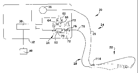
Fig. 1, the ophthalmic surgical system 20 includes a disposable surgical
microkeratome 22 and a remotely located control assembly 24. The control
assembly includes a flexible suction line or tube 26 and at least one
(preferably
only one) control cable 28. The suction tube and the control cable connect the
control assembly to the microkeratome for remotely controlling and driving the
microkeratome. (As described herein, the term "microkeratome" is used to
describe the complete device between the control cable and/or the suction tube
and the patient.)
The control assembly 24 is substantially contained within a housing 32 and
includes a drive assembly 34, a suction pump 36, and a controller 38. The
controller 38 can include an electronic circuit for controlling the operation
of the
6
CA 02421268 2008-06-16
71932-92
drive assembly and the pump. The control assembly also has one or more input
devices 40 connected to the controller, such as a touch screen, an ONIOFF
switch, a suction pedal or switch, a two-position drive pedal or switch,
and/or a
flap length adjustment selector, also referred to as a flap hinge positioning
system. The one or more input devices permit the surgeon to control several
variables in the operation of the system 20, as further described herein. For
more
detail conceming the operation of an exemplary control assembly, reference may
be had to commonly owned Intemational Application No. PCT/US98/24785, filed
November 20, 1998 (published as Intemational Publication No. W099126568 on
io June 3, 1999).
The drive assembly 34 is substantially similar to the drive assembly
disclosed in the aforementioned International Publication No. W099/26568, with
the exception thnt thn rotary and axial cables in the aforementioned
publicaticui
have been replaced in the ilkistrated system with a single control cable that
transmits both rotational and axial motion to the microkeratome. The drive
assembly 34 includes a linear drive motor 60 connected to slide member 62 by a
rack 64 and pinion 66, for example, for moving the slide member through a
range
of linear motion corresponding to the desired cut length. The slide member is
mounted on a pair of parallel rods 72 that act as slide guides for directing
or
guiding the linear motion of the slide member 62. A rotary drive motor 70 is
carried on the slide member for movement therewith. A control cable 28 having
a
control shaft 76 is connected to the rotary motor 70. Because the rotary drive
motor 70 is mounted on the slide member 62, the drive shaft can simultaneously
transfer rotational motion (created by the rotary drive motor) and linear
axial
motion (created by the linear drive motor through the slide member) to the
microkeratome.
The control shaft 76 is disposed in a flexible sheath 78 and the shaft and
the sheath together form the control cable 28. Consequently, the control cable
connects the rotary drive motor 70 and the axial drive motor 60 (through the
slide
member 62) to the microkeratome 22. The control shaft rotates and/or moves
axially within the sheath 70 as the slide member 62 moves through its range of
motion.
7
CA 02421268 2003-03-03
WO 02/17834 PCT/US01/27167
The control shaft 76 may have a monocoil, double wound or triple wound
construction over a central wire or mandrel with the winds pitched in opposite
directions to provide torsional rigidity. The triple wound construction
provides bi-
directional properties as well as flexibility for increased endurance life of
the shaft,
as compared to a monocoil or double wound construction. The torsional
stiffness
of the triple wound construction generally is equivalent to or greater than
the
double wound construction but its flexural stiffness generally is less than
about
half that of the double wound construction. However, this is desirable because
the shaft may rotate in a relatively sharp bend at high speed. The control
shaft
1o can be made of stainless steel due to the strength and endurance limit of
stainless steel. Furthermore, the exemplary control shaft is coated with a
thin
wall shrink tubing in order to provide a smooth surface to minimize or
eliminate
vibration. The inside of the sheath 78 may be coated with a material, such as
polytetrafluoroethylene (PTFE), to reduce friction between the sheath and the
shaft. Alternatively, the entire sheath may be formed of PTFE. The control
shaft
is designed to rotate at speeds up to 20,000 revolutions per minute and to
provide the necessary torque to drive the microkeratome 22.
An enlarged view of the microkeratome 22 is shown in Fig. 2 and includes
a base 90 for mounting the microkeratome on the eye of a patient, a carriage
92
mounted to the base for movement relative to the base, and a cutting blade 94
carried in the carriage. The base includes a main portion and an extension at
one end that synergistically provides a handle 96 for holding the
microkeratome.
The handle holds and guides a control shaft (Fig. 11) that is connected to the
carriage for movement therewith. The distal or terminal end of the handle is
connectable to the control cable 28 for transferring the motion from the
remotely
located drive assembly 34 to the cutting blade. The handle includes few,
simple
components, is disposable as part of the microkeratome, and can be
preassembled and connected to the base and the carriage. The handle can be
cast or molded as a single piece and may be formed as a unitary part of the
base.
As shown in Figs. 3a and 3b, the main portion of the base 90 has a
substantially flat top surface 100 on which the carriage 92 (Fig. 2) rests.
Extending from the top surface, the base includes a pair of spaced apart
parallel
8
CA 02421268 2003-03-03
WO 02/17834 PCT/US01/27167
guides 102. The guides generally have an inverted L-shape and oppose each
other to cooperatively form a track or guideway. The guides cooperate with the
carriage to restrain and guide the carriage for linear movement along the base
in
a cutting direction parallel to the length of the guides. The track also
functions to
hold the carriage to the top surface of the base against any significant
separation
therefrom, thereby maintaining the carriage in stable sliding engagement with
the
top surface of the base.
At a front end 104 of the base 90 opposite the handle 96 and between the
guides 102, a generally circular opening 106 is provided for receiving a
cornea
1o therethrough. The opening communicates through the top surface (and
actually
top wall) of the base to a substantially cylindrical suction chamber 107
provided at
the underside of the base. The chamber is formed in part by a cylindrical
suction
ring 108 which depends from the top wall of the base and generally is larger
than
the opening in the base. The suction ring~and the opening are adapted for
engaging and sealing against the surface of an eye to provide an air tight
enclosed space therebetween, or are otherwise configured to form a tight seal
with the eye so that a partial vacuum can be drawn to hold the base to the
eye.
The base 90 also is designed such that when suction is applied to the suction
chamber 107, the cornea protrudes through the opening and above the top
surface 100 of the base.
As an alternative to the illustrated suction ring, the suction ring may be
formed of one or more circumferentially arranged suction devices that hold the
microkeratome in a stable relation to the eye.
The base 90 has extending upwardly at the forward end thereof a fixture
(or fitting) 110. The illustrated fixture is angled away from the opening 94
to
provide an unobstructed view of the cornea. The fixture acts as a suction pipe
for
attachment of the suction tube 26 (Fig. 1) to the base. The opposite end of
the
suction tube is connected to the suction pump 36 (Fig. 1). The fitting has a
passage 112 extending therethrough to the suction chamber 107. The pump and
the tube supply suction to the suction chamber to pump air out of the chamber
to
create a partial vacuum that retains the base 86 in a stable and fixed
position
relative to the eye.
9
CA 02421268 2003-03-03
WO 02/17834 PCT/US01/27167
The surgeon can use the handle 96 to facilitate positioning the
microkeratome on an eye and to hold the microkeratome 22 in place until a
partial
vacuum is drawn in the suction chamber 107. Consequently, the handle must
extend sufficiently for an adult surgeon to place two fingers and a thumb on
the
handle, and more preferably extends at least about 2 inches (about 5 cm), at
least about 3 inches (about 7.6 cm), at least about 4 inches (about 10 cm) or
more. Referring briefly to Fig. 1, the suction tube 26 can advantageously be
retained adjacent the handle with a clip 114 to help keep the suction tube out
of
the way during the operation.
As an alternative to the base 90 shown in Figs. 3a and 3b, the fixture may
be rotated to an alternate position to move the suction tube 26 (Fig. 1)
further out
of the way, for example as shown in the embodiment illustrated in Figs. 4a and
4b. In this embodiment, the base 90' is substantially similar to the base 90
shown
in Figs. 3a and 3b, and thus like elements will be identified with like
reference
numbers. In this embodiment, the base 90' includes a fixture 115 which is
solid,
but omitting a passage therethrough to the suction chamber 107. A separate
suction pipe 116 is formed in the base with a passage 118 extending from the
suction chamber toward a rear end of the base and toward the handle 96 (Fig.
2),
away from the fixture. This allows the suction tube to be run underneath the
top
surface 100 of the base, thereby substantially eliminating any opportunity for
the
suction tube to obscure the surgeon's view of the operation.
Referring now to Figs. 5a-5c, the carriage 92 (Fig. 2) includes a cutting
head 120 which is substantially similar to the cutting assembly disclosed in
the
aforementioned international patent publication. A pair of parallel guide
rails 122
extend from the sides of the cutting head to cooperatively engage the guides
102
(Fig. 3a) on the base 90 (Fig. 3a).
The cutting head 120 has formed therein a substantially vertical slot 124
that opens from an angled blade guide surface 126 underneath the cutting head.
The slot sliding receives and guides a blade holder 128 (Fig. 6a) for
transverse
3o reciprocating movement therein. The width of the slot is transverse to the
direction of the motion of the carriage 92 (Fig. 2) and is wider in the
transverse
direction than the blade holder to permit the blade holder to oscillate within
the
CA 02421268 2003-03-03
WO 02/17834 PCT/US01/27167
slot. The transverse oscillation of the blade holder creates a transverse
oscillation of the cutting blade 94 (Fig. 2) which is connected to the blade
holder
as described below.
The blade holder 128 is illustrated in Figs. 6a-6d. The blade holder has a
protrusion 132 which closely fits through an opening in the cufting blade 94
(Fig.
2). The blade holder also includes a vertical slot 134 which is perpendicular
to
the width of the blade holder. When the blade holder is assembled in the
cufting
head 120 (Fig. 3a) the slot 134 is aligned with a substantially horizontal
opening
or passage 150 (Figs. 6a-6c) in the cutting head. The passage generally is
1o perpendicular to the slot in the blade holder. Note that the protrusion is
angled
from the body of the blade holder to extend substantially perpendicularly to
the
blade guide surface 126 (Fig. 5b) and the cufting blade 94 (Fig. 2) in the
illustrated embodiment.
The cufting blade 94 (Fig. 2) is held between the angled blade guide
surface 126 of the cufting head 120 (Fig. 3a) and a parallel angled top
surface of
a wedge 130. The wedge is illustrated in Figs. 7a-7c and supports the cufting
blade and the blade holder 128 (Fig. 5a) in the carriage 92 (Fig. 2). The
wedge
has an approximately triangular cross-section with a thinner portion facing
forward. The angled top surface 136 supports the cutting blade as it
oscillates
with the blade holder. The wedge also. includes a transverse recess 140 in the
top surface thereof that receives a portion of the protrusion 132 (Fig. 6c) of
the
blade holder that extends beyond the cufting blade. When the wedge is
assembled in the carriage the recess extends substantially coextensively with
the
vertical slot 124 (Fig. 5b) in the cutting head 120 (Fig. 5b) that receives
the blade
holder.
The wedge also includes at least one protrusion 142, extending from a top
surface. The protrusion is adapted to be press fit or otherwise secured in
corresponding recesses 144 (Fig. 5c) in the underside of the cufting head. The
recesses cooperate with the protrusions to locate and hold the wedge in place.
3o The wedge locks the cufting blade and the blade holder in the cufting head
while
allowing the cufting blade and the blade holder to oscillate therebetween.
Although the nature of the fit should hold the wedge in place, a surgical
adhesive
11
CA 02421268 2003-03-03
WO 02/17834 PCT/US01/27167
or medical grade epoxy may be used to ensure that the wedge remains in place.
Alternative means for positively locating the wedge relative to the carriage
and
locking it in place may be used as well, or in the alternative.
The carriage (except the cutting blade) and at least the main portion of the
base may be composed of transparent materials, such as a transparent molded
plastic, to maximize the surgeon's view of the operation.
Referring briefly back to Figs. 3a and 3b, the base 90 has at a back end
thereof one or more openings 146 for fixing the handle 96 thereto by suitable
means, such as one or more anchor clips 148 integrally formed in the handle.
1o Any suitable method of attachment may be used, including the illustrated
clips
which snap into position when pressed into the corresponding openings, as
shown more clearly in Figs. 8-11. As a result, the handle also functions,
along
with the fixture 110 to retain the carriage 92 (Fig. 2) between the guides 102
and
prevent the carriage from disengaging the guides and the base. As noted above,
the handle may alternatively be formed as a unitary piece of the base.
As shown in Fig. 2, the handle 96 is connected to both the base 90 and the
carriage 92 through a linkage 149. A forward portion of the microkeratome 22
has been enlarged in Figs. 8-10 to better illustrate the connection between
the
handle, the base and the carriage, and thus only a portion of the handle is
shown.
At a forward end of the handle 96, a drive shaft 152 extends therefrom and
is connected to the carriage 92. The drive shaft is removed from the
microkeratome 22 in Fig. 8 to more clearly illustrate its shape. The drive
shaft
has a fitting 154 on a proximal end thereof, from which an eccentric 156
extends.
The fitting passes through the horizontal passage 150 in the carriage to
extend
the eccentric into the slot 124 in the cutting head 120 to engage the vertical
slot
134 in the blade holder 128. The fitting also has a distended portion 158
which
has a greater thickness than the rest of the fitting and the drive shaft. The
cutting
head has a corresponding annular recess 160 in the passage 150. The fitting
can be press fit into the passage until the distended portion of the fitting
snaps or
locks the fitting into the annular recess, thereby securing the fitting in the
carriage
such that it can freely rotate while providing a positive connection between
the
drive shaft and the carriage for advancing and retracting the carriage along
the
12
CA 02421268 2003-03-03
WO 02/17834 PCT/US01/27167
base 90. Alternative means for connecting the drive shaft to the carriage may
be
used, including a bayonet coupling, for example. However, the illustrated
method
requires fewer parts and is faster and less expensive to assemble.
In connecting the handle 96 to the base 90 and the carriage 92, the
carriage generally is advanced toward the forward end 104 of the base toward
its
distal portion, and the drive shaft 152 is extended from the forward or
proximal
end of the handle to snap the fitting 154 thereon into the passage 150 in the
carriage. The anchor clips 148 on the handle are then snapped into the
openings
146 (Fig. 3a) in the base, as shown in Fig. 10. The drive shaft can then be
1o retracted to move the carriage toward its proximal position at the rear of
the base,
as shown in Fig. 9. The assembly of the microkeratome 22 is thus complete.
An assembled microkeratome 22 is shown in Fig. 11. As can be seen, the
drive shaft 152 generally is enclosed within the hollow handle 96, which can
be
formed economically of a molded plastic material. The drive shaft can be a
flexible shaft, similar to the control shaft 76 (Fig. 1) of the control cable
28, or it
can be formed of a solid rod or bar, such as a metal rod. The fitting 154
(Fig. 8)
at the forward end of the drive shaft can be formed of a metal or other
material
which can be economically formed into the desired shape to perform the
intended
function of positively engaging the carriage 92 and transmitting rotational
motion
2o as well as forward and reverse motion to the carriage.
As shown in Fig. 11, the drive shaft 152 may include an annular flange 162
at an intermediate point along the length of the shaft. The flange is shown
resting
against a forward side of a retaining wall (or retaining members) 164
extending
into the interior of the handle 96. From the annular flange, the drive shaft
extends
through a compression spring 166, such as a nylon spring, and a forward wall
168 in the handle. The flange, the forward and retaining walls, and the spring
cooperate to form an automatic return device 169 or biasing element. The
spring
acts on the flange to resist forward movement of the drive shaft and biases
the
flange toward the retaining wall when insufficient force is applied to the
drive shaft
to overcome the spring force. As a result, the microkeratome 22 having such a
handle automatically retracts the drive shaft and the carriage from its
extended
position to its retracted position under the influence of the spring at the
end of the
13
CA 02421268 2003-03-03
WO 02/17834 PCT/US01/27167
cutting operation when the forward power is reduced. Automatic retraction is
particularly advantageous when there is a loss of power to the system.
An alternative embodiment of an automatic return device is illustrated in
Fig. 12, where angled wall portions 170 extend into the interior of the handle
96.
The wall portions function as spring-like members which simultaneously resist
the
passage of the annular flange 162 on the drive shaft 152 toward the forward
end
of the handle and automatically retract the drive shaft and the carriage 92 in
the
event of a loss of power. Consequently, in this embodiment the wall portions
170
combine the functions of the compression spring 166 and the retaining wall 164
in
1o the embodiment shown in Fig. 11
In either embodiment, the drive shaft 152 transmits the rotational and axial
motion of the control shaft 76 from the control cable 28 to the carriage 92.
The
control cable is connected to the rear of the handle 96. Any means for
connecting the control cable to the drive shaft is acceptable, as long as it
provides
a positive connection for the transmission of rotational motion and both
forward
and reverse axial motion.
One type of connection is shown in the embodiments illustrated in Figs. 11,
and 13a-14b, for example. The sheath 78 of the control cable 28 is held in a
fitting 172 on the end thereof and through which the control shaft 76 passes.
The
2o end of the fitting abuts the rear end of the handle 96 and is attached
thereto by a
bayonet coupling 176, for example, held in place by another spring 178. The
control shaft passes through the fitting and the spring into the handle where
it
abuts and engages the rear end of the drive shaft 152. The positive contact
between the control shaft and the drive shaft permits the drive shaft to
transmit
forward axial motion of the control shaft to the carriage, with the automatic
return
device 169 biasing the drive shaft toward the control shaft to provide reverse
axial
motion of the drive shaft and retraction of the carriage 92.
In Figs. 13a and 13b, one configuration is shown for transferring rotational
motion from the control shaft 76 to the drive shaft 152. In this embodiment,
the
control shaft has a fitting or key 180 on an end thereof with an eccentric 182
which mates with a semi-circular slot 184 in a corresponding fitting 186 on
the
rear end of the drive shaft. The slot 184 can be larger than the eccentric 182
to
14
CA 02421268 2003-03-03
WO 02/17834 PCT/US01/27167
facilitate locating the eccentric therein. Since the control shaft generally
only
rotates in one direction, a loose fit can be utilized between the eccentric
and the
slot. Another embodiment is illustrated in Figs. 14a and 14b, wherein the
fitting
188 on the control shaft and the fitting 190 on the drive shaft have
male/female
faces which mate together to provide a positive interlock for transferring
rotational
motion. However, both of these embodiments require the use of an automatic
return device, such as those described above with respect to Figs. 11 and 12.
An alternative means for connecting the control cable 28 to the
microkeratome 22 (Fig. 11) is illustrated in Figs. 15a-15d. This type of
connection
lo does not require internal components in the handle 96 other than the drive
shaft
152, and does not require the drive shaft to have an intermediate flange,
thereby
reducing manufacturing costs, while at the same time improving the reliability
of
the system and providing positive control over the return motion of the
carriage 92
(Fig. 11). In this embodiment fittings 192, 194 on the drive shaft and the
control
1s shaft, respectively, partially axially and transversely overlap and
interlock to
advantageously provide positive transfer of rotational motion as well as both
forward and reverse axial motion.
In this embodiment the sheath 78 of the control cable 28 is swaged into a
cylindrical fitting 196 through which the control shaft 76 extends. The distal
end
20 of the fitting has an annular flange 198 which retains a threaded nut 200
which
fits over the fitting and the sheath. The nut is retracted, as shown in Figs.
15a
and 15b, for the connection of the control shaft fitting 194 to the drive
shaft fitting
192. The fitting on the drive shaft protrudes from the handle 96 and has a
shape
which partially overlaps and mates with the fitting on the control shaft. The
25 control shaft can be extended to push the drive shaft into the handle and
to draw
the sheath fitting 196 toward the handle.
A portion 202 of the rear end of the handle 96 also is threaded, or includes
a threaded part mounted thereto, which mates with the threaded nut 200 on the
control cable. Once the control shaft 76 and the drive shaft 152 are
connected,
30 the control shaft is advanced to move the sheath fitting 196 and the nut
into
engagement with the rear end of the handle, and the threaded parts are screwed
together, as shown in Fig. 15c, to secure the sheath to the handle and to lock
the
CA 02421268 2003-03-03
WO 02/17834 PCT/US01/27167
shafts together. The control shaft can then be retracted to positively retract
the
drive shaft along with it, as shown in Fig. 15d.
Referring now to Figs. 9 and 10 as well, in order to remotely drive the
carriage 92 relative to the base 90 (Fig. 1), the control cable 28 (Fig. 1)
relies on
relative movement between the control shaft 76 and the sheath 78, with no
resulting change in the displacement of the end of the sheath from the base of
the microkeratome 22 as the control shaft moves axially within the sheath. In
this
embodiment, the sheath is held in the fitting, which is positively secured to
the
rear end of the handle, which in turn is connected to the base. As a result,
1o forward and reverse axial motions of the control shaft moving through the
sheath
results in forward and reverse motion of the drive shaft and the carriage
without
the necessity for any gears or rotational motion on the part of the control
shaft to
effect movement of the carriage 92 relative to the base 90 (Fig. 1).
In addition, the rotational motion of the control shaft 76 is transmitted to
the
carriage 92 (Fig. 11) by the drive shaft 152 where the cutting head 120 (Fig.
5b),
the blade holder 128 (Fig. 6c), and the wedge 130 (Fig. 7a) cooperate to
transform the rotational motion into an oscillating motion for driving the
cutting
blade 94 (Fig. 2) in an oscillating side-to-side motion.
The surgeon or other member of the surgical staff will select the optimum
microkeratome from a plurality of microkeratomes, with different
microkeratomes
providing different size openings in the base and/or providing for different
depths
of cut. The different microkeratomes are useful for accomodating different
size
eyes and different depths of cut relative to the different size eyes. The
microkeratome comes preassembled and requires no assembly in the operating
room. Someone from the surgical staff removes the packaging and connects the
microkeratome to the control assembly. The assembler generally double-gloves
to maintain sterility, removing one set of gloves after touching reusable
components of the control assembly. A new, and sterile, suction tube is
connected to the fixture and the suction pump, and the control cable is
connected
to the handle of the microkeratome. The microkeratome can be handed to the
surgeon and the surgeon can position the microkeratome on the eye by holding
the handle without contacting any other component of the system. Operation of
16
CA 02421268 2003-03-03
WO 02/17834 PCT/US01/27167
the microkeratome is substantially automatic and proceeds in substantially the
same manner as the operation of the microkeratome described in the
aforementioned Published Application No. WO 99/26568. The surgeon can
control the microkeratome with foot pedals, without touching the control
assembly
with his hands, further maintaining sterility. Once the operation is complete,
the
microkeratome can be disconnected from the control assembly and discarded. A
new microkeratome is selected for a subsequent operation.
The control assembly includes components that are expensive to produce
and are intended to be reused for surgery on many patients, whereas the
1o microkeratome includes relatively inexpensive components and is intended to
be
used for a single eye of a single patient and then discarded. Because the
control
assembly is removed from the patient and thus away from the surgical area,
contamination of the control assembly by the patient and vice versa, is
minimized
or prevented.
Although the present invention has been described with reference to an
embodiment that uses a single cable, the invention also includes a
microkeratome having a handle for connecting more than one cable. For
example, the invention includes a microkeratome connectable to a first cable
used to control movement of the blade relative to the carriage and a second
cable
used to control movement of the carriage relative to the base.
An exemplary microkeratome is formed primarily of a few molded plastic
parts that are easy to manufacture and assemble such that the microkeratome is
inexpensive to produce. Consequently the microkeratome may be considered
disposable, thereby obviating problems of cleaning and sterilizing the
microkeratome between patients. The microkeratome is provided completely
assembled, sterilized, and ready for use. Since only the microkeratome comes
into contact with the patient, the microkeratome does not require extensive
assembly by the surgical staff immediately prior to surgery, and the
microkeratome is only used once, the surgical area is more easily and more
3o effectively maintained in a clean and sterile condition. Maintaining such a
high
degree of confidence in the sterility of a microkeratome has been a problem
that
is problem for which the present invention provides an improved solution.
17
CA 02421268 2003-03-03
WO 02/17834 PCT/US01/27167
In summary, since the axial and rotary motors are independently
controllable, the system can independently control the oscillation and speed
of
advance of the cutting blade. The system of the present invention also
advantageously uses a single control cable to drive the microkeratome. In
addition, the microkeratome includes a handle that facilitates a quick
connection
between the carriage and the control cable at a location remote from the main
portion of the base, and thus the patient's eye. The present invention also
provides a microkeratome having means for automatically retracting the cutting
blade, a particular advantage in the event of a sudden loss of power. The
1o present invention clearly provides significant improvements over the prior
art.
Although the invention has been shown and described with respect to
certain illustrated embodiments, equivalent alterations and modifications will
occur to others skilled in the art upon reading and understanding the
specification
and the annexed drawings. In particular regard to the various functions
performed by the above described integers (components, assemblies, devices,
compositions, etc.), the terms (including a reference to a "means") used to
describe such integers are intended to correspond, unless otherwise indicated,
to
any integer which performs the specified function (i.e., that is functionally
equivalent), even though not structurally equivalent to the disclosed
structure
which performs the function in the herein illustrated embodiments of the
invention. In addition, while a particular feature of the invention may have
been
described above with respect to only one of several illustrated embodiments,
such a feature may be combined with one or more other features of the other
embodiment, as maybe desired and advantageous for any given or particular
application.
18