Note: Descriptions are shown in the official language in which they were submitted.
CA 02421957 2003-03-11
WO 02/22852 PCT/CAO1/01297
METHOD FOR MEASURING ANTITHROMBIN ACTIVITY
FIELD OF INVENTION
This invention relates to a method of measuring antithrombin activity.
BACKGROUND OF INVENTION
Blood coagulation or clotting results when a series of inactive enzymes in
blood are
activated to generate, at the end of the cascade, a clot at the site of the
wound. The intrinsic
blood coagulation pathway is activated upon contact with the surface of a
foreign matter
which initiates the sequential activation of factors XII to XIIa,
prekallikrein to kallikrein,
kininogen to kinin, XI to XIa, IX to IXa, X to Xa, and II (prothrombin) to Ha
(thrombin).
Tissue thromboplastin initiates the extrinsic blood coagulation pathway by
activation of
factor X, which in turn results in the activation of prothrombin to form
thrombin. In both
pathways, the final enzyme in the cascade is thrombin, a serine protease which
cleaves the
soluble protein fibrinogen to form fibrin. Fibrin molecules crosslink to form
a clot which
reduces the flow of blood from the wound.
Antithrombin or antithrombin III (AT) is an important regulator of blood
coagulation. AT, which is produced in the liver, is a serine protease
inhibitor with a
molecular weight of approximately 60,000 Daltons and circulates in the blood
at a
concentration of 150 to 200 micrograms per millilitre, or 2.5 to 3.4
micromoles per litre.
AT has a broad specificity, and inhibits most of the coagulation factors
involved in the
intrinsic and the extrinsic pathways and is the principle regulator of
thrombin. The
inhibition of most coagulation enzymes by AT is significantly augmented in the
presence of
heparin.
AT deficiency is associated with an increased risk of thrombosis. The
condition
may be congenital, or acquired as a result of underlying conditions such as
liver disease,
kidney disease or disseminated intravascular coagulation. AT deficiency may be
related to
reduced levels of AT or reduced AT activity. For example, in congenital AT
deficiency
1
CA 02421957 2003-03-11
WO 02/22852 PCT/CAO1/01297
type II, the AT concentration is normal but the activity is reduced due to the
presence of a
dysfunctional AT. Successful clinical diagnosis and management of patients
with AT
deficiency therefore demands a specific, sensitive and a simple laboratory
assay of the AT
activity.
Current methods to determine AT deficiency can be divided into three classes:
immunoassays, amidolytic-based activity assays and clot-based activity assays.
Immunoassay techniques measure the concentration of AT in a sample through
methods such as radial immunodiffusion, nephelometry and enzyme-linked
immunosorbant
assays (ELISAs). These assays are very specific and quite sensitive, but can
be time-
consuming to perform. As well, concentration measurements of AT do not always
correlate
with AT activity levels since inactive forms of AT or AT-enzyme complexes may
still
exhibit immunoreactivity in these assays. This may lead to inappropriately
high test results
for some patients with reduced AT activity as in the case of type II
deficiency.
Amidolytic-based activity assays work on the principle of incubating a fixed
quantity of a single purified enzyme, usually thrombin or factor Xa, with a
diluted test
sample and heparin. The residual enzyme activity is measured by determining
the endpoint
or kinetic rate of cleavage of synthetic chromogenic or fluorogenic
substrates. These types
of assays are currently the most widely used methods to determine AT activity
levels.
However, these assays tend to be susceptible to interference from other
coagulation
inhibitors such as a2-macroglobulin, heparin cofactor II and al-antitrypsin.
As well,
measurements of AT activity vary depending on which purified enzyme is used
for the
assay. Costs for these assays can be fairly high due to the use of purified
enzymes and
synthetic substrates, and the requirement for a spectrophotometer or high-end
coagulation
analyser to detect the reaction endpoint.
Clot-based activity assays may be performed as either two-stage or one-stage
assays.
The two-stage assays involve incubating a fixed quantity of purified enzyme,
such as
thrombin, with defibrinated test serum or plasma. Residual enzyme activity is
measured by
determining clotting activity upon the addition of plasma or purified
fibrinogen instead of.
2
CA 02421957 2003-03-11
WO 02/22852 PCT/CAO1/01297
determining the amidolytic activity as described above. Drawbacks of these
assays include
artifactual reduction of AT levels if heat denaturation is used to defibrinate
the plasma.
Also, these methods tend to be cumbersome and labour intensive, time-
consuming, and
susceptible to interference by other progressive coagulation inhibitors.
A one-stage clot-based assay is described in U.S. Patent No. 5,093,237 to
Enomoto.
In this assay, the test specimen is mixed with AT-free plasma containing the
extrinsic
coagulation factors, heparin and a prothrombin time measuring reagent and the
coagulation
time resulting from the activation of the extrinsic coagulation pathway is
measured. The
prothrombin time test is known to be extremely insensitive to heparin-enhanced
inhibition
by AT and the Enomoto assay discloses the use of a high concentration of
heparin (12
U/ml). This is far above the optimal concentration of heparin for AT and at
such high
concentrations, it is known that the efficiency of inhibition by AT is
reduced. Moreover,
while Enomoto discloses that the extrinsic coagulation reaction is utilized to
avoid the many
potential errors in the intrinsic reaction pathway, the assay generates only
two enzymes,
activated Factor X and thrombin, upon which AT can exert its inhibitory effect
and does not
best reflect the full spectrum of in vivo physiological AT activity.
It is apparent therefore, that there remains to be developed a sensitive,
specific yet
simple clot-based laboratory assay for AT activity.
SUMMARY OF INVENTION
The present invention provides a method for measuring AT activity in samples
containing AT, such as in a patient plasma sample. The method of the invention
includes
the step of mixing a test sample with an AT-deficient substrate plasma, an
activator of the
contact phase of the intrinsic coagulation pathway and a phospholipid. The AT-
deficient
plasma contains the enzymes of the intrinsic coagulation pathway and may also
contain an
AT augmenting compound, such as heparin. The AT augmenting compound if not
present
in the substrate plasma is added separately to the test sample. Following
addition of calcium
ions to the mixture, the coagulation time is measured. By comparing the
coagulation time
to a reference standard, the AT activity level of the test sample can be
determined.
3
CA 02421957 2010-11-30
Thus in one aspect, there is provided a method for determining antithrombin
activity
in a sample comprising mixing in a reaction mixture a dilution of the sample
with an
antithrombin deficient plasma, an activator of contact phase of intrinsic
coagulation pathway,
a phospholipid and an antithrombin augmenting compound wherein the
antithrombin
deficient plasma comprises intrinsic coagulation enzymes; introducing calcium
ions to the
reaction mixture; measuring coagulation time; and comparing the coagulation
time to a
reference standard.
In another aspect, there is provided a method for determining antithrombin
activity in
a sample comprising mixing in a reaction mixture a dilution of the sample with
an
antithrombin deficient plasma, an activator of contact phase of intrinsic
coagulation pathway
and a phospholipid wherein the antithrombin deficient plasma comprises
intrinsic coagulation
enzymes and an antithrombin augmenting compound; introducing calcium ions to
the
reaction mixture; measuring coagulation time; and comparing the coagulation
time to a
reference standard.
In another aspect, the invention provides a kit for determining AT activity in
a sample
which kit includes an AT-deficient substrate plasma, wherein the plasma
comprises intrinsic
coagulation enzymes, an AT augmenting compound, an APTT reagent and a calcium
salt
solution.
BRIEF DESCRIPTION OF DRAWINGS
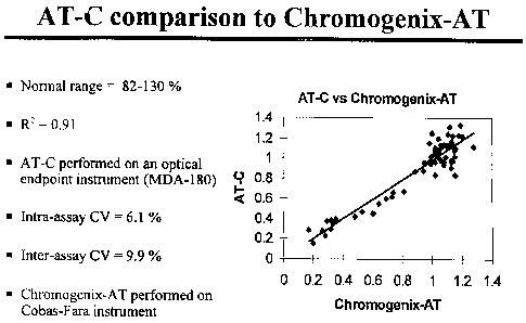
Figure 1 depicts a graphical comparison and correlation between the results
obtained
from an assay performed according to the present invention (AT-C) and an
amidolytic-based
activity assay performed according to the chromogenic AT assay from
Chromogenix
(Chromogenix-AT).
DETAILED DESCRIPTION OF THE INVENTION
The present invention provides a simple one-stage in vitro method for
measuring AT
activity levels in any sample containing AT. The invention therefore can be
used to measure
AT activity levels in samples from patients such as plasma or serum to detect
a
4
CA 02421957 2009-10-30
deficiency of AT activity or to monitor antithrombin therapy using
antithrombin
concentrates. The invention can also be used to monitor the level of AT during
production procedures such as cell culture or during purification of AT from
plasma or
culture fluid. If protease inhibitors other than AT are present at greater
than physiological
concentrations, controls must be run to determine the effect of these other
inhibitors on
the assay. Such controls may include testing the vehicle (plasma, culture
fluid or buffer
solution that is being tested for AT activity) in the absence of AT at
dilutions equivalent
to those used for the assay of AT, as well as the addition of a known quantity
of AT to
the vehicle and calculating its recovery.
In carrying out the method, a diluted test sample is mixed with an AT
deficient
substrate plasma, an activator of the contact phase of the intrinsic
coagulation pathway
and a phospholipid. An activated partial thromboplastin time (APTT) reagent
may also be
used to provide the contact phase activator and the phospholipid to the
reaction mixture.
An AT-augmenting compound, if not present in the AT-deficient substrate plasma
is also
added to
4a
CA 02421957 2003-03-11
WO 02/22852 PCT/CAO1/01297
the test sample. A calcium ion containing solution is then added and the
clotting time
determined by mechanical or optical means.
The term contact phase refers to that part of the intrinsic coagulation
pathway which
does not require calcium ions for activation and which is presently understood
to include the
activation of factor XII, prekallikrein, kininogen and factor XI. Contact
phase activators
are known and 'include ellagic acid, silica and kaolin. The term APTT reagent
is
understood, and intended to refer to a reagent which contains at least a
contact phase
activator and a phospholipid. A phospholipid, which may be synthetic or plant
or animal
tissue extract derived, is required to support assembly of activated
coagulation factor
complexes by acting as a template. Different APTT reagents are widely
available
commercially. For optimal results, an APTT reagent which provides a clotting
time of
between 80 and 140 seconds for a dilution of reference plasma representing
100% AT
activity, and between 40 and 60 seconds for a dilution of a reference plasma
representing
approximately 6.3% AT activity should be selected.
For optimum results, the reaction mixture, prior to the introduction of
calcium ions
is incubated at a time and temperature sufficient to activate the contact
phase of the intrinsic
coagulation pathway, typically, for about 2 to 30 minutes at a temperature of
about 20 to
40 C. An optimal incubation time and temperature may vary depending on the
APTT
reagent selected, and in some cases, may not be necessary provided the
variability in the
clotting time is within an acceptable limit for the particular application of
the assay.
The calcium ions may be introduced to the mixture by addition of calcium salt
solution with a stock concentration between 0.01 and 0.1 M. The optimal
calcium
concentration is that which provides the shortest clotting time and may be
determined by
titration. The final concentration of calcium ions in the clotting mixture
which the inventors
have found optimal is between 4 and 10 mM. For example, one volume of 0.02 M
calcium
chloride solution may be added to a three volume reaction mixture to achieve a
concentration of 5mM.
5
CA 02421957 2003-03-11
WO 02/22852 PCT/CAO1/01297
The optimal dilution range of the test sample which may vary depending on the
APTT reagent sensitivity and the instrumentation used, can be readily
determined by those
skilled in the art. The inventors have generated a reference curve using
reference plasma
dilutions of 1/10, 1/20, 1/40, 1/80, 1/160 representing AT concentrations of
approximately
100%, 50%, 25%, 12.5% and 6.3%, respectively, referenced against a World
Health
Organization (W.H.O.) - traceable calibrator.
The substrate plasma used in this method is an AT-deficient plasma that may be
prepared by the known methods including immuno-affinity chromatography, or
affinity
chromatography using immobilised sulphated polysaccarides such as heparin as
previously
described (Hoogendoorn, 1980), or some combination of these techniques, to
remove AT
while retaining the coagulation factor activities of intrinsic coagulation. To
ensure that the
only component influencing the clotting time is due to the level of AT in the
test sample,
other factors which may influence the clotting time should be present in
sufficient
concentrations in the AT-deficient substrate plasma to minimize any influence
of small and
variable quantities present in the test sample. An AT-deficient substrate
plasma for use in
the invention should therefore have a normal clotting time in an ATPP based
assay, in the
absence of an AT augmenting compound such as heparin described below.
For optimum results, the substrate plasma should contain less than about 1% of
normal AT levels as determined by an assay method that can accurately detect
AT levels of
less than 2%, for example, an antigen assay or an activity assay. The
substrate plasma
should contain normal levels of other coagulation inhibitors such as heparin
cofactor H, a2-
macroglobulin and al-antitrypsin. Additionally, optimum results may be
obtained when at
least 40% of normal activity levels of coagulation factors XII, XI, IX, VIII,
X, V and II as
measured by a one-stage clotting activity assay, and at least 1 gram per litre
of fibrinogen
are present in the substrate plasma. Normal activity levels of coagulation
factors and
coagulation inhibitors refer to those determined from a WHO traceable
standard.
An AT-augmenting compound as that term is used in this invention is a compound
capable of prolonging the APTT of normal plasma but not of AT-deficient
plasma. For the
purposes of this invention, a prolongation of the APTT of less than 20 seconds
is not
6
CA 02421957 2003-03-11
WO 02/22852 PCT/CAO1/01297
considered significant. An AT augmenting compound is most typically heparin or
a heparin
derivative, but also includes other sulphated compounds such as a
glycosaminoglycan, a
sulphated oligosaccharide or a polysulphone.
The AT-augmenting compound may be added to the AT-deficient substrate plasma
or mixed directly with the test sample either before or at the time of
introducing calcium
ions to the mixture. In the case of heparin, optimum results may be achieved
with an AT-
deficient plasma which has an APTT in the normal range and which increases by
less than
20 seconds in the presence of heparin. Yet still, optimum results may be
achieved when
heparin is present in a concentration sufficient to prolong the APTT by 2- to
4-fold in the
presence of diluted AT in the test sample and such concentration is typically
in the range of
0.5 to 2 international units per millilitre, or 2.5 to 10 micrograms per
millilitre. Addition of
small amounts of AT in the diluted sample, when mixed with heparin or with the
substrate
plasma containing heparin, causes a substantial and dose dependent increase in
the clotting
time when an APTT reagent is added.
In one specific embodiment, one volume of AT deficient substrate plasma is
mixed
with one volume of test or reference sample diluted 1/10 and then mixed with
one volume
of APTT reagent. The sample is diluted using a buffer such as 0.01 to 0.1 M
imidazole,
Tris or HEPES or other suitable buffers known in the art. The mixture is
incubated at 37 C
for 180 seconds at which time one volume of 20 mM CaC12 is added and the clot
time
recorded. The clotting time is then compared to the clotting times of a
reference plasma
(such as a WHO traceable reference) containing a known amount of AT to obtain
a measure
of the AT activity in the test sample. For this comparison, a reference curve
can be
generated using different dilutions of the reference plasma. A typical curve
may include
readings from reference plasma diluted 1:10, 1:20, 1:40, 1:80 ad 1:160,
representing AT
activity levels of 100, 50, 25, 12.5 and 6.5%, respectively, referenced
against a WHO
traceable calibrator.
The present method is easy and relatively quick to perform in that it requires
no pre-
treatment of samples, such as defibrination. Additionally, there is no
requirement for
specialised detection equipment, as the results may be read manually. As the
method can be
7
CA 02421957 2003-03-11
WO 02/22852 PCT/CAO1/01297
performed on most all automated or semi-automated coagulation analysers, it
may be
automated using existing laboratory instrumentation and software.
The method of the invention measures the activity of AT on a wider range of
endogenously generated coagulation factors than that possible for the
prothrombin time
method disclosed in Enomoto. As such, the present method is believed to
provide a better
measure of the physiological AT activity in vivo. Furthermore, given the
greater sensitivity
of the inhibition of the intrinsic coagulation activation to heparin (Nordfang
et al, 1993
Thrombosis and Haemostasis 70 (3) 448-453), relatively low levels of heparin
can be used.
The present method therefore avoids the reduced efficiency of inhibition by AT
that can
occur at higher heparin concentrations.
Table 1 below shows typical APTT clot times obtained according to the
invention
for a reference plasma and for known normal control and known abnormal control
plasmas.
TABLE 1
Sample Dilution Clotting Value
time (sec) obtained
Reference plasma (AT value of 96%) 1:10 86.2" NA
(CCNRP #7020, Precision BioLogic) 1:20 67.9"
1:40 58.2"
R2 = 0.9987 1:80 52.3"
1:160 48.9"
Normal plasma 1:20 68.3" Mean = 102%
(NP97-05 Precision BioLogic) 1:40 59.1"
Abnormal plasma 1:20 53.4" Mean = 32%
(ARPI #8020, Precision BioLogic) 1:40 50.1"
With reference to Figure 1, the results of the present method (AT-C assay)
correlate
well with the results obtained with the currently popular amidolytic based
activity and
further demonstrates the specificity of the method according to the invention.
Moreover, as shown below in Table 2, no significant difference is seen between
results of the two methods for test samples from patients receiving heparin
therapy. These
results further demonstrate that the method of present invention is as
specific as the
8
CA 02421957 2003-03-11
WO 02/22852 PCT/CAO1/01297
currently popular method. Data are reported as AT units per millilitre using a
WHO
reference plasma containing known amounts of AT as a reference calibrator.
TABLE 2
Sample AT-C Assay Chromogenix AT assay
(U/mL) (U/nnL)
H58 0.64 .76
H71 .83 0.91
Hep 1 .62 0.8
Hep 2 0.75 0.78
Hep 3 .76 0.72
Hep 8 1.1 1.07
Hep 9 0.85 0.8
Hep 10 0.85 0.83
Hep 11 1.16 0.96
Hep 12 0.71 0.7
Hep 9.1 0.59 0.61
Hep 10.1 0.76 0.7
Hep Brown 0.73 0.98
Hep 70 0.83 0.84
Hep EVA 0.53 55
Hep 58 0.82 0.82
1003HC49 0.84 1.01
Hep 40 0.54 0.67
Hep Mou 0.59 0.81
Hep 7 0.75 0.73
Hep 50 0.83 0.84
MEAN 0.77 0.77
Standard Deviation 0.16 0.16
Table 3 shown below illustrates the effect of addition of heparin cofactor II
(HCII)
to HCII immune-deficient plasma at a concentration gradient of 0 to 200 % of
normal
concentration. The AT activity of the resulting plasmas as measured according
to the
invention changed less than 10% even at high HCII concentration demonstrating
that the
present method is insensitive to interference from other coagulation
inhibitors. It is
9
CA 02421957 2003-03-11
WO 02/22852 PCT/CAO1/01297
believed that dilution of the test plasma and the use of a substrate plasma
with normal levels
of these inhibitors minimizes the effect of modest additions of these
inhibitors from the
reference or test plasmas on the clotting time endpoint. The specificity may
also be
attributed to the wide spectrum of coagulation enzymes inhibited by AT in the
present
method.
TABLE 3
Sample Relative Dilution AT-C assay
HCII (%) value
HCII-DP alone <1% 1:20 140%
HCII-DP + 25 gg/ml purified HCII 50% 1:20 144%
HCII-DP + 50 gg/ml purified HCII 100% 1:20 136%
HCII-DP + 100 g/ml purified HCII 200% 1:20 138%
The present method is sensitive to AT activity levels as low as 12% of the
normal
activity. Moreover, no interference has been observed in plasma samples from
patients on
coumadin therapy or on heparin therapy or with lupus anticoagulant inhibitors.
The reagents of the present method may be conveniently packaged and the
invention
therefore also contemplates a kit for determining AT activity levels. For ease
of packaging
and storage, the substrate plasma in the kit may be lyophilized. In this
instance, the kit may
include a diluent for dissolving the lyophilized substrate plasma. The diluent
may be any
suitable buffer for example containing about 0.01 to 0.1 M imadazole, Tris or
HEPES or
other suitable buffers as would be known to a skilled person in the art,
including buffers
described by Good et al (Biochemistry 5 (1966), pp.467). The kit may also
include a
reference or control plasma which may also be lyophilized.
One skilled in the art can readily appreciate that various modifications can
be made
to the described embodiments without departing from the scope and spirit of
the invention.
Such modifications are also intended to be within the scope of the invention.