Note: Descriptions are shown in the official language in which they were submitted.
CA 02423064 2003-03-21
Docket No. DEP0675
?NOVEL EARLY INTERVENTION SPINAL TItEATMLENT I~IETIIODS AND
s DE~iCES FOR USE THEREIN
BACKGROUND OF THE IN'VENTIOI1'
One of the leading causes of spine-related pain is t:he ntpture or
degeneration of
discs located between lumbar vertebrae (''lumbar intervertebral discs"). Pain
in the lower
~o extremities may be caused by compression of spinal nen~e roots by such
damaged discs,
while low back pain may be caused by collapse of these discs and by tine
adverse effects
of bearing weight through a damaged, unstable vertebral joint. One
conventional method
of managing this problem is to treat the problernatie intelvertebral disc with
energy.
Tn some instances, the disc is globally heated. US Patent No. 5,433,739
("Sluitjer
15 !") proposes inserting an RF electrode or other heating electrode into the
intervereebral
disc and globally heating the entire intervertebral disc to a temperature
significantly
above body temperature. See col. 2, lines 52-56. Sluijter 1 teaches that this
process can
denervate the neural structures within the disc on a global or semi-global
basis, thus
relieving the patient of back pain related to stress of the disc and its
surface. See col. 5,
20 line 65 - col. 6, line 2. Sluijter I further notes that the precise
anatomical mechanism of
this pain relieving process has not been totally clarified, and discloses not
only that
anatomical material changes within the disc material itself and the resulting
volumetric
changes may play some role, but also that the spread of heat to large neural
structures in
the proximity of the disc may be additional contributory factors of signif
cance. See col.
25 13, lines 28-37.
In some instances, only the nucleus of the disc is treated. For example, Choy
et
ai., S ine, 17:8 (1992), pp. 949-956, discloses using a laser to evaporate the
nucleus
pulposus. It is believed that evaporation of the nucleus reduces the pressure
within the
disc, thereby relieving the pressure upon the nerves therein.
30 In some instance, only the inner wall of the annulus fibrosus portion of
the disc is
heated. US Patent Na. 6,261,311 ("Sharkey") proposes using a floppy wand-like
probe
for fully contacting and resistively heating a portion of the inner wall of
the annulus
fibroses to a temperature of about 45-70 °C. It is believed that
heating the inner wall in
CA 02423064 2003-03-21
this rrtanner may coa~.tlate andlor densrvate the eollagenous annular wall
leading to
increased stability of the vertebral joint andiar eliminating the ability of
the annular ~.~all
to sense pam.
US Patent No. 6,105,581 {"Eggers") discloses a radiofrec~uency ("RF") probe
s which uses an electrically conductive fluid to help ablate tissue within the
spine. Eggers
discloses coating portions of the probe with an electrically insulative
coating t~ produce
"non-active portions of the probe which help the surgeon selectively ablate
tissue". See
cal. 4, line 60 to col. 5, line L0. FICr.lSb of Eggers discloses a transverse
cross-section
view of a loop electrode wherein active portions of electrode 194 Lie in
essentially the
~a same plane. Eggers further recites selected spinal applications for this
device, including
laminectomy/discectomy procedures for treating herniated discs, decompressivc
laminectomy for stenosis in the lumbosacral and cervical. spine, posterior
luanhrosacral
and cervial spine fi~sions, treatment of scoliois associated with vertebral
disease,
foraminotomies to remove the roof of on the intervertebrai foramina to relieve
nerve root
i5 compression and anteror cervical and lumbar discectomies. Norse of these
applications
inv~lve the therapeutic treatment of ari intervertebral disc which results in
an essentially
intact disc.
In some instances, the nucleus pulposus is subjected to two power Levels of RF
energy. For example, US Published Patent Application ~(o. 200110029370
discloses a
2o rrrethod of treatment whereby a single probe ablates a portion of the
nucleus putposus as
it advances through the nucleus pulposus, and then heats the nucleus pulposus
(using
bipolar RF energy ) as it is withdrawn from the nucleus pulposus in order to
coagulate
the collagen within the nucleus pulposus.
Some investigators have proposed using ultrasound technology as a means for
25 heating the intervertebral disc. For example. US Patent No. 5,571,147
("Sluijter II")
proposes using an ultrasound probe to heat the intervertebral disc
US Patent No. 5,620,479 ("Diedrich I") discloses an ultrasound applicator for
thermal therapy of tumors and benign tissues using heat generated from
acoustic energy,
which includes a segmented array of individually coc~troliable tubular
ultrasound
3o transducers through which an inner probe extends: See col. 3, lines 6-10.
The segmented
array disclosed by Diedrich I in FIG.1 is a linear array of ultrasound sources
along the
2
CA 02423064 2003-03-21
longitudinal axis of the probe. Typically, ultrasound probes are configured to
emit
ultrasound waves in a full 360° radius about the axis of the probe.
Diedrieh 1 also
proposes masking a portion of the radius so that ultrasound is emitted through
only a
portion of the probe circumference.
s L1S Patent No. 5,391,197 ("Burdette") proposes r~adially segmenting the
ultra
sound transducers (as in FIG.~3a) in order to adjust the power distribution in
the angular
expanse (see col. 10, line 17).
Some investigators have focused upon nerves contained within the vertebral
bodies which are adjacent the problematic disc. For example, PC'T Patent
Publication
to No. WO OI/O1S76SS ("Heggeness") discloses ablating nerves contained within
the
vertebral body by first boring into the vertebral body with a probe, and then
ablating the
nerves therein with the probe. Heggeness discloses using Iaser devices,
electricity
transmitting devices, fluid transmitting devices and therrrtal devices, and
devices far
carrying either chemotherapeutic or radioactive substances as candidate
ablation devices.
is EPO Patent Published Patent Application No. FP 1 ~S9C~67 A1 ("Gosman")
discloses ablative treatment of metastatic bone tumors, including those within
the spine.
Pain relief is reportedly achieved by penetrating the bone wall with a
suitable probe, and
applying heat through the probe to ablate either the bone tumor or the tissue
near ehe bone
tumor. Cosman also teaches that the treatment may also be used to ablate the
nerves and
z0 nerve ratnifications in and/or around the bone to desensitize them against
further tumor
encroachment. See col. 1 I, lines 7-11.
In general, the prior art methods for treating back pain appear to focus upon
identifying a single problematic tissue and treating only that tissue with
energy. In
addition, the prior art does not disclose or appreciate any need for treating
back pain by
25 tzeating two different tissue sites (e.g., both the intervertebral disc and
the adjacent
vertebral body portions of the adjacent vertebrae) within the same therapeutic
procedure.
In addition, the prior art does not disCloSe Or appreciate any need for probes
which are
adapted for therapeutically treating different tissue sites associated with
back pain.
SUIYII~ARY OF °fHE IN'i~ENTION
3n The present inventors have recognized that back pain may be generated
within an
individual patient from a multitude of different tissue sites. For example,
back pain
3
CA 02423064 2003-03-21
within a single patient may be generated nat only by nerves within that
patient's
vertebrae, but also by nerves with that patient's intervertebral discs_
Simply, there may
nvt be just a single tissue site responsible for a patient's back pain.
Accordingly, in one
aspect of the present invention, there is provided a method of treating back
pain wherein
s at least two separate tissue sites (e.g., both the intervertebral disc and
at least one adjacent
vertebra) are therapeutically treated within the same procedure, preferably
with a single
probe.
In particular, in accordance with the present invention, there is provided a
device for
therapeutically treating back pain, comprising:
t0 a) a probe having a proximal portion and a distal pardon,
b) first and second treatment sources, each source located in the distal
portion of
the probe,
wherein the first treatment source is adapted to therapeutically treat a first
tissue site,
the second treatment source is adapted to therapeutically treat a second
different tissue
~ s site, and
wherein the first and second different tissue sites are selected from the
group consisting
of
l) a first intervertebral
disc,
ii) a first vertebra,
~o iii) a first spinal ligament,
and
iv) a first spinal facet joint
capsule,
v) a second intervertebral
disc,
vi) a second vertebra,
vii) a second spinal ligament,
and
25 viii) a second spinal facet
joint capsule.
Also in accordance with the present invention, there is provided a method of
treating
back pain, comprising the step of therapeutically treating first and second
different tissue
sites with a single device, each tissue site being selected from the group
consisting of
3o l) a first intervertebral disc,
ii) a first vertebra,
4
CA 02423064 2003-03-21
iii) a first spinal ligament,
iv) a first spinal facet joine capsule,
v) a second intervertebral disc,
vi) a second vertebra,
vii) a second spinal ligament, and
viii) a second spinal facet joint capsule.
In addition, the inventors have recognized that back pain rnay be generated
within
an individual patient from a multitude of different components within a single
spinal
tissue site. For example, back pain within a single patient may be generated
not only by
t o the nerves within the annulus ~brosus of that patient's intervertebral
disc, but also by the
nucleus puiposus with that patient's same intervertebral disc. Simply, there
may not be
just a single component within a tissue site which is responsible for a
patient's back pain.
Accordingly, in one aspect ofthe present invention, there is provided a method
of treating
back pain wherein at least two separate components of the same tissue site
(e.g., both the
t 5 annulus ~brosus and the nucleus pulposus within the same intervertebral
disc) are
therapeutically treated within the same procedure, preferably with a single
probe.
In particular, in accordance with the present invention, there is provided a
device
for therapeutieaily treating back pain, comprising:
2o a) a probe having a proximal portion and a distal portion,
b) frst and second treatment sources adapted to therapeutically treat an
intenre~tebrxl disc, each source located in the distal portion of the probe,
wherein the farst treatment source is adapted to therapeutically treat a first
component of
the intervertebral disc, and
25 the second treatment source is adapted to therapeutically treat a second
different
component of the intervertebral disc.
Also in accordance witfz the present invention, there is provided a device for
therapeutically treating back pain, comprising:
3o a) a probe having a proximal portion and a distal portion,
5
CA 02423064 2003-03-21
b) first and second treatment sources adapted to therapeutically treat a
vertebra,
each source located in the distal portion of the probe,
wherein the first treatment source is adapted to therapeutically treat a first
component of
the vertebra, and
the second treatment source is adapted to therapeutically treat a second
different
component of the vertebra.
Also in accordance with the present invention, there is provided a device for
therapeutically treating back pain, comprising:
to a) a probe having a proximal poation and a distal portion.
b) first and second treatment sources adapted to therapeutically treat a
spinal
facet joint capsule, each source located in the distal portion of the probe,
wherein the first treatment source is adapted to therapeutically treat a first
component of
the spinal facet joint capsule, and
t5 the second treatment source is adapted to therapeutically treat a second
different
component of the spinal facet joint capsule.
Also in accordance with the present invention, there is provided a method of
treating back pain, comprising the step of therapeutically treating first and
second
2o components within an intervertebral disc with a single device_
Also in accordance; with the present invention, there is provided a method of
treating back pain, comprising the step of therapeutically treating first and
second
different components within a vertebra with a single device.
Also in accordance with the present invention, there is provided a method of
treating back pain, comprising the step of therapeutically treating first and
second
dif~'erent components within a spinal facet joint capsule with a single
device.
o In one prefeaed embodiment which treats different tissue sites, the
inventors have
recognized that back pain may be generated within an individual patient from
each of the
CA 02423064 2003-03-21
vertebral bodies which are adjacent a problematic disc. Accordingly, in one
aspect of the
present invention, there is provided a method of treating back gain wherein at
least two
separate vertebral bodies {e.g., the vertebral bodies on either side of a
problematic disc)
are therapeutically treated within the same procedure, preferably with a
single probe.
In particular, in accordance with the present invention, there is provided a
device for
therapeutically treating back pain, comprising:
a) a probe having a proximal portion and a distal portion,
b) first and second treatment sources, each source located in the distal
portion of
the probe,
~ o wherein the first treatment source is adapted to therapeutically treat a
first vertebral body,
the second treatment source is adapted to therapeutically treat a second
vertebral body .
Also in accordance with the present invention, there is provided a method of
treating back pain, comprising the step of therapeutically treating first and
second
vertebral bodies with a single device.
DESCRIPTi4N ~F T>RE DRAWINGS
FIG.I discloses a side view of an embodiment of a device ofthe present
inventian.
FIG.2 discloses a Gross-sectional view of a device of the present invention
having sources
which emit energy in different directions.
F1G.3 discloses a cross-sectional view of a device of the present invention
having sources
which emit energy at different dispersion angles.
FIG.4 discioses a cross-sectional view of a device of the present invention
having sources
which emit energy with different intensities.
FIGS discloses a cross-sectional view of a device of the present invention
having sources
which emit energy with different frequencie.
FIG.6 discloses a cross-sectional view of a device of the present invention
having sources
which emit different forms of energy.
FIGS. 7a and 7b disclose respectively a cross-suction of an embodiment of the
present
invention whose sources emit energy in directions which form a 90 degree
angle, and its
operation within a disc.
CA 02423064 2003-03-21
FIGS. 8a and Rb discIose respectively a cross-section of an embodiment of the
present
invention having three sources, and its operation within a disc.
FIGS. 9a and 9b disclose respectively a cross-section of an embodiment of the
present
invention having three sources, and its operation within a disc.
FIGS. 10a and 1 Ob disclose respectively a cross-section of an embodiment of
the present
invention having four sources, and its operation within a disc.
FIGS. 1 la-1 Ic disclose respectively a crass-section of an embodiment of the
present
invention having two sources emitting energy in opposite directions, and its
operation
within a disc.
t0 FIGS. 12a-12c disclose respectively a cross-section of an embodiment of the
present
invention having one concave arid one convex source emitting energy in the
same
direction, and its operation within a disc.
FIG.12d discloses a cross-section of art embodiment of the present invention
having two
concave and two convex sources, and its operation within a disc.
t s FIGS. 13a-13d disclose respectively a cross-section of an embodiment of
the present
invention having two sources, each of whose focal length can be adjusted, and
its
operation within a disc.
FIGS. 14a-14b disclose respectively a cross-section of an embodiment of the
present
invention having two lateral sources and one intermediate source, and its
operation within
20 a disc.
FIGS. 15a-1 Sb disclose respectively a cross-section of a device of the
present invention
having sources which emit energy at different angles, and its operation within
a disc.
FIGS. I 6a- l 6b discloses a cross-sectional view Qf a device of the present
invention
having radially symmetric sources, and its operation within a disc.
25 FIG.17 is a cross sectional view of a conventional spine.
DETAILED DESCRLP'I"I~N C)F THE INYENTI~N
For the purposes of the present invention, a "cross-section" of a probe is
normal
to the longitudinal axis of the probe; and "healthy bone" is bone which is
essentially non-
tumorous. ''Healthy bone" further includes osteoporotic bone, non-osteoporotic
bone,
3o fractured bone and intact bone. A nerve which is "denervated" no longer
performs its
sensing function.
s
CA 02423064 2003-03-21
"Different tissue sites" include not only sites having different physiologic
structures (e.g., a disc and a vertebra), but also sites having the same
physiologic
str,.~cture which are located in different places (e.a., first and second
vertebrae).
"Different components" within a single vertebra include: a) the basivertebral
s nerve trunk located in the eaneellous portion of the vertebral body portion
of the vertebra,
b) the basivertebrai nerve endings located in the endplate portion of the
vertebral body
portion of the vertebra, and c) the nerve endings located in a facet portion
of the vertebra.
"Different components" within a single intervertebral disc include: a) nerve
fibrils
within the inner portion of the annulus iibrosus, b) eollagenous iigament5 of
the inner
to portion of the annulus fibroses, c) the nucleus pulposus, d) name fibrils
within the outer
portion of the annulus fibroses, e) collagenous ligaments of the outer portion
of the
annulus f brosus,
"Different components" within a Single spinal facet joint capsule includes a)
the
synovial fluid, b) nerve fibrzls within each cartilagenous articular surface,
c) nerve fibrils
t 5 within each capsular ligament, and d) collagenous fibers of each capsular
ligament.
In some embodiments, one tissue site selected far therapeutic treatment is the
intervcrtcbral disc. Without wishing to be tied to a theory, it is believed
that damage to or
degeneration of the intervertebral disc may contribute to back or leg pain in
at least one
of the following ways:
2o a) innervation of its annulus fibroses component, leading to chemical and
mechanical
sensitization of the nociceptors contained therein;
b) mechanical instability due to a fissure in its annulus fibroses component;
and
c) contained herniation of its nucleus pulposus component.
Accordingly, when the intervertebral disc is so selected as the target tissue,
the step of
zs therapeutically treating the intervertebral disc may comprise any one of i)
coagulating the
collagen contained within an annulus fibroses portion of the disc, ii)
denervating the
nociceptors contained within an annulus fibroses portion of the disc, and iii)
removing
mass from the nucleus pulpo5us component within the disc, or a combination
thereof.
a
CA 02423064 2003-03-21 ...
In some embodiments, one tissue site selected for eherapeucic treatment is the
vertebra, Without wishing to be tied to a theory, it is believed that the
vertebra may
contribute to back or leg pain in at least one of the following ways:
a) mechanical or chemical sensitization of the basivertebral nerve trunk
component
located in the cancellous portion of the vertebral body portion of the
vertebra;
b) mechanical or chemical sensitization of nerve endings located in the
endplate portion
of the vertebral body portion of the vertebra; and
c) mechanical or chemical sensitization of nerve endings located in the facet
portion of
the vertebra.
to Accordingly, when the vErtebra is selected for therapeutic treatment, the
step of
therapeutically treating the vertebra comprises denervating at least a portion
of the
basivertberal nerve trunk locaxed within the cancellous portion of the
vertebral body
portion of the vertebra, denea~ating nerves located within the endplate
portion of the
vertebral body portion of the vertebra, or denervating nerves located within
the facet
is portion of the vertebra.
In some embodiments, one tissue site selected for thE~rapeutic treatment is a
spinal
ligament selected from the group consisting of a posterior longitudinal
ligament ("PLL"),
an anterior longitudinal ligament ("ALL") and an interspinous ligament, and
preferably is
2o selected from the group consisting of PLL and ALL. Without wishing to be
tied to a
theory, it is believed that a spinal ligament may contribute to back or leg
pain in at least
one of the following ways:
a) mechanical or chemical sensitization of nerve fibril corrtponent contained
within the
ligament, or
25 b) loosening of the ligament, leading to instability.
Accordingly, when a spinal ligament is so selected as the target tissue, the
step of
therapeutically treating the spinal ligament may comprise any one of i)
denervating the
nerve fbril component of the spinal ligament, ii) shrinking the loose collagen
fiber
component of the ligament, or a combination thereof.
10
CA 02423064 2003-03-21
In some embodiments, one tissue site selected for therapeutic treatment is the
spinal facet joint capsule. Without wishing to be tied to a, theory, it is
believed that a
spinal facet joint capsule may contribute to back or Leg pain in at Least one
of the
following ways:
s a) mechanical or chemical sensitization of nerve fibrils contained within
the collagenous
ligaments of the spinal (acct joint capsule,
b) loosening of the collagenous ligaments of the spinal facet joint capsule,
or
c) mechanical or chemical sensitization of nerve fibrils contained within the
cartilagenous articular surfaces of the spinal facet joint capsule.
Accordingly, when the spinal facet joint capsule is so selected as the target
tissue site, the
step of therapeutically treating the Spinal facet joint capsule may comprise
either l)
denenrating the nerve fibrils within the ligament portion of the spinal facet
joint capsule,
ii) shrinking the loose collagen fiber portion of the ligament portion of the
spinal facet
! 5 joint capsule, or iii) denetvating the nerve fibrils contained within the
cartilagenous
artieular surfaces of the spinal facet joint eapsu:le.
in particularly preferred embodiments, the first tissue site is an
intervertebral disc
and the second tissue site is a vertebra. When these tissue sites are so
selected, the step of
2o therapeutically treating may comprise heating the annulus fabrosus portion
of the disc
and denervating at least a portion of the nerves in the vertebra. In some
embodiments,
this method Further comprises the step of b) removing at least a portion of
the nucleus
pulposus of the disc. The step of removing at least a portion of the nucleus
pulposus may
include the step of liquefying the nucleus pulposus.
In some preferred embodiments, the first tissue site is an intervertebral disc
and
the second tissue site is a spinal ligament. Preferably, the spinal ligament
is selected from
the group consisting of PLL and ALL.
CA 02423064 2003-03-21
In some embodiments, the fast tissue site is a vertebra and the second tissue
site is
a spinal ligament. Preferably, the spinal ligament is selected from the group
consisting of
PLL and ALL.
Although in some instances, a surgeon may choose to practice the present
invention by therapeutically treating two different tissue sites, in other
embodiments, the
surgeon may choose to practice the present invention by therapeutically
treating two
different components of the same tissue site.
For example, in disc-related embodiments, the first treatment source is
adapted to
therapeutically treat a first component of the intervertebral disc, and the
second treatment
t 0 source is adapted to therapeutically treat at Ieast a second different
component of the
same intervertebral disc_ In some embodiments thereof, the Fnt treatment
source is
adapted to therapeutically treat at least a portion of an annulus fibroses
within an
intervertebral disc, and the second treatment source is adapted to
therapeutically treat at
least a portion of a nucleus pulposus within the intervertebra.l disc, In
other embodiments.
t s the first treatment source is adapted to therapeutically treat at least an
interior component
of the intervertebral disc (such as the nucleus pulposus or the inner wall of
the annulus
fibroses), and the second treatment source is adapted to therapeutically treat
an exterior
component of the intervcrtebral disc (such as the outer wall of the annulus
fibroses).
2o In some embodiments wherein the vertebral body portion of the vertebra is
treated, the first treatment source is adapted to therapeutically treat a
first component of
the vertebral body, and the second treatment source is adapted to
therapeutically treat at
least a second different component of the same vertebral body. In some
embodiments
thereof, the first treatment source is adapted to therapeutically treat at
least a portion of
2s the basivertebral nerve trunk located in the cancellous bone portion of the
vet~ebral body,
and the second treatment source is adapted to therapeutically treat a nerve
ending located
in the endplate portion of the vertebral body.
In ligament-related embodiments, the first treatment source is adapted eo
therapeutically treat a first component of the ligament, and the second
treatment source is
3o adapted to therapeutically treat at least a seeond different component of
the same
ligament. In some embodiments thereof, the frst treatment source is adapted to
12
CA 02423064 2003-03-21
therapeutically treat a nerve fibril of the spinal ligament., and the second
treatment
source is adapted to therapeutically shrink the loose collagen fiber component
of the
ligament.
In facet joint-related embodiments, the first treatment source is adapted to
therapeutically treat a first component of the spinal facet joint capsule ,
and the second
treatment source is adapted to therapeutically treat at least a second
different component
of the same spinal facet joint capsule. In some embodiments thereof, the first
treatment
source is adapted to therapeutically treat a nee fibril contained within the
ligamentous
to portion of the spinal facet joint capsule, and the second treatment source
is adapted to
therapeutically treat nerves in the cartilagenous articular surfaces in the
capsule. 1n other
embodiments thereof, the first treatment source is adapted to therapeutically
treat the
collagen tibcr portion of the ligaments in the capsule, and the second
treatment source is
adapted to therapeutically treat the nerves in the cartilagenous articular
surfaees_
Is In general, the device should have a shape which causes nnlnin7al
disruption to the
patient's internal anatomy. It should be made of or coated with biocompatible
materials
such as polyimide. Since the device will enter the human body, it should be
sterile.
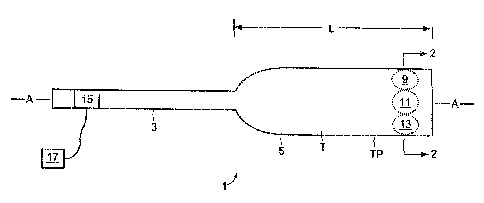
Now referring to FIG.1, preferably, the device is shaped for insertion into
and
withdraw) from the human body. As such, the device typically comprises a probe
1
2o having a shape suitable for entry into and withdraw) from the body. The
probe may
comprise a proximal portion 3 and a distal portion 5. The proacimal portion of
the probe
may include a power lead 1 S for activating the treatment sources. Typically,
distal
portion 5 comprises l) a tubular portion T having a tube perimeter TP and ii)
a plurality of
treatment sources (such as sources 9, 11 and 13) located on yr within the tube
T. The
25 function of tubular portion T is to essentially transport th.e treatment
sources. and it can
be solid or hollow.
Together, the proximal and distal portions of the probe may define a
longitudinal
axis A and a cross-section CS, as shown in FIG.). In many embodiments, the
distal
portion of the probe has length L (measured along axis A) and a cross-section
CS
3o whereby the length L of the distal portion is at least 10 times longer than
the cross-section
CS of the distal portion, preferably at least 100 times longer.
13
CA 02423064 2003-03-21
In some embodiments, the probe is shaped for insertion into an intervertebral
disc.
As such, it has a shape suitable for forming a bore in the disc for both entry
into and
withdraw) from the intervertebral disc, Preferably, the cross section of the
distal portion
of the device is less than the height of the targeted disc.
s in some embodiments, the probe is shaped for insertion into a vertebral body
portion of the vertebra. As such, it has a shape suitable for forming a bore
in the
vertebral body, and for entry into and withdraw) from the vertebral body.
In some embodiments, the probe is shaped for insertion between two vertebral
bodies, but outside the intervertebral disc.
to In selected embodiments, the two treatment sources provided within the same
probe may therapeutically act upon i) different tissue sites, or ii) different
components of
the same tissue site by embodiments including but not limited to the
embodiments
disclosed in the following:
Now referring to FIG.2, in some embodiments, a treatment source may be
radially
I S biased so as to emit energy in a preferred radial direction. For example,
source 9 may
emit energy preferentially in the 12 o°eloek direction whip source 13
may emit energy
preferentially in the 6 o'clock direction. Providing a source with a preferred
radial
emission direction may be accomplished, for example, by masking a portion of
the
circumference of a cylindrical source (see Deardorf, Ultrasound applicators
with internal
2o cooling for interstitial thermal therapy, SP1E Conference of Thermal
Treatment of Tissue
with Tmage Guidance, January 1999), or by using segmented emission sources, as
in
F1G.19B of Burdette, the specification of which is incorporated by reference.
When the
source is designed so as to produce an elliptical energy pattern, the emission
pattern is
considered to be bi-directional along the longitudinal axis of the ellipse.
z5 Now refernng to FIG.3, in some embodiments, the sources may emit energy in
different dispersion angles. For example, source 9 may have a concave surface
so that
energy emitted therefrom focuses upon relatively small volume V, while source
13 may
have a convex surface so that energy emitted therefrom can couple with a broad
surface
S.
14
CA 02423064 2003-03-21 '
Now referring to F1G.4, in some embodiments, the sources may emit energy with
different intensities. For example, sources 9 and I3 may provide a high
intensity emission
H white source 11 may provide a low intensity emission L_
Now referring to FIG., in some embodiments, the sources may emit energy with
different frequencies. For example, sources 9 and 13 may have a high frequency
a
emission while source 11 may have a low frequency ~ emission.
Now referring to FIG.6, in some embodiments, the sources may emit different
types of energy. For example, source 9 may emit ultrasound energy (US) while
source 1 1
may emit microwave energy (NIW).
0 The treatment source of the present invention includes alI forms of energy
which
may have a therapeutic effect. Such sources include but are not limited to
energy output
devices (such as electrical sources, Sight sources, and acoustic sources) and
chemical
delivery sources.
Examples of electrical sources include a) sources which produce resistive
heating,
t5 and b) radiofrequency sources. Examples of therapeutic devices having
sources which
produce resisitive heating can be found in US Patent No. 6,261,311, the
specification of
which is incorporated by reference. When radiofrequency sources are used,
either
monopolar or bipolar sources may be employed. Preferably, the radiofrequency
source is
bipolar. Examples of therapeutic devices having radiofrequency sources can be
found in
2o US Patent Nos_ 6,105,581 and 5,458.96, the specification of which is
incorporated by
reference.
Examples of light sources include UV sources, visible light sources, and
diffused
laser light sources. Preferably, the light source provides interstitial
penetration of the
target tissue to achieve a desired depth of penetration. 7Exatnples of
therapeutic devices
2s having light sources can be found in US Patcnt Nos. ~~,437,661 and
5,084,043, the
specifications of which are incorporated by reference.
Examples of acoustic sources include ultrasonic transducers. Examples of
therapeutic devices having ultrasonic transducers can be found in US Patent
Nos.
5,620,479 ("Diedrich I") and 5,733,315("Lax"), the specifications of which are
3o incorporated by reference.
CA 02423064 2003-03-21
Examples of chemical delivery sources include a pair of chemicals which when
combined produce an exothermic reaction. 1_n one embodiment, the chemical
sources
comprises a monomer and a chemical which, when combined with the monomer,
produces an exothermic cross-linking reaction to produce a polymer.
s In some embodiments, the first treatment source provides therapy by a form
of
energy which is different than that of the second treatment source_ For
example, the first
treatment source may be an ultrasonic transducer and the second treatment
source may be
a resistive heating element. In another example, the first treatment source
may be an
ultrasonic transducer and the second treatment source may be a microwave
heatinb
t o source. In another example, the first treatment source may be an
ultrasonic transducer and
the second treatment source may be an RF heating source, preferably a bipolar
RF source.
In another example, the first treatment source may be an energy source (such
as an
ultrasonic transducer) and the second treatment source may be chemical
delivery source
(preferably a source which delivers a pair of chemicals which when combined
have an
t 5 exothermic reaction}.
In some embodiments, at least one of the treatment sources comprises an
ultrasound transducer. One advantage of ultrasonic transducers is the ability
to focus the
ultrasound energy upon a small volume of tissue far away from the transducer.
Accordingly, a device having an ultrasound transducer may be inserted either
a) into the
2o intervertebral disc or b) between two adjacent vertebrae but outside the
disc, and the
ultrasound energy from this transducer may be focused in such a way as to heat
the
adjacent vertebrae, a spinal facet joint capsule, or a portion of the adjacent
PLL or ALIT
spinal ligament. At the same time, the second treatment source (which also may
be
ultrasound or may be another type of treatment source such as a resistive
heating
25 element) may be used to heat the annulus fibrousus portion of the disc or
to coagulate or
liquify the collagen component of the nucleus puiposus therein.
in many embodments, the ultrasound transducer comprises a ceramic component,
and typically is a sintered, polycrystalline ceramic component. In some
embodiments
using two ultrasound transducers to heat different tissue sites, the ceramic
component of
3U the first ultrasonic transducer has a composition which is different than
the ceramic
component of the second ultrasonic transducer. The different ceramic component
m
CA 02423064 2003-03-21
compositions can produce different frequencies of ultrasound given the same
energy
input. The different frequencies will in turn couple selectively to different
tissue
structures. This difference in frequencies produces a different acoustic
penetration of
ultrasound, and the surgeon can exploit this difference by using a particular
transducer
type to treat a particular tissue site. For example, if the surgeon desires to
treat a first Soft
tissue structure (such as an intcrvcrtcbral disc or a spinal ligament), the
surgeon would
select a ceramic component which produces a relatively high frequency.
Conversely, if
the surgeon desires to treat a second hard tissue structure (such as a
vertebral body), the
surgeon would select a ceramic component which produces a relatively low
frequency.
~o In some embodiments using two ultrasound transducers to heat different
tissue
structures, the wall thickness of the first ultrasonic transducer ceramic
component is
different than the wall thickness of the second ultrasonic transducer ceramic
component .
This difference produces different frequencies of ultrasound given the same
energy input,
and the surgeon can exploit this difference by using a particular transducer
to
advantageously coupe a particular frequency with a particular tissue site. Far
example,
if the surgeon desires to treat a first soil tissue structure, the surgeon
would desire a
relatively high frequency and so would select a ceramic component having a
relatively
thin wall thickness. Likewise, if the surgeon desires t~o treat a second hard
tissue
structure, the surgeon would select a ceramic component having a relatively
thick wall
zo thickness. In some embodiments, the wall thickness of the first ceramic
component is at
least 2~% thicker (and in some embodiments at least 50% thicker) than the wall
thickness
of the second ceramic component.
In many embodiments, the ceramic Component of the ultrasound transducer has a
coating thereon. These coatings are typically made of a material selected from
the group
?5 consisting of a metal (such as gold) or a polymer. These coatings may be
employed to
alter the properties of the ultrasound wave emitted from the ceramic
component. They are
typically acoustically absorbent. However, the coating may also be used to
modify other
acoustic outputs. Far example, the coating may change the dispersion pattern
of the
ultrasound emission (by, for example, masking), change its frequency, or
change its
3o focus. Therefore, in some embodiments using two ultrasound transducers to
heat
different tissue structures, each transducer has a coating, and the coating
upon the first
m
CA 02423064 2003-03-21
ultrasonic transducer ceramic component is different than the coating upon the
second
ultrasonic transducer ceramic component. In other embodiments using two
ultrasound
transducers to heat different tissue structures, only one transducer has a
coating. In some
embodiments using two ultrasound transducers to heat different tissue
structures, each
s transducer has a coating, and the coating upon the first ultrasonic
transducer ceramic
component is the same a_c the coating upon the second ultrasonic transducer
ceramic
component.
In same embodirrments, at least one of the treatment sources comprises a
resistive
heating element. A resistive heating element provides the advantage of being
able to heat
t o by surface conduction. Accordingly, a resistive heating element is most
advantageous
when the surgeon is seeking to heat a surface, such as the internal wall of
the annulus
fibroses portion of the disc or the external wall of the annulus fibroses.
In some embodiments, at least one of the treatment sources comprises a
radiofrequency heating element. A radiofrequency heating clement provides the
t 5 advantage of being able to heat or ablate. Accordingly, a radiofrequency
heating clement
1S mOSt advantageous when the surgeon is seeking to ablate a tissue..
preferably, the radiofrequency heating element is bipolar. A bipolar RF
element
provides the advantage of being able to Localize current flow, and thereby
ablate without
producing substantially high temperatures in the surrounding tissues.
Accordingly, a
2U radiofrequency heating element is most advantageous when the surgeon is
seeking to
ablate a particular tissue without disturbing nearby tissues.
In some embodiments wherein ablation of a portion of the basivertebral nerve
is
desired, an RF element is desirably selected.
In some embodiments, a single treatment source is dynamically controllable
(e,g.,
25 its particular output can be changed during a procedure). When the source
is an
ultrasonic transducer, the acoustic output may be dynamically controlled by
changing the
power intensity, the frequency, the angle of dispersion, the focus, or other
dynamically
controllable parameters. In some embodiments using ultrasound, the device may
further
comprise a feedback monitor which monitors the changes in the acoustic
properties of the
30 treated tissue, and provides feedback to the device which then directs an
adjustment of
the ultrasound emission. In some embodiments using RF, the device may further
1s
CA 02423064 2003-03-21
comprise a feedback monitor which anonitors the changes in the itnpedenee of
the treated
tissue, and provides feedback to the device which directs an adjustment of the
RF
emasston.
in some embodiments, different treatnnent sources are individually
controllable
s (e.g., a first source has a different output than a second source). When the
source is an
ultrasonic transducer, the acoustic output may be controlled by intensity, by
frequency,
by angle of dispersion, by focus, or by other controllable parameters, such as
inputs- for
example, the device may comprise two different channel boxes which drive
respective
sources at dif3ferent freduencies.
In this way, the intensity and quality of the acoustic output may be tailored
for the
particular application. For example, in some embodiments, now referring to
FIG.7a, each
of treatment sources 71 and 72 (which are preferably ultrasound transducers)
preferentially emit energy in respective first A and second B directions
defining an angle
B of about 90 degrees therebetween. When this device is placed into an
intervertebral
t s disc (as in FIG.76), transducer 71 faces the upper endplate 73 of
vertebral body 74 while
transducer 72 faces the collagenous annulus fibroses 75 (preferably the
posterior inner
wall thereof). Because bane couples much more efficiently with ultrasound than
with
collagen, endplate 73 will heat up much more quickly than the annulus fibroses
7S.
Accordingly, controlling the output of bone-directed transducer 71 so that it
produces less
zo ultrasound energy than the collagen-directed transducer '72 wily allow the
surgeon to
provide enough energy to both Lhe bone and annulus fibroses so that eaeh
tissue heats up
to its desired temperature in about the same time. This ability to produce
such a tailored
results provides an advantage over conventional devices.
Therefore, in some embodiments, the first transducer is adapted to heat the
25 endplate while the second transducer is adapted to heat the annulus
fibroses, and the
energy flux from the first transducer is less than that of the second
transducer.
Now refern.ng to FIG.8a, in some embodiments, transducer 76 which emits
energy substantially iz~ the G direction is added to the device of FIG.7a
dirceEly opposite
from transducer 71 so that both the upper 73 and lower 81 endplates can be
heated at the
3n same time (as in FIG.8b).
!9
CA 02423064 2003-03-21
Now referring to FIG.9a, in some embodiments, transducer 77 which emits
energy substantially in the D direction is added. to the device of FIG.7a so
that both the
posterior 78 and anterior 79 walls of the annulus fbrosus can be heated at the
same time
(as in FIG.9b).
Now referring to F1G_ 1 Oa, in some embodiments, both treatment sources 76 and
77 (which are preferably both ultrasound transducers) are added to the device
of FIG.7a
so that both endplates and both the posterior and anterior walls of the
annulus fibrosus
can be hEated at the samc time (as is FIG.IOb). When the device comprises at
least four
treatment sources (preferably at least two and more preferably all four being
ultrasound
to sources), the device can simultaneously treat upper and lower vertebral
bodies and
posterior and anterior portions of the annulus fibrosis from a location within
the
intervertebral disc. Preferably, the Erst, second, third and fourth sources
emit energy in
first, second, third and fourth directions, the directions respectively
defning angles y, b
and E therebetween, wherein angle ~y is between 80 and l Old degrees, angle 8
is between
t ~ 170 and 190 degrees, angle s is between 260 and 284 degrees.
In another instance which demonstrates the advantage of having individually
controllable sources, now referring to FIG.11 a, each of treatment sources 71
and 76 are
preferably ultrasound transducers which emit energy substantially in the
respective first
A and second B directions defining an angle B of about 18C~ degrees
therebetween. When
2o this device is placed into the intervertebral disc as shown an the F1G.11
b, transducer 71
faces the endplate 73 of upper vcrtcbral body 74 while transduceu 76 faces the
endplate
81 of lower vertebral body 80. In this instance, the surgeon can frost
simultaneously treat
sources of pain in the opposing vertebral bodies by using a first power level
which will
effectively therapeutically treat the vertebral bodies. Then, the surgeon can
rotate the
25 device 90° so that transducers 71 and 76 now face the opposing wall
portions 7$ and 79
of the annulus fibrosus {as in FIG.l 1 c)_ The surgeon can then treat sources
of pain in the
annulus fibrosus by using a second power level which will effectively hcat the
walls of
the annulus fibrosus. The second power level may be sufficient to coagulate
collagen
within the walls, or may be sufficient to denervate the nerves within the
walls. This
3o ability to produce such a tailored results provides an advantage over
conventional
devices.
CA 02423064 2003-03-21
In some embodiments, the treatment steps are reversed whereby the annulus
fibrosus is first treated, and then the vertebral bodies are then treated.
In another instance which demonstrates the advantage of having indiv ideally
focused sources, now referring to FIG. I2a, each of treatment sources 7 t and
76 are
preferably ultrasound transducers which emit energy in substantially the same
direction.
Whereav treatment source 71 is concave and produces a focused energy pattern
within
volume V, treatment source 76 is convex and produces a dispersed energy
pattern. When
the device of F1G.12a is placed into the intervertebral disc as shown in the
FIG.l2b, each
0 transducer 71 and 76 faces upper vertebral body 74. HowEVer, whereas upper
transducer
71 is focused to treat sources of pain deep within the upper vertebral body
(such as nerve
trunk NT), lower transducer 76 focuses its energy upon the vertebral endplate
73 (eo treat
the nerve endings located therein). In use, activation of source 76 will cause
a
temperature rise within the endplate sufficient to denervate the plurality of
nerve endings
located in or near the endplate, while activation of source i'1 will cause a
temperature rise
in the vicinity of nerve trunk NT sufficient to dene:rvate the basivertebral
nerve
substantially at its source.
After so treating the upper vertebral body, the surgeon may then flip the
probe
180 degrees (as in FTG.l2c), in order to similarly treat the lower vertebral
body.
zU Now referring to FIG_12d, in another embodiment, two concave sources 91, 92
and two convex sources 93, 94 are disposed within th.e probe so that
simultaneous
treatment of both the endplates and the nerve trunks may occur. In particular,
the
concave sources can heat the respective nerve trunks while the convex sources
heat the
vertebral endplates.
In some instances, the temperature rise within an endplate treated with the
device of 1~ 1Gn 12a may also be sufficient to cause heat to radiate from the
vertebral
endplate to the intervertebral disc 95 in an amount sufficient to cause at
least one of
collagen shrinkage or nerve denervation within the intervertebral disc.
In another instance which demonstrates the advantage of having individually
3o controllable sources, now referring to F1G.13a, each of treatment sources
101 and 103
emit energy in first and second directions defining an .angle 8 of about 180
de~ees
21
CA 02423064 2003-03-21
therebetween. When this device is placed into the intervertebral disc as shown
in the
FIGS. 13 b and 13d, source 101 faces the endplate 73 of upper vertebral body
74, while
source 10:i faces the endplate 8I of lower vertebral body S0. Now referring to
FIG.13b,
the surgeon can first treat sources of pain deep within the vertebral bodies
(such as nerve
trunks NT) by using a first focus which will effectively direct substantially
alI the energy
to the nerve root. Now refernng to FIG.l3c, the; surgeon can adjust the foci
of the
sources in situ to a second broader focus. Now referring to Fl.G.l3d, the
surgeon can
then focus all the energy upon the vertebral endplates 73 and 81, thereby
heating up each
endplate to a desired temperature.
The sequence steps of this embodiment may also be reversed.
In some embodiments, the probe may have a single in-situ focus adjustable
source, thereby requiring the probe to be flipped as described above in order
to treat each
adjacent vertebral body.
In some instances, the temperature rise within the endplate will be sufficient
to
denervate the plurality of nerve endings located in or near the endplate. In
some
instances, the temperature rise within the endplate will be sufficient to
cause heat transfer
from the vertebral endplate to the intcrvertebral disc in an amount sufficient
to cause at
least one of collagen shrinkage or nerve denervation within the intervertebral
disc.
This ability to produce such a tailored results provides an advantage over
zo conventional devices.
In another instance which demonstrates the advantage of having individually
controllable sources, now referring to FIG.l4a, lateral treatment sources 11 l
and 115 are
ultrasound transducers which emit energy in first A and second B directions
defining an
angle of about 180 degrees thecebetween_ On the other hand, middle local
treatment
source 113 is a source for more localized heating, such as a resistive heating
element, a
bipolar RF heating element, or a concave ultrasound transducer having a short
focus.
When this device is placed into the nucleus pulposus of an intervertebral disc
as shown in
FIG.l4b, transducer 1 I S faces the right portion of the disc's annulus
fbrosus portion 78,
while transducer I 1 1 faces the Ieft portion of the disc's annulus fibrosus
portion 79. In
one embodiment, the surgeon can first depressurize the nucleus pulposus by
activating
the local source 113 which will effectively locally deposit alt of its energy
into the
22
CA 02423064 2003-03-21
nucleus pulposus and vaporize at least a portion of it. Next, the surgeon can
activate the
lateral ultrasound transducers to deposit their energy upon the respective
lateral walls of
the annulus fibrosus 78 and 79, thereby heating up these walls to a desired
temperature.
In some instances, the temperature rise within the walls will be sufficient to
denervate the
plurality of nerve endings located in or near the walls. In some instances,
the temperarurt
rise within the walls will be sufficient to cause collagen shrinkage within
the walls.
Alternatively, the sequence of treatment steps in this procedure may be
reversed,
wherein the surgeon frst treats the annulus fibrous and then treats the
nucleus pulposus_
Alternatively, the surgeon can activate all of the sources at the same time.
no This ability to produce such a tailored results provides an advantage over
conventional devices.
In some embodiments wherein the first and second treatment sources are of the
same type (e.g_, both are ultrasonic transducers), the surface energr,~ flux
of the frst
source may be different than the surface energy flux of the second source. In
preferred
1 s embodiments, the surface energy flux of the first source is at last twice
as large as the
surface energy flux of the second source. This e:mbodirnent would have find
advantageous in simultaneously treating two different tissues types (e.g., the
vertebra
and the annulus fibrosus of the intervertebral disc) which couple to
ultrasound differently.
Sirnitarly, in some embodiments wherein the first and second treatment sources
20 are of the same type (e.g., both are ultrasonic transducers)., the surface
energy flux density
of the first source may be different than the surface energy flux density of
the second
source. In preferred etxtbodiments, the surface enery flux density of the
first source is at
least twice as large as the surface energy flux density of the second source.
Now referring to 1~ lG.7a, in some embodiments, first 71 and second 72
treatment
25 sources emit energy in first and second directions defining an angle 8 of
between 45 and
135 degrees, preferably between 60 and 12U degrees, more preferably between $0
and
100 degrees, most preferably about 30 degrees. In this rnost preferred
embodiment, the
emissions from the sources are substantially orthogonal. Such a device would
be useful
for simultaneously treating both a vertebral body and a wall of the annulus
fibrosus from
3o the middle of the intervertebral disc, or from between adjacent vertebrae
but outside the
disc.
23
CA 02423064 2003-03-21
Now referring to FIG.IIa, in other embodiments, the first 71 and second 72
sources emit energy in first and second directions defining an angle ~y of
between morc
than 135 and 225 degrees, preferably between 160 and 200 degrees, more
preferably
between 175 and 185 de~ees, most preferably about I80 degrees. In this most
preferred
embodiment, the emissions from the sources are substantially linear. Such a
devioe would
be useful for simultaneously treating adjacent vertebral bodies from the
middle of the
intervertebral disc, or from beriveen adjacent vertebrae but outside the disc.
Now referring to FIGS. 15a and b, in some embodiments, first 12I and second
t 23 treatment sources have respective dispersion angles a and (3, and wherein
a is
different than Vii. Such a device would be useful in simultaneously treating
the endplates
of a vertebral body and the walls of the annulus fibrosu5 from within the
intervertebral
disc, or from between adjacent vertebrae but outside the disc. In this
particular design,
since the endplates are relatively wide and the annulus f brosus is relatively
short. the
wide angle a is preferably much greater than the angle Vii. In some
embodiments, the
t5 wide angle a is at least 3 Mmes greater than angle (3, more preferably at
least 10 times
greater than angle ~3.
As noted above, Heggeness discloses a method of treating back pain which
includes
ablating the basivertebral nerve within the vertebral body portion of a
vertebra.
However, the Heggeness disclosure requires boring a hole in the vertebral body
in order
2o to gain unobstructed access to that nerve. Boring such a hole is
undesirable because it is
a time consuming process and leaves a hole in the treated vertebral body. In
addition, the
excavated bone must somehow be managed, thereby adding to the complexity of
the
procedure.
Therefore, also in accordance with the present invention, there is provided a
method
25 of denervating nerves in a vertebral body, comprising the steps of
a) providing a device having a treatment source,
b) placing the treatment source inside the human body, and
c) activating the treatment souroe to cause energy to~ flow from the treatment
source
and into the vertebral body in an amount sufficient to denervate nerves in the
30 vertebral body.
24
CA 02423064 2003-03-21
In some embodiments, the treatment source is placed within the intervertebral
disc. Ln other, it is placed between adjacent vertebrae but outside the disc.
Preferably, the
treatment source is an ultrasound transducer. Ultrasound is of particular
advantage in this
regard in that it can heat the vertebra from outside the mertebra~
Accordingly, the time
and invasiveness and bone management issues present: in the method disclosed
by
Heggeness are avoided. Preferably. the ultrasonic transducer may be focused to
provide
localized energy substantially to an interior portion of the vertebral body,
more preferably
to an area housing the trurttc of the basivertebral nen~e. However, in some
embodiments,
the activation step causes energy to ilow from the treatment source and
substantially into
~o the vertebral endplate in an amount sufficient to denervate a nerve ending
in the vertebral
endplate. In such instance, the treatment source need not necessarily be an
ultrasound
transducer.
In some embodiments which treat the vertebral body from within the
intervertebral
disc, the device is the device of FIC.1.1 la which provides essentially co-
linear emission
t s from the treatment sources, wherein the first and second sources are
oriented so that the
first source faces the upper adjacent vertebral endplate and the second source
faces the
lower adjacent vertebral endplate during step a} above. This method may
further
comprise the steps of
a) providing a device comprising first and second treatment sources forming a
co-
ao linear envision pattern,
b) placing the treatment source into an intervertebral disc so that the first
treatment
sotcrce faces a first vertebral body and the second treatment source faces a
second
vertebral body, and
c) activating the first treatment source to cause energy to flow from the
first
25 treatment source and into a first vertebral body in an amount sufficient to
denervate a nerve in the first vertebral body, and.
d) activating the second treatment source to cause energy to flow from the
second
treatment source and into the second vertebral body in an amount sufficient to
denervate a nerve in the second vertebral body.
3o This device may then be rotated to treat the annulus fibrosus, so that the
method may
further include the steps of
CA 02423064 2003-03-21
e) rotating the device approximately 90° so that the fiast source faces
a first portion of
the annulus fibroses, (and preferably the second source faces a second portion
of the
annulus fibroses),
f) energising the fast source and second source so that the first source heats
the first
portion of the annulus fibroses and the second source heats the second portion
of the
annulus fibroses.
In some embodiments, the treatment sequence is reversed so that this device
first acts
upon the annulus Fbrflsus, i~ rotated 90 degrees, and then acts upon the
adjacent vertebral
body or bodies.
In some embodiments, the device is the device of FlCi.7a which provides
orthogonal
emission from the treatment sources. In this case, the method of denervating
nerves in a
vertebral body comprises the steps of
a) providing a device comprising first and second treatment sources forming an
orthogonal emission pattern,
~5 b) placing the treatment source into an intervertebral disc so that the
first treatment
source faces a vertebral body and the second treatment source faces an annulus
f brosus, and
e) activating the first treatment source to cause e-nergy to flow $rom the
first
treatment source and into the vertebral body in an amount sufficient to
denervate
2o a nerve in the vertebral body, and.
d} activating the second treatment source to cause energy to flow from the
second
treatment source and into the first portion of the annulus fibroses in an
amount
sufficient to denervate a nerve and/or shrink the collagen within the annulus
fibroses.
25 The device may then be rotated to treat the second adjacent vertebra and
the opposing
face of the annulus fibroses, so that the method further comprises the steps
of
e) rotating the device approximately 90° so that the first source faces
a second portion
of the annulus fibroses and the second source faces thc; second adjacent
vertebra,
f)activating the first treatment source to cause energy I_o fl~w from the
first treatment
30 source and into the second portion of the annulus fibroses in an amount
sufficient to
denervate the nerves andJor shrink the collagen within the annulus fibroses,
and.
zb
CA 02423064 2003-03-21
g) activating the second hreatmertt source to cause energy to flow from the
second
treatment source and into the second vertebral body vertebral body in an
amoune
sufficient to denervate the nerves in the second vertebral body.
h) energizing the first source so that the fast source heats the first portion
of the
S annulus fibrosus.
Although many embodiments of the present invention employ energy sources
having preferred directional emissions, the present invention is not limited
to preferred
directional emissions. For example, and now referring to FIG.l6a, there is
provided a
probe have a first energy source 201 which spans the full circumference of the
probe and
t0 provides a first radially unifornn emission 309, and a second energy source
203 which
spans the full circumference of the probe and provides a radially uniform
emission 313.
blow referring to FIG. 16b, when this probe is inserted into the disc, the
first energy
source may have a first frequency which preferentially couples with bone (and
thereby
preferentially heats the adjacent vertebral bodies), while the second energy
source may
~ 5 have a second frequency which preferentially couples with collagen (and
thereby
preferentially heats the collagen in the annulus fibrosus).
As noted above, lieggeness discloses treating the nerves in bone with many
different ablation sources, but does not disclose sources such as ultrasound,
microwave,
UV source, or an exothermic chemical source, each of which can act upon the
nerve of
20 interese from long range. Accordingly. 1-leggeness' procedure must bore
into the
vertebral body to a point near the nerve of interest in order to et~ect
treatment. Cosman
discloses treating only vertebral bodies which are tumorous, and so offers
little help for
the treatment of healthy vertebral bodies whose nerves ma;y be the source of
pain.
Therefore, in accordance with the present invention, there is provided a
method of
zs denervating nerves in a healthy vertebral body, comprising; the steps of-.
a) providing a device comprising at least one treatment source selected from
the
group consisting of an ultrasound transducer, a microwave source, a L1V
source,
and a eXOtherTillC Chemical Source,
b) placing the treatment source into the healthy vertebral body, and
o c) activating the device to cause energy to flow from the treatment source
in an
amount sufficient to denervate a nerve in the healthy vertebral body.
27
CA 02423064 2003-03-21
In some embodiments, the nerve of interest includes; the trunk of the
basivertebral
nerve. In other embodiments, the nerve of interest includes a nerve ending
located in the
endplate portion of the vertebral body.
Because this inventive method allows the use of long range sources such as
ultrasonic
s energy a_s a way of heating, it can focus energy from a far away location.
Therefore, the
method does not require that the treatment source be placed in close prohimity
to the
nerve of interest. Therefore, this method is advantageous i.f a) the approach
to a nerve of
known location is problematic, b) if the location of the nerve is not well
known, c) if the
surgeon desires to heat either the entire vertebral body or just the nerves of
the vertebral
2o endplate. However, care mu.5t be taken to select an appropriate frequency
which allows
sufficient energy transmission through the vertebral body.
As noted above, both Heggeness and Cosman disclo >es treating the nerves in
bone
with many different treatment sources, but do not disclose treatment sources
which effect
envlrOnrnental cooling.
15 Therefore, in accordance with the present invention, there is provided a
method of
denervating nerves in a healthy vertebral body, comprising the steps of:
a) providing a device comprising a cooling source,
b) placing the cooling source in the healthy vertebral body, and
c) activating the device to cool the healthy vertebral body in an amount
sufficient to
zo denervate the nerves in the healthy vertebral body.
In one embodiment, the cooling source comprises liquid nitrogen. In another,
the cooling
source comprises heat pipe technology.
The many conventional methods for heating the annulus fibrosus or nucleus
pulposus
disclose essentially direct methods of heating these tissues. Unfortunately,
due to the low
25 heat capacity of these tissues, the heat imparted to them dissipates
relatively quickly,
thereby leading to relatively short treatment times and relatively localized
treatment.
Therefore, in accordance with the present invention, there is provided a
method of
treaeing an interveztebral disc, comprising the steps of:
a) providing a device comprising at least one ultrasound transducer,
3o b) placing the ultrasound transducer into the human body, and
zs
CA 02423064 2003-03-21
e) energizing the ultrasound transducer to cause energy to flow from the
energy
output source and into the vertebral ea~dplate in an amount sufficient to heat
the
vertebral endplate to a temperature which causes :heat transfer from the
vertebral
endplate to the intervertebral disc in an amount sufficient to cause at least
one of
collagen shrinkage or nerve denervation within the intervertebral disc.
In this inventive method, the vertebral endplate acts as a heat capacitor.
This provides
two advantages. First, the heat in the vertebral endplate spreads evenly over
the endplate,
thereby providing a uniform heating source which spans the width of the
annulus
fibrosus. Second, the high heat capacity of the vertebral body allows it to
effectively
conduct and/or radiate hEat for a relatively long period of time. Thereby
allowing for
prolonged treatment of the annulus fibrosus.
In some embodiments, this method comprises the step of
a) energizing an ultrasound transducer to cause energy to flow from the
ultrasound
t 5 transducer and into the vertebral endplate in an amount sufficient to heat
the
vertebral endplate to a temperature which causes heat transfer from the
vertebral
endpEate to the intervertebral disc in an amount sufficient to cause at least
one of
collagen shrinkage or nerve denervation within the intervertebral disc.
In other embodiments, the device is an implant. These implants have the
2o advantage of requiring only a single invasive procedure. Since it is
believed that many of
the energetic treatments, such as material removal, provide only temporary
relief to the
patient, providing an implant having a treatment sources whiclx can be
activated from
outside the body provides a distinct advantage over conventional probe-based
technologies which require invasive procedure far each treatment.
z5 Accordingly, the implant is shaped for substantially permanent residence
within
the human body. When the implant is placed within the intervertebral disc, it
preferably
has a height which is less than the height of the disc. Also preferably, it
has a foot print
which is less than the foot print of the disc. Also preferal>ly, the width of
the implant is
less than the width of the disc. In some instances, the implant is shaped to
substantially
3o reside within the space occupied by the nucleus pulposus.
z9
CA 02423064 2003-03-21
Therefore, in some embodiments, the step c~f therapeutically treating
comprises the
steps of
a) placing an implant comprising a treatment source substantially completely
within
the human body (preferably. within the disc, more preferably ~~ithin the
nucleus
pulposus}, arid
b) activating the treatment source to treat a first tissue site.