Note: Descriptions are shown in the official language in which they were submitted.
DYE PAIR FOR FLUORESCENCE RESONANCE ENERGY TRANSFER (FRET) MEASUREMENTS
The present invention concerns new fluorescent dye systems especially for
fluorescence resonance energy transfer determinations for example combined
with
the time-resolved measurement of the resulting fluorescence. The invention
also
concerns the use of these dyes to label biomolecules and for the homogeneous
determination of interactions between biomolecules, for example in the
detection of
an analyte.
Binding partners that can specifically bind to the biomolecule that is to be
detected or
examined are very often used to determine biomolecules. A basic
differentiation is
made between heterogeneous and so-called homogeneous assays, the latter being
characterized in that one or more washing steps are necessary to carry out the
test.
In so-called heterogeneous assays at least one biomolecule is provided with a
marker
group. The concentration of the analyte molecule to be examined is ultimately
determined by measuring this marker group. Of course this determination is
only of
practical use when the bound and unbound labelled binding partners have been
separated by means of a suitable washing step before carrying out the
measurement.
In conventional homogeneous assays the test conditions are selected such that
measurable signal changes due to turbidity effects or scattered light effects
which
depend on the concentration of the analyte molecule that is present are
generated.
Particulate carrier materials are often used to amplify the generated signals
in such
assays.
Only in recent years has it become possible to carry out homogeneous
determinations
i.e. determinations that do not necessarily require an intermediate washing
step, even
when using marker groups. Further developments in the field of homogeneous
immunoassays are all based on the interaction of at least two molecules which
only
CA 02426479 2003-04-22
-2-
occurs when these molecules are in direct proximity to one another.
Homogeneous
assays have become well-known that are based on the principles of "cloned
enzyme
donor immunoassays" = CEDIA (Microgenics Inc. USA), fluorescence polarization
= FPIA (Syva Co. USA) or scintillation proximity assays (Amersham UK).
However,
special mention is made here of those methods that are based on the principle
of
fluorescence resonance energy transfer (FRET). Always at least two dye
molecules
are required for fluorescence energy transfer. A first dye which acts as an
energy
donor and a second dye which acts as an energy acceptor. The energy transfer
between the donor and acceptor occurs non radiatively i.e. without emitting
radiation.
The efficiency of FRET is very dependent on the distance between the donor and
acceptor dye. FRET only occurs efficiently when the donor and acceptor are
very
close together.
Usually the donor molecule as well as the acceptor molecule are each bound to
one
partner of a bioaffine binding pair. If the carrier biomolecules interact and
associate
for example to form an antigen-antibody complex, the donor and acceptor
molecule
are also then in close proximity and FRET is possible.
The energy acceptors can either be selected such that they suppress the energy
released by the donor which are referred to as quenchers or the fluorescence
resonance energy acceptors can themselves release fluorescent energy i.e. they
fluoresce, these are referred to as fluorophore groups or briefly as
fluorophores. It is
known from the prior art that metallic complexes are suitable as fluorescence
energy
donors as well as fluorescence energy acceptors.
As mentioned above fluorophore groups are also known as acceptors in FRET
systems. In particular dyes from the group of allophycocyanins (APCs) are used
as
such fluorophores. A high quantum yield and very good absorbance properties
are
among some of the known properties of APCs (e.g. EP 076 695).
CA 02426479 2003-04-22
-3-
However, phycobiliproteins have disadvantages and thus for example due to the
high
molecular weight of more than 100,000 Da it is not possible to selectively
couple
them to a biomolecule i.e. via a predetermined position in the APC molecule.
This
coupling is usually chemical and hence statistical, or the binding is indirect
using
binding systems such as the streptavidin/biotin system known to a person
skilled in
the art. Also the sensitivity i.e. the lower limit of detection of these
systems appears
to be in need of improvement for example when detecting analyte molecules in
low
concentrations.
Commercial systems are available from Wallac, Oy, Turku, Finland and Packard
Instrument Company, Meriden, USA, which use lanthanide chelates as the donor
label and dyes from the phycobiliprotein class e.g. allophycocyanin as the
acceptor
label. The lanthanide chelates have a luminescence lifetime in a range up to
several
milliseconds i.e. the acceptor emission can be observed for a corresponding
length of
time. Hence the energy released by lanthanide chelates is usually measured in
a time
window between 400 - 600 microseconds. This also inevitably means that there
are
also relatively long dead times. The stability of the lanthanide chelates is
reduced
under certain test conditions; thus for example a re-chelation can occur when
complexing agents such as EDTA (ethylene-di-amino-tetra-acetic acid) are
added.
US 5,998,146 describes the use of lanthanide chelate complexes in particular
of
europium and terbium complexes combined with fluorophores or quenchers. It
also
underscores the advantageous properties of the long-lived lanthanide chelate
complexes.
Blomberg et al., Clinical Chemistry 45(6) (1999) 855 ff describe the use of
europium
or terbium complexes as donors and a rhodamine dye as an acceptor in new FRET
pairs. The sensitivity of the detection of (3hCG ((3 subunit of human
chorionic
gonadotrophin) is stated as 0.43 g/L. Thus the FRET assays based on europium
or
CA 02426479 2003-04-22
-4-
terbium chelate complexes do not lead to a major improvement with regard to
the
sensitivity of the assays.
The use of ruthenium complexes for time-resolved fluorescent measurement is
described for example in EP-A2-439 036 where lumazine is used as the energy
donor
and a ruthenium complex is used as the energy acceptor.
Joun et al., Analytical Biochemistry 232 (1995) 24-30 use fluorescent
ruthenium
complexes as energy donors for homogeneous determinations based on the FRET
principle. The dye well-known under the trivial name "reactive blue" is used
as the
resonance energy acceptor. Reactive blue suppresses the fluorescence emitted
by the
ruthenium complex and hence the quantification is based on the suppressed
fluorescence signal which was originally emitted by the ruthenium complex.
WO 00/47693 also describes the use of ruthenium chelate complexes as
fluorescence
energy donors in combination with quenchers. The ruthenium complex known under
the trivial name "Fair Oaks RedTM" was used as the energy donor. This dye was
coupled to an antibody to human serum albumin. The antigen human serum albumin
was labelled with a non-luminescent dye known as "light green yellowish". As
with
Joun et al., (see above) the analyte concentration (human serum albumin) was
ultimately determined from the extent of signal suppression.
For the assay of biomolecules it is in very many cases also necessary to
detect small
quantities of the analyte molecule to be examined. The term lower limit of
detection
is often used to characterize the sensitivity of a measuring system. The lower
this
detection limit the more sensitive is the test system.
However, there is still considerable potential for improvement with regard to
the
simplicity of coupling FRET dyes to biomolecules and above all with regard to
the
sensitivity of tests that are based on the FRET principle. More sensitive
CA 02426479 2003-04-22
-5-
homogeneous test procedures based on FRET measurements would have a broader
and more diverse practical use and would therefore be very desirable.
Hence the object of the present invention was to search for and optionally to
describe
new FRET pairs which can overcome the known disadvantages of the prior art
e.g.
with regard to the lower limit of detection.
Surprisingly it was found that metallic chelate complexes based on metal ions
of
groups VII and VIII of the transition elements can be used to great advantage
as
energy donors in combination with low-molecular fluorophores as energy
acceptors
for example in sensitive methods for determining the interaction between
biomolecules based on the FRET principle.
Brief description of the invention:
The invention concerns methods for determining the interaction between
biomolecules labelled with a donor or acceptor based on the principle of
fluorescence
energy transfer and measurement of the resulting fluorescence which are
characterized in that metallic chelate complexes based on metal ions of groups
VII
and VIII of the transition elements are used as energy donors in combination
with
low molecular fluorophores having a molecular weight between 300 Da and 3000
Da
as energy acceptors.
It was surprisingly found that low molecular fluorophores in particular from
the dye
classes rhodamines, xanthenes, cyanins and oxazines are excellently suitable
for
FRET measurements especially in combination with metallic chelate complexes
based on metal ions of groups VII and VIII of the transition elements in
particular
ruthenium chelate donors, to construct improved FRET assays.
CA 02426479 2003-04-22
-6-
The FRET pairs according to the invention are especially suitable for the time-
resolved measurement of the resulting energy (TR-FRET).
The dye combinations according to the invention are also suitable for methods
for
determining the interaction between biomolecules labelled with a donor or
acceptor
which are based on the principle of fluorescence modulation.
The new FRET pairs can now be used in a very advantageous manner to examine
interactions of biomolecules especially when the biomolecules labelled with
the
FRET partners are firstly in very close spatial proximity and are further
apart after
interaction or conversely when they can be brought very close to one another
for
example by forming a complex between the partners of a bioaffine binding pair.
The invention improves and extends the possibilities for detecting numerous
analyte
molecules especially in so-called homogeneous test procedures and therefore
also
encompasses test kits for detecting an analyte in a sample which contain at
least one
biomolecule labelled with a metal chelate complex based on metal ions of
groups VII
and VIII of the transition elements, especially a biomolecule labelled with
the
ruthenium chelate complex, and a biomolecule labelled with a low molecular
fluorophore acceptor.
Detailed description of the invention:
The invention essentially concerns methods for determining the interaction
between
biomolecules labelled with a donor or acceptor based on the principle of
fluorescence
energy transfer and measurement of the resulting fluorescence which are
characterized in that metallic chelate complexes based on metal ions of groups
VII
and VIII of the transition elements are used as energy donors in combination
with
low molecular fluorophores having a molecular weight between 300 and 3000 Da
as
energy acceptors.
CA 02426479 2003-04-22
-7-
Interaction in the sense of the invention means changes in the distance
between
biomolecules that can be detected by FRET measurement. In order to detect this
interaction, it is necessary that a FRET donor as well as a FRET acceptor are
coupled
to a biomolecule or each is coupled to one partner of a binding pair and that
the
interaction leads to a change in the distance between the donor and acceptor.
In a preferred embodiment one partner of a bioaffine binding pair is labelled
with the
donor and the other is labelled with the acceptor. The donor and acceptor come
into
very close proximity as a result of the formation of the binding complex
between the
partners of the bioaffine binding pair and FRET becomes possible.
Known examples of bioaffine binding pairs are in particular complementary
nucleic
acid sequences (DNA, RNA or peptidic nucleic acids), ligands and receptors,
antigen
or hapten and antibodies, or lectins and sugars. Under suitable conditions the
partners
of these binding pairs associate to form complexes.
The extent of complex formation is preferably used to determine the
concentration of
an analyte molecule. For this purpose the reaction conditions are selected in
a manner
known to a person skilled in the art such that depending on the test format
either an
increase or decrease in the signal occurs which depends on the analyte
concentration.
Another preferred embodiment of an interaction in the sense of the invention
is when
the donor and acceptor are originally present under conditions which allow
FRET but
the FRET is interrupted for example by enzymatic activity between the donor
and
acceptor coupling site.
Fluorescence energy transfer is understood as the transfer of energy from a
donor dye
to an acceptor dye during which the donor emits the smallest possible amount
of
measurable fluorescent energy. In this method a fluorescent dye donor is for
example
excited with light of a suitable wavelength. Due to its spatial vicinity to a
suitable
CA 02426479 2003-04-22
-8-
second dye, the acceptor, this results in a so-called non-radiative i.e.
radiation-free
energy transfer to the acceptor (Van der Meer, et al., Resonance Energy
Transfer
VCH (1994)). If the second dye is a fluorophore or luminophore, the light
emitted by
this molecule at a particular wavelength can be used for a qualitative as well
as for a
quantitative determination.
In many test systems based on this FRET principle the luminophore group acting
as a
donor is excited and converted by absorption of a photon from a ground state
into an
excited state. If the excited donor molecule is close enough to a suitable
acceptor
molecule, the excited state can be transferred from the donor to the acceptor.
This
energy transfer results in a decrease in the fluorescence or luminescence of
the donor
and, if the acceptor is itself luminescent, results in an increased
luminescence. If the
acceptor is a quencher, it of course exhibits no fluorescence.
The efficiency of the energy transfer depends very strongly on the distance
between
the donor and the acceptor molecule. The decrease in signal depends on the
sixth
power of the separation distance. Due to this dramatic effect of the distance
between
the donor and acceptor, FRET pairs (also referred to as FRET systems) can be
used
to examine how many donor and acceptor molecules are present in close
proximity to
one another. This property is used for example to determine the presence of an
analyte by contacting it with a specific partner. Numerous applications of
FRET
systems are known from the prior art. For this reference is in particular made
to
WO 00/47693, EP 76 695; Hemmila, Chemical Analysis 117, John Wiley & Sons,
Inc. (1991) 135-139; and Van der Meer et al., Resonance Energy Transfer VCH
(1994) supra.
Preferred embodiments of FRET systems are detection methods which additionally
utilize a time-delayed measurement of the signal from a FRET system. Basic
devices
and methods for determining time-resolved FRET signals are described in the
prior
art. The principle of time-resolved FRET measurements is essentially based on
CA 02426479 2003-04-22
CA 02426479 2003-04-22
-9-
selecting a measuring window such that interfering background fluorescence
that
may for example be due to interfering substances in the sample, is not co-
detected,
but rather only the fluorescence generated or suppressed by the energy
transfer is
measured.
The resulting fluorescence of the TR-FRET system is determined by means of
appropriate measuring devices.
Such time-resolved detection systems use for example pulsed laser diodes,
light
emitting diodes (LEDs) or pulsed dye lasers as the excitation light source.
The
measurement occurs after an appropriate time delay i.e. after the interfering
background signals have decayed. The basic design of such measuring equipment
is
shown in fig. 1. Commercially available measuring systems e.g. based on Xenon
flash lights such as VictorTM from Wallac Oy, are not suitable for the
sensitive
determination of time-delayed fluorescence in the range of a few s as is
required for
FRET pairs according to the invention but only for FRET systems having a
lifetime
of more than 10 s.
The detection can preferably also be carried out using a phase modulation
technique.
In this case the intensity of the excitation light is modulated with a high
frequency
and likewise the intensity of the emission also. The lifetime results in a
phase shift
and demodulation of the fluorescence emission. Explicit reference is herewith
made
to relevant information on the corresponding systems (WO 00/47693; French et
al.,
SPIE BiOS in Proc. SPIE v 3259 (1998) 209-218 and French, et al., SPIE BiOS in
Proc. SPIE v 3603 (1999) 272-280). A person skilled in the art will find the
necessary information in these references to successfully use the dye
combinations
according to the invention also in such fluorescence modulation systems. The
following mainly relates only to TR-FRET but it is obvious to a person skilled
in the
art that he can also use the phase modulation technique to measure the FRET
pairs
according to the invention.
-10-
FRET systems based on metallic complexes as energy donors and dyes from the
class of phycobiliproteins as energy acceptors are known in the prior art (EP
76 695;
Henunila, Chemical Analysis 117, John Wiley & Sons, Inc., (1991) 135-139).
Established commercial systems (e.g. from Wallac, OY or Cis Bio Packard) use a
FRET pair consisting of a lanthanide chelate as the metallic complex and a
phycobiliprotein.
The advantageous properties of the lanthanide-chelate complexes in particular
of
europium or terbium complexes are known and can be used in combination with
quenchers as well as in combination with fluorophores. The combination of such
lanthanide-chelate complexes with low molecular fluorophores does not appear
to
result in a substantial improvement in the sensitivity (US 5,998,146 and
Blomberg et
al., supra).
Ruthenium complexes per se are used as fluorophores or luminophores especially
for
electro-chemoluminescence. Preferred ruthenium-chelate complexes are for
example
known from EP 178 450 and EP 772 616 in which preferred methods for coupling
these complexes to biomolecules are also described. Their use as energy donors
in
FRET systems is not discussed there.
Allophycocyanins have excellent properties such as unusually high extinction
coefficients (about 700 000 L/M cm) and also extremely high emission
coefficients.
These are ideal prerequisites for their use as fluorophore acceptors in FRET
systems.
Moreover these dyes are known to be readily soluble in water and stable.
The term low molecular fluorophore refers to fluorophoric dyes having a
molecular
weight between 300 and 3000 Da. Such low molecular fluorophoric groups such as
xanthenes, cyanins, rhodamines and oxazines have considerable disadvantages
compared to the APCs with regard to important characteristics. Thus for
example their
extinction coefficients are substantially lower and are in the range of ca.
100 000 L/M
CA 02426479 2003-04-22
CA 02426479 2003-04-22
-11-
cm. It is also known that unspecific binding due to the hydrophobic properties
of these
chromophores is a potential disadvantage for these dyes as acceptors in FRET
systems.
It has now been surprisingly found that the FRET pairs according to the
invention
consisting of a metal chelate complex containing metal ions from the VIIth and
VIIIth group of the transition elements, preferably rhenium, osmium, iridium
or
ruthenium, particularly preferably ruthenium, on the one hand, and a low
molecular
fluorophore on the other hand, have major advantages in FRET measurements
especially with regard to sensitivity.
Surprisingly it was found that the dye combinations described in this
invention lead
to very sensitive test systems and even to an improvement of the lower limit
of
detection e.g. in measurement procedures based on the principle of time-
delayed
measurement in FRET systems.
It was also found that those dyes from the above-mentioned dye classes which
have
an absorption maximum at a wavelength between 600 nm and 750 nm are
particularly suitable. Consequently a preferred embodiment of the present
invention
is a method for determining the interaction between biomolecules labelled with
a
donor or acceptor based on the principle of fluorescence energy transfer and
for
example the time-delayed measurement of the resulting fluorescence which is
characterized by the combined use of metal chelate complexes as described
above
and low molecular fluorophores having an absorption maximum between 600 nm
and 750 nm.
If a ruthenium complex such as that described in EP 178 450 or EP 772 616 is
used
as a donor in a FRET system, a particularly suitable acceptor molecule should
have
an absorption maximum at wavelengths between 600 nm and 750 nm and especially
preferably in the wavelength range between 630 nm and 700 nm.
-12-
In a particularly preferred embodiment of the invention the low molecular
fluorophore molecule is further characterized in that it has a molecular
weight of less
than 2000 Da or preferably less than 1500 Da or particularly preferably less
than
1000 Da. In this context the molecular weight of e.g. 1000 Da relates to the
dye
component per se i.e. not to additional linker structures or other coupling
products.
The low molecular fluorophore preferably has a molecular weight of at least
300 Da
and preferably of at least 350 Da.
Dyes from the following classes of substances have proven to be particularly
suitable: xanthenes, cyanins, rhodamines and oxazines. Hence in a particularly
preferred embodiment of the invention the low molecular fluorophoric dye is
selected
from a group comprising xanthenes, cyanins, rhodamines and oxazines.
Dyes from the rhodamine group are described in detail in EP 567 622. This
application also describes which measures can be used to obtain rhodamine dyes
whose absorption maximum is shifted towards light of longer wavelengths.
Fluorophores from the class of rhodamines of the following general formula
(formula
I) are particularly preferred as low molecular fluorophores.
Formula I: rhodamines
R9 R16 R8 R7 R6R5 R18
R10 R4
~
R17 R19
R2
+
~
R1A12 N O- ~ N" 'R3
R13 14 R15 R1
in which R1 and R13 are the same or different and denote: hydrogen, alkyl with
1 to
20 carbon atoms, polyoxyhydrocarbyl units, phenyl, phenylalkyl with 1 to 3
carbon
atoms in the alkyl chain wherein the alkyl or/and phenyl residues can be
substituted
CA 02426479 2003-04-22
CA 02426479 2003-04-22
-13-
by one or more hydroxy, halogen, sulphor, carboxy or alkoxycarbonyl groups in
which alkoxy can have 1 to 4 carbon atoms; R7 denotes an alkyl group
substituted by
at least one halogen with 1 to 20, preferably 1 to 7 carbon atoms or a phenyl
group
which is substituted by a carboxy or alkoxycarbonyl group in the o-position
relative
to the carbon atom bound to the pentacyclic ring system and by at least one
halogen
and wherein alkoxy can have 1 to 4 carbon atoms, or a carboxyalkyl group or a
carboxymethyleneoxy-alkyloxy group;
the two bonds marked by the dashed lines mean that the two carbon atoms linked
by
the dashed bond can be linked together by a single or double bond; wherein
R14,
R15 and the other positions of the pentacyclic basic structure that are not
labelled
with specific symbols can be substituted by alkyl groups with 1 to 20 carbon
atoms;
wherein X is a counterion and wherein at least one of the residues R1, R7
or/and R13
is coupled to a biomolecule.
Those rhodamines are especially preferred in which the residue R7 is a strong
electron-attracting group. Such electron-attracting groups on R7 are
preferably
polyhalogencarboxyphenyl and perfluoroalkyl residues. Tetrachlorocarboxyphenyl
residues and polyhalogenphenyl residues are especially preferred at position
R7.
These exhibit a particularly good stability over a broad pH range. The fine
tuning of
the wavelength of the rhodamine dyes can be achieved by introducing double
bonds
and additionally by methyl substituents on the residues R2, 3, 11, 12, 5
and/or 9.
They are preferably linked to biomolecules via the residues Rl or R13. In
addition
the linker lengths can also be optimized for test performance.
The hydrophilicity of such pentacyclic rhodamine dyes can also be modified
over a
wide range by substitution with appropriate hydrophilic groups. Sulfonic acid
groups
are preferably used which can in principle be introduced at any positions. If
the
sulfonic acid group is introduced at one of the positions Rl and R13, the
other
position is then preferably substituted with carboxyalkyl.
-14-
In a further preferred embodiment oxazines of the following general formula
are used
as fluorescence resonance energy acceptors.
Formula II: oxazines
R6 R5
R7 / N ~ R4
R8,+i / I / ,R3
N O N
I 1
R9 R10 RI R2
in which R1, R4, R5, R6, R7, R10 denote hydrogen, alkyl, hydroxy, halogen,
carboxyl, sulfonyl or amino and
R2, R3 denote hydrogen, alkyl, alkoxy, polyoxyhydroxycarbonyl units, phenyl,
phenylalkyl which can be substituted by hydroxy, sulfonyl, carboxy, amino,
alkoxycarbonyl, and in which R2 and Rl or R3 and R4 can form a saturated or
unsaturated C4 or C5 bridge and
R8, R9 denote hydrogen, alkyl, alkoxy, polyoxyhydroxycarbonyl units, phenyl or
phenylalkyl which can be substituted by hydroxy, sulfonyl, carboxy, amino,
alkoxycarbonyl, in which R2 and Rl or R3 and R4 can form a saturated or
unsaturated C4 or C5 bridge and
wherein at least one of the residues R2, R3, R8 or R9 represents a non-bridge
forming residue that is coupled to a biomolecule and wherein at least one of
the
residues R2, R3, R8 and R9 represents a bridge-forming residue which can be
optionally substituted by alkyl.
Oxazines and their coupling to biomolecules are described in EP 747 447.
Reference
is herewith made to its description of oxazine dyes and the preferred
embodiments.
CA 02426479 2003-04-22
-15-
Oxazine dyes in which R3, R4 and/or R7, R8 can form a ring structure are
particularly preferred since this results in a substantial improvement in the
quantum
yield. The absorption wavelength and the hydrophilicity can be fine tuned as
described above for rhodamines.
Other preferred dyes are selected from the classes of cyanins (see Mujumdar et
al.,
Bioconjugate Chem. 7, 1996, 356-362) or xanthenes (EP 1 054 039).
A large number of test configurations for determining the achievable
sensitivity of a
FRET pair to be examined, are conceivable and feasible depending on the test,
analyte and binding or detection reagent. However, such systems obviously lack
comparability with one another and transferability to other systems.
It is expedient to use the biotin-streptavidin system as a standard system for
determining the lower limit of detection (LLD). The low molecular biotin is
bound
very strongly by streptavidin. This high affinity binding pair enables a
reproducible
comparative determination of the sensitivities that can be achieved by
different FRET
pairs.
It is preferable to use the biotin-streptavidin system to determine the lower
limit of
detection for a FRET pair to be examined. For this the FRET energy donor
complex
is bound to streptavidin as described in example lb). The low molecular
acceptor dye
is coupled to biotin using diamino-dioxa-octane (DADOO) as a linker (cf.
examples
1 d) and 1 e)). The sensitivity is preferably determined as described in
example 3.
The procedure described above can be used to select FRET pairs according to
the
invention in such a manner that the lower limit of detection is improved.
Those
FRET pairs are preferred which are composed of metal ions of the VIIth and
VIIIth
group of the transition elements as donors and low molecular acceptors which
have a
lower limit of detection of 3.0 x 10"13M under the conditions defined above.
Preferred
CA 02426479 2003-04-22
CA 02426479 2003-04-22
-16-
FRET pairs have a lower limit of detection of 2 x 10-13M, particularly
preferred
FRET pairs have a lower limit of detection of 1 x 10-13M.
Hence the invention also concerns an improved method for determining the
interaction between biomolecules labelled with a donor or acceptor based on
the
principle of fluorescence resonance energy transfer and measurement of the
resulting
fluorescence characterized in that metallic chelate complexes containing metal
ions
of groups VII and VIII of the transition elements are used as energy donors in
combination with low molecular fluorophores having a molecular weight between
300 Da and 3000 Da as energy acceptors.
A preferred embodiment of the invention concerns a homogeneous test with
improved sensitivity for determining the interaction between biomolecules
labelled
with a donor or acceptor based on the principle of fluorescence resonance
energy
transfer and measurement of the resulting fluorescence characterized in that
metallic
chelate complexes containing metal ions of groups VII and VIII of the
transition
elements are used as energy donors in combination with low molecular
fluorophores
having a molecular weight between 300 Da and 3000 Da as energy acceptors.
A person skilled in the art can very easily select optimal combinations of
donor and
acceptor for his purposes based on the present invention.
The novel sensitive FRET pairs according to the invention are particularly
preferably
used to determine molecular interactions. Examples of such interactions are in
particular hybridizing reactions of nucleic acids, binding of biomolecules to
corresponding receptors and interactions between antigen or hapten and
antibody, or
other bioaffine binding pairs e.g. between lectin and sugar.
However, FRET pairs can also be used to for example measure the distance
between
the donor and acceptor molecule. Changes in the distances between such
molecules
-17-
can for example be used to document an enzymatic activity. The use of metallic
chelate complexes as energy donors in combination with low molecular
fluorophores
as energy acceptors to determine interactions between biomolecules is
therefore also
a particularly preferred embodiment of this invention.
A particular advantage of ruthenium complexes is their lifetime in the range
of 50 ns
- ca. 10 s which allows a high repetition rate as well as a short dead time
in the
measurement. Lifetime is understood as the time which elapses until half of
the
energy of a FRET system has been radiated. The short lifetime of the dye pairs
according to the invention using ruthenium complexes as donors is particularly
advantageous because repetitive, i.e. multiple, measurements can be carried
out. If
for example europium chelate complexes are used as donors it is usual to
select a
measuring window in a time range of ca. 300 s to 1 ms and it is usual to
measure
over a time period of ca. 200 s. This procedure is due to the long lifetime
of the
excited europium complexes which would make shorter measuring windows
disadvantageous. In contrast the dye pairs according to the invention, for
example
using ruthenium complexes as donors, have major advantages. The ruthenium
complexes usually have a lifetime of ca. 50 ns - 10 s. Since the low
molecular
fluorophores have very short lifetimes, the lifetime of the metal chelate
complex is
decisive for the optimal time window for measuring the FRET pairs according to
the
invention. An individual measuring cycle can be completed within ca. 100 s or
less
and the measuring cycles can be repeated several times. This leads to a
considerable
improvement in sensitivity. FRET dye pairs using ruthenium complexes as donors
which have lifetimes of 50 ns to 10 s are therefore particularly preferred.
Pairs
having a lifetime of 100 ns to 8 s have proven to be especially preferred.
The FRET pairs according to the invention are preferably used to for example
determine the presence or concentration of a biomolecule. In this case it is
particularly preferred that one partner of the FRET pair is bound to a binding
partner
for the said biomolecule while another partner of the FRET pair is bound or
becomes
bound directly or indirectly to the said biomolecule. A simple system of this
kind for
CA 02426479 2003-04-22
CA 02426479*2003-04-22
-18-
example uses antigen labelled with metal chelate and fluorophore-labelled
antibody
(or vice versa). Corresponding models and examples are described in the method
section.
As already mentioned a particular advantage of FRET systems and especially of
TR-
FRET systems is that interactions between biomolecules can be determined
without
washing steps i.e. in so-called homogeneous methods of determination. Hence
homogeneous methods of determination using the dye combinations according to
the
invention are particularly preferred.
The two partners of the dye pairs according to the invention i.e. metal
(transition
elements of group VII or VIII)-chelate donor on the one hand and low molecular
fluorophore on the other hand can be coupled in a known manner to biomolecules
as
described for example in EP 178 450 and EP 772 616 (hydrophilic metal
complexes)
or in EP 567 622 or EP 747 447. These coupling products are readily soluble in
water
and very stable under transport or storage conditions. Hence they are very
well suited
for preparing test or reagent kits which enable the detection of an analyte in
a sample
wherein at least one biomolecule labelled with a metal chelate complex and at
least
one other biomolecule that is labelled with a low molecular fluorophore are
contained in this reagent kit.
A preferred embodiment of the invention is a reagent or a reagent combination
for
determining the interaction between the partners of a bioaffine binding pair
characterized in that one partner of a bioaffine binding pair is labelled with
a metal
chelate complex containing metal ions from the VIIth and VIIIth group of the
transition elements and another partner of this bioaffine binding pair is
labelled with
a low molecular fluorophore having a MW between 300 Da < 3000 Da.
Another preferred embodiment of the invention is a reagent kit which, in
addition to
the biomolecules labelled with the FRET partners, also contains other useful
reagents
CA 02426479 2003-04-22
-19-
which are used to carry out the analyte determination, and are for example
certain
buffers and control reagents.
The following examples, the cited publications, the sequence protocol, the
formulae
and the figure further elucidate the invention whose protective scope is
derived from
the patent claims. The described methods are to be understood as examples
which
still describe the subject matter of the invention even after modification.
Figures
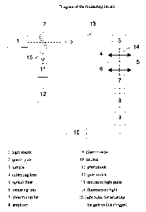
Figure 1: Diagram of the measuring device
Schematic representation of the measuring apparatus used within the scope of
the
present invention for time-resolved fluorescence measurement. This measuring
arrangement and its use is described and elucidated in more detail in the
following
examples.
-20-
Examples:
Abbreviations used
DADOO = 1,8 diamino-3,6-dioxaoctane
batho = bathophenanthroline disulfonic acid
bpy = 2,2'-bipyridyl-4-methyl-4'-butylcarboxylic acid
APC = allophycocyanin
HA = human haemagglutinin
HA peptide = YPYDVPDYA
Osu = 0-succinimide
Strept. = streptavidin
Ru = ruthenium
Eu = europium
Example 1: Syntheses and labelling of biomolecules
a) Synthesis ofRu(batho)Zbpy-Osu
50 mg (3* 10-5 mol) Ru(batho)Zbpy is dissolved in DMF, 12 mg (6* 10-5 mol) EDC
(N-
(3-dimethylaminopropyl)-N'-ethylcarbodiimide hydrochloride) and 7 mg (6*10-5
mol)
NHS (N-hydroxysuccinimide) are added and stirred overnight at room
temperature. The
reaction product is precipitated by adding acetone. The precipitate is
separated by
filtration and dried in a vacuum. It is purified by means of HPLC. The final
product is
analysed by means of MALDI-MS and corresponds to formula 111.
CA 02426479 2003-04-22
CA 02426479 2003-04-22
-21-
SO3Na ---~~ /_ SO3Na
- // \ -
N N
N Ru 2+ N SO3Na N\ SO3Na
0= 0
N
O_
Formula III: Ru(batho)2bpy-OSu
b) Synthesis of streptavidin Ru(batho),pby
mg (1.9* 10"' mol) streptavidin is dissolved in 1 ml N NaHCO3 solution and 2
mg
(1.3* 10' mol) Ru(batho)2bpyOSu (dissolved in 0.5 ml of an aqueous 0.1 N
NaHCO3
solution) is added dropwise. It is stirred for one hour at room temperature
and the
mixture is chromatographed on a Sephadex LH2O column (eluent 0.1 N NaHCO3
solution). The product fractions, i.e. the fractions which contain the
labelled
streptavidin (protein O.D. 280 nm), are dialysed overnight against HZO and
subsequently freeze-dried.
The degree of labelling was determined by comparing the absorption at 280 nm
(protein) and 460 nm (Ru2+ complex) and determined as ca. 3.6 Ru2+ complexes
per
streptavidin. Streptavidin-ruthenium conjugates having an average of 3 to 5
RuZ+
complexes per streptavidin molecule are suitable for determining the
sensitivity.
CA 02426479 2003-04-'22
-22-
c) Coupling of Ru(batho),pby to MAB-anti M
1 mg (0.65* 10-6 mol) Ru(batho)ZbpyOSu (dissolved in 1.0 ml of an aqueous 0.1
N
NaHCO3 solution) is added dropwise to 300 l (3 mg) MAB-anti HA. It is stirred
for
one hour at room temperature and the mixture is chromatographed on an LH2O
column (eluent 0.1 N NaHCO3 solution). The product fractions, i.e. the
fractions that
contain the labelled antibody (O.D. 280 nm), are pooled and frozen as 0.1 N
NaHCO3
solutions.
In the present experiment an average of about 7 ruthenium complexes per
antibody
were bound.
d) Preparation of biotin-JA286
10.2 mg (3* 10"5 mol) biotin-DADOO and 17.2 mg (3* 10"5 mol) JA 286 are
dissolved in 1 ml DMF, 15 l triethylamine is added and cooled to 0 C. After
adding
l DECP (diethyl cyanophosphonate), the mixture is stirred for 1 hour at 0 C
and
subsequently overnight at room temperature. After evaporating to dryness the
crude,
product is purified by means of HPLC. The final product was characterized by
MALDI-MS and corresponds to formula IV:
,/-o,s
N 0
~ O
+N N O 0 N H ~ H
H Fl H~
O HN -f NH
0
formula IV: biotin-JA286
CA 02426479 2003-04=22
-23-
e) Synthesis of biotin-JA198
mg (5.3* 10-6 mol) JA198-OSu and 2 mg (5.3* 10-6 mol) biotin-DADOO are
dissolved in 800 l phosphate buffer pH 7.5 and stirred overnight at room
temperature in the dark. Subsequently the mixture is purified by HPLC. The
product
was examined with ESI-MS and corresponds to formula V:
cl
ci CI
CI COO
O O N
HN ~ NH O
H H
H ,~'\/ NO O N
S H O O-s=0
0
Formula V: biotin-JA198
fi Coupling of JA133 to the synthetic HA peptide
5 mg (4.5* 10-6 mol) HA peptide (YPYDVPDYA = SEQ ID NO: 1) is dissolved in
1 ml acetonitrile/phosphate buffer pH 7.5 (1:1) and a solution of 4 mg (4.5*
10-6 mol)
JA133-OSu and 500 l acetonitrile are added while stirring. The mixture is
stirred
overnight at room temperature.
After evaporating to dryness in a vacuum, it is purified by means of HPLC. The
product was examined by means of ESI-MS and corresponds to formula VI:
CA 02426479 2003-04-22
-24-
ci
ci ci
~
ci Tc0o
N O N
O~
YPYDVPDYA
Formula VI: HA peptide-JA133
Example 2: Measuring device and description thereof
The measuring apparatus used in the present invention for time-resolved
fluorescence
measurement is described in the following and elucidated by a diagram (figure
1).
The pulsed light source (1) - nitrogen laser or dye laser - excites the donor
marker of
the measuring sample (3) with light pulses of a suitable wavelength (13), the
width of
the light pulse t = 0.7 ns being much shorter than the decay time of one of
the
fluorescent markers. The acceptor marker is then excited to fluoresce by
energy
transfer. This fluorescence radiation (14) is guided with an optical system
(4,6)
through an optical filter (cut-off filter / band pass filter) (5) which allows
the
emission wavelength of the acceptor marker to pass, onto the photocathode of a
photomultiplier (7). The individual detected photons generate current pulses
which
can be counted digitally (10) (photon counting method) after amplification (8)
and
standardization (9). A part of the excited light (15) is deflected by a quartz
plate (2)
to a photodiode (11) which controls a gate switch (12) which starts the
counter (10)
after a preset delay time of preferably 1 s and stops the counting process
again after
an adjustable opening time of the measuring window which is preferably 100 s.
The
delay time is selected such that scattered light effects and background
fluorescence
-25-
have almost completely decayed within this time. In this manner the number of
counted current pulses is proportional to the intensity of the marker
fluorescence
which is measured separately from the background. The procedure for such
measurements is described in detail in examples 3 and 4 for two new TR-FRET
pairs. The other new FRET pairs described in example 5 are measured in a
similar
manner or the europium-APC systems of example 5 are quantified using known
instruments and methods from the prior art.
Example 3: Procedure for measuring the sensitivity of a FRET system
comprising a RuZ+ complex as the donor and JA-286 as the fluorophoric
acceptor
400 l of a solution (10 mM Na phosphate, 150 mM NaCI, pH 7.2) containing
100 nM streptavidin labelled with the Ru2+ complex and 300 nM biotin-DADOO-
JA-286 is pipetted into a measuring cuvette.
The measurement is carried out with the apparatus descibed in example 2 and
shown
schematically in figure 1. The donor is excited by a 460 nm dye laser. The
light pulse
duration is 1 ns. The fluorescence of the system is detected by a combination
of an
optical cut-off filter KV550 + RG645 and the photomultiplier according to the
described experimental arrangement using the photon counting technique.
The delay time is set to 1 s and the measuring window is 100 s. The
background is
determined by a separate measurement of the buffer under the same conditions.
The
sensitivity (lower limit of detection) is given by a limiting concentration
which is
determined by the following relationship:
2B
Co = Cs,
S
CA 02426479 2003-04-22
-26-
in which Co is the limiting concentration in [M], B is the background in
[counts], S is
the signal in [counts] and Cs is the sample concentration in [M].
The lower limit of detection of this system is 7.4 - 10-14 M streptavidin.
Example 4: Procedure for measuring the sensitivity of a FRET system comprising
RuZ+ complex as the donor and JA-133 as an acceptor in an antigen-antibody
reaction
The FRET partners in this example are the Ru2+ complex from example 1 a) as
the
donor and JA-133 as the acceptor. A monoclonal antibody to HA is labelled with
the
donor as described under 1 c). The antigen (HA peptide) is labelled with the
acceptor
according to 1 f). The donor-acceptor pair are brought sufficiently closely to
one
another by the antigen-antibody reaction such that FRET is possible and
measurable.
After 10 minutes incubation at room temperature, 400 l of a solution (10 mM
Na
phosphate, 150 mM NaCI, pH 7.2) containing 100 nM anti-HA labelled with the
RuZ+
complex and 100 nM HA-JA-133 is pipetted into the measuring cuvette.
The measurement is carried out with the apparatus described in example 2. The
donor is excited at 460 nm by a 460 nm dye laser. The light pulse duration is
1 ns.
The fluorescence of the system is detected by a combination of an optical cut-
off
filter KV550 + RG630 and the photomultiplier according to the described
experimental arrangement using the photon counting technique.
The delay time is set to 1 s and the measuring window is 100 s. The
background is
determined by a separate measurement of the buffer under the same conditions.
The
sensitivity is given by a limiting concentration Co in [mol/litre] as stated
in example 1.
The sensitivity of this system is 7.2 - 10-14 M HA.
CA 02426479 2003-04-22
-27-
Example 5: Combination of some new FRET pairs and comparison with FRET
pairs from the prior art
a) TR-FRET pair 1: Streptavidin-Bu(batho)2bpy/biotin-APC
The labelled streptavidin from example lb) was used at a concentration of 100
nM
and APC biotin (commercial product: APC-XL biotin from Europa Bioproducts
Ltd.,
Cambridge, GB) was used at a concentration of 300 nM. The excitation
wavelength
(kX) was at 460 nm and the emission or measuring wavelength (kM) was at 634
nm.
A lower limit of detection of 4.1 x 10⁻¹³ mol/litre (= M) was determined with
this dye
and reagent pair.
CA 02426479 2003-04-22
-28-
b) R-FRET pair 2: streptavidin-Ru(batho)2bpy/biotin-JA198
S03Na - S03Na
- i) ~ -
N N
N Ru2+ N
SO3Na N N - SO3Na
0= L
Streptavidin
ci
ci .. ~ ci
ci c0o
O -~~ NO ~N~
HN NH O
H H
~ O N
s
O O-S=0
O
CA 02426479 2003-04-22
-29-
c) TR-FRET pair 3: streptavidin-Ru(batho)2bpy/biotin-JA286
SO3Na - SO3Na
N N
\ / \ N Ru2+ N,~
SO3Na - SO3Na
0=
[Streptavidin
03S
N 0
O
H N S
,~ H
+N ~~ N O
O
H~
O HN I NH
O
CA 02426479 2003-04-22
-30-
d) TR-FRET pair 4: MAB-anti-HA-Ru(batho)2bpy/HA-JA133
S03Na S03Na
-- i, ~ -
N N
2+
\ / \ /N Ru N\
SO3Na N N - / SO3Na
0
Anti-HA
ci
ci ci
ci coo
4N ~ p' / N
O,\
YPYDVPDYA
CA 02426479 2003-04-22
CA 02426479 2003-04-22
-31-
e) +J) Additional FRET pairs using commercially available raw materials.
Commercially available streptavidin derivatives labelled with europium from
Wallac,
Oy (Strept-Eu0062: AD0062 streptavidin-W 1024; Strept-Eu0060: AD0060
streptavidin-W8044) were used for the FRET pairs 5 and 6 in table 1.
Commercially
available-biotin-APC described in example 5a) was used as the acceptor.
Table 1: Lower limit of detection for some examined FRET pairs
No. System Concentration XEX XEM Lower limit of
[ M] [nm] [nm] detection (LLD)
strept./biotin [mol/litre]
1 strept.-Ru(batho)2bpy / 0.100 / 0.300 460 6 4.1 - 10-13
biotin-APC 34
2 strept.-Ru(batho)2bpy / biotin- 0.150 / 0.150 460 660 2.6 = 10-13
JA198
3 strept.-Ru(batho)2bpy / biotin- 0.100 / 0.300 460 703 7.4 - 10-14
JA286
4 anti-HA-Ru-(batho)2bpy / 0.100 / 0.100 460 631 7.2 - 10-14
HA-JA133
5(#) strept.-Eu0062 / biotin-APC 0.030 / 0.090 337 660 2.5 - 10-11
6(#) strept.-Eu0060 / biotin-APC 0.030 / 0.090 337 660 7.3 - 10-12
The sensitivity was measured as follows:
1 s delay and 100 s measuring window determined according to 2B
Co = CS,
[M],
S
in which Co is the limiting concentration in [M], CS is the concentration used
in [M],
B is the background (of the buffer) in [counts] and S is the signal in
[counts].
-32-
(#): measuring window: 400 s - as described in the prior art for Eu-APC
complexes.
Table 1 clearly shows that the novel FRET pairs are at least equivalent to or
superior
to the known systems in the prior art. The FRET pair No. 3 according to the
invention has an LLD of 7.4 = 10-14 which is considerably lower than the LLD
for
examples No 5 or No 6 according to the prior art.
CA 02426479 2003-04-22
CA 02426479 2003-04-22
-33-
List of references
EP 178 450
EP 567 622
EP 747 447
EP 076 695
EP 772 616
EP 439 036
EP 1 054 039
US 5,998,146
WO 00/47693
Blomberg, et al., Clinical Chemistry 45 (1999) 855 ff.
French, et al., SPIE BiOS in Proc. SPIE v 3259 (1998) 209-218
French, et al., SPIE BiOS in Proc. SPIE v 3606 (1999) 272-280
Hemmila, Chemical Analysis 117
John Wiley & Sons, Inc., (1991) 135-139
Joun et al., Analytical Biochemistry 232 (1995) 24-30
Mujumdar, et al., Bioconjugate Chem. 7, 1996, 356-362
Van der Meer, et al., Resonance Energy Transfer VCH (1994)
CA 02426479 2003-08-20
-34-
Sequence Listing
<110> F. Hoffmann-La Roche AG
<120> Dye pair for fluorescence resonance energy transfer
(FRET)measurements
<130> PAT 54393W-1
<140> PCT/EP01/13152
<141> 2001-11-14
<150> EP 00124995.2
<151> 2000-11-16
<160> 1
<170> PatentIn Ver. 2.1
<210> 1
<211> 9
<212> PRT
<213> artificial sequence
<220>
<223> description of the artificial sequence: HA peptide
<400> 1
Tyr Pro Tyr Asp Val Pro Asp Tyr Ala
1 5