Note: Descriptions are shown in the official language in which they were submitted.
CA 02426688 2011-03-28
53940-14
-1-
SYSTEMS AND METHODS FOR REDUCING FRACTURED
BONE USING A FRACTURE REDUCTION CANNULA
FIELD OF THE INVENTION
This invention relates to the treatment of bone
conditions of the human and other animal body systems and,
more particularly, to systems and methods for correcting
such conditions.
BACKGROUND OF THE INVENTION
Bone -fractures, particularly osteoporotic bone
fractures, are common in older adults. Due to the nature of
osteoporotic bone, standard methods of fracture fixation
yield unsatisfactory results. Such methods cannot
adequately place the broken fragments back to their pre-
fracture state. For instance, with a non-osteoporotic bone
fracture, common practice includes inserting rods, pins
and/or screws into the bone in order to reduce the fracture
and/or fix the fracture fragments to plates. Osteoporotic
bone generally cannot support such a method. Another common
method for non-osteoporotic bone fractures involves
maintaining the bone in a cast for several weeks.
osteoporotic bone that has suffered a crush fracture, such
as a Colles' fracture of the distal radius, will not heal
properly if placed in a cast; the bone mechanics are altered
such that the bone is shortened and/or subsides. Yet
another non-osteoporotic fracture reduction method involves
using an external fixation device. However, when used in
CA 02426688 2003-04-24
WO 02/34148 PCT/US01/45589
- 2 -
elderly patients, the fixation pins may not remain within
the weakened bone. Moreover, such a device typically
increases the likelihood of infection at the treatment site.
Further, because casts and/or an external fixation devices
must be left in place for several weeks in order for the
bone to heal, the lack of joint movement in the affected
area often results in painful arthritis in the immobilized
joints of the elderly patient.
Even where osteoporosis is not present, it is typically
necessary to immobilize a fractured bone to allow the bone
to properly heal. This often requires immobilization of the
joints adjacent to the fractured bone - often for extended
periods of time. However, such immobilization often causes
the joints to degenerate over time. Often, such treatment
can result in temporary or permanent loss of joint motion.
At the very least, such immobilization of the joints
requires extensive and often painful rehabilitation for an
individual to recover the full range of their joint motion.
SUMMARY OF THE INVENTION
Because of the problems associated with treating distal
radius fractures such as Colles' fractures, and other bone
fractures similar thereto, there is a need for a method and
apparatus that will improve the existing protocol for
treating such fractures such as reducing the pain resulting
from the fracture fixation method used, reducing the chance
that an infection will occur at the site, improving the
likelihood that the fracture will heal properly and
minimizing degeneration of the adjacent joints and allows
for sooner resumption of activity. The present invention
provides apparatus and a method of fracture reduction which
satisfies this need.
This invention provides a system that fixes or reduces
osteoporotic and non-osteoporotic fractures in human and
other animal body systems. Moreover, by immediately
reducing and/or reinforcing the fractured bone, thereby
CA 02426688 2003-04-24
WO 02/34148 PCT/US01/45589
3 -
rendering the bone capable of bearing limited loads, the
present system promotes healing of the fractured bone while
minimizing degeneration of the adjacent joints. It is
particularly well suited for fractures of long bones such as
the human distal radius.
One aspect of the invention provides a tool for
establishing a percutaneous path into bone. The tool is a
cannula having a side wall defining an internal bore aligned
along an axis. The cannula has a distal end. A
circumferential opening is defined in the side wall. The
circumferential opening has a distal terminus. The
circumferential opening extends partially about the side
wall and is elongated along the axis. The circumferential
opening is adapted to accommodate passage of an expandable
structure from within the bore. In one embodiment, the bore
is solid between the distal terminus of the circumferential
opening and the distal end of the cannula.
In an alternate embodiment of the above described tool,
the bore is open between the distal terminus of the
circumferential opening and the distal end of the cannula.
The cannula has a distal opening in the distal end
communicating with the bore. The opening in the distal end
can accommodate passage of a guide pin.
In an alternate embodiment of the above described tool,
the cannula desirably has a surface on its distal end to
anchor the distal end in bone.
Another aspect of the invention provides an assembly
for treating bone, including a cannula as described above.
The cannula has a distal opening in the distal end
communicating with the bore. The opening in the distal end
can accommodate passage of a guide pin. The assembly also
includes an expandable structure. The expandable structure
is adapted for insertion through bone into the cannula and
expansion through the circumferential opening.
Another aspect of the invention provides an assembly
CA 02426688 2003-04-24
WO 02/34148 PCT/US01/45589
4 -
for treating bone, including a cannula as described above.
Desirably, the bore is solid between the distal terminus of
the circumferential opening and the distal end of the
cannula. The assembly also includes an expandable
structure. The expandable structure is adapted for
insertion through bone into the cannula and expansion
through the circumferential opening.
Another aspect of the invention provides an assembly
for treating bone, including a cannula as described above.
Desirably, the cannula has a surface on its distal end to
anchor the distal end in bone. The assembly also includes
an expandable structure. The expandable structure is
adapted for insertion through bone into the cannula and
expansion through the circumferential opening.
Another aspect of the invention provides an assembly as
described above. Desirably, the expandable structure has
radio opaque markers. The markers allow one to locate the
expandable structure within a circumferential opening in a
cannula.
Another aspect of the invention provides a method for
treating bone. The method includes providing a cannula and
inserting the cannula into cancellous bone. The method also
includes inserting an expandable structure through the
cannula until the structure is in registration with a
circumferential opening in the cannula. The method further
includes expanding the expandable structure through the
circumferential opening into contact with cancellous bone.
Another aspect of the invention provides a method for
treating bone, including a step of expanding an expandable
structure. The expansion compacts cancellous bone.
Another aspect of the invention provides a method for
treating bone, including a step of compacting cancellous
bone. The compaction of cancellous bone forms a cavity.
Another aspect of the invention provides a method for
treating bone, including a step of conveying a material into
CA 02426688 2010-03-11
50749-17
-5-
a cavity.
Another aspect of the invention provides a method for treating bone,
including a step of expanding an expandable structure such that the expansion
moves fractured cortical bone.
Another aspect of the invention provides an assembly for treating
bone comprising: a cannula having a side wall defining an internal bore
aligned
along an axis, the cannula having a distal end; a distal opening in the distal
end
communicating with the bore to accommodate passage of a guide pin; a
circumferential opening in the side wall extending partially about the side
wall and
being elongated along the axis; and an expandable structure sized and shaped
for
insertion through the cannula into the bone and expansion through the
circumferential opening, the expandable structure having a distal projection
that
engages with a distal portion of the cannula adjacent the bore to anchor the
expandable structure within the bore during expansion through the
circumferential
opening.
Another aspect of the invention provides an assembly for treating
bone comprising: a cannula having a side wall defining an internal bore
aligned
along an axis, the cannula having a distal end; a circumferential opening in
the
side wall, the circumferential opening having a distal terminus, and the
circumferential opening extending partially about the side wall and being
elongated along the axis; the cannula being solid between the distal terminus
of
the circumferential opening and the distal end of the cannula; and an
expandable
structure sized and shaped for insertion through bone into the cannula and
expansion through the circumferential opening, the expandable structure having
a
distal projection that engages with a surface of the solid portion of the
cannula
adjacent the bore to anchor the expandable structure within the bore during
expansion through the circumferential opening.
Another aspect of the invention provides an assembly for treating
bone comprising: a cannula having a side wall defining an internal bore
aligned.
along an axis, the cannula having a distal end with a distal opening in
communication with the bore; the distal opening having a cross-sectional
CA 02426688 2009-05-12
50749-17
- 5a -
diameter; a circumferential opening in the side wall, the circumferential
opening
extending partially about the side wall and being elongated along the axis; a
guide
pin sized and shaped for passage through the bore and the distal opening; and
an
expandable structure sized and shaped for insertion into the bore and
expansion
through the circumferential opening, the expandable structure having a cross-
sectional diameter larger than the cross-sectional diameter of the distal
opening,
the expandable structure having a distal projection that engages with a distal
portion of the cannula adjacent the bore to anchor the expandable structure
within
the bore during expansion through the circumferential opening.
Another aspect of the invention provides an assembly for treating
bone comprising: a cannula comprising: a side wall defining an internal bore
aligned along an axis, the cannula having a distal end; a circumferential
opening
in the side wall, the circumferential opening having a distal terminus, and
the
circumferential opening extending partially about the side wall and being
elongated along the axis; a segment of the cannula being solid between the
distal
terminus of the circumferential opening and the distal end of the cannula; and
an
expandable structure sized and shaped for insertion through the bore and
expansion through the circumferential opening, the expandable structure having
a
distal projection that engages with the segment of solid cannula to anchor the
expandable structure within the bore during expansion through the
circumferential
opening.
Another aspect of the invention provides an assembly for treating
bone comprising: a cannula for establishing a percutaneous path into a bone
comprising: a side wall defining an internal bore aligned along an axis, a
circumferential opening in the side wall, the circumferential opening
extending
partially about the side wall and being elongated along the axis, a distal end
with a
distal opening in communication with the bore, the distal opening having a
cross-
sectional diameter, an external anchoring surface between the distal terminus
of
the circumferential opening and the distal end of the cannula to anchor the
cannula in bone; and an expandable structure sized and shaped for insertion
into
CA 02426688 2009-05-12
50749-17
- 5b -
the cannula and expansion through the circumferential opening such that when
expanded a portion of the expandable structure expands and remains within the
bore, the expandable structure having a cross-sectional diameter larger than
the
cross-sectional diameter of the distal opening.
CA 02426688 2009-05-12
50749-17
- 5c-
BRIEF DESCRIPTION OF THE DRAWINGS
Figure I is an anatomic view that shows bones of a
human forearm;
Figure 2 is an anatomic view that shows bones of the
forearm including an ulna and a fractured distal radius;
Figure 3 is an enlarged section view of the distal
radius showing cancellous bone and cortical bone in a
fractured condition;
Figure 4 is a plane view showing a kit containing a
system of instruments used to treat bones and that embodies
features of the invention;
Figure 5 is a perspective view of an obturator
instrument that is contained in the kit shown in Fig. 4,
Figure 6 is a perspective view of a percutaneous
cannula that is contained in the kit shown in Fig. 4;
Figure 7 is a perspective view of a drill bit
instrument that is contained in the kit shown in Fig. 4;
Figure 8 is a perspective view of a fracture reduction
cannula that is contained in the kit shown in Fig. 4,
showing a distal end, a proximal end, and a circumferential
opening;
Figure 8A is a perspective view of an alternate
embodiment of a fracture reduction cannula constructed in
accordance with the teachings of the present invention;
_ Figure 8B is a perspective view of another alternate
embodiment of a fracture reduction cannula constructed in
accordance with the teachings of the present invention;
Figure 9 is a side view of the fracture reduction
cannula of Figure 8 showing an end interior bore
therethrough;
CA 02426688 2003-04-24
WO 02/34148 PCT/US01/45589
6
Figure 10a is an enlarged view of the distal end of the
fracture reduction cannula, the distal end being solid;
Figure 10b is an enlarged view of the distal end of the
fracture reduction cannula of Figure 8, the distal end being
open to accommodate passage of a guide pin;
Figure 11 is a perspective view of an instrument
carrying an expandable structure, the instrument being
contained in the kit shown in Fig. 4;
Figure 12 is an enlarged perspective view of an
instrument, showing the expandable structure in an
unexpended state and, in broken lines, the expandable
structure in an expanded state;
Figure 13 is a perspective view of a tamp that is
contained in the kit shown in Fig. 4;
Figure 14 is a perspective view of a handle that is
contained in the kit shown in Fig. 4; showing recesses
therein;
Figure 15 is a perspective view showing the obturator
instrument inserted into the handle, the handle being
grasped by a hand;
Figure iSa is a side section view showing the obturator
instrument inserted into the handle and advanced to the
distal radius;
Figure 16 is a side section view showing the
percutaneous cannula inserted over the obturator instrument
and advanced to the distal radius;
Figure 17 is a side section view showing the drill bit
instrument within the percutaneous cannula and advanced to
the distal radius, and further showing the distal radius
fracture and cancellous bone;
Figure 18 is a side section view showing the fracture
reduction cannula within the percutaneous cannula and
advanced into the cancellous bone of the distal radius, and
further showing the circumferential opening facing the
fracture;
CA 02426688 2003-04-24
WO 02/34148 PCT/US01/45589
7 -
Figure 19 is an enlarged view showing the fracture
reduction cannula seated within cortical bone;
Figure 20 is an enlarged view showing the fracture
reduction cannula seated within cortical bone and containing
the unexpanded expandable structure;
Figure 21 is an enlarged view showing the fracture
reduction cannula seated within cortical bone, containing
the expanded expandable structure, and compressing
cancellous bone and/or moving cortical bone;
Figure 21A is an enlarged view showing a fracture
reduction cannula seated within cortical bone, with the
expanded expandable structure compressing cancellous bone
and/or moving cortical bone and creating a cavity which
extends across a fracture line in the targeted bone;
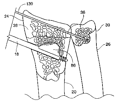
Figure 22 is an enlarged view showing the fracture
reduction cannula seated within cortical bone and containing
the expanded expandable structure, showing compressed
cancellous bone, displaced cortical bone, and a reduced
fracture, and further showing a pin placed through the
distal radius and into the ulna;
Figure 22A is an enlarged view showing a fracture
reduction cannula seated within cortical bone and containing
the expanded expandable structure, showing compressed
cancellous bone, displaced cortical bone, a reduced
fracture, and a cavity extending across a fracture line in
the cortical bone, and further showing a pin placed through
the distal radius and into the ulna;
Figure 23 is a top view showing a patient's forearm on
a rolled towel, with horizontal finger traps on the
patient's fingers, the instrument inserted through the
handle and into the percutaneous cannula, with the fraction
reduction cannula hidden from view, and the pin inserted
into the patient's wrist;
Figure 24 is an enlarged view showing a cavity created
by expansion of the expandable structure in the distal
CA 02426688 2003-04-24
WO 02/34148 PCT/US01/45589
8
radius, the pin in place, the fracture reduction cannula,
and the cavity ready to receive a bone filling material;
Figure 25 is an enlarged view showing the filling
material beginning to fill the cavity;
Figure 26 is an enlarged view showing the tamp urging
the filling material fully into the cavity;
Figure 27 is an enlarged view showing the filled cavity
with the fracture reduction cannula and tamp removed; and
Figure 28 is an enlarged view showing an alternate
embodiment of the fracture reduction cannula with a guide
pin placed therethrough.
DETAILED DESCRIPTION OF THE PREFERRED EMBODIMENTS
The invention may be embodied in several forms without
departing from its spirit or essential characteristics. The
scope of the invention is defined in the appended claims,
rather than in the specific description preceding them. All
embodiments that fall within the meaning and range of
equivalency of the claims are therefore intended to be
embraced by the claims.
The preferred embodiment describes improved systems and
methods that embody features of the invention in the context
of treating bones. This is because the new systems and
methods are advantageous when used for this purpose.
However, aspects of the invention can be advantageously
applied for diagnostic or therapeutic purposes in other
areas of the body.
The new systems and methods will be more specifically
described in the context of the treatment of long bones such
as the human distal radius. Of course, other human or
animal bone types can be treated in the same or equivalent
fashion.
I. ANATOMY OF THE RADIUS
The human forearm consists of two bones, the radius and
the ulna. As shown in Figs. 1 and 2, the radius 20 is a
long bone that is situated on the thumb side of the forearm,
CA 02426688 2003-04-24
WO 02/34148 PCT/USO1/45589
9 -
while the ulna 26 is located at the little finger side. The
radius 20 lies side by side with the ulna 26, and it exceeds
the ulna 26 both in length and in size.
The upper, or proximal end 22 of the radius 20 is small
and articulates with a part of the elbow joint, including
the proximal ulna 28. The distal end 24 of the radius 20 is
large and articulates with two bones of the wrist, or
carpus, known as the lunate 21 and scaphoid 27 bones. The
inner, or medial side 25 of the distal radius 24 contains an
ulnar notch 32 that articulates with the ulna 26.
II. BONE FRACTURES
The systems and methods of the present invention are
especially suited for treating fractures of long bones. One
type of bone fracture that may be so treated is known as a
Colles' fracture or transverse wrist fracture. As shown in
Fig. 2, such a fracture 34 generally occurs less than one
inch from the distal end 24 of the radius 20. Colles'
fractures are commonly noted in children and the elderly
where the person tries to break or stop a fall by using his
or her hands and arms. Colles' fractures in children are
often associated with sports such as skateboarding and in-
line skating. in the elderly, Colles' fractures are
commonly caused by osteoporosis and/or in connection with a
fall.
Osteoporosis is a disease of the bone that is most
commonly found in the middle-aged and elderly, particularly
women. It is characterized by a gradual loss of a type of
bone tissue known as cancellous bone 36. As shown in Fig.
3, cancellous bone 36, also referred to as trabecular bone,
is a spongy bone tissue located within the harder outer or
cortical bone. Cancellous bone 36 comprises most of the
bone tissue of the extremities of long bones such as the
radius 20.
In contrast to cancellous bone 36, cortical bone 38
tissue is much harder and denser. Cortical bone 38 is
CA 02426688 2003-04-24
WO 02/34148 PCT/US01/45589
- 10 -
layered over cancellous bone 36, and provides a protective
layer and support for long bones such as the radius 20, as
shown in Figs. 1 and 2. At the ends of such bones, however,
the cortical bone 38 layer becomes thinner. Where
osteoporosis has significantly weakened the cancellous bone
36, such regions at the ends of long bones become especially
prone to fracture and/or collapse.
It may be indicated, due to disease or trauma, to
reduce fractured cortical bone 38 and/or compress cancellous
bone 36 within long bones such as the radius 20. The
compression, for example, can be used to form an interior
cavity 35, which receives a filling material 99, e.g., a
flowable material that sets to a hardened condition, such as
poly(methylmethacrylate), as well as a medication, or
combinations thereof, to provide improved interior support
for cortical bone 38 or other therapeutic functions, or
both. The compaction of cancellous bone 36 also exerts
interior force upon cortical bone 38, making it possible to
elevate or push broken and compressed bone back to or near
its original pre-fracture, or other desired, condition.
III. THE INSTRUMENTS
Figure 4 shows instruments, arranged as a kit 200,
which are usable in association with each other to reduce
fractured bone. The number and type of instruments can
vary. Fig. 4 shows seven representative instruments, each
having a different size and function.
In Fig. 4, the kit 200 includes an obturator instrument
12 for penetrating soft tissue and bone; a percutaneous
cannula 14 that functions as a guide sheath; a drill bit
instrument 16 that is used for drilling into bone; a
fracture reduction cannula 18 used in reducing fractures and
that is inserted into bone and designed to receive an
expandable structure; a bone compaction instrument 80 that
functions to deliver a filling material 99 into a cavity 35
in bone and that carries an expandable structure 86 that may
CA 02426688 2003-04-24
WO 02/34148 PCT/US01/45589
- 11 -
be expanded in bone; a tamp 81 functions to urge residual
bone filling material into bone; and a handle 13 with
recesses that receives instruments 12, 14, 16 and 18.
Instruments 12, 14, 16, and 18 share some common
features, although they are intended, in use, to perform
different functions. Instruments 12, 14, 16, and 18 each
comprise an elongated, cylindrical body 40 having a proximal
end 42 and a distal end 44. Instruments 12, 14, 16, and 18
are each made of a rigid, surgical grade plastic or metal
material.
A. The Obturator Instrument
The first instrument 12 functions as an obturator. As
shown in Fig. 5, its distal end 44 is tapered to present a
penetrating surface 50. In use, the surface 50 is intended
to penetrate soft tissue and/or bone in response to pushing
or twisting forces applied by the physician at the proximal
end 42. In a preferred embodiment, the proximal end 42 of
the obturator instrument 12 mates with a handle 13, to be
described in detail later.
The proximal end 42 of the obturator instrument 12
presents a flanged surface 52. The flanged surface 52 is
designed to fit securely into a recess in the handle 13,
such that pushing or twisting forces applied to the proximal
end 42 of the obturator 12 instrument will not displace the
obturator instrument 12. The flanged surface 52 tapers from
a larger outer diameter to a smaller outer diameter in the
direction of the proximal end 42. The flanged surface 52
includes an array of circumferentially spaced teeth 54 with
intermediate flutes 56.
An interior bore 60 extends through the obturator
instrument 12 from the distal end 44 to the proximal end 42.
Desirably, the interior bore 60 is sized to accommodate a
conventional surgical guide pin 108 component to aid in its
deployment, as will be described in greater detail later.
The obturator instrument 12 has an outer surface 142
CA 02426688 2003-04-24
WO 02/34148 PCT/US01/45589
- 12 -
that is sized such that one may slide a percutaneous cannula
14 over the obturator instrument 12 as described below.
B. The Percutaneous Cannula
The second instrument 14 functions as a percutaneous
cannula or guide sheath. It also serves to protect soft
tissue and nerves, ligaments, muscle and vasculature from
the use of a drill bit instrument 16, which will be
described in greater detail later.
As shown in Fig. 6, the percutaneous cannula 14 is
somewhat larger in diameter than, and is not as long as, the
obturator instrument 12. In one embodiment, the cannula 14
is approximately 2 inches long, although it could be various
other lengths, depending upon the thickness of the patient's
soft tissue at the surgical site. Desirably, the
percutaneous cannula 14 is made of metal, and contains
markings 120 along its outer surface 142 to indicate the
depth at which it is placed into a patient's distal radius
24.
The proximal end 42 of the percutaneous cannula 14
presents a tapered flange 52, as Fig. 6 shows. The flanged
surface 52 is designed to fit securely into a recess in the
handle 13, such that forces applied to the proximal end 42
of the percutaneous cannula 14 will not displace the
percutaneous cannula 14. The tapered flange 52 changes from
a larger diameter to a smaller diameter in the direction of
the proximal end 42. The tapered flange 52 of the
percutaneous cannula 14 also includes an array of
circumferentially spaced teeth 54 with intermediate flutes
56. The form and orientation of the teeth 54 and flutes 56
on the percutaneous cannula 14 correspond to the form and
orientation of teeth 54 and flutes 56 on the fracture
reduction cannula 18.
As shown in Fig.6, the percutaneous cannula 14 includes
an interior bore 60 that extends from its distal end 44 to
its proximal end 42. Desirably, the interior bore 60 is
CA 02426688 2003-04-24
WO 02/34148 PCT/US01/45589
- 13 -
sized to accept the obturator instrument 12. The size of
the interior bore 60 permits a physician to slide and rotate
the percutaneous cannula 14 relative to the obturator
instrument 12, and vice versa, as will be described in
greater detail later.
The distal end 44 of the percutaneous cannula 14
presents an end surface 62. Desirably, the surface of the
distal end 44 is designed to penetrate soft tissue. In use,
the end surface 62 of the percutaneous cannula 14 is
intended to penetrate soft tissue surrounding the obturator
instrument 12, in response to pushing or twisting forces
applied at the proximal end 42. If desired, the end surface
62 can incorporate one or more teeth (not shown) which
anchor the cannula 14 to the surface of the targeted bone.
C. The Drill Bit Instrument
The third instrument functions as a drill bit. As
shown in Fig. 7, The drill bit instrument 16 has generally
the same physical dimensions as the obturator instrument 12.
Like the obturator instrument 12, the drill bit instrument
16 is intended, in use, to fit for sliding and rotational
movement within the interior bore 60 of the percutaneous
cannula 14.
The distal end 44 of the drill bit instrument 16
includes machined cutting edges 64, as shown in Fig. 7. In
use, the cutting edges 64 are intended to penetrate hard
tissue in response to rotation and longitudinal load forces
applied at the proximal end 42 of the drill bit instrument
16.
As further shown in Fig. 7, the proximal end 42
presents a tapered flange 52, substantially identical to the
flange 52 on the obturator instrument 12, as Fig. 5 shows.
The flanged surface 52 is designed to fit securely into a
recess in the handle 13, such that forces applied to the
proximal end 42 of the drill bit instrument 14 will not
displace the drill bit instrument 14. Like the obturator
CA 02426688 2003-04-24
WO 02/34148 PCT/US01/45589
- 14 -
instrument 12, the tapered flange 52 changes from a larger
diameter to a smaller diameter in the direction of the
proximal end 42. The tapered flange 52 of the drill bit
instrument 16 also includes an array of circumferentially
spaced teeth 54 with intermediate flutes 56. The form and
orientation of the teeth 54 and flutes 56 on the drill bit
instrument 16 correspond to the form and orientation of the
teeth 54 and flutes 56 on the obturator instrument 12.
D. The Fracture Reduction Cannula
The fourth instrument functions as a fracture reduction
cannula 18. As shown in Fig. 8, the fracture reduction
cannula 18 is somewhat smaller in diameter than, and is
longer than, the percutaneous cannula 14. In one
embodiment, the fracture reduction cannula 18 is
approximately 3 34 inches in length, although it could be
various other lengths depending on the size of the patient
and the desired location within the targeted bone. Like
both the obturator instrument 12 and the drill bit
instrument 16, the fracture reduction cannula 18 is
intended, in use, to fit for sliding and rotational movement
within the interior bore 60 of the percutaneous cannula 14.
The proximal end 42 of the fracture reduction cannula
18 presents a flanged surface 52. The flanged surface 52 is
designed to fit securely into a recess in the handle 13,
such that pushing or twisting forces applied to the proximal
end 42 of the obturator 12 instrument will not displace the
fracture reduction cannula 18. Like the percutaneous
cannula 14, the flanged surface 52 of the fracture reduction
cannula 18 tapers from a larger outer diameter to a smaller
outer diameter in the direction of the proximal end 42. The
flanged surface 52 includes an array of circumferentially
spaced teeth 54 with intermediate flutes 56.
The fracture reduction cannula 18 is sized to fit
within the interior bore 60 of the percutaneous cannula 14.
The size of the interior bore 60 permits a physician to
CA 02426688 2003-04-24
WO 02/34148 PCT/US01/45589
- 15 -
slide and rotate the fraction reduction cannula relative to
percutaneous cannula 14, and vice versa, as will be
described in greater detail later.
As further shown in Fig. 8, the fracture reduction
cannula 18 includes a side wall 66 that defines an interior
bore 68 that extends from the distal end 44 of the fracture
reduction cannula 18 to its proximal end 42. The interior
bore 68 is adapted to allow passage of, among other things,
an expandable structure 86. In a preferred embodiment, the
distal end 44 of the interior bore 68 is solid, as shown in
Fig. 10a. In an alternate embodiment, the distal end 44 of
the bore 68 is not solid, but rather, it is open to
accommodate passage of an instrument such as a guide pin
108, as shown in Fig. lob. As another alternative, the
distal end of the bore 68 could be hollow, such that a
portion of the expandable structure could extend into the
distal end 44 of the cannula 18.
The fracture reduction cannula 18 further includes a
circumferential opening 70 in the side wall 66. In one
embodiment, the circumferential opening 70 extends
approximately one-half inch in length along its longitudinal
axis, although the size of this opening could vary depending
upon the dimensions of the targeted bone and the size of the
expandable structure. The circumferential opening 70 is
sized to accommodate an expandable structure 86. The
circumferential opening 70 desirably also allows a filling
material 99 to be placed near and/or into the fracture site.
Figure 8A depicts one alternate embodiment of a
fracture reduction cannula 18A constructed in accordance
with the teachings of the present invention. Because many
of the disclosed components are similar to those previously
described, like reference numerals will be used to denote
similar components. In this embodiment, the distal end 44A
of the cannula 18A is not solid, but rather extends along
the side wall 66A, with one or more longitudinally extending
CA 02426688 2011-03-28
53940-14
- 16 -
teeth 120 disposed at the distal end 44A.
E. The Handle
The handle 13, which can be made from a molded or cast
rigid plastic or metal material.
As shown in Fig. 14, the handle has a smooth
upper side 17. Its lower side 29 contains recesses 15 and
19. The flanged surfaces of the obturator instrument 12,
the drill bit instrument 16, the percutaneous cannula 14,
and the fracture reduction cannula 18 mate with the handle
13. Recess 15 is adapted to accept the obturator 12 and the
drill bit instrument 16 while recess 19 is adapted to accept
the fracture reduction cannula 18. If desired, another
recess can be provided (not shown) sized to accept the
percutaneous cannula 14 in a similar manner.
F. The Bone Compaction and/or Displacement Instrument
Fig. 11 shows an instrument 80 for accessing bone. for
the purpose of compacting cancellous bone 36 and/or
displacing cortical bone 38.
The instrument 80 includes a catheter tube assembly 82,
as shown in Fig. 11_ The distal end 84 of the catheter tube
assembly 82 carries an expandable structure 86. In use, the.
expandable structure 86 is deployed and expanded inside
.bone, e.g., in the. radius 20 as shown in Figs. 20, 21, and
22, to compact cancellous'bone 36 and/or displace cortical
bone 38, as, will be described later.
As further shown in Fig. 11, the instrument 80 includes
.an outer catheter body 88, and an inner catheter body 90
which extends through the outer catheter body 88. The
proximal ends 92 of' the outer 88 and inner 90 catheter
.bodies are coupled to-a y-shaped adaptor/handle 94. The y-
CA 02426688 2009-05-12
50749-17
- 17 -
shaped adaptor/handle 94 carries a first port 96 and a
second port 98 at its proximal end 92. The first port 96 is
adapted to be coupled with an inflation syringe 101, the
syringe 101 in the present case being used to deliver a
pressurized liquid into the expandable structure 86. The
second port 98 is adapted for insertion of a stiffening
stylet (not shown) to facilitate insertion of the distal end
84 of the instrument 80.
As Fig. 11 shows, the expandable structure 86 is
coupled at its proximal end 95 to the distal end 93 of the
outer catheter body 88. Likewise, the expandable structure
86 is coupled at its distal end 87 to the distal end 84 of
the inner catheter body 90.
The, outer catheter body 88 defines an interior bore,
through which the inner catheter body 90 extends. The
interior bore, in use, conveys a pressurized liquid, e.g., a
radio-opaque solution such as CONRAY solution, or another
fluid into the expandable structure 86 to expand it.
The material from which the expandable structure 86 is
made should possess various physical and mechanical
properties to optimize its functional capabilities to
compact cancellous bone 36, and to move cortical bone 38.
Desirably, the expandable structure 86 has the capability to
move cortical bone 38 from a fractured condition to a pre-
fractured or other desired condition, or.both. The three
most important properties of expandable structure 86 are the
ability to expand its volume; the ability to deform in a
desired way when expanding and assume a desired shape inside
bone; and the ability to withstand abrasion, tearing, and
puncture when in contact with cancellous bone 36.
The desired properties for the structure material, and
the description for creating a pre-formed structure, are
more fully set out in U.S. Patent No. 6,607,544 issued on August 19, 2003.
As shown in Fig. 11, the expandable structure 86
CA 02426688 2003-04-24
WO 02/34148 PCT/US01/45589
- 18 -
carries radio-opaque markers 91 located at a distal end 102
and at a proximal end 104 of segmented shaped regions 100 of
the expandable structure 86. The radio opaque markers 91
function to indicate, under fluoroscopic or other real-time
monitoring, the location of the segmented shaped regions 100
in relation to the circumferential opening 70 of the
fracture reduction cannula 18.
Fig. 12 illustrates the expandable structure in a
collapsed state (solid lines) and an expanded state (broken
lines).
G. The Pin
One or more conventional smooth Steinman pins 130 or
Kirschner ("K") wires may be provided to assist in aligning
and/or stabilizing fracture fragments, as will be described
in greater detail later.
H. The Filling Material Instruments
The filling material 99 instruments include a tamp 81
as shown in Fig. 13, and a standard syringe. The filling
material 99 is introduced through the syringe and into the
fracture reduction cannula 18. Residual filling material 99
may be urged through the fracture reduction cannula 18 by
employing the tamp 81, as will be described in greater
detail later.
I. The Kit
As shown in Fig. 4, a kit 200 is provided, including
instruments 12, 13, 14, 16, 18, 80, and 81. The kit 200 and
the instruments contained therein are sterile and are sealed
until an instance of use.
IV. ILLUSTRATIVE USE OF THE SYSTEM
The size and shape of the access tools and/or
expandable structure(s) 86 to be used, and the amount of
bone to be moved, are desirably selected by the physician,
taking into account the morphology and geometry of the site
to be treated. The shape of the joint, the bones and soft
tissues involved, and the local structures that could be
CA 02426688 2009-05-12
50749-17
- 19 -
harmed if moved inappropriately, are generally understood by
medical professionals using textbooks of human anatomy along
with their knowledge of the site and its disease and/or
injury. The physician is also desirably able to select the
desired shape and size of the expandable structure 86, the
cavity 35 and their placement based upon prior analysis of
the morphology of the targeted bone and joint using, for
example, plain film x-ray, fluoroscopic x-ray, or MRI or CT
scanning. The shape, size and placement are desirably
selected to optimize the strength and ultimate bonding of
the fracture relative to the surrounding bone and/or tissue
of the joint.
In a typical procedure, a patient is placed under local
anesthesia, although general anesthesia may instead be
employed. Where a fracture 34 is that of a distal radius
24, a physician makes an incision of approximately one (1)
centimeter on the radial aspect of the distal radius 24. in
an alternate embodiment, one may access the distal radius 24
by an approach through the ulna 26. The distance between
the incision and the fracture 34 is approximately 0.5
centimeter. Of course, while the present procedure is
described in the context of a minimally invasive surgery,
various other surgical approaches, including percutaneous,
subcutaneous, non-open, partially open and/or completely
open surgical approaches may be utilized in accordance with
the teachings of the present invention.
After making the incision, the physician spreads the
soft tissue by using a small clamp designed to avoid injury
to nearby nerves, muscles, and vasculature. The physician
then acquires the obturator instrument 12 and the handle 13.
The obturator instrument 12 may have at its proximal end 42
a flanged surface 52 that mates with a recess 15 within the
handle 13. Use of the handle 13 with the obturator
instrument 12 will produce axial as well as radial movement,
as shown in U.S. Patent No. 6,468,279 issued on October 22, 2002.
CA 02426688 2009-05-12
50749-17
- 20 -
The physician then fits the proximal end
42 of the obturator instrument 12 into recess 15 in the
handle 13, as shown in Fig. 15.
The physician next twists the handle 13 while applying
longitudinal force to the handle 13. In response, the
tapered surface of the obturator instrument 12 rotates and
penetrates soft tissue through the incision, as shown in
Fig. 15a. The physician may also tap the handle 13, or
otherwise apply appropriate additional longitudinal force to
the handle 13, to advance the obturator instrument 12
through soft tissue.
Under fluoroscopic monitoring or other real-time
monitoring, the physician advances the obturator instrument
12 through soft tissue down to the distal radius 24, as Fig.
15a shows. The obturator instrument 12 is inserted distal
to proximal from the radial side of the radius 20 to the
ulnar side of the radius 20. The obturator instrument 12 is
introduced into the radius 20. Desirably, the obturator
instrument 12 is introduced at an angle between minus 10
degrees and 45 degrees to the radio-carpal joint. More
desirably, the obturator instrument 12 is introduced at an
angle between zero degrees and 30 degrees to the radio-
carpal joint. Most desirably, the obturator instrument 12
is introduced at an angle equal to the angle of the radio-
carpal joint, i.e., approximately 23 degrees. Of course, if
,desired, the physician may utilize various other approach
paths to access the bone, including a dorsal approach.
The physician next removes the handle 13 from the
obturator instrument 12 and places the proximal end 42 of
the percutaneous cannula 14 in a recess 19 in the handle 13.
The physician slides the percutaneous cannula 14 over the
obturator instrument 12, distal end 44 first. The physician
then twists the handle 13 while applying longitudinal force
to the handle 13, in order to seat the percutaneous cannula
14 against and/or into the external cortical bone 38, as
CA 02426688 2003-04-24
WO 02/34148 PCT/US01/45589
- 21 -
shown in Fig. 16. Once the percutaneous cannula 14 is
seated in the cortical bone 38, the obturator instrument 12
is removed, proximal end 42 first.
In an alternate embodiment, instead of using the
obturator instrument 12 to access external cortical bone 38,
the physician may instead insert a conventional spinal
needle, the needle having an outer sheath and a stylus, into
the bone. Upon puncturing the bone, the physician removes
the stylus and inserts a guide pin 108 through the outer
sheath. The sheath is then removed and the fracture
reduction cannula 18 is deployed over the guide pin 108.
The physician then fits the proximal end 42 of the
percutaneous cannula 14 into a recess 19 in the handle 13
and slides the assembly, distal end 44 first, over the
fracture reduction cannula 18, as shown in Fig. 28.
Subsequently, the guide pin 108 is removed, proximal end
first.
After removing the obturator instrument 12, or the
guide pin 108 as in the case of the alternate embodiment
described above, the handle 13 is removed from the
percutaneous cannula 14. As shown in Fig. 15, the proximal
end 42 of a drill bit instrument 16 is then placed in a
recess in the handle 13. The preferred size of the drill
bit 16 is 3.2 millimeters. The physician slides the drill
bit assembly distal end 44 first through the bore 60 of the
percutaneous cannula 14. Using manual pressure, the drill
bit instrument 16 is advanced down to and into the distal
radius 24. As an alternate embodiment, instead of using
manual pressure, the physician could connect the proximal
end 42 of the drill bit instrument 16 to a conventional
motor-driven drill. The physician directs the drill bit
instrument 16 to penetrate the cortical bone 38 and the
cancellous bone 36 of the distal radius 24, as shown in Fig.
17.
After drilling through cortical bone 38 and into
CA 02426688 2003-04-24
WO 02/34148 PCT/US01/45589
- 22 -
cancellous bone 36, the physician removes the drill bit
instrument 16 from the handle 13. The fracture reduction
cannula 18 is then inserted, distal end 44 first, into the
bore of the percutaneous cannula 14, as shown in Fig. 18.
The distal end 44 of the fracture reduction cannula 18
extends beyond the distal end 44 of the percutaneous cannula
14. In an alternate embodiment, the physician may at this
point remove the percutaneous cannula 14, leaving only the
fracture reduction cannula 18 in place. In one embodiment,
it is preferred to employ a fracture reduction cannula 18
that has screw threads 71 on its distal end 44 as shown in
Fig. 9, thereby enabling the fracture reduction cannula 18
to be anchored to an interior surface of cortical bone 38 in
response to rotation of the fracture reduction cannula 18,
e.g., by using the handle 13. In an alternative embodiment
(see Fig. 83), the physician may employ a fracture reduction
cannula 18 that has a blunt, tapered distal end 44 instead
of screw threads 71 on the distal end 44. If such a
fracture reduction cannula 18 is employed, the physician may
choose to drill a hole in cortical bone 38 in which to seat
the blunt, tapered distal end 44. Desirably, if the distal
end 44 is blunt and tapered, the fracture reduction cannula
18 may be adapted to rotate independently from the distal
end 44. As another alternative, a cannula 18A as depicted
in Figure 8A could be inserted into the targeted bone as
previously described, with the teeth 120 anchoring the
distal end 44A of the cannula 18A to the cortical wall (not
shown) of the targeted bone region. With, this embodiment,
it would not be necessary to drill a hole through the
cortical wall to anchor the distal end 44a of the cannula
18A.
In another embodiment, the access path can be made
directly through the one or more fracture lines in the
targeted bone. Such an arrangement minimizes trauma to the
fractured bone (by reducing additional damage to healthier
CA 02426688 2003-04-24
WO 02/34148 PCT/US01/45589
- 23 -
sections of the bone) and permits the creation of a cavity
35 which extends to each side of the fracture line.
The fracture reduction cannula 18 is placed into the
cancellous bone 36 of the distal radius 24 such that the
circumferential opening 70 is facing towards the fracture,
as shown in Fig. 18. The fracture reduction cannula 18 is
checked radiologically to ensure that the circumferential
opening 70 is contained entirely within the cancellous bone
38 of the radius 20. In one embodiment, one or more
markings (not shown) can be provided on the proximal end 42
of the cannula 18, allowing the physician to visually gauge
the orientation of the cannula 18. In one embodiment, the
fracture reduction cannula 18 is approximately 3 to 4 inches
in length.
The physician can now acquire the catheter tube
assembly 82 for placement into the bore 68 of the fracture
reduction cannula 18. In one embodiment, the uninflated
expandable structure 86 carried by the catheter tube
measures 12 millimeters in length from its proximal end to
its distal end, although structures 86 of varying lengths
could be used, including expandable structures 86 of 15 mm
or 20 mm, depending upon the size of the patient, the size
and location of the fracture 34, the size of the opening 70
and the cavity 35 size and shape and/or displacement of bone
desired. The catheter tube assembly 82 is now introduced
into the bore 68 of the fracture reduction cannula 18.
The physician guides the catheter tube assembly 82
through the fracture reduction cannula 18 until the
expandable structure 86 enters and lies adjacent to the
circumferential opening 70 of the fracture reduction cannula
18, as shown in Fig 20. In one embodiment, the distal end
44 of the fracture reduction cannula 18 is solid, as shown
in Fig. 9, thus preventing an expandable structure 86 from
emerging from the distal end 44 of the fracture reduction
cannula 18. The placement of the expandable structure 86
CA 02426688 2003-04-24
WO 02/34148 PCT/US01/45589
- 24 -
within the circumferential opening 70 can be determined by
radio opaque markers 91 located on the expandable structure
86, as shown in Fig. 11. The expandable structure 86 is
passed into bone through the fracture reduction cannula 18
in a normally collapsed and non-inflated condition. The
expandable structure 86 is now aligned with cancellous bone
36.
The physician, after verifying that the expandable
structure 86 is adjacent the circumferential opening 70,
conveys a pressurized fluid, such as a radio opaque fluid,
through the catheter tube assembly 82 and into the
expandable structure 86. The expandable structure 86 now
expands into cancellous bone 36, as shown in Fig. 21. The
fracture reduction cannula 18 desirably directs the
expanding structure 86 towards the fracture 34. Progress of
the expandable structure 86 is evaluated both on A-P, or
anterior-posterior, and lateral x-rays. Preferably, the A-P
x-ray is used until the distal end 24 of the radius 20
begins to move, at which point both A-P and lateral views
are obtained. The pressurized fluid is used to inflate the
expandable structure 86 and expand it through the
circumferential opening 70 in order to compress cancellous
bone 36 and/or displace cortical bone 38. The expandable
structure 86 will desirably form an interior cavity 35 in
the cancellous bone 36, as shown in Fig. 24. Desirably, the
compressed cancellous bone 36 will seal any fractures 34
and/or cracks in the targeted bone through which the filling
material 99, to be described later, can flow out of the
targeted treatment area.
The compression of cancellous bone 36, as shown in Fig.
22, can also exert an interior force upon the surrounding
cortical bone 38. The interior force will elevate or push
broken and compressed bone back to or near its original pre-
fracture, or other desired, condition. once the fracture 34
is well aligned, it is preferred to introduce one or more
CA 02426688 2003-04-24
WO 02/34148 PCT/US01/45589
- 25 --
smooth "Steinman" pins 130 or K-wires proximal to the joint
surface of the radius 20 and distal to the inflated
expandable structure 86. The pins 130 can be placed across
the distal end 24 of the radius 20 and into the distal ulna
30, as shown in Figs. 22 and 24-27. Alternatively, the
pin(s) 130 can be secured into the radius 20 without
penetrating the ulna 26. The pin 130 desirably prevents the
fracture 34 from displacing upon further manipulation of the
wrist and/or contraction of the expandable structure 86. If
desired, additional pins 130 can be used to manipulate
and/or secure other cortical bone fragments, or can be used
to further secure a single bone fragment.
In one or more alternate embodiments, the pins 130 can
be introduced once a bone fragment has been displaced to a
prior position, but prior to completion of the inflation
steps. For example, where inflation of the balloon
displaces a fragment to a desired position, but addition
cavity creation is desired, the fragment may be secured in
position using one or more pins 130, and then the balloon
can be further inflated to create a larger cavity 35 and/or
compress additional cancellous bone 36.
As shown in Fig. 23, in one preferred embodiment, the
patient's fingers of the affected arm can be placed in
horizontal finger traps 132, with the patient's palm facing
the treatment table. A rolled towel 133 may be placed under
the patient's wrist. By grasping the finger traps 132 and
gently pulling on them, the physician can extend the
patient's arm and thus reduce any pressure that may be
exerted at the fracture site. This approach potentially
allows for an improved correction of the volar tilt (15
degrees) of the distal radius 24. If desired, this can be
accomplished prior to, during or after fracture reduction
has been accomplished.
Once the interior cavity 35 is formed and any desired
pins 130 set in place, the expandable structure 86 is
CA 02426688 2003-04-24
WO 02/34148 PCT/US01/45589
26 -
collapsed and the catheter tube assembly 82, with the
collapsed expandable structure 86, is removed,. As shown in
Fig. 27, the cavity 35 is now in a condition to receive a
filling material 99 through the fracture reduction cannula
18. The filling material 99 can be any of a number of
available bone filling materials, which include, but are not
limited to, resorbable and/or remodelable bone cements,
calcium phosphates, allograft tissue, autograft tissue,
poly(methylmethacrylate) or Norian SRSOO bone matrix. The
filling material may be introduced into the fracture
reduction cannula by means of a syringe (not shown). The
filling material 99 progresses through the fracture
reduction cannula 18 and into the circumferential opening 70
of the fracture reduction cannula 18. The filling material
99 desirably provides improved interior structural support
for cortical bone 38. Desirably, the filling material 99
extends proximal to any cortical defects created by the
drill bit instrument 16 and by the fracture reduction
cannula 18. In one embodiment, approximately two (2) to
seven (7) cubic centimeters of filling material 99 can be
injected into the cavity 35.
After the filling material 99 is introduced, a tamp 81
may be inserted into the fracture reduction cannula 18 as
shown in Fig 26, for the purpose of urging residual filling
material 99 into the interior cavity 35. Tamping of the
filling material 99 may also cause the material to
interdigitate into the surrounding cancellous bone 36,
further supporting the cancellous 36 and cortical bone 38.
The fracture reduction cannula 18 and (if still present) the
percutaneous cannula 14 are removed. If desired, any void
remaining subsequent to removal of the cannula 18 can be
filled with filling material 99. The patient should be kept
immobile for ten to fifteen minutes. After the
immobilization, the pin(s) 130 and finger traps 132 can be
removed and the hand of the patient is checked for motion.
CA 02426688 2003-04-24
WO 02/34148 PCT/US01/45589
27 -
The entry site is covered with appropriate antibiotics and
an adhesive strip is applied.
Figures 21A and 22A depict an alternate embodiment in
which the expandable structure 86 is expanded within the
fractured bone to create a cavity 35 which extends across at
least one fracture line in the bone. In this embodiment,
the filling material 99 ultimately introduced into the
cavity 35 can extend across the fracture line and desirably
interdigitate into the cancellous bone of the fragmented
section(s). This will desirably anchor the fractured
sections to the bone, thereby permitting the bone to undergo
significant distractive and/or torsional loading without
slippage along the fracture line(s) and/or subsequent re-
fracture of the treated bone.
If desired, the disclosed systems and methods could be
used with equal utility in reducing and/or reinforcing
fractures in bones of younger individuals and/or individuals
not having osteoporosis. In such patients, the present
systems and methods would allow for an immediate resumption
of activity, reducing the opportunity for degradation of
adjacent joints and promoting healing of the fracture.
The features of the invention are set forth in the
following claims.