Note: Descriptions are shown in the official language in which they were submitted.
CA 02426910 2003-04-24
WO 02/36193 PCT/SE01/02362
SYSTEM AND METHOD FOR PHYSIOLOGICAL DRAINAGE
Field of invention
This invention concerns a device to be applied on a blood vessel in such a way
that it creates a new chazmzel together with the vessel. The new channel runs
essentially parallel with the vessel and conducts fluid from a fluid-
conducting
catheter to the blood vessel without contact between the device or the fluid
conducting catheter and the blood flow in the vessel. The invention also
comprises
an instrument.for the application of the device and a support instrument for
the
vessel during an operation connected to the application. Such a device is of
particular use when treating (shunting) hydrocephalus, where it is desirable
to create
a channel to conduct CSF (cerebrospinal fluid) from the fluid conducting
catheter to
the deep neck vein without contact between the device, the catheter or any
foreign
body and the blood flow in the vessel.
The invention also comprises the use of such an invention to connect the CSF
conducting catheter to the human deep neck vein and a way to apply the device.
Background
In individual patients the natural resorption of CSF is blocked or reduced
because of malformation or following bleeding or infection. If the missing
natural
resorption is not compensated for the patient will develop hydrocephalus
(increased
intracranial pressure with brain damage). To avoid this the patient usually
needs a
. lifelong drainage of CSF to the venous system or the abdomen.
When the infusion of fluid in the venous system is long-lasting as when
shunting hydrocephalus there is an increased risk of thrombosis or infection
caused
by the foreign material of the catheter. Thrombosis lead to stop in the blood-
flow
and infection makes it necessary to remove the catheter and treat with
antibiotics
before putting in a new catheter. The problem with thrombosis is reduced by
letting
the fluid conducting catheter end in the right atrium of the heart where the
fast blood
. flow reduces the risk of thrombosis, but the rislc for infection_ is still
the same.
In the end of the 50's, when useful materials for catheters and pressure
regulating valves became available; the most common method became draining CSF
to the heart. When draining to the heart the catheter runs from the
ventricular system
of the brain under the skin and via a pressure or flow regulating valve that
prevents
backfJow and further down under the skin into the deep neck vein and into the
right
atrium of the heart. Trials have been done where the catheter has been
conxzected to
a narrow branch of the neck vein in the hope that the fluid from the brain
would
prevent contact between the catheter and the blood. However, this did not
function
well.
CA 02426910 2003-04-24
WO 02/36193 PCT/SE01/02362
2
To be able to regulate the blood flow from the brain and the pressure in the
brain the veins in the neck are soft and have a variable volume. This results
in back
flow of blood, thrombosis and stop of the flow when the catheter ends in the
blood
in such aJvein.
Today the usual method is draining to the abdomen. This removes the risk of
thrombosis and is of great advantage when shunting children, as a long
catheter in
the abdomen can coxilpensate for growth. In adult patients, draining to the
abdomen
has an important disadvantage with overdrainage and siphon effect caused by
the
increased pressure gradient in upright position compared with lying down: This
problem is not fully solved by using antsiphon valves or other types of
pressure and
flow regulating valves. Another disadvantage in shunting to the abdomen is
that
abdominal disease, especially infection, may hinder the use of the shunt.
Obviously there exists a need to improve the methods now in use for shunting
hydrocephalus. The ideal method would be to drain CSF to the sagittal sinus,
the
stiff vein in the middle inside the skull bone, and trials with this are in
progress.
However, with the conventional shunt systems of today it should be easier and
less
risky to find a way to drain to the deep neck vein and from there to the
heart, but
then without contact between the blood and any foreign material. In this way a
catheter in the heart would be avoided as well as'-the use of X-ray, ECG or
other
means fox this positioning.
Prior art includes a number of patent documents.
US 3,894,541 (El-Shafei et al.) discloses a method of treating hydrocephaulus
preventing the contact between a shunting catheter and the circulating blood
by
inserting a venous end of a venous catheter into, the proximal segment of the
ligated
neck vein against the direction of blood flow.
US 3,738,365 (Schulte) discloses an extensible catheter comprising a flexible
metallic helical spring and slidable conduit sections. .
US 3,769,982 (Schulte) discloses a physiological drainage system with closure
means responsive to downstream suction comprising a flexible.diaphragm
extending
across a control cavity.
Summary of the invention
Therefore an object of the present invention is to provide a,device for
conducting fluid from a fluid-conducting catheter to a blood vessel without
contact
between the device ~or the catheter and the blood flow in the vessel and to
provide
instrumentation for the application and a method for applying the device. When
there is no contact between the blood and the foreign material the risk of
thrombosis
is diminished and infection is reduced.
CA 02426910 2003-04-24
WO 02/36193 PCT/SE01/02362
3 ..
In accordance with the invention this object is obtained by the means of the
features of the device that are evident in the independent claim 1, of the
applicator
in claim 12, of the support instrument in claim l 5, and of the method of
applying in
claim 13. Additional objectives and advantages are provided by means of the
5' features in the dependent claims.
The invention concerns a device that can be used to conduct fluid,
preferentially-CSF, from a fluid-conducting catheter to a blood vessel without
contact between the device or the catheter and the blood flow in the blood
vessel.
This can be obtained by applying the device on a blood vessel in such a way
that a
part of this blood vessel, in cross section, creates a new, narrow channel
with
invariable volume and essentially running in parallel with the length of the
blood
vessel and the blood flow. CSF flows into this channel via a fluid-conducting
catheter that is connected to the device and out of this channel via its
opening into
the vein. Because the channel has a constant volume and is filled with CSF the
blood flowing in the blood vessel is prevented from passing into the new
channel
and come in contact with the device.
Part of the inventive concept is also an integral support instrument for
capturing and compressing a vessel during the operation, comprising a handle
member that has an elongated handle member connected to a fork-like vessel-
capturing element having a first and a second prong; wherein the first prong
is
essentially U-shaped, and the second prong is shorter than said first prong
and being
essentially L-shaped, and wherein a vessel-capturing slit is formed between
said
prongs.
Brief description of the drawings
The following section describes preferred embodiments of the invention in
detail, with reference to the accompanying drawings. .
Figure la shows a midline section of the preferred design of the device
according to the invention, seen from the side;
Figure 1b shows a cross section of the device in figure la, along the line S-S
in
figure 1 a;
Figure 2 shows a midline section of the inner tube-like part of the device,
seen
from the side;
Figure 3 shows the outer tube of the device, a) from the side, b) seen from
below, c) seen from above;
Figure 4 illustrates the resulting blood vessel channel after applying the
device,
a) midline section, b) cross section;
Figure 5 shows the applicator system seen from the side, and
Figure 6 - 9 illustrate four steps in the procedure.
CA 02426910 2003-04-24
WO 02/36193 PCT/SE01/02362
4
Figure 1.0 a show in perspective a support instrumezit for keeping a vessel in
place during an operation.
Figure 10 b shows in a view from above a detail of a fork-lilce element of the
instrument in fig 10a.
Figure lOc shows in a side view, the instrument of fig. 10a.
Detailed description of preferred embodiments
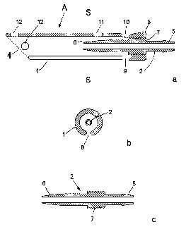
Figure 1 a shows a midline section of a preferred design of device A according
to the invention.
The device A is composed of two parts: one outer tube-like part 1 with a first
3
and a second 4 end and.one inner tube-like part 2 with a first 5 and a second
6 end.
The two tube-like parts are essentially concentric and parallel. The two tube-
like
parts 1 and 2 are joined near the end 3 of the outer part 1 and at an
intermediate part
7 of the inner part 2.
Figure 1b shows a cross section of the device along the line S - S in figure
la.
Here it is obvious that the two tube-like parts 1 and 2 are concentric. It is
also
obvious that the outer part 1 has a slit &., which will be mentioned later on.
Figure 2 shows in closer detail a rriidline section of an embodiment of the
inner
part 2. Part 2 is essentially axially symmetric with exception for the thread
of screw
on the intermediate part 7.
The first end 5 of the inner part 2 is arranged to be connected to a fluid
conducting catheter. In the shown design, this is achieved by shaping end 5 as
an
external nipple.
The other end 6 ~of the inner part 2 is designed to facilitate its passing
through
an opening in the vessel wall and at the same time give a close connection
between
the blood vessel wall and said part 2. This is achieved by making this part
slightly
conical, pointing to the end 6.
The outer part 1 and the izmer part 2 are in the illustrated embodiment joined
with threads. The inner part 2 is supplied with external threads along the
outer
intermediate part 7, and the outer part 1 is supplied with corresponding
internal
threads at an internal part close to the fzrst end 3.
Alternatively, the outer part 1 and the inner part 2 can be joined by conical
surfaces. In this case the inner part 2 has a slightly conical form pointing
either to
the first end, 5 or the second end 6, while the outer part 1 at its first end
3 has a
corresponding internal conical form.
The two parts can if desired also be joined by glue.
As yet an alternative, the outer part 1 and the inner part 2 can be solidly
joined
from the beginning, instead of being two parts meant to join later on. The
device has
been designed to be possible to manufacture in one piece by injection
moulding.
CA 02426910 2003-04-24
WO 02/36193 PCT/SE01/02362
However, there axe some advantages to malce the device A in two separate
parts, as it may allow simple manufacturing and combination of different
materials
and designs.
Figure 3a) - c) shows in closer detail the outer tube-like part 1 of the
device
5 seen from a) the side, b) from below, c) from above.
The outer tube-like part I has along a part of its length from the second end
4 a
lengthwise slit 8 for entrance of a corresponding length of the blood vessel.
When the outer and inner part 1 and 2 are joined, as illustrated in figure l,
the
second end 6 of the inner tube-like part 2 is wholly or partly surrounded by
the split
~ equipped part of the outer part 1.
The lengthwise slit 8 has essentially a width corresponding to the double
thickness of the blood vessel wall. The intention with the slit 8 is to limit,
in the
cross section, the part of the blood vessel that enters the lengthwise slit 8.
This part
of the blood vessel will, before it enters the lengthwise slit 8, be punctured
in such a
I S way (see procedure figure 6) that the end 6 of the inner part 2 can pass
the vessel
wall in this part of the vessel when the vessel enters the slit 8. After this,
the inner
part 2 from the end 6 to the intermediate part 7 will be inside the part of
the vessel
that has entered the slit 8. In this way, as well as by the moulding effect of
the
infusion needle 24 when the glue is injected (see procedure figure 6 - 8) a
new
narrow and stiff channel will be created along the blood vessel, constituting
a
channel bf living tissue from the end 6 of the inner tube-like part 2 to the
end 4 of
the outer tube-like part 1. Figure 4a shows the appearance of the new channel
401
when the device A has been applied on a blood vessel. The outer part 1 has
been
removed in the drawing for reasons of clarity. Figure 4b shows the same
situation in
cross section.
After the device A has been applied on a blood vessel there is thus a double
blood vessel wall in slit 8, part of the circumference as well as part of the
length
(corresponding to the length of the slit 8) of the blood vessel is situated
inside the
outer part 1. The end 6 of the inner tube-like part 2 passes the blood vessel
wall that
is inside the outer tube-like part 1. The width of slit 8 is enough for
nutrition of the
vessel wall inside part 1, but does not allow fluid to pass through i.e.
between, the
compressed vessel walls in the slit 8. Too narrow a slit will prevent
nutrition and too
wide a slit will allow blood to pass through and come into contact with
foreign
material (the end 6 of the inner part. 2).
The second end 4 of the outer tube-like part 1 is cut oblique in such a way
that
the side running alongside slit 8 is the shortest. This facilitates passing of
the device
onto the vessel and gives a better anatomy for passage of the new channel into
the
blood vessel (the deep neclc vein). The outer tube-like part 1 has two cross
openings
9~and IO near the end 3. The opening 9 is joined with and crosses the
lengthwise slit
CA 02426910 2003-04-24
WO 02/36193 PCT/SE01/02362
6
8, and the other opening 10 is arranged opposite the opening 9. The crossing
opening
is featured in the same way. All edges and corners are .rounded to adjust to
the
anatomy of the tissue. ~ . -
The openings 9 and 10 give opportunity to fix the vessel wall to the device .
5 with one or more sutures. The opening 10 can also, if desirable, serve as an
opening
for a suture thread tied to the vessel. This makes.it possible to pull the
vessel wall in
between the inner part 2 and the outer part 1 of the device. Said opening 10
can also
be used to checlc that the blood vessel wall is in the proper position and
that the glue
flows out as intended. The glue used is preferably Tisseel°, a two-
component glue.
10 The fibrinogen and the thrombin of such a glue give a rubberlike tissue
that is later
organised as the surrounding tissue.
The end 4 of the outer tube-like part 1 has at least one hole 12 to allow
fixing
the vessel wall to the outer part 1 with suture. The glue will also fix the
vessel wall
to the inside of the outer part 1 partly by adhering directly to the inside of
the outer
part 1, partly by filling the openings 9 - 12 of the part 1, thereby
mechanically fixing
the vessel to said part 1.
The outer tube-lilce part 1 has an additional hole 11 to fix the device A to
the
applicator (at the end of a glue conducting tube 27) and to apply glue. As the
glue is
injected from the syringe on the applicator it passes hole 11 and fills the
space
between the inside of the outer part 1 and the vessel wall.
Preferably, the whole device A is made out of firm tissue-compatible materials
like plastics, nylon, ceramics or metals. A suitable plastic is
polyetheretherketone
(PEEK). The various parts can be made out of different materials that are firm
and
compatible. It is an advantage if the material used is resistant and stable to
allow
repeated sterilization procedures, e.g. to withstand steam and air at
140°C, to allow
sterilization by heat and/or to resist ethylene oxide at 50 degrees Celsius as
this is
another procedure of sterilization.
Figure 5 a) shows in a side view, an applicator 20 for applying the device A.
The Applicator 20 facilitates the procedure when applying the device A in
accordance with the invention on a blood vessel and especially when applying
the
device A on the deep neck vein when shunting a patient with hydrocephalus.
The applicator 20 consists of a frame 2lmade of anodised (eloxidized)
aluminium or other suitable material. The frame 21 carnes the puncture needle
23, a
blunt infusion needle 24 that is connected to a tube 25, a glue conducting
tube 27
and the glue syringe 26.
.The glue conducting tube 27 is at its lower end arranged fox solid and dense
connection to the hole 11 of the device, but the device A may still easily be
removed.
The device A is lcept in place on the frame 21 also by help of the force
created from
the infusion needle 24 and from that said needle is guided by a hoolc-shaped
guide
CA 02426910 2003-04-24
WO 02/36193 PCT/SE01/02362
7
S 1. Said guide S 1 is advantageously arranged to apply a little force on said
needle 24
towards the frame 21 creating a force helping to keep the device A in place at
the
frame 21. The upper end of the tube 27 is arranged to be connected to the glue
syringe 26 via its angled needle S8 that has a piece of catheter to prevent
leakage.
S The puncture needle 23 is fixed to the frame 21 by means of a lock 41. The .
lock 4lconsists of a long, narrow part that has a hook 42 and two small
projections
43, 44, each with a hole fitting the puncture needle 23. This lock is arranged
to beep
the puncture needle 23 in the correct position and yet be easy to remove.
When using the applicator 20 a piece 28 of a silicon catheter is connected to
the end S of the inner part 2 of the device A to prevent leakage. This
catheter 28 will
later on in the procedure be connected to the catheter from the shunt valve
via a
connector or be removed to allow the catheter from the shunt valve to be
connected
directly to the nipple of the device A.
The infusion needle 24 passes through the catheter 28 and through the inner 2
1 S and outer 1 parts of the device A to a point where it meats the puncture
needle 23,
that is applied in such a way that the two needles form a
sharp angle and the opening of the puncture needle covers the end of the
infusion .
needle. This arrangement allows the infusion needle 24 to be blunt to avoid
undue
puncturing. The tube 2S that is connected to the other end of the infusion
needle is
used to check the function, of the new channel when the device A is applied on
the
deep neck vein and the new channel 401 has been created. The infusion needle
is
withdrawn to a reference point 29 to allow for this check. In this position,
the
infusion needle ends inside the inner tube-like part 2 close to its end 6 and
the new
channel is empty and ready to check for free passage. When the needle is fully
2S . retracted, the device A will be released from the applicator 20 and the
catheter 28
should then be held with forceps to prevent air embolism or backflow. It is
advantageous to provide a mark 60 on the applicator 20 that indicates
remaining
length of catheter on the infusion needle 24.
The following section will describe how a device according to the invention
can be used to connect a CSF conducting catheter to the deep neck vein in a
human.
The device A and the applicator 20 are symmetric and their use is .independent
of whether the person who uses them is right- or left-handed.
Figure S b) shows the applicator likewise from the side and also showing a
number of cross sections. The figure emphasises on the cross sections of the
frame
3S at positions AA to D and G as seen from the left refernng to the side view
of figure
Sb. In cross section AA is shown a docl~ing site S4 of the frame 21 for the
device A.
In cross section B is shown how the tube 27 extend into the docl~ing site S4.
In C is
shown how the hook-shaped guide S 1 is arranged for guiding the infusion
needle 24
as described above. In D is shown a guide opening S6. The views E and F shows
a
CA 02426910 2003-04-24
WO 02/36193 PCT/SE01/02362
g
locking wheel for the (double) syringe seen from two views orthogonal to the
side
view, i.e., from behind and from above. In G is shown a cross section of the
frame at
position G having a profile looping like ari inverted T.
Figure 6 illustrates the first part of an application procedure. An
appropriate
length of the deep neck vein is dissected~free, lifted and ftxed by a support
structure
31. The device A can be applied in a direction parallel or anti-parallel to
the blood
stream. A double suture 33 is placed proximally and distally relatively to the
intended puncture site. The puncture will be facilitated by lifting the vessel
wall of
the vein with the suture needle. After the puncture, the puncture needle 23 is
removed
Figure 7 illustrates how the infusion needle 24 with the device A is brought
fuxther onto the vein by stretching the suture threads via the slit 8 through
the
opening 9 in the opposite direction relatively to the direction of the
infusion needle
24. In this way, the inner tube 2 of the device A is passed more easily
through the
vessel wall of the vein.
The proper position of the vein in the device A can be checked in three ways:
A. The vessel wall of the vein can be seen in the upper opening 10.
B. The suture threads should be situated clearly opposite to the upper opening
10.
C. The slit 8 should to its whole extent be filled with vessel wall.
Figure 8 illustrates that the double suture 33 is tied around the inner tube
and
by an extra suture incision also around the outer tube. Figure 8 also shows a
suture
32 that attaches the wall of the vein vessel to the device A near the hole 12.
When the vein wall is in proper position in the device A and fixed with
sutures, glue is injected with force. Glue from the syringe 26 is injected via
the tube
27 through the hole 11 in the outer part of the device in such a way that the
space
between the inside of the outer part and the vessel wall and all openings are
filled
with glue. The infusion needle functions as a mould for the new channel that
will
get the same length as slit 8.
Figure 9 illustrates the fourth part of the procedure.
When after a few minutes the glue has settled, the glue syringe is removed and
the infusion needle is withdrawn to a position where the needle ends inside
the inner
part 2 of the device A. Infusion via the infusion needle will now wash out
blood
from the new channel and its fraction can be checlced. After this, the
infusion
needle is fully withdrawn and the device A is released from the applicator 20
and
can be connected to the catheter coming from the shunt valve.
After possibly adding additional glue on the outside of the device, the wound
is sutured to give additional support to the device A.
By means of the device A corresponding to the invention a part of the deep
neck vein is now transformed into a narrow, stiff channel with invariable
volume.
CA 02426910 2003-04-24
WO 02/36193 PCT/SE01/02362
9
The walls of this channel are living vein walls with good nutrition and the
channel is
filled with CSF. Backflow of blood in this channel is prevented by the stiff
blood
vessel walls and by the tightness in the slit and by the shunt valve. The new
channel
has to be narrow (about I millimetre in diameter) to allow the small volume of
CSF
(less than 500 millilitre per 24 h) to remove the blood that mixes with the
CSF in the
opening into the vein.
Accordingly, this invention malces it possible to drain CSF to the deep neck
vein and further to the heart without contact between the fluid-conducting
catheter
or any foreign material and the blood flow in the vein and at the same time
avoid the
use of X-ray, ECG or other means to secure the proper position of the catheter
in the
heart.
The invention also excludes the risk of thrombosis and decreases the risk of
infection since neither the device corresponding to the invention; the fluid-
conducting catheter or any other foreign material comes in contact with the
blood
flow in the vein. Draining to the deep neck vein gives a natural antisiphon
effect.
Figure 10 a) shows a view of a support, or a support instrument 1000, for
supporting fixing and shutting off of a vessel. The support instrument 1000 is
especially advantageous for supporting and fixing a neck vein when connecting
a
hydrocephalus shunt according to an embodiment of the invention to the vein,
and
the above mentioned support structure 31 in fig. 6 could advantageously be
equal to
this support instrument 1000.
An embodiment of the support comprises a handle 1001, a bend 1003, an
extending common part 1004, a bifurcation 1005 and two elongated members, one
concave or U-shaped 1010, 1012, 1015 and one L-shaped 1020, 1025. The support
is preferably devised with essentially flat parts, i.e., at least the common
part 1004
and a distal part 1015 of the U-shaped member 1010, 1012, 1015 are devised
having
essentially rectangular cross sections, with a thickness substantially smaller
than a
corresponding width. In a typical embodiment the thickness is approximately
0.5 to
2.5 mm and the width of the common part is approximately S to 15 mm.
The support is devised for capturing a blood vessel,between the U-shaped
member 1015 and the L-shaped member 1025, in the. opening 1030 that is formed
between said members; said opening 1030 continues as a lengthwise slit 1031
between said members 1012, 1025. Said lengthwise slit 1031 between these
members has essentially a width corresponding to the double thickness of the
blood
vessel wall for the vessel in question, i.e., embodiments of the support
instrument
provides different sizes of said slit, and of course also of the instrument
itself. The
reason for having a slit of'that dimensions is that~when a vessel is captured
in the
slit, the vessel is flattened and compressed so that the blood-flow is
reduced. Typical
widths include 1 to 1.5 mm. The reduction is in most eases complete so that
the
CA 02426910 2003-04-24
WO 02/36193 PCT/SE01/02362
.
blood flow is shut off. The U-shaped and the L-shaped members preferably have
a
rectangular or quadratic cross section, and are so arranged that said opening
1030
and slit formed between theportions 1012 and 1025 is wide enough to let the
vessel
slide gently into place when the support is twisted a bit. The members have
rounded
5 edges, so that the risk of hurting the vessel or other tissues during proper
use of the
support instrument is reduced or eliminated. The opening 1030 is preferrably
arranged so that a free end 1026 of the member .1025 are placed opposite a
bend
1014 between the distal parts of the U-shaped member 1012, 1015. Said free end
1026 is triangularly shaped so that the opening 1030 is wide at start and
narrows as
10 it transitions to the slit 1031.
The support is devised to lock itself into place at the operating wound as the
distal part of the U-shaped member and the common part 1004 or the part of the
U-
shaped and L-shaped members 1010, 1020 immediately following the bifurcation
1005 are arranged to make contact to the wound edges, and because the cross
section of said members and part is flat or rectangular. Because the vessel
itself,
when caught in the slit between the members 1025, 1012 or just resting above
both
members, will exert-a certain force on the support instrument 1000 directed
downwards to the bottom of the operating wound, the instrument will come
neatly
into place.