Note: Descriptions are shown in the official language in which they were submitted.
CA 02427749 2003-05-20
D1AGNOSTIC ASSAYS USING SOLUBLE ENDOTHELIAL CELL
PROTEIN CIACTIyATED PROTEIN C RECEPTOR
Background of the lnvention
The present invention is generally in the area of assays involving
detection and/or measurement of endothelial cell protein Clactivated protein
C receptor or soluble forms thereof derived either by proteolysis or by
alternative splicing.
The activation of protein C to its active serine protease, activated
protein C (APC), initiates a series of events that play a key role in the
regulation of blood coagulation. The clinical importance of the protein C
pathway is evidenced by the multitude of dysfunctions in this pathway that
result in thrombosis (Esmon and Schwarz. 1995. Trends Cardiovasc. Med.
5:141-148; Reitsma, et al. 1995. Thromb. Haemost. 73:876-879). Patients
deficient in protein C usually exhibit life threatening
thrombotic-complications in infancy (Seligsohn et al. , 1984. N. Engl. J. Med.
310, 559-562; Esmon, 1992. Trends Cardiovasc. Med. 2, 214-220) that are
corrected by protein C administration (Dreyfus et al., 1991. N. Engl. J. Med.
325, 1565-1568).
Protein C and APC have also been implicated in the regulation of
the host response to inflammation. Activated protein C (APC) can prevent
the lethal effects of E. colt in baboon models of gram negative sepsis (Taylor
et al., 1987. J. Clin. Invest. 79; U.S. Patent No. 5,009,889 to Taylor and
' Esmon) and preliminary clinical results suggest that protein C is effective
in
treating certain forms of human septic shock (Gerson et al., 1993. Pediatrics
91, 418-422). Inhibition of protein S, an important component of the protein
C pathway, exacerbates the response of primates to sublethal levels of E. colt
and augments the appearance of TNF in the circulation (Taylor et al., 1991.
CA 02427749 2003-05-20
2
Blood 78, 357-363). These results suggest that protein C may both control
coagulation and influence inflammation.
' Protein C is activated when thrombin, the terminal enzyme of the
coagulation system, binds to an endothelial cell surface protein,
thrombomodulin (Esmon, 1989. J. Biol. Chem. 264, 4743-4746; Dittman and
Majerus, 1990. Blood 75, 329-336; Dittman, 1991. Trends Cardiovasc. Med.
1, 331-336). In cell culture, thrombomodulin transcription is blocked by
exposure of endothelial cells to tumor necrosis factor (TNF) (Conway and
Rosenberg, 1988. Mol. Cell. Biol. 8, 5588-5592) and thrombomodulin
activity and antigen are subsequently internalized and degraded (Lentz et al.,
1991. Blood 77, 543-550, Moore, et.al., 1989. Blood 73, 159-165). C4bBP,
a regulatory protein of the complement system, binds protein S to form a
complex that is functionally inactive in supporting APC anticoagulant activity
in vitro (Dahlback, 1986. J. Biol. Chem. 261, 12022-12027) and in vivo
(Taylor,et al., 1991). C4bBP behaves as an acute phase reactant (Dahlback,
1991. Thromb. Haemostas. 66, 49-61). Thus, proteins of this pathway-not
only appear to regulate inflammation, but they also interact with components
that regulate inflammation, and they themselves are subject to down
regulation by inflammatory mediators.
Endothelial cells play a critical role in the protein C pathway in that
they express two of the known receptors responsible for efficient protein C
activation, thrombomodulin and the endothelial protein C/APC receptor
(EPCR) (Fukudome and Esmon. 1994. J. Biol. Chem. 269:26486-26491;
Steams-Kurosawa, et al. 1996. Proc. Nat!. Acad. Sci. (USA) 93:10212-
10216). Thrombomodulin (CD141) is a transmembrane cofactor that binds
circulating thrombin with high affinity and the resultant enzyme-cofactor
complex is required for physiologically relevant protein C activation rates .
(Esmon and Owen. 1981. Proc. Natl. Acad. Sci. (USA) 78:2249-2252;
Dittrnan, W. A. 1991. Trends Cardiovasc. Med. 1:331-336).
CA 02427749 2003-05-20
3
EPCR is a recently identified receptor with significant homology to
the CD11MHC class 1 family (Fukudome and Esmon, 1994; Fukudome, et
al. 1996. J. Biol. Chem. 271:17491-17498; Regan, et al. 1996. J. Biol.
Chem. 271:17499-17503). The cloning and biological role of the endothelial
cell receptor for protein C was described in PCTIUS95109636 by Oklahoma
Medical Research Foundation, entitled "Cloning and Regulation of an
Endothelial Cell Protein C/Activated Protein C Receptor" . The protein was
predicted to consist of 238 amino acids, which includes a 15 amino acid
signal sequence at the N-terminus, and a 23 amino acid transmembrane
region which characterizes the receptor as a type 1 transmembrane protein.
EPCR binds both protein C and APC with similar affinity (Kd~,~ - 30
nM) (Fukudome, et al., 1996) in the presence of calcium and facilitates
protein C activation by presenting the protein C substrate to the thrombin-
thrombomodulin activation complex on cell surfaces (Steams-Kurosawa, et
I S al . , I 996) . Both endothelial cell receptors are type 1 transmembrane
proteins
in which the ligand binds to an extracellular domain and both have a short
intracellular cytoplasmic tail (Fukudome, et al. 1996; Jackman, et al. 1987.
Proc. Natl. Acad. Sci. (USA) 84:6425-6429; Wen, et al. , 1987. Biochemistry
26:4350-4357; Suzuki, et al. 1987. EMBO J. 6:1891-1897). In addition,
their in vitro cell surface expression is down-regulated similarly by tumor
necrosis factor-a(Fukudome and Esmon 1994). However, the characteristics
of soluble forms of thrombomodulin and EPCR differ in several respects.
Recombinant soluble thrombomodulin has reduced cofactor activity relative to
the membrane form (Calvin, et al. 1987. J. Biol. Chem. 262:2199-2205;
Parkinson, et al. 1990. J. Biol. Chem. X65:12602-12610). With both
purified components and with cells, the changes in thrombin's substrate
specifically induced by thrombomodulin result from competition for a shared
binding domain on thrombin as well as conformational alterations in the
active site pocket (Ye, et al. 1991. J. Biol. Chem. 266:23016-23021; Lu, et
CA 02427749 2003-05-20
4
al. 1989. J. Biol. Chem. 264:12956-12962; Ye, et al. 1992. J. Biol. Chem.
267:11023-11028; Hofsteenge, et al. 1986. Biochem. J. 237:243-251;
Mathew>;, 1994. Biochemistry 33:13547-13552; Esmon, et al. 1982. J. Biol.
Chem. 257:7944-7947; Sadler, et al. 1993. Haemostasis 23:183-193).
S Soluble thrombomodulin also accelerates inactivation of thrombin by a
variety of inhibitors (Bourin and Lindahl. 1993. Biochem. J. 289:313-330;
Rezaie, 1995. J. Biol. Chem. 270:25336-25339). Both plasma and urine
contain detectable thrombomodulin (Takano, et al. 1990. Blood. 76:2024-
2029; Ishii and Majerus. 1985. J. Clin. Invest. 76:2178-2181) and because
the thrombomodulin gene does not contain introns (Jackman, et al., 1987),
these soluble forms are due to proteolysis of the extracellular domain at the
cell surface.
Soluble degradation products of thrombomodulin in plasma are a
known marker of endothelial cell damage in a variety of disease states
(Takano, et al . , 1990; Tanaka, et al. 1991. Clin. Chem. 37:269-272;
Takahashi, et a1. 1991.Am. J. Hematol. 38:174-177; Asakura, et al. 1991.
Am. J. Hematol. 38:281-287; Wada, et a1. 1992. Am. J. Hematol. 39:20-24;
Takahashi, et al. 1992.Am. J. Hematol. 41:32-39; Ohdama, et al. 1994.
Chest 106:666-671 ) and are comprised of a mixture of thrombin-binding
fragments with varying reduced affinities, as well as non-binding fragments
(Takano, et al., 1990).
In contrast, recombinant soluble EPCR (rsEPCR), truncated just
before the transmembrane domain, binds both protein C and APC with an
affinity similar to that observed for intact cell-surface expressed EPCR
(Fukudome, et al. 1996). APC anticoagulant activity is inhibited effectively
when bound to rsEPCR (Regan, et al., 1996), presumably because both
rsEPCR and factor Va share binding determinants in a groove reminiscent of
the anion binding exosite I in thrombin occupied by thrombomodulin
(Mather, et al. 1996. EMBO J. 15:6822-6831). However, rsEPCR does not
CA 02427749 2003-05-20
S
appear to influence proteolysis of small synthetic substrates by APC, nor
inactivation of APC by al-antitrypsin or protein C inhibitor (Regan, et al.,
1996). Unlike membrane-bound EPCR which enhances protein C activation
(Steams-Kurosaw, at al., 1996), rsEPCR has little effect on protein C
activation by the soluble thrombin-thrombomodulin complex (Regan, et al.,
1996), suggesting that any soluble farms of EPCR might inhibit protein C
activation by competing with membrane-associated EPCR for protein C.
Immunohistochemistry indicates that EPCR is present primarily on
the surface of endothelial cells from large vessels and is absent or present
at
low levels on most capillary endothelial cells.
It is therefore an object of the present invention to identify
therapeutic and diagnostic uses for naturally occurring soluble EPCR.
It is a further object of the present invention to characterize
naturally occurring soluble EPCR.
Summary of the Invention
Plasma EPCR (has been isolated, characterized and shown to block
cellular protein C activation and APC anticoagulant activity. Plasma EPCR
appears to be about 43,000 daltons and circulates at approximately 100 ng/ml
(98.4 ~ 27.8 ng/ml, n = 22). Plasma EPCR was purified from human
citrated plasma using ion-exchange, immunoaffinity, and protein C affinity
chromatography. Flow cytometry experiments demonstrated that plasma
EPCR bound activated protein C with an affinity similar to that previously
determined from recombinant truncated EPCR (KdaPP approximately 30 nM),
defined as EPCR not including the transmembrane and cytoplasmic domains.
Furthermore, plasma EPCR inhibited both protein C activation on an
endothelial cell line and APC anticoagulant activity in a one-stage factor as
clotting assay. Soluble EPCR has also been detected in human urine.
Cloning of the gene encoding EPCR demonstrates that at least human EPCR
can be alternatively spliced, yielding a truncated soluble EPCR including an
CA 02427749 2003-05-20
6
insert unique to the alternatively spliced form (sEPCR). These results.
indicate that plasma EPCR can be derived either by proteolysis at the cell
surface or by alternative splicing.
If the local concentrations of plasma EPCR are sufficiently high,
particularly in disease states, the data indicates that the truncated soluble
plasma EPCR could attenuate the membrane-bound EPCR augmentation of
protein C activation and the anticoagulant function of activated protein C.
As demonstrated by the examples comparing normal plasma EPCR with
levels of EPCR from patients with an autoimmune disease (systemic lupus
erythernatosus, SLE) and sepsis (a disorder involving both inflammation and
coagulation abnormalities), levels of soluble EPCR appear to be correlated
with inflammation and disease states associated with abnormal coagulation.
Assays are described based on measurement of soluble EPCR which are
indicative of disease conditions involving coagulation, inflammation, and
large vessel disease. Assay reagents are described, including isolated
purified soluble EPCR, recombinant truncated soluble EPCR, and antibodies
to the soluble EPCRs.
Brief Description of the Drawings
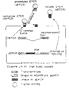
Figure 1 is a schematic of the two known mechanisms for
producing a soluble receptor as applied to EPCR, by proteolysis of the
membrane-bound receptor to release an extracellular domain and leave the
membrane anchor behind, and by alternative splicing of the m.RNA, showing
the sequences unique to membrane bound EPCR (mEPCR) and to
proteolyzed plasma EPCR (pEPCR), and the sequence unique to soluble
EPCR (sEPCR).
Figure 2 is a schematic comparing mEPCR and sEPCR, showing
the nucleotide insert and encoded amino acid sequence unique to sEPCR.
Figure 3 shows the sequence inserted into human, bovine, murine,
and baboon EPCR by alternative splicing.
CA 02427749 2003-05-20
7
Figure 4a is a graph showing that soluble plasma EPCR binds to
human protein C and APC. EA.hy926 cells were incubated with 60 nM fl-
APC in 'the presence of 0 - 500 nM rsEPCR ( ~ ) or plasma EPCR ( O ) for
30 minutes on ice. The cells were washed and cell-bound fluorescence was
determined by flow cytometry as described. The intrinsic cell fluorescence
in the absence of added fl-APC is indicated by the arrow. The mean cell
fluorescence (MCF) plotted represents the average of duplicate MCF
determinations.
Figures 4b and 4c are graphs showing soluble plasma EPCR and
rsEPCR inhibit protein C activation on cell surfaces. In Figure 4b,
EA.hy926 cell monolayers were pre-incubated for 15 minutes at room
temperature with 0.1 ~.M protein C alone ( O ) or with 1 ~uM rsEPCR ( ~ ), or
2 ~,glml 1494 mAb (O). Protein C activation was initiated by the addition
of thrombin (2 nM final) and the reactions were stopped at the indicated
times. Activated protein C was determined with an amidolytic assay and the
activity rates in mOD/min are plotted for each time point. Control wells
without added thrombin were included ( ~ ). Each data point represents the
average of triplicate well determinations. In Figure 4c, EA.hy926 cell
monolayers were pre-incubated for 15 minutes at room temperature with 0.1
~.M protein C and the indicated concentrations of plasma EPCR (O) or
rsEPCR ( ~ ). Thrombin (final 2 nM) was added and the activation
proceeded for 60 minutes at room temperature. The supernatants were added
to a mixture of antithrombin and heparin and activated protein C activities
(mOD/min) were determined with an amidolytic assay. Each data point
represents the average of triplicate well determinations.
Figure 4d is a graph showing soluble plasma EPCR inhibits APC
anticoagulant activity
The anticoagulant activity of APC (25 nM) was determined with a one-stage
Xa clotting assay in the presence of 460 nM plasma EPCR or rsEPCR. The
effect was reversed when either soluble EPCR was pre-incubated for 5
CA 02427749 2003-05-20
minutes with 42 pglrnl of 1496 mAb which blocks binding of APC to..EPCR.
The data represent the average of 4-6 determinations ~ S.D.
' Figure S is a graph comparing levels of soluble plasma TM to
soluble plasma PCR in lupus patients, demonstrating that there is no
correlation between TM and EPCR values, but that the majority of lupus
patients exhibit extremely elevated levels of soluble plasma EPCR.
Figure 6 is a graph of soluble receptor concentration (ng/ml) for
sTM (normal), sTM (sepsis), sEPCR (normal), and sEPCR (sepsis). No
correlation between sTM and sEPCR, v2 = 0.034.
Detailed Description of the Invention
Definitions
Endothelial Protein C Receptor, EPCR.
Soluble, in solution and not bound to a cell surface.
Truncated, not including the transmembrane and cytoplasmic
domains; can be a result of either proteolysis or alternative splicing.
Detection and Characterization of Soluble EPCR; Physiological
Role and Utility as a Marker
Previous investigations into the function of EPCR found that
protein C binding to the membrane form of EPCR resulted in facilitation of
protein C activation by the thrombin-thrombomodulin complex on cell
surfaces (Steams-Kurosawa, et al., 1996), but that soluble recombinant
EPCR inhibited APC anticoagulant activity (Regan, et al. 1996). These
observations, along with the knowledge that soluble thrombomodulin
degradation products in plasma are a marker of endothelial damage in various
disease states, led to the question of whether a soluble circulating forms) of
EPCR existed and, if so, what role it may have in the protein C pathway.
The examples demonstrate that a soluble form of EPCR circulates
in plasma and is present in urine. In a healthy donor population, the plasma
EPCR level was about 100 ng/ml and it appeared to be a single antigen
CA 02427749 2003-05-20
9
species of approximately 43,000 daltons. Subsequent purification of the
soluble EPCR from plasma and functional studies determined that it was
capable of binding both protein C and APC with an affinity similar to intact
membrane-bound EPCR. The in vitro studies using an endothelial cell line
demonstrated that plasma EPCR inhibited protein C activation at near
physiological concentrations of protein C and thrombin. In addition, direct
addition of purified plasma EPCR to plasma resulted in inhibition of APC
anticoagulant activity that was reversed with monoclonal antibodies to
rsEPCR.
The identification of the purified plasma protein as being EPCR
was based on comparison with the properties of rsEPCR. These proteins
both reacted with the same battery of monoclonal and polyclonal antibodies,
had the same amino-terminal sequence, bound to immobilized protein C in a
Ca2+-dependent fashion, and blocked protein C activation and APC
anticoagulant activity with similar dose response curves. In addition, the
affinities of both protein C and APC for rsEPCR and plasma EPCR are
similar to the affinity of intact membrane-bound EPCR. These properties
appear to be unique to EPCR.
Previous studies demonstrated that membrane-bound EPCR
expressed on endothelial cells augments protein C activation by a factor of
between three and five fold, whereas the examples demonstrate that the
soluble form of EPCR purified from plasma inhibits protein C activation on
endothelial cells and APC anticoagulant activity. This predicts that EPCR
could modulate the protein C pathway in several ways. First, in the larger
vessels where thrombomodulin concentration is low to the microcirculation,
EPCR expression is correspondingly increased (Laszik, et al., Circulation
1997). Immunohistochemistry shows that in most organs, EPCR expression
is most intense on large vessels and decreases progressively with decreasing
vessel size, with little or no expression in the most abundant endothelial
cell
type, the capillary endothelium. EPCR expression may play a critical role in
CA 02427749 2003-05-20
capturing the protein C substrate from the circulation and presenting ii
to.the
thrombin-thrombomodulin complex for activation. This is supported by in
vitro ol3servations that both the EA.hy926 endothelial cell line and human
umbilical vein endothelial cells have at least six times more surface-
expressed
5 EPCR antigen than thrombomodulin. In the microcirculation where
thrombomodulin concentration is high and EPCR is low, one would predict
little influence on protein C activation. Finally, circulating soluble EPCR
may reduce the generation of APC and the ability of APC to inactivate factor
Va.
10 In a healthy individual, the soluble EPCR levels are about 2.5 nM,
a concentration.well below both the Kde~,(approximately 30 nM) and the 80
nM protein C concentration in the circulation. Both of the effects of soluble
plasma EPCR (inhibition of APC anticoagulant activity and protein C
activation) required considerably higher concentrations than that present in
normal plasma, leaving the question of the physiological role of the plasma
EPCR uncertain. Patients with soluble EPCR levels that exceed 40 nM have
been identified, as described in Example 3 (lupus). Thus, if the local
concentration near the endothelial cell surface exceeds the systemic
concentration, the soluble EPCR concentration would reach levels that would
attenuate both APC generation and activity, contributing to thrombotic risk.
A soluble form of a receptor can be produced by proteolytic
cleavage of the membrane-bound receptor or by alternative splicing
mechanisms. Proteolysis at the membrane surface releases soluble
thrombomodulin, and receptors for TNF, IL-1, IL-2, M-CSF, PDGF, and
NGF (Heaney, et al. 1996. Blood 87:847-857). Soluble receptors have a
multitude of potential functions including acting as antagonists of the
membrane receptor, stabilizing the ligand, initiating ligand-mediated
signaling, downmodulation of the membrane form, and binding to receptor
inhibitors to indirectly facilitate receptor-ligand activity. The latter
mechanism is used by the IL-1 receptor system in which the soluble isoforms
CA 02427749 2003-05-20
11
of both IL-1 receptors are generated by proteolytic cleavage and tightly
regulate the responsiveness to IL-la and IL-lQ (Arend, et al. 1994. J.
Immunol. 153:4766-4774). The EPCR genomic structure contains an
alternative splicing site which would code for a soluble protein truncated
just
before the transmembrane domain (Fukudome and Esmon. 1995. J. Biol.
Chem. 270:5571-5577), as discussed below. Soluble IL-6 receptors appear to
be generated by both proteolytic and alternative splicing mechanisms
(Mullberg, et al. 1994. J. Immunol. 152:4958-4968; Lust, et al. 1992.
C~tokine 4:96-100; Horiuchi, et al. 1994. Eur. J. Immunol. 24:1945-1948).
This cleavage site can also be useful in recovering large quantities of
soluble
EPCR, by constructing an expression vector encoding the truncated EPCR
immediately followed by a peptide sequence to which an antibody is
specifically directed, as described in U.S. Patent No. 5,298,599 to Morrissey
and Esmon, the teachings of which are incorporated herein. The epitope will
then be cleaved by proteolysis, before or after administration to a patient.
See also U.S. Patent No. No.4,782,137 to Hopp et al.
Immunohistochemical studies have indicated that EPCR is located
primarily on endothelium of large vessels and is barely detectable in
capillaries. Plasma EPCR derived from membrane-bound EPCR, can
therefore serve as a marker of large vessel disease processes. Plasma EPCR
may serve as a useful comparison with plasma thrombomodulin levels which
have been shown to be modulated in a variety of disease states, but which
would reflect both large and small vessel disease processes, but probably
would be dominated by small vessel contributions since most endothelium is
microvascular.
Nucleotide and Predicted Protein Structure Analysis of EPCR
The cDNA for EPCR is predicted to code for a protein of 238
amino acids (Sequence 1D No. 2), which includes a 15 amino acid signal
sequence (von Heijne, (1986) Nucleic Acids Res. 14, 4683-4690) at the
CA 02427749 2003-05-20
12
N-terminal. Therefore, the mature protein is predicted to contain 223 amino
acids. Direct sequencing of the recombinant protein showed that the mature
proteiw started at Serl8. Sequence ID No. 2 is the predicted amino acid
sequence of EPCR. Amino acids 1-15 of Sequence ID No. 2
(MLTTLLPILLLSGWA) are the putative signal sequence determined by the
method of von Heijne (von Heijne, 1986). Amino acids 211-236 of
Sequence ID No. 2 (LVLGVLVGGFIIAGVAVGIFLCTGGR) are the
putative transmembrane domain. Potential N-glycosylation sites are present
at amino acids 47-49, 64-66, 136-138, and 172-174 of Sequence ID No. 2.
Extracellular cysteine residues are present at amino acids 17 (removed in
plasma EPCR), 114, 118, and 186 of Sequence ID No. 2. A potential
transmembrane region (Engelman et al., (1986) Annu. Rev. Biophys.. Chem.
15, 321-53 ) consisting of 23 amino acids was identified at the C-terminal
end (beginning at amino acid 211 of Sequence ID No. 2).
The protein is a type 1 transmembrane protein. The extracellular
domain contains four potential N-glycosylation sites and three Cys residues.
Glycosylation is not essential for activity, as shown by N-glycanase
digestion. The cytoplasmic region contains only three amino acids and
terminates with a Cys, which is palmitoylated. If the terminal cysteine is not
properly palmitoylated, the protein may be secreted. Altering the sequence
of the EPCR to replace this cysteine with another amino acid thereby
provides a means for making an essentially full length EPCR which is
secreted instead of being membrane bound.
As used herein, the nucleotide sequences encoding the receptor
include the sequence shown in Sequence ID No. 1, and sequences having
conservative substitutions, additions or deletions thereof which hybridize to
Sequence ID No. 1 under stringent conditions. As used herein, the amino
acids sequences constituting the receptor include the sequence shown in
Sequence ID No. 2, and sequences having conservative substitutions,
additions or deletions thereof which form a receptor having functionally
CA 02427749 2003-05-20
13
equivalent biological activity. It is well known to those skilled in theart
what constitutes conservative substitutions, additions or deletions, and which
could be readily ascertained as encoding, or forming, a functionally
equivalent receptor molecule using the functional assays described herein.
This is further illustrated by reference to Figure 3, discussed below.
Alternative Splicing
Receptors are most often visualized as being proteins anchored in a
cell membrane with the portion exposed to the outside of a cell responsible
for binding a specific ligand to generate a physiological response. In many
cases, a soluble form of the receptor exists that frequently is quite capable
of
binding its ligand, despite the fact that it is no longer restricted to a
cell.
Ligand binding to the soluble receptor isoform can also generate a response
which takes many forms, including up- or down-modulation of the
membrane-bound receptor interactions, or propagating a response by
transporting the ligand to a cell that normally is not responsive (I-Ieaney,
ML
and DW Golde. Soluble cytokine receptors. Blood 87:847-857, 1996).
There are two known mechanisms for producing a soluble receptor:
by proteolysis of the membrane-bound receptor to release an extracellular
domain and leave the membrane anchor behind, and by alternative splicing of
the mRNA {Figure 1). The latter mechanism can take many forms, but the
simplest is when the reading frame continues through an exon-intron
boundary and terminates with a stop codon before reaching the sequence
coding for the transmembrance anchor. This creates a protein that is similar
to the membrane form, but with important differences. It is made, and
secreted, as a soluble protein and will have a unique carboxyl-terminal tail.
This tail was formed by reading a portion of the intron mRNA sequence that
is ignored in the formation of the membrane-bound receptor. Generation of a
soluble receptor by alternative splicing can also be modulated independently
from the membrane-bound receptor, despite the fact that they both originate
CA 02427749 2003-05-20
14
from the same mRNA template (Heaney, et al. Proc. Natl. Acad. Sci._ U.S.A.
92:2365-2369, 1995).
' To demonstrate that a soluble receptor is generated by alternative
splicing mechanisms, one must know the genomic sequence and intron-exon
boundaries of the relevant region. It is also helpful to link the soluble
receptor with a physiological response to distinguish it from aberrant mRiVA
splicing, found fairly frequently including during expression of the protein C
and protein S genes (Berg, et al. Blood Coag Fibrinol. 7:625-631, 1996).
Details of the following studies and results are described in the
examples. Human plasma contains about 100 ng/ml of soluble EPCR (Table
1). This was measured by an enzyme linked immunoassay (ELISA) using
two monoclonal antibodies (1494 mAb and 1495 mAb) and standard
techniques. Significantly elevated soluble EPCR levels were found in
patients with systemic lupus erythematosus and sepsis. These levels seemed -
fairly high for a membrane-bound receptor that is present, with few
exceptions, only on the surface of the large blood vessels. To put this iiI
perspective, thrombomodulin (TM) is expressed on all endothelium, as well
as some non-vascular cells, yet normal soluble TM levels are only about 10-
40 ng/ml (Takano, et al., Blood 76:2024-2029, 1990). The soluble TM
levels were elevated in the patients with lupus, but riot sepsis. Importantly,
there was no correlation between the plasma EPCR and TM levels in these
patient groups (rz=0.028. and 0.034, respectively).
CA 02427749 2003-05-20
Table 1. Plasma soluble receptor levels -
Plasma EPCR Plasma TM
~ ng/ml ng/ml
Normal volunteers, 133.4 ~ 53.4 35.5 ~ 20.4
' n=20
Systemic lupus 262.1 ~ 154.5* 104.7 ~ 77.5*
5 erythematosus, n=40 (P=0.0004) (P=0.0008)
Sepsis, n=24 224.9 ~ 74.5* 39.9 ~ 73.1
(P=0.00009)
* igru scant di ference between the means relatme to normal;
unpaired Student's t test.
The TM genomic structure does not contain introns (3ackman, et al.
10 Proc. Natl. Acad. Sci. (USA) 84:6425-6429, 1987), so the only way to create
a soluble TM isoform is by proteolysis of the membrane-bound receptor.
Proteolysis of endothelial TM by neutrophil elastase and cathepsin G has
been shown in vitro, suggesting that the elevated soluble TM levels found in
a variety of disease states result from proteolysis mediated by products of
15 activated inflammatory cells at the endothelial surface (Boehme, et al.,
Immunology 87:134-140, 1996 and Abe, et al. J. Lab. Clin. Med. 123:874-
881, 1994).
The lack of correlation between the plasma EPCR and TM levels
and the high plasma EPCR concentration is consistent with the concept that
plasma EPCR originates from both proteolytic and alternative splicing
mechanisms. The genomic structure of human EPCR contains four exons,
separated by introns. Review of this sequence reveals an in-frame reading
sequence after the exon 1II - intron III boundary (at the 5' GT) that includes
a TAA stop codon at position 7527. Since this stop codon is upstream of
exon IV that codes for the transmembrane domain, the predicted protein
would contain a unique 48 residue carboxyl-terminal tail (coded for by the
intron sequence) and would not contain a transmembrane anchor.
CA 02427749 2003-05-20
16
Figure 1 is a diagram of two potential ways truncated EPCB can be
derived: by proteolysis immediately before the transmembrane domain or by
alternarive splicing. As shown by Figure 2, alternative splicing results in
inclusion of a peptide sequence in the alternatively spliced truncated EPCR.
As shown by Figure 3, this sequence is highly conserved between species,
although slight differences exist, resulting in a new carboxyl-terminal tail
of
48 residues for human and bovine EPCR, S 1 residues for murine EPCR, and
22 residues for baboon EPCR.
Screening of_patient samples for expression of receptor proteins.
Patient samples can be screened for the presence of, and amount
of, sEPCR or EPCR, using antibodies to either EPCR, the unique insert
present in the alternatively spliced insert in EPCR, or antibodies which bind
with greater affinity to either EPCR or sEPCR due to conformational
differences. Samples can also be screened using other standard techniques to
specifically quantitate proteins which are present.
Generation of Antibodies for Diagnostic or Therapeutic
Use
Antibodies to EPCR, and in particular, soluble EPCR ("sEPCR"),
and recombinant soluble EPCR ("rsEPCR") can be generated which are
useful in detection, characterization or isolation of receptor proteins, as
well
as for modifying receptor protein activity, in most cases, through inhibition
of ligand binding. Antibodies are generated by standard techniques, using
human or animal purified or recombinant receptor proteins or fragments
thereof as the immunogen.
Monoclonal antibodies to EPCR were obtained as described for
' other proteins by Esmon, et al. , 1993. Methods Enzymol. 222:359-385. The
antibodies referred to as 1494, 1495, and 1496 mAbs are IgGIK antibodies
that bind to recombinant soluble EPCR and to cell surface-expressed EPCR.
The 1494 and 1496 mAbs block the binding of protein C and APC to EPCR,
and inhibit the ability of cellular EPCR to facilitate protein C activation by
CA 02427749 2003-05-20
17
the thrombin-thrombomodulin complex. The 1495 mAb does not block
ligand binding to EPCR, does not alter cell surface protein C activation, and
has a binding epitope distinct from that for 1494 or 1486 mAb. The
antibodies can be labelled using standard techniques, such as radiolabeiling,
enzyme labelling, fluorescent labels such as fluorescein, gold particles,
dyes,
and other means for detection of the antibodies. For example, antibody can
be biotinylated with biotinamidocaproate N-hydroxysuccinimide ester using
standard procedures. Antibody can be immobilized to a solid support for use
in immunoassays, for example, AffiGel-10'~"', nitrocellulose, or rnicrotiter
wells, or use in solution phase immunoassays.
In a preferred embodiment, EPCR is measured using microtiter
plates (MaxisorpT"', NUNC NS, Roskilde, Denmark) coated with 50
microliters of 4 micrograms/ml 1495 mAb in 15 mM NaZC03, 35 mM
NaHC03, pH 9.6, at 4'C overnight. At room temperature, the plates are
then washed three times with 20 mM Tris-HCI, 0.1 M NaCI, 0.05 % Tween
20, pH 7.5 (assay buffer), and blocked with assay buffer containing 0.1 %
(wt/vol) gelatin for at least one hour. The wells are then washed, 50
microliter samples added in triplicate wells, and the plates incubated for one
hour. The wells are aspirated, washed three times with assay buffer, and 50
microliters of 2 micrograms/ml biotin-1494 mAb added. The plates are
incubated for 1 hour, washed three times, and 50 microliters of 0.25
micrograms/ml streptavidin-alkaline phosphatase conjugate (G1BC0 BRL)
added and incubated for an additional hour. The wells are washed five
times, and the substrate and amplifier reagents from an ELISA amplification
kit (GIBCO BRL) added sequentially at 15-min intervals according to the
manufacturer's directions. The color development is stopped with 0.3 M
H,SO" and the endpoint absorbance read at 490 nm on a V",ex microplate
reader. Standards in triplicate wells are from 1.5 to 100 ng rsEPCR/ml in
20 mM Tris-HC1, 0.1 M NaCI, and 1 mM EDTA, 0.1 % gelatin, pH 7.5.
The standard curve is linear from 1.5 to 12.5 nglml, and samples are diluted
CA 02427749 2003-05-20
18
with the same buffer to fall within the linear range. Studies show that
between one and two percent plasma does not affect the linearity of the assay
or the sensitivity of the standard curve. Plasma samples from healthy
volunteers were diluted with assay buffer containing 1 mM EDTA to a final
2 % plasma, and EPCR antigen levels are calculated from the average of
triplicate wells by reference to standard curve determined on the same plate.
Disorders
The assay for soluble EPCR is useful in detection and analysis of
coagulation and inflammatory states and disorders as discussed herein, such
as autoimmune diseases like lupus, in transplant monitoring, sepsis, shock,
pre-eclampsia, diabetes, cardiopulmonary bypass, unstable angina, restenosis,
angioplasty (i.e., vascular disease), kidney or liver disease. For example,
the EPCR is a marker for large blood vessels, and therefore for damage to
large blood vessels. An increase in the amount of soluble EPCR is indicative
of large vessel injury, resulting either in proteolysis of EPCR or stimulation
of sEPCR synthesis. The ratio of EPCR to thrombomodulin can also be'
determined, based on either blood or urine samples, which is indicative of
the relative extent of microvascular versus large vessel. The relative amounts
of EPCR to cytokines, leukocyte activation markers and complement factors
or activation markers can also be used to indicate disease state.
Since EPCR is present on endothelial cells, it is useful as a marker
of endothelial cell damage. It can be used as an indicator of drug effect,
both toxicity as well as efficacy. For example, in lupus patients, drugs
effectively minimizing inflammatory/coagulation mediated, large vessel injury
would result in decreasing EPCR levels.
The present invention will be further understood by reference to the
following non-limiting examples.
CA 02427749 2003-05-20
19
Example 1: Identification of Functional Endothelial Protean C
Receptor in Human Plasma
The following abbreviations are used: rsEPCR, recombinant soluble
EPCR with the HPC4 epitope inserted in place of the transmembrane domain
and cytosolic tail; mAb, monoclonal antibody; SDS-PAGE, sodium
dodecylsulfate polyacrylamide gel electrophoresis.
METHODS
Materials. The following reagents were purchased from the indicated
vendors:
Porcine intestinal mucosal heparin, diisopropyl fluorophosphate,
biotinamidocaproateN-hydroxysuccinimide ester, bovine serum albumin,
Sigma (St. Louis, MO); Spectrozyme PCa, American Diagnostica
(Greenwich, CT); ELISA amplification kit, GibcoBRL (Gaithersburg, MD);
AffiGel-10, BioRad (Hercules, CA); Hank's balanced salt solution, 3-(N-
morpholine)propane sulfonic acid (MOPS), Fisher Scientific (Fair Lawn,
NJ). All other reagents were of the highest quality commercial available:
Proteins. Human protein C (Esmon, et al. 1993. Methods Enryrnol.
222:359-385), bovine thrombin (Owen, et al. 1974. J. Biol. Chem. 249:594-
605), and bovine antithrombin (Esmon 1977. "Factors regulating the
inhibition of thrombin by antithrombin III. In Chemistry and Biology of
Thrombin". R. L. Lundblad, J. W. Fenton, II, and K. G. Mann, editors.
Ann Arbor Science, Ann Arbor. 403-411 ) were purified as described.
Recombinant soluble EPCR, rsEPCR, consists of the extracellular domain of
EPCR truncated at residue 210 just before the transmembrane domain,
followed by a 12 residue sequence that permits calcium-dependent
immunoaffinity purification on the HPC4 monoclonal antibody (Takahashi, et
al. 1992; Stearns, et al. 1988. J. Biol. Chem. 263:826-832). The
construction, purification, and protein C/APC binding characteristics of
rsEPCR (Fukudome, et al. 1996). Goat preimmune serum and polyclonal
antiserum to rsEPCR was prepared and the IgG purified (Fukudome, et a!.
CA 02427749 2003-05-20
1996). Goat anti-rsEPCR polyclonal antibody was biotinylated with _
biotinamidocaproate N-hydroxysuccinimide ester using standard procedures.
Monoclonal antibodies. Monoclonal antibodies (mAb) against rsEPCR were
obtained as described for other proteins (Esmon, et al. 1993). The 1494,
5 1495, and 1496 mAb are IgGlk antibodies that bind to rsEPCR and to cell
surface-expressed EPCR. The 1494 and 1496 mAb block the binding of
protein C and APC to EPCR and inhibit the ability of cellular EPCR to
facilitate protein C activation by the thrombin-thrombomodulin complex
(Steams-Kurosawa, et al. 1996). The 1495 mAb does not block ligand
10 binding to EPCR, does not alter cell surface protein C activation and has a
binding epitope distinct from that for 1494 or 1496 mAb. The 1494 and
1495 mAbs were biotinylated with biotinamidocaproate N-
hydroxysuccinimide ester using standard procedures. The 1494 mAb was
coupled to AffiGel-10, according to the manufacturer's directions, for
15 immunoaffinity purification of plasma EPCR. The screening of anti-EPCR
mAb was done using methods described by Stearns-Kurosawa, et al. (1996);
Fukudome, et al. (1996).
Clotting Assay. The effect of rsEPCR or purified plasma EPCR on APC (25
nM) anticoagulant activity in a one-stage factor Xa clotting assay was
20 performed (Regan; et al. 1996) in the presence or absence of 83 ~,glml 1496
mAb, an antibody that blocks APC-EPCR interaction (Stearns-Kurosawa, et
al. 1996). The soluble EPCRs and 1496 mAb were pre-incubated for 15
minutes before assay.
Cell Culture. All human cell lines were maintained as described previously
(Fukudome, et al. 1996). EA.hy926 cells, a transformed human endothelial
cell line (Edgell, et al. 1983. Proc. Natl. Acad. Sci. (USA) 80:3734-3737),
were kindly provided by Cora-Jean Edgell (University of North Carolina at
Chapel Hill).
CA 02427749 2003-05-20
21
Flow C~tometric Analysis. To serve as a fluorescent probe, APC was labeled
with fluorescein in the active site (fl-APC) as described (Fukudome and
Esmon, 1994; Bock, P. E. 198$. Biochemistry 27:6633-6639). The effect of
rsEPCR or plasma EPCR on APC binding to EA.hy926 cells was studied by
flow cytometry (Fukudome, et al. 1996). Briefly, harvested cells were
incubated for 30 min on ice with 60 nM fl-APC in the absence or presence of
increasing concentrations of either soluble EPCR preparation, washed, and
cell-bound fluorescence was determined by flow cytometry with 10,000
events counted per sample. All assays were done in Hank's balanced salt
solution supplemented with 1 % bovine serum albumin, 3 mM CaCh, 0.6
mM MgClz, and 0.02 % sodium azide.
Cell surface protein C activation. EA.hy926 cells were cultured in 96-well
tissue culture dishes (Stearns-Kurosawa, et al. 1996). The confluent
monolayers were washed three times with Hank's balanced salt solution
supplemented with 1 %a (w/v) bovine serum albumin, 3 mM CaCI,, 0.6 mM
MgCl2, and 0.02 % sodium azide. All assays were done at room temperature
in the same buffer in 60 ~,1 final volume, and all protein concentrations
represent the final concentration in the assay. Protein C was added (0.1 ~cM)
in the absence or presence of rsEPCR, plasma EPCR, or 1494 mAb at the
indicated concentrations and pre-incubated with the cells for 15 minutes.
Thrombin was added to the mixtures (2 nM) to start the activation reactions.
At the indicated time, 50 ~l aliquots were removed and added to 10 td of
antithrombin (0.7 ~M final) and heparin (5 U/ml final) in a 96-well
microtiter plate. APC amidolytic activity was determined by addition of
Spectrozyme PCa substrate (0.2 mM) and the rate of change in absorbance at
405 nm (mOD/min) was measured on a Vmax kinetic microplate reader
(Molecular Devices, Menlo Park, CA). All assay points were done in
triplicate wells and less than 10% of the protein C substrate was activated as
determined by reference to a standard curve of fully activated protein C
versus mOD/min.
CA 02427749 2003-05-20
22
Plasma and Serum Collection. Whole blood was collected from normal adult
volunteers (12 females and 10 males) by venipuncture into 3.8°~
buffered
citrate 'solution or into tubes without anticoagulant (Vacutainer tubes;
Becton
Dickinson, Franklin Lakes, NJ). No screening of donors was attempted with
respect to age, diet or other variables. Ail volunteers were informed of the
study and gave their written consent. The blood was centrifuged at 1160 x g
for 10 min. The plasma and serum were aliquoted and stored frozen at -
80°C until assay.
ELISA for quantitation of plasma EPCR. An enzyme-linked immunosorbent
assay for detection of EPCR antigen in plasma was developed. Microtitre
plates (Maxisorp; Nunc, Roskilde, Denmark) were coated with 50 ~.1 of 4
~cg/ml 1495 mAb in 15 mM Na,C03, 35 mM NaHC03, pH 9.6 at 4°C
overnight. The following steps were done at room temperature. The wells
were washed three times with 20 mM Tris-HCI, 0.1 M NaCI, 0.05 % Tween
20, pH 7.5 (assay buffer) and blocked with assay buffer containing 0.1 %
(w/v) gelatin for at least one hour. The wells were washed, 50 ~,1 samples
were added in triplicate wells, and the plates were incubated for one hour.
The wells were aspirated, washed three times with assay buffer and 50 ~.1 of
2 ~.g/ml biotin-1494 mAb was added. The plates were incubated for one
hour, washed three times and 50 ~1 of 0.25 ~,glml streptavidin-alkaline
phosphatase conjugate (GibcoBRL) was added and incubated for an additional
one hour. The wells were washed five times and the substrate and amplifier
reagents from an ELISA amplification kit (GibcoBRL) were added
sequentially at 15 minute intervals according to the manufacturer's
directions.
The color development was stopped with 0.3 M H2S04 and the endpoint
absorbance at 490 nm was read on a Vmax microplate reader. Each plate
contained standards in triplicate wells from 1.5 - I00 ng/ml rsEPCR in 20
mM Tris-HCI, 0.1 M NaCI, 1 mM EDTA, 0.1 % gelatin, pH 7.5. The
standard curve was linear (r=0.99) from 1.5 - 12.5 ng/ml and plasma
samples were diluted with the same buffer to fall within the linear range.
CA 02427749 2003-05-20
23
Preliminary experiments determined that a final concentration of 1-2 %a human
plasma did not affect the linearity or sensitivity of the standard curve.
Plasma samples from healthy volunteers were diluted with assay buffer
containing 1 mM EDTA to a final 2 % plasma and EPCR antigen levels were
calculated from the average of triplicate wells by reference to a standard
curve determined on the same plate.
An alternative assay was developed in which the coating and
detecting antibodies were reversed (1494 mAb coating; biotin-1495 mAb
detecting) and antibody binding was detected with the Blue Phos substrate
(KPL Laboratories; Gaithersburg, MD). this method was used to assay
plasma EPCR in the sepsis patients. This assay was more sensitive, probably
because of affinity differences, but both assays gave qualitatively similar
results.
Western Blor. Sodium dodecyl sulfate polyacrylamide gel electrophoresis
(SDS-PAGE) of plasma or serum samples was done with 10 % acrylamide
gels with the Laemmli buffer system (Nature 227:680-685) under non-
reducing conditions using standard procedures. Gels were transferred to
polyvinylidine membranes (PVDF; Millipore, Bedford, MA), the membranes
blocked, and then incubated for 30 minutes with either pre-immune goat IgG
(50 ~.g/ml) or a goat anti-rsEPCR polyclonal IgG (50 ~.g/ml). After washing,
membranes were incubated with mouse anti-goat IgG-horseradish peroxidase
conjugate (Pierce, Rockford, IL) at a 1:20,000 dilution for 30 minutes.
Membranes were washed and bound antibody-enzyme conjugate was detected
with an enhanced chemiluminescent substrate (Pierce) according to the
manufacturer's instructions.
Immunoadsorption. Serum or citrated plasma samples (400 ~,1) from healthy
volunteers were incubated with 50 ~.1 of 1495 mAb conjugated to AfftGel-10
(5 mg IgG/ml resin) overnight at 4°C with mixing. The samples were
centrifuged, the supernatant was removed, and the resin was washed three
times with 1 ml of 20 mM Tris-HCI, 0.1 M NaCI, 0.02% sodium azide, pH
CA 02427749 2003-05-20
24
7.5. SDS-PAGE sample buffer containing a final 20 mM dithiothreitQl was
added to the washed resin, the samples were boiled for three minutes, and
processdd for SDS-PAGE and Western blotting. Membranes were probed
with biotinylated-goat anti-rsEPCR polyclonal antibody at 4 p,g/ml and bound
antibody was detected with a streptavidin-horseradish peroxidase conjugate
(Pierce) and enhanced chemiluminescent detection system. Preliminary
experiments determined that pre-adsorption of samples with 100 ~cl of Tris-
inactivated AffiGel-10 resin for 1-4 hours at room temperature, followed by
overnight immunoadsorption with the 1495 mAb-AffiGel-10 gave identical
Western blotting results.
Purification of plasma EPCR. Plasma EPCR as purified from human citrated
plasma (Oklahoma Blood Institute) using a combination of ion-exchange
chromatography, anti-rsEPCR mAb immunoaffinity chromatography, and
chromatography on protein C affinity columns. Two preparations were done
in slightly different ways.
In the first preparation, plasma (1L) was diluted with an equal
volume of 20 mM Tris-HCI, pH 7.5, 10 mM benzamidine, 400 units sodium
heparin and batch-adsorbed for 1 hour with 1 g pre-swollen QAE resin.
After settling, the resin was processed for purification of protein C (Esmon,
et al. 1993). Solid ammonium sulfate was added to the supernatant at
4°C to
40 % saturation, centrifuged, and additional ammonium sulfate was added to
that supernatant to achieve 70% saturation. After centrifugation, the soft
pellet was placed in dialysis bags and dialyzed overnight against 12 L of 20
mM Tris-HCI, 0.02 % sodium azide, pH 7.4. The dialysate was applied to a
1496 mAb-AffiGel-10 immunoaffinity column (6 ml resin; 5 mg IgG/ml
resin) equilibrated in 20 mM Tris-HCI, 0.1 M NaCI, 0.02% sodium azide,
pH 7.4. The column was washed with more than 12 ml of the same buffer
and eluted with 50% (v/v) ethylene glycol in 20 mM Tris-HCI, pH 7.4 (Jun
Xu, unpublished observations). The peak fractions from the elution were
pooled (0.37 total ODZHO), concentrated (Centriprep 30, Millipore), and the
CA 02427749 2003-05-20
buffer exchanged to 20 mM Tris-HCI, 0.1 M NaCI, 3 mM CaCl2, 0.~ mM
MgCl2, 0.02 % sodium azide, pH 7.4. This material was applied to a protein
C affinity column that had been previously prepared by applying the purified
protein C (3 mg) to an HPC4-AffiGel-10 column (S mg IgG/ml resin; 0.9 x
5 8 cm) in the same buffer. The HPC4 mAb binds the protein C activation
region in a calcium-dependent fashion (Esmon, et al. 1993; Stearns, et al.
1988) and does not interfere with subsequent binding of EPCR to protein C.
After applying the sample containing plasma EPCR, the column was washed
with approximately 12 ml of buffer and eluted with 20 mM Tris-HCI, 0.1 M
10 NaCI 5 rnM EDTA, 10 mM MOPS, 0.02 %a sodium azide, pH 7.5. Fractions
were monitored for absorbance at 280 nm and for EPCR antigen using the
ELISA described above. The eluate containing both protein C and plasma
EPCR was applied to an FPLC (Pharmacia-LKB, Uppsala, Sweden) Mono Q
column and the column developed with a linear gradient of 0.1-1 M NaCI in
15 20 mM Tris-HCI, pH 7.5. About half of the plasma EPCR did not bind to
the Mono Q column, half eluted at about 0.2 M NaCI, and the protein C
eluted at approximately 0.5 M NaCi. Both ionic species of plasma EPCR
appeared identical on SDS-PAGE gels under reducing or non-reducing
conditions with silver staining, with Coomassie BB staining, or with gold
20 staining (Pierce) after transfer to PVDF membranes, and on Western blots
with the biotin-polyclonal anti-rsEPCR antibody probe.
The second preparation of plasma EPCR was done starting with 4L
of plasma to purify enough protein for functional studies. In this case, the
1496-AfftGel-10 resin (20 ml of 5 mg IgGImI resin) was added directly to
25 the citrated plasma, along with final concentrations of 10 mM benzamidine,
1
mM diisopropylfluorophosphate, and 0.5 unitslml sodium heparin. The
plasma was batch-adsorbed overnight at 4°C with gentle mixing. After
the
resin settled, the supernatant was processed for protein C purification
(Esmon, et al. 1993). The resin was packed into a 2.5 x 30 cm column,
washed extensiveiy with 20 mM Tris-HC1, 0.1 M NaCI, 0.02% sodium
CA 02427749 2003-05-20
26
azide, pH 74 and eluted with SO% ethylene glycol in 20 mM Tris-HC.1, pH
7.4. The eluate was pooled and concentrated (5.5 total OD28o), applied to a '
Mono Q column and the two ionic species (breakthrough and 0.2 M NaCI
eluate peak) were re-applied to the 1496-AffiGel-10 resin (1.5 x 11 cm).
The column was eluted with 50 % ethylene glycol as before. The eluate
(0.71 ODs) was concentrated and the buffer exchanged to 20 mM Tris-HCI,
0.1 M NaCI, 3 mM CaCl2, 0.6 mM MgCl2, 0.02% sodium azide with a
Centriprep 30. This material was then applied to an affinity column in which
protein C (2.9 mg) had been initially applied in the same buffer to an HPC2-
AffiGel-10 column (0.6 x 17 cm). The HPC2 mAb binds to the protein C
serine protease domain and does not interfere with EPCR binding
(Fukudome, et al. 1996). The bound EPCR was eluted with buffer
containing S mM EDTA. Contaminating serum amyloid P (from the protein
C sample) was removed by ion-exchange chromatography on the FPLC
Mono Q column. The sample was applied in 0.2 M NaCI, so that the plasma
EPCR did not bind, and was separated from the contaminants which eluted at
0.4-0.5 M NaCI. The resultant purified plasma EPCR (0.193 OD28o)
appeared homogenous by SDS-PAGE with silver staining and by Western
blotting with polyclonal anti-rsEPCR. This material was used for the
functional studies and amino-terminal sequence analysis.
Protein Sequencing. The amino-terminal sequence analysis of soluble plasma
EPCR was performed in Dr. Kenneth Jackson's laboratory at the Molecular
Biology Research Facility, W.K. Warren Medical Research Institute,
Oklahoma City. Amino acids are designated by the standard one letter code.
RESULTS
As a first approach, plasma and serum samples from three healthy
volunteers were diluted (4% v/v), run on 10% SDS-PAGE gels under non-
reducing conditions, and processed for Western blotting using a goat
polyclonal antibody raised against rsEPCR. Plasma and serum samples (4 %
v/v) from healthy volunteers were processed for SDS-PAGE on 10% gels
CA 02427749 2003-05-20
27
under non-reducing conditions, transferred to membranes and the membranes
probed with goat anti-rsEPCR polyclonal antibody. Results were compared
to rsEPCR (0.2 ng). Bound antibody was detected with mouse anti-goat IgG
and an enhanced chemiluminescence detection system. Plasma samples from
two healthy volunteers were immunoadsorbed with 1495 AffiGel-10 resin.
The washed resin was eluted and processed for SDS-PAGE under reducing
conditions. Western blotting was done using biotin-goat anti-rsEPCR as a
probe.
Plasma EPCR purity was determined from silver stained SDS-
PAGE 10 J gels and western blots of membranes probed with biotin-goat
anti-rsEPCR (reduced and non-reduced). A single band of approximately
43,000 Da appears in both the serum and plasma samples after the membrane
is probed with the polyclonal antibody. The size of the protein detected
appears slightly larger than the rsEPCR. The other bands detected were
background binding of IgG as judged by probing with preimmune IgG and
longer exposure times. Overnight incubation of plasma samples with the
anti-EPCR 1495 mAb coupled to AffiGel-10 resin, followed by washing and
elution of bound antigen under reducing conditions, resulted in a single band
detected by Western blotting with biotin-goat anti-rsEPCR polyclonal
antibody.
Determination of soluble EPCR antigen in plasma from healthy
volunteers by ELISA using mAb 1495 as the coating antibody found antigen
levels of 91.1 +/- 24.5 ng/ml in females (n=12) and 107.2 +I- 30.2 ng/ml
in males (n=10). When calculated together, the average plasma EPCR
antigen level was 98.4 +/- 27.8 ng/mI. The value for males appeared to be
slightly higher than for females, similar to thrombomodulin (Quehenberger,
. et al. Thromb. Haemost. 76: 729-734), although the population studied was
too limited for statistical analysis and this study was not designed to assess
differences due to gender, age, diet or other variables.
CA 02427749 2003-05-20
28
Since the plasma EPCR appeared to be a single species at _
approximately 100 ng/ml, it became important to determine whether the
circularing EPCR could bind protein C and APC. Soluble EPCR was
purified from human plasma by a combination of ion-exchange
chromatography, precipitation with ammonium sulfate, and
immunoadsorption by anti-EPCR 1496 mAb-AffiGel-10 column
chromatography as described in Experimental Procedures.
This plasma EPCR (approximately 110 ~,g) was applied to a protein
C affinity column prepared by applying protein C (3 mg) to an anti-protein C
HPC4 mAb-AffiGel-10 column in buffer containing 3 mM CaCI=, 0.6 mM
MgCl2 The column was washed and plasma EPCR was applied at fraction
19. The column was washed and eluted with buffer containing 5 mM EDTA
starting at fraction 35. Absorbance at 280 nm and EPCR antigen was
determined for the fractions. EPCR antigen was determined by ELISA.
1 S More than 98 % of the applied plasma EPCR antigen bound to the
protein C affinity column. The absorbance profile indicates co-elution of
EPCR and protein C from the antibody column, consistent with the calcium-
dependence of protein C binding to this antibody (Steams, et al. 1988).
To purify sufficient protein for functional and structural studies,
EPCR was purified from 4L of plasma using a similar, but slightly modified
procedure. After elution from a protein C-antibody affinity column, residual
contaminating proteins were removed by ion-exchange chromatography on an
FPLC Mono Q column. The resultant preparation of plasma EPCR appeared
homogenous on SDS-PAGE 10% gels with silver staining and identical
results were obtained with western blots probed with biotin-goat anti-rsEPCR
polyclonal antibody under both reducing and non-reducing conditions.
Amino-terminal sequence analysis of the purified protein yielded only one
sequence, S-Q-D-A-S-D, which is identical to the amino-terminal sequence of
recombinant soluble EPCR (Sequence ID No. 2). This is the first amino-
terminal sequence determination of EPCR from a natural source.
CA 02427749 2003-05-20
29
The ability of plasma EPCR to bind to APC was assessed bx
competition studies in which plasma EPCR was allowed to compete with
cellular EPCR for APC, and the resultant free APC that could bind to
cellular EPCR was assessed by flow cytometry (Figure 4a). APC labeled
with fluorescein in the active site (fl-APC) was incubated with EA.hy926
cells in the presence or absence of either plasma EPCR or rsEPCR. The
EPCR concentration dependence for inhibition of APC binding to the cells
was similar for both soluble forms of EPCR. This observation indicates that
the affinity of plasma EPCR for binding APC is similar to that previously
determined for the rsEPCR-APC binding interaction (Kd,~Papproximately 30
While rsEPCR has little effect on protein C activation in a soluble
system (Regan, et al. 1996), membrane-bound EPCR has a very potent
ability to facilitate activation on cell surfaces (Steams-Kurosawa, et al.
1996). The current data demonstrating the existence of a circulating form of
EPCR capable of binding protein C and APC suggested that plasma EPCR
has the potential to alter cell-surface activation of protein C. The thrombin-
dependent activation of an approximately physiological level of protein C
(0.1 ~M) on EA.hy926 cells was inhibited by excess rsEPCR almost to the
level of that observed with the anti-rsEPCR 1494 mAb that blocks the EPCR-
protein C binding interaction, as shown by Figure 4b. Previous studies have
demonstrated that rsEPCR has no effect on APC amidolytic activity using
small synthetic substrates (Regan, et al. 1996). The plasma EPCR was
slightly more effective in its ability to inhibit cell-surface protein C
activation
on the EA.hy926 cells relative to the rsEPCR, as shown by Figure 4c.
In a one-stage factor Xa clotting assay, purified plasma and soluble
recombinant EPCR inhibited the APC prolongation of clotting times similarly
(Figure 4d). Inhibition of APC anticoagulant activity by rsEPCR had been
observed previously (Regan, et al. 1996). As expected, the 1496 mAb
reversed this effect by blocking the APC-plasma EPCR binding interaction.
CA 02427749 2003-05-20
Example 2: Detection of soluble EPCR in urine. _
To address the question of whether soluble EPCR is present in
urine, fbur urine samples were collected (first morning void) and analyzed
for the presence of soluble EPCR by western blotting and ELISA.
5 Undiluted pediatric urine samples were compared to a 4 % normal
plasma and recombinant soluble EPCR (1 ng). The samples were incubated
with biotin-goat-anti-rsEPCR and a streptavidin-alkaline phosphatase
detection system.
The western blot indicates that a) soluble EPCR is present in urine,
10 and b) the soluble EPCR antigen is present at a size similar to that
observed
in plasma. Obvious degradation is not observed.
The amount of soluble EPCR in the four samples as quantified by ELISA
was 40.3, 6.1, 35.6, and 90.1 ng/ml.
Example 3: Measurement of Plasma EPCR from Lupus Patients.
15 Normal human plasma EPCR concentration are about 100 ng/ml
(98.4 ~ 27.8 ng/ml; 2.5 nM), as discussed above. A panel of samples from
patients with lupus erythematosus (n = 54) was assayed 'and soluble EPCR
levels were found to range from non-detectable levels to greater than 1,700
ng/ml. Fifteen patients had soluble EPCR levels greater than 200 ng/ml.
20 Previous studies have shown elevated soluble plasma TM levels in
lupus patients due to endothelial damage and the current lupus patient
samples were assayed for plasma TM as a reference. It was found that their
soluble TM levels had absolutely no correlation with their soluble EPCR
levels, as shown by Figure 5. This is an important observation that suggests
25 that the source of the soluble plasma EPCR is not simply from randomly
damaged endothelium. In contrast to TM, membrane-bound EPCR
expression in humans and primates is restricted primarily to the endothelium
of large vessels, with capillaries expressing little EPCR. The distinctive
localization of EPCR is expected to augment protein C activation locally to
30 prevent large vessel thrombosis. The primary localization of membrane-
CA 02427749 2003-05-20
31
bound EPCR to the large vessels points to a targeted thrombotic risk in the'
large vessels that may be predicted by soluble plasma EPCR concentrations.
Example 4: Plasma soluble EPCR in septic shock patients.
Sepsis (accplsccm consensus conference, chest 1992; 101:1644-
1655) is defined as the systemic inflammatory response to infection,
including, but not limited to, more than one of the following clinical
manifestations:
1 ) body temperature greater than 38' C or less than 36' C;
2) heart rate greater than 90 beats per minute;
3) tachypnea manifested by:
a) respiratory rate greater than 20 breaths per minute;
b) hyperventilation as from PaCO~ of less than 32 mm Hg;
4) WBC count greater than 12,000/mm' or less than 4,OOO/mm3, or presence
of more than 10% immature neutrophils (bands).
Samples were obtained from patients with post-surgical
complications with or without severe sepsis, as defined by sepsis associated
with organ dysfunction, hypoperfusion or hypotension. Perfusion
abnormalities may include lactic acidosis, oliguria, or acute alterations in
mental status. Septic shock refers to sepsis with hypotension requiring
vasoactive drugs for more than 24 hours in spite of adequate fluid
resuscitation and the absence of cardiogenic shock.
All the patients included in the study fullfilled the following
criteria:
a) admission to the intensive care unit because of sepsis andlor
post surgical complications requiring respiratory (controlled ventilation for
more than 24 hours) and or hemodynamic support (requirement of inotropic
drugs, dopamine or dobutamine at greater than or equal to 5
micrograms/Kg/min and/or vasoactive amines, epinephrine or nor-
epinephrin) ;
b) age between 1$ and 75 years;
CA 02427749 2003-05-20
32
c) antithrombin activity Less than 70% (tested locally). _
Patients were excluded if they had polytrauma, liver cirrhosis or acute liver
failure, ,cancer in terminal phase, immunodeftciency, leukemia, pregnancy, or
heparin therapy.
Patient blood samples were taken at time 0 (entry into the Intensive
Care Unit, ICU) and at two days and six days after treatment with anti-
thrombin II (ATIII) or a placebo. Plasma soluble EPCR and soluble
thrombomoduIin {TM) were assayed only on time 0 samples.
sEPCR:
normal: 133.4 ~ 53.4 ng/ml (mean ~ SD)
sepsis: 224.9 ~ 74.5 ng/ml
Significant difference between the means, P=0.00009
sTM
normal: 35.5 ~ 20.4 ng/ml (mean ~ SD)
sepsis: 39.9 ~ 73.1 ng/ml
No significant difference between the means, P=0.81
No correlation between sEPCR and sTM levels in plasma, r2 =. 0.34.
These results are shown graphically in Figure 6. As in the lupus
patients, patients with sepsis show very significant elevations in plasma
EPCR levels, not correlated with soluble TM levels.
The observation that soluble plasma EPCR inhibits both protein C
activation and activated protein C anticoagulant activity indicates that the
elevated plasma EPCR levels in these patients poses an additional thrombotic
risk and marks evidence of vascular injury/responsiveness. Examples of
conditions these are indicative of include disorders associated with
endothelial
cell stimulation, atherogenesis, leukocyte adhesion and plaque rupture.
Example 5: ldentification of Alternatively Spliced forms of
EPCR in Baboon and Human Tissues.
As an initial approach to determine whether a soluble EPCR
isoform could be generated by alternative splicing mechanism, RIvIA was
CA 02427749 2003-05-20
33
isolated from human and baboon tissues and reverse transcriptase-PCR (RT-
PCR) performed with gene-specific primers. Although the baboon EPCR
genomic sequence is not known, primers based on the human sequence were
used based on the reasoning that baboons and humans are closely related on
the evolutionary scale.
In the RT-PCR procedure generally, total RNA is isolated from
homogenized tissue. The RNA is mixed with a specific antisense primer,
nucleotides and the reverse transcriptase enzyme. In the mix, the RNA
serves as a template for the reverse transcriptase to create a first strand
cDNA. This new cDNA template is then amplified by conventional PCR
using specific primers and Taq polymerase. Primers that would amplify both
the membrane form of EPCR (424 bp) and the predicted alternatively spliced
product (674 bp) were chosen. Products corresponding to both forms of
EPCR were amplified from a variety of baboon tissues (Figure 4) and human
lung and placenta. Possible contamination with genomic DNA was unlikely
as judged by controls without reverse transcriptase and the lack of a 1,885 by
band in the reactions with the tissues.
To confirm that the baboon genomic DNA sequence has the
appropriate exon-intron boundary and the intron-inframe reading sequence for
alternative splicing, the intron sequence from baboon kidney genomic DNA
was amplified by conventional PCR. The assumption was made that the
genomic structure was retained between the species and primers were used
(human sequence) that flank intron III. This is the intron (human sequence)
believed to contain the alternatively spliced sequence. Baboon kidney tissue
was homogenized and the DNA extracted. The DNA was mixed with the
specific primers and a product amplified by PCR. The DNA product was
purified and electrophoresed on an agarose gel.
Procedural Details:
A. EPCR ELISA: The coating antibody is 1494 mAb that binds to the
ligand binding domain of EPCR. The detecting antibody is biotinylated 1495
CA 02427749 2003-05-20
34
mAb, which does not block protein C/APC binding, and does not cross-react
with 1494 mAb. The detection system is streptavidin-alkaline phosphatase
and Blu~Phos substrate (from KPL).
B. RT-PCR of tissues: Tissues (50-100 mg) were homogenized in
Trizol (Gibco BRL). The upper phase containing RNA was extracted with
chloroform, precipitated with isopropanol, washed and solubilized in DEPC-
water. RNA (1-S p,g) was mixed with nucleotides, the CREA antisense
primer, and reverse transcriptase in the appropriate buffer according to the
manufacturer's directions (SuperscriptT"' Preamplification system for first
strand cDNA synthesis, Gibco BRL). The cDNA product was amplified by
conventional PCR using the CRES and CREA primers for 30 cycles. The
cDNA products were purified by chloroform extraction and alcohol
precipitation, solubilized in water and electrophoresed in a 2% agarose gel
using standard procedures. Gels were stained with Vistra Green (Amersham)
1~ and imaged on a phosphoimager (StormT"' scanner, Molecular Dynamics,
Inc.).
C. PCR of baboon genomic DNA: Baboon kidney DNA (82 mg) was
homogenized in Trizol reagent. The lower phase containing DNA was
extracted, precipitated and solubilized in sterile water. The DNA was
amplified by conventional PCR in a mix with buffer, nucleotides, and the
HRT-1 and HRT-2 primers for 30 cycles. The amplified DNA was
extracted, precipitated, solubilized in sterile water and electrophoresed on a
2 % agarose gel using standard procedures. The single band (465 bp) was
visualized with ethidium bromide, cut out and the PCR product purified on a
spin column according to the manufacturer's directions (Qiagen). The PCR
product was sequenced using the same primers,
CA 02427749 2003-05-20
D. Primer Sequences: _
CRES : 5'-TCGTGCGCCTGGTGCACCAGGAGC-3'
(5' sense primer near end of exon II)
CREA: 5'-CGCCGTCCACCTGTGCACAGGAAG-3'
S (3' antisense primer within exon IV)
HRT-1: 5'-AGCAGCTCAATGCCTACAACCG-3' ''
(5' sense primer near end of exon III)
HRT-2: 5'-CCGTAGAAGGACACGTGTCCACCTGCCGC-3' '.:
(3' antisense primer within exon IV)
10 Results with Baboon Tissues:
There was a single band amplified from teh kidney genomic DNA
that was cut out of the gel, purified and sequenced. The sequence was 92%
identical to the human sequence and the exon-intron boundaries were
conserved. The high level of similarity in this intron sequence is notable,
15 because intron sequences are typically not well conserved between species.
There was also an in-frame reading sequence within the intron that contained
a stop codon, predicting a unique 22 residue carboxyl-terminal tail in the
baboon alternatively spliced soluble protein.
The observation that the predicted soluble EPCR isoforms will have
20 unique carboxyl terminal tails provides a structural difference for
distinguishing between the isoforrns using isoform-specific antibodies. The
working model is that plasma levels of proteolyzed soluble EPCR will report
endothelial injury, whereas levels of alternatively spliced soluble EPCR will
report an endothelial response to stimuli. It is anticipated that the relative
25 plasma levels of the soluble EPCR isofonms will provide infonmation on
large
vessel endothelial dysfunction and injury in specific pathologies.
Results with Human Tissues:
RT-PCR products from human tissues: placenta, lung, and tongue,
were electrophoresed using the CRES/CREA primers specific for EPCR.
CA 02427749 2003-05-20
36
The procedures were the same as used for the baboon tissues. Products
corresponding to the membrane isoform of EPCR (mEPCR) and the
alternatively-spliced soluble EPCR isoform (sEPCR) were observed. The
products look essentially the same as that seen using the baboon tissues. The
only difference is that the placental tissue appears to have additional
products.
CA 02427749 2003-05-20
37
SEQUENCE LISTING
(1) GENERAL INFORMATION:
(I) APPLICANTS: Oklahoma Medical Research Foundation
(ii) TITLE OF INVENTION: DIAGNOSTIC ASSAYS USING SOLUBLE
ENDOTHELIAL CELL PROTEIN C/ACTIVATED PROTEIN C RECEPTOR
(iii) NUMBER OF SEQUENCES: 8
(iv) CORRESPONDENCE ADDRESS:
(A) ADDRESSEE: BERESKIN & PARR
(B) STREET: 40 King Street West
(C) CITY: Toronto
(D) STATE: Ontario
(E) COUNTRY: Canada
(F) ZIP: M5H 3Y2
(v) COMPUTER READABLE FORM:
(A) MEDIUM TYPE: Floppy disk
(B) COMPUTER: IBM PC compatible
(C) OPERATING SYSTEM: PC-DOS/MS-DOS
(D) 'SOFTWARE: PatentIn Release #1.0, Version #1.25
(vi) CURRENT APPLICATION DATA:
(A) APPLICATION NUMBER: 2,294,647
(B) FILING DATE: 26-JUN-1998
(C) CLASSIFICATION:
(viii) ATTORNEY/AGENT INFORMATION:
(A) NAME: Gravelle, Micheline
(B) REGISTRATION NUMBER: 4189
(C) REFERENCE/DOCKET NUMBER: 5208-177
(ix) TELECOMMUNICATION INFORMATION:
(A) TELEPHONE: (416) 364-7311
(B) TELEFAX: (416) 361-1398
(2) INFORMATION FOR SEQ ID N0:1:
(I) SEQUENCE CHARACTERISTICS:
(A) LENGTH: 1302 base pairs
(B) TYPE: nucleic acid
(C) STRANDEDNESS: single
(D) TOPOLOGY: linear
(ii) MOLECULE TYPE: cDNA
(iii) HYPOTHETICAL: NO
(iv) ANTI-SENSE: NO
(ix) FEATURE:
(A) NAME/KEY: misc_feature
(B) LOCATION: 1..1302
(D) OTHER INFORMATION: /note= "Nucleotides 25 through 738
encode the Endothelial Cell Protein Receptor of Sequence
ID No. 2."
(xi) SEQUENCE DESCRIPTION: 5EQ ID NO:1:
CAGGTCCGGA GCCTCAACTT CAGGATGTTG ACAACATTGC TGCCGATACT GCTGCTGTCT 60
GGCTGGGCCT TTTGTAGCCA AGACGCCTCA GATGGCCTCC AAAGACTTCA TATGCTCCAG 120
ATCTCCTACT TCCGCGACCC CTATCACGTG TGGTACCAGG GCAACGCGTC GCTGGGGGGA 180
CACCTAACGC ACGTGCTGGA AGGCCCAGAC ACCAACACCA CGATCATTCA GCTGCAGCCC 240
CA 02427749 2003-05-20
38
TTGCAGGAGC CCGAGAGCTG GGCGCGCACG CAGAGTGGCC TGCAGTCCTA CCTGCTCCAG 300
TTCCACGGCC TCGTGCGCCT GGTGCACCAG GAGCGGACCT TGGCCTTTCC TCTGACCATC 360
CGCTGCTTCC TGGGCTGTGA GCTGCCTCCC GAGGGCTCTA GAGCCCATGT CTTCTTCGAA 420
GTGGCTGTGA ATGGGAGCTC CTTTGTGAGT TTCCGGCCGG AGAGAGCCTT GTGGCAGGCA 480
GACACCCAGG TCACCTCCGG AGTGGTCACC TTCACCCTGC AGCAGCTCAA TGCCTACAAC 540
CGCACTCGGT ATGAACTGCG GGAATTCCTG GAGGACACCT GTGTGCAGTA TGTGCAGAAA 600
CATATTTCCG CGGAAAACAC GAAAGGGAGC CAAACAAGCC GCTCCTACAC TTCGCTGGTC 660
CTGGGCGTCC TGGTGGGCGG TTTCATCATT GCTGGTGTGG CTGTAGGCAT CTTCCTGTGC 720
ACAGGTGGAC GGCGATGTTA ATTACTCTCC AGCCCCGTCA GAAGGGGCTG GATTGATGGA 780
GGCTGGCAAG GGAAAGTTTC AGCTCACTGT GAAGCCAGAC TCCCCAACTG AAACACCAGA 840
AGGTTTGGAG TGACAGCTCC TTTCTTCTCC CACATCTGCC CACTGAAGAT TTGAGGGAGG 900
GGAGATGGAG AGGAGAGGTG GACAAAGTAC TTGGTTTGCT AAGAACCTAA GAACGTGTAT 960
GCTTTGCTGA ATTAGTCTGA TAAGTGAATG TTTATCTATC TTTGTGGAAA ACAGATAATG 1020
GAGTTGGGGC AGGAAGCCTA TGCGCCATCC TCCAAAGACA GACAGAATCA CCTGAGGCGT 1080
TCAAAAGATA TAACCAAATA AACAAGTCAT CCACAATCAA AATACAACAT TCAATACTTC 1140
CAGGTGTGTC AGACTTGGGA TGGGACGCTG ATATAATAGG GTAGAAAGAA GTAACACGAA 1200
GAAGTGGTGG AAATGTAAAA TCCAAGTCAT ATGGCAGTGA TCAATTATTA ATCAATTAAT 1260
AATATTAATA AATTTCTTAT ATTTAAAAAA A,~~i~,AAAAAAA AA 1302
(2) INFORMATION FOR SEQ ID N0:2:
(i) SEQUENCE CHARACTERISTICS:
(A) LENGTH: 238 amino acids
(B) TYPE: amino acid
(D) TOPOLOGY: linear
(ii) MOLECULE TYPE: protein
(iii) HYPOTHETICAL: NO
(ix) FEATURE:
(A) NAME/KEY: misc_feature
(B) LOCATION: 1..365
(D) OTHER INFORMATION: /note= "Endothelial Cell Protein
Receptor encoded by nucleotides 1 through 1302 of
Sequence ID No. 1."
(ix) FEATURE:
(A) NAME/KEY: Modified-site
(B) LOCATION: 1..15
(D) OTHER INFORMATION: /note= "Amino acids 1-15 represent
a putative signal sequence."
(ix) FEATURE:
(A) NAME/KEY: Domain
(B) LOCATION: 211..236
(D) OTHER INFORMATION: /note= "Amino acids 211-236
represent a putative transmembrane domain."
(ix) FEATURE:
(A) NAME/KEY: Active-site
(B) LOCATION: 47..174
(D) OTHER INFORMATION: /note= "Amino acids 47-49, 69-66,
CA 02427749 2003-05-20
39
136-138 and 172-174 represent potential
N-glycosylation sites."
(ix) FEATURE:
(A) NAME/KEY: Active-site
(B) LOCATION: Cys 17
(D) OTHER INFORMATION: /note=immediately preceeds amino
acid cleavage site
(ix) FEATURE:
(A) NAME/KEY: Active-site
(B) LOCATION: Gly 201
(D) OTHER INFORMATION: /note=peptide inserts in
alternatively spliced EPCR
(ix) FEATURE:
(A) NAME/KEY: Modified-site
(B) LOCATION: 17..186
(D) OTHER INFORMATION: /note= "Amino acids 17, 114, 118
and 186 represent extracellular cysteine residues."
(xi) SEQUENCE DESCRIPTION: SEQ ID N0:2:
Met Leu Thr Thr Leu Leu Pro Ile Leu Leu Leu Ser Gly Trp Ala Phe
1 5 10 15
Cys Ser Gln Asp Ala Ser Asp Gly Leu Gln Arg Leu His Met Leu Gln
20 25 30
Ile Ser Tyr Phe Arg Asp Pro Tyr His Val Trp Tyr Gln Gly Asn Ala
35 40 45
Ser Leu Gly Gly His Leu Thr His Val Leu Glu Gly Pro Asp Thr Asn
50 55 60
Thr Thr Ile Ile Gln Leu Gln Pro Leu Gln Glu Pro Glu Ser Trp Ala
65 70 75 80
Arg Thr Gln Ser Gly Leu Gln Ser Tyr Leu Leu Gln Phe His Gly Leu
85 90 95
Val Arg Leu Val His Gln Glu Arg Thx' Leu Ala Phe Pro Leu Thr Ile
100 105 110
Arg Cys Phe Leu Gly Cys Glu Leu Pro Pro Glu G1y Ser Arg Ala His
115 120 125
Val Phe Phe Glu Val Ala Val Asn Gly Ser Ser Phe Val Ser Phe Arg
130 135 140
Pro Glu Arg Ala Leu Trp Gln Ala Asp Thr Gln Val Thr Ser Gly Val
145 150 155 160
Val Thr Phe Thr Leu Gln Gln Leu Asn Ala Tyr Asn Arg Thr Arg Tyr
165 170 175
Glu Leu Arg Glu Phe Leu Glu Asp Thr Cys Val Gln Tyr Val Gln Lys
180 185 190
His Ile Ser Ala Glu Asn Thr Lys Gly Ser Gln Thr Ser Arg Ser Tyr
195 200 205
Thr Ser Leu Val Leu Gly Val Leu Val Gly Gly Phe Ile Ile Ala Gly
210 215 220
Val Ala Val Gly Ile Phe Leu Cys Thr Gly Gly Arg Arg Cys
225 230 235
(2) INFORMATION FOR SEQ ID N0:3:
CA 02427749 2003-05-20
(i) SEQUENCE CHARACTERISTICS:
(A) LENGTH: 48 amino acids
(B) TYPE: amino acid
(D) TOPOLOGY: linear
(ii) MOLECULE TYPE: protein
(iii) HYPOTHETICAL: NO
(xi) SEQUENCE DESCRIPTION: SEQ ID N0:3:
Met Met Gly Arg Gly Pro Gly Lys Gln Ala Gly Glu Arg Ala Gly Ser
1 5 10 15
Arg Gln Met Asp Gly Pro Glu Gly Trp Met Pro Arg Ala Thr Arg Gly
20 25 30
Pro Gln Lys Gly Val Trp Asp Arg Thr His Ala Ala Ser Val Ser Trp
35 40 45
(2) INFORMATION FOR SEQ ID N0:4:
(i) SEQUENCE CHARACTERISTICS:
(A) LENGTH: 148 base pairs
(B) TYPE: nucleic acid
(C) STRANDEDNESS: single
(D) TOPOLOGY: linear
(ii) MOLECULE TYPE: cDNA
(iii) HYPOTHETICAL: NO
(iv) ANTI-SENSE: NO
(xi) SEQUENCE DESCRIPTION: SEQ ID N0:4:
ATGATGGGAC GGGGCCCAGG CCTGCAAGCT GGGGAGAGGG CGGGTTCCAG ACAAATGGAT 60
GGACCTGAAG GATGGATGCC TAGAGCAACA AGAGGCCCAC AGCTGGGGGT TTGGGACAGA 120
ACACACGCAG CTTCAGTCAG TTGGTAAA 148
(2) INFORMATION FOR SEQ ID N0:5:
(i) SEQUENCE CHARACTERISTICS:
(A) LENGTH: 24 base pairs
(B) TYPE: nucleic acid
(C) STRANDEDNESS: single
(D) TOPOLOGY: linear
(ii) MOLECULE TYPE: cDNA
(iii) HYPOTHETICAL: NO
(iv) ANTI-SENSE: NO
(ix) FEATURE:
(A) NAME/KEY: misc_feature
(B) LOCATION: 1..24
(D) OTHER INFORMATION: /note= "5' sense primer near end of
exon II."
(xi) SEQUENCE DESCRIPTION: SEQ ID N0:5:
TCGTGCGCCT GGTGCACCAG GAGC 24
(2) INFORMATION FOR SEQ ID N0:6:
(i) SEQUENCE CHARACTERISTICS:
CA 02427749 2003-05-20
41
(A) LENGTH: 24 base pairs
(B) TYPE: nucleic acid
(C) STRANDEDNESS: single
(D) TOPOLOGY: linear
(ii) MOLECULE TYPE: cDNA
(iii) HYPOTHETICAL: NO
(iv) ANTI-SENSE: NO
(ix) FEATURE:
(A) NAME/KEY: misc_feature
(B) LOCATION: 1..24
(D) OTHER INFORMATION: /note--- "3' antisense primer within
exon IV."
(xi) SEQUENCE DESCRIPTION: SEQ ID N0:6:
CGCCGTCCAC CTGTGCACAG GAAG 24
(2) INFORMATION FOR SEQ ID N0:7:
(i) SEQUENCE CHARACTERISTICS:
(A) LENGTH: 22 base pairs
(B) TYPE: nucleic acid
(C) STRANDEDNESS: single
(D) TOPOLOGY: linear
(ii) MOLECULE TYPE: cDNA
(iii) HYPOTHETICAL: NO
(iv) ANTI-SENSE: NO
(ix) FEATURE:
(A) NAME/KEY: misc_feature
(B) LOCATION: 1..22
(D) OTHER INFORMATION: /note= "5' sense primer near end of
exon III."
(xi) SEQUENCE DESCRIPTION: SEQ ID N0:7:
AGCAGCTCAA TGCCTACAAC CG 22
(2) INFORMATION FOR SEQ ID N0:8:
(i) SEQUENCE CHARACTERISTICS:
(A) LENGTH: 29 base pairs
(B) TYPE: nucleic acid
(C) STRANDEDNESS: single
(D) TOPOLOGY: linear
(ii) MOLECULE TYPE: cDNA
(iii) HYPOTHETICAL: NO
(iv) ANTI-SENSE: NO
(ix) FEATURE:
(A) NAME/KEY: misc_feature
(B) LOCATION: 1..29
(D) OTHER INFORMATION: /note= "3' antisense primer within
exon IV."
(xi) SEQUENCE DESCRIPTION: SEQ ID N0:8:
CCGTAGP.AGG ACACGTGTCC ACCTGCCGC 29