Note: Descriptions are shown in the official language in which they were submitted.
- CA 02432493 2003-06-16
!?
1
Real time radiation treatment planning system.
DESCRIPTION
The invention relates to a real time radiation treatment
planning system for use in effecting radiation therapy of a pre-selected
anatomical portion of an animal body, comprising:
A a stepper for automatically positioning imaging means
for
generating image data corresponding to the anatomical portion;
means for inserting under the guidance of a template at
least one hollow needle at a position into said anatomical portion;
radiation delivery means for defining a plurality of
positions having a spatial relationship within a volume of said
anatomical portion and for inserting at least one energy emitting source
through said at least one hollow needle at said plurality of positions
into said anatomical portion;
o processing means for generating a radiation treatment
plan
for effecting said radiation therapy, said treatment plan including
information concerning:
the number, position, direction and estimation of the best
way of placement of one or more of said hollow needles within the
anatomical portion and volume of said anatomical portion to be treated;
the amount of radiation dose to be emitted.
The last decade has seen major changes in the way radiation
treatments are delivered. The century-old objective of radiation therapy,
i.e. to deliver a curative dose to the target, e.g. a tumour, while
preserving normal tissues of the animal body can now be aimed at with a
high degree of sophistication. However, despite of major improvements
achieved with three-dimensional imaging techniques, that allow the
anatomy to be properly defined, brachytherapy treatments have not yet
fully benefited from these important new pieces of information.
For brachytherapy using high dose rate (HDR) energy
emitting sources, catheters or hollow needles are placed in a target
CA 02432493 2003-06-16
2
volume within an animal body and it is assumed that if the dose
distribution covers the catheters, it should also cover the anatomy.
Imaging is commonly used to set the treatment margins, but optimized dose
distributions are based on considerations, such as the catheter positions
and desired dose and limited to a few defined points. This necessarily
results in an approximation of the shape of the anatomical portion to be
treated.
For the case of treatments of the prostate, volume
optimization results in a dose distribution that is essentially
cylindrically shaped. With a cylindrically shaped approximation of the
prostate it is possible to assure the complete coverage of the prostate
volume with the radiation emitted by the source or sources. Only a
conformal dose distribution delivered to the anatomical portion with an
adequate margin around the prostate will encompass all affected,
cancerous tissue.
The methods described in the prior art (e.g. Etienne
Lessard , Med. Phys. 28. (5), May 2001) are using the concept of inverse
planning to obtain an anatomy-based optimization of the dose
distribution. Without any manual modification to deliver conformal OR
prostate treatment and knowing the exact location of the applicators
(catheters/hollow needles), due to modern imaging techniques, it is easy
to determine the possible stopping position of the radioactive source
within a catheter or hollow needle present in the animal body. The
possible source positions are considered given. The system has to
determine based on a HOR inverse planning dose optimization governed
entirely from anatomy and clinical criteria to decide the best dwell time
distribution.
In United States Patent No. 5,391,139 in the name of G.K.
Edmundson a real time radiation treatment planning system according to
the preamble is disclosed. With this system image data of the anatomical
portion, e.g. the prostate is obtained for planning purposes and the
medical personnel chooses an arbitrary number of needle locations using
CA 02432493 2003-06-16
3
predetermined placement rules, which have been empirically determined
from experience. The planning system develops a treatment plan based on
these arbitrary needle positions after which the medical personnel has to
examine the planning results and decide whether these results are
suitable for the performing the actual radiation treatment. In case the
medical personnel finds the planning results unsatisfactorily the virtual
needle positions have to be altered and using the repositioned needles a
new treatment plan is generated. This trial-and-error approach is
repeated until a treatment plan is developed that satisfies the actual
intended radiation treatment.
Subsequently the catheters or needles are inserted via a
template into the animal body according to the generated treatment plan.
Conventional dose optimization algorithms are single
objective, i.e. they provide a single solution. This solution is found by
a trial-and-error search method as in Edmundson's U.S. Patent No.
5,391,139, by modifying importance factors of a weighted sum of
objectives, e.g. by repositioning the virtual needles or by changing the
radiation dose to be delivered. This problem has been addressed currently
and some methods have been proposed to find an optimal set of importance
factors.
Conventional optimization methods combine the target
objectives and the objectives for the surrounding healthy tissue and of
critical structures into a single weighted objective function. The weight
or importance factor for each objective must be supplied. The obtained
solution depends on the value of importance factors used. One goal of a
treatment planning system is the ability to assist the clinician in
obtaining good plans on the fly. Also it should provide all the
information of the possibilities given the objectives of the treatment.
In order to explore the feasible region of the solution space with
respect to each objective, different values for the importance factors in
the aggregate objective function must be given.
Furthermore, the appropriate values of these importance
CA 02432493 2003-06-16
- =
.= I
4
factors differ from clinical case to clinical case. This implies that for
any new clinical case a lot of effort is necessary for their
determination.
While current optimization methods are single weighted
objective methods the dose optimization problem is a true multi-objective
problem and therefore multi-objective optimization methods should be
used.
The gradient-based algorithm due to its efficiency allows
the construction of the so-called Pareto or trade-off surface which
contains all the information of the competition between the objectives
which is necessary for the planner to select the solution which best
fulfills his requirements.
One problem of this algorithm is that the weighted sum as
used in all conventional dose optimization algorithms cannot provide
solutions in possible non-convex parts of the Pareto tradeoff surface,
because a convex weighted sum of objectives converges only to the convex
parts of the Pareto front. Another major limitation of the algorithm is
its restriction to convex objective functions for which gradients can be
calculated. In this case according to the Kuhn-Tucker theorems a global
optimum can be obtained and the entire Pareto front is accessible from
the weighted sum.
When searching for an optimal set of importance factors
dividing each importance factors in n points, then the number of
combinations for k objectives is approximately proportional to n'' and
the shape of the entire trade-off surface require a very large
computational time. Most realistic problems require the simultaneous
optimization of many objectives. It is unlikely that all objectives are
optimal for a single set of parameters. If this is so, then there exist
many, in principle infinite solutions.
A multi-objective algorithm does not provide a single
solution, but a representative set of all possible solutions. Out of
these representative solutions a single final solution has to be
CA 02432493 2013-10-07
selected. It is a
complex problem to automatically select such a
solution and such methods have been proposed but then a planner
would not know what alternatives solutions could instead be
selected. In problems where different sets of objectives have to be
compared this information is valuable, since it shows the
possibilities a planner has for each such set.
A time analysis of the optimization with available
commercial systems based on e.g. 35 clinical cases shows that even
if a single optimization run requires only a few seconds the actual
optimization requires 5.7 4.8 minutes. The evaluation of the
results requires additional 5.8 2.5 minutes. This
shows that the
result of a single optimization run is not always satisfactorily and
most of the time is spent in a manual trial-and-error optimization.
The invention aims to obviate the above described
problems and proposes a new real time radiation treatment planning
system according to the above preamble, where the possible positions
of the energy emitting sources are not considered given and the
location of the needles are not predetermined based on rules, which
have been empirically determined.
In accordance with an aspect of the present invention,
there is provided a real time radiation treatment planning system
for use in effecting radiation therapy of a pre-selected anatomical
portion of an animal body, comprising:
A a
stepper for automatically positioning imaging means for
generating image data corresponding to the anatomical portion;
means for inserting under the guidance of a template at least
one hollow needle at a position into said anatomical portion;
o
radiation delivery means for defining a plurality of positions
having a spatial relationship within a volume of said anatomical
portion and for inserting at least one energy emitting source
through said at least one hollow needle at said plurality of
positions into said anatomical portion;
processing means for generating a radiation treatment plan
for effecting said radiation therapy, said treatment plan including
information concerning:
CA 02432493 2013-10-07
5a
- the number, position, direction and estimation of the best way
of placement of one or more of said hollow needles within the
anatomical shape and volume of said anatomical portion to be
treated;
- the amount of radiation dose to be emitted;
wherein
said processing means are provided with a three-dimensional
imaging algorithm and a three-dimensional image segmentation
algorithm for at least the specific organs within said anatomical
portion and the needles for converting the image data obtained with
said imaging means into a three-dimensional image of the anatomical
portion, whereby by using at least one single or multi-objective
anatomy based genetic optimization algorithm
for pre-planning or virtual simulation purposes said
processing means are arranged to determine in real time the optimal
number and position of at least one of said hollow needles, the
position of said energy emitting source within each hollow needle as
well as the dwell times of said energy emitting source at each
position using said at least one single or multi-objective anatomy
based genetic optimization algorithm; whereas
for post planning purposes said processing means are arranged
to determine based on three-dimensional image information in real
time the real needle positions and the dwell times of said energy
emitting source for each position using said at least one single or
multi-objective anatomy based genetic optimization algorithm, and
wherein
for generating each treatment plan said processing means are
arranged to generate a set of multiple sampling points using said
three-dimensional imaging algorithm and said three-dimensional image
segmentation algorithm and to calculate the optimal radiation dose
distribution for each of said sampling points by using a gradient-
based algorithm and wherein
the number of operations for the calculation of optimal
radiation dose distribution is independent on the number of sampling
points.
In accordance with another aspect of the present
invention, there is provided a method for generating a radiation
CA 02432493 2013-10-07
5b
treatment plan for use in effecting radiation therapy of a selected
anatomical portion of an animal body of a patient, whereby at least
one hollow needle is inserted under the guidance of a template or
guidance tool at a position into said anatomical portion and whereby
at least one energy emitting source is delivered through said at
least one hollow needle into said anatomical portion using radiation
delivery means, comprises the steps of:
imaging the anatomical portion to be treated;
generating a treatment plan for effecting said radiation
therapy, said treatment plan including virtual information
concerning:
- the number and position of one or more of said hollow
needles within the anatomical shape and volume of said
anatomical portion to be treated;
- the amount of radiation dose to be emitted;
which further comprising the steps of :
converting the image data obtained with said imaging means
into a three-dimensional image of the anatomical portion using
a three-dimensional imaging algorithm and a three-dimensional
image segmentation algorithm for the specific organs within
said anatomical portion, the needles and the energy emitting
source,
using at least one single or multi-objective anatomy based
genetic optimization algorithm for determining for pre-
planning or virtual simulation purposes in real time the
optimal number and position of at least one of said hollow
needles, the position of said energy emitting source within
each hollow needle as well as the dwell times of said energy
emitting source at each position; and whereby
for generating each treatment plan generating a set of
multiple sampling points using said three-dimensional imaging
algorithm and said three-dimensional image segmentation
algorithm and
calculating the optimal radiation dose distribution for each
of said sampling points by using s gradient-based algorithm
and wherein
CA 02432493 2014-07-21
5c
the number of operations for the calculation of optimal
radiation dose distribution is independent on the number of
sampling points.
In accordance with another aspect of the present invention
there is provided a method for generating a radiation therapy
treatment plan comprising:
configuring an imaging device to
generate image data corresponding to an anatomical portion of a
patient; imaging the anatomical portion to be treated to obtain
imaging data; generating a treatment plan for effecting the
radiation therapy, said treatment plan Including virtual information
including:
the number and position of one or more hollow needles within
the anatomical shape and volume of said anatomical portion to be
treated; and
the amount of radiation dose to be emitted;
inserting under the guidance of one of a template and a guidance
tool at least one hollow needle to a position in said anatomical
portion; delivering at least one energy emitting source through said
at least one hollow needle into said anatomical portion using a
radiation delivery device; converting the image data obtained with
said imaging device into a three-dimensional image for the one or
more specific organs within the anatomical portion, the plurality of
needles, and the energy emitting source by using a three-dimensional
imaging algorithm and a three-dimensional image segmentation
algorithm; determining in real time an optimal number and position
of at least one of said hollow needles, a position of said energy
emitting source within at least one of the hollow needles, and the
dwell time of at least one energy emitting source at the position;
generating a set of multiple sampling points using said three-
dimensional imaging algorithm and said three-dimensional image
segmentation algorithm; calculating an optimal radiation dose
distribution for each sampling point in a set of sampling points
using a gradient-based algorithm, wherein a number of operations for
the calculation of objective functions is independent of the number
of sampling points in the anatomical portion; and determining, based
on the three-dimensional image information, the real hollow needle
CA 02432493 2015-07-23
5d
positions and dwell times of said energy emitting source for each
position.
In accordance with another aspect of the present invention
there is provided a use of at least one hollow needle insertable in
an anatomical portion of a patient under the guidance of one of a
template and a guidance tool and at least one energy emitting source
deliverable through said at least one hollow needle into said
anatomical portion with a radiation delivery device for generating a
radiation therapy treatment plan, wherein generating the radiation
therapy treatment plan comprises:
configuring an imaging device to generate image data
corresponding to the anatomical portion;
imaging the anatomical portion to be treated to obtain imaging
data;
generating a treatment plan for effecting the radiation
therapy, said treatment plan including virtual information
including:
- the number and position of one or more hollow needles
within the anatomical shape and volume of said anatomical portion to
be treated; and
- the amount of radiation dose to be emitted;
converting the image data obtained with said imaging device
into a three-dimensional image for the one or more specific organs
within the anatomical portion, the plurality of needles, and the
energy emitting source by using a three-dimensional imaging
algorithm and a three-dimensional image segmentation algorithm;
determining in real time an optimal number and position of at
least one of said hollow needles, a position of said energy emitting
source within at least one of the hollow needles, and the dwell time
of at least one energy emitting source at the position;
generating a set of multiple sampling points using said three-
dimensional imaging algorithm and said three-dimensional image
segmentation algorithm;
calculating an optimal radiation dose distribution for each
sampling point in a set of sampling points using a gradient-based
algorithm, wherein a number of operations for the calculation of
CA 02432493 2015-07-23
5e
objective functions is independent of the number of sampling points
in the anatomical portion; and
determining, based on the three-dimensional image information,
the real hollow needle positions and dwell times of said energy
emitting source for each position.
It is also an object of an aspect of the present invention to
describe a new real time radiation treatment planning system and
method, which will allow a significant speed-up of single and multi-
objective anatomy based dose optimization and inverse planning
procedures for HDR interstitial brachytherapy.
More in particular the invention aims to generate in real time
a treatment plan, which will be presented to the medical personnel
instantly and can also immediately used as the radiation treatment.
According to the invention said processing means are provided
with a three-dimensional imaging algorithm and a three-dimensional
image segmentation algorithm for at least the specific organs within
said anatomical portion and the needles for converting the image
data obtained with said imaging means into a three-dimensional image
of
CA 02432493 2003-06-16
6
the anatomical portion, whereby by using at least one single or multi-
objective anatomy based genetic optimization algorithm for pre-planning
or virtual simulation purposes said processing means are arranged to
determine in real time the optimal number and position of at least one of
said hollow needles, the position of said energy emitting source within
each hollow needle as well as the dwell times of said energy emitting
source at each position; whereas for post planning purposes said
processing means are arranged to determine based on three-dimensional
image information in real time the real needle positions and the dwell
times of said energy emitting source for each position.
The term needles in this application also covers e.g.
catheters, guide tubes or other elements to be implanted in an animal
body for positioning an energy emitting source inside that animal body.
The three-dimensional image segmentation algorithm used in the treatment
planning system according to the invention may use all or part of these
elements (hollow needles, catheters, and guide tubes, e.g.).
The use of a three-dimensional imaging algorithm and a
three-dimensional image segmentation algorithm allows a real time and
fast generation of a treatment plan without the necessity of determining
certain objectives as a starting point for the treatment planning step,
such as the positioning of one or more needle inside the anatomical
portion.
In fact with the treatment planning system according to the
invention for generating a treatment plan said anatomy based genetic
optimization algorithm uses specific animal body related data and/or
system related data and/or radiation related data, wherein said animal
body related data are data concerning the shape and localization of said
anatomical portion and/or the shape and localization of specific organs
near or within said anatomical portion.
Said system related data may be data concerning the
template and its position in relation to said anatomical portion of said
animal body and/or the dimensions of the needles used and/or the minimum
CA 02432493 2003-06-16
7
displacement distance of said energy emitting source through said
radiation delivery means, whereas said radiation related data are data
concerning the prescribed radiation dose to said anatomical portion of
said animal body, the maximum radiation exposure dose to said specific
organs near or within said anatomical portion.
These specific data are used as boundary conditions and
entered into the anatomy based genetic optimization algorithm by the
medical personnel or determined/established by said algorithm from e.g.
said image data obtained with the imaging means.
Dose optimization has to consider many objectives in
conflict, such as the coverage of the pre-selected anatomical portion or
planning target volume (PTV) to be treated with a specified dose and the
dose protection of the surrounding tissue and specific delicate organs
(OAR = organs at risk), such as the bladder and urethra, when treating
prostate cancer. The objectives are combined into a single objective
function fTot formed by a weighted sum of the individual objective
functions. The optimal value f*, for the ith objective found by a
optimization algorithm depends on the weights (importance factors) used
and may not be the best possible result as the mapping from importance to
objective space is complex, especially for three and more objectives. In
cases where the solution is not satisfactory the treatment planner is
required to repeat the optimization with a different set of importance
factors. One method is to increase the importance factors of the
objectives for which the solution does not provide a satisfactory result.
Practical only a very small number of combinations can be
tested and with this approach the treatment planner cannot gain all the
information about the range of possible values and the degree of
competition, which are required to select the "best solution". In order
to get the "best" possible result avoiding trial-and-error methods the
invention proposes and uses a gradient based multi-objective optimization
algorithm.
The importance of this has been recognized by the United
CA 02432493 2003-06-16
8
States Patent No. 6,327,490, where a method is presented to save and
compare different treatment plans. What is not recognized in this patent
is that not only a modification of the energy emitting source positions
is sometimes necessary, but also a modification of the importance
factors/boundary conditions, which determine the quality of the solution.
In clinical practice the reality is often that the optimization required
is multi-objective rather than single-objective and this increases even
further the time required for the calculation process. Consequently there
is a real need in anatomy based optimization techniques and inverse
planning procedures for very fast dose calculation methods.
The dose d1(x) at the i" sampling point is calculated by:
Nd
d (x) =
.1 =1
where Nd is the number of sources, x52 is the dwell time of
the j" source dwell position and ary the kernel value for the i" dose
calculation point and jth source dwell position. Dose calculation look-up
tables (LUT) of ary are calculated and stored in a preprocessing step.
The calculation of the dose for Ns sampling points requires NsNd
multiplications and Ns(Nd-1) additions.
A Fast Fourier Transform (FFT) based convolution technique
for the calculation of a three-dimensional dose distribution has been
proposed by Boyer and Mok in 1986, which tries to reduce the calculation
time. Employing FFT based convolution methods the time needed for the
calculation of a dose distribution with this method is independent of the
number of sources. While unable to cope with angular asymmetric kernels,
an advantage of the FFT based method is that the computational time is
practically independent of the form of the dosimetric kernel used. Our
analysis has shown that this method is comparable with conventional
methods only if the number of sources is much larger than 300. The
avoidance of wrap around effects can only be avoided with zero padding in
CA 02432493 2003-06-16
9
each dimension which increases the transform size to N = 8.Ns for Ns
sampling points and requires 8.Ns.ln(8.Ns) operations.
The aim of HDR brachytherapy dose optimization according to
the invention is to cover the anatomical portion (PTV) to be treated with
at least some dose value, and to protect the specific delicate organs
(OAR) and the surrounding normal tissue with dose values above some
specific level. For variance based objective functions dose values above
a critical dose value are penalized quadratic. The objectives are such
that the isodose of the optimal dose distribution of the prescription
dose coincides with the surface of the anatomical portion. With this
approach, the use of an additional objective for the surrounding normal
tissue is not necessary. For this the dose variance fs of the sampling
points (dose points) as uniformly distributed on the surface of the
anatomical portion should be as small as possible. The avoidance of
excessive high dose values inside the anatomical portion, e.g. the
prostate is controlled by the dose distribution variance f, inside the
anatomical portion.
Normalized variances are used:
1 x-,Ns(c/is¨ms)2 Nv (dr mv)2
N
is = 9 'iv =
S )=1 N m-
v mv
where in and in are the average dose values on the surface
of the anatomical portion and within the anatomical portion respectively,
and Nõ N, the corresponding numbers of sampling points. The objective
space of (L, .0 is convex and gradient-based algorithms converge to the
global Pareto front. If specific, delicate organs are to be considered
then an additional objective is included for each specific organ (OAR):
1 Non, o(dOAR Dc0AR ms)(dipAR DeOARms)2
f __________________ E ________________________________________ o x < o 6(x)
{1 X0
OAR
0,4R 1=1 (D OAR m s)2
where Nom is the number of sampling points in the specific
organ and Dc"R is the corresponding critical dose as a fraction of the
CA 02432493 2003-06-16
prescription dose or reference dose, which dose equals in this model the
average dose on the surface of the anatomical portion. The objective
functions for the specific delicate organs are of the same form as for
the anatomical organs, but involve the dose variances versus the critical
5
dose values, which are specific only to those particular specific organs.
The derivatives are:
afs 4; 3dis im S 2 s _ dms)
k k
aXk N sms
of, 4x ,v
k 3 L (nlvd'ikv -dryfrikv)
axk N,mv
afOAR Ltxk 2 3 c 20AR Dc0AR
nis)(dioAR DeoAR
Ins)(msa:k AR eAR Mks)
coARs
axk N OAR (D ) in
Where the following relations are used:
Nd 1 =1\kLIS d 2,3 __ Ns iS MS Zad"1
2.: 1E
;71'S 1
d
1=1 NS 1=1
S 1=1
Nd 1NV 1 Nv
cl,Y , rn, = ________ , fn-kv =N __ E
dL k
Ny 1=1 v 1=1
Na
diOAR
where di.s, div and dioAR is the dose rate at the ith sampling
point on the surface of the anatomical portion, within the anatomical
portion and within a specific organ respectively. jils, jiloAR is the dose
kernel for the ith sampling point and the lth source dwell position for
the sampling points on the surface of the anatomical portion, within the
anatomical portion and in the specific organ respectively. Nd is the
number of source dwell positions.
For a conventional method to calculate objective values and
CA 02432493 2003-06-16
11
ivs
derivatives the lookup table Was a NsxNd matrix A- are considered:
( d; 2 ' s1 1s disNd
cis
Es= "21 "21 "2Nd
a-s ;Ts
\\. Ns1 "Ns2 NsNd)
If (PT .(dsi,ds2,...,dsAfs) is the vector of dose values di' and
(t1,t21¨, tNd (X12 X22='
X N2 d)
the vector of the dwell times, then
js =Est.
The conventional approach is to calculate the dose values
and then the objective values and their derivatives. The number of
operations to calculate ds requires Nd.N, multiplications and (Nd -
additions. The storage required for ks is Nd.N, floating points. The
storage of this pre-computed matrix is desired because of the significant
gain in the optimization speed. The calculation of the objective
functions and the derivatives for N, sampling points and Nd source dwell
positions requires therefore an order of Nd.N, operations.
The New method of the calculation of the objectives and derivatives using
dose kernel look-up tables.
The objective function _I's and its Nd derivatives can be
written also as:
1 s 2 af 4x s 4xk .1 N s
fs = _2=(cl, ) 1, s = k2 a, ac (d, r k=1,2,...,Nd
Nsms 1
aXk N m N
s s s s 2=.1
It is possible to reduce the number of operations of the
objective value and its derivatives to approximately 0(Nd.Nd) operations.
CA 02432493 2003-06-16
12
NS ,
With dsr=rksTwe have y(disi =d-ST -cis =rgsTirt=trDst
Ds is a
symmetric (Dasfl=DigSa) Nd x Nd matrix. We call Esthe first order and DS
the second order dose kernel matrix.
AT,
The terms mõ m: and IdiTios can be calculated from
5using the relations:
m =gsrEst
rn
_2 = v.TESTgSySTESt- = TTASASTr
S
Ns
Ediswi,s = krizq
i=1
_sT 1 /
where s = __________ (1,1,1,...,1) is a N, dimensional vector and els'. = STS
Ns
From the matrix representation the following terms can be
written analytically as:
Ns Nd
gadSd =Ex.,2Dasfl
(1)
1=1 p=1
Ns NaNa Nd Nd Nd
I(cliS )2 = EE xa2xfl2DaSde = xa,E4Das, = xa2 ga
(2)
a=1 /3=1 a=1 )3=1 a=1
Nd
152 ¨S
= ZaX an 1 a ( 3 )
a =1
Ns
where Dasfl=EWisciats . For the objective f,, the corresponding
matrices Ew and Dv are required. DS and Dv can be calculated once in a
preprocessing step which requires only 1-2 seconds.
CA 02432493 2003-06-16
13
The equations (1)-(3) show us that it is possible to
increase the number of sampling points in the anatomical portion without
increasing the optimization time as the right side of each equation does
not directly depend on the number of sampling points. This means that we
can increase accuracy without increasing the computation costs.
With the new approach the number of operations is
independent on the number of sampling points. It is not necessary to
store the matrix Ks and Ir. Only the matrices Ds, Dv and the Nd
dimensional vectors EsTys and xvre are required for the dose
optimization. The matricesks and
vrequire Ns.Nd and Nv.Nd numbers to be
stored, whereas both symmetric matrices Ds and 17 require Nd (Nd+1) numbers
to be stored, i.e. the storage is independent on the number of sampling
points.
It is of importance to mention that this novel algorithm
can be applied in general for objectives (and their derivatives) of the
following types commonly used in brachytherapy (HDR and seeds) and
external beam radiotherapy:
(4)
N i=1
1
f2 = ¨Id, (5)
N
I
f3=¨Ea412 (6)
N
where Di is the desired dose for the ith sampling point and
N the number of sampling points. Objectives of the form h and h are used
for the surrounding normal tissue, where the dose has to be minimized. L
is a measure of the integral dose, whereas with h high dose values are
penalized stronger than with L.
For external beam radiotherapy and intensity modulated beam
radiotherapy (IMRT) due to the sparse matrix nature of the kernel matrix
CA 02432493 2003-06-16
t
14
we have a benefit only if the sampling point density is such that the
average number of sampling points per beamlet is larger than the number
of beaml ets.
Furthermore it is to mention that since the kernel of an
inverse planning system is based on the calculation of anatomy related
objective functions, it is obvious that the same benefit is expected when
this novel method is implemented as described in the Equations 1 to 3
above.
In order to optimize for the specific organ's the objective
function and the derivatives are build up only by terms for which
dR >D ARms, which is expressed by Cs(dR ¨ cDoARm) s, .
A large fraction of
the sampling points has dose values that are smaller than D,'Rms . It is
possible to avoid the calculation of a fraction of these dose values
using the Cauchy-Schwarz-Bunjakovski inequality:
N N N
I ctibi ... ,\Ia /b2
For the dose di of the ith sampling point we have
Na !Nd iNd
d i =Ix 12 du ... lIE x 14 ill 4
. 1 1, v 1,
INd
The quantity r = 11E4 is calculated only once in each
/.1
,\INd
iteration while the Ars constants pi = Ec7i,2 ,i =1,..., N s can be calculated
/.1
and stored in a pre-processing step. If r = p, <leARms it follows
di </rRms, otherwise it is necessary to calculate di. Even if the
estimation of the dose by the inequality is not very good it is possible
to avoid the calculation of the dose values of a large fraction of the
sampling points in the specific organs using only one multiplication per
CA 02432493 2003-06-16
point.
A better estimate can be obtained using the relation:
Nd INdl INdl I Nd _______ I Nd __ Nd INd
d xi2 4 d2+ x
L X/4 E d,i2, iN
1.1 =\ 1=1 ' Nd1+1 1=Nd1+1 1=1 \ 1=1
where Nd is divided into 2 approximately equal terms N1, N2, i.e. Nd = NI
5
Ncri Nd
The two terms Ex/4 and ,\1 1x/4 can be calculated in
each
1=1 1=Nd1+1
INdl Nd
iteration once while the 2N5 terms ,\IEd:12 and ,\ 1a//2 are calculate once
\1 1=1 1=Nd1+1
and stored in a pre processing step before the optimization.
This method can be extended to objective functions of the
10 type
f (x) = ¨1E (d ,(x) ¨ D H)(d (x) ¨ DH Y
N
For a=2 we obtain the quadratic type of objectives, for a=1
the Lessard-Pouliot objectives and for a=0 the DVH based objectives.
Using the inequality we can avoid the calculation of the dose of a
15 fraction of the sampling points if
r=pi <DH. For the rectum, bladder
and normal tissue this approach avoids the calculation of a significant
fraction of the dose values.
The main idea of the proposed dose optimization speed-up
method is that the objective functions presented here and commonly used
in brachytherapy and their derivatives can be calculated without the
calculation of the individual dose values or a fraction of them. For
objectives of the type given by equation (4)-(6) the number of operations
for the calculation of the objective function values and their
derivatives is independent of the number of sampling points, if we ignore
the pre processing time to calculate and store ysTir and Ds = ySTIC=
CA 02432493 2003-06-16
16
This method allows us to increase the number of sampling
points in the anatomical portion up to a few thousands, improving thus
the accuracy without any loss of optimization speed. In comparison to
standard dose optimization the new method is faster the more sampling
points we have. The speed-up is also significant for implants with a
small number of source dwell positions, where the term Nd2 is much smaller
than /Vs.Nd equired previously for the calculation of the dose values.
A multi-objective optimization with 100 sources using up to
100 solutions and up to 5000 sampling points in the anatomical portion
with a 2 GHz PC is possible in less than 10 s. The storage for Ns
sampling points and Na sources can be reduced by a factor of
approximately 2Ns/Na.
A speed-up is expected not only for deterministic
algorithms but also for stochastic algorithms such as genetic algorithms
or simulated annealing.
For high dose limits objectives an estimation of the dose
value using only two multiplications and one addition per sampling point
avoids the necessity of the calculation of the dose value of a fraction
of the sampling points.
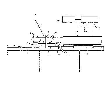
Figure 1 shows in very schematic form various elements of a
known device for implanting an energy emitting source, e.g. radioactive
seeds into a prostate gland. A patient 1 is shown lying in lithotomy
position on a table 2. Fixedly connected to the table 2 is a housing 3.
Housing 3 comprises a drive means 4 to move rod 4a stepwise. A template 5
is connected or mounted to the table 2, which template is provided (not
shown) with a plurality of guiding holes through which holes hollow
needles 9, 10 can be positioned relative to the patient. By means of a
holder 6 a transrectal imaging probe 7 is fixedly connected to said rod
4a, which is moveable in a direction towards and from the patient by
means of the drive means 4. The imaging probe 7 can be an ultrasound
probe.
A needle 9 is used for fixing the prostate gland 11 in
CA 02432493 2003-06-16
17
position relative to the template 5. A number of needles 10 is fixed into
position through the template 5 in the prostate gland 11. The template 5
determines the relative positions of the needles 10 in two dimensions.
The needles 10 are open at their distal ends and are sealed of by a plug
of bio-compatible, preferably bio-absorbable wax. In said housing 3 a
seed loading unit 8 is present.
A well-known therapy planning module 12a is provided for
determining the number and relative positions of seeds in each needle for
implantation in the prostate gland 11. Such therapy planning module 12a
usually comprises a computer programmed with a therapy planning program.
The therapy planning module 12a is connected to the seed loading unit 8
through a control device 12 for controlling the number of seeds for each
needle. Control device 12 may be a separate device or may be an
integrated part either of the seed loading unit 8 or of the therapy
planning module 12a or may be embodied in the software of the therapy
planning module 12a or of the seed loading unit 8.
The known device shown in Fig. 1 operates as follows. A
patient 1 is under spinal or general anesthesia and lies on the operating
table 2 in lithotomy position. The (ultrasound) imaging probe 7 is
introduced into the rectum and the probe is connected via signal line 7a
with a well known image screen, where an image may be seen of the inside
of the patient in particular of the prostate gland 11 as seen from the
point of view of the imaging probe 7. The template 5 is attached to the
drive means 4, thereby insuring the correlation of the ultrasound image
geometry and the template 5. The prostate gland 11 is fixed relative to
the template 5 and the drive means 4 and the imaging probe 7 by means of
one or more needles 9, 10. Subsequently further needles 10 are introduced
in the body and the prostate gland under ultrasound guidance one by one.
Moving the imaging probe with the drive means 4
longitudinally within the rectum controls the needle depths of each
needle 10. After all needles 10 have been placed, their positions
relative to the prostate gland 11 are determined in at least one of
CA 02432493 2003-06-16
18
several known ways. In a known way the therapy planning module 12a
determines how the needles 10 are to be placed in the prostate and how
many radioactive seeds are to be placed in what order in each of the
needles 10. The information about the desired placement of the
radioactive seeds in the needles 10 is used to control the seed loading
unit 8.
According to the invention said therapy treatment planning
module generates at least one treatment plan as it is provided with a
three-dimensional imaging algorithm and a three-dimensional image
segmentation algorithm for the specific organs within said anatomical
portion, the needles and the tubes for converting the image data obtained
with said imaging means into a three-dimensional image of the anatomical
portion, whereby by using at least one single or multi-objective anatomy
based genetic optimization algorithm for pre-planning or virtual
simulation purposes said processing means are arranged to determine in
real time the optimal number and position of at least one of said hollow
needles, the position of said energy emitting source within each hollow
needle as well as the dwell times of said energy emitting source at each
position; whereas for post planning purposes said processing means are
arranged to determine based on three-dimensional image information in
real time the real needle positions and the dwell times of said energy
emitting source for each position.
In Figure 2 a template is disclosed for use in a real time
radiation treatment planning system according to the invention.
Especially the template 20 is detachable from a template frame 25, which
frame is connected with the stepper means for displacing the imaging
means as described in connection with Figure 1.
According to the invention template 20 has a grid
configuration with needle holes 22 at an intermediate distance of 3.5 mm
seen in diagonal direction. In another embodiment the template has a grid
configuration with needle holes at an intermediate distance of 2.5 mm
seen in orthogonal direction.
CA 02432493 2015-07-23
19
In a specific embodiment the template is a motorized
template without holes and the needles are guided with a guiding
tube, whereas the guiding tube can be positioned in each position of
the virtual template grid. As this embodiment does not use holes,
the absence of a grid does not limit the positioning of the needles
in relation to the template and the anatomical portion to be
treated. In fact with a template without holes the grid
configuration is only limited to the diameter of the needles used.
A more specific embodiment of the template is disclosed
in Figure 2, where said template 20 is detachable from the frame 25.
Frame 25 is connected with the stepper means as described above. For
a good connection and orientation of the frame 25 and template 20 in
relation to the device of Figure 1 the frame 25 is provided with
alignment pins 27 which cooperate with corresponding openings (not
shown) in the device of Figure 1.
The template 20 has a saddle shaped body 20a, which fits
with the frame 25 as shown in Figure 2. For alignment purposes the
template 20 is provided with notches lla-llb which cooperate with
corresponding holes 26a-26b present in the circumference of frame
25.
It is an another object of an aspect of the invention to
describe the catheters or needles inserted in the body through which
the HDR source is travelling with their real geometrical dimensions.
As a direct result of this it is a next object of an aspect of the
invention that sampling points for dose evaluation, which are lying
inside the needles or catheters are excluded. This will contribute
to the reduction of the number of sampling points in the anatomical
portion compared with other conventional methods and to the increase
of the speed.
As shown in Figure 3 a catheter or hollow needle is
defined by catheter describing points. These points are connected
with cylinders and at each catheter describing lengths and
diameters. The set of catheters cylinders and spheres are used to
describe the geometry of a catheter that may be either metallic
linear or plastic and curved.
CA 02432493 2003-06-16
For generating each treatment plan the processing means of
the radiation treatment planning system according to the invention are
arranged to generate a set of multiple sampling points using said three-
dimensional imaging algorithm and said three-dimensional image
5
segmentation algorithm and to calculate the optimal radiation dose
distribution for each of said sampling points by using a gradient-based
algorithm.
The quality of the results depends on the distribution of
the sampling points as generated. According to the invention the dose
10
distribution inside the anatomical portion (PTV), critical structures,
such as specific delicate organs (OAR) and the surface of the anatomical
portion is estimated from the dose of a small number of points (sampling
points).
As shown in Figure 4 and 5 the generated sampling points
15
are distributed on the contours and on the triangulated surface of the
anatomical portion to be treated. For the contour based method no points
are on both ends of the anatomical portion. Therefore a large part of the
surface is undefined for the optimization algorithm and the resulting
isodose is bounded only by the contours of the anatomical portion.
20
Sampling points in the volume are generated from low
discrepancy sequences or quasi-random distributed sampling points.
It is an another objective of the invention that in
contrast to pseudo-random distributed sampling points voids and
clustering are avoided. Monte-Carlo generated quantities convergence much
more rapidly than a conventional pseudo-random sequence. Sampling points
inside catheters are excluded. This reduces the influence of very large
dose values of sampling points that occasionally are produced very close
to the source dwell positions. Statistical values obtained from the
sampling points are calculated therefore with a higher accuracy.
According to the invention two treatment planning steps are
performed:
Pre-planning or inverse planning: Given the geometry of the anatomical
CA 02432493 2003-06-16
21
portion (PTV) to be treated, the specific organs (OAR) near or within
said anatomical portion, a template and its position the optimal number
and position of needles, the dwell positions and the dwell times of the
energy emitting source are determined, so that the resulting dose
distribution satisfies various criteria such as coverage of the
anatomical portion with the prescription dose, avoidance of dose values
above some critical values in the specific organs, etc.
Postplanning: Given the geometry of the anatomical portion (PTV) and the
specific organs (OAR) and a given number and position of needles and the
position of the energy emitting source in each needle the dwell times of
the energy emitting source at each position are determined, so that the
resulting dose distribution satisfies various criteria such as coverage
of the anatomical portion with the prescription dose, avoidance of dose
values above some critical values in the specific organs, etc.
Template based Inverse Planning
Figure 6 defines the template and catheter characteristics.
The planning software is run on a personal computer or laptop computer
and allows the setting of certain objectives/boundaries/parameters prior
to the generation of a treatment plan. The displacement step of the
source within a needle is set at 5.0 mm in the afterloader parameters,
since a 2.5 mm value produces a large number of sources and the
optimization using the algorithm according to the invention may take more
time and since then 512 MB RAM are recommended. By pressing ,the button
Auto-activation the dialog of Figure 7 appears, which may contain other
organs (or VOIS i.e. Volumes Of Interest)
This dialog is used for the auto-activation algorithm. The
anatomical organ (PTV) and the specific organs (OAR) to be protected
against too much radiation exposure are listed as wels as the minimum
distance of the source dwell positions from the corresponding VOI in mm.
It is used to select only source dwell positions that are at a distance
to a corresponding VOI larger than a specified value.
CA 02432493 2003-06-16
=
22
VOIs for which the corresponding button is pressed only
will be considered. In this example the rectum is ignored since it is
outside the anatomical portion (PTV).
Now the program moves all catheters/needles, which inside
the anatomical portion taking the specific organs and the geometry of the
anatomical portion into account. The user has now to take only a subset
of these catheters. In principle this will be done automatically by using
an optimization methods which are flexible and robust. This will be the
true inverse planning.
In Figure 8 the Source Parameter dialog for setting the
prescription dose is disclosed. This dialog is used to define the source
strength or activity and the prescription dose. These parameters have to
be supplied for the use of the optimization algorithms. The source is
characterized by its strength in units of U or as activity in units of
GBq or Ci. The prescription dose is specified in cGy.
Subsequently the Inverse planning dialog of Figure 9 and
Figure 10 Template View and Loading appears. Figure 10 shows the grid of
the template, the catheters and VOIs at various distances from the
template. The selected catheters are shown in dark. The catheters which
can be selected in light gray. By moving the z-slide a plane parallel is
moved along the normal to the template and at some given distance from
the template defined by distance z. In the Template View the intersection
of the VOIs with that plane is shown in the anatomy window.
By selecting one of the buttons shown in Figure 11 (and
Figure 10) the catheter density can be selected.
By selecting with the mouse cursor one or more of the
catheters or needles (except those which are light gray) the selected
catheter can be switched on or off. So the user can select the catheters
he wants to use during treatment planning. For example a set of catheters
on the periphery and an additional set of catheters inside the anatomical
portion can be selected. It is perferred to limit the numer of selected
CA 02432493 2003-06-16
23
catheters or needles to 15-20 in order to limit the number of
calculations to be performed.
Subsequently the dialog of Figure 12 Geometry and sampling
appears which contains information about the sampling points, the number
of source dwell positions and the number of catheters. Subsequently a
optimization modus has to be selected and the dialog of Figure 13
appears. Here the optimization method can be selected e.g. a
deterministic optimization method.
After selecting Set Optimization Options in Figure 13 the
dialog of Figure 14 appears. The specific organs (OAR=Organs At Risk)
which are to be considered during the radiation treatment planning have
to be selected. In this case the rectum is located outside the anatomical
region to be treated and it can be ignored. However in this example
prostate cancer is to be treated and therefore the Urethra button is
selected as the urethra is present inside the prostate. The critical dose
value to which the specific organ may be exposed to as fraction of the
prescription dose. In this case it is decided by the medical personnel
that the urethra does not to receive more than 50% of the prescription
dose. Therefore the factor 1.50 is entered.
With this dialog a single or multi-objective optimization
can be performed. The multi-objective optimization is selected. After the
optimization 20 solutions are presented to the user (medic personnel) of
the treatment planning system. The treatment planning system Plato
developed and commercialized by the present applicant Nucletron B.V. or
other systems use a single set of importance factors which is not
recommended because there can not be such a single set of importance
factors for all cases. There is not a single solution but in principle
infinite solutions.
The treatment planning system according to the invention
tries to produce a representative set of multiple treatment planning
solutions. The deterministic method is the most simple approach.
CA 02432493 2003-06-16
4
24
Recommended is of course the evolutionary algorithm which is more
flexible and produces much more solutions out of which the best for each
case can be found. It is not always possible to have similar results.
Even if only prostate cases are considered.
After initialization the treatment planning system of the
invention calculates the volumes of the anatomical portion and the
specific organs, it generates the sampling points and look-up tables are
filled. In this case the optimization algorithm repeats 20 times with 20
different sets of importance factors.
After the optimization step the Decision button has to be
selected in order to select a solution and see the results. The dialog of
Figure 15 appears. When the button Show results of all solutions is
pressed the dialog of Figure 16 appears. By moving the slider to the DVH
values are made visible, as these values are used in the decision making
of the final treatment plan.
CA 02432493 2003-06-16
By selecting the column the DVH(1.500) urethra, the values
in that column are sorted in descending order. See Figure 18.
In Figure 18 the best radiation dose coverage of the
anatomical portion (PTV) is in this example 92.13%, while 10.785% of the
5 urethra receives a dose value above 1.5 times the prescription dose. If
the medic personnel want a dose exposure of the urethra below 1% (in
Figure 18 0.77%), then the best coverage for the anatomical portion
(prostate) is 86.115%. By pressing the Histogram button the distributions
e.g. are displayed (Figure 19).
10 The deterministic algorithms use a mean on the dose
normalization for the surface of the anatomical portion and it is
therefore not as flexible as the evolutionary algorithms. But the
examples still show the differences between the treatment planning
solutions obtained with different importance factors/boundary conditions,
15 which can be quite large. So one method would be to consider first the
specific organs (OARs), then the dose coverage of the anatomical portion
(PTV) and finally the dose in the surrounding tissue. Whatever the
preferences of the planner are the algorithm according to the invention
generates all possible solutions and the planner can select which
20 treatment solution is the best solution.
In the event that it is decided that 1% of urethra may to
receive more than the critical dose value, then the treatment solution
no. 15 is selected in Figure 20 (see also Figure 16 and 18). When the
solution 15 in the list is selected the Accept single solution button is
25 to be pressed and for seeing the isodose distributions the Iso-Dose of
Selected Solution button is to be pressed in Figure 20.
By selecting the 3D button in Figure 21 the isodose values
are marked, which are to be displayed (here the isodose for lx the
prescription and 2x the prescription). Subsequently two 3D isodose
distributions will be displayed.
CA 02432493 2003-06-16
26
=
Post Implant Optimization
Post Implant Optimization assumes that the source dwell
positions are given. This is in principle what Nucletrons PLATO systems
calls inverse planning. After activating the Post Implant Optimization
the treatment planning system loads the VOIS and catheters and the
Autoactivation dialog of Figure 22 appears.
After pressing the OK button the Source parameters dialog
of Figure 23 is displayed. After selecting the source parameters and
pressing on OK the system directly continues with the optimization step
of Figure 13. Analogue to the pre-planning step the deterministic
optimization algorithm can be selected. The steps of generating multiple
treatment solutions are then the same as with the pre-planning step.