Note: Descriptions are shown in the official language in which they were submitted.
CA 02433432 2003-06-27
WO 02/056018 PCT/EP02/00334
1
SURFACE CHEMICAL MODIFICATION OF OPTICAL ELEMENTS
The invention relates to devices suitable for the investigation of ligand-
receptor
interactions, in particular for the investigation of biological or chemical
molecules and organic
components and their interaction with suitably modified surfaces.
More in particular the invention concerns methods of chemical surface
activation and
covalent grafting of ATR (Attenuated Total Internal Reflection) elements and
their use in
FTIR (Fourier Transform Infra Red) devices for the detection,
characterization, and dosage
of biological molecules and organic compounds.
In another aspect the invention also relates to the grafted ATR elements as
such.
Background of Art
Biosensors (for a detailed review, see: Leech D, Chem. Soc. Rev., 1994, 23,
205-
213) are devices based on the specific recognition of an analyte of interest
by a target such
as a biological component, for example a receptor, an antibody, an enzyme, a
membrane, a
cell or cell containing media, a molecule and the subsequent transformation of
this
interaction into an electrical, optical, or other signal. Biosensors have
already attracted
intensive interest in many different fields such as medical diagnostics and
control,
environmental analysis, and monitoring of biotechnological processes.
Different surface sensitive techniques can be applied to detect ligand-
receptor
interactions, depending on the nature of the sensor supports. They are
piezoelectric
methods, impedance spectroscopy, microscopy, and surface plasmon resonance
(SPR)
spectroscopy, in the case of gold or other metal surfaces, on the one hand,
and integrated
optics and fluorescence spectroscopy, in the case of glass (or Ti02) surfaces,
on the other
hand.
Amongst the previous techniques, SPR spectroscopy has been successfully used
by
BIACORE International AB (Stockholm) to develop a commercial instrument (Lofas
S,
Malmqvist M, Ronnberg I, Stenberg E, Liedberg B, Lundstrom I, Sensors
Actuators B, 1991,
5, 79; Malmqvist M, Nature, 1993, 361, 186). Surface plasmon resonance is a
surface
sensitive technique able to probe molecular interactions in real time, and on-
line (for selected
examples, see: Kooyman RPH, de Bruijn HE, Eenink RG, Greve J, J. Mot. Struct.,
1990, 218,
345; Liedburg B, Nylander C, Lundstrom I, Sensors Actuators B, 1983, 4, 299;
Karlsson R,
Michaelsson A, Mattsson L, J. ImmunoL Methods, 1991, 145, 229; Green RJ,
Davies J,
CONFIRMATION COPY
CA 02433432 2003-06-27
WO 02/056018 PCT/EP02/00334
2
Davies MC, Roberts CJ, Tendler SJB, Biomaterials, 1997, 18, 405; Liedberg B,
Lundstrom I,
Stenberg E, Sensors Actuators B, 1993, 11, 63; Lahiri J, Isaacs L, Tien J,
Whitesides GM,
Anal. Chem., 1999, 71, 777).
Biosensors based on the SPR optical technique make use of the changes in the
refractive index of a medium near a thin film of metal (gold) deposited on a
substrate (glass).
Modifications in refractive index occur when an analyte such as a molecule or
a protein, for
instance is adsorbed or fixed to the surface substrate; consequently, the
angle of minimum
intensity of reflected light (the resonance angle) is affected. Intrinsically,
this detection
technique, i.e. measuring mass loading of the surface cannot provide
structural and
1o mechanistic information about the interacting analyte. The access to
chemical information,
such as molecular structure, packing, orientation,... and time-resolved
information, such as
conformational transitions accompanying the ligand-receptor interactions, are
practically
inaccessible. Moreover, the SPR technique requires the deposition of a metal
film, which is
usually gold on a support, and the subsequent immobilization of the targets or
biological
receptors of interest via the method of self-assembled monolayers (SAMs) based
on the
chemisorption of thiol-containing surfactants (for selected examples, see:
Mrksich M,
Whitesides GM, ACS Symp. Ser., 1997, 680, 361; Deng L, Mrksich M, Whitesides
GM, J.
Am. Chem. Soc., 1996, 118, 5136; Prime KL, Whitesides GM, J. Am. Chem. Soc.,
1993,
115, 10714) or via the interaction with an amorphous Dextran matrix. The
presence of this
metal film (about 200 nm in depth) dramatically restricts the possibilities of
surface detection
by highly sensitive techniques, such as infra-red spectroscopy or fluorescence
techniques
that can be applied in aqueous media. Accordingly, novel concepts and devices
have to be
found to improve the biosensor supports and the related detection techniques.
Fourier transform infra-red (FTIR) spectroscopy is an extremely powerful
analytical
technique, particularly well adapted to the characterization of organic
molecules and
biological systems. Quantitative structural and conformational information can
be recorded.
The method has been applied to the investigation of mono- and multilayers of
bio-organic
samples displayed on the optical elements, by using the attenuated total
internal reflection
(ATR) configuration (Scheuing DR), Fourier Transform Infra-Red Spectroscopy in
Colloid and
Interface Science, ACS Symposium Series n 447, Am. Chem. Soc., Washington,
1991;
Mirabella Jr FM (editor), Internal Reflection Spectroscopy, Theory and
Applications, Marcel
Dekker, New York, 1993). Interestingly, the ATR configuration allows the study
of analytes
such as biological components and molecules or proteins, for instance on
surfaces in contact
with water-containing media. The examination of protein adsorption on
biomaterial surfaces
CA 02433432 2003-06-27
WO 02/056018 PCT/EP02/00334
3
is one of the relevant applications (Chittur KK, Biomaterials, 1998, 19, 357).
Recently, Vogel
and coworkers reported the effect of the reduction of the thickness of the
metal layer
deposited at the surface of an ATR element on the infrared detection of
biomolecules (Liley,
M.; Keller, T.A.; Duschl, C.; Vogel, H. Langmuir 1997, 13, 4190-4192). Later,
an international
patent application has appeared which discloses methods and devices for the
detection and
investigation of biological molecules at (or in) self-assembled monolayers on
metal surfaces
using infra-red spectroscopy in the attenuated total internal reflection
configuration (Vogel H,
Keller T, Liley M; WO 99/05509). This disclosure is based on the use of a very
thin film of
gold (5 to 30 nm) deposited on an ATR element, and the subsequent formation of
SAMs
susceptible to immobilize biomolecules. Thus, the relative transparency of the
thin metal film
in the infra-red is exploited, whereby the internal reflection of the IR beam
at the interface
between the ATR element and the metal produces an evanescent field which
penetrates
through the metal film and into the aqueous phase on the other side. This
allows sampling of
SAMs and fixed biomolecules at the metal-water interface.
Other techniques are available to immobilize molecules of interest on ATR
crystals,
such as the coating with thin layers of synthetic polymers, such as
polyurethanes,
polystyrene, polyethylene by spin-casting (Lenk TJ, Ratner BD, Chittur KK,
Gendreau RM,
Langmuir, 1991, 7, 1755; Lenk TJ, Ratner BD, Chittur KK, Gendreau RM, J.
Biomed. Mater.
Res., 1989, 23, 549), and the coating with bioceramic, such as hydroxyapatite
by ion
bombardment (Ong JL, Chittur KK, Lucas LC, J. Biomed. Mater. Res., 1994, 28,
1337). In all
cases, i.e. metal, polymer or ceramic materials, the thickness of the
deposited layer appears
to be a crucial point because it is not allowed to significantly change the
nature and the
intensity of the evanescent field.
The main disadvantage of the use of an intermediate film of metal, polymer, or
ceramic on the surface of the ATR element is the formation of a barrier for
light transmission,
which results in a drastic restriction of the possibilities to detect ligand-
receptor interactions
taking place at the ATR surface. Therefore, there is a further need for a
method of surface
modification of ATR elements which minimizes the effect of the deposition of
an intermediate
film of metal, polymer, or ceramic, between the crystal and the organic film
displaying the
biological receptors.
Summary of the invention
In the present inventive application, the disturbing intermediate film is
omitted and
replaced by a modification of the ATR element surface involving a process
consisting of two
CA 02433432 2003-06-27
WO 02/056018 PCT/EP02/00334
4
steps. The first step consists of chemically activating and modifying the ATR
element surface
by wet chemistry. Preferably, ATR elements according to the invention are made
of silicon or
germanium crystals. The direct chemical modification of silicon surfaces by
silanization
methods is well known (Patai S and Rappoport Z (editors), The chemistry of
Organic Silicon
Compounds, John Wiley, Chichester, 1989; Mittal KL (editor), Silanes and other
Coupling
Agents, VSP, Utrecht, 1992; Auner N and Weis J (editors), Organosilicon
Chemistry 11, from
Molecules to Materials, VCH, Weinheim, 1996); but this technique was never
used for the
elaboration of FTIR-ATR elements of biosensors.
The second step consists of grafting of the activated surface with an organic
molecule, such as a silane derivative, through covalent coupling. The
anchoring of a silane
derivative through its silane moiety on the surface of an internal reflection
element has been
described previously (Stefan I and Scherson D, Langmuir, 2000, 16, 5945-5948).
EP 0 596
421 discloses a coating of dielectric Ti02 waveguides with elements capable of
recognizing
biological molecules in the formation of a biosensor. The coating consists of
an organic
support layer, to which the receptor molecules are bonded, the support layer
comprising an
ordered monomolecular layer which is bonded via a Si atom directly to a Ti02
waveguide or
if desired via an intermediate layer to a Ti02 waveguide. However, none of
these
constructions disclose a chemical prior modification of the surface of the
optical element. In
present invention, pretreatment of the surface device by chemicals improves
the wettability
and the reactivity of the surface. Chemical activation of the ATR surface
allows an easier and
direct grafting of organic components on this surface. This results in a more
accurate and
efficient measuring of ligand-receptor interactions via infra-red
spectroscopy.
The present invention is directed to a device suitable for the investigation
of ligand-
receptor interactions, in particular for the investigation of an analyte
target interaction such as
biological and chemical molecules and organic components and their interaction
with
surfaces, consisting of an attenuated total internal reflection element,
transparent in the infra-
red and of which at least one surface is chemically activated and covalently
grafted with an
organic molecule able to immobilize the receptor.
In a preferred embodiment the organic molecule is a silane derivative of the
general
formula
X3Si - (CH2)n - (CF2)1' - Y, X2(R1)Si - (CH2)11- (CF2)n' - Y or
X(R1)(R2)Si - (CH2)1- (CF2)n'- Y,
CA 02433432 2003-06-27
WO 02/056018 PCT/EP02/00334
wherein X is halogen, preferably Cl, Br or C1-C6 alkoxy, preferably OMe, OEt;
n is 1 to 20;
n' is 0 to 20;
R1, R2 are independently C1-C6 alkyl;
5 Y is Me, CF3, CHF2, CH2F, CH=CH2, ON, CH=O, epoxide, halogen, SH, NH2, OH,
N=C=O, N=C=S, CO2H or derived esters thereof.
In another preferred embodiment the organic molecule is a silane derivative
covalently coupled with an multifunctional spacer-arm of the general formula
Z1-(CH2)n-Z2 wherein n is 2 to 12;
Z1-CH2-(O-CH2-CH2-)n'-O-CH2-Z2 wherein n' is 0 to 5;
wherein Z1,Z2 are independently chosen from Aryl-N3 (photoactivable
substituents),
CO2H and activated forms thereof such as N-hydroxysuccinimidyl ester, CH2NH2
and activated derivatives such as N-maleimide, CH2OH and activated forms such
as
tosylates, CH2SH and activated forms such as dithiane derivatives, CH2N=C=O or
CH2N=C=S.
The invention also provides for a method of construction of said device
including the
steps of:
- surface activation of at least one surface of an attenuated total internal
reflection
element,
- surface grafting with an organic molecule of the activated surface obtained
in the
previous step, and
- coupling a receptor via covalent fixation on the organic molecule.
Preferred embodiments are described in the sub-claims.
Description of the figures
In the drawings schematic views and graphs of spectrum embodiments of the
invention are presented.
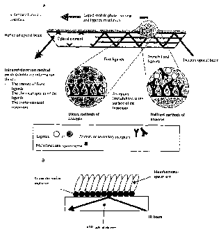
Figure 1 shows a schematic view of (A) a biosensor principles and related
methods of
analysis and detection (B) surface-modified ATR element for FTIR detection of
biomolecules
according to the invention.
CA 02433432 2003-06-27
WO 02/056018 PCT/EP02/00334
6
Figure 2 shows (A) FTIR-ATR spectrum of a silicon crystal activated and
grafted with
undecyl trichlorosilane; (B) FTIR-ATR spectrum of a germanium crystal
activated and grafted
with octadecyl trimethoxysilane.
Figure 3 shows a FTIR-ATR spectrum of a silicon crystal activated and grafted
with
APTES.
Figure 4 shows a synthetic scheme for the coupling of a spacer-arm (example
2).
Figure 5 shows a FTIR-ATR spectrum of a silicon crystal equipped with the
spacer-
arm.
Figure 6 shows FTIR spectra of a PE-biotin film exposed to a buffer solution
containing streptavidin (125 pg/ml). 100 pg of PE-biotin were applied on the
surface of the
germanium crystal. A persistaltic pump was used for recirculating the
streptavidin aqueous
solution into a waterproof vertical ATR flow cell (4 ml/min). Spectra were
recorded in the
course of the binding of streptavidin on the PE-biotin film. The first ten
spectra were recorded
every 5 minutes, then every 50 minutes.
Figure 7 shows FTIR-ATR spectra, recorded as a function of time, of a
germanium
crystal displaying PE-biotin and submitted to a flux of streptavidin solution
(125 pg/ml).
Intensities of the absorption bands at 1634.7 cm-1 (amide I) and 1543.4 cm-1
(amide II).
Figure 8 shows a graph of calibration obtained by the multivariate analytical
technique
PLS. Known concentrations of streptavidin vs predicted ones.
Figure 9 shows FTIR-ATR spectra recorded at each step of the construction of
the
"biotin-streptavidin" sensor. A = crystal grafted with APTES (base line); B =
crystal grafted
with the spacer-arm (1740 cm-1); C = crystal having fixed the protein (1634.7
and 1543.4
cm-1).
Figure 10 shows a graph of the regeneration of the ATR crystal. A = Crystal
with the
spacer-arm and the coupled protein, having fixed PE-biotin (1640 cm-1), as
indicated by the
black arrow; B = crystal with the spacer-arm, recovered after application of a
streptavidin
solution.
Figure 11 shows a synthetic scheme for the coupling of a spacer-arm (examples
3
and 4).
Figure 12 shows the specificity of ligand/receptor binding. lysozyme (Lys)
(130 pg/ml)
was added first, then streptavidin (SA) 30 pg/ml was flown in the system. The
cell was then
washed with 2 mM Hepes, pH 7.5 (hps). Further addition of streptavidin (SA)
did not result in
any further binding.
CA 02433432 2003-06-27
WO 02/056018 PCT/EP02/00334
7
Detailed description of the Invention
The invention provides a device suitable for the investigation of ligand-
receptor
interactions, in particular for the investigation of biological molecules and
organic
components and their interaction with surfaces, consisting of an attenuated
total internal
reflection element, transparent in the infra-red and of which at least one
surface is chemically
activated and covalently grafted with a organic molecule able to immobilize a
receptor. An
embodiment of said device is schematically depicted in figure 1.
In another embodiment the invention provides a method for activating a surface
of a
attenuated total internal reflection element, by wet chemistry using oxidation
/ hydroxylation /
reduction in an acid or alkaline environment.
In general, the present invention provides a method to activate the surface of
an
optical element, transparent in the infra-red, particularly a silicon or
germanium ATR device,
typically a crystal, an optical fiber or a rod made of such materials, and to
further covalently
fix functionalized organic molecules able to immobilize biological components
and molecules,
including biological and non-biological receptors, proteins, antibodies,
membranes,... This
purposely modified ATR element provides a method to study ligand-receptor
interactions
occurring at the solvent ATR element interface, particularly, at the water-
containing media -
ATR element interface, by using attenuated total internal reflection (ATR)
infra-red (IR)
spectroscopy, preferably Fourier transform infra-red spectroscopy (FTIR).
In another embodiment the invention relates to a method, wherein the surface
grafting is performed through covalent coupling with a silane derivative. The
technique is
based on the surface modification, preferably on the
oxidation/hydroxylation/reduction, of the
ATR element followed by the covalent grafting of organic molecules presenting
a reactive
moiety at the one terminus for the surface anchorage, typically a silanyl or
germanyl
functional group, and a functional group at the other terminus for the
covalent coupling of
biological receptors, either directly, or via multifunctional spacer-arms.
In another embodiment the invention provides a method for studying ligand-
receptor
interactions, in particular biological molecules or organic components or
their interactions or
complexations or reactions with biological molecules or organic components or
water-soluble
molecules at or in the grafted organic molecule, using a surface-activated and
covalently-
grafted ATR element, comprising the steps of
- installation of the ATR element in a FTIR cell
CA 02433432 2003-06-27
WO 02/056018 PCT/EP02/00334
8
- conducting a flux of potential ligands, preferably a water-containing
solution, on the
ATR surface
- analysis of the infra-red spectrum obtained after submitting a FTIR beam
through
the cell
- regeneration of the ATR surface by application of a solution of free ligand.
In general, the inventive method allows the study of specific ligands
interacting with
the receptor fixed on the ATR element. The element is placed in a low pressure
cell allowing
a liquid flux to pass on its surface. A solution of potential ligands to be
analyzed is passed
through the cell submitted to FTIR beam. Due to the low penetration depth of
the evanescent
field, ligands in solution do not significantly contribute to the IR spectrum.
On the contrary,
ligands fixed on the receptors, close to the surface of the ATR element,
increase
considerably the local concentration at the interface, and contribute to the
IR spectrum. The
ATR element is preferably a crystal, more preferably having a trapezoidal,
fiber or rod
shaped geometry. These types of crystals are easy to use and allow good
detection.
The quantitative analysis of fixed ligands is an object of the invention.
The time-resolved study of ligand-receptor interaction is a further object of
the
invention.
The regeneration of the ATR element is another object of the invention.
Methods for carrying out the invention
1. Activation of the ATR element (germanium and silicon)
The activation results from the surface oxidation/hydroxylation by any
available
technique (physical or chemical), preferably the wet-chemistry technique using
a solution of
an oxidant in acidic or basic media, such as H2O2/H2SO4, H2O2/TFA, H202/HF,
K2Cr2O7/H2SO4, oxone/H2SO4, H202/NH4OH, or in organic media, such as an
organic
peracid, Br2 in solution. The activation may also be carried out by dipping
the crystals in
sequences of solutions of an oxidant in acidic or basic media. Suitable
solutions of an oxidant
in acidic or basic media, are e.g. H202/H2SO4, H202/TFA, H2O2/HF,
K2Cr2O7/H2SO4,
oxone/H2SO4, H202/NH4OH, or e.g. in organic media, such as an organic peracid,
Br2 in a
suitable solution, or a combination of these solutions in specific sequences,
such as HF in
water followed by H202 in water iterated for several times (e.g. number of
repetitions:
between 1 and 20, preferably 3) or NH4OH/H2O2 in water followed by HCI/H202 in
water (e.g.
number of repetitions: between 1 and 20, preferably 1).
CA 02433432 2003-06-27
WO 02/056018 PCT/EP02/00334
9
The temperature is preferably comprised between -15 C and +150 C and the
duration of the
treatment comprised between a few seconds to several hours.
2. Grafting of a silane derivative on the activated ATR element
The covalent grafting on the activated element is obtained by contacting a
solution of
silane derivative chosen from:
X3SI - (CH2)n - (CF2)n' - Y, X2(R1)Si - (CH2)n - (CF2)n'- Y or X(R1)(R2)Si -
(CH2), - (CF2)n' - Y,
wherein X is halogen, preferably Cl, Br or C1-C6 alkoxy, preferably OMe, OEt;
n is 1 to 20;
n' is 0 to 20;
R1, R2 are independently C1-C6 alkyl;
Y is Me, CF3, CHF2, CH2F, CH=CH2, CN, CH=O, epoxide, halogen, SH, NH2, OH,
N=C=O,
N=C=S, CO2H or derived esters thereof.
Other suitable parameters in this reaction are preferably:
- solution in organic solvent, such as toluene, dichloromethane, 1,2-
dichloroethane,
chloroform, acetonitrile
- concentration of 0.01 % to 5%
- temperature of -15 C to 80 C
- reaction time of a few minutes to several hours
3. Coupling of a multifunctional spacer-arm
The covalent coupling on the modified ATR element is obtained by contacting a
solution of multifunctional molecules of the formula:
Z1-(CH2)n-Z2 wherein n is 2 to 12;
Z1-CH2-(O-CH2-CH2-)n'-O-CH2-Z2 wherein n' is 0 to 5;
CA 02433432 2003-06-27
WO 02/056018 PCT/EP02/00334
wherein Z1,Z2 are independently Aryl-N3, CO2H and activated forms thereof such
as N-
hydroxysuccinimidyl ester, CH2NH2 and activated derivatives such as N-
maleimide, CH2OH
and activated forms such as tosylates, CH2SH and activated forms such as
dithiane
derivatives, CH2N=C=O or CH2N=C=S.
5
The same experimental conditions (solvent, concentration, temperature, time)
as above
apply.
In the case of Z1, Z2 equal to Aryl-N3, light activation is applied (X between
200 and 400 nm).
10 4. Coupling of a silane derivative equipped with the spacer-arm
(alternative route to 2.
followed by 3.)
The molecules presented in 2. and 3. are reacted to give the following
molecules in
which X, R1, R2, and Z2 are defined as above :
X3Si-(CH2)n-W-(CH2)n"-Z2
X3Si-(CH2)n-W-CH2-(O-CH2-CH2-)n"--O-CH2-Z2
wherein n and n" are identical or different from 0-20;
X3Si can be replaced with X2(R1)Si or X(R1)(R2)Si;
W is -NHCO-, -CONH-CH2-, -OCH2-, -NHCH2-, -SCH2-, -S-S-CH2 or -CH=CH-.
The covalent grafting on the activated ATR element is obtained by contacting a
solution of the compounds.
Again the experimental conditions (solvent, concentration, temperature, time)
are the
same as described under 2 and 3.
5. Covalent fixation of a receptor
The ATR element resulting from steps 3. or 4. is placed in contact with an
water-
containing solution of receptor, preferentially proteins, peptides,
membranes,...
CA 02433432 2009-05-05
11
The concentration is preferably in the range 10-6 mgr/ml to 103 mgr/ml, the
temperature is
comprised between -15 C and 150 C and the interaction time is from a few
seconds to
several hours (or up to several hours, if accepted).
The activated functions of the surface modified ATR element react as such; it
is the
case, for instance, for isocyanate, isothiocyanate, ester of N-
hydroxysuccinimide, N-
maleimide. The non activated functions, such as free acid, amine, alcohol, are
previously
activated in situ by using the classical methods of peptide synthesis.
6. Ligand-receptor interaction and FTIR-ATR detection and quantification
The ATR element obtained in step 5, is placed in the FTIR cell and submitted
to a flux
of potential ligands, preferably in a water-containing solution.
7. Regeneration of the ATR element surface
The fixed ligand (previous step) is displaced from the ATR element surface by
the
application of a solution of free ligand.
Examples
Example 1: surface grafting of molecules on a germanium crystal and their FTIR-
ATR
detection
Step 1: surface activation
A germanium crystal was activated by surface treatment with an acid/oxidant
mixture
at elevated temperature. Typically, a sulfochromic mixture (8 g/I) at 90 C
during 1 to 3 hours,
preferably 3 hours, was used. The germanium crystal provided with an activated
surface is
then abundantly rinsed with milliQTM-water and dried under a flux of nitrogen.
Step 2: surface grafting with silane derivatives
The activated germanium crystal of step 1 was exposed to ozone and UV
radiation (in
an oven) for 30 min, then treated with a solution of alkyl trichlorosilane or
alkyl trialkoxysilane
in toluene at 20 C. Typically, octadecyl trimethoxysilane (0.05 to 4%,
preferably 0.5% in
toluene) was reacted during 1 to 16 h, preferably during 2 h to furnish a
grafted layer of 4.5
nm in depth (as measured by ellipsometry), corresponding to a water contact
angle of 95 .
The FTIR-ATR spectrum of this surface-modified crystal showed typical bands
between 2850
CA 02433432 2009-05-05
12
and 2950 cm-1 due to the CH2 chain, and a band at 2900 cm-1 due to the
terminal CH3
group (Figure 2B).
Aminopropyl triethoxysilane (APTES) was similarly reacted with the activated
germanium crystal (0.5% in toluene, 1-16 h, 20 C) to furnish a grafted layer
of 1.2 nm in
depth (as measured by ellipsometry), corresponding to a water contact angle of
42 .
Other molecules are undecyltrichiorosilane (C11H23SiCI3),
octadecyltrichiorosilane
(C1BH37SiC13), octadecyltrimethoxysilane (C18H37Si(OCH3)3). These molecules
are brought in
a solution hexadecane and carbon tetrachloride 3/7 (v/v) when trichiorosilanes
are used and
in a solution of toluene when trialkoxysilanes are used. Grafting was
performed in a time
to period varying from 1 to 16 hours, preferably during 2 hours for
alkoxysilanes and 1 h30 for
the trichiorosilanes. The grafted substrates were subsequently rinsed in a
chloroform bath
during 3 minutes and then in an acetone bath during 5 minutes.
Example 2: surface grafting of molecules and functionalized spacers on a
silicon crystal and
their FTIR-ATR detection (method A)
Step 1: surface activation
A silicon crystal was surface-activated by treatment with an acid/oxidant
mixture at
high temperature. Preferably, H2S04/H2O2 in ratio 7/3 (v/v) at 150 C during 8
min was
used.
Similarly, the crystal was surface-activated by immersion during 5 minutes in
a mixture
composed out of NH4OH (25%), oxygenated water H202 (30%) and MilIiQTM H2O in a
ratio 1/1/5
(v/v), heated up to 80 C and during agitation, followed by rinsing with
MilIiQTM H2O and finally
an immersion during 5 minutes in a mixture composed out of HCI (15M), H202
(30%) and
MilIiQTM H2O in a ratio 1/1/5 (v/v), heated up to 80 C during agitation.
The silicon crystal provided with an activated surface is then abundantly
rinsed with
MilIiQTM H2O and dried under a flux of nitrogen.
Step 2: surface grafting with silane derivatives
The activated silicon crystal of step 1 was exposed to ozone and UV radiation
(in an
oven) for 30 min, then treated with a solution of alkyl trichiorosilane or
alkyl trialkoxysilane in
toluene at 20 C. Typically, undecyl trichlorosilane ( 0.08 % in a mixture
composed out of CCI4
and decane in a ratio 3/7 (v/v)) was reacted during 1 to 16 h at low
temperature and low
relative humidity, preferably 1.5 h at 12 C and 30%-40% RH, preferably 35%, to
furnish a
CA 02433432 2009-05-05
13
grafted layer of 12 A in depth (as measured by ellipsometry), corresponding
to a water
contact angle of 108 . The FTIR-ATR spectrum of this surface-modified crystal
showed the
characteristic C-H bands around 2900 cm-1 (Figure 2A).
Aminopropyl triethoxysilane (APTES) was similarly grafted on the activated
silicon
crystal (0.5% in toluene, 20 C; water contact angle: 50 ). The FTIR-ATR
spectrum of this
crystal showed a broad band centered at 3200 cm-1 (NH2) and sharp bands
between 2850
and 2950 cm-1 (CH2) (Figure 3).
Similarly, other molecules were used : undecenyltrichlorosilane CH2=CH-
C9H,8SiCl3,
octadecyltrichlorosilane C55H37SiC13i octadecyltrimethoxysilane
C18H37Si(OCH3)3. These
molecules are brought in a solution and a mixture of hexadecane (for
octadecyltrichlorosilane) or decane (for undecenyltrichlorosilane) and of
carbon tetrachloride
in a ratio 3/7 (v/v) when trichlorosilanes are used and in a solution of
toluene when the
trialkoxysilanes are used. Grafting is performed during a time period varying
from 1 to 16
hours, preferably during 2 hours for the alkoxysilanes and about 1 h30 for the
trichlorosilanes.
Subsequently, these grafted silane surfaces are sonicated in a chloroform bath
during
3 minutes and subsequently immersed in an acetone bath during 5 minutes.
Step 3: coupling of a bifunctional spacer-arm
The silicon crystal grafted with APTES, as obtained in step 2, was treated
with the
bis-activated ester of a a,w-diacid derivative dissolved in dry organic
solvent, at 20 C.
Typically, this preferred method used the bis-N-hydoxysuccinimidyl ester of
3,6-dioxaoctane-
1,8-dioic acid (0.05 - 2 %, preferably 1% in acetonitrile; 1 to 16 hours) to
furnish a crystal
surface exposing succinimidyl groups able to covalently fix the receptors of
interest. The
FTIR-ATR spectrum of this crystal showed a typical carbonyl band at 1700 cm-1
(Figure 5).
Rinsing is performed with chloroform, aceton or preferably with acetonitrile
or
dichloromethane. The activated germanium crystal was consequently rinsed with
Mi11iQTM H2O
and dried under a nitrogen flux. An example of a synthetic scheme for the
coupling of a
spacer arm according to the invention is illustrated in figure 4.
CA 02433432 2009-05-05
14
Example 3: surface grafting of molecules and functionalized spacers on a
germanium wafer
and their detection by ellipsometry
Step 1: surface activation
A germanium wafer was activated by surface treatment with a acidic/oxydant
sequence at low temperature. Typically, sequences similar to the following one
were used :
(a) HF(48%) diluted in water (final concentration between 1 % and 20 %,
preferably 10%),
during 1 to 600 seconds, preferably 10 seconds, at 15 C to 25 C, preferably
20 C and (b)
H202 (30%) diluted in water (final concentration between 1 % and 20 %,
preferably 10%),
1o during 1 to 600 seconds, preferably 15 seconds, at 15 C to 25 C,
preferably 20 C. The
sequence (a), (b) was repeated between 2 and 10 times, preferably 3 times. The
germanium
wafer provided with an activated surface is then abundantly rinsed with
milliQTM-water and dried
under a flux of nitrogen.
Step 2: surface grafting with silane derivatives
The activated germanium wafer of step 1 was treated with a solution of alkyl
trichlorosilane or alkyl trialkoxysilane in toluene at 20 C. Typically,
octadecyl
trimethoxysilane (0.5 % in toluene) was reacted during 16 h, then the wafer
was rinsed
successively in a chloroform bath during 3 min. and in an acetone bath during
5 min., to
leave a grafted layer of 4 - 5 nm in depth (as measured by ellipsometry).
Step 3: photochemical coupling of a bifunctional spacer-arm (Figure 11)
The germanium wafer grafted with octadecyl groups, as obtained in step 2, was
treated with an arylazide under light activation. Typically, this preferred
method used
4-(4-azidophenyl)butyric acid (Carnazzi E., Aumalas A., Barberis C., Guillon
G., Seyer R.,
J. Med. Chem. 1994, 37, 1841) or the corresponding N-hydroxysuccinimidyl ester
(called
activated ester) dissolved in an ether (diethyl ether, or preferably
tetrahydrofurane (THF))
(solution at 1 % to 5 %). An aliquot of the previous solution was deposited on
the germanium
wafer with a pipette and evaporated in the dark in order to obtain an amount
of 0.01 mg to 1
mg of coated azide per square centimeter of substrate, preferably 0.1 mg to
0.3 mg. The
substrate was then irradiated at 254 nm during 0.1 to 6 hours (preferably 2
h), rinsed
successively with chloroform (5 min.) and THE (10 min.), and air dried to
furnish a total
grafted layer of 7 - 8 nm in depth (as measured by ellipsometry).
CA 02433432 2003-06-27
WO 02/056018 PCT/EP02/00334
Example 4: surface grafting of molecules and functionalized spacers on a
silicon crystal and
5 their FTIR-ATR detection (method B)
Step 1: surface activation
As described in example 2.
10 Step 2: surface grafting with octadecyl trimethoxysilane (OTS)
As described in example 2.
Step 3: photochemical coupling of a bifunctional spacer-arm (Figure 11)
As described in example 3.
The FTIR - ATR spectrum of the resulted surface-modified crystal showed the
characteristic
C=O band of the activated ester at 1740 cm'.
Example 5: surface coating of a germanium crystal with phosphatidyl
ethanolamine coupled
to biotin (PE-biotin) and FTIR-ATR quantification of the streptavidin fixation
Step 1: surface fixation of biotin
A germanium crystal (activated or not) was coated with a membrane made of
phosphatidyl ethanolamine coupled to biotin; this PE-biotin layer is stable
under an aqueous
flux.
Step 2: surface recognition by streptavidin
An aqueous flux of a solution of streptavidin (125 pg/ml) was passed on the PE-
biotinylated crystal, and FTIR-ATR spectra were recorded every 5 min during 50
min, then
every 50 min. The protein contribution was increasingly visible at 1634.7 cm-1
(amide I) and
1543.4 cm-1 (amide II) (Figure 6). After 250 min, the system was saturated
when using a
streptavidin solution of 250 pg/ml (Figure 7).
CA 02433432 2003-06-27
WO 02/056018 PCT/EP02/00334
16
Step 3: quantitative analysis
Although the equilibrium has been reached after 250 min, the streptavidin
quantification could be already realized during the ascending phase of the
curve (for
instance, after 20 min). Five aqueous solutions of streptavidin with
concentrations comprised
between 50 to 500 pg/ml have been used for calibrating the quantitative
analysis of the
ligand(biotin)-protein interaction. FTIR-ATR spectra corresponding to the five
solutions have
been recorded as a function of time of streptavidin adsorption. We have chosen
the
multivariate analytical technique PLS (partial least squares) to develop a
model from the set
of reference spectra (Haaland DM, Thomas EV, Anal. Chem., 1988, 60, 1193 and
1202;
Dousseau F, Pezolet M, Biochemistry, 1990, 29, 8771). Two spectral domains
(1654.7-1602
cm-1 and 1566.4-1499.8 cm-1), representative of the characteristic bands of
the protein,
have been considered for this calibration. The correlation obtained between
the known
concentrations of streptavidin and the predicted ones according to the PLS
model is
illustrated in the Figure 8.
Example 6: surface modification of a silicon crystal for the fixation of a
specific receptor
(streptavidin) and regeneration of the surface
Step 1: surface activation
as described in example 2
Step 2: surface grafting with APTES
as described in example 2
Step 3: coupling of the spacer-arm
as described in example 2
Step 4: streptavidin fixation
An aqueous solution of streptavidin (250 gg/ml) was placed in contact with the
crystal
surface resulting from step 3 (20 min, 20 C). The recorded FTIR-ATR spectrum
showed the
typical bands of amide 1 (1634.7 cm-1) and amide 11 (1543.4 cm-1) of the
protein (Figure 9).
The covalently fixed protein could not be eliminated by washing.
CA 02433432 2003-06-27
WO 02/056018 PCT/EP02/00334
17
Step 5: interaction with biotin
A flux of PE-biotin solution was passed on the sensor surface obtained in step
4. The
ligand fixation was visible at 1640 cm-1 in the FTIR-ATR spectrum (Figure 10).
Step 6: surface regeneration
A flux of streptavidin solution was passed on the surface of the sensor
obtained in
step 5. According to the FTIR-ATR spectrum, the crystal surface of the step 3
has been
recovered.
Example 7.= lipid membrane adsorption on an OTS-grafted germanium ATR crystal
Step 1: surface activation :
as described in example 2
Step 2: surface grafting with OTS :
as described in example 2
Step 3: adsorbing a membrane
Lipid vesicles (DDP/PE-biotine 10/1 w:w) 2 mg/ml were incubated overnight in
the
presence of the coated silicium crystal. After rinsing for 60 min at 0.5
ml/min the crystal was
heated at 45 C for 1 hour. The surface was then rinsed again in the same
conditions.
Step 4: measurements
The adsorbed membrane film was shown to bind specifically streptavidin.
Concentrations as low as 0.3 pg/ml were readily detected. The recorded FTIR-
ATR spectrum
showed the typical amide I band of streptavidin (1634 cm) characteristic of
its beta-sheet
secondary structure (Figure 12). Data reported on figure 12 demonstrate that
binding is
specific. When lysozyme (Lys) was added, no significant binding occurred. When
streptavidin (SA) was flown into the system, binding to its receptor present
on the surface
prepared in step 3 resulted in a large absorbance change. Washing the cell
with 2 mM
Hepes, pH 7.5 (hps) did not remove the bound streptavindin. Further addition
of streptavidin
(SA) did not result in any further binding.