Note: Descriptions are shown in the official language in which they were submitted.
CA 02437737 2003-08-14
WO 02/065093 PCT/US02/05713
METHODS AND COMPOSITIONS OF AMPLIFYING RNA
[0001] The present invention claims priority to USSN 60/268,664 entitled "A
Novel Method
to Amplify RNA" filed February 14, 2001; USSN 60/268,645 entitled "Detection
of Gene
Expression in Histologically Stained Tissues and Cells" filed February 14,
2001; USSN
60/306,216 entitled "Method and Composition of Amplifying mRNA through
Terminal
Continuation" filed July 18, 2001; USSN unknown entitled "RNA Amplification
Method",
filed November 7, 2001; USSN unknown entitled "Detection of Gene Expression in
Histologically Stained Tissues and Cells," filed November 7, 2001; and USSN
60/350,176
entitled "Method and Composition of Amplifying Nucleic Acid through Terminal
Continuation" filed November 9, 2001; all of which are incorporated by
reference herein in
their entirety.
FIELD OF THE INVENTION
[0002] The present invention is directed to methods to amplify a nucleic acid
molecule, such
as an RNA molecule. Specifically, the methods are directed to increasing the
efficiency of
second strand cDNA s5mthesis utilizing the mechanism of terminal continuation
prior to
further RNA amplification with an RNA polymerase. More specifically, the
methods are
directed to provide a double stranded (ds) cDNA molecule for ifz vitro
transcription. In other
embodiments, the present invention regards methods related to detection of
gene expression,
particularly from a histologically stained tissue.
BACKGROUND OF THE INVENTION
[0003] Contemporary gene expression profiling or "molecular fingerprinting" is
typically
performed using cDNA array technology. Essentially, a gene array allows the
investigation of
multiple (e.g., hundreds to thousands) of genes simultaneously. However,
fairly large
quantities of tissues are needed for subsequent RNA extraction due to the lack
of sensitivity
of the methodology. The low sensitivity of methodology may be problematic in
two aspects.
First, the sources of tissues may be limited and, second, arrays can only be
performed on a
heterogeneous cell population since collection of large numbers of homogeneous
tissues
and/or cell types is often complicated.
[0004] Antisense RNA synthesis has been used to amplify genetic signals from
limited
amounts of tissues or cells (Van fielder et al., 1990; Eberwine et al., 1992;
U.S. Patent No.
5,545,522). However, the antisense RNA synthesis method presently in use has a
low
1
CA 02437737 2003-08-14
WO 02/065093 PCT/US02/05713
efficiency in amplifying the genetic signals. Therefore, the overall
sensitivity and reliability
of the method is not optimal. The main obstacle for increasing the efficiency
of the method is
the problematic second strand cDNA synthesis. There are two procedures
currently in use for
second strand cDNA synthesis, self priming and replacement synthesis. Self
priming uses the
hairpin formed at the 3' of first strand cDNA to self prime the synthesis of
second strand
cDNA (Sambrook et al., 199). However, the loop formed at the end has to be
removed using
S 1 nuclease digestion. It is a poorly controlled reaction and invariably
leads to the loss of the
5' signal. In addition, self priming ca~1 only be performed with Klenow
fragment of E. coli
DNA polymerase I, which is an enzyme with relatively low processivity. This
factor further
decreases the efficiency of the methodology. The replacement synthesis avoids
S1 nuclease
digestion altogether and has been used in RNA amplification. The reaction
employs multiple
enzynes, RNAse H, E. coli DNA polymerase I and bacteriophage T4 DNA ligase to
digest
RNA in a DNA:RNA complex, synthesize DNA fragments, and ligate them. In
general, the
reaction suffers from a low efficiency, lilcely caused by the multiple
enzymatic steps
involved. In summary, one lcey factor to increase of efficiency of RNA
amplification is to
increase the efficiency of second strand cDNA synthesis.
[0005] U.S. Patent No. 5,545,522, Van fielder et al. (1990), and Eberwine et
al. (1992) are
directed to synthesis of a cDNA from an RNA primed by a single complementary
primer in
the reaction, wherein the primer is linl~ed to sequence of an RNA polymerase
promoter
region. Antisense RNA is transcribed from the cDNA by an RNA polymerase.
[0006] U.S. Patent No. 5,962,272 regards preparing a DNA molecule using a
template
switching oligonucleotide. An RNA is contacted with a cDNA synthesis primer
which
anneals to the RNA, and the cDNA molecule is reverse transcribed to generate a
mRNA-
cDNA hybrid. A template switching oligonucleotide hybridizes to the 5' CAP
site aald serves
as a short, extended template for CAP-dependent extension of the 3'-end of the
ss cDNA that
is complementary to the template-switching oligonucleotide.
[0007] PCT application WO 00/75356 is directed to an RNA polymerase chain
reaction
wherein a poly (dT) primer primes a reverse transcription reaction to
synthesize a first strand
cDNA. The reaction is then followed by a terminal transferase tailing reaction
to incorporate
dGTPs to the 3' end of the first strand cDNA, a second strand cDNA synthesis
reaction, and
transcription.
[0008] Furthermore, the fwctional states) of tissues and cells have been
studied by
morphological observation for over a century. The study of optimally prepared,
i.e., fixed,
sectioned, and/or stained tissues has long been a principal method for
histological and
2
CA 02437737 2003-08-14
WO 02/065093 PCT/US02/05713
histopathological investigation. Several histological staining methods were
developed
empirically on the basis of their capacity to increase the contrast of
specific tissue
constituents to enable the visualization of distinct cell types. Although
histological stains are
in most cases not specific to an individual cell type or protein, much
information can be
gleaned by utilizing classical histochemical preparations in conjunction with
contemporary
protein (e.g., immunocytochemistry) and molecular biological methodologies.
However, the
information gathered through morphological observation and molecular
biological methods
are often difficult to compare and correlate with each other. The problem
arises mainly from
the fact that the methods for morphology and molecular studies have been
thought to be
mutually exclusive. Thus, morphological observation and molecular procedures
such as RNA
amplification could not be performed on the same tissue section or cell. This
limitation
hinders direct examination, and ultimately, hypothesis testing, of the
rriorphological features
of tissues and distinct cell types with simultaneous examinations at a
molecular level.
[0009] Contemporary gene expression profiling or "molecular fingerprinting" is
typically
performed using complementary deoxyribonucleic acid (cDNA) array technology.
Essentially, a gene array allows the investigation of multiple (e.g., hundreds
to thousands) of
genes simultaneously. However, fairly large quantities of tissues are needed
for subsequent
RNA extraction due to the lacy of sensitivity of the methodology. The low
sensitivity of
methodology may be problematic in two aspects. First, the sources of tissues
may be limited
and, second, arrays can only be performed on a heterogeneous cell population
since
collection of large numbers of homogeneous tissues and/or cell types is often
complicated.
(0010] Reverse-transcriptase polymerise chain reaction (RT-PCR) has been the
method of
choice to amplify genetic signals when only limited starting materials are
available. However,
RT-PCR distorts the quantitative relationships between members of a gene
population
because it amplifies genes non-linearally (Phillips and Eberwine, 1996). As a
result, PCR
preferably amplifies abundant genes over rare genes and the wear signals of
later populations
may be further obscured by PCR amplification. Attempts to avoid this bias in
PCR
amplification include limiting the cycles of PCR. However, the amplification
capacity of
limited cycles of PCR reaction is greatly decreased. Ifs vitro RNA
transcription amplifies
genes in a linear manner (Ginsberg et al., 1999; Ginsberg et al., 2000).
Therefore, the original
quantitative relationship of members in an amplified gene population is
preserved. Amplified
RNA is the method of choice for gene expression profiling when only a small
quantity of
starting material is available. The present invention describes a methodology
that is useful for
3
CA 02437737 2003-08-14
WO 02/065093 PCT/US02/05713
amplifying the genetic signals from histologically stained tissues and cells
using the method
of ifa vitro RNA transcription.
[0011] Saito et al. (1997) describe detection of RNA from liver tissue by
extracting RNA
from histologically stained sections, subjecting the RNA to strand-specific
reverse
transcription double PCR (Chu et al., 1994) and Southern blotting.
[0012] To et al. (1998) describe a teclnuque to analyze mRNA from
microdissected frozen
tissue sections without RNA isolation. Lesions are microdissected from frozen
tumor
sections, sections are stained and immersed in a freezing solution, followed
by RT-PCR
analysis in the absence of further purification methods.
[0013] Florell et al. (2001) describe a protocol for preservation of RNA to
maintain the
integrity of tissue for pathologic diagnosis and to provide RNA for molecular
analyses.
Freshly excised tissue was treated with RNAlate~TM, a RNA storage solution,
total RNA was
extracted, followed by microarray analysis and northern analysis.
[0014] Thus, there is a void.in the art using non-PCR-based methods to
linearly amplify
genetic signals from histologically stained tissues. The present invention is
directed to
provide methods and compositions for fulfilling such a void.
SUMMARY OF THE INVENTION
[0015] The present invention describes a new procedure which results in the
addition of a
sequence complementary to an oligonucleotide to the 3' region of a synthesized
nucleic acid
strand. This process is described as "terminal continuation". The
oligonucleotide used to add
its complement to the 3' region of the synthesized nucleic acid strand
contains at least one
specific nucleotide, preferably a guanine or deoxyguanine, or cytosine or
deoxycytosine, at
the 3 ° end of the oligonucleotide. This oligonucleotide is described
as the "terminal
continuation oligonucleotide". The complementary sequence of the
oligonucleotide can be
added to the 3' end of the synthesized nucleic acid strand by a polymerise
reaction using one
primer and one terminal continuation oligonucleotide. One primer, the "first
strand synthesis
primer", anneals to the 3' end, or upstream of the 3 ° end, of a target
nucleic acid strand to
initiate a polymerise-dependent synthesis of a nucleic acid strand, the "first
strand nucleic
acid", that contains the complementary sequence of the target nucleic acid
strand. The
"terminal continuation oligonucleotide" is added so that a polymerise adds
nucleotides
complementary to the terminal continuation oligonucleotide at the 3' end of
the first strand
nucleic acid synthesis reaction. As a result, second strand nucleic acid
synthesis can be
primed with the terminal continuation oligonucleotide or a part thereof. Thus,
"terminal
4
CA 02437737 2003-08-14
WO 02/065093 PCT/US02/05713
continuation" may add the complementary sequence of an oligonucleotide to the
3' region of
first strand nucleic acid, allowing the use of a primer comprising all or part
of the
oligonucleotide sequence for second strand synthesis.
[0016] A spilled artisan recognizes that by providing a known sequence at the
3' region of
first strand cDNA and a primer complementary to it, hairpin loops will not
form, avoiding
use of the destructive S 1 nuclease digestion step associated with the "self
priming" method.
Thus, the reaction of "terminal continuation" is highly efficient and offers
improved
sensitivity, as compared to the relatively low efficiency "self priming" or
"replacement"
synthesis of second strand cDNA. Furthermore, the synthesis of the second
strand cDNA can
be performed with robust enzynes such as Taq polymerase, which further
improves the
efficiency of the method.
[0017] When the target nucleic acid is RNA, the method of terminal
continuation may
incorporate the complementary sequence of a terminal continuation
oligonucleotide to the 3 °
end of a first strand nucleic acid which is cDNA. This may be achieved through
the use of
reverse transcriptase as the "polymerase", a poly(dT) oligonucleotide as the
"first strand
synthesis primer", and a terminal continuation oligonucleotide. In this
embodiment, the
sequence complementary to the terminal continuation oligonucleotide is
incorporated to the
3' end of first strand cDNA, where the sequence of first strand cDNA is the
complementary
sequence of the target RNA strand. The terminal continuation oligonucleotide
may then be
used as the primer to initiate second strand synthesis of cDNA through the use
of a DNA
polymerise.
[0018] Thus as described, the methods of the present invention are directed to
the
amplification of ail RNA molecule. In a specific embodiment, the methods of
the present
invention increase the efficiency of second strand cDNA synthesis by utilizing
the
mechanism of terminal continuation prior to further RNA amplification with an
RNA
polymerise. In another specific embodiment, and in contrast to other methods
known in the
art, the methods are directed to provide a ds cDNA molecule for ih vitYO
transcription. In an
additional specific embodiment, and in contrast to other methods known in the
art, the
methods laclc a terminal transferase tailing reaction and instead utilize an
intrinsic activity of
reverse transcriptase to incorporate deoxycytidine into the 3' end of the
first strand cDNA.
[0019] In addition, a transcription promoter such as an RNA synthesis promoter
can be
attached to the 5' region of cDNA utilizing the same "terminal continuation"
mechanism.
That is, as the complementary sequence of the terminal continuation
oligonucleotide is
incorporated to the 3' end of first, strand cDNA, second strand cDNA
synthesis, using the
CA 02437737 2003-08-14
WO 02/065093 PCT/US02/05713
terminal continuation oligonucleotide containing the transcriptional promoter
as a primer,
results in a transcriptional promoter at the 5 ' end of second straald cDNA.
Therefore, ifZ vitro
transcription using this second strand cDNA as a template is possible,
resulting in the RNA
amplification of sense-strand RNA.
[0020] The orientation of RNAs subsequently transcribed and amplified will
have an
orientation of either "sense" or "antisense" direction depending on which
strand a promoter
is attached to. This may be accomplished by designing the terminal
continuation
oligonucleotide to possess a transcriptional promoter, and to design the first
strand cDNA
synthesis primer with a different transcriptional promoter. Compared to the 3'-
promoter
attachment, the RNA synthesized from a 5' promoter avoids the shortcomings of
antisense
RNA synthesis presently in use and preferentially preserves the 5' sequence of
mRNAs. This
advantage is even more significant when more than one round of amplification
is needed.
Furthermore, sense RNA can be used as a protein translation template,
providing an
additional powerful methodology for downstream proteomic investigations.
[0021] The present invention provides a highly efficient means for the
synthesis of second
strand cDNA by providing a sequence-specific priming method. The RNA
amplification is
subsequently performed by RNA transcription driven by a bacteriophage promoter
attached
to cDNA. Using tlus methodology, even a small amount of starting RNA will be
amplified
linearly, and can be utilized for many downstream applications. The downstream
applications
of amplified RNA include, but are not restricted to, gene expression
profiling, cDNA
microarray analysis, cDNA library construction, and subtraction library
construction
following the conversion of amplified RNA to double stranded cDNA. The
synthesized sense
RNA of a total starting mRNA population can also be used as template for ifz
vitro protein
translations. A variety of reagent bits for the procedures are developed as a
result of, and are
inclusive under, the present invention.
[0022] Another obstacle to increase the sensitivity of current RNA
amplification method is
the location of the RNA synthesis promoter. A critical component of the
method, the
bacteriophage transcriptional promoter, is attached to the 3' end of, fox
example, a mRNA
through a primer comprising of a DNA sequence complementary to poly(A+)
sequence of
mRNA and a promoter. The subsequent amplification step amplifies the 3'
sequence, whereas
the informative protein coding sequence tends to be localized to the 5'
regions of mRNAs.
However, the sensitivity of the method is an improvement on other known
methods, reducing
the loss of informative protein coding sequence,
6
CA 02437737 2003-08-14
WO 02/065093 PCT/US02/05713
[0023] The reaction of "terminal continuation" is highly efficient. The
method, when used in
conjunction with RNA amplification, offers improved sensitivity as compared to
the
relatively inefficient "replacement" sylthesis of second strand cDNA
synthesis.
Furthermore, the synthesis of the second strand cDNA can be performed with any
robust
DNA polymerase, further improving the efficiency of the method.
[0024] Furthermore, this invention further produces multiple experimental
advantages over
l~nown methods in the art, including: 1). Providing a suitable platfornz for
the correlation
between morphology and "molecular fingerprinting", thus facilitating direct
comparison and
evaluation of disease states and genetic alterations; 2). Only limited target
tissues or cells
from a wide variety of sources (for example, but not limited to, fresh tissues
and archival
paraffin-embedded tissues) are needed. Thus, it is possible to study gene
expression in a
homogeneous cell population, even a single cell (Ginsberg et al., 1999;
Ginsberg et al.,
2000); 3). Gene expression levels can be investigated from tissue sections
used for diagnostic
purposes; 4). When utilized in combination with other molecular methods, such
as library
construction and/or recombinant protein expression, the applicability can be
further extended
to subtractive hybridization, cloning of novel gene targets, and ultimately,
generating probes
and expression of recombinant proteins.
[0025] A spilled artisan recognizes, based on the methods and compositions
described herein,
that the amplification of the RNA from the histologically stained tissue does
not include
polymerase chain reaction. Specifically, the genetic signals are amplified
through RNA
synthesis by in vit~~o transcription, a method distinct from polymerase chain
reaction.
[0026] An object of the present invention is a method to amplify an RNA
molecule,
comprising obtaining the RNA molecule; introducing to the mRNA molecule a
first primer,
wherein the first primer comprises a region that hybridizes under suitable
conditions to a
complementary region of the RNA molecule; introducing to the RNA molecule and
the first
primer a second primer, wherein the second primer comprises at least one
riboguanine at. the
3' end of the primer; synthesizing a first complementary nucleic acid molecule
to the RNA
molecule by extending the first primer using reverse transcriptase under
conditions wherein
the synthesis results in there being more than one cytosine at the 3' end of
the first
complementary nucleic acid molecule, wherein the synthesis results in an RNA-
first
complementary nucleic acid molecule hybrid comprising the first primer and its
extension
product bound to the second primer and the RNA; removing the RNA molecule and
the
second primer from the hybrid; synthesizing a second complementary nucleic
acid molecule
to the first complementary nucleic acid molecule, wherein the synthesis
results in a first
7
CA 02437737 2003-08-14
WO 02/065093 PCT/US02/05713
complementary nucleic acid molecule and second complementary nucleic acid
molecule
hybrid, wherein the hybrid further comprises both a third primer with a
sequence
substantially similar to the second primer and an extension product of the
third primer bound
to the first complementary nucleic acid molecule; and transcribing at least
one mRNA
molecule from the first complementary nucleic acid molecule and second
complementary
nucleic acid molecule hybrid. In a specific embodiment, the RNA molecule is an
mRNA
molecule. Iri a specific embodiment, the RNA is a tRNA molecule. In another
specific
embodiment, the RNA is a rRNA molecule. In an additional specific embodiment,
the RNA
molecule is obtained from a plurality of RNA molecules. In another specific
embodiment,
the plurality of RNA molecules comprises mRNA, tRNA, rRNA, or a combination
thereof.
In an additional specific embodiment, the first primer further comprises a
region comprising
at least two poly(dT)s. In another specific embodiment, the first primer is a
short primer of
random sequence. In a further specific embodiment, the first primer further
comprises a
region selected from the group consisting of a promoter region, a restriction
enzyme digestion
sequence, aald a combination thereof. In another specific embodiment, the
first primer further
comprises a promoter region. In an additional specific embodiment, the
promoter is a
bacteriophage transcription promoter. hl another specific embodiment, the
bacteriophage
transcription promoter is selected from the group consisting of T7 RNA
polymerise
promoter, T3 RNA polymerise promoter, SP6 RNA polymerise promoter, and a
recombinant
promoter. In another specific embodiment, the second primer comprises a random
sequence
at it 5' end and at least one riboguanine at its 3' end. In another specific
embodiment, the
second primer further comprises a region selected from the group consisting of
a promoter
region, a protein translation start region, a restriction enzyme digestion
sequence, and a
combination thereof. In an additional specific embodiment, the second primer
further
comprises a promoter. In another specific embodiment, the promoter is a
bacteriophage
transcription promoter. In a further specific embodiment, the bacteriophage
transcription
promoter is selected from the group consisting of T7 RNA polymerise promoter,
T3 RNA
polymerise promoter, SP6 RNA polymerise promoter, and a recombinant promoter.
In a
further specific embodiment, the reverse transcriptase is selected from the
group consisting of
Tack reverse transcriptase, Moloney Murine Leul~emia Virus reverse
transcriptase, Moloney
Murine Leul~emia Virus reverse transcriptase lacl~ing RNAseH activity, Avian
Myeloblastosis Virus reverse transcriptase, Avian Myeloblastosis Virus reverse
transcriptase
lacl~ing RNAseH activity, huma~l T-cell leul~emia virus type I (HTLV-I), Rous-
associated
vines 2 (RAV2), bovine leulcemia virus (BLV), Rous sarcoma virus (RSV), HIV-1
reverse
8
CA 02437737 2003-08-14
WO 02/065093 PCT/US02/05713
transcriptase, TERT reverse transcriptase, and Ttl2 reverse transcriptase. In
another specific
embodiment, the method further comprises at least one step of reverse
transcribing the
mRNA molecule from the transcription step, wherein the reverse transcription
results in
generating at least one cDNA molecule. In an additional specific embodiment,
the reverse
transcribing step is primed by at least one random primer. In another specific
embodiment,
the reverse transcribing step is primed by a primer attached to the first
complementary
nucleic acid molecule, the second complementary nucleic acid molecule, or a
combination
thereof. In an additional specific embodiment, the cDNA molecule comprises at
least one
promoter sequence. In another specific embodiment, the promoter is a
bacteriophage
transcription promoter. In a specific embodiment, the bacteriophage
transcription promoter is
selected from the group consisting of T7 RNA polymerise promoter, T3 RNA
polymerise
promoter, SP6 RNA polymerise promoter, and a recombinant promoter. In a
further specific
embodiment, the RNA is removed by RNAase digestion. In an additional specific
embodiment, the RNA is removed by RNAse digestion, by heating in solution
comprising a
low concentration of MgCl2, or by a combination thereof.
[0027] In another embodiment of the present invention, there is a method to
amplify an
mRNA molecule, comprising obtaining the mRNA molecule; introducing to the mRNA
molecule a first primer, wherein the first primer comprises at least two
poly(dT)s; and
random sequences; introducing to the mRNA molecule and the first primer a
second primer,
wherein'the second primer comprises at least one riboguanine at the 3' end of
the primer; and
a bacteriophage promoter sequence; synthesizing a first complementary nucleic
acid
molecule to the mRNA molecule by extending the first primer using reverse
transcriptase
under conditions wherein the synthesis results in there being more than one
cytosine at the 3
end of the first complementary nucleic acid molecule, wherein the synthesis
results in an
mRNA-first complementary nucleic acid molecule hybrid comprising the first
primer and its
extension product bound to the second primer and the mRNA; removing the mRNA
molecule
and the second primer from the hybrid; synthesizing a second complementary
nucleic acid
molecule to the first complementary nucleic acid molecule, wherein the
synthesis results in a
first complementary nucleic acid molecule and second complementary nucleic
acid molecule
hybrid, wherein the hybrid further comprises both a third primer with a
sequence
substantially similar to the second primer and an extension product of the
third primer bound
to the first complementary nucleic acid molecule; and transcribing at least
one mRNA
molecule from the first complementary nucleic acid molecule and second
complementary
nucleic acid molecule hybrid.
9
CA 02437737 2003-08-14
WO 02/065093 PCT/US02/05713
[0028] In another embodiment of the present invention there is a method to
amplify an
mRNA molecule, comprising obtaining the mRNA molecule; introducing to the mRNA
molecule a first primer, wherein the first primer comprises at least two
poly(dT)s; and
[0029] a bacteriophage promoter sequence; introducing to the mRNA molecule and
the first
primer a second primer, wherein the second primer comprises at least one
riboguanine at the
3' end of the primer; synthesizing a first complementary nucleic acid molecule
to the mRNA
molecule by extending the first primer using reverse transcriptase under
conditions wherein
the synthesis results in there being more than one cytosine at the 3' end of
the first
complementary nucleic acid molecule, wherein the synthesis results in an mRNA-
first
complementary nucleic acid molecule hybrid comprising the first primer and its
extension
product bound to the second primer and the mRNA; removing the mRNA molecule
and the
second primer from the hybrid; introducing to the complementary nucleic acid
molecule an
oligo (dNTP) primer with substantially the same sequence as the second primer;
synthesizing
a second complementary nucleic acid molecule to the first complementary
nucleic acid
molecule, wherein the synthesis results in a first complementary nucleic acid
molecule and
second complementary nucleic acid molecule hybrid; and transcribing at least
one mRNA
molecule from the first complementary nucleic acid molecule and second
complementary
nucleic acid molecule hybrid, wherein the at least one mRNA molecule is an
antisense
mRNA.
[0030] In an additional embodiment of the present invention, there is a method
to amplify an
mRNA molecule, comprising obtaining the mRNA molecule; introducing to the mRNA
molecule a first primer, wherein the first primer comprises at least two
poly(dT)s or a short
primer of random sequence; introducing to the mRNA molecule and the first
primer a second
primer, wherein the second primer comprises at least one riboguanine at the 3'
end of the
primer; and a bacteriophage promoter sequence; synthesizing a first
complementary nucleic
acid molecule to the mRNA molecule by extending the first primer using reverse
transcriptase under conditions wherein the synthesis results in there being
more than one
cytosine at the 3' end of the first complementary nucleic acid molecule,
wherein the synthesis
results in an mRNA-first complementary nucleic acid molecule hybrid comprising
the first
primer and its extension product bound to the second primer and the mRNA;
removing the
mRNA molecule and the second primer from the hybrid; introducing to the
complementary
nucleic acid molecule an oligo (dNTP) primer with substantially the same
sequence as the
second primer; synthesizing a second complementary nucleic acid molecule to
the first
complementary nucleic acid molecule, wherein the synthesis results in a first
complementary
CA 02437737 2003-08-14
WO 02/065093 PCT/US02/05713
nucleic acid molecule and second complementary nucleic acid molecule hybrid;
and
tra~lscribing at least one mRNA molecule from the first complementary nucleic
acid molecule
and second complementary nucleic acid molecule hybrid, wherein the at least
one mRNA
molecule is a sense mRNA molecule.
[0031] In another embodiment of the present invention there is a kit for
amplifying an RNA
molecule using the method of claim l, wherein the lcit is in a suitable
container and comprises
the first primer, the second primer, the third primer, or a combination
thereof. In a specific
embodiment, the first primer is a short primer of random sequences. In another
specific
embodiment, the first primer further comprises a region selected from the
group consisting of
a promoter, a restriction enzyne digestion sequence, and a combination thereof
In another
specific embodiment, the second primer further comprises a region selected
from the group
consisting of a promoter, a restriction enzyme digestion sequence, and a
combination thereof.
[0032] In an additional embodiment of the present invention, there is a method
of providing a
substrate for rya vitoo transcription, comprising obtaining the mRNA molecule;
introducing to
the mRNA molecule a first primer, wherein the first primer comprises a region
which anneals
under suitable conditions to a complementary region of the mRNA molecule;
introducing to
the mRNA molecule and the first primer a second primer, wherein the second
primer
comprises at least one riboguanine at the 3 ° end of the primer;
synthesizing a first
complementary nucleic acid molecule to the mRNA molecule by extending the
first primer
using reverse transcriptase under conditions wherein the synthesis results in
there being more
than one cytosine at the 3' end of the first complementary nucleic acid
molecule, wherein the
synthesis results in an mRNA-first complementary nucleic acid molecule hybrid
comprising
the first primer and its extension product bound to the second primer and the
mRNA;
removing the mRNA molecule and the second primer from the hybrid; synthesizing
a second
complementary nucleic acid molecule to the first complementary nucleic acid
molecule,
wherein the synthesis results in a first complementary nucleic acid molecule
and second
complementary nucleic acid molecule hybrid, wherein the hybrid further
comprises both a
third primer with a sequence substantially similar to the second primer and an
extension
product of the third primer bound to the first complementary nucleic acid
molecule; and
transcribing at least one mRNA molecule from the first complementary nucleic
acid molecule
and second complementary nucleic acid molecule hybrid.
[0033] In an embodiment of the present invention, there is a method to amplify
an RNA
molecule, comprising obtaining said RNA molecule; introducing to said mRNA
molecule a
first primer, wherein said first primer comprises a region that hybridizes
under suitable
11
CA 02437737 2003-08-14
WO 02/065093 PCT/US02/05713
conditions to a complementary region of said RNA molecule; introducing to said
RNA
molecule and said first primer a second primer, wherein said second primer
comprises at least
one riboguanine at the 3' end of said primer; synthesizing a first
complementary nucleic acid
molecule to said RNA molecule by extending said first primer using reverse
transcriptase
under conditions wherein said synthesis results in there being more than one
cytosine at the
3' end of said first complementary nucleic acid molecule, wherein said
synthesis results in an
RNA-first complementary nucleic acid molecule hybrid comprising the first
primer and its
extension product bound to the second primer and the RNA; removing said RNA
molecule
and said second primer from said hybrid; synthesizing a second complementary
nucleic acid
molecule to said first complementary nucleic acid molecule, wherein said
synthesis results in
a first complementary nucleic acid molecule and second complementary nucleic
acid
molecule hybrid, wherein the hybrid further comprises both a third primer with
a sequence
substantially similar to the second primer and an extension product of the
third primer bound
to the first complementary nucleic acid molecule; and transcribing at least
one mRNA
molecule from said first complementary nucleic acid molecule and second
complementary
nucleic acid molecule hybrid. In a specific embodiment, the RNA molecule is an
mRNA
molecule, a tRNA molecule, or a rRNA molecule. In another specific embodiment,
the RNA
molecule is obtained from a plurality of RNA molecules. In a further specific
embodiment,
the plurality of RNA molecules comprises mRNA, tRNA, rRNA, or a combination
thereof.
In an additional specific embodiment, the first primer further comprises a
region comprising
at least two poly(dT)s. In an additional specific embodiment, the first primer
is a short
primer of random sequence. In an additional specific embodiment, the first
primer further
comprises a region selected from the group consisting of a promoter region, a
restriction
enzyme digestion sequence, and a combination thereof. In a further specific
embodiment, the
first primer further comprises a promoter region. In another specific
embodiment, the
promoter is a bacteriophage transcription promoter. In an additional specific
embodiment,
the bacteriophage transcription promoter is selected from the group consisting
of T7 RNA
polymerase promoter, T3 RNA polymerase promoter, SP6 RNA polymerase promoter,
and a
recombinant promoter. In another specific embodiment, the second primer
comprises a
random sequence at it 5' end and at least one riboguanine at its 3' end. In a
fitrther specific
embodiment, the second primer further comprises a region selected from the
group consisting
of a promoter region, a protein translation start region, a restriction enzyme
digestion
sequence, and a combination thereof. In an additional specific embodiment, the
second
primer further comprises a promoter. In another specific embodiment, the
promoter is a
12
CA 02437737 2003-08-14
WO 02/065093 PCT/US02/05713
bacteriophage transcription promoter. In a further specific embodiment, the
bacteriophage
transcription promoter is selected from the group consisting of T7 RNA
polymerase
promoter, T3 RNA polymerase promoter, SP6 RNA polymerase promoter, and a
recombinant
promoter. In a specific embodiment, the reverse transcriptase is selected from
the group
consisting of Tai reverse transcriptase, Moloney Murine Leukemia Virus reverse
transcriptase, Moloney Murine Leukemia Virus reverse transcriptase laclcing
RNAseH
activity, Avian Myeloblastosis Virus reverse transcriptase, Avian
Myeloblastosis Virus
reverse transcriptase lacking RNAseH activity, human T-cell leukemia virus
type I (HTLV-
I), Rous-associated virus 2 (RAV2), bovine leukemia virus (BLV), Rous sarcoma
virus
(RSV), HIV-1 reverse transcriptase, TERT reverse transcriptase, and Ttla
reverse
transcriptase. In another specific embodiment, the method further comprises at
least one step
of reverse transcribing said mRNA molecule from said transcription step,
wherein said
reverse transcription results in generating at least one cDNA molecule. In an
additional
specific embodiment, the reverse transcribing step is primed by at least one
random primer.
In a further specific embodiment, the reverse transcribing step is primed by a
primer attached
to said first complementary nucleic acid molecule, said second complementary
nucleic acid
molecule, or a combination thereof. In another specific embodiment, the cDNA
molecule
comprises at least one promoter sequence. In a further specific embodiment,
the promoter is
a bacteriophage transcription promoter. In an additional specific embodiment,
the
bacteriophage transcription promoter is selected from the group consisting of
T7 RNA
polymerise promoter, T3 RNA polymerise promoter, SP6 RNA polymerise promoter,
and a
recombinant promoter. In another specific embodiment, the RNA is removed by
RNAase
digestion. In a further specific embodiment, the RNA is removed by RNAse
digestion, by
heating in solution comprising a low concentration of MgCh, or by a
combination thereof.
[0034] In an embodiment of the present invention, there is a method to amplify
an mRNA
molecule, comprising obtaining said mRNA molecule; introducing to said mRNA
molecule a
first primer, wherein said first primer comprises at least two poly(dT)s; and
random
sequences; introducing to said mRNA molecule and said first primer a second
primer,
wherein said second primer comprises at least one riboguanine at the 3
° end of said primer;
and a bacteriophage promoter sequence; synthesizing a first complementary
nucleic acid
molecule to said mRNA molecule by extending said first primer using reverse
transcriptase
under conditions wherein said synthesis results in there being more than one
cytosine at the
3' end of said first complementary nucleic acid molecule, wherein said
synthesis results in an
mRNA-first complementary nucleic acid molecule hybrid comprising the first
primer and its
13
CA 02437737 2003-08-14
WO 02/065093 PCT/US02/05713
extension product bound to the second primer and the mRNA; removing said mRNA
molecule and said second primer from said hybrid; synthesizing a second
complementary
nucleic acid molecule to said first complementary nucleic acid molecule,
wherein said
synthesis results in a first complementary nucleic acid molecule and second
complementary
nucleic acid molecule hybrid, wherein the hybrid further comprises both a
third primer with a
sequence substantially similar to the second primer and an extension product
of the third
primer bound to the first complementary nucleic acid molecule; and
transcribing at least one
mRNA molecule from said first complementary nucleic acid molecule and second
complementary nucleic acid molecule hybrid.
[0035] In another embodiment of the present invention, there is a method to
amplify an
mRNA molecule, comprising obtaining said mRNA molecule; introducing to said
mRNA
molecule a first primer, wherein said first primer comprises at least two
poly(dT)s; axed a
bacteriophage promoter sequence; introducing to said mRNA molecule and said
first primer a
second primer, wherein said second primer comprises at least one riboguanine
at the 3' end of
said primer; synthesizing a first complementary nucleic acid molecule to said
mRNA
molecule by extending said first primer using reverse transcriptase under
conditions wherein
said synthesis results in there being more than one cytosine at the 3' end of
said first
complementary nucleic acid molecule, wherein said synthesis results in an mRNA-
first
complementary nucleic acid molecule hybrid comprising the first primer and its
extension
product bound to the second primer and the mRNA; removing said mRNA molecule
and said
second primer from said hybrid; introducing to said complementary nucleic acid
molecule an
oligo (dNTP) primer with substantially the same sequence as said second
primer;
synthesizing a second complementary nucleic acid molecule to said first
complementary
nucleic acid molecule, wherein said synthesis results in a first complementary
nucleic acid
molecule and second complementary nucleic acid molecule hybrid; and
transcribing at least
one miRNA molecule from said first complementary nucleic acid molecule and
second
complementary nucleic acid molecule hybrid, wherein said at least one mRNA
molecule is an
antisense mRNA.
[0036] In an additional embodiment of the present invention, there is a method
to amplify an
mRNA molecule, comprising obtaining said mRNA molecule; introducing to said
mRNA
molecule a first primer, wherein said first primer comprises at least two
poly(dT)s or a short
primer of random sequence; introducing to said rnRNA molecule and said first
primer a
second primer, wherein said second primer comprises: at least one riboguanine
at the 3' end
of said primer; and a bacteriophage promoter sequence; synthesizing a first
complementary
14
CA 02437737 2003-08-14
WO 02/065093 PCT/US02/05713
nucleic acid molecule to said mRNA molecule by extending said first primer
using reverse
transcriptase under conditions wherein said synthesis results in there being
more than one
cytosine at the 3' end of said first complementary nucleic acid molecule,
wherein said
spzthesis results in an mRNA-first complementary nucleic acid molecule hybrid
comprising
the first primer and its extension product bound to the second primer and the
mRNA;
removing said mRNA molecule and said second primer from said hybrid;
introducing to said
complementary nucleic acid molecule an oligo (dNTP) primer with substantially
the same
sequence as said second primer; synthesizing a second complementary nucleic
acid molecule
to said first complementary nucleic acid molecule, wherein said synthesis
results in a first
complementary nucleic acid molecule and second complementary nucleic acid
molecule
hybrid; and transcribing at least one mRNA molecule from said first
complementary nucleic
acid molecule and second complementary nucleic acid molecule hybrid, wherein
said at least
one mRNA molecule is a sense mRNA molecule.
[0037] In an additional embodiment of the present invention, there is a kit
for amplifying an
RNA molecule using the method of claim 1, wherein said kit is in a suitable
container and
comprises said first primer, said second primer, said third primer, or a
combination thereof.
In a specific embodiment, the first primer is a short primer of random
sequences. In another
specific embodiment, the first primer further comprises a region selected from
the group
consisting of a promoter, a restriction enzyme digestion sequence, and a
combination thereof.
In a further specific embodiment, the second primer further comprises a region
selected from
the group consisting of a promoter, a restriction enzyme digestion sequence,
and a
combination thereof.
[0038] In an additional embodiment of the present invention, there is a method
of providing a
substrate for ifa vitro transcription, comprising obtaining said mRNA
molecule; introducing to
said mRNA molecule a first primer, wherein said first primer comprises a
region which
anneals under suitable conditions to a complementary region of said mRNA
molecule;
introducing to said mRNA molecule and said first primer a second primer,
wherein said
second primer comprises at least one riboguanine at the 3' end of said primer;
synthesizing a
first complementary nucleic acid molecule to said mRNA molecule by extending
said first
primer using reverse transcriptase under conditions wherein said synthesis
results iw there
being more than one cytosine at the 3' end of said first complementary nucleic
acid molecule,
wherein said synthesis results in an mRNA-first complementary nucleic acid
molecule hybrid
comprising the first primer and its extension product bound to the second
primer and the
mRNA; removing said mRNA molecule and said second primer from said hybrid;
CA 02437737 2003-08-14
WO 02/065093 PCT/US02/05713
synthesizing a second complementary nucleic acid molecule to said first
complementary
nucleic acid molecule, wherein said synthesis results in a first complementary
nucleic acid
molecule and second complementary nucleic acid molecule hybrid, wherein the
hybrid
further comprises both a third primer with a sequence substantially similar to
the second
primer and an extension product of the third primer bomid to the first
complementary nucleic
acid molecule; and transcribing at least one mRNA molecule from said first
complementary
nucleic acid molecule and second complementary nucleic acid molecule hybrid.
[0039] In another embodiment of the present invention, there is a method of
detecting an
RNA from a histologically-stained cell, comprising obtaining the cell;
extracting RNA from
the cell; and amplifying the RNA. In a specific embodiment, the cell is in a
tissue.
[0040] In another embodiment of the present invention, there is a method of
detecting an
RNA from a cell, comprising obtaining the cell; histologically staining the
cell; extracting
RNA from the cell; and amplifying the RNA. In a specific embodiment, the cell
is in a
tissue. In a further specific embodiment, the tissue is fresh tissue or fixed
tissue. In another
specific embodiment, the tissue is fixed by acetone, aldehyde derivatives,
ethanol, or
combinations thereof. In a specific embodiment, the cell is from a
physiological body fluid, a
pathological exudate, or a pathological transudate. In a further specific
embodiment, the
physiological body fluid is blood, cerebrospinal fluid, urine, sweat, semen,
or saliva. In an
additional specific embodiment, the cells are in blood, bone marrow,
cerebrospinal fluid, or
any other physiological body fluids or any pathological exudates or
transudates. In a further
specific embodiment, the cell is from bone marrow. In an additional specific
embodiment,
the cell is from iya vitf°o cultured cells. Tn another specific
embodiment, the histological stain
identifies cellular structures. Tn a further specific embodiment, the cellular
structures are
mitochondria, centrioles, rough endoplasmic reticulum, smooth endoplasmic
reticulum,
peroxisomes, endosomes, lysosomes, vesicles, Golgi apparatus, nucleus,
cytoplasm, or a
combination thereof. In a further specific embodiment, the histological stain
identifies tissue
structures. In an additional specific embodiment, the tissue structures axe
structures of
lamina, matrix, or a combination thereof. In a further specific embodiment,
the histological
stain is Acid blacl~ 1, Acid blue 22, Acid blue 93, Acid fuchsin, Acid green,
Acid green 1,
Acid green 5, Acid magenta, Acid orange 10, Acid red 26, Acid red 29, Acid red
44, Acid red
51, Acid red 66, Acid red 87, Acid red 91, Acid red 92, Acid red 94, Acid red
101, Acid red
103, Acid roseine, Acid rubin, Acid violet 19, Acid yellow 1, Acid yellow 9,
Acid yellow 23,
Acid yellow 24, Acid yellow 36, Acid yellow 73, Acid yellow S, Acridine
orange,
Acriflavine, Alcian blue, Alcian yellow, Alcohol soluble eosin, Alizarin,
Alizarin blue 2RC,
16
CA 02437737 2003-08-14
WO 02/065093 PCT/US02/05713
Alizarin carmine, Alizarin cyanin BBS, Alizarol cyanin R, Alizarin red S,
Alizarin purpurin,
Aluminon, Amido black 10B, Amidoschwarz, Aniline blue WS, Anthracene blue SWR,
Auramine O, Azocarmine B, Azocarmine G, Azoic diazo 5, Azoic diazo 48, Azure
A, Azure
B, Azure C, Basic blue 8, Basic blue 9, Basic blue 12, Basic blue 15, Basic
blue 17, Basic
blue 20, Basic blue 26, Basic brown l, Basic fuchsin, Basic green 4, Basic
orange 14, Basic
red 2, Basic red 5, Basic red 9, Basic violet 2, Basic violet 3, Basic violet
4, Basic violet 10,
Basic violet 14, Basic yellow l, Basic yellow 2, . Biebrich scarlet, Bismarck
brown Y,
Brilliant crystal scarlet 6R, Calcium red, Carmine, Carminic acid, Celestine
blue B, China
blue, Cocluneal, Coelestine blue, Chrome violet CG, Chromotrope 2R, Chromoxane
cyanin
R, Congo corinth, Congo red, Cotton blue, Cotton red, Croceine scarlet,
Crocin, Crystal
ponceau 6R, Crystal violet, Dahlia, Diamond green B, Direct blue 14, Direct
blue 58, Direct
red, Direct red 10, Direct red 28, Direct red 80, Direct yellow 7, Eosin B,
Eosin Bluish,
Eosin, Eosin Y, Eosin yellowish, Eosinol, Erie garnet B, Eriochrome cyanin R,
Erythrosin B,
Ethyl eosin, Ethyl green, Ethyl violet, Evans blue, Fast blue B, Fast green
FCF, Fast red B,
Fast yellow, Fluorescein, Food green 3, Gallein, Gallamine blue, Gallocyanin,
Gentian violet,
Haematein, Haematine, Haematoxylin, Helio fast nubin BBL, Helvetia blue,
Hematein,
Hematine, Hematoxylin, Hoffman's violet, Imperial red, Ingrain blue, Ingrain
blue 1, Ingrain
yellow 1, INT, Kermes, Kennesic acid, Kernechtrot, Lac, Laccaic acid, Lauth's
violet, Light
green, Lissamine green SF, Luxol fast blue, Magenta 0, Magenta I, Magenta II,
Magenta III,
Malachite green, Manchester brown, Martius yellow, Merbromin, Mercurochrome,
Metanil
yellow, Methylene azure A, Methylene azure B, Methylene azure C, Methylene
blue, Methyl
blue, Methyl green, Methyl violet, Methyl violet 2B, Methyl violet 10B,
Mordant blue 3,
Mordant blue 10, Mordant blue 14, Mordant blue 23, Mordant blue 32, Mordant
blue 45,
Mordant red 3, Mordant red 11, Mordant violet 25, Mordant violet 39 Naphthol
blue blaclc,
Naphthol green B, Naphthol yellow S, Natural black 1, Natural red, Natural red
3, Natural red
4, Natural red 8, Natural red 16, Natural red 25, Natural red 28, Natural
yellow 6, NBT,
Neutral red, New fuchsias, Niagara blue 3B, Night blue, Nile blue, Nile blue
A, Nile blue
oxazone, Nile blue sulphate, Nile red, Nitro BT, Nitro blue tetrazolium,
Nuclear fast red, Oil
red O, Orange G, Orcein, Pararosanilin, Phloxine B, Picric acid, Ponceau 2R,
Ponceau 6R,
Ponceau B, Ponceau de Xylidine, Ponceau S, Primula, Purpurin, Pyronin B,
Pyronin G,
Pyronin Y, Rhodamine B, Rosanilin, Rose bengal, Saffron, Safranin O, Scarlet
R, Scarlet red,
Scharlach R, Shellac, Sirius red F3B, Solochrome cyanin R, Soluble blue,
Solvent black 3,
Solvent blue 38, Solvent red 23, Solvent red 24, Solvent red 27, Solvent red
45, Solvent
yellow 94, Spirit soluble eosin, Sudan III, Sudan IV, Sudan black B, Sulfur
yellow S, Swiss
17
CA 02437737 2003-08-14
WO 02/065093 PCT/US02/05713
blue, Tartrazine, Thioflavine S, Tluoflavine T, Thionin, Toluidine blue,
Toluyline red,
Tropaeolin G, Trypaflavine, Trypan blue, Uranin, Victoria blue 4R, Victoria
blue B, Victoria
green B, Water blue I, Water soluble eosin, Xylidine ponceau, or Yellowish
eosin.
[0041] In a specific embodiment, the extracting step further comprises
dissection of the cell
from the tissue. In a specific embodiment, the dissection is from a
micropipette on a
micromanipulator or by laser capture microdissection. In a further specific
embodiment, the
amplifying step further comprises synthesis of cDNA from the RNA. In a
specific
embodiment, the synthesis of cDNA further comprises synthesizing the cDNA by
reverse
traaiscriptase with an oligonucleotide that binds the RNA. In an additional
specific
embodiment, the RNA amplification method is in vitYO transcription. In a
fixrther specific
embodiment, the amplification is by a method which comprises introducing to
said RNA
molecule a first primer, wherein said first primer comprises a region that
hybridizes under
suitable conditions to a complementary region of said RNA molecule;
introducing to said
RNA molecule and said first primer a second primer, wherein said second primer
comprises
at least one riboguanine at the 3' end of said primer; synthesizing a first
complementary
nucleic acid molecule to said RNA molecule by extending said first primer
using reverse
transcriptase under conditions wherein said synthesis results in there being
more than one
cytosine at the 3' end of said first complementary nucleic acid molecule,
wherein said
s5rathesis results in an RNA-first complementary nucleic acid molecule hybrid
comprising the
first primer and its extension product bound to the second primer and the RNA;
removing
said RNA molecule and said second primer from said hybrid; synthesizing a
second
complementary nucleic acid molecule to said first complementary nucleic acid
molecule,
wherein said synthesis results in a first complementary nucleic acid molecule
a~ld second
complementary nucleic acid molecule hybrid, wherein the hybrid further
comprises both a
third primer with a sequence substantially similar to the second primer and an
extension
product of the third primer bound to the first complementary nucleic acid
molecule; and
transcribing at least one mRNA molecule from said first complementary nucleic
acid
molecule and second complementary nucleic acid molecule hybrid.
[0042] A lcit, housed in a suitable container, for the detection of RNA from a
cell in a
histologically-stained tissue, comprising dye/histological stain, RNA
extraction reagent, RNA
precipitation carrier, oligo (dT) primer, reverse transcriptase, DNA
polymerise, RNA
polymerise, RNAse inactivating agent, terminal continuation oligonucleotide,
dNTPs, NTPs,
or a combination thereof. In a specific embodiment, the RNA polymerise is T7
RNA
polymerise, T3 RNA polymerise, or SP6 RNA polymerise. In a further specific
18
CA 02437737 2003-08-14
WO 02/065093 PCT/US02/05713
embodiment, the lcit further comprises a vector, a ligase, or a combination
thereof. In an
additional specific embodiment, the dyelhistological stain is Acid blaclc l,
Acid blue 22, Acid
blue 93, Acid fitchsin, Acid green, Acid green 1, Acid green 5, Acid magenta,
Acid orange
10, Acid red 26, Acid red 29, Acid red 44, Acid red 51, Acid red 66, Acid red
87, Acid red
91, Acid red 92, Acid red 94, Acid red 101, Acid red 103, Acid roseine, Acid
rubin, Acid
violet 19, Acid yellow 1, Acid yellow 9, Acid yellow 23, Acid yellow 24, Acid
yellow 36,
Acid yellow 73, Acid yellow S, Acridine orange, Acriflavine, Alcian blue,
Alcian yellow,
Alcohol soluble eosin, Alizarin, Alizarin blue 2RC, Alizarin carmine, Alizarin
cyanin BBS,
Alizarol cyanin R, Alizarin red S, Alizarin purpurin, Aluminon, Amido black
10B,
Amidoschwarz, Aniline blue WS, Anthracene blue SWR, Auramine O, Azocarmine B,
Azocarmine G, Azoic diazo 5, Azoic diazo 48, Azure A, Azure B, Azure C, Basic
blue 8,
Basic blue 9, Basic blue 12, Basic blue 15, Basic blue 17, Basic blue 20,
Basic blue 26, Basic
brown 1, Basic fuchsin, Basic green 4, Basic orange 14, Basic red 2, Basic red
5, Basic red 9,
Basic violet 2, Basic violet 3, Basic violet 4, Basic violet 10, Basic violet
14, Basic yellow 1,
Basic yellow 2, Biebrich scarlet, Bismarck brown Y, Brilliant crystal scarlet
6R, Calcium red,
Carmine, Carminic acid, Celestine blue B, China blue, Cochineal, Coelestine
blue, Chrome
violet CG, Chromotrope 2R, Chromoxane cyanin R, Congo corinth, Congo red,
Cotton blue,
Cotton red, Croceine scarlet, Crocin, Crystal ponceau 6R, Crystal violet,
Dahlia, Diamond
green B, Direct blue 14, Direct blue 58, Direct red, Direct red 10, Direct red
28, Direct red
80, Direct yellow 7, Eosin B, Eosin Bluish, Eosin, Eosin Y, Eosin yellowish,
Eosinol, Erie
garnet B, Eriochrome cyanin R, Erythrosin B, Ethyl eosin, Ethyl green, Ethyl
violet, Evans
blue, Fast blue B, Fast green FCF, Fast red B, Fast yellow, Fluorescein, Food
green 3,
Gallein, Gallamine blue, Gallocyanin, Gentian violet, Haematein, Haematine,
Haematoxylin,
Helio fast rubin BBL, Helvetia blue, Hematein, Hematine, Hematoxylin,
Hoffinan°s violet,
Imperial.red, Ingrain blue, Ingrain blue 1, Ingrain yellow 1, 1NT, Kermes,
I~ermesic acid,
Kernechtrot, Lac, Laccaic acid, Lauth's violet, Light green, Lissamine green
SF, Luxo1 fast
blue, Magenta 0, Magenta I, Magenta II, Magenta III, Malachite green,
Manchester brown,
Martius yellow, Merbromin, Mercurochrome, Metanil yellow, Methylene azure A,
Methylene
azure B, Methylene azure C, Methylene blue, Methyl blue, Methyl green, Methyl
violet,
Methyl violet 2B, Methyl violet 10B, Mordant blue 3, Mordant blue 10, Mordant
blue 14,
Mordant blue 23, Mordant blue 32, Mordant blue 45, Mordant red 3, Mordant red
11,
Mordant violet 25, Mordant violet 39 Naphthol blue black, Naphthol green B,
Naphthol
yellow S, Natural black 1, Natural red, Natural red 3, Natural red 4, Natural
red 8, Natural red
16, Natural red 25, Natural red 28, Natural yellow 6, NBT, Neutral red, New
fuchsin, Niagara
19
CA 02437737 2003-08-14
WO 02/065093 PCT/US02/05713
blue 3B, Night blue, Nile blue, Nile blue A, Nile blue oxazone, Nile blue
sulfate, Nile red,
Nitro BT, Nitro blue tetrazolium, Nuclear fast red, Oil red O, Orange G,
Orcein,
Pararosanilin, Phloxine B, Picric acid, Ponceau 2R, Ponceau 6R, Ponceau B,
Ponceau de
Xylidine, Ponceau S, Primula, Purpurin, Pyronin B, Pyronin G, Pyronin Y,
Rhodamine B,
Rosanilin, Rose bengal, Saffron, Safranin O, Scarlet R, Scarlet red, Scharlach
R, Shellac,
Sirius red F3B, Solochrome cyanin R, Soluble blue, Solvent blacle 3, Solvent
blue 3~, Solvent
red 23, Solvent red 24, Solvent red 27, Solvent red 45, Solvent yellow 94,
Spirit soluble
eosin, Sudan III, Sudan IV, Sudan black B, Sulfur yellow S, Swiss blue,
Tartrazine,
Thioflavine S, Thioflavine T, Thionin, Toluidine blue, Toluyline red,
Tropaeolin G,
Trypaflavine, Trypan blue, Uranin, Victoria blue 4R, Victoria blue B, Victoria
green B,
Water blue I, Water soluble eosin, Xylidine ponceau, or Yellowish eosin.
[0043] In an embodiment of the present invention, there is a method of
incorporating a
nucleic acid sequence to a 3 ° region of a synthesized nucleic acid
strand comprising
incubating a target nucleic acid strand with a terminal continuation
oligonucleotide, and a
first strand synthesis primer which is complementary to a region at the 3
° end or a region
upstream of the 3 ° end of the target nucleic acid strand under
conditions that facilitate
hybridization of the first strand synthesis primer to the target nucleic acid
strand; and
extending the primer, wherein the extending is carried out with a polymerase
such that
extension synthesizes a nucleic acid strand comprising the first strand
synthesis primer, a
complementary sequence of the target nucleic acid strand, and a complement of
the terminal
continuation oligonucleotide. In a specific embodiment, the terminal
continuation
oligonucleotide contains at least one guanine, deoxyguanine, cytosine, or
deoxycytosine at
the 3 ° end of the terminal continuation oligonucleotide. In a further
specific embodiment, the
target nucleic acid strand is RNA and the polymerase is reverse-transcriptase,
such that the
nucleic acid synthesized in the extending step is a first strand cDNA
comprising the first
strand synthesis primer, a complement of the target nucleic acid strand, and a
complement of
the terminal continuation oligonucleotide at the 3 ' end. In a specific
embodiment, the RNA
is inRNA. In another specific embodiment, the first strand synthesis primer
comprises at
least two thymidine residues at its 3 ' end. In a further specific embodiment,
the first strand
synthesis primer comprises a random hexamer sequence of nucleic acid. In
another specific
embodiment, the terminal continuation oligonucleotide comprises at least two
nucleotides
selected from a group consisting of guanine, deoxyguanine, cytosine ar
deoxycytosine bases.
In a further specific embodiment, the mehod further comprises the additional
steps incubating
the first strand cDNA with the terminal continuation oligonucleotide under
conditions that
CA 02437737 2003-08-14
WO 02/065093 PCT/US02/05713
facilitate hybridization of the terminal continuation oligonucleotide to the
first strand cDNA;
and extending the terminal continuation oligonucleotide, wherein said
extending is carried
Out with a DNA polymerise such that extension synthesizes a second strand cDNA
comprising the sequence of the terminal continuation oligonucleotide and a
complementary
sequence of the first strand cDNA. In a specific embodiment, the DNA
polymerise is Taq
polymerise. In another specific embodiment, the first strand synthesis primer
comprises a
transcriptional promoter sequence. In an additional specific embodiment, the
terminal
continuation oligonucleotide comprises a transcriptional promoter sequence and
at least one
guanine, deoxyguanine, cytosine, or deoxycytosine at the 3' end of the
terminal continuation
oligonucleotide. In an additional specific embodiment, the terminal
continuation
oligonucleotide comprises a transcriptional promoter sequence and at least one
guanine or
cytosine at the 3' end of the terminal continuation oligonucleotide. In a
further specific
embodiment, the method comprises the additional steps incubating the second
strand cDNA
with a RNA polymerise ~ capable of binding to the transcriptional promoter
sequence; and
transcribing the second strand cDNA wherein the transcribing synthesizes a RNA
traazscript
complementary in sequence to the second strand cDNA.
[0044] In another specific embodiment, the method further comprises the
additional steps
incubating the first strand cDNA with a RNA polymerise capable of binding to
the
transcriptional promoter sequence; and transcribing the first strand cDNA
wherein the
transcribing synthesizes a RNA transcript complementary in sequence to the
first strand
cDNA. In a specific embodiment, the first strand synthesis primer comprises a
transcriptional promoter sequence and wherein the terminal continuation
oligonucleotide
comprises at least one guanine, deoxyguanine, cytosine, or deoxycytosine at
its 3' end and a
transcriptional promoter sequence different from the transcriptional promoter
sequence in the
first strand synthesis primer. In a specific embodiment, the method further
comprises the
additional steps incubating the first strand cDNA with a RNA polymerise
capable of binding
to the transcriptional promoter sequence located on the first strand cDNA;
transcribing the
first strand cDNA wherein the transcribing synthesizes a RNA transcript
complementary in
sequence to the first strand cDNA; incubating the second cDNA strand with a
RNA
polymerise capable of binding to the transcriptional promoter sequence located
on the second
strand cDNA; and transcribing the second strand cDNA wherein the transcribing
synthesizes
a RNA transcript complementary in sequence to the second strand cDNA. In a
specific
embodiment, the synthesized RNA transcripts are used as templates for ih.
vitro translation.
21
CA 02437737 2003-08-14
WO 02/065093 PCT/US02/05713
BRIEF DESCRIPTION OF THE FIGURES
[0045] The following drawings form part of the present specification and are
included to
further demonstrate certain aspects of the present invention. The invention
may be better
understood by reference to one or more of these drawings in combination with
the detailed
description of specific embodiments presented herein.
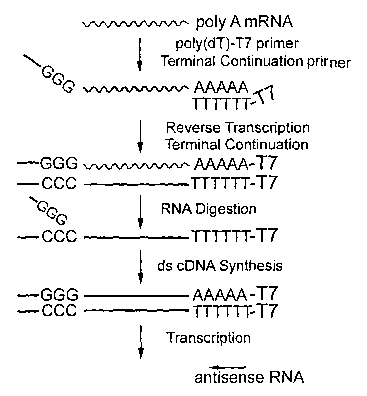
[0046] FIG. 1 is a schematic surmnary of the method of the present invention
demonstrating
attachment of a T7 promoter to the 3' region of mRNA and the mechanism of
terminal
continuation.
[0047] FIG. 2 is a schematic summary of the method of the present invention
demonstrating
attachment of a T7 promoter to the 5' region of mRNA and the mechanism of
terminal
continuation.
[0048] FIG. 3 is a schematic sunnnary of the method of the present invention
demonstrating
attachment of a T7 promoter to the 5' region and a SP6 promoter to the 3'
region of mRNA
and the mechanism of terminal continuation.
[0049] FIG. 4 shows a diagram of RNA amplification based cDNA library
construction.
[0050] FIG. 5 illustrates a schematic summary of the method regarding
detection of RNA
from a histologically stained sample.
[0051] FIG. 6 shows microdissection of cells from tissue sections. Individual
cells are
microdissected with a micropipette under the guidance of a micromanipulator.
The cell can
be physically attached to the tip of the micropipette (as shown in this
schematic) or aspirated
into the fluid-filled pipette tip. Laser capture microdissection can also be
used to isolate one
or more cells from tissue sections adhered to glass slides or coverslips.
[0052] FIG. 7 demonstrates expression profiles of normal (NCI) and Alzheimer's
diseased
(AD) tissues using methods of the present invention.
[0053] FIG. 8 shows amplification and detection of various genes of two
adjacent regions
from the same tissue by present method versus a~NA method in the art. The
relative
hybridization signal intensity of the low, moderate, and higher expressing
genes using the
new methodology of present invention are improved compared to aRNA method
l~nown in
the art.
[0054] FIGS. 9A through 9C show the methods of the present invention. FIGS. 9A
and 9B
schematically illustrate the method. FIG. 9C demonstrates robust linear
amplification.
[0055] FIGS. 10A through lOC demonstrate amplification with the methods of the
present
invention. FIG. 10A utilizes biological samples of RNA extracted from a
variety of brain
22
CA 02437737 2003-08-14
WO 02/065093 PCT/US02/05713
sources including post morten hippocampus and basal forebrain. FIG. 10B shows
a
comparison of different extraction methods. FIG. lOC shows a scatter plot
demonstrating a
liilear relationship between TC RNA input concentration and mean hybridization
signal
intensity of all cDNA clones and an individual clone (CREB) on a custom-
designed cDNA
array.
[0056] FIGS. 11A and 11B demonstrates that methods of the present invention
has increased
sensitivity for the threshold of detection of genes with low hybridization
signal intensity.
FIG. 1 1A demonstrates a dot blot assay showing increased sensitivity for
genes with relative
low abundance. FIG. 11B shows a quantitation in total, normalized
hybridization signal
intensity for custom-designed cDNA array.
[0057] FIG. 12 presents a microscopic field during the microdissection of
mouse dentate
gyrus granule cells described in Example 1. Arrows in frames B & C show the
aspiration
device removing a single cell.
[0058] FIG. 13 presents microarray expression data of Example 8. The top panel
shows
representative raw microarray data of mRNA expression of GluRl, R2, R3, R4, R6
and R7
genes. Vehicle is a negative control experiment, and KA 1 DPL and IAA SDPL are
two
different experiments using intracerebral injection of l~ainate. The bottom
panels show the
average of mRNA expression levels from multiple experiments.
[0059] FIG. 14 presents microarray expression data of Example 9. The top panel
shows
representative microaxray data of mRNA expression of synaptic marl~er genes
from neurons
of subjects with either no cognitive impairment (NCI) or Alzheimer's disease
(AD). The
bottom panel shows the average mRNA expression levels for these genes from
multiple
experiments.
[0060] FIG. 15 presents a schematic of the instrument used for LCM. In section
A, cells are
identified for isolation through microscopy. These targeted cells are then
primed for
separation from tissue by an ultraviolet or infrared laser beam. A transfer
film attached to
either a microfuge cap or membrane adheres the cells) of interest for removal.
The
microfuge cap or membrane containing the cells) of interest is then removed
from the
instrument. Section B shows the part of the apparatus that is responsible for
the transfer of
cells.
[0061] FIG. 16 depicts a comparison of methods of the present invention with
different
histochemical stains from adjacent tissue sections.
23
CA 02437737 2003-08-14
WO 02/065093 PCT/US02/05713
[0062] FIG. 17 is a quantitative analysis using methods of the present
invention for total
signal intensity from adjacent sections stained with an antibody
(neurofilament) and
histologically (cresyl violet).
[0063] Other objects, features and advantages of the present invention will
become apparent
from the following detailed description. It should be understood, however,
that the detailed
description and the specific examples, while indicating preferred embodiments
of the
invention, are given by way of illustration only, since various changes and
modifications
within the spirit and scope of the invention will become apparent to those
slcilled in the art
from this detailed description.
DETAILED DESCRIPTION OF THE INVENTION
[0064] As used herein the specification, "a" or "an" may mean one or more. As
used herein
in the claim(s), when used in conjunction with the word "comprising", the
words "a" or "an"
may mean one or more than one. As used herein "another" may mean at least a
second or
more.
I. Definitions
[0065] The term "histologically-stained tissue" as used herein is defined as
tissue sections or
cells stained by any of a great variety of combinations of dyes that color
various constituents
more or less selectively, or the application to histological preparations of
physical and
chemical methods of analysis that permit identification of chemical substances
in their
normal sites in tissues.
[0066] The term "irZ vitro transcription" as used herein is defined as
generation of an RNA
molecule from a DNA template under conditions outside of a living cell.
[0067] The term "laser capture microdissection" as used herein is defined as
the use of an
infrared (IR) laser beam to remove a desired cell from a nondesired cell. In
preferred
embodiments, the desired cell is a cancer cell and the nondesired cell is a
normal cell.
[0068] The term "oligonucleotides" as used herein are short-length, single-
stranded
polydeoxynucleotides that are chemically synthesized by l~nown methods (such
as
phosphotriester, phosphite, or phosphoramidite chemistry, using solid phase
techniques such
as described in EP 266,032, or via deoxynucleoside H-phosphonate intermediates
as
described by Froehler et al. (1986), followed by purification, such as on
polyacrylamide gels.
In a specific embodiment, an oligonucleotide is a primer.
24
CA 02437737 2003-08-14
WO 02/065093 PCT/US02/05713
[0069] The term ''primer," as used herein, is meant to encompass any nucleic
acid that is
capable of priming the synthesis of a nascent nucleic acid in a template-
dependent process.
[0070] The term ".a short primer of random sequence" as used herein is defined
as an
oligonucleotide primer having the general formula dNl-dN2-...dNq, wherein dN
represents a
deoxyribonucleotide selected randomly from among dAMP, dCMP, dGMP, and dTMP
and q
represents integer 6 and above, preferably from 6 to 10.
[0071] The teen "recombinant promoter" as used herein refers to a nucleic acid
sequence
which regulates expression of a particular nucleic acid sequence, wherein the
promoter is
genetically engineered through the application of recombinant DNA technology.
[0072] The term "template continuation (TC) oligonucleotide" as used herein is
defined as an
oligonucleotide used in a process of template-dependent synthesis of a
complementary strand
of DNA °by a DNA polymerase using two templates in consecutive order
and which are not
covalently linl~ed to each other by phosphodiester bonds. The synthesized cDNA
strand is a
single continuous strand complementary to both templates. In a specific
embodiment of the
present invention, the first template is poly (A)+ RNA and the second template
is a template
continuation oligonucleotide which preferably comprises at least two
riboguanines at its 3'
end. It has a general formula dNl-dN~-...dNq- rNl_~, where dN represents a
deoxyribonucleotide selected from among dAMP, dCMP, dGMP, and dTMP and q
represents
integer 6 and above, preferably from 6 to 70, and rN represents a
ribonucleotide, preferably
riboguanine nucleotide. It typically provides a template for continuous
synthesis of the first
strand cDNA by attaching at the 3' terminus of first strand cDNA through its
sequence
complementary to the 3' terminal sequence of the first strand cDNA.
[0073] The term "terminal continuation reaction" as used herein is defined as
a process of
synthesizing the first strand cDNA using two templates. The first strand cDNA
synthesis
continues using a terminal continuation oligonucleotide as the second template
at the
termination of the first template. The synthesized cDNA is a single strand
continuous
molecule complementary to both first and second templates. In a specific
embodiment of the
present invention, the first template is RNA and the second template is a
terminal
continuation oligonucleotide which preferably comprises at least one
riboguanine at the 3'
end. In some embodiments, at least two riboguanines are present at the 3' end.
CA 02437737 2003-08-14
WO 02/065093 PCT/US02/05713
II. The Present Invention
A. General Embodiments
[0074] The present invention relates to 'a method of adding a nucleic acid
sequence
complementary to a "terminal continuation oligonucleotide", to the 3' end of a
synthesized
nucleic acid strand that is complementary to a target nucleic acid strand. The
method
comprises incubating the target nucleic acid strand in the presence of a
terminal continuation
oligonucleotide and a primer, the "first strand synthesis primer", which is
complementary to a
sequence at the 3' end, or upstream of the 3' end, of the target nucleic acid
strand. The first
strand synthesis primer anneals or hybridizes to its complementary sequence on
the taxget
nucleic acid strand, which allows a polymerase to begin the synthesis of a
nucleic acid strand
complementary to the target nucleic acid strand. The polymerase also
facilitates
incorporation of sequence complementary to the terminal continuation
oligonucleotide into
the 3' end of the synthesized nucleic acid strand by using the terminal
continuation
oligonucleotide as a template.
[0075] When using the above method to generate cDNA, the target nucleic acid
strand is
preferably RNA, more preferably mRNA. If mRNA is the target nucleic acid
strand, then the
first strand synthesis primer may preferably contain poly(dT). Random primers,
for example
random hexamers, and specifically designed primers may also be used as the
first strand
synthesis primer. With the addition of the first strand synthesis primer,
terminal continuation
oligonucleotide and reverse-transcriptase, a first-strand cDNA is synthesized
that is
complementary to the sequence of the target RNA strand sequence. In addition,
the
synthesized first strand cDNA contains the complementary sequence of the
terminal
continuation oligonucleotide at its 3' end and the sequence of the first
strand synthesis primer
at its 5 ° end.
[0076] The present invention provides a highly efficient method for the
synthesis of second
strand cDNA by being able to provide a sequence-specific priming method. As
the
complementary sequence of the terminal continuation oligonucleotide is
incorporated into the
3 ° end of first strand cDNA, second strand cDNA sy~ithesis may be
primed by the terminal
continuation oligonucleotide. This obviates the need for inefficient second
strand
polymerases, such as I~lenow and DNA Pol I, because the second strand
synthesis is initiated
by a primer, and not for example, by a hairpin loop. Therefore, the present
invention
26
CA 02437737 2003-08-14
WO 02/065093 PCT/US02/05713
provides for the use of robust polymerises, for highly efficient second strand
cDNA
synthesis.
[0077] Any polymerise may be used in the present invention, including but not
limited to,
polymerises from the following six families of polynerases: Pol I, Pol alpha,
Pol beta,
DNA-dependent RNA polymerises, reverse transcriptases, and RNA-dependent RNA
polymerises (U.S. Patent No.: 5,614,365). Representative examples of Pol I-
type DNA
polymerises are: bacteriophage T7, T3, T4, T5, Spo 1, Spot and SP6 DNA
polymerises, E.
coli DNA polymerise I, I~lenow fragment of E. coli DNA polymerise I, Tlae~mus
aquaticus
DNA polymerise I (Taq), Bacillus stea~othe~tnophilus DNA polymerise (Bst),
TlZer~mus
they°naophilus DNA polymerise (Ttla), Pyrococcus fm°iosus DNA
polymerise (Pfu),
They°f~aococcus lito~alis DNA polymerise (Vent), and TheYrnus flavus
DNA polymerise I. W
addition to Taq, Vent, Bst, Ttla and Pfu, other thermostable DNA polymerises
are also
included in the present invention. Examples of Pol-alpha or Polymerise II-type
polymerises
include E. coli DNA polymerise II and S. cej°evisiae DNA polymerise II.
Representative
examples of RNA polymerises include: bacteriophage T7, T3 and SP6 RNA
polymerises, E.
coli RNA polymerise holoenzyme, E. coli RNA polymerise core enzyme, and human
RNA
polymerise I, II, III, and human mitochondrial RNA polymerise.
[0078] The present invention further provides the incorporation of cis-
regulatory elements
into synthesized nucleic acid strands through the use of the terminal
continuation method.
Cis-regulatory elements that may be introduced into nucleic acids, include but
are not limited
to, transcriptional promoters, bacteriophage transcriptional promoters,
enhancers, silencers,
methylation sites, origins of replication, matrix attachment regions, locus
control regions and
recombination signal sequences. Other similar elements known in the art may
also be used.
[0079] The present invention also provides the incorporation of nucleic acids
into
synthesized nucleic acid strands by terminal continuation, where the
incorporated nucleic
acids may encode amino acids, stretches of amino acids and antigenic epitopes.
The present
invention further provides the incorporation of nucleic acids into synthesized
nucleic acid
strands by terminal continuation, where the incorporated nucleic acids may
serve to function
as modification signals.
[0080] In one embodiment of the invention, the terminal continuation
oligonucleotide and/or
the first strand synthesis primer are designed to contain a transcriptional
promoter, preferably
a bacteriophage transcriptional promoter. In this embodiment of first strand
cDNA synthesis,
the cDNA strand may contain a transcriptional promoter at its 5' end due to
the annealing of
a first strand synthesis primer that has a complementary sequence to the 3'
region of RNA in
27
CA 02437737 2003-08-14
WO 02/065093 PCT/US02/05713
addition to the sequence that comprises the transcriptional promoter. The
first strand cDNA
may also contain a sequence complementary to a transcriptional promoter at its
3' end if a
terminal continuation oligonucleotide is desig~led to contain a
transcriptional promoter.
Alternatively, the first strand cDNA may contain a sequence of a
transcriptional promoter at
its 3 ' end if a terminal continuation oligonucleotide is designed to contain
the complementary
sequence of a transcriptional promoter. A second strand of cDNA complementary
to the first
synthesized strand of cDNA may be synthesized using the first strand of cDNA
as a template,
and the terminal continuation oligonucleotide as a primer. Sense and/or
antisense RNA
amplification reaction may be subsequently performed by i~r vitf°o RNA
transcription, as both
the first strand and second strand of cDNA may contain transcriptional
promoters
incorporated at either the 5' end, 3'end or both ends.
[0081] Using this methodology, even a small amount of starting RNA amplified
linearly,
such as RNA from a single cell, can be used for many~downstream applications.
Following
the conversion of amplified RNA to double stranded cDNA, the down stream
applications of
amplified RNA include, but are not restricted to, probe generation, gene
expression profiling,
genetic polymorphism amplification and/or detection, cDNA microarray analysis,
cDNA
library construction, expression library construction, single cell cDNA
library construction,
subtraction library construction and competitive array hybridization. The
synthesized sense
RNA of a total starting RNA population can also be used as a template for ifs
vitro protein
translations, where the resultant protein may then be used for further
downstream
applications. A variety of reagent bits for the procedures may be developed as
a result of,
and are encompassed in, the present invention.
[0082] Any source of nucleic acid can be used as starting material, including
but not limited
to, DNA, RNA, ribosomal RNA, mitochondrial DNA, mitochondrial RNA, synthetic
DNA,
and synthetic RNA. Preferably, total RNA or poly (A)+ mRNA is used as starting
material.
A small amount (as low as picograms) of total RNA or mRNA extracted from
single cells is
sufficient for subsequent amplification. Sources of RNAs can include synthetic
sources or
biological sources, such as tissues from in vitro and in vivo preparations,
including, but not
restricted to, biopsy samples and post mortem tissues from a variety of
species ranging from
invertebrates to mammals including humans and genetically altered subj ects.
RNA from
microbial genomes is also a source of starting genetic material. RNAs are
extracted using
standard molecular biological methods. Care must be tal~en to avoid RNase
contamination
along with inactivation of endogenous RNase activity.
2$
CA 02437737 2003-08-14
WO 02/065093 PCT/US02/05713
[0083] Thus, the present invention concerns compositions and methods for
amplification of
RNA, preferably mRNA. The compositions and methods employ terminal
continuation
oligonucleotides described herein. The methods of the present invention
comprise contacting
RNA with a primer which can amleal to the RNA, a reverse transcriptase, and a
terminal
continuation oligonucleotide under conditions sufficient to permit the
template-dependent
extension of the annealed primer to generate an mRNA-cDNA hybrid, which is
then followed
by second strand cDNA synthesis.
[0084] First strand synthesis is preferably primed with an oligonucleotide
primer, the "first
strand synthesis primer", containing the sequence complement of a sequence at
the 3' end of
the target nucleic acid. First strand synthesis may also be primed with an
oligonucleotide
primer containing the sequence complement of a sequence located upstream of
the 3' end of
the target nucleic acid. If the target nucleic acid is RNA, examples of first
strand synthesis
primers include, but are not limited to, polythymidylate [poly(dT)s] or random
sequences,
such as random hexamer. In addition, the first strand synthesis primer can
also include other
desirable sequences, such as for example, a transcription promoter sequence,
or a designed
restriction enzyme digestion sequence (FIGS. 1 and 2).
[0085] It is preferred that a second primer, the "terminal continuation
oligonucleotide", is
also present in the first strand syithesis reaction mixture. In addition, a
sequence of a desired
bacteriophage promoter, such as T7, T3, or SP6 or other functional sequences
may optionally
be a component sequence of the terminal continuation oligonucleotide (FIGS. 1
and 2).
[0086] It is preferred that the "terminal continuation oligonucleotide"
contains at least one
guanine or deoxyguanne (G or dG), or cytosine or deoxycytosine (C or dC) at
its 3' end,
most preferably at least two G or dG or C or dC at its 3' end. The terminal
continuation
oligonucleotide may alternatively contain at least one adenosine or
deoxyadenosine (A or
dA), or thymidine or deoxythymidine (T or dT) at its 3'end. The terminal
continuation
oligonucleotide may also consist of a random sequence or nucleotide. It is
preferred that the
total length of the terminal continuation oligonucleotide is between about 8-
100 nucleotides,
more preferably about 15-75 nucleotides, most preferably about 20-50
nucleotides.
[0087] One reason for the preference that the "terminal continuation
oligonucleotide"
contains a short stretch of at least one guanine or deoxyguanine (G or dG), or
cytosine or
deoxycytosine (C or dC) at its 3' end, is due to the efficiency in terminal
continuation
function. Both of the aforementioned structures have comparable efficiency in
terminal
continuation function. A complete or partial replacement of G, dG, C, dC at
the 3' end of a
terminal continuation oligonucleotide with A, dA, T, dT decreases the
efficiency of a
29
CA 02437737 2003-08-14
WO 02/065093 PCT/US02/05713
terminal continuation reaction slightly. However, tlus reaction also produces
terminal
continuation products. The number of nucleotides and the sequence at the 3'
end of the
terminal continuation oligonucleotide may be optimized empirically, and can
readily be
determined by the skilled artisan.
[0088] It is desirable for the method to match a primer with the appropriate
promoter. For
example, the same RNA transcription promoter is preferably not added to both
the 5' and 3'
termini of cDNA. However, two different promoters, such as T7 and T3, may be
added at
both the 5' and 3' ends of cDNA and direct either "sense" or "anti-sense" RNA
synthesis.
(FIG. 3). It is within the scope of the invention, that any promoter capable
of initiating
transcription can be used.
[0089] The second strand cDNA synthesis is preferably primed by an oligo(dNTP)
with the
sequence complementary to at least a portion of the terminal continuation
oligonucleotide. In
the embodiment where the synthesized cDNA strands contain transcriptional
promoters,
RNA may be transcribed with an RNA polymerase corresponding to the promoter.
For
example, T7 RNA polymerase may be used to transcribe RNA driven by a T7
promoter,
whereas SP6 RNA polymerase may be used to transcribe RNA driven by a SP6
promoter.
When two different promoters are attached at both ends of the cDNA, the RNA
polymerase is
chosen according to the "sense" or "antisense" orientation of the transcribed
RNA desired.
[0090] More than one round of RNA amplification may be performed when
necessary.
During subsequent amplifications, the total population of RNA is reverse
transcribed back
into cDNA. The reverse transcription is primed either with specific primers
attached to
cDNA previously, by random primers, or by primers designed to amplify specific
internal
regions. In this embodiment of the invention, it is preferred that at least
one RNA
transcription promoter is incorporated into the subsequently synthesized
double stranded
cDNA.
[0091] The cDNAs can be further engineered or altered by appropriate enzymatic
maxiipulations prior to downstream applications. The downstream uses of the
nucleic acid
produced by the present method may include, for example, probe generation,
gene expression
profiling, genetic polymorphism profiling, cDNA library constructioy (FIG. 4),
expression
library construction, subtraction library construction, competitive array
hybridization, iya vitro
translation, and clinical diagnostics independently or in combination with
morphological
examination.
[0092] The present invention may be conveniently developed into appropriate
reagent kits for
research or diagnostic purposes.
CA 02437737 2003-08-14
WO 02/065093 PCT/US02/05713
[0093] Thus, in a specific embodiment, the process of the present invention
comprises at
least the following steps: .
[0094] 1. Incubating a sample of poly(A)+RNA or total RNA with a poly (dT)
primer or a
short primer of random sequence which can ameal to mRNA and an enzyme that
possesses
reverse transcriptase activity under conditions sufficient to permit the
template-dependent
extension of the primer to generate an mRNA-cDNA hybrid. In some embodiments,
the poly
(dT) primer also comprises a bacteriophage promoter sequence, such as T7 RNA
polymerase,
T3 RNA polymerase, or SP6 RNA polymerase. In some embodiments, a small amount
of
total RNA or mRNA extracted from single cells is sufficient for subsequent
amplification.
[0095] 2. Incubating the first-strand cDNA synthesis mixture obtained from
step 1 with a
terminal continuation oligonucleotide of the present invention. The terminal
continuation
oligonucleotide has at least one riboguanine residue at its 3'-end, a
nucleotide sequence at its
5'-end which may be variable, and in some embodiments a restriction enzyme
digestion site,
an RNA synthesis promoter, a protein translation start signal, or a
combination thereof.
[0096] 3. Second strand cDNA synthesis.
[0097] 4. In vit>"o transcription.
[0098] Using the methods of the present invention with conventional
procedures, first-strand
cDNA synthesis is caiTied out using RNA as a template for reverse
transcription. A primer is
annealed to RNA forming a primer:RNA complex. Extension of the primer is
catalyzed by
reverse transcriptase, or by a DNA polymerase possessing reverse transcriptase
activity, in
the presence of adequate amounts of other components necessary to perform the
reaction, for
example, deoxyribonucleoside triphosphates dATP, dCTP, dGTP and dTTP, Mg2+,
and
optimal buffer. A variety of reverse transcriptases can be used. Preferably,
the reverse
transcriptase is isolated from Moloney murine leukemia virus (M-MLV) (U.S.
Pat. No.
4,943,531) or M-MLV reverse transcriptase lacl~ing RNaseH activity (U.S. Pat.
No.
5,405,776), avian myeloblastosis virus (AMV), hwnan T-cell leukemia virus type
I (HTLV-
I), Rous-associated virus 2 (RAV2), bovine leukemia virus (BLV), Rous sarcoma
virus
(RSV), human irmnunodeficiency virus (HIV) or Thermus aquaticus (Taq) or
Tlzerzzzus
thermophilus (Ttlz) (U.S. Pat. No. 5,322,770). These reverse transcriptases
may be isolated
from an organism itself or, in some cases, obtained commercially. Reverse
transcriptases
useful with the subject invention can also be obtained from cells expressing
cloned genes
encoding the enzyme. As a starting material for cDNA synthesis, poly(A)+RNA or
total RNA
from yeast and higher organisms such as plants or animals can be used. The
first-strand
cDNA synthesis step of the subject method can include terminal continuation
31
CA 02437737 2003-08-14
WO 02/065093 PCT/US02/05713
oligonucleotides of the present invention in the reaction mixture, but are not
a necessary
component for carrying out first-strand cDNA synthesis. Thus, it is understood
that terminal
continuation oligonucleotide molecules can be included in the first-strand
reaction
composition (for example, during the first primer annealing to RNA or when
contacting the
RNA with an enzyme possessing reverse transcriptase activity) or the
oligonucleotides can be
added in the course of, or after completion of, the first-strand cDNA
synthesis reaction.
[0099] In an alternative embodiment, in lieu of a poly (dT) primer, a primer
to an inner, non-
poly(A)+ portion of the mRNA is utilized. These oligonucleotide primers) have
the general
formula dNl -dN2 - . . . dNq, where dN represents a deoxyribonucleotide
selected from
among dAMP, dCMP, dGMP, and dTMP and q represents integer 6 and above.
[0100] In an alternative embodiment, a population of short primers of random
sequences can
be used. The primers are sufficiently short, preferably 6-10 deoxyoligo
nucleic acids, and the
sequences are sufficiently variable that every RNA present has at least one
primer that has the
sequence complementary to it and anneals to it to prime the synthesis of a
first strand cDNA.
[OlOI] Following the complete synthesis of the first strand cDNA, the terminal
transferase
activity of reverse transcriptase adds a few additional nucleotides, primarily
deoxycytidine
and/or deoxyguanine, to the 3' end of the newly synthesized cDNA strand
independent of
template. The terminal continuation oligonucleotide, which in some embodiments
has an
oligo (rG) sequence at its 3' end, base pairs with the deoxycytidine-rich
stretch of nucleotides
present on the first cDNA straild, creating an extended template. Reverse
transcriptase then
continues synthesis of cDNA complementary to the terminal continuation
oligonucleotide
attached to the terminal of the first stranded cDNA. Thus, the full extension
product of the
first cDNA synthesis comprises both sequences complementary to the RNA and to
the
terminal continuation oligonucleotide.
[0102] Replacement of the RNA portion of the mRNA:cDNA hybrid with a second-
strand
cDNA entails removal of the RNA strand in RNA:DNA molecules, and also include
DNA
synthesis by a DNA polymerase. In a specific embodiment, RNAse H is utilized.
In an
alternative embodiment, heating in the presence of appropriate concentration
(such as in a
range of 0.001 mM to 0.15 mM) of magnesium chloride. DNA synthesis is
continuous and
no ligation step is necessary.
[0103] The second strand cDNA synthesis is primed by an oligo (dNTP) with the
sequence
identical to whole or a portion of the terminal continuation oligonucleotide.
A variety of
DNA polymerases can be used, such as E. coli DNA polymerase I, bacteriophage
T4 DNA
polymerase, bacteriophage T7 DNA polymerase, and large fragment of E. coli DNA
32
CA 02437737 2003-08-14
WO 02/065093 PCT/US02/05713
polymerase I (I~lenow fragment). In a specific embodiment, a thermostable and
robust DNA
polymerase, Taq DNA polymerase, is used for second strand cDNA synthesis.
[0104] In other embodiments of the present invention, the present invention is
directed to
amplification and detection of RNA from a histologically-stained tissue. Until
now, the
amplification of RNA by ire vitro transcription from the same presently
lustologically-stained
source of tissue has not been lrnown, although methods to amplify genetic
signals by PCR
based methods are known. That is, it is known to use PCR methods, which are
exponential,
to amplify a dsDNA molecule or to amplify an mRNA by RT-PCR, but the
amplification of
an RNA molecule derived from the dsDNA molecule, particularly in a linear
fashion, is
uWcnown. In a preferred embodiment, the RNA is amplified by aRNA methods (Van
fielder
et al. (1990); Eberwine et al. (1992); U.S. Patent No. 5,545,522), all of
which are
incorporated herein by reference in their entirety) or by other ifa
vitf°o transcription methods,
such as are the subject of the present invention.
[0105] In an object of the present invention, the amplified RNA population is
used as a
clinical diagnostic tool independently or in combination with morphological
examination,
such as regarding the treatment and/or diagnosis of an individual.
[0106] The present invention describes a method for amplification of RNA
populations from
histologically stained tissues and cells through ii2 vitro transcription (FIG.
5). The amplified
RNAs could be fuxther genetically manipulated for the applications of down
stream
investigations, including, but not restricted to, RNA amplification, cDNA
microaxray
analysis, subtractive hybridization, RT-PCR, library constt-uctions, and
clinical molecular
diagnoses.
[0107] 1) Biological tissues from ira vitro and ih vivo preparations can be
used, including, but
not restricted to, biopsy samples and post mortem tissues from a variety of
species ranging
from invertebrates to mammals, including genetically altered subjects and
humans.
[0108] 2) The sample for the present invention is directed to any cellular
material including
but not limited to muscle, connective tissue, skin, brain, liver, urine, bone
marrow, touch
preps of surgical specimens, fine needle aspirates and all cellular body
fluids, including
cerebrospinal fluid, blood, mucus, saliva, nipple aspirates, urine, sweat, and
feces. Ti1 addition
samples can include any pathological tissue including but not limited to
tumors, lymph nodes,
lesions, blood vessels, and traumatic injured tissues.
[0109] 3) The fixation conditions are flexible, as both fresh tissues and
fixed tissues can be
utilized. The samples can be fixed by a wide variety of reagents, including
but not restricted
to, acetone, aldehyde derivatives, ethanol, and combinations therein. The
critical step for the
33
CA 02437737 2003-08-14
WO 02/065093 PCT/US02/05713
fixation is use of RNAse-free conditions and buffers, prompt accession of
tissues, and low
temperature. RNAs are preserved best under these conditions. Frozen tissues
and various
cross-linl~ing and precipitating fixatives such as fonnalin, paraformaldehyde,
acetone and
ethanol are utilized. A spilled artisan recognizes that the present invention
is utilized to
amplify RNA from cells/tissues as well as body fluids, (e.g., cerebrospinal
fluid, blood,
saliva, urine, feces, sweat).
[0110] 4) After fixation, tissues are sectioned and histological stains
applied for cellular
visualization and diagnostic prediction prior to the extraction of RNA. The
histological stains
include all preparations that depict cellular, regional, laminar, and nuclear
structures within
tissue samples. Examples of histological stains that can be utilized by this
invention include:
hematoxylin and eosin, thionin, cresyl violet, acridine orange, and reduced
silver
preparations. When the presence of RNA is in doubt, acridine orange staining
can be used to
visualize RNA (Ginsberg et al., 1997; Ginsberg et al., 1998) in the tissues
and cells) of
interest before RNA extraction and subsequent amplification.
[0111] 5) Individually-identified cells or populations of cells are dissected
from tissues using
a micropipette attached to a micromanipulator or by laser capture
microdissection (FIG. 6).
[0112] 6) Microdissected cells should be immediately merged into chaotropic
cell lysis
buffers to inactivate RNase activity instantaneously. Commercially available
RNA extraction
reagents (such as trizol) can also be used. In general, no homogenization step
is necessary.
The usage of an inert Garner, such as glycogen or linear acrylamide, is
helpful for maximum
RNA precipitation.
[0113] 7) RNAs of such minute amount will almost always have to be amplified
first prior to
desired down stream usage. The first step of the RNA amplification is to
synthesize ds-cDNA
templates. This first strand cDNA is synthesized with a reverse transcriptase
primed by an
oligonucleotide that anneals to RNAs. In some embodiments, a TC primer is
included in the
first strand cDNA synthesis mixture, which will serve as a template at the 3'
terminal of the
synthesized first strand cDNA. The second strand cDNA is synthesized by a DNA
polymerise using first strand cDNA as template and primed by a primer with the
sequence
substantially similar to TC primer.
[0114] 8) RNA extracted from lustologically stained tissues or cells is
amplified through iya
vitYO RNA amplification. In practice, ira vitT°o RNA transcription
needs a promoter to drive the
reaction. The best promoter candidates are the bacteriophage promoters T7, T3,
and SP6.
[0115] 9) A transcription promoter can be annealed to the 3' of first strand
cDNA by priming
mRNA with a specific poly(T) primer that contains the promoter sequence.
Alternatively, a
34
CA 02437737 2003-08-14
WO 02/065093 PCT/US02/05713
promoter can be attached to the 5' of first strand cDNA through terminal
continuation (U.S.
Patent Application filed February 14, 2001 entitled "RNA Amplification
Method.")
[0116] 10) This procedure can be developed conveniently into reagent lcits
with the essential
component of histological stains, an RNA extraction reagent, an RNA
precipitation Garner,
primers and enzymes for the synthesis of ds-cDNA template and enzymes irz
vitro RNA
transcription.
[0117] 11) Various modifications or changes in light thereof will be suggested
to persons
spilled in the art, and are to be included in the scope of the invention.
[0118] Also within the scope of the present invention is a method of
hybridization using
probes generated from an amplified RNA population. RNA probes generated
according to
the present invention will be labeled, either by radioisotopes, fluorescent
dye, biotin and other
reporter groups by conventional chemical or enzymatic labeling procedures. On
the other
hand, a complementary cDNA can be further synthesized and labeled using RNA
generated
in present invention as a template. Labeled RNA or cDNA can then be used in
standard
hybridization assays pnown in the art, i.e., the labeled RNA or cDNA is
contacted with the
defined oligonucleotide/polynucleotides corresponding to a particular set of
the genes
immobilized on a solid surface for a sufficient time to permit the formation
of patterns of
hybridization on the surfaces caused by hybridization between certain
polynucleotide
sequences in the hybridization probe with the certain immobilized defined
oligonucleotide/polynucleotides. The hybridization patterns using available
conventional
techniques, such as scintillation counting, autoradiography, fluorescence
detection,
colorimetric assays, optical density assessments, or light emission
measurement. Techniques
and conditions for labeling, hybridization and detection are well known in the
art (see, e.g.
Sambroop et al., 1989; Ausubel et al., 1994). ,
[0119] In a preferred embodiment, a microarray is probed with RNA or cDNA
generated by
methods of the present invention. A microarray is usually a solid support,
either a glass slide
or a membraxie, with hundreds or even thousands pnown genes or DNAs printed on
it. As
used herein, the term "solid support" refers to any lcnown substrate which can
be used for the
immobilization of a binding ligand or oligonucleotide/polynucleotide sequences
by any
known method. A distinct pattern of hybridization will be generated by probing
a microaxray
with RNA or cDNA generated with the present invention, which leads to the
establishment of
a gene expression profile of the tissue from wluch RNA is extracted.
j0120] In another embodiment, a RNA or cDNA generated with the present
invention can be
separated in an agarose gel, transferred to a solid support, such as a nylon
or a nitrocellulose
CA 02437737 2003-08-14
WO 02/065093 PCT/US02/05713
membrane, and probed with a labeled l~nown RNA or DNA as in Northern or
Southern
hybridization analysis.
[0121] Also within the scope of the present invention is a method for
generating libraries
containing cDNAs generated from amplified RNAs. Conventional methods used to
generate
cDNA libraries require either haxge quantities starting materials or a PCR
step to amplify
small quantity of starting materials. Both methods are not suitable for the
generation of
cDNA from a homogeneous population of cells due to the difficulty of obtaining
large
quantities of pure material from a homogeneous population. Moreover, a low
copy gene can
rendered undetectable during PCR amplification. The present method provides an
improved
alternative to generate cDNA libraries from a homogeneous cell population.
[0122] In another aspect, the invention provides methods wherein the resulting
cDNA
product generated can be used as a starting material for use with cDNA
subtraction methods.
Specifically, the method of the subject invention can be used in conjunction
with cDNA
subtraction procedures to prepare a cDNA population containing highly enriched
representation of cDNA species that are present in one DNA population (the
tester
population), but that are less abundant or absent in another DNA population
(the driver
population). Tester and driver ds cDNA amplified by the methods of the present
invention
can be used in combination with suppression subtractive hybridization
technology described
previously (see e.g. U.S. Pat. No. 5,565,340 and U.S. Pat. No. 5,436,142).
[0123] A person familiar with the art of the field will be able to devise
modifications of the
above method for the detection of genes present in the RNA population
generated in the
present invention.
[0124] Thus, the use of the terminal continuation method provides a
substantially improved
sensitivity and efficiency of linear RNA amplification. The benefit of the
improvement is the
detection of the presence and the quantity of multiple genes from minimum
quantity of
starting materials.
III. Ih vitro Transcription
[0125] In vitro transcription xequires a purified, linear ds cDNA template,
such as is
generated with the methods of the present invention, containing a promoter,
ribonucheotide
triphosphates, a buffer system that preferably includes DTT and magnesium, and
an
appropriate bacteriophage RNA polymerase. A shrilled artisan recognizes that
the exact
conditions used in the transcription reaction depend on the quantity and
quality of RNA
needed for a specific application (the reaction conditions will be different
for generating
36
CA 02437737 2003-08-14
WO 02/065093 PCT/US02/05713
labeled RNA as hybridization probes compared to those reaction conditions for
obtaining
large quantity of RNAs).
[0126] The common RNA polynerases used in irl vitro transcription reactions
are SP6, T7
and T3 polymerases, named for the bacteriophages from which they were cloned.
The genes
for these proteins have been overexpressed in Escherichia coli, and the
polymerases have
been purified and are commercially available. RNA polymerases are DNA template-
dependant with distinct and very specific promoter sequence requirements. The
promoter
consensus sequences for each of the phage RNA polymerases are as follows,
wherein the first
base incorporated into the transcript is bolded, and the minimum sequence
required for
efficient transcription is underlined:
T7: 5'-TAATACGACTCACTATAGGGAGA-3' (SEQ ID NO:1)
SP6: 5'-ATTTAGGTGACACTATAGAAGNG-3' (SEQ ID NO:2)
T3: 5'-AATTAACCCTCACTAAAGGGAGA-3' (SEQ ID N0:3)
[0127] After the RNA polymerase binds to its double-stranded DNA promoter, the
polymerase separates the two DNA strands and uses the 3' to 5' strand as
template for the
synthesis of a complementary 5' to 3' RNA strand. Depending on the orientation
of DNA
sequence relative to the promoter, as generated by the methods described
herein, the template
may be designed to produce sense strand or antisense strand RNA. Specifically,
a
transcription promoter has to be attached to a dsDNA template through the
mechanism of
terminal continuation when sense RNA is to be synthesized, whereas a
transcription promoter
has to be attached to a ds RNA template through amlealing a poly(dT) primer
containing a
promoter sequence to an mRNA molecule when antisense RNA is to be synthesized.
When
designing a transcription template, it must be decided whether sense or
antisense transcripts
are needed. If the RNA is to be used as a probe for hybridization to messenger
RNA (e.g. ih
situ hybridization, or nuclease protection assays), complementary antisense
transcripts are
required. In contrast, sense strand transcripts are used when performing
expression, structural
or functional studies or when constructing a standard curve for RNA
quantitation using an
artificial sense strand RNA. Either sense or antisense RNA can be used in
microarray
analysis or reverse northern hybridization.
[0128] By convention, the single strand of a DNA sequence shown in scientific
journals and
databases is the coding, (+), or "sense strand", identical in sequence (with
T's changed to
U's) to its mRNA copy. The +1 G of the RNA polymerase promoter sequence in the
DNA
37
CA 02437737 2003-08-14
WO 02/065093 PCT/US02/05713
template is the first base incorporated into the transcription product (see
above). To make
sense RNA, the 5' end of the coding strand must be adjacent to, or just
downstream of, the +1
G of the promoter. For antisense RNA to be transcribed, the 5' end of the
noncoding strand
must be adjacent to the +1 G.
IV. Nucleic Acid Detection
[0129] In some embodiments of the present invention, detection of nucleic
acids, particularly
those amplified by the methods described herein, is desired. In a preferred
embodiment, a
microarray is probed with RNA generated by methods of the present invention.
A. Hybridization
[0130] The use of a probe or primer of between 13 and 100 nucleotides,
preferably between
17 and 100 nucleotides in length, or in some aspects of the invention up to 1-
2 kilobases or
more in length, allows the formation of a duplex molecule that is both stable
and selective
(Sambrook et al., 1989). Molecules having complementary sequences over
contiguous
stretches greater than 20 bases in length are generally preferred, to increase
stability and/or
selectivity of the hybrid molecules obtained. One will generally prefer to
design nucleic acid
molecules for hybridization having one or more complementary sequences of 20
to 30
nucleotides, or even longer where desired. Such fragments may be readily
prepared, for
example, by directly synthesizing the fragment by chemical means or by
introducing selected
sequences into recombinant vectors for recombinant production.
[0131] Accordingly, the nucleotide sequences of the present invention may be
used for their
ability to selectively form duplex molecules with complementary stretches of
DNAs and/or
RNAs or to provide primers for amplification of DNA or RNA from samples.
Depending on the
application envisioned, one would desire to employ varying conditions of
hybridization to
achieve varyiilg degrees of selectivity of the probe or primers for the target
sequence.
[0132] For applications requiring high selectivity, one will typically desire
to employ
relatively high stringency conditions to form the hybrids. For example,
relatively low salt
and/or high temperature conditions, such as provided by about 0.02 M to about
0.10 M NaCI
at temperatures of about 50°C to about 70°C. Such high
stringency conditions tolerate little,
if any, mismatch between the probes and target sequences would be particularly
suitable for
isolating specific genes or for detecting specific mRNA transcripts. It is
generally
appreciated that conditions can be rendered more stringent by the addition of
increasing
amounts of formamide.
38
CA 02437737 2003-08-14
WO 02/065093 PCT/US02/05713
[0133] For certain applications, it is appreciated that lower stringency
conditions are
preferred. Under these conditions, hybridization may occur even though the
sequences of the
hybridizing strands are not perfectly complementary, but are mismatched at one
or more
positions. Conditions may be rendered less stringent by increasing salt
concentration and/or
decreasing temperature. For example, a medium stringency condition could be
provided by
about 0.1 to 0.25 M NaCI at temperatures of about 37°C to about
55°C, while a low
stringency condition could be provided by about 0.15 M to about 0.9 M salt
(such as NaCI),
at temperatures ranging from about 20°C to about 55°C.
Hybridization conditions can be
readily manipulated depending on the desired results.
[0134] In other embodiments, hybridization may be achieved under conditions
of, for
example, 50 mM Tris-HCl (pH 8.3), 75 mM KCI, 3 mM MgCl2, 1.0 mM
dithiothreitol, at
temperatures between approximately 20°C to about 37°C. Other
hybridization conditions
utilized could include approximately 10 mM Tris-HCl (pH 8.3), 50 mM KCI, 1.5
mM MgCl2,
at temperatures ranging from approximately 40°C to about 72°C.
In a specific embodiment,
50% formamide solutions with 6 XSSPE, KCl, MgCl2, SX Denhardt's. 1M NaPPi, and
200
ng/ml sheared salmon sperm DNA are used.
[0135] In certain embodiments, it will be advantageous to employ nucleic acids
of defined
sequences of the present invention in combination with an appropriate means,
such as a label,
for determiiung hybridization. A wide variety of appropriate indicator means
are known in
the art, including fluorescent, radioactive, enzymatic or other ligands, such
as avidin/biotin,
which are capable of being detected. In preferred embodiments, one may desire
to employ a
fluorescent label or an enzyme tag such as urease, alkaline phosphatase or
peroxidase, instead
of radioactive or other environmentally undesirable reagents. In the case of
enzyme tags,
colorimetric indicator substrates are lmown that can be employed to provide a
detection
means that is visibly or spectrophotometrically detectable, to identify
specific hybridization
with complementary nucleic acid containing samples.
[0136] In general, it is envisioned that the probes or primers described
herein will be useful
as reagents in solution hybridization, as in PORT"", for detection of
expression of
corresponding genes, as well as in embodiments employing a solid phase. In
embodiments
involving a solid phase, the test DNA (or RNA) is adsorbed or otherwise
affixed to a selected
matrix or surface. This fixed, single-stranded nucleic acid is then subj ected
to hybridization
with selected probes under desired conditions. The conditions selected will
depend on the
particular circumstances (depending, for example, on the G1-C content, type of
target nucleic
39
CA 02437737 2003-08-14
WO 02/065093 PCT/US02/05713
acid, source of nucleic acid, size of hybridization probe, etc.). Optimization
of hybridization
conditions for the particular application of interest is well l~nown to those
of slcill in the a~.-t.
After washing of the hybridized molecules to remove non-specifically bound
probe
molecules, hybridization is detected, and/or quantified, by determining the
amount of bound
label. Representative solid phase hybridization methods are disclosed in U.S.
Patent Nos.
5,843,663, 5,900,481 and 5,919,626. Other methods of hybridization that may be
used in the
practice of the present invention are disclosed in U.S. Patent Nos. 5,849,481,
5,849,486 and
5,851,772. The relevant portions of these and other references identified in
this section of the
Specification are incorporated herein by reference.
B. Amplification of Nucleic Acids
[0137] In the present invention, it is desirable to first convert an RNA to a
complementary
DNA, and in a specific embodiment, the resultant cDNA is amplified, such as
with primers.
Typically, primers are oligonucleotides from ten to twenty and/or thirty base
pairs in length,
but longer sequences can be employed. Primers may be provided in double-
stranded and/or
single-stranded form, although the single-stranded fore is preferred.
[0138] The technique of "polymerase chain reaction," or "PCR," as used herein
generally
refers to a procedure wherein minute amounts of a specific piece of nucleic
acid, RNA and/or
DNA, are amplified as described in U.S. Pat. No. 4,683,195. Generally,
sequence information
from the ends of the region of interest or beyond needs to be available, such
that
oligonucleotide primers can be designed; these primers will be identical or
similar in
sequence to opposite strands of the template to be amplified. The 5' terminal
nucleotides of
the two primers may coincide with the ends of the amplified material. PCR can
be used to
amplify specific RNA sequences, specific DNA sequences from total genomic DNA,
a~ld
cDNA transcribed from total cellular RNA, bacteriophage or plasmid sequences,
etc. See
generally Mullis et al. (1987); Erlich, ed., PCR Technology, Stocl~ton Press,
N.Y., (1989). As
used herein, PCR is considered to be one, but not the only, example of a
nucleic acid
polynerase reaction method for amplifying a nucleic acid test sample,
comprising the use of
a lmown nucleic acid (DNA or RNA) as a primer and utilizes a nucleic acid
polymerase to
amplify or generate a specific piece of nucleic acid or to amplify or generate
a specific piece
of nucleic acid that is complementary to a particular nucleic acid.
[0139] Pairs Qf primers designed to selectively hybridize to nucleic acids are
contacted with
the template nucleic acid under conditions that permit selective
hybridization. Depending
upon the desired application, high stringency hybridization conditions may be
selected that
CA 02437737 2003-08-14
WO 02/065093 PCT/US02/05713
will only allow hybridization to sequences that are completely complementary
to the primers.
In other embodiments, hybridization may occur under reduced stringency to
allow for
amplification of nucleic acids contain one or more mismatches with the primer
sequences.
Once hybridized, the template-primer complex is contacted with one or more
enzymes that
facilitate template-dependent nucleic acid synthesis. Multiple rounds of
amplification, also
referred to as "cycles," are conducted until a sufficient amount of
amplification product is
produced.
[0140] A number of template dependent processes are available to amplify the
oligonucleotide sequences present in a given template sample. One of the best
known
amplification methods is the polymerase chain reaction (referred to as PCRTM)
which is
described in detail in U.S. Patent Nos. 4,683,195, 4,683,202 and 4,800,159,
and in Innis et
al., 1988, each of which is incorporated herein by reference in their
entirety.
[0141] A reverse transcriptase PCRTM amplification procedure may be performed
to quantify
the amount of in.RNA amplified. Methods of reverse transcribing RNA into cDNA
are well
lcnown (see Sambrook et al., 1989). Alternative methods for reverse
transcription utilize
thermostable DNA polymerases. These methods are described in WO 90/07641.
Polymerase
chain reaction methodologies are well known in the art. Representative methods
of RT-PCR
are described in U.S. Patent No. 5,882,864.
[0142] Another method for amplification is ligase chain reaction ("LCR"),
disclosed in
European Application No. 320 308, incorporated herein by reference in its
entirety. U.S. Patent
4,883,750 describes a method similar to LCR for binding probe pairs to a
target sequence. A
method based on PCRTM and oligonucleotide ligase assay (OLA), disclosed iii
U.S. Patent
5,912,148, may also be used.
[0143] Alternative methods for amplification of target nucleic acid sequences
that may be
used in the practice of the present invention are disclosed in U.S. Patent
Nos. 5,843,650,
5,846,709, 5,846,783, 5,849,546, 5,849,497, 5,849,547, 5,858,652, 5,866,366,
5,916,776,
5,922,574, 5,928,905, 5,928,906, 5,932,451, 5,935,825, 5,939,291 and
5,942,391, GB
Application No. 2 202 328, and in PCT Application No. PCT/US89/01025, each of
which is
incorporated herein by reference in its entirety.
[0144] Qbeta Replicase, described in PCT Application No. PCT/US87/00880, may
also be used
as an amplification method in the present invention. In this method, a
replicative sequence of
RNA that has a region complementary to that of a target is added to a sample
in the presence of
an RNA polymerase. The polyinerase will copy the replicative sequence which
may then be
detected.
41
CA 02437737 2003-08-14
WO 02/065093 PCT/US02/05713
[0145] An isothermal amplification method, in which restriction endonucleases
and ligases
are used to achieve the amplification of target molecules that contain
nucleotide 5'-[alpha-
thio]-triphosphates in one strand of a restriction site may also be useful in
the amplification of
nucleic acids in the present invention (Walker et al., 1992). Strand
Displacement
Amplification (SDA), disclosed in U.S. Patent No. 5,916,779, is another method
of carrying
out isothermal amplification of nucleic acids which involves multiple rounds
of strand
displacement a~.id synthesis, i.e., nick translation.
[0146] Other nucleic acid amplification procedures include transcription-based
amplification
systems (TAS), including nucleic acid sequence based amplification (NASBA) and
3SR
(I~woh et al., 1989; Gingeras et al., PCT Application WO 88/10315,
incorporated herein by
reference in their entirety). European Application No. 329 822 disclose a
nucleic acid
amplification process involving cyclically synthesizing single-stranded RNA
("ssRNA"),
ssDNA, and double-stranded DNA (dsDNA), which may be used in accordance with
the
present invention.
[0147] PCT Application WO 89/06700 (incorporated herein by reference in its
entirety)
disclose a nucleic acid sequence amplification scheme based on the
hybridization of a
promoter regionprimer sequence to a target single-stranded DNA ("ssDNA")
followed by
transcription of many RNA copies of the sequence. This scheme is not cyclic,
i.e., new
templates are not produced from the resultant RNA transcripts. Other
amplification methods
include "race" and "one-sided PCR" (Frohman, 1990; Ohara et al., 1989).
C. Detection of Nucleic Acids
[0148] Following any amplification, it may be desirable to separate the
amplification product
from the template and/or the excess primer. In one embodiment, amplification
products are
separated by agarose, agarose-acrylamide or polyacrylamide gel electrophoresis
using
standard methods (Sambrook et al., 1989). Separated amplification products may
be cut out
and eluted from the gel for further manipulation. Using low melting point
agarose gels, the
separated band may be removed by heating the gel, followed by extraction of
the nucleic
acid.
[0149] Separation of nucleic acids may also be effected by chromatographic
teclnuques
known in art. There are many kinds of chromatography which may be used in the
practice of
the present invention, including adsorption, partition, ion-exchange,
hydroxylapatite,
molecular sieve, reverse-phase, column, paper, thin-layer, and gas
chromatography as well as
HPLC.
42
CA 02437737 2003-08-14
WO 02/065093 PCT/US02/05713
[0150] In certain embodiments, the amplification products are visualized. A
typical
visualization method involves staining of a gel with etludium bromide and
visualization of
bands under UV light. Alternatively, if the amplification products are
integrally labeled with
radio- or fluorometrically-labeled nucleotides, the separated amplification
products can be
exposed to x-ray film or visualized under the appropriate excitatory spectra.
[0151] In one embodiment, following separation of amplification products, a
labeled nucleic
acid probe is brought into contact with the amplified marl~er sequence. The
probe preferably
is conjugated to a chromophore but may be radiolabeled. In another embodiment,
the probe
is conjugated to a binding partner, such as an antibody or biotin, or another
binding partner
carrying a detectable moiety.
[0152] In particular embodiments, detection is by Southern blotting and
hybridization with a
labeled probe. The techniques involved in Southern blotting are well l~nown to
those of shill
in the art (see Sambrool~ et al., 1989). One example of the foregoing is
described in U.S.
Patent No. 5,279,721, incorporated by reference herein, which discloses an
apparatus and
method for the automated electrophoresis and transfer of nucleic acids. The
apparatus
permits electrophoresis and blotting without external manipulation of the gel
and is ideally
suited to carrying out methods according to the present invention.
[0153] Other methods of nucleic acid detection that may be used in the
practice of the instant
invention are disclosed in U.S. Patent Nos. 5,840,873, 5,843,640, 5,843,651,
5,846,708,
5,846,717, 5,846,726, 5,846,729, 5,849,487., 5,853,990, 5,853,992, 5,853,993,
5,856,092,
5,861,244, 5,863,732, 5,863,753, 5,866,331, 5,905,024, 5,910,407, 5,912,124,
5,912,145,
5,919,630, 5,925,517, 5,928,862, 5,928,869, 5,929,227, 5,932,413 and
5,935,791, each of
which is incorporated herein by reference.
43
CA 02437737 2003-08-14
WO 02/065093 PCT/US02/05713
V. Kits
[0154] All of the essential materials and/or reagents required for amplifying
mRNA
according to the methods of the present invention in a sample may be assembled
together in a
lcit. This generally will comprise a probe or primers designed to hybridize
specifically to
individual nucleic acids of interest in the practice of the present invention.
In specific
embodiments, the terminal continuation primer, a short random primer, and/or a
poly (dT)
primer are included in the kit. Also included may be enzymes suitable for
amplifying nucleic
acids, including various polymerases (reverse transcriptase, Taq, etc.),
deoxynucleotides and
buffers to provide the necessary reaction mixture for amplification. Such kits
may also
include enzymes and other reagents suitable for detection of specific nucleic
acids or
amplification products. Such bits generally will comprise, in suitable means,
distinct
containers for each individual reagent or enzyme as well as for each probe or
primer pair.
VI. Primer Synthesis
[0155] In the present invention, oligonucleotide synthesis for primers
necessary to practice
methods of the present invention may be performed according to one or more of
the standard
methods described in the art. See, for example, Itakura and Riggs (1980).
Additionally, U.
S. Patent No. 4,704,362; U. S. Patent No. 5,221,619; and U. S. Patent No.
5,583,013 each
describe various methods of preparing synthetic structural genes.
[0156] Oligonucleotide synthesis is well known to those of skill in the art.
Various different
mechanisms of oligonucleotide synthesis have been disclosed in for example,
U.S. Patents.
4,659,774, 4,816,571, 5,141,813, 5,264,566, 4,959,463, 5,428,148, 5,554,744,
5,574,146,
5,602,244, each of which is incorporated herein by reference.
[0157] Basically, chemical synthesis can be achieved by the diester method,
the triester
method polynucleotides phosphorylase method and by solid-phase chemistry.
These methods
are discussed in further detail below.
A. Diester method
[0158] The diester method was the first to be developed to a usable state,
primarily by
Khorana and co-worlcers (Khorana, 1979). The basic step is the joining of two
suitably
protected deoxynucleotides to form a dideoxynucleotide containing a
phosphodiester bond.
The diester method is well established and has been used to synthesize DNA
molecules
(Khorana, 1979).
44
CA 02437737 2003-08-14
WO 02/065093 PCT/US02/05713
S. Triester method
[0159] The main difference between the diester and triester methods is the
presence in the
latter of an extra protecting group on the phosphate atoms of the reactants
and products
(Italcura et al., 1975). The phosphate protecting group is usually a
chlorophenyl group, which
renders the nucleotides and polynucleotide intermediates soluble in organic
solvents.
Therefore purification's are done in chloroform solutions. Other improvements
in the
method include (i) the block coupling of trimers and larger oligomers, (ii)
the extensive use
of high-performance liquid chromatography for the purification of both
intermediate and final
products, and (iii) solid-phase synthesis.
C. Polynucleotide phosphorylase method
[0160] This is an enzymatic method of DNA synthesis that can be used to
synthesize many
useful oligodeoxynucleotides (Gillam et al., 1978; Gillam et al., 1979). Under
controlled
conditions, polynucleotide phosphorylase adds predominantly a single
nucleotide to a short
oligodeoxynucleotide. Chromatographic purification allows the desired single
adduct to be
obtained. At least a trimer is required to start the procedure, and this
primer must be obtained
by some other method. The polynucleotide phosphorylase method works and has
the
advantage that the procedures involved are familiar to most biochemists.
D. Solid-phase methods
[0161] Drawing on the technology developed for the solid-phase synthesis of
polypeptides, it
has been possible to attach the initial nucleotide to solid support material
and proceed with
the stepwise addition of nucleotides. All mixing and washing steps are
simplified, and the
procedure becomes amenable to automation. These syntheses are now routinely
carried out
using automatic DNA synthesizers.
[0162] Phosphoramidite chemistry (Beaucage and Lyer, 1992) has become by far
the most
widely used coupling chemistry for the synthesis of oligonucleotides. As is
well lmown to
those skilled in the art, phosphoramidite synthesis of oligonucleotides
involves activation of
nucleoside phosphoramidite monomer precursors by reaction with an activating
agent to form
activated intermediates, followed by sequential addition of the activated
intermediates to the
growing oligonucleotide chain (generally anchored at one end to a suitable
solid support) to
form the oligonucleotide product.
VII. Cell samples
[0163] The cell samples to be subjected to methods of the present invention
are, in an object
of the present invention, from an individual with an unknown or uncertain
medical condition
CA 02437737 2003-08-14
WO 02/065093 PCT/US02/05713
or whose medical condition is known but means of therapy remains to be
determined. In an
alternative embodiment, the cell samples are from individuals whose cells are
being tested for
inclusion in a database for genomics analysis.
[0164] The sample for the present invention is directed to any cellular
material including but
not limited to urine, bone marrow, blood, touch preps of surgical specimens,
fme needle
aspirates and all cellular body fluids, including cerebrospinal fluid, blood,
mucus, saliva,
urine sweat, a~ld feces. In one specific embodiment, the cell is fixed, such
as by fixatives
known in the art, including acetone, aldehyde derivatives, ethanol, and
combinations thereof.
In an alternative embodiment, the cell is from fresh tissue. Regardless, it is
preferable to
maintain the cell sample in RNAse-free conditions and buffers wherein the RNA
is
preserved.
[0165] A skilled artisan recognizes that touch prep specimens are generated by
smearing or
pressing onto a slide, applying pressure to the tissue, and fixing in ethanol
under cool
temperatures. In a specific embodiment, the tissue is extracted surgically and
smeared onto a
glass slide by applying relatively weak pressure to tumor tissue and
relatively strong pressure
to normal tissue, followed by fixing in about 100% ethanol for approximately
10 minutes at
about 4°C. In another specific embodiment, the samples to be analyzed
by methods of the
present invention are originally frozen in liquid nitrogen and stored at about-
~0°C.
[0166] In a specific embodiment, the sample to be analyzed contains primarily
a cancer cell,
an epithelial cell, a bone marrow cell, a red blood cell, a white blood cell,
a muscle cell, a
bone cell, a comzective tissue cell, a nerve cell andlor a brain cell.
[0167] Specimens, or samples, of a cellular body fluid or material are
received and may be
concentrated and/or diluted, depending on the source. In a specific
embodiment, the samples
axe further processed or prepared. For example, cell suspensions may be
purified by standard
techniques including ficoll-hypaque density centrifugation. Microscopic slides
are prepared
using the concentrated or processed specimen to optimize cellular content and,
in a preferred
embodiment, are stained with propidium iodide for DNA content and with stains
or markers
for additional cell characteristics such as cytokeratin, CD19, CD34, CD3,
annexin V, and a
combination thereof.
VIII. Histological Staining
[0168] In particular embodiments of the present invention, the tissue or cell
from which the
RNA is amplified is histologically stained at some point prior to the genetic
signal analysis.
46
CA 02437737 2003-08-14
WO 02/065093 PCT/US02/05713
The histological stains include all preparations that depict cellular,
regional, and laminar
structures within tissue samples. The histological stains also include all
preparations that
depict nuclear, cytoplasmic, mitochondria, centrioles, rough endoplasmic
reticulum, smooth
endoplasmic reticulum, Golgi apparatus structures, peroxisomes, endosomes,
lysosomes and
carbohydrates, glycoproteins, lipids and nucleoproteins components. The
examples of
staining methods include hematoxylin and eosin, Congo red, Gallyas silver,
thioflavin,
Masson's trichrome, Movat's pentachrome, Verhoeff van Gieson, Ricinus communis
lectin,
phosphorungstic acid hematoxylin, Prussian blue, Oil red O, Sudan, Fontana-
Masson,
bleached granules, Giemsa, Mucicarmine, alcian blue-PAS, Luxol fast blue,
toluidine blue,
Hohnes, Hicks, methyl green-pyronine, thionin, cresyl violet, acridine orange,
and reduced
silver preparations as opposed to protein mediated, e.g. immunohistochemistry,
or nucleic
acid mediated, e.g. in situ hybridization or in situ PCR mediated staining.
[0169] A skilled artisan recognizes that there are a variety of histological
stains lrnown in the
art, examples of which are listed in Table 1.
TABLE 1: COMMONLY USED HISTOLOGICAL STAINS
Name Class Common name
Acid black 1 Disazo ido black 10B
Acid blue 22 TriarylmethaneWater blue I
Acid blue 93 Triarylmethaneeth 1 blue
Acid fuchsin TriarylmethaneAcid fuchsin
Acid green TriarylmethaneLi ht een SF ellowish
Acid green 1 itroso a hthol een B
Acid green 5 TriarylmethaneLi ht een SF ellowish
Acid magenta TriarylmethaneAcid fuchsin
cid orange 10 Monoazo Oran a G
cid red 26 Monoazo X lidine onceau
Acid red 29 zo Chromotro a 2R
cid red 44 Azo onceau 6R
Acid red 51 luorone E hrosin B
Acid red 66 isazo iebrich scarlet
cid red 87 Fluorone Eosin Y ws
Acid red 91 luorone Eosin B
Acid red 92 Fluorone Phloxine B
cid red 94 luorone Rose ben al
Acid red 101 Quinone-ImineAzocannine B
cid red 103 Quinone-Imineocarmine B
Acid roseine Triarylmethanecid fuchsin
Acid robin Triaryhnethanecid fuchsin
Acid violet 19 TriarylmethaneAcid fuchsin
47
CA 02437737 2003-08-14
WO 02/065093 PCT/US02/05713
Name Class Common name
Acid yellow 1 itro Na hthol ellow
S
Acid yellow 9 Nitro Fast ellow
Acid yellow 23 Azo Tartrazine
Acid yellow 24 Nitro artius ellow
Acid yellow 36 Azo Metanil ellow
cid yellow 73 Fluorone Fluorescein
Acid yellow S itro a hthol ellow S
Acridine orange Acridine Acridine oran a
Acriflavine Acridine Acriflavine
Alciaii blue PhthalocyanineAlcian blue
lcian yellow Azo Alcian ellow
lcohol soluble Fluorone Eth 1 eosin
eosin
lizarin Anthraquinonelizarin
lizarin blue 2RC AnthraquinoneAnthracene blue
SWR
Alizarin carmine AnthraquinoneAlizarin red S
lizarin cyanin AnthraquinoneAlizarin c anin
BBS BBS
lizarol cyanin TriarylmethaneEriochrome c anin
R R
Alizarin red S Anthraquinonelizarin red S
Alizarin purpurin Anthraquinonea urin
Aluminon TriphenylinethaneChrome violet CG
ido blacl~ lOB isazo ido blaclc 10B
idoschwarz Disazo nido blaclc 10B
iline blue WS Triarylmethanedine blue WS
Anthracene blue AnthraquinoneAnthracene blue
SWR SWR
Auramine O DiarylmethaneAuramine O
Azocarmine B Quinone-hnineocarmine B
Azocarmine G Quinone-finineAzocarmine B
zoic diazo 5 Diazonium ast red B
salt
Azoic diazo 48 Diazonium Fast blue B
salt
Azure A Thiazin Azure A
Azure B Thiazin Azure B
zure C Thiazin Azure C
asic blue 8 TriaryhnethaneVictoria blue 4R
Basic blue 9 Thiazin eth lene blue
asic blue 12 Oxazin Nile blue A
Basic blue 15 TriarylmethaneNi ht blue
asic blue 17 Thiazin Toluidine blue
O
asic blue 20 Triarylmethaneeth 1 reen
asic blue 26 TriarylmethaneVictoria blue B
asic brown 1 Disazo Bismarcl~ brown
Y
Basic fuchsin Triarylinethaneasic fuchsin
asic green 4 Triarylmethanealachite een
asic orange 14 Acridine cridine oran a
Basic red 2 Safranin Safranin O
Basic red 5 Eurhodin eutral red
48
CA 02437737 2003-08-14
WO 02/065093 PCT/US02/05713
Name Class Common name
Basic red 9 TriarylmethanePararosanilin
Basic violet 2 TriaryhnethaneNew fuchsin
Basic violet 3 TriaryhnethaneC stal violet
Basic violet 4 TriarylmethaneEth 1 violet
asic violet 10 Rhodamine Rhodamine B
asic violet 14 TriarylmethaneRosanilin
asic yellow 1 Thiazole Thioflavine T
Basic yellow 2 iarylmethane Auramine O
iebrich scarlet Disazo iebrich scarlet
ismarck brown Y isazo ismarck brown Y
Brilliant crystal zo Ponceau 6R
scarlet 6R
Calcitun red AnthraquinoneNuclear fast red
Carmine Natural Carmine
Carminic acid atural Carmine
Celestine blue Oxazin Celestine blue
B B
China blue Aniline blue
Cochineal atural Carmine
Coelestine blue Oxazin Celestine blue
B
Chrome violet CG TriphenylmethaneChrome violet CG
Chromotrope 2R Azo Chromotro a 2R
Chromoxane cyanin TriaiylmethaneEriochrome c anin
R R
Congo corinth Disazo Con o corinth
Congo red isazo Con o red
Cotton blue Triarylmethaneeth 1 blue
Cotton red Disazo Con o red
Croceine scarlet iazo iebrich scarlet
Crocin atural S affron
Crystal ponceau Azo Ponceau 6R
6R
Crystal violet TriarylmethaneC stal violet
Dahlia Triarylmethaneoffinan's violet
Diamond green B Triarylmethanealachite een
irect blue 14 iazo T an blue
Direct blue 58 isazo Evans blue
Direct red Disazo Con o red
Direct red 10 isazo Con o corinth
irect red 28 isazo Con o red
irect red 80 Tetrakisazo Sirius red F3B
irect yellow 7 Thiazole Thioflavine S
Eosin B Fluorone Eosin B
Eosin Bluish luorone Eosin B
Eosin luorone Eosin Y ws
Eosin Y luorone Eosin Y ws
Eosin yellowish luorone Eosin Y ws
Eosinol Fluorone Eosinol
Erie garnet B Disazo Con o corinth
49
CA 02437737 2003-08-14
WO 02/065093 PCT/US02/05713
Name Class Common name
Eriochrome cyaiun TriarylinethaneEriochrome c anin
R R
Erythrosin B Fluorone E hrosin B
Ethyl eosin Fluorone Eth 1 eosin
Ethyl green TriarylmethaneEth 1 een
Ethyl violet TriarylmethaneEth 1 violet
Evans blue isazo Evans blue
ast blue B iazonium saltast blue B
ast green FCF Triarylmethaneast een FCF
ast red B iazoniiun ast red B
salt
ast yellow itro ast ellow
luorescein luorone luorescein
ood green 3 Triarylmethaneast reen FCF
Gallein luorone Gallein
Gallamine blue Oxazin Gallamine blue
Gallocyanin Oxazin Galloc anin
Gentian violet Triarylinethaneeth 1 violet 2B
aematein atural ematein
aematine atural ematein
aematoxylin atural ematox lin
elio fast robin Anthraquinoneuclear fast red
BBL
elvetia blue Triarylmethaneeth 1 blue
ematein Natural ematein
ematine atural ematein
ematoxylin atural ematox lin
offman's violet Triarylinethaneoffinan's violet
Imperial red luorone Eosin B
Ingrain blue hthalocyanineAlcian blue
Ingrain blue 1 hthalocyaninelcian blue
Ingrain yellow Azo lcian ellow
1
INT Tetrazolium Iodonitrotetrazolium
salt
~ermes atural Kermes
~ermesic acid Natural I~ermes
ernechtrot thraquinone uclear fast red
Lac Natural Laccaic acid
Laccaic acid atural Laccaic acid
auth's violet Thiazin Thionin
Light green TriarylmethaneLi ht reen SF ellowish
Lissamine green TriarylmethaneLi ht reen SF ellowish
SF
Luxol fast blue hthalocyanineLuxol fast blue
MBS
agenta 0 Triarylmethaneararosanilin
agenta I TriarylinethaneRosanilin
agenta II Triaryhnethanea enta II
Magenta III Triarylmethaneew fuchsin
alachite green Triarylmethanealachite een
anchester brown isazo Bismarck brown
Y
SO
CA 02437737 2003-08-14
WO 02/065093 PCT/US02/05713
Name Class Common name
Martius yellow Nitro artius ellow
erbromin luorone Mercurochrome 220
ercurochrome luorone Mercurochrome 220
etanil yellow Azo etanil ellow
ethylene azure Thiazin Azure A
A
ethylene azure Thiazin Azure B
B
ethylene azure Thiazin Azure C
C
ethylene blue Thiazin Meth lene blue
ethyl blue TriarylmethaneMeth 1 blue
Methyl green TriarylmethaneMeth 1 een
ethyl violet Triarylmethaneeth 1 violet 2B
ethyl violet 2B TriarylmethaneMeth 1 violet 2B
ethyl violet l TriarylmethaneC stal violet
OB
Mordant blue 3 TriarylmethaneEriochrome c anin
R
Mordant blue 10 Oxazin Galloc anin
Mordant blue 14 Oxazin Celestine blue
B
ordant blue 23 AnthraquinoneAlizarin c anin
BBS
ordant blue 32 AnthraquinoneAnthracene blue
SWR
ordant blue 45 Oxazin Gallamine blue
ordant red 3 AnthraquinoneAlizarin red S
ordant red 11 AnthraquinoneAlizarin
Mordant violet Fluorone Gallein
25
ordant violet 39 TriphenylmethaneChrome violet CG
aphthol blue blackisazo Amido black lOB
aphthol green B itroso a hthol reen B
aphthol yellow itro a hthol ellow S
S
atural black 1 atural Hematein
atural red thraquinone urin
Natural red 3 atural I~ermes
atural red 4 atural Carmine
atural red 8 AnthraquinonePu urin
atural red 16 Anthraquinonea urin
atural red 25 atural Laccaic acid
atural red 28 atural Orcein
atural yellow 6 atural Saffron
T Tetrazolium itro blue tetrazolium
salt
eutral red Eurhodin eutral red
ew fuchsin Triaryhnethaneew fuchsin
iagara blue 3B iazo T an blue
fight blue Triarylinethanei ht blue
file blue Oxazin file blue A
file blue A Oxazin file blue A
file blue oxazone Oxazone file red
file blue sulphateOxazin file blue A
file red I Oxazone Nile red
51
CA 02437737 2003-08-14
WO 02/065093 PCT/US02/05713
Name Class Common name
itro BT Tetrazolium itro blue tetrazolium
salt
itro blue tetrazoliumTetrazolium Nitro blue tetrazolium
salt
uclear fast red AnthraquinoneNuclear fast red
Oil red O Disazo Oil red O
Oraalge G Monoazo Oran a G
Orcein Natural Orcein
ararosanilin Triarylmethaneararosanilin
hloxine B luorone hloxine B
icric acid Nitro icric acid
onceau 2R onoazo lidine onceau
onceau 6R Azo onceau 6R
Ponceau B isazo Biebrich scarlet
onceau de XylidineMonoazo lidine onceau
onceau S isazo Ponceau S
rimula Triarylmethaneoffinan's violet
urpurin Anthraquinoneurin
yronin B yronin onin B
Pyronin G Pyronin onin Y
yronin Y yronin onin Y
odamine B odamine odamine B
osanilin Triarylmethaneosanilin
ose bengal Fluorone ose ben al
S affron atural S affron
S afranin O S afranin S afranin O
Scarlet R isazo Sudan IV
Scarlet red isazo Sudan IV
Scharlach R isazo Sudan IV
Shellac atural Laccaic acid
Sirius red F3B Tetrakisazo Sirius red F3B
Solochrome cyanin TriarylinethaneEriochrome c anin
R R
Soluble blue /A Aniline blue
Solvent black 3 isazo Sudan black B
Solvent blue 38 PhthalocyanineLuxol fast blue
MBS
Solvent red 23 isazo Sudan III
Solvent red 24 isazo Sudan IV
Solvent red 27 Disazo Oil red O
Solvent red 45 luorone Eth 1 eosin
Solvent yellow luorone Fluorescein
94
Spirit soluble luorone Eth 1 eosin
eosin
Sudan III isazo Sudan III
Sudan IV isazo Sudan IV
Sudan black B isazo Sudan black B
Sulfur yellow S itro a hthol ellow S
Swiss blue Thiazin eth lene blue
Tartrazine Azo Tartrazine
52
CA 02437737 2003-08-14
WO 02/065093 PCT/US02/05713
Name Class Common name
Thioflavine S Thiazole Thioflavine S
Thioflavine T Thiazole Thioflavine T
Thionin Thiazin Thionin
Toluidine blue Thiazin Toluidine blue
O
Toluyline red Eurhodin Neutral red
Tropaeolin G Azo etanil ellow
Trypaflavine Acridine Acxiflavine
Trypan blue Diazo T an blue
ranin luorone luorescein
Victoria blue 4R TriaryhnethaneVictoria blue 4R
ictoria blue B Triarylmethaneictoria blue B
Victoria green TriarylmethaneMalachite een
B
Water blue I TriarylmethaneWater blue I
Water soluble eosinluorone Eosin Y ws
Xylidine ponceau onoazo X lidine onceau
Yellowish eosin uorone ~ Eosin Y ws
~
[0170] Furthermore, a skilled artisan recognizes which stains and their
related methods are
useful for the characterization of pauticular tissues, cells, subcellular
structures, and so forth,
examples of which are illustrated in Table 2.
TABLE 2: INDEX OF METHODS AND STAINS
STAIN TISSUEICELL
Acridine orange fluorescence ungi
(Chick stain)
cridine orange/acriflavine fluorescentungi
Schiff
criflavine fluorescent PAS Carbohydrates
lcian blue, Lendrum, Slidders Amyloid
& Fraser
Alcian yellow toluidine blue elicobacter pylori
Leung & Gibbon
dersons alum hematoxylin uclei
derson's iron hematoxylin Acid resistant nuclear stain
and others
ennhold's Congo red Amyloid
urns, Pennock & Stoward's Thioflavineyloid
T
Congo red fluorescence Amyloid
astwood and Coles Congo red Amyloid
i hman's Congo red Amyloid
Lendrum, Slidders & Fraser's Amyloid
Alcian blue
Llewell 1's sirius red Amyloid
uchtler, Sweat and Levines CongoAmyloid
red
Stokes' Congo red yloid
Sweat and Puchtler's sirius red Amyloid
Vassax & Cullin 's tluoflavine Amyloid
T
Apathys alum hematoxylin uclei
53
CA 02437737 2003-08-14
WO 02/065093 PCT/US02/05713
STAIN TISSUE/CELL
ennett's alum hematoxylin uclei
Bennhold's Congo red Amyloid
Bensley's trichrome Collagen, muscle
ohmer's alum hematoxylin Nuclei
rillmeyer's trichrome Collagen, muscle
Burns, Pennock & Stoward's tluoflavineAmyloid
T
ullard's alum hemxtoxylin uclei
Carazzi's alum hematoxylin uclei
Carbohydrates eriodic acid fluorescent Schiff
Cason's trichrome Collagen, muscle
Chick stain (acridine orange fluorescence)ungi
Chromic acid fluorescent Schiff ungi
Cole's alum hematoxylin uclei
icro-fuchsia variants Collagen
uchtler's Picro-sirius red Collagen
Van Gieson's icro-fuchsia Collagen
I~ohashi's trichrome Collagen, elastic
ollendorf's trichrome Collagen, muscle
ollier's trichrome Collagen, elastic
a uin & Goddard's Trichrome Collagen, elastic
asini's Trichrome Collagen, elastic
Walter's Trichrome Collagen, elastic
Garve et. al. Collagen, elastic, fibrin
Garve -Movat entachrome Collagen, elastic, fibrin,
mucin
ollande's trichrome Collagen, mitoses, keratin
Bensle 's trichrome Collagen, muscle
Casons trichrome Collagen, muscle
Gomori's trichrome Collagen, muscle
Heidenhain's Azan trichrome Collagen, muscle
'cheski's trichrome Collagen, muscle
Ladewi 's trichrome Collagen, muscle
Lee-Brown's trichrome Collagen, muscle
Lillie's trichrome Collagen, muscle
allo 's trichrome Collagen, muscle
asson's trichrome, standard type Collagen, muscle
asson's trichrome, original Collagen, muscle
asson's trichrome, original variantCollagen, muscle
asson's trichrome, yellow variantCollagen, muscle
Milli an's trichrome Collagen, muscle
Pata 's Trichrome Collagen, muscle
Lendrum, Slidders & Fraser's trichromeComlective tissue
Shoobrid e's of chrome Connective tissue and more
Congo red, Bennhold Amyloid
Congo red, Eastwood and Cole Amyloid
Congo red fluorescence Amyloid
Conogo red, Highman Amyloid
54
CA 02437737 2003-08-14
WO 02/065093 PCT/US02/05713
STAIN TISSUE/CELL
Congo red, Puchtler, Sweat and Amyloid
Levine
Congo red, Stoles Amyloid
Crossman's trichrome Collagen, muscle
Culling & Vassar's thioflavine Amyloid
T
Cunningham & Engel's (Gomori's) Muscle fibres, types I and II
trichrome
Cytology - vaginal cells (cancer Papanicolaou's alcoholic trichrome
screening)
Cytology - vaginal cells apanicolaou's trichrome
ebiden's alum hematoxylin Nuclei
de Groot's alum hematoxylin uclei
Delafield's alu~.n hematoxylin uclei
Dupres' magenta uclei (red)
Dupres' trichrome Collagen, muscle
Eastwood and Cole's Congo red Amyloid
Chrlich's alum hematoxylin Nuclei
art's Iron resorcin fuchsia Elastic fibres
Humberstone's Iron resorcin dye Elastic fibres
Wei ert's Iron resorcin fuchsia Elastic fibres
I~ohashi's trichrome Elastic fibres, collagen
ollier's trichrome Elastic fibres, collagen
a uin & Goddard's Trichrome Elastic fibres, collagen
asini's Trichrome Elastic fibres, collagen
Walters Trichrome Elastic fibres, collagen
Garve et. al. Elastic, fibrin, collagen
Garve -Movat entachrome Elastic, fibrin, collagen, mucin
uscle fibres, types I & II En e1 & Cunnin haan's (Gomori's)
trichrome
Counterstain to alum hematoxylin Eosin, Meter's
a anicolaou's alcoholic trichromeExfoliated vaginal cells (cancer
screening)
a anicolaou's trichrome Exfoliated vaginal cells
Acid resistant nuclear stain and Faures iron hematoxylin
others
erls Prussian blue Ferric iron
Garve et. al. Fibrin, elastic, collagen
Garve -Movat entachrome Fibrin, elastic, collagen, mucin
luorescent Congo red Amyloid
luorescent Gridley (chromic acid ungi
Schiff)
Fluorescent periodic acid Schiff Carbohydrates
riedlander's alum hematoxylin uclei
Chiclc stain (fluorescent) ungi
Chromic acid - fluorescent Schiffungi
Periodic acid - fluorescent SchiffFungi
Gadsdon's alum hematoxylin uclei
Gage's alum hematoxylin uclei
Gallego's carbol fuchsia uclei, blue-blaclc
Garvey's alum hematoxylin uclei
Garvey et. al. Elastin, fibrin, collagen
Garvey-Movat pentachrome Elastic, fibrin, collagen, mucin
CA 02437737 2003-08-14
WO 02/065093 PCT/US02/05713
STAIN TISSUE/CELL
Gibbon & Leung Helicobacter pylori
Gill's alum hematoxylin Nuclei
Goddard & Paquin's Trichrome Elastic fibres, collagen
Goldman's iron hematoxylin Protozoa
Goldner's trichrome Collagen, muscle
Gomori's trichrome Collagen, muscle
(Gomori) Engel & Cunningham's Muscle fibres, types I and
trichrome II
Gridley - Fluorescent ungi
Groot's (de Groot's) alum hematoxylinuclei
amilton alum hematoxylin uclei
ansens iron hematoxylin Acid resistant nuclear stain
and others
arris's alum hematoxylin uclei
arris & Power's alum hematoxylin uclei
Hart's iron resorcin fuchsin Elastic fibres
aug's alum hematoxylin Nuclei
aythorne's trichrome Collagen, muscle
Heidenhain's Azan trichrome Collagen, muscle
Heidenhain's iron hematoxylin Acid resistant nuclear stain
and others
Held's iron hematoxylin Acid resistant nuclear stain
and others
Leun & Gibbons Alcian yellow toluidineelicobacter pylori
blue
Sa eed's PAS-toluidine blue elicobacter pylori
Toluidine blue elicobacter pylori
Hematox lin formula index
Anderson ematoxylin, alum
ath ematoxylin, alum
ennett ematoxylin, alum
ohmer ematoxylin, alum
ullard Hematoxylin, alum
Carazzi Hematoxylin, alum
Cole ematoxylin, alum
ebiden ematoxylin, alum
a Groot ematoxylin, alum
elafield Hematoxylin, alum
Ehrlich ematoxylin, alum
riedlander ematoxylin, alum
Gadsdon Hematoxylin, alum
Ga a Hematoxylin, alum
Garve ematoxylin, alum
Gill ematoxylin, alum
amilton Hematoxylin, alum
Harris Hematoxylin, alum
arris & Power ematoxylin, alum
au Hematoxylin, alum
~rutsa ematoxylin, alum
~leinenber ematoxylin, alum
Lan eron ~ ematoxylin, alum
56
CA 02437737 2003-08-14
WO 02/065093 PCT/US02/05713
STAIN TISSLTE/CELL
Launo Hematoxylin, alum
Lee Hematoxylin, alum
Lillie ematoxylin, alum
cLachlan Hematoxylin, alum
Martinotti ematoxylin, alum
Mallo ematoxylin, alum
Mann ematoxylin, alum
asson ematoxylin, alum
Ma er Hematoxylin, alum
a amiltiades ematoxylin, alum
use ematoxylin, alum
awitz ematoxyliri, alum
Sass Hematoxylin, alum
Schmorl ematoxylin, alum
Watson ematoxylin, alum
eter's eosin Hematoxylin counterstain
Anderson ematoxylin, iron
Faure ematoxylin, iron
Goldma~z Hematoxylin, iron
ansen Hematoxylin, iron
Heidenhain ematoxylin, iron
Held ematoxylin, iron
Janssen Hematoxylin, iron
efalas ematoxylin, iron
Asian ematoxylin, iron
La Manna ematoxylin, iron
Lillie Hematoxylin, iron
Lillie & Earle ematoxylin, iron
asson eidenhain ematoxylin, iron
orel & Bassal ematoxylin, iron
urra eidenhain ematoxylin, iron
a uin & Goddard Hematoxylin, iron
Rozas Hematoxylin, iron
ematoxylin van Gieson Collagen
Hollande's trichrome itoses, l~eratin, collagen
iglnnan's Congo red Amyloid
~,Humberstone's iron resorcin Elastic fibres
dye
iInclusions, acitophil Laidlaw's trichrome
Inclusions, acidophil Lendrum's phloxine tartrazine
Iron - fernc erls' Prussian blue
Iron resorcin dye - Humberstone Elastic fibres
Iron resorcin fuchsin - Hart Elastic fibres
Iron resorcin fuchsin - Weigert Elastic fibres
eratin, mitoses, collagen Hollande's trichrome
Janssen's iron hematoxylin Acid resistant nuclear stain
and others
IT~efalas's iron hematoxylin cid resistant nuclear stain
and others
57
CA 02437737 2003-08-14
WO 02/065093 PCT/US02/05713
STAIN TISSUE/CELL
ohashi's trichrome Collagen, elastic
Koneff's trichrome Pituitary cells
Krajian's iron hematoxylin Acid resistant nuclear stain
and others
Krichesl~i's trichrome Collagen, muscle
I~rutsay's alum hematoxylin Nuclei
K.leinenberg's alum hematoxylin Nuclei
Ladewig's trichrome Collagen, muscle
Laidlaw's trichrome Acidophil cell inclusions
Landeron's alum hematoxylin uclei
Launoy's alum hematoxylin Nuclei
Lee's alum hematoxylin Nuclei
Lee-Brown's trichrome Collagen, muscle
Lendrum'sphloxine tartrazine Acidophil cell inclusions
Lendrum & McFarlane's trichrome Collagen, muscle
Lendrum, Slidders & Fraser's Amyloid
Alcian blue
Lendrum, Slidders & Fraser's Connective tissue
trichrome
Leung & Gibbon's alcian yellow-toluidineelicobacter pylori
blue
Lewis & Miller's trichrome ituitary cells
Lillie's alum hematoxylin uclei
Lillie's trichrome Collagen, muscle
Llewellyn's sirius red Amyloid
McFarlane's trichrome, one-step Collagen, muscle
McFarlane's trichrome #1 Collagen, muscle
cFarlane's trichrome #2 Collagen, muscle
cLachlan alum hematoxylin uclei
agenta, Dupres uclei (red)
allory's alum hematoxylin Nuclei
Mallory's trichrome Collagen, muscle
Manns alum hematoxylin uclei
Masson's alum hematoxylin uclei
asson's iron hematoxylin (Heidenhain)Acid resistant nuclear stain
and others
Masson's trichrome, standard Collagen, muscle
type
Masson's trichrome, original Collagen, muscle
Masson's trichrome, original Collagen, muscle
variant
asson's trichrorne, yellow variantCollagen, muscle
Martinotti's alum hematoxylin Nuclei
Mayer's alum hematoxylin uclei
Meter's eosin Counterstain to alum hematoxylin
Miller & Lewis' trichrome ituitary cells
illigan's trichrome Collagen, muscle
itoses, l~eratin, collagen Hollande's trichrome
ollendorf's trichrome Collagen, muscle
Mollier's trichrome Collagen, elastic
orel & Bassal's iron hematoxylinAcid resistant nuclear stain
and others
Movat-Garvey pentachrome Elastic, fibrin, collagen, mucin
IMucin, elastic, fibrin, collagenGarvey-Movat pentachrome
58
CA 02437737 2003-08-14
WO 02/065093 PCT/US02/05713
STAIN TISSUEICELL
array's iron hematoxyliiz (Heidenhain)Acid resistant nuclear stain
and others
ensle 's trichrome Muscle, collagen
Canon's trichrome Muscle, collagen
Gomori's tricluome Muscle, collagen
eidenhain's Azan trichrome Muscle, collagen
Krichesl~i's trichrome Muscle, collagen
Ladewi 's trichrome Muscle, collagen
La Manna's iron hematoxylin Acid resistant nuclear stain
and others
Lee-Brown's trichrome Muscle, collagen
Lillie & Earle's iron hematoxylinAcid resistant nuclear stain
and others
Lillie's iron hematoxylin Acid resistant nuclear stain
and others
Lillies trichrome uncle, collagen
alto 's trichrome uscle, collagen
Masson's trichrome, standard Muscle, collagen
type
asson's trichrome, original uncle, collagen
assons trichrome, original variantuncle, collagen
asson's trichrome, yellow variantuscle, collagen
ollendorf's trichrome uncle, collagen
Milli an's trichrome uncle, collagen
ata 's Trichrome uncle, collagen
En e1 & Cunnin ham's (Gomori's) uncle fibres, types I & II
trichrome
eutral red comlterstain uclei
Celestine blue-hemalum uclei, acid resistant
Galle o's carbol fuchsin uclei, blue-blacl~
Du res magenta uclei, red
eutral red uclei, red counterstain
Lendrums hloxine tartrazine aneth cell granules
Papamiltiades's alum hematoxylinNuclei
Papanicolaou's alcoholic trichromeExfoliated vaginal cells (cancer
screening)
apanicolaou's trichrome Exfoliated vaginal cells
AS-toluidine blue, Sayeed elicobacter pylori
again & Goddard's iron hematoxylincid resistant nuclear stain
and others
Paquin & Goddard's Trichrome Elastic fibres, collagen
Pasini's Trichrome Elastic fibres, collagen
atay's Trichrome Collagen, muscle
entachrome, Garvey-Movat ucin, elastic, fibrin, collagen
Periodic acid fluorescent SchiffCarbohydrates
erls Prussian blue erric iron
Phloxine tartrazine - Lendrum Acidophil cell inclusions
Picro-fuchsias, van Gieson Collagen and muscle
icro-fuchsias variants Collagen and muscle
ituitary cells ~oneff's trichrome
Pituitary cells Lewis & Miller's trichrome
ollalc's trichrome Collagen, muscle
olychrome - Shoobridge Connective tissue and more
Protozoa Goldman's iron hematoxylin
59
CA 02437737 2003-08-14
WO 02/065093 PCT/US02/05713
STAIN , TISSUE/CELL
uchtler's Picro-sirius red Collagen
Puchtler and Sweat's Sirius red Amyloid
Puchtler, Sweat and Levine's CongoAmyloid
red
Pusey's alum hematoxylin Nuclei
Rawitz' alum hematoxylin Nuclei
Rozas' iron hematoxylin Acid resistant nuclear stain
and others
Sass's alum hematoxylin uclei
Sayeed's PAS-toluidine blue Helicobacter pylori
Schmorl's alum hematoxylin uclei
Shoobridge's polycluome Connective tissue and more
Sirius red, Llewellyn Amyloid
Sinus red-picric acid, Puchtler Collagen
Sirius red, Sweat and Puchtler Amyloid
Stoles Congo red A~nyloid
Sweat and Puchtler's sirius red Amyloid
Thioflavine T, Burns, Pennoclc Amyloid
& Stoward
Thioflavine T, Vassar & Culling Amyloid
Toluidine blue Helicobacter pylori
Toluidine blue alcian yellow, Helicobacter pylori
Leung & Gibbon
Trichrome methods - Index to methods
Trichrome methods - Comparison
chart
Trichrome, Lendrum, Slidders & Connective tissue
Fraser
Vaginal cells, exfoliated (cancerPapanicolaous alcoholic trichrome
screening)
Vaginal cells, exfoliated Papanicolaous trichrome
an Gieson Collagen
Vassar & Culling's tluoflavine Amyloid
T
erhoeff - Garvey et. al. Elastin, fibrin, collagen
Virus inclusions, acidophil Laidlaw's trichrome
Virus inclusions, acidophil Landrum's phloxine tartrazine
Wallart & Honette trichrome Collagen, muscle
Walter's Trichrome Collagen, muscle
Watson's alum hematoxylin uclei
Weigert's iron resorcin fuchsias Elastic
Weiss' trichrome Collagen, muscle
IX. Iiistological Preparation
[0171] Because living cells are minute and relatively translucent, little of
their inner structure
can be seen without applying one or more histological stains. Pathologists
routinely examine
tissues after the most commonly used histochemical staining of the tissues,
e.g. hematoxylin
and eosin staining. The processing involves a series of steps: fixation,
dehydration,
embedding, and subsequent sectioning with an instrument such as a microtome.
These steps
are time consuming and often alter the cell structure in subtle ways. For
example, fixing cells
CA 02437737 2003-08-14
WO 02/065093 PCT/US02/05713
with formaldehyde will preserve the general organelle structure of the cell
but may destroy
agents such as antigens and enzymes which are intracellularly located.
[0172] Pathologists routinely examine tissues which have been fixed in
formaldehyde and
embedded in paraffin wax prior to sectioning. The process requires a minimum
of 24 hours,
which is crucial when a diagnosis of benign or malignant cancer is at issue.
Valuable time
can be saved by freezing the tissue in a modified microtome, such as the
cryostat, and
omitting the fixation and dehydration steps required for paraffin embedding.
Additionally,
frozen sections will more often retain their enzyme and antigen functions.
Although the use
of frozen sections can reduce the processing time, it is inadequate for long
term preservation
of the tissues, and the formation of ice crystals within the cells destroys
subcellular features.
Given that frozen sections do not section as thin as paraffin, they are also
thiclcer. This results
in poor microscopic resolution and poor images of remaining subcellular
structures. If time or
enzyme function is critical, frozen sections are the preferred process. If
subcellular detail is
important, other procedures must be used. Selection of the correct procedure
depends on what
the analyst is analyzing. The histologist must choose among hundreds of
procedures to
prepare tissues in a manner that is most appropriate to the task at hand.
A. Fixation
[0173] Since cellular decomposition begins immediately after the death of an
organism,
biologists must fix the cells to prevent alterations in their structure
through decomposition.
Routine fixation involves the chemical cross-linking of proteins (to prevent
enzyme action
and digestion) and the removal of water to further denature the proteins of
the cell. Heavy
metals may also be used for their denaturing effect.
[0174] A typical laboratory procedure involves the use of an aldehyde as the
primary
fixative. Glutaraldehyde is used for transmission electron microscopy (TEM),
and
formaldehyde is used for routine light microscopy. The formaldehyde solution
most often
employed was originally formulated by Baker in 1944.
[0175] Baker's Formalin Fixative contains: calcium chloride 1.0 g, cadmium
chloride 1.0 g,
formalin, concentrated 10.0 ml , and distilled water 100.0 ml. Blocks of
tissue (liver, lcidney,
pancreas, and so forth) of approximately 1 cm are rapidly removed from a
freshly killed
organism and placed in the fixative. They are allowed to remain in the
fixative for a
minimum of four hours but usually overnight. The longer the blocks remain in
the fixative,
the deeper the fixative penetrates into the block and the more protein cross--
linking occurs.
The fixative is therefore termed progressive. Blocks may remain in tlus
fixative indefinitely,
61
CA 02437737 2003-08-14
WO 02/065093 PCT/US02/05713
although the tissues will become increasingly brittle with long exposures and
will be more
difficult to section. While it is not recommended, sections have been cut from
blocks left for
years in fonnalin.
[0176] Formalin has lately been implicated as a causative agent for strong
allergy reactions
(contact dermatitis with prolonged exposure) and may be a carcinogen -- it
should be used
with care and always in a well ventilated environment. Fonnalin is a 39%
solution of
formaldehyde gas. The fixative is generally used as a 10% formalin or the
equivalent 4%
formaldehyde solution.
B. Dehydration
[0177] Fixatives, such as formaldehyde, have the potential to further react
with any staining
procedure which may be used later in the process. Consequently, any remaining
fixative is
washed out by placing the blocks in running water overnight or by successive
changes of
water and/or a buffer. There are myriad means of washing the tissues (using
temperature, pH
and osmotically controlled buffers), but usually simple washing in tap water
is sufficient.
[0178] If the tissues are to be embedded in paraffin or plastic, all traces of
water must be
removed: water and paraffin are immiscible. The removal of water is
dehydration. The
dehydration process is accomplished by passing the tissue through a series of
increasing
alcohol concentrations. The blocks of tissue are transferred sequentially to
30%, 50%, 70%,
80%, 90%, 95%, and 100% alcohols for about two hours each. The blocks are then
placed in
a second 100% ethanol solution to ensure that all water is removed. Note that
ethanol is
hydroscopic and absorbs water vapor from the air. Absolute ethanol is only
absolute if steps
are taken to ensure that no water has been absorbed.
C. Embedding
[0179] After dehydration, the tissues can be embedded in paraffin,
nitrocellulose or various
formulations of plastics. Paraffin is the least expensive and therefore the
most commonly
used material. More recently, plastics have come into increased use, primarily
because they
allow thinner sections (about 1.5 microns compared to 5--7 microns for
paraffin).
D. Paraffin
[0180] For 'paraffin embedding, first clear the tissues. Clearing refers to
the use of an
intermediate fluid that is miscible with ethanol and paraffin, since these two
compounds are
immiscible. Benzene, chloroform, toluene or xylol are the most commonly used
clearing
agents, although some histologists prefer mixtures of various oils (cedarwood
oil, methyl
62
CA 02437737 2003-08-14
WO 02/065093 PCT/US02/05713
salicylate, creosote, clove oil, amyl acetate or Cellosolve). Dioxane is
fiequently used and has
the advantage of short preparation times.
[0181] The most often used clearing agent is toluene. It is used by moving the
blocks into a
50:50 mixture of absolute ethanolaoluene for two hours. The blocks are then
placed into pure
toluene and then into a mixture of toluene and paraffin (also 50:50). They are
then placed in
an oven at 56 - 58° C (the melting temperature of paraffin).
[0182] The blocks are transferred to pure paraffin in the oven for 1 hour and
then into a
second pot of melted paraffin for an additional 2--3 hours. During this time
the tissue block is
completely infiltrated with melted paraffin.
[0183] Subsequent to infiltration, the tissue is placed into an embedding mold
and melted
paraffin is poured into 'the mold to form a bloclc. The blocks are allowed to
cool and are then
ready for sectioning.
E. Plastic
[0184] More recent developments in the formulation of plastic resins have
begun to alter the
way sections are embedded. For electron microscopy that requires ultrathin
sections, paraffin
is simply not suitable. Paraffin and nitrocellulose are too soft to yield thin
enough sections.
[0185] Instead, special formulations of hard plastics are used, and the basic
process is similar
to that for paraffin. The alterations involve placing a dehydrated tissue
sample of about 1 mm
into a liquid plastic which is then polymerized to form a hard bloclc. The
plastic block is
trimmed and sectioned with an ultramicrotome to obtain sections of a few
hundred
Angstroms.
[0186] Softer plastics are also being used for routine light microscopy. The
average thickness
of a paraffin-sectioned tissue is between 7 and 10 microns. Often this will
consist of two cell
layers and, consequently lack definition for cytoplasmic structures. With a
plastic such as
Polysciences JB--4 it is possible to section tissues in the 1--3 micron range
with increased
sharpness. This is particularly helpful if photomicrographs are to be taken.
With the decrease
in section thickness, however, comes a loss of contrast, and thin sections (1
micron) usually
require the use of a phase contrast microscope as well as special staining
procedures.
[0187] Soft plastics can be sectioned with a standard steel microtome blade
and do not
require glass or diamond knives, as with the harder plastics used for EM work.
F. Sectioning
[0188] A microtome is a simple device consisting of a stationary knife
holder/blade and a
specimen holder which advances by pre-set intervals with each rotation of the
flywheel
63
CA 02437737 2003-08-14
WO 02/065093 PCT/US02/05713
mounted on the right-hand side. In operation, it is similar to the meat and
cheese dicers found
within delicatessens. A control bnob adjusts internal cams which advance the
paraffin blocle
with each strobe. It is relatively easy to section paraffin at 10 microns but
requires a lot of
shill and practice to cut at 5 microns. Since each section comes off of the
bloclc serially, it is
possible to align all of the sections on a microscope slide and produce a
serial section from
one end of a tissue to the other.
[0189] The Ultramicrotome is the offspring of the standard microtome, in that
it also is a
mechanical device that involves a stationary bnife (glass or diamond) and a
moving
specimen. The specimen, or block, is a plastic embedded tissue that advances
in nanometers
rather than microns. Operationally, the only difference is that smaller
samples are handled,
which in turn requires a binocular dissecting microscope mounted over the
blade. The tissue
sections are too thin to see their thickness with the naked eye, one usually
estimates thickness
by the color of the diffraction pattern on the section as it floats off the
knife onto the surface
of a water bath. The sections are also too thin to be handled directly, and
they are therefore
transferred with wire loops, or picked off the water directly onto an EM grid.
This process
requires a good light source mounted to cast the light at just the correct
angle to see the color
pattern.
[0190] Since the plastics are hard enough to breab steel bnives, freshly
prepared glass knives
or connnercially available diamond knives are used. A glass knife costs
several dollars each,
while a good dia~.nond lmife will cost in excess of $3,000. Either can be
permanently
damaged with a single careless strolce by the operator. Diamond knives aa-e
used in research
laboratories by trained technicians because they have the advantage of a
consistent bnife edge
(unlike glass which varies with each use) and can Last for years if treated
properly. They can
usually be resharpened several times before discarding.
[0191] To minimize vibrations (which lead to uneven sections) ultramicrotomes
are cast in
heavy metal, are mounted on shocb absorbent tables and, preferably, kept in
draft free
environments of relatively constant temperature. To further minimize
vibrations, some
manufacturers have replaced the block's mechanical advance mechanism with a
thermal bar,
which advances the tissue by heating a metal rod. Tlus can be exquisitely
precise and is the
ultimate in thin sectioning. Of course with this advancement comes increased
cost and
maintenance, and decreased ability to withstand rough treatment. The
mechanically advanced
ultramicrotome remains as the workhorse of the cell biology laboratory.
G. The Cryostat
64
CA 02437737 2003-08-14
WO 02/065093 PCT/US02/05713
[0192] Whether the sectioning is performed with a microtome or an
ultramicrotome, one of
the major delays in preparing a tissue section is the time required to
dehydrate and embed the
tissue. This can be overcome by direct sectioning of a frozen tissue.
Typically a piece of
tissue can be quick frozen to about -15 to -20 °C (for light
microscopic worle) and sectioned
immediately in a device termed a cryostat. The cryostat is merely a microtome
mounted
within a freezer box.
[0193] A piece of tissue is removed from an organism, placed onto a metal stub
and covered
with a viscous embedding compound to keep it in a form convenient for
sectioning. The stud
and tissue are placed within the cryostat and quick frozen. This method has
the advantage of
speed, maintenance of most enzyme and immunological functions (fixation is
mnecessary)
and relative ease of handling (far fewer steps to manipulate). It has the
disadvantage that ice
crystals formed during the freezing process will distort the image of the cell
(bursting
vacuoles and membra~ies for example) and the blocks tend to freeze-dry or
sublimate. Thus,
the blocks must be used immediately and great care must be taken to guard
against induced
artifact from the freezing process.
[0194] When temperature-sensitive (or lipid-soluble) molecules are to be
studied, or where
speed is of the essence (such as pathological examination during an operation)
this is the
preferred method. Sectioning operation with the cryostat is similar to that of
the microtome,
with the exception that one handles single frozen sections and thus all
operations must be
handled at reduced temperatures.
X. Laser Capture Microdissection
[0195] Developments in gene sequencing and amplification techniques, among
others, now
allow detailed molecular analysis of normal as well as diseased samples. The
efficacy of
these sophisticated genetic testing methods, however, depends on the purity
and precision of
the cell populations being analyzed. Simply homogenizing large tissue samples
results in an
impure combination of healthy and diseased cells or the cells of different
populations. Using
mechanical tools to manually separate cells of interest from the histologic
section is time-
consuming and extremely labor-intensive. None of these methods offers the
ease, precision
and efficiency necessary for modem molecular diagnosis.
[0196] The process of laser capture microdissection (LCM) circumvents many
problems in
the art regarding accwacy, efficiency and purity. A laser beam focally
activates a special
transfer film which bonds specifically to cells identified and targeted by
microscopy within
the tissue section. The transfer film with the bonded cells is then lifted off
the thin tissue
CA 02437737 2003-08-14
WO 02/065093 PCT/US02/05713
section, leaving all unwanted cells behind (which would contaminate the
molecular purity of
subsequent analysis). Tlle transparent transfer film is applied to the surface
of the tissue
section. Under the microscope, the diagnostic pathologist or researcher views
the thin tissue
section through the glass slide on which it is mounted and chooses microscopic
clusters of
cells to study. When the cells of choice are in the center of the field of
view, the operator
pushes a button which activates a near IR laser diode integral with the
microscope optics. The
pulsed laser beam activates a precise spot on the tratzsfer film immediately
above the cells of
interest. At this precise location the film melts and fuses with the
underlying cells of choice.
When the film is removed, the chosen cells) are tightly held within the
focally expanded
polymer, while the rest of the tissue is left behind. This allows multiple
homogeneous
samples witlun the tissue section or cytological preparation to be targeted
and pooled for
extraction of molecules and analysis.
[0197] In a commercial system, such as with the instruments and methods of
Arcturus
(Mountain View, CA) (http://www.arctur.com/), the film is permanently bonded
to the
underside of a transparent vial cap. A mechanical arm precisely positions the
transfer surface
onto the tissue. The microscope focuses the laser beam to discrete sizes
(presently either 30
or 60 micron diameters), delivering precise pulsed doses to the targeted film.
Targeted cells
are transferred to the cap surface, and the cap is placed directly onto a vial
for molecular
processing. The size of the targeting pulses is selected by the operator. The
cells adherent to
the film retain their morphologic features, and the operator can verify that
the correct cells
have been procured.
[0198] Examples of LCM with, for example, breast tissue include those
available at
http:l/www.arctur.com/technology/lcm examples/ex breast.html.
[0199] Methods regarding the specific preparations and techniques associated
with LCM are
well l~nown in the art and are provided at
(http://www.arctur.com/technology/protocols.html),
including: Paraffin-Embedded Tissue, Frozen Tissue, White Blood Cell Cytospin,
De-
Paraffinization of Tissue Sections, Hematoxylin and Eosin Staining,
hnmunohistochemical
Staining (IHC), Intercalator Dye Staining (Fluorescence), Methyl Green
Staining, Nuclear
Fast Red Staining, and Toluidine Blue O Staining.
[0200] An example of Laser Capture Microdissection steps, particularly for use
with Acturus
instruments, includes the following:
[0201] 1. Prepare. Follow routine protocols for preparing a tissue or smear on
a standard
microscope slide. Apply a Prep StripTM to flatten the tissue and remove loose
debris prior to
LCM.
66
CA 02437737 2003-08-14
WO 02/065093 PCT/US02/05713
[0202] 2. Place. Place a CapSureTM HS onto the tissue in the area of interest.
The CapSureTM
HS is custom designed to keep the transfer film out of contact with the
tissue.
[0203] 3. Capture. Pulse the low power infrared laser. The laser activates the
transfer film
which then expands down into contact with the tissue. The desired cells)
adhere to the
CapSureTM HS transfer film.
[0204] 4. Microdissect. Lift the CapSureTM HS film carrier, with the desired
cells) attached
to the film surface. The surrounding tissue remains intact.
[0205] 5. Extract. Snap the ExtracSureTM onto the CapSureTM HS. The
ExtracSureTM is
designed to accept low volumes of digestion buffer while sealing out any non-
selected
material from the captured cells. Pipette the extraction buffer directly into
the digestion well
of the ExtracSureTM. Place a microcentrifuge tube on top.
[0206] 6. Analyze. Invert the microcentrifiige tube. After centrifuging, the
lysate will be at
the bottom of the tube. The cell contents, DNA, RNA or protein, are ready for
subsequent
molecular analysis.
XI. Enzymes and Nucleic Acids: Modifying Enzymes
[0207] In specific embodiments of the present invention, an enzyme, such as
one described
as follows are utilized in the methods of the present invention, including a
kit for the
methods.
A. Restriction Enzymes
[0208] Examples of restriction enzymes are provided in the following Table 3.
TABLE 3:RESTRICTION ENZYMES
AatII GACGTC
Acc65I GGTACC
Acc I GTMKAC
Aci I CCGC
Acl I AACGTT
Afe I AGCGCT
AfIII CTTAAG
AfIIII ACRYGT
Age I ACCGGT
Ahd I GACT~JNNNGTC
Alu I AGCT
Alw I GGATC
AIwN CAGNNNCTG
I
Apa I GGGCCC
ApaL GTGCAC
I
Apo I RAATTY
Asc I GGCGCGCC
67
CA 02437737 2003-08-14
WO 02/065093 PCT/US02/05713
Asewl . ' ATTAAT
AvaI CYCGRG
Ava II GGWCC
Avr II CCTAGG
Bae I NACNNNNGTAPyCN
Ba~nH I GGATCC
Ban I GGYRCC
Ban II GRGCYC
BbsI GAAGAC
Bbv I GCAGC
BbvC I CCTCAGC
Bcg I CGATI~~T~tNNNTGC
BciV I GTATCC
Bcl I TGATCA
Bfa I CTAG
B g1 I GCCT1I~IVNNGGC
BglII AGATCT
Blp I GCTNAGC
Bmr I ACTGGG
Bpm I CTGGAG
BsaA I YACGTR
BsaB I GATNNNNATC
BsaH I GRCGYC
Bsa I GGTCTC
BsaJ I CCNNGG
BsaW I WCCGGW
BseR I GAGGAG
Bsg I GTGCAG
BsiE I CGRYCG
BsiHKA I GWGCWC
BsiW I CGTACG
Bsl I CCT~2~T:NNNNNGG
BsmA I GTCTC
BsmB I CGTCTC
BsmF I GGGAC
Bsm I GAATGC
BsoB I CYCGRG
Bsp1286I GDGCHC
BspD I ATCGAT
BspE I TCCGGA
BspH I TCATGA
BspM I ACCTGC
BsrB I CCGCTC
BsrD I GCAATG
BsrF I RCCGGY
BsrG I TGTACA
Bsr I ACTGG
BssH II GCGCGC
BssK I CCNGG
Bst4C I ACNGT
68
CA 02437737 2003-08-14
WO 02/065093 PCT/US02/05713
~BssS CACGAG
I
BstAP GC?d~NNNNTGC
I
BstB I TTCGAA
BstE TI GGTNACC
BstFSI GGATGNN
BstN I CCWGG
BstU I CGCG
BstX I CC~.l~I:NINNNNTGG
BstY I RGATCY
B stZ GTATAC
17 I
Bsu36I CCTNAGG
Btg I CCPuPyGG
Btr I CACGTG
Cac8I GCNNGC
Cla I ATCGAT
Dde I CTNAG
Dpn I GATC
Dpn II GATC
Dra I TTTAAA
Dra III CACNNNGTG
Drd I GACTIT~tNNNNGTC
Eae I YGGCCR
Eag I CGGCCG
Ear I CTCTTC
Eci I GGCGGA
EcoN I CCTNNNNNAGG
Eco0109I RGGNCCY
EcoR I GAATTC
EcoR V GATATC
Fau I CCCGCNNNN
Fnu4H GCNGC
I
Fol~ I GGATG
Fse I GGCCGGCC
Fsp I TGCGCA
Hae II RGCGCY
Hae III GGCC
Hga I GACGC
Hha I GCGC
Hinc II GTYRAC
Hind III AAGCTT
Hinf I GANTC
HinP 1 GCGC
I
Hpa I GTTAAC
Hpa II CCGG
Hph I GGTGA
Kas I GGCGCC
Kpn I GGTACC
Mbo I GATC
Mbo II GAAGA
Mfe I CAATTG
69
CA 02437737 2003-08-14
WO 02/065093 PCT/US02/05713
MIuI ACGCGT
Mly I GAGTCNNNNN
MnII CCTC
MscI TGGCCA
Mse I TTAA
MsII CA~'l~NNNRTG
M- spAl CMGCKG
I
Msp I CCGG
Mwo I GCI~~~:NNNNNGC
NaeI GCCGGC
Nar I GGCGCC
Nci I CCSGG
NcoI CCATGG
NdeI CATATG
NgoMI GCCGGC
V
Nlze GCTAGC
I
Nla III CATG
Nla IV GGNNCC
Not I GCGGCCGC
Nru I TCGCGA
Nsi I ATGCAT
NspI RCATGY
Pac I TTAATTAA
PaeR7I CTCGAG
Pci I ACATGT
PflFI GACNNNGTC
PflM CCANNNNNTGG
I
PleI GAGTC
Pme I GTTTAAAC
Pml T CACGTG
PpuM RGGWCCY
I
PshA GACNNNNGTC
I
Psi I TTATAA
PspG CCWGG
I
PspOM GGGCCC
I
Pst I CTGCAG
Pvu I CGATCG
Pvu II CAGCTG
Rsa I GTAC
Rsr II CGGWCCG
Sac I GAGCTC
Sac II CCGCGG
S al GTCGAC
I
Sap I GCTCTTC
Sau3A GATC
I
Sau96I GGNCC
Sbf I CCTGCAGG
Sca I AGTACT
ScrF CCNGG
I
SexA ACCWGGT
I
CA 02437737 2003-08-14
WO 02/065093 PCT/US02/05713
SfaN I GCATC
Sfc I CTRYAG
Sfi I GGCCT~~NNNGGCC
Sfo I GGCGCC
'
SgrA I CRCCGGYG
.
SmaI CCCGGG
SmII ' CTYR.AG
SnaB~I~ TACGTA
Spe I~ ACTAGT
Sph I GCATGC
.
Ssp II AATATT -
Stu I AGGCCT
Sty I CCWWGG
SwaI ATTTAAAT
Taq I TCGA
Tfi I GAWTC
Tli I CTCGAG
Tse I GCWGC
Tsp45I GTSAC
Tsp509I AATT
TspR I CAGTG
Tth111I GACNNNGTC
Xba I TCTAGA
CC TG
Xcm I G
Xho I CTCGAG
XmaI CCCGGG
X~nn I G~~~NNNNTTC
[0100] The term "restriction enzyne digestion" of DNA as used herein refers to
catalytic
cleavage of the DNA with an enzyme that acts only at certain locations in the
DNA. Such
enzymes are called restriction endonucleases, and the sites for which each is
specific is called
a restriction site. The various restriction enzymes used herein are
commercially available and
their reaction conditions, cofactors, and other requirements as established by
the enzyme
suppliers are used. Restriction enzymes commonly are designated by
abbreviations composed
of a capital letter followed by other letters representing the microorganism
from which each
restriction enzyme originally was obtained and then a number designating the
particular
enzyme. In general, about 1 ~.g of plasinid or DNA fragment is used with about
1-2 units of
enzyme in about 20 ~.1 of buffer solution. Appropriate buffers and substrate
amounts for
particular restriction enzymes are specified by the manufacturer. Incubation
of about 1 hour
at 37°C. is ordinarily used, but may vary in accordance with the
supplier's instructions. After
incubation, protein or polypeptide is removed by extraction with phenol and
chloroform, and
the digested nucleic acid is recovered from the aqueous fraction by
precipitation with ethanol.
71
CA 02437737 2003-08-14
WO 02/065093 PCT/US02/05713
Digestion with a restrictibn enzyme may be followed with bacterial alkaline
phosphatase
hydrolysis of the terminal.5' phosphates to prevent the two restriction
cleaved ends of a DNA
fragment' from "circularizing". or forming a closed loop that would impede
insertion of
another DNA fragment at the restriction site. Unless otherwise stated,
digestion of plasmids is
not ~ followed . by 5 ' terminal dephosphorylation. Procedures and reagents
for
dephosphorylation are conventional as described in Sambroolc et al. (1989).
B. Polymerases and Rever se Transcriptases
Thermostable DNA Polymerases
OmniBaseTM Sequencing Enzyme
Pfu DNA Polymerase
Taq DNA Polymerase
Taq DNA Polymerase, Sequencing Grade
Taq Mini Kit
TaqBeadTM Hot Start Polymerase, 1.25u/bead, Nonbarner
Tfl DNA Polymerase
Tfl DNA Polymerase Mini Fits
Tli DNA Polymerase
Tth DNA Polymerase
DNA Polymerases
DNA Polymerase I, Klenow Fragment, Exonuclease Minus
DNA Polymerase I
DNA Polymerase I Large (Klenow) Fragment
DNA Polymerase I Large (Klenow) Fragment Mini Kit
Terninal Deoxynucleotidyl Transferase
T4 DNA Polymerase
RNA Polymerases
SP6 RNA Polymerase
T3 RNA Polymerase
T7 RNA Polymerase
Reverse Transcriptases
AMV Reverse Transcriptase
M-MLV Reverse Transcriptase
C. DNA/RNA Modifying Enzymes
Ligases
T4 DNA Ligase
T4 RNA Ligase
72
CA 02437737 2003-08-14
WO 02/065093 PCT/US02/05713
I~inases
T4 Polynucleotide Kinase
Nucleases
Exonuclease III
Mung Bean Nuclease
Nuclease BAL 31
Ribonuclease H
RNase ONETM Ribonuclease
RQ1 RNase-Free DNase
S 1 Nuclease
Phosphatases
[0210] All~aline Phosphatase, Calf Intestinal (CIAP)
EXAMPLES
[0211] The following examples are included to demonstrate preferred
embodiments of the
invention. It should be appreciated by those of skill in the art that the
techniques disclosed in
the examples which follow represent techniques discovered by the inventor to
function well
in the practice of the invention, and thus can be considered to constitute
preferred modes for
its practice. However, those of shill in the art should, in light of the
present disclosure,
appreciate that many changes can be made in the specific embodiments which are
disclosed
and still obtain a life or similar result without departing from the spirit
and scope of the
invention.
EXAMPLE 1
AMPLIFICATION OF SENSE AND ANTISENSE RNA BY
TERn~iINAL CONTINUATION
[0212] The method of terminal continuation allows for the efficient linear
amplification of
nucleic acids, including sense and antisense strand RNA. Current methods of
RNA
amplification either distort the quantitative relationship between gene
populations or are
limited to inefficiently synthesizing antisense RNA.
[0213] mRNA is purified using standard methods that prevent RNA degradation.
Small
amounts of mRNA, as low as picogram amounts, are used as the target nucleic
acid strand.
First strand synthesis primers containing poly(dT) and ail SP6 transcriptional
promoter at its
5' end, terminal continuation oligononucleotides having the T7 transcriptional
promoter
73
CA 02437737 2003-08-14
WO 02/065093 PCT/US02/05713
sequence and three deoXyguaiunes at the 3' end, and reverse-transcriptase
enzyme are added
to the mRNA. The poly(dT) sequence of the first strand synthesis primer
anneals to the
poly(A) tail of mRNA, serving as a primer for reverse-transcriptase to
synthesize first strand
cDNA. At the 3' end .of the first strand cDNA, reverse-transcriptase adds the
nucleic acid
sequence that is complementary to the terminal continuation oligonucleotide,
in this case, the
complementary sequence to T7 transcriptional promoter-GGG (FIG. 3). The 5' end
of first
strand cDNA has the SP6 promoter followed by a poly(T) stretch, as this
sequence was used
as the primer for first strand synthesis.
[0214] RNA digestion or heat denaturation is used to disassociate the mRNA
with the first
strand cDNA. mRNA::first strand cDNA cori~plex may now be isolated for use as
a reagent
in other biological applications. To the disassociated first strand cDNA, the
terminal
continuation oligonucleotide is added to serve as a primer for Taq polymerise
for second
strand cDNA synthesis. The terminal continuation primer 'anneals to its
complementary
sequence at the 3' end of first strand cDNA. The Taq polymerise then
synthesizes the
second strand cDNA, which contains the sequence of the terminal continuation
primer at its
5' end and the complementary sequence of first strand cDNA. Thus at this
point, a double-
strand cDNA molecule has been formed which contains a functional T7
transcriptional
promoter at the 5' end of second strand cDNA and a functional SP6
transcriptional promoter
at the 5' end of first strand cDNA.
[0215] Ifa vitro RNA transcription is conducted using the second strand cDNA
and/or first
strand cDNA as a template. With the addition of T7 polymerise and rNTPs, T7
polymerise
initiates transcription at the 5' end of second strand cDNA. With the second
strand cDNA as
the template of transcription, sense strand RNA is amplified. With the
addition of SP6
polymerise, SP6 polymerise initiates transcription from the 5' end of first
strand cDNA.
With the first strand cDNA as the template of transcription, antisense strand
RNA is
amplified. The amplified RNA can be reverse-transcribed to generate abundant
amounts of
cDNA. In addition, the amplified sense strand RNA may be used as templates for
in vitYo
translation.
EXAMPLE 2
METHOD TO LINEARLY AMPLIFY RNA
[0216] The amplification of RNA through ih vitr°o transcription has the
advantage over RT-
PCR because of its ability to better preserve the quantitative relationship
between different
genetic signals, wluch is a feature that males it a preferred method for gene
profiling.
74
CA 02437737 2003-08-14
WO 02/065093 PCT/US02/05713
[0217] The l~ey step of the procedure is the synthesis of ds cDNA template for
the subsequent
RNA transcription. Traditional methods use either self priming (Van Gelder et
al., 1990;
Eberwine et al., 1992; U.S. Patent No. 5,545,522) or replacement methods to
prime the
second strand synthesis. However, both suffer frorri the low efficiency in
generating ds
cDNA template for subsequent RNA traalscription. The methods and materials of
the present
invention significantly increase the efficiency of ds cDNA template synthesis.
The flow chart
of FIG. 1, describes the "terminal continuation" technology for the synthesis
of ds cDNA
template. Some obvious modifications in protocol, e.g., the choice of first
or/and second
primer to attach the promoters, the choice of different promoters, the
reduction or addition of
functional sequences, such as restriction enzyne digestion sequences or
protein synthesis
starting sequences, all fall within the scope of the present invention.
Step 1. First strand synthesis.
[0218] 10 pmol of first primer (ohigo d(T) primer)
[0219] 5'-d(T)24VN-3' (where V=G or A or C; N=G or A or T or C) and
[0220] 10 pmol of second primer (terminal continuation (TC) primer)-
[0221] 5'd(AAACGACGGCCAGTGAATTGTAATACGACTCACTATAGGCGCDAGAG)
r(GGGG)-3' (SEQ ID N0:4) (TC primer contains a T7 RNA synthesis promoter) are
annealed to total RNA from a single neuron (containing approximately 0.1-1 pg
mRNA) in
volume of 7 ~.l of RNase free water, by heating the mixture at 85°C for
2 minutes, followed
by cooling on ice for at least 2 minutes. First-strand cDNA synthesis is
initiated by adding to
the annealed primer-RNA 200 units of M-MLV RNase H- reverse transcriptase in a
final
volume of 20 ~.1, containing 50 xnM Tris-HCI, pH 8.3, 75 mM ICI; 3 mM MgCh2; 1
mM
DTT; and 1 mM each of dATP, dGTP, dCTP, and dTTP. The first strand synthesis
reaction
is incubated at 42°C for 60 minutes.
[0222] Primers other than listed above, e.g. an ohigo d(T) primer containing a
RNA synthesis
promoter, or short primers of random sequences can also be used as the first
primer in the
reaction; and an oligo with a RNA synthesize promoter other than T7 promoter
or a primer
with random sequence at its 5' and multiple rG at 3' can be used as TC
primers.
Step 2. Second strand cDNA synthesis.
[0100] Second strand cDNA synthesis is initiated by mixing 5 units of Tack DNA
polymerase
with the first strand synthesis reaction in a final volume of 100 ~.1,
containing 1 unit of RNase
H, 25 mM Tris-HCl, pH 8.3, 65 mM KCI, and 2 mM MgClz. The reaction is
performed in a
thermocycler with these sequential temperature changes; 37°C for 10
minutes, 95°C for 3
CA 02437737 2003-08-14
WO 02/065093 PCT/US02/05713
minutes, 50°C for 3 minutes and, finally, 75°C for 30 minutes.
The.reaction is terminated by
extracting with phenol/chloroform/isoamyl alcohol (25:24:1) once and the
synthesized ds
cDNA is precipitated with 2.5 M of armnonium acetate (final concentration),
and 1 ml cold
100% ethanol. Ten ~g linear acrylamide is added to facilitate the
precipitation. The ds cDNA
is pelleted by centrifugation at 14,000 rpm at room temperature in a tabletop
microfuge and
the pellet then air-dried. The cDNA is then drop dialyzed to rid excess salt
for 2 hours at
room temperature and the final volume adjusted as determined by the desired by
dovmstream
experiments.
[0224] Step 3. IfZ vitro RNA amplification. In a suitable condition, each ds
cDNA template is
used to transcribe hundreds to thousands copies of RNA through ih vitro
transcription, which
leads to the amplification of the original genetic signals. Ifz vitro
transcription was done by
adding 1,000 units of T7 RNA polymerise to the reaction mixture in a final
volume of 20 ~,l
containing 40 mM Tris-HCl, pH 7.5, 7 mM MgCl2, 10 mM NaCI, 2 mM spermidine, 5
mM
DTT, 20 units of RNase inhibitor and 0.5 mM of each of ATP, GTP, CTP and UTP.
The
reaction is done at 37°C for four hours. In some applications, the
transcribed RNA was
subjected to the further amplification before the downstream processing. In
this situation, the
above steps 1, 2 and 3 can be repeated at least once.
EXAMPLE 3
DETECTION OF WEAK GENETIC SIGNALS BY
HYBRIDIZATION OF AMPLIFIED RNA PROBES
[0225] Methods and materials of the present invention are used effectively to
generate RNA
probes to detect genetic signals. Following the amplification steps
illustrated in Example 1,
genetic signals, especially wear signals, are substantially amplified.
Therefore, the signals
too weak to be detected without amplification can be detected readily. This
feature is
especially useful when the supply of starting material is limited, e.g.
clinical samples or
specific cell types such as tumor cells or discrete neuronal populations. It
will be apparent to
those spilled in the art that each individual step or material used for the
procedure, e.g.
reporter group used to label RNA probe, supporting materials or hybridization
procedures,
can be varied without changing the final result of the procedure. Any such
variations in the
preferred protocol, which are based on using methods and materials of the
subject invention,
are within the scope of the invention. .
Step 1. Generation of Amplified Hybridization Probes.
[0100] The generation of amplified RNA probes requires first converting
original RNA
population into ds cDNA template as described in Example 1. In some
applications, RNA
76
CA 02437737 2003-08-14
WO 02/065093 PCT/US02/05713
was directly labeled during the transcription by incorporating radioisotope,
e.g. 40 ~,Ci 33P-
UTP, to generate the RNA probe for hybridization. To increase the specific
activity of the
labeled RNA probe, unlabeled UTP is adjusted to final concentration of 5 p,M.
[0227] W an alternative embodiment, a cDNA probe is generated with a reverse
transcription
procedure in the presence of labeled deoxyribonucleic acid. Briefly, 0.5 ~g
random hexomers
hexamers are annealed to amplified RNA in volume of 7 ~.1 of RNase free water,
by heating
the mixture at 72°C for 2 minutes, followed by cooling on ice for 2
minutes. The reverse
transcription is initiated by adding into the annealed primer:RNA mixture 200
units of M-
MLV RNase H- reverse transcriptase in a final volume of 20 ~1, containing 50
mM Tris-HCl,
pH 8.3, 75 mM ICI; 3 mM MgCl2; 1 mM DTT; 6.5 ~,M fluorescent Cy3 Cy5 labelled
dCTP,
1 mM each of dATP, dGTP, dTTP, and 0.1 mM dCTP. The probe synthesis reaction
is
incubated at 42°C for 60 minutes. One unit of RNase H is then added,
and the reaction
mixture is incubated at 37°C for 10 minutes. The probe is purified
using a Qiagen
commercially available PCR purification kit.
Step 2. Hybridization.
[0228] In a specific embodiment, the generated RNA probes are used in reverse
Northern
hybridization analysis. Genes of known DNA sequences are arrayed or directly
spotted on a
solid support, which is subjected to prehybridization for four hours at
42°C prior to addition
of the RNA probe. When a nylon membrane is used, the pre-hybridization step is
performed
in a final vohune of 10 ml prehybridization solution containing 50% formamide,
6x SSPE, Sx
Denhardt's solution, 0.1% SDS and 10 mM Na2PPi and 200 ng/ml salmon sperm DNA.
After
a labeled RNA probe is added into the prehybridization solution, the
hybridization continues
for another eighteen hours. The membrane blots are washed sequentially with 10
ml 2x SSC,
0.1% SDS, lx SSC, 0.1% SDS and 0.5 SSC, 0.1% SDS at 42°C for 15
minutes.
Hybridization signal intensity is detected by a phosphorimager.
[0229] In an alternative embodiment, Cy3 or Cy5 labeled probes are used in
cDNA
microarray analysis. When glass slides are used, the prehybridization step is
performed by
immersing the glass slides in 0.2% SDS in room temperature for 5 minutes, 3
times followed
by H20 at 95°C for 2 minutes and drying with nitrogen gas. The
hybridization is performed
in Sx SSC, 0.2% SDS, 65°C for four hours. The slides are washed
sequentially with 3x SSC,
0.2% SDS for 5 minutes at 65°C, 0.1x SSC 0.2% SDS for 5 minutes at room
temperature and
O.lx SSC and room temperature for 30 seconds. The slides are dried and imaged
using a laser
scamung apparatus.
77
CA 02437737 2003-08-14
WO 02/065093 PCT/US02/05713
EXAMPLE 4
RNA AMPLIFICATION BASED cDNA LIBRARY CONSTRUCTION
[0230] Conventional procedures for constructing a cDNA library starts with
obtaining an
mRNA population from tissues of interest, which is then converted into first
strand cDNA by
reverse transcription. Double stranded cDNA can usually be generated through a
single step
second strand synthesis or PCR when an amplification of the cDNA is necessary.
However,
the conventional procedures are not suitable for constructing a cDNA library
from a
homogeneous cell population, especially when the quantity of starting
materials is limited.
Although some genetic signals can be amplified by PCR, genes of low copy
number in a
minority cell population of a tissue can easily be obscured and/or lost after
the amplification
of PCR. With the methods of the present invention, minute amounts of mRNAs
harvested
from a variety of different tissues can be amplified linearly before
constructing a library.
Therefore, cell specific genes, especially genes of low copy nwnber, are
enriched and
subsequently identified.
[0231] The amplified RNA population is generated through the three steps
illustrated in
Example 1, which was subjected to the following further treatment.
(illustrated in FIG. 4)
Step 1. First-strand synthesis-terminal continuation.
[0232] 100 ng First primer 5'-d(CCCAGAATTC(T)ZOVN)-3 ° (SEQ ID N0:5)
[0233] 100 ng terminal continuation primer 5'-d(GGGCAATTCAAGCCTA)r(GGG)-3'
(SEQ ID N0:6) are annealed to the amplified RNA in a volume of 7 ~1
RNase/DNase free
water by heating the mixture for 2 minutes at 85°C, followed by cooling
on ice for 2 minutes.
First-strand cDNA synthesis is iutiated by mixing the annealed primer-RNA with
200 units
of M-MLV RNase H-reverse transcriptase in a final volume of 20 ~,1, containing
50 mM Tris-
HCI, pH 8.3, 75 mM KCl; 3 mM MgCl2;1 uM DTT; and 1 mM each of dATP, dGTP,
dCTP,
and dTTP. The first strand synthesis reaction is incubated at 42°C for
60 minutes.
Step 2. Second strand cDNA synthesis.
[0234] The second-strand cDNA synthesis is initiated by mixing 5 units of Taq
DNA
polymerase with the first-strand synthesis reaction in a final volume of 100
~1, containing 1
unit of RNase H, 25 mM Tris-HCI, pH 8.3, 65 mM I~Cl, and 2 mM MgCl2. The
reaction is
performed in a thermocycler with the following steps; 37°C for 10
minutes, 95°C for 3
minutes, 50°C for 3 minutes, and 75°C for 30 minutes. Five uW is
of EcoR I restriction
enzyme are then added to the reaction and incubated in room temperature for 30
minutes. The
reaction is terminated by extraction with phenol:chloroform once and the
synthesized ds
78
CA 02437737 2003-08-14
WO 02/065093 PCT/US02/05713
cDNA is precipitated by adding 2.5 M of ammonium acetate (final
concentration), and 1 ml
cold 100% ethanol. Ten mg linear acrylamide is added to facilitate
precipitation. The pellet
is washed once with 1 ml 95% ethanol and air-dried.
Step 3. Ligation the double stranded DNA into a suitable cloning vector.
j0235] The EcoR I restriction enzyme digested ds cDNA is ligated into a
suitable cloning
vector using standard protocols (e.g., lambda ZAP II vector (Stratagene; La
Jolla, CA) and
packaged with Gigapack III gold Extract (Stratagene; La Jolla, CA) according
to
manufacturer's instructions).
EXAMPLE 5
MICROARRAY DETECTION USING METHODS OF THE PRESENT INVENTION
[0236] FIG. 7 illustrates how the methods of the present invention are useful
for
amplification and detection using high-density arrays. In FIG. 7, an Incyte
life grid
microarray having approximately 8,400 ESTs was obtained from Ambion (Austin,
TX). FIG.
7 shows significant signal intensity and distribution, as well as some
poignant differences
between normal (7A a~ld 7C; NCI) and Alzheimers's disease (7B and 7D; AD).
EXAMPLE 6
COMPARISON OF METHODS OF THE PRESENT INVENTION
[0100] FIG. 8 illustrates the comparison of two identical aliquots of RNA
extracted from the
same tissue section amplified by methods of the present invention versus aRNA
methods in
the art (Van Gelder et al., 1990; Eberwine et al., 1992; Miyashiro et al.,
1994). The relative
hybridization signal intensity of the low, moderate, and higher expressing
genes using the
new methodology of the present invention are improved using the new methods of
the present
invention compared to aRNA methods knoml in the art. All other steps in the
procedure
were performed identically, such as hybridization time, identical washing
regimens, and
source of the array. Significant gene expression levels are detected for (3-
act, tau44, nestin,
utrophin, GluRl, GluR3, and GluRS-7.
EXAMPLE 7
AMPLIFICATION OF RNA
[0238] Materials and methods for this example are as follows:
[0239] RNA preparation. RNAs, either total or mRNAs, are extracted from
tissues, single
cells, or bodily fluids (Van Deerlin et al., 2002; Ginsberg et al., 2001). The
TC method is
especially useful when employed in conjunction with single cell (or population
cell) laser
capture microdissection or microaspiration. For optimal extraction from fixed
tissues, single
cells or populations are incubated in 250 ~,l of Proteinase K solution
(Ambion, 50 ~g/ml) for
79
CA 02437737 2003-08-14
WO 02/065093 PCT/US02/05713
12 hours at 37.~°C..prior to extraction. RNA caal be extracted using
conventional organic
methods (e.g. Trizol reagent, Invitrogen) or semi-automated magnetic mRNA
extraction
methods (e.g., KingFishBr, ThermoLabsystems). .
[0240] RNA amplification. Amplification of genetic signals includes '
synthesizing first
strand cDNA complementary to the RNA template, subsequently generating second
strand
cDNA complementary to the first strand cDNA, and finally ire vitro RNA
transcription using
the ds cDNA as template. For synthesis of the first strand cDNA complementary
to template
mRNA, two oligonucleotide primers are used, a poly d(T) primer and a TC
primer. The poly
d(T) primer used in TC RNA amplification is similar to conventional primers
that exploit the
poly A+ sequence present on most mRNAs, typically containing 24 TTPs (plus a
bacteriophage promoter sequence for antisense amplification; see Table 4).
TABLE 4. OLIGONUCLEOTIDE SEQUENCES UTILIZED FOR THE POLY D(T) AND TC
PRIMERS FOR THE TC RNA AMPLIFICATION METHOD.
Antisense RNA orientation
poly d(T)-T7 primer (66 bp): 3'- AAA CGA CGG CCA GTG AAT TGT AAT ACG ACT
CAC TAT AGG CGC TTT TTT TTT TTT TTT TTT TTT TTT -5' (SEQ ID NO:S)
TC primer (17 bp): 5'- TAT CAA CGC AGA GTC CC -3' (SEQ ID N0:6)
Sense RNA orientation
poly d(T) primer (18 bp): 3'- TTT TTT TTT TTT TTT TTT -5' (SEQ ID N0:7)
TC-T7 primer (51 bp): 5 '- AAA CGA CGG CCA GTG AAT TGT AAT ACG ACT
CAC TAT AGG CGC GAG AGC CCC-3' (SEQ ID N0:8)
[0241] The TC primer consists essentially of two parts, an oligonucleotide
sequence at the 5'
terminus and a short span of three cytosines (CTPs) at the 3' terminus. An
advantage of using
this methodology is that in vitro transcription can be directed either in a
'sense' or 'antisense'
(or both sense and antisense) orientation, depending on where the
bacteriophage promoters)
are attached (Table 4). Specifically, for antisense RNA amplification (similar
to the
conventional aRNA), the bacteriophage promoter (i. e., T7, T3, SP6) sequence
is placed on
the poly d(T) primer. For the novel sense orientation, the bacteriophage
sequence is attached
to the TC primer (FIG. 9A).
[0242] Extracted RNAs are reverse transcribed in the presence of the poly d(T)
primer (100
ng/~,l) and TC primer (200 ngl~,l) in 1X first strand buffer (Invitrogen;
Carlsbad, CA), 1 mM
dNTPs, 5 mM DTT, 20 U of RNase inhibitor (Ambion; Austin, TX) and 5 U reverse
CA 02437737 2003-08-14
WO 02/065093 PCT/US02/05713
transcriptase (Superscript II; Invitrogen; Carlsbad, CA) in a final volume of
20 ~.1. The
synthesized single stranded (ss) cDNAs are converted into ds cDNAs by adding
into the
reverse transcription reaction the following: 10 mM Tris (pH 8.3), 50 mM KCI,
1.5 rnM
MgCl2, 0.5 U RNase H (Invitrogen), arid 5 U Taq polynerase (PE Biosystems) in
a total
volume of 100 ~.1. The samples are placed in a thermal cycler and second
strand synthesis
proceeds as follows: RNase H digestion step 37 °C, 10 min.;
denaturation step 95 °C, 3 min.,
annealing step 50 °C, 3 min; elongation step 75 °C, 30 min. The
reaction is terminated with
SM ammonium acetate. The samples are then extracted in
phenol:chloroform:isoamyl
alchohol (25:24:1) and ethanol precipitated.. The cDNAs are pelleted in a
tabletop centrifuge
and washed once with 95% ethanol and air-dried. The cDNAs are then resuspended
and drop
dialyzed on 0.025 qm filter membranes (Millipore) against 50 ml of RNase-free
H20 for 2
hours. The sample is collected off the dialysis membrane and hybridization
probes are
synthesized by in vitro transcription using radiolabel, fluorescent, or biotin
incorporation. For
example, radiolabeling with 33P occurs in the following solution: 40 mM Tris
(pH 7.5), 7 mM
MgCl2, 10 mM NaCI, 2 mM spermidine, 5 mM of DTT, 0.5 mM of ATP, GTP, and CTP,
10
p,M of cold UTP, 20 U of RNase inhibitor, and 40 p.Ci of 33P-UTP (Amersham
Biosciences).
The reaction is performed at 37 °C for 4 hours. The synthesized
radioisotope-labeled RNA
probes are added into the prehybridization solution directly without further
purification.
[0243] cDNA array analysis. Labeled probes can be used for a variety of
downstream
applications including expression profiling in combination with a myriad of
cDNA array
platforms. We typically utilize single cell microdissection in conjunction
with TC RNA
amplification to hybridize to custom-designed cDNA arrays consisting of (220-
384) cDNAs
and ESTs for analysis of neurodegeneration-related paradigms in mouse brain
and human
postmortem brain tissues (Ginsberg et al., 1999; Ginsberg et al., 1999;
Ginsberg et al., 2000).
Specifically, 1 ~.g of linearized cDNA purified from plasmid preparations is
adhered to arrays
using high-density nitrocellulose (Hybond XL, Amersham Biosciences). Each
cDNA/EST on
the custom-designed cDNA arrays is verified by restriction digestion and
sequence analysis.
Mouse, rat, and human clones axe successfully employed on the arrays. Arrays
are
prehybridized (12 hours) and hybridized (48 hours) in a solution consisting of
6X SSPE, SX
Denhardt's solution, 50% formamide, 0.1% sodium dodecyl sulfate (SDS), and
denatured
salmon sperm DNA (200 ~g/ml) at 42 °C in a rotisserie oven (Ginsberg et
al., 2001; Ginsberg
et al., 2000; Ginsberg et al., 1999). Following hybridization, arrays are
washed sequentially
with 2X SSC/0.1% SDS, O.SX SSC/0.1% SDS and O.1X SSC/0.1% SDS for 20min each
at
81
CA 02437737 2003-08-14
WO 02/065093 PCT/US02/05713
42°C. TC~ hybridization signal intensity is detected by phosphor
imaging. Specific signal
intensity (minus baclcground using the empty vector pBs) of TC amplified RNA
bound to
e~ch_linearized cDNA. is expressed as a ratio of the total hybridization
signal intensity of the
array, thereby minimizing variations due to differences in the specific
activity of the probe
aiid the absolute quantity of probe present. Data analyzed in this maimer does
not allow the
absolute quantitation of mRNA levels, but generates an expression profile of
the relative
changes. in mRNA levels. Relative changes in individual mRNAs are analyzed
using
ANOVA with .post-hoc analysis (Newman-I~euls test) for individual comparisons.
Differentially . expressed genes are also clustered into functional protein
categories for
multivariate coordinate gene expression analysis.
[0244] TC provides reproducible, linear RNA amplification. To evaluate the
ability of the
TC method to amplify RNA species, yield and size distribution profiles are
estimated by
bioanalysis (2100 Bioanalyzer, Agilent Technologies) using a RNA6000 LabChip
(Agilent
Technologies). This assay utilizes a capillary device and a sensitive
fluorescent RNA dye for
electrophoretic separation and detection of RNA profiles. Using a 7.5 Kb
purified control
poly(A+) obtained commercially (Invitrogen), highly reproducible, robust
linear amplification
is demonstrated (FIG. 9C). Concordance analysis of amplification using
aliquots of the
control poly(A+) mRNA as staxting template (n=6; series run twice in
triplicate) is r2=0.97,
also indicates a high level of reproducibility. Amplification efficiency (as
estimated using
bioanalysis) of approximately 2500-3000 fold is demonstrated with the control
poly(A+)
mRNA. Amplification of approximately 1000-1500 fold is demonstrated using
biological
samples of RNA extracted from a variety of brain sources including post mortem
hippocampus and basal forebrain (FIG. 10A). The efficiency of RNA
amplification appears
independent of the method of RNA extraction, as both conventional
phenol:chloroform
extraction and semi-automated magnetic bead extraction both yield high quality
transcripts
for subsequent TC RNA amplification (FIG. 10B). In addition, scatter plots
demonstrate a
linear relationslup between TC RNA input concentration and mean hybridization
signal
intensity of all cDNA clones (n=96) and an individual clone (CREB is depicted)
on a custom-
designed cDNA array (FIG. 10C). These observations are strikingly similar to
linearity data
obtained by this group using an aRNA amplification methodology (Ginsberg et
al., 2000).
[0245] TC has increased sensitivity. The TC RNA amplification methodology
produces
robust and reproducible hybridization signal intensity after one round of
amplification. The
threshold of detection of genes with low hybridization signal intensity is
also greatly
increased. For example, several genes that are at the limit of detection using
conventional
82
CA 02437737 2003-08-14
WO 02/065093 PCT/US02/05713
aRNA can be readily observed with the TC method (FIG. 11A). An approximate 3.5-
4 fold
increase in total, normalized hybridization signal intensity is observed on
custom-designed
cDNA arrays (FIG, 11B). Importantly, the increased sensitivity appears
greatest for genes
with relative low abundance (FIG. 11A). Genes with a relatively high
hybridization signal
intensity display the nearly the same normalized signal value as in
conventional aRNA
methodology. This apparent aspnptote of highly expressed genes can be readily
explained by
an overall increase in total hybridization signal intensity of all genes using
the TC RNA
amplification method. Thus, the denominator for normalization becomes larger
and
normalized signal values become greater for the lower expressed genes and
remain
approximately the same for highly expressed genes.
[0246] TC is effective in a variety of tissue sources. The TC methodology has
been shown
to worlc with total tissues as well as fixed regions such as paraffin-embedded
postmortem
hippocampus (FIG. 10A; lanes 1-2). Further, single cells and populations of
single cells
obtained through laser capture microdissection or microaspiration can be
utilized with one
round of amplification (FIG. 10A; lanes 3-5). Individual cells can be
identified in paraffm-
embedded tissues as well as fixed, frozen sectioned tissues using a variety of
histochemical
stains (e.g., cresyl violet, thionin, hematoxylin & eosin, and others) as well
as
ixmnunohistochemical methods.
[0247] TC allows for amplification in 'antisense° and °sense'
orientations. A
bacteriophage transcription promoter drives linear amplification of genetic
signals, either
attached to 3' of mRNA through hybridization of the poly(A+) tail with a poly
d(T)-promoter,
similar to conventional methods, or the transcription promoter can be attached
to the 5° end
of transcripts using the TC method, directing RNA synthesis in the sense
direction. To date,
no overall quantitative differences have been detected in total hybridization
signal intensity
between 3' and 5' TC RNA amplification reactions (FIG. 11B). However,
individual genes
have been identified that are expressed differentially. For example, the
neurofilament genes
NF-M and NF-H display a relative increase in the 3' TC amplification as
compared to the 5'
TC RNA amplication version using single neurofilament-immunoreactive CA1
pyramidal
neurons from normal human hippocampus (Table 5).
83
CA 02437737 2003-08-14
WO 02/065093 PCT/US02/05713
TABLE 5. GENE EXPRESSION ANALYSIS OF INDIVIDUAL, NEUROFILAMENT-
IIVVIMUNOREACTIVE CAl PYRAMmAL NEURONS (N=25) FROM ADULT HUMAN
BRAINS (N=5 BRAINS; 5 CAl NEURONS APIECE) USING 3' AND 5' TC RNA
AMPLIFICATION COMBINED WITH CUSTOM-DESIGNED cDNA ARRAYS (230 cDNAS).
A. Classes of transcripts that do not vary . between 3' and S' TC RNA
amplification procedures:
acetylcholine receptors/synthesis (14), Alzheimer's disease associated genes
(n=16), ~catehcolamine synthesis/transporters (10), cell death/transcriptional
activators (n=1S),
. cytoskeletal elements (n=20), dopamine receptors/synthesis (n=8), GABA
receptors/synthesis
(n=1S), glial-enriched proteins (n=7), glutamate receptors/interacting
proteins (n=24),
phosphatases/kinases (n=21), neuropeptides (1S), neurotrophins/neurotrophin
receptors
(n=12), synaptic/vesicular proteins (n=16), potassium/sodium chamlels (n=14),
and others
(n=6). . . . .
- B. cDNA~s that have a significantly higher hybridization signal intensity
following 3' TC RNA amplification versus S' TC RNA amplification (n=7)
include: fos B,
GluR3, KA2, Kv 1.2, NF-M, NF-H, and nestin.
C. cDNAs that have a significantly higher hybridization signal intensity
following S' TC RNA amplification versus 3' TC RNA amplification (n=6)
include:
ocCAMI~II, D2, GABA Aal, GABA Ay3, nAch rocl, and nAch roc7.
[0248] In contrast, the nicotinic acetylchoilne receptor subunits nAchr ocl
and nAchr a7
display a relative increase iri the 'S ° TC amplification versus the 3'
TC RNA amplification.
Therefore, hybridization signal intensity of individual genes and/or
~cDNAs/ESTs can vary
between 3' and S' TC RNA amplification, yet total populations of mRNAs have
similar
expression levels, indicating relatively equivalent signal detection
efficiency.
[0249] Mechanism of TC primer annealing to 5' regions of transcripts. In one
specific
embodiment of the present invention, there is a mechanism for the ability of
the TC primer to
anneal preferentially to the S' regions of transcripts, which was investigated
using cloning to
evaluate the S' regions of genes from a variety of brain tissue sources,
including post mortem
human brains and mouse brains. In a specific embodiment, the TC primer, with
its span of
C's (or G's) anneals preferentially within CpG islands. CpG islands are
nonrnethylated GC-
rich regions of the genome that tend to include the S' end of genes. Estimates
suggest that
upwards of 60% of all human genes are located near CpG islands (Antequera et
al., 1993;
84
CA 02437737 2003-08-14
WO 02/065093 PCT/US02/05713
Cross et al., 1999). By annealing to regions that contain the 5' regions of
genes, the TC
primer potential yields the highest lilcelihood of amplifying the overall
population of genes,
and accounts for~the laxge transcript lengths following TC RNA amplification
(FIGS. 9 and
10) and high sensitivity and hybridization signal intensity using cDNA arrays
(FIG. 11).
[0250] Thus; as illustrated in this example, gene profiling is a powerful tool
to examine the
expression of multiple genes simultaneously. This paradigm can provide
valuable insight into
the pathophysiology of disease, tools for diagnosis, and guidance for the
development of new
pharnacotherapeutic interventions. However, one significant obstacle for the
most effective
application of gene profiling technology is the relative difficulty in
utilizing small samples
for subsequent downstream genetic analysis. The development of techniques such
as laser
capture microdissection (Emmert-Bucl et czl., 1996; Bonner et al., 1997) and
single cell
microaspiration (Ginsberg et czl., 2001; Hemby et czl., 2001) has allowed for
the accession of
minute amounts of starting materials including single cells as well as
clusters of
homogeneous cells ifa vitro and iya vivo. However, an RNA amplification
procedure is
requisite to generate significant hybridization signal intensity for cDNA
microarray
platforms. PCR is not suitable for this application because exponential
amplification cannot
preserve optimally the quantitative relationships between the expressed genes,
a. parameter
that is critical for gene profiling. The TC RNA amplification method is a
protocol that meets
both requirements of amplifying genetic signals as well as preserving the
quantitative
relationships between expressed genes. Essentially, the TC method amplifies
genetic signals
stepwise through ih viti°o RNA transcription. Therefore, transcripts
can be amplified in linear
fashion, preserving iutial quantitative relationships) between the amplified
genes. Compared
to conventional RNA amplification methodologies, the TC method is more robust
(approximately 3.5-4 fold stronger signal intensity) and significantly less
laborious (the
procedure tales approximately two days to complete).
[0251] A critical component of the TC RNA amplification method is the highly
efficient
second strand cDNA synthesis. Traditionally, this step is inefficient when the
5 ° sequence of
the first strand cDNA is not mown. Under these conditions, a sequence-specific
primer can
not be generated to prime the second strand synthesis. Therefore, the
generation of non-
sequence specific primers by either self priming or replacement strategies
have been
employed. In contrast, the TC method can attach an oligonucleotide primer of
mown
sequence to 3 ° of synthesized the first strand cDNA, thus providing a
specific sequence
platform for the priming of the second strand synthesis. As with the majority
of mRNA
amplification procedures, the first strand synthesis of the TC method is
primed by a poly d(T)
CA 02437737 2003-08-14
WO 02/065093 PCT/US02/05713
oligonucleotide primer. Following reverse transcription, along with the
presence of the
second (TC) primer, however, the reverse transcriptase continues DNA synthesis
using the
second primer as template. Therefore, the synthesized first strand cDNA will
have a short
stretch of oligonucleotides at the 3' end that are complementary to the second
(TC) primer.
This paradigm enables the l~aowledge of the overhang 3' sequence for first
straald cDNA (at
the 5' end), thus a specific oligonucleotide acts to prime the synthesis of
the second strand
cDNA. Essential structural requirements of the second (TC) primer include a
shout stretch of
c osines or ~ua~zosines at the 3' of the second primer. Replacement of the c
osines and
guanosines with adenines or thymidines vastly diminishes the terminal
continuation effect of
the second primer.
[0252] In specific embodiments, the second primer has to base pair with the
complementary
C's or G's at the termination site of the reverse transcription reaction in
order to provide a
short template for DNA synthesis to continue. Several potential locations have
been
implicated for this complementary interaction to occur. Fox example, the
reverse transcriptase
reaction will add a few d(C)'s nonspecifically at the end of mRNA template. It
has been
observed that both d(C)'s and d(G)'s are added by reverse transcriptase
activity that may base
pair with the TC primer oligonucleotide sequence. Based upon the present
results, however, a
short stretch of C's and G's in mRNAs can also base pair with the G's and
C°s at the 3' end
of the second primer, thus providing a continuous template platform for
reverse transcription
under the appropriate conditions. Short regions of CG-rich CpG islands are
prevalent at the 5'
region of approximately 60% of all human genes, and are found at a
significantly less
frequency (CpGs are 25% less frequent than predicted) throughout the rest of
the genome
(Antequera et al., 1993; Cross et al., 1999 ; Bird et al., 1987). CpG islands
may represent a
site whereby TC primers preferentially anneal, and would explain the long
transcripts that are
synthesized during the RNA amplification procedure. Further, replacement of
G's or C's with
A's or T's will almost completely abolish the efficacy of the TC primer. In
addition, random
base pairing of A's and T's with complementary T's and A's in mRNAs may
interrupt a
proper reverse transcription process that is essential for generation of the
first strand cDNA.
[0253] The present series of results are primarily from brain tissues accrued
from post
mortem human samples and animal models of neurodegeneration. The brain is an
obvious
site for single cell RNA exploratory studies, due to the plethora of cell
types and intricate
connectivity of regions. The TC RNA amplification methodology, however, has
much
broader applications. Virtually an in vivo or ih vitro setting can be employed
for TC RNA
amplification and subsequent downstream genetic analysis. Disciplines include,
but are not
86
CA 02437737 2003-08-14
WO 02/065093 PCT/US02/05713
restricted to, cancer biology, development, musculosl~eletal, and a myriad of
other sources of
RNA. Current tissue sources include human, monlcey, rat, and mouse tissues,
and other
sources are being investigated. The requirement appears to be polyadenylation
on the 3' end
r
(no different than standard RNA amplification and a stretch of C's or G's on
the 5' region
(either through CpG islands or other structures).
[0254] Thus, TC RNA amplification provides a technical means to amplify minute
amounts
of mRNAs for subsequent microarray or proteomic-based analyses. Conceivably,
the
downstream applications of synthesized RNA are expanded and the direction of
RNA can be
chosen according to the need. For example, antisense orientation may be
selected for
plasmid-designed cDNA microarray analysis, whereas a sense orientation may be
selected for
library construction or transcription for downstream proteomic applications
and
oligonucleotide-based microarray platforms.
EXAMPLE 8
cDNA MICROARRAY ANALYSIS OF SINGLE MOUSE DENTATE GYRUS GRANULE
CELLS USING TERMINAL CONTINUATION FOR RNA AMPLIFICATION
[0255] In this example, the efficiency of terminal continuation for downstream
RNA
amplification allowed for the study of individual neuron gene expression with
relation to
synaptic plasticity and neuronal remodeling following injury. Two paradigm
methods were
utilized in this example to cause synaptic and dendritic reorganization of
adult mouse dentate
gyros granule cells: a unilateral perforant path (PP) transection and an
intracerebral injection
of lcainate (IAA).
[0256] After injury through PP or IAA, RNA was isolated at a short term after
injury (1-5
days post-lesion) and at a long term after injury (10-90 days post-lesion),
and after no injury
(control). PP or KA was performed on adult C57BL/6 mice. Histology was
conducted on
brain sections of these mice to identify mouse dentate gyros granule cells
that have
undergone synaptic and dendritic reorganization. Single cells were
microdissected from the
tissue section slides using single cell microdissection (FIG. 6). The cells of
interest were
identified through microscopy and recovered through a microaspiration device
(FIG. 12).
RNA was subsequently isolated using standard techniques. Single cell RNA was
then
amplified using the terminal continuation method (FIG. 3).
[0257] Amplified RNA was used to generate cDNA microarray probes to screen
high-density
(8,400 ESTs) and custom-designed (>225 cDNAs) cDNA array platforms. The
results in
FIG. 13 indicate a significant downregulation of GluRl, GluR2, GluR6 and GluR7
receptor
subunits following both PP transections and IAA injections. These expression
profiles may
87
CA 02437737 2003-08-14
WO 02/065093 PCT/US02/05713
provide early biomarl~ers for synaptic and dendritic changes and reveal novel
targets for
pharnacotherapeutic intervention.
EXAMPLE 9
SINGLE CELL AND REGIONAL CDNA MICROARRAY ANALYSIS OF CHOLINERGIC
BASAL FOREBRAIN NEURONS USING TERMINAL CONTINUATION FOR RNA
AMPLIFICATION
[0258] The invention of terminal continuation allows the combination of
precise tissue
microdissection, RNA amplification, and expression profiling to test
hypotheses that are
difficult to attempt by assessing single genes or proteins in larger amounts
of starting
material. In this example, the principal goal was to utilize expression
profiling methods to
evaluate gene regulation in vulnerable cell types early in the pathogenesis of
Alzheimer's
disease (AD) for pharmacotherapeutic intervention.
[0259] RNA was isolated from individual neurons harvested from the various
subfields of the
cholinergic basal forebrain (CBF). Cholinergic neurons from the subfields of
the CBF was
obtained postmortem from subjects either with no cognitive impairment (NCI) or
with
Alzheimer's disease (AD).
[0260] Cholinergic basal forebrain tissues were sectioned and fixed.
Histological stains were
conducted to observe sections microscopically to identify a cell or
cells/regions of interest.
Individual cholinergic neurons were thus identified, and isolated using the
single cell
microdissection cell aspiration method (FIG. 12).
[0261] RNA was amplified using the terminal continuation method (FIG. 3), and
cDNA was
subsequently synthesized in sufficient amounts using the amplified RNA as
templates.
Resultant cDNA from NCI subj ects or AD subj ects was used to generate custom-
designed
cDNA microarrays and probes for use with these microarrays. Such single cell
analyses
revealed alterations between NCI and AD subjects in relevant classes of
transcripts including
neurotroplun receptors, protein phosphatases and l~inases, and synaptic
marleers (synapsin I,
synaptophysin, synaptotagmin, synaptobrevin, SNAP-29, FIG. 14). In FIG. 14,
the
expression of synaptic marl~ers was significantly reduced in cells recovered
from subjects
with Alzheimer°s disease. These studies provided novel regional and
single cell molecular
fingerprints of vulnerable cells to neurodegeneration that may help to define
early biomarl~ers
and mechanisms of pathogenesis of AD and related dementia disorders.
88
CA 02437737 2003-08-14
WO 02/065093 PCT/US02/05713
EXAMPLE 10
PROFILE OF GENE EXPRESSION FROM MICRODYSGENIC CORTICAL NEURONS
USING TERMINAL CONTINUATION RNA AMPLIFICATION
[0262] Intractable seizures during childhood are frequently associated with
cellular
neuropathology. For example, neuromigrational abnormalities resulting in
microdysgenesis
are a common feature. A gene expression profile of microdysgenic neurons was
created by
use of terminal continuation based RNA amplification, microdissection and
microarray
analysis.
[0263] Microdysgenic neurons were obtained from a biopsy resection of the
temporal cortex
of a child with an intractable seizure disorder. The epileptic focus was
surgically removed to
control the seizures. Neuropathological observation of the resected tissue
indicated extensive
microdysgenesis within the temporal cortex in addition to a ganglionglioma.
[0264] Tissue from the dysgeuc temporal cortex was removed in accordance with
standard
approved surgical procedures and processed for further neuropathological
analysis. Thin
paraffin sections were immunohistochemically stained with anti-NeuN antibodies
to reveal
the location of neurons. Abnormal neurons that appeared to be in direct
contact with each
other ("clustered neurons") were isolated using the laser capture
microdissection cell
aspiration method (FIG. 6). Laser capture microdissection (LCM) uses a
microscopy based
instrumentation (FIG. 15). Essentially, cells , of interest are identified
using the microscopy
part of the LCM instrument, and then these cells are transferred either to a
microfuge cap or
membrane (section B, FIG.12) through the use of a laser, either infrared or
ultraviolet
(section A, FIG. 12). Normal neurons (non-clustered) were also isolated from
surrounding
and adjacent cortical areas for use as controls.
[0265] Terminal continuation based RNA amplification was performed in
combination with
custom-designed cDNA arrays for the simultaneous analysis of over 200 genes
relevant
towards neurodegeneration and brain function. Five pairs of "clustered"
neurons and 5 pairs
of "non-clustered" control neurons were processed for analysis with 96 blot
gene arrays. As
expected, a dynamic range of gene expression levels was observed across the
207 genes
studied. Preliminary results indicated that several subsets of genes from
distinct cellular
pathways were differentially regulated between clustered and non-clustered
cells. These data
provided an initial molecular fingerprint of microdysgenetic cells from a
human biopsy
sample that will be relevant towards the study of the molecular
pathophysiology of
migrational and seizure disorders.
89
CA 02437737 2003-08-14
WO 02/065093 PCT/US02/05713
EXAMPLE 11
GENE EXPRESSION ANALYSIS FROM ADJACENT TISSUE STAINED
DIFFERENTIALLY
[0266] FIG. 16 illustrates gene expression profiles from serial adjacent 6 ~,m-
thick tissue
sections (paraffin embedded, 70% ethanol buffered with 150 mM sodium chloride)
from the
same human hippocampus stained with different stains. All of the arrays were
synthesized
concomitantly, and the RNA amplification was performed simultaneously. No
apparent
differences are detectable using tissue aspirated from the different staining
conditions.
Nissl stain: cresyl violet:
1. Deparaffinize slides and hydrate to ddH20 (xylenes 2x 5 min,
100%EtOH 2x 1 min, 95%, 80%, 70%, ddH20 1 min each).
2. Immerse sections in filtered 1 % cresyl violet for 2 min.
3. Differentiate sections in 95% EtOH until only cell bodies are
visualized and background is low. Check background level of each slide under
microscope
before step 4.
4. Irmnerse sections in 100% EtOH for 30 seconds.
Hemotoxylin and eosin stain:
. 1. Deparaffinize slides and hydrate to ddH20 (xylenes 2x 5 min,
100%EtOH 2x 1 min, 95%, 80%, 70%, ddH20 1 min each).
2. Immerse sections in filtered undiluted Hematoxylin (Gills #2) 1
min and rinse in ddH20.
3. Immerse sections in 1% lithium carbonate for approximately 30
seconds and rinse in ddH20.
4. Immerse sections in 1% eosin solution for 1 minute,
differentiate in 80% EtOH and rinse in ddH20.
Acridine orange stain:
1. Deparaffnuze slides and hydrate to ddH20 (xylenes 2x 5 min,
100%EtOH 2x 1 min, 95%, 80%, 70%, ddH20 1 min each).
2. Immerse sections in 0.2 M dibasic .sodium phosphate/0.1 M citric acid
(SC buffer; pH 4.0) solution for 5 min.
3. Immerse sections in acridine orange solution (10 ~,g/ml in SC buffer)
for 15 min.
CA 02437737 2003-08-14
WO 02/065093 PCT/US02/05713
4. Rinse the sections SC buffer (3x 1 min) and immerse sections in 50%
ethanol in phosphate-buffered saline (PBS; 0.12 M; pH 7.4) for 2 min.
[0267] In FIG. 16, Nissl stains include cresyl violet and thionin, and H&E
stands for
hemotoxylin and eosin The neuxofilament section is stained with an antibody
against
neurofilaments by standard methods in the art.
EXAMPLE 12
COMPARISON OF TOTAL SIGNAL INTENSITY USING DIFFERENT STAINS
[0268] The methods of the present invention were utilized to compare adjacent
sections
stained with an antibody (neurofilament, NF) and a histological stain (cresyl
violet, CV)
(FIG. 17). Total hybridization signal intensity on the array (220 cDNAs) is
presented with
means and standard deviations. No significant differences are seen in antibody
versus
histological stained sections, particularly given that cresyl violet did not
render the RNA
inaccessible by, for example, the primer.
[0269] Arrays are generated using high-density nitrocellulose, 96 well slot
blot apparatus,
and a 12-channel micropipettor. One microgram of linearized cDNA purified from
plasmid
preparations is adhered to nitrocellulose membranes in a final volume of 50
p1. cDNA
clones/ESTs (approximately 220) corresponding to specific subgroups include:
glutamate
receptors/transporters (n=22), glutamate receptor interacting proteins (n=6),
synaptic/vesicular proteins (n=10), immediate early/cell death genes (n=19),
GABA
synthesis/receptors/transporters (n=17), cytoskeletal elements (n=15), protein
phosphatases/lcinases (n=23), neurotrophins/neurotrophin receptors (n=I2), AD-
Iinked genes
(n=12), calcium binding proteins/calcium channels (n=7), glial/microglial
enriched markers
(n=6), monoamine synthesis/transporters (n=7), dopamine receptors/transporters
(n=6),
neuropeptides/neuropeptide receptors (n=15), acetylcholine synthesis/receptors
(n=15),
potassium/sodium channels (n=11), positive controls (n=2), negative controls
(n=2), and
others (n=15). ). Each cDNA/EST on the custom-deigned .cDNA arrays is verified
by
restriction digestion and sequence analysis. Arrays are prehybridized (12
hours) and
hybridized (48 hours) in a solution consisting of 6X SSPE, SX Denhardt's
solution, 50%
formamide, 0.1% sodium dodecyl sulfate (SDS), and denatured salmon sperm DNA
(200
~g/ml) at 42 °C in a rotisserie oven. Following hybridization, anays
are washed sequentially
with 2X SSC/0.1% SDS, O.SX SSC/0.1% SDS and O.1X SSC/0.1% SDS for 20min each
at
42°C. aRNA hybridization signal intensity is detected by phosphor
imaging. The
91
CA 02437737 2003-08-14
WO 02/065093 PCT/US02/05713
hybridization signal intensity of the empty vector pBs (double spotted on the
arrays) serves to
identify bacl~ground. The specific signal intensity (minus baclcground) of
aRNA bound to
each linearized cDNA is expressed as a ratio of the total hybridization signal
intensity of the
array, thereby minimizing variations due to differences in the specific
activity of the probe
and the absolute quantity of probe present. Data analyzed in this manner does
not allow the
absolute quantitation of mRNA levels, but generates an expression profile of
the relative
changes in mRNA levels. Relative changes in individual mRNAs are analyzed
using
ANOVA with post-hoc analysis (Newman-Keuls test) for individual comparisons.
REFERENCES
[0270] The following references, to the extent that they provide exemplary
procedural or
other details supplementary to those set forth herein, are specifically
incorporated herein by
reference.
PATENTS
European Application No. 320 308
European Application No. 329 822
PCT Application No. PCT/US87/00880
PCT Application No. PCT/LTS89/01025
PCT Application WO 88/10315
PCT Application WO 89/06700
PCT Application WO 90/07641
U.S. Patent No. 4,683,195
U.S. Patent No. 4,883,750
U.S. Patent No. 4,946,773
U.S. Patent No. 5,279,721
U.S. Patent No. 5,840,873
U.S. Patent No. 5,843,640
U.S. Patent No. 5,843,650
U.S. Patent No. 5,843,651
U.S. Patent No. 5,843,663
U.S. Patent No. 5,846,708
U.S. Patent No. 5,846,709
U.S. Patent No. 5,846,717
92
CA 02437737 2003-08-14
WO 02/065093 PCT/US02/05713
U.S. Patent No. 5,846,726
U.S. Patent No. 5,846,729
U.S. Patent No. 5,846,783
U.S. Patent No. 5,849,481
U.S. Patent No. 5,849,483
U.S. Patent No. 5,849,487
U.S. Patent No. 5,849,497
U.S. Patent No. 5,849,546
U.S. Patent No. 5,849,547
U.S. Patent No. 5,851,770
U.S. Patent No. 5,853,990
U.S. Patent No. 5,853,992
U.S. Patent No. 5,853,993
U.S. Patent No. 5,856,092
U.S. Patent No. 5,858,652
U.S. Patent No. 5,861,244
U.S. Patent No. 5,863,732
U.S. Patent No. 5,863,753
U.S. Patent No. 5,866,331
U.S. Patent No. 5,866,337
U.S. Patent No. 5,866,366
U.S. Patent No. 5,882,864
U.S. Patent No. 5,905,024
U.S. Patent No. 5,910,407
U.S. Patent No. 5,912,124
U.S. Patent No. 5,912,145
U.S. Patent No. 5,912,148
U.S. Patent No. 5,916,776
U.S. Patent No. 5,916,779
U.S. Patent No. 5,919,630
U.S. Patent No. 5,922,574
U.S. Patent No. 5,925,517
U.S. Patent No. 5,925,525
U.S. Patent No. 5,928,862
U.S. Patent No. 5,928,869
93
CA 02437737 2003-08-14
WO 02/065093 PCT/US02/05713
U.S. Patent No. 5,928,870
U.S. Patent No. 5,928,905
U.S. Patent No. 5,928,906
U.S. Patent No. 5,929,227
U.S. Patent No. 5,932,413
U.S. Patent No. 5,932,451
U.S. Patent No. 5,935,791
U.S. Patent No. 5,935,825
U.S. Patent No. 5,939,291
U.S. Patent No. 5,942,391
U.S. Patent No.4,683,202
U.S. PatentNo.4,800,159
U.S. Patent No.5,849,486
U.S. Patent No.5,851,772
U.S. Patent No.5,900,481
U.S. Patent No.5,919,626
GB Application No. 2 202 328
94
CA 02437737 2003-08-14
WO 02/065093 PCT/US02/05713
PUBLICATIONS
Bellus, 1994.
Eberwine, J., Crino, P. and Dichter, M., Single-cell mRNA amplification:
implications for basic and clinical neuroscience, The Neuroscientist, 1 (1995)
200-211.
Eberwine, J., Yeh, H., Miyashiro, K., Cao, Y., Nair, S., Finnell, R., Zettel,
M. and
Colemaaz, P., Analysis of gene expression in single live neurons, Proc. Natl.
Acad. Sci. USA,
89 (1992) 3010-3014.
Florell SR, Coffin CM, Holden JA, Zimmermann JW, Gerwels JW, Summers BK,
Jones DA, Leachman SA. 2001. Preservation of RNA for functional genomic
studies: a
multidisciplinary tumor bank protocol. Mod. Pathol. 14(2):116-128.
Frohman, Ih: PCR PROTOCOLS. A GUIDE TO METHODS AND APPLICATIONS,
Academic Press, N.Y., 1990.
Ginsberg, S.D., Expression profile of dentate gyrus granule cells following
perforant
path lesions in adult mice using gene microarrays, Proc. Soc. Neurosci., 26
(2000) 607.
Ginsberg, S.D., Crino, P.B., Hemby, S.E., Weingarten, J.A., Lee, V.M.-Y.,
Eberwine,
J.H, and Trojanowski, J.Q., Predominance of neuronal mRNAs in individual
Alzheimer's
disease senile plaques, Ann. Neurol., 45 (1999) 174-181.
Ginsberg, S.D., Hemby, S.E., Lee, V.M.-Y., Eberwine, J.H. and Trojanowski,
J.Q.,
Expression profile of transcripts in Alzheimer's disease tangle-bearing CAI
neurons, Ann.
Neurol., 48 (2000) 77-87.
Ginsberg, S.D., Schmidt, M.L., Crino, P.B., Eberwine, J.H., Lee, V.M.-Y. and
Trojanowski, J.Q., Molecular pathology of Alzheimer's disease and related
disorders. In A.
Peters and J.H. Morrison (Eds.), Cerebral Cortex, vol. 14. Neurodegenerative
and Age-related
Changes in Structure and Function of Cerebral Cortex, Kluwer Academic/Plenum,
New
York, 1999, pp. 603-653.
Hemby, S.E., Ginsberg, S.D., Brunk, B., Anlold, S.E., Overton, C.,
Trojanowski, J.Q.
and Eberwine, J.H., A mRNA expression profile for schizophrenia: single-neuron
transcription patterns from the entorhinal cortex, Submitted for publication
(2001).
Innis et al., "DNA sequencing with Thermus aquaticus DNA polymerase and direct
sequencing of polymerase chain reaction-amplified DNA," P3°oc Natl Acad
Sci U S' A.
85(24):9436-9440, 1988.
Kwoh et al., "Transcription-based amplification system and detection of
amplified
human irmnunodeficiency virus type 1 with a. bead-based sandwich hybridization
format,
P~oc Natl Acad Sci USA. 86(4):1173-1177, 1989.
Lillie, R.D. (1969), Conn's Biological Stains, 8th ed., Williams and Wilkins
Company, Baltimore, Maryland
Luzzi V, Holtschlag V, Watson MA. 2001. Expression profiling of ductal
carcinoma
in situ by laser capture microdissection and high-density oligonucleotide
arrays. Am J Pathol
158:2005-2010.
Merclc Index, 12 ed., Susan Budavari, (ed.), Merck & Co., NJ, USA.
CA 02437737 2003-08-14
WO 02/065093 PCT/US02/05713
Mufson, E.J. and Ginsberg, S.D., Expression profile of cholinergic basal
forebrain
neurons in mild Alzheimer's disease using gene microarrays, Proc. Soc.
Neurosci., 26 (2000)
232.
Ohara et al., "One-sided polymerase chain reaction: the amplification of
cDNA,"
Phillips J, Eberwine JH. 1996. Antisense RNA Amplification: A Linear
Amplification
Method for Analyzing the mRNA Population from Single Living Cells. Methods
Enzymol.
Suppl. 10:283-288.
Saito I~, Sullivan D, Haruna Y, Theise ND, Thung WN, Gerber MA. 1997.
Detection
of hepatitus C virus RNA sequences in hepatocellular carcinoma and its
precursors by
microdissection polymerase chain reaction. Arch Pathol Lab Med 121:400-403.
Sambroolc et al., If2: Molecular Clofii~2g: A Labof°atory Maf2ual, Vol.
l, Cold Spring
Harbor Laboratory Press, Cold Spring Harbor, NY, Ch. 7,7.19-17.29, 1989.
To MD, Done SJ, Redston M, Andrulis IL. 1998. Analysis of mRNA from
microdissected frozen tissue sections without RNA isolation. Am. J. Pathol.
153(1):47-51.
VanGelder, R., von Zastrow, M., Yool, A., Dement, W., Barchas, J. and
Eberwine, J.,
A~.nplified RNA (aRNA) synthesized from limited quantities of heterogeneous
cDNA, Proc.
Natl. Acad. Sci. USA, 87 (1990) 1663-1667.
Walker et al., "Strand displacement amplification--an isothermal, in
vits°o DNA
amplification technique," Nucleic Acicls Res. 20(7):1691-1696, 1992
[0271] It will be apparent to those slcilled in the art that some
modifications, such as
modifications of sequences in first and' second primers in the preferred
protocol can lead to
the expand applications of amplified RNA population, such as constructing
subtractive cDNA
libraries, and expression libraries. Therefore, it should be understood that
the examples and
embodiments described herein are for illustrative purposes only and that
various
modifications or changes in light thereof will be suggested to persons skilled
in the art and
are to be included within the spirit and purview of this application and the
scope of the
claims.
[0272] One skilled in the art readily appreciates that the patent invention is
well adapted to
carry out the obj ectives and obtain the ends and advantages mentioned as well
as those
inherent therein. Methods, procedures, techniques, and kits described herein
are presently
representative of the preferred embodiments and are intended to be exemplary
and are not
intended as limitations of the scope. Changes therein and other uses will
occur to those
skilled in the art which are encompassed within the spirit of the invention or
defined by the
scope of the pending claims.
96