Note: Descriptions are shown in the official language in which they were submitted.
CA 02438005 2003-08-07
WO 02/062250 PCT/CHO1l00087
Device and method for intraoperative navigation
The invention relates to a device for intraoperative navigation in surgery,
particularly for placing a medical implant or prosthesis according to the
introductory clause of patent claim 1 and to a method for navigation in
surgery
according to the introductory clause of patent claim 5.
Computers are often used at the present in surgery for image processing and
position determination devices are employed for intraoperative position
measurement of surgical instruments, tools and implants as well as the
position of
relevant bones or bone fragments of the patient. Such devices (CAS systems =
computer-assisted surgery systems) serve, for one thing, to show the surgeon
on a
screen X-ray images taken before or during the operation in case of minimal
invasive operations where the surgeon has no direct line of sight because of
the
small incisions in the tissue around the relevant bone. Should image data
gathering be performed via computer whereby the images can be presently stored
in digital form as a matrix of typically 1282 to 10242 pixels, then pictorial
representation of bones or bone fragments can be produced from these X-ray
images on a screen or through other projection means, such as full views,
perspective illustrations or sectional views.
Should the implantation of the prosthesis take place with the aid of computer-
assisted navigation, then, the images must be registered in-situ before the
operation with the patient's bone or the bone fragment to be treated whereby
the
images are used in the planning of the surgery. The registration process
serves
thereby to determine a geometric transformation between the position of points
on
the actual bone of the patient relative to the three-dimensional coordinate
system
in the operating room and the position of identical points on a virtual bone
storexl
in the computer in form of a data set relative to the coordinate system of the
images.
CA 02438005 2003-08-07
WO 02/062250 PCT/CFi01/00087
One method for implanting a knee prosthesis by means of computer-assisted
navigation is disclosed in US 5,582,886. Images of the pertinent body portions
of
the patient are generated by means of a radiation source and a receiver and
then
stored in the computer as a data set. A three-dimensional computer model of
the
recorded body section is generated by means of a computer. Employed as
radiation source are preferably a CT scanner, an MRI device (magnetic
resonance
imaging), or an X-ray source. A conventional scanning protocol is employed in
the use of the preferred CT scanner to gather image data. The images generated
by computed tomography are two-dimensional, cross-sectional images of the bone
or the body portion. Such cross-sectional images are taken through this
protocol
in a plurality of axially juxtaposed layers whereby the layer thickness is 1.5
mm,
for example. The number of images to be taken depends on the length of the
bone
itself. The operator produces subsequently a three-dimensional computer model,
preferably a surface reconstruction, which must be registered with the actual
bone
or body portion of the patient before the start of the surgery. This
registration can
be performed by scanning several anatomical landmarks on the body of the
patient and by determining the corresponding points on the screen. After the
completed registration, the measured position of the respectively used
surgical
instrument or tool is illustrated in a perspective view or as a section of the
computer model of the bone so that the surgeon can optically observe on the
screen the relative position, e.g. the invisible bones in-situ and instrument
parts.
A disadvantage in this known method is the costly and complicated method of
producing the pictorial representation of the bone via computed tomography.
The invention has the aim to find a remedy in this regard. The invention is
based
on the object to provide a device and a method for surgical navigation that is
based on a reference coordinate system foamed by only a few anatomical
landmarks. Expenditures for the determination of a reference system for
surgical
navigation are considerably reduced through the inventive method wherein the
computer-assisted navigation of the surgical instrument used in operations can
be
performed with clearly lower costs.
2
CA 02438005 2003-08-07
WO 02/062250 PCT/CHO1/00087
The invention achieves the stated object with a device for intraoperative
navigation having the characteristics of claim 1 and with a method for
intraoperative navigation having the characteristics of claim S.
The device according to the invention serves for intraoperative navigation in
surgery, particularly placing of a medical implant or a prosthesis with the
aid of a
medical imaging device and it comprises essentially a mobile medical imaging
device having at least one radiation source and at least one receiving unit
for rays
emitted by at least one radiation source, and at least one surgical instrument
or
implant, a position determination device and a computer connected to said
position determination device, as well as a screen. At least one radiation
source
as well as at least one receiving unit are fixed relative to each other and
are
connected to a mobile receiving unit in.the operating room. A reference
element
is attached to said receiving unit and to at least one surgical instrument,
respectively, whereby said reference element's spatial position and
orientation can
be determined relative to a spatially-fixed coordinate system by means of a
position determination device. The computer comprises furthermore a screen
whereon there can be displayed by means of the imaging device preoperative or
intraoperative images taken or full views, perspective illustrations, or
sectional
views of virtual bones or bone fragments stored in the processor or memory as
data set. The surgeon obtains with the aid of surgical navigation numeric
and/or
graphic feedback about angles and positions or depths of the surgical
instrument
and possible superposition of the instrument position with a medical image
data
set. This medical image data set can be a representation of a bone or a bone
fragment and it can consist, for example, of intraoperative X-ray images
taken,
and it can be stored in the memory of the computer in form of a data set.
In the preferred embodiment of the inventive device, the mobile imaging device
comprises a wheeled frame movable at floor level of an operating room and an
CA 02438005 2003-08-07
WO 02/062250 PCT/CHO1/00087
imaging unit which is movable relative to the spatially-fixed coordinate
system in
three superposed axial directions and which is rotatable about said axes.
The intraoperative navigation together with the employment of surgical
instruments and feedback about the position of the surgical instrument
relative to
the bone require a reference system connected closely with the bone whereby
the
position of said reference system must be defined in the spatially-fixed
coordinate
system. Said surgical instruments can be pinpointed relative to their position
in
the spatially-fixed coordinate system by the position determination device.
This
referencing of the spatially fixed coordinate system, together with the
reference
system on the bone, may be conducted at low costs through the method according
to the invention.
The inventive method for navigation in surgery, particularly planing of a
medical
implant or prosthesis, includes essentially the following steps:
A) Defining and measuring of three reference points arranged non-linear on a
bone of a patient. The position of these reference points may be detenx~ined
percutaneously by means of a pointer. In place of a pointer, there can also be
employed an ultrasonic device or some other device for three-dimensional
locating of points, such as an X-ray device, for example. A reference element
is
fastened to this device (pointer, ultrasonic device or X-ray device) to
measure the
reference points relative to a spatially-fixed coordinate system whereby the
position of said reference element can be detected relative to the spatially-
fixed
coordinate system by the position determination device and the computer. The
position of the reference point relative to the spatially-faced coordinate
system can
be determined from the known position of the pointer tip or the ultrasonic
source
or the plane of the image in the image-producing X-ray grocess relative to the
respective reference system.
B) Creating a reference system from the measured reference points according to
step A). The reference points are anatomical points so that the anatomy of the
bone is known relative to the reference system.
4
CA 02438005 2003-08-07
WO 02!0b2250 . PCTlCHOIl0008?
C) Performing a surgical operation step with a surgical instrument, implant or
prosthesis.
D) Measuring of the position of the surgical instrument, the implant, or the
prosthesis relative to its position to the spatially-fixed coordinate system
and
transferring the position into the reference system.
In the preferred embodiment of the inventive method there is one axis X', Y'
of
the reference system identical to the longitudinal axis, and the other axis
X', Y' is
identical to the transverse axis of the patient whereby the sagittal plane,
the
transverse plane, and the coronal plane can be determined.
The advantages achieved by the invention are essentially shown in the fact
that:
- the radiation exposure is considerably reduced and the cost are considerably
reduced thereby as well; and
- the reference system can be determined also without additional preoperative
steps (e.g. establishing a preoperative image data set or planning).
The invention and the development of the invention are described now in the
following with the aid of the partially schematic illustrations of several
embodiment examples.
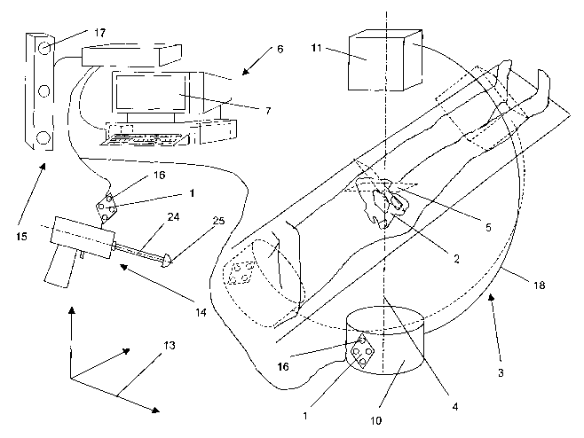
FIG.1 shows one embodiment of the inventive device for intraoperative
navigation in surgery;
FIG. 2 shows a hipbone with the reference system determined according to the
inventive method;
FIG. 3 shows the definition of the angles of inclination and anteversion; and
FIG. 4 shows the display of the axis of a surgical instrument in the instance
of an
acetabulum operation with surgical navigation.
S
CA 02438005 2003-08-07
WO 021062250 PCT/CHO1/00087
FIG. 1 shows a device for surgical navigation in the example of an implant of
an
artificial hip socket with the aid of a mobile medical imaging device 3. Such
an
imaging device 3, for instance an X-ray device, comprises essentially one or
several radiation sources 10 and one or several receivers 11, which are
arranged
along a central axis 4 and which have a projection plane 5. The device
comprises
essentially a position detern>ination device 15 for the spatial measurement of
reference elements 1 relative to a spatially-fixed three-dimensional
coordinate
system 13, a computer 6, which includes display means 7 and which is connected
to said position determination device 15, and it comprises reference elements
1
measurable by the position determination device 15. Such reference elements 1
are attached to the imaging device 3 and to the corresponding surgical
instnunent
14. The reference elements 1 comprise four markers 16 recorded by the cameras
17 of the position determination device 15 so that there can be determined the
position and the spatial orientation of the reference elements 1 relative to
the
coordinate system 13 in-situ. The position of the acetabulum 27, the direction
of
axis 24 of the surgical instrument 14, and the position of its tip 25 can be
determined relative to the coordinate system 13 through measuring of the
position
and the spatial orientation of the reference elements 1, and computed and
shown
on the display means 7 can be from this the numeric values of the relevant
momentarily in-situ set angles of anteversion 36 (FIG. 3) and inclination 35
(FIG.
3) of axis 24 of the surgical instrument 14. During the operation, the surgeon
can
make the correction of the direction of axis 24 of the surgical instrument 14
based
on the size of the angles of anteversion 36 and inclination 35 shown on the
display means 7 or their deviation to a possible plan. An evacuation tool to
work
on the acetabulum is exemplary shown here as a surgical instrument 14.
The reference elements 1 include at least three markers 16 that are not
arranged in
a straight line. The markers 16 as well as the position-finding means 17 of
the
position determination device 17 may be in the form of acoustic or electro-
6
CA 02438005 2003-08-07
WO 02/062250 . PCT/CHO1/00087
magnetic means in their effect whereby the embodiment shown here has an opto-
electric position determination device 15.
FIG. 2 shows a hipbone 2 with the acetabulum 27 and an artificial joint socket
28
with the axis 26 of the acetabulum 27 which extends through the center of the
joint socket and is oriented perpendicular to the face of the joint socket.
According to the inventive method, the position of the three reference points
19,
20, 21 on the hipbone 2 is measured relative to a coordinate system 13.
Suitable
as reference points 19, 20, 21 on the hipbone are, for example:
reference point 19: right spine iliac anterior superior;
reference point 20: center of pubis; and
reference point 21: left spine iliac anterior superior.
The reference system can then be established as coordinate system 23 from the
coordinates of the three reference points 19, 20, 21 whereby its x-axis X'
corresponds to the longitudinal axis 37 of the patient (FIG. 3) and whereby
the
patient's y-axis Y' corresponds to the transverse axis 38 of the patient. The
relevant angles of inclination and anteversion can be determined by means of
said
coordinate system 23.
The position of the three reference points 19, 20, 21 can be percutaneously
determined by means of a pointer (not illustrated) whose tip is measured
spatially.
An ultrasonic device or an image-producing device, e.g. an X-ray device, may
be
employed in place of said pointer.
FIG. 3 serves to explain the two angles of anteversion 36 and inclination 35
within a reference system, which includes the sagittal plane 29, the
transverse
plane 30 and the coronal plane 31, whereby the longitudinal axis 37 of the
patient
lies in the coronal plane 31.
7
CA 02438005 2003-08-07
WO 02/062250 PCT/CH01/00087
Axis 26 of the acetabulum 27 is projected by a first projection line 32 into
the
sagittal plane 29, through a second projection line 33 into the coronal plane
31,
and through a third projection line 34 into the transverse plane 30. The
operative
definition is illustrated here in regard to the definition of anteversion 36
and
inclination 35. According to D.W. Murray "The Definition and Measurement of
Acetabular Orientation" in The Journal of Bone and Joint Surgery, 1993, page
228 and following pages there are three different definitions common for
anteversion and inclination:
a) Operative definition:
The operative inclination 35 is the angle between the second projection line
33
and the sagittal plane 29, while the operative anteversion 36 is the angle
between
the first projection line 32 and the longitudinal axis 37 of the patient.
b) Anatomical definition:
The anatomical inclination is the angle between the axis 26 of the acetabulum
and
the longitudinal axis 37 of the patient, while the anatomical anteversion is
the
angle between the third projection line 34 and the transverse axis 38.
c) Radiographical definition:
The radiagraphical (X-ray) inclination is the angle between the second
projection
line 33 and the longitudinal axis 37 of the patient, while the radiagraphical
anteversion 36 is the angle between the axis 26 of the acetabulum 27 and the
coronal plane 31.
These differently defined angles can also be fittingly converted in a
corresponding
manner according to D.W. Murray "The Definition and Measurement of
Acetabular Orientation" in The Journal of Bone and Joint Surgery, 1993, page
228 and following pages.
8
CA 02438005 2003-08-07
WO 02/062250 . PCT/CHO1/00087
FIG. 4 shows an embodiment of means suitable for intraoperative surgical
navigation in the intraoperative use of the angle display of anteversion 36
{FIG. 3)
and inclination 35 (FIG. 3) on the basis of the refcrence system determined by
means of the inventive method. These means comprise essentially a computer 6
and display means 7 connected thereto. The display means 7 consist here of a
screen, but they can include other embodiments, for instance a head-mounted
display. A graphic illustration of the surgical instrument 14 is shown on the
display means 7 having an axis 24 and a tip 25. Furthermore, the numerical
values of the relevant angles of inclination 35 and anteversion 36 are shown
on
the display means 7. In addition, a scale can be inserted in said display
means 7
to display the depth betwcen the surface of the acetabulum and the tip 25 of
the
surgical instrument 14.' Should an image-producing device be intraoperatively
employed, e.g. a mobile X-ray device 3 (FIG. 1), then a projection of the
acetabulum 22 can be additionally shown on the display means 7.
9
