Note: Descriptions are shown in the official language in which they were submitted.
CA 02439115 2003-08-22
WO 02/067861 PCT/US02/05300
NOVEL ONCOLYTIC ADENOVIRAL VECTORS
This application claims the benefit of US Patent Application No. 60/270,922,
filed February 23,
2001; US Patenf'Application 60/295,037, filed June 1, 2001; and US Patent
Application No.
60/348,670, filed January 14, 2002, which are herein incorporated by
reference.
FIELD OF THE INVENTION
The present invention generally relates to substances and methods useful for
the treatment of
neoplastic disease. More specifically, it relates to oncolytic adenoviral
vectors and their use in
methods of gene therapy.
BACKGROUND OF THE INVENTION
Adenoviruses that replicate selectively in tumor cells are being developed as
anticancer agents
("oncolytic adenoviral vectors"). Such oncolytic vectors amplify the input
virus dose due to viral
replication in the tumor, leading to spread of the virus throughout the tumor
mass. In situ
replication of adenoviruses leads to cell lysis. This in situ replication may
allow relatively low, non-
toxic doses to be highly effective in the selective elimination of tumor
cells.
One approach to achieving selectivity is to introduce loss-of-function
mutations in viral genes that
are essential for growth in non-target cells but not in tumor cells. This
strategy is exemplified by
the use of Add11520, which has a deletion in the E1b-55KD gene. In normal
cells, the adenoviral
E1 b-55KD protein is needed to bind to p53 to prevent apoptosis. In p53-
deficient tumor cells, E1 b-
55K binding to p53 is unnecessary. Thus, deletion of E1b-55KD should
theoretically restrict vector
replication to p53-deficient tumor cells.
Another approach is to use tumor-selective promoters to control the expression
of early viral
genes required for replication (US patent 5,998,205 (Hallenbeck et al.,
1999)). Thus, in this
approach the adenoviral vectors will specifically replicate and lyse tumor
cells if the gene that is
essential for replication is exclusively under the control of a promoter or
other transcriptional
regulatory element which is tumor-specific.
It is an object of the present invention to provide novel oncolytic adenoviral
vectors for the
treatment of neoplastic disease, which exhibit a high degree of tumor
selectivity, therapeutic
efficacy, and safety when administered to a host organism.
CA 02439115 2003-08-22
WO 02/067861 PCT/US02/05300
BRIEF DESCRIPTION OF THE DRAWINGS
Figure 1: Cleavage and polyadenylation process for the SV40 early poly(A) site
(SEQ ID N0:1 ).
Figure 2: E1A transcription control region (SEQ ID N0:2).
Figure 3: Sequence of Ar6pAE2fF from left and right ends of viral DNA. Regions
of Ar6pAE2fF
confirmed by DNA sequencing. Panel A. Regions in first 1802 nucleotides are
the inverted
terminal repeat (ITR) (nucleotides 1-103), poly-adenylation signal
(nucleotides 116-261), a
human E2F-1 promoter (nucleotides 283-555), E1A gene (nucleotides 574-1647)
and a portion
of the E1 b gene (nucleotides 1648-1802) are indicated (SEQ ID N0:3). Panel B.
Regions in the
last 531 nucleotides are the Pacl restriction site (nucleotides 33967-33974)
(underlined), the
packaging signal (nucleotides 34020-34217 and the ITR (34310-34412).
Figure 4: Sequence of Ar6F from left end of viral DNA (SEQ ID N0:4). The first
660 nucleotides
at the left end of Ar6F. The ITR (nucleotides 1-103), a multiple cloning site
(MCS) (nucleotides
104-134) and a portion of the E1A gene (nucleotides 135-660) are shown.
Figure 5: Sequence of Ar6pAF from left end of viral DNA. The first 660
nucleotides at the left
end of Ar6pAF. The ITR (nucleotides 1-103), the SV40 early polyA signal
(nucleotides 104-134)
and a portion of the E1A gene (nucleotides298-660) are shown.
Figure 6: Schematic diagram of Ar6pAF and Ar6pAE2fF vectors. The backbone
adenoviral
sequences are derived from the pAr6pAF and pAr6pAE2fF infectious plasmids. The
intermediate vector backbone adenoviral sequences are derived from Add1327, an
E3-deleted
adenovirus type 5, in which the packaging signal is located immediately
upstream of the right
ITR. The Ar6pAF vector backbone is deleted in the E1A promoter and the SV-40
poly(A) signal
is inserted after the left ITR. The Ar6pAE2fF vector backbone contains, after
the SV-40 poly(A)
signal sequences, a E2F-1 promoter (bp-212 to +51 ), a DNA segment of four
intact E2F, one
NF-kB and four Sp-1 consensus sequences.
Figure 7: Comparison of body weight change after administration of vectors
Add1327,
AvPAE1A09i, Ar6F, Ar6pAF, Add1312.
Figure 8: Backbones of vectors Add1327, AvE1A09i, AvPAE1A09i, Ar6F, Ar6pAF,
Add1312
CA 02439115 2003-08-22
WO 02/067861 PCT/US02/05300
3
Figure 9: Mean H460 tumor volume after intratumoral injections of Ar6pAE2fF.
Comparison of in
vivo growth of H460 tumors after five consecutive daily (study day 1-5)
intratumoral injections
of Ar6pAE2fF at 5x10$(n=13), 5x10⁹(n=13), or 5x10'° (n=12)
particles/dose/day. Control
animals were treated IT with HBSS (n=13) or Add1327 (n=12, 5x10'°
particles/dose/day) also for
consecutive days. Data is expressed as mean tumor volume + SE. *p < 0.05
compared to
HBSS treated controls using one-way RM ANOVA with Tukey's test for multiple
comparison.
Figure 10: Survival of tumor-bearing animals after intratumoral injections of
Ar6pAE2fF. Survival
of tumor bearing animals after treatment with Ar6pAE2fF. Animals were observed
until study
day 32. Numbers of animals in each treatment group were as follows: HBSS, n =
13;
Ar6pAE2fF at 5x10a, n = 13; 5x10⁹, n = 13; and 5x10'°
particles/dose/day, n=12; and Add1327
at 5x10'° particles/dose/day, n. =12. The survival of animals was
analyzed by the Mantel-
Haenszel logrank test.
Figure 11: Mean Hep3B tumor volume after intratumoral injections of Ar6pAE2fF.
Comparison
of in vivo growth of Hep3B tumors after five consecutive daily (study day 1-5)
intratumoral
injections of Ar6pAE2fF at 5x10⁸(n=11 ), 5x10⁹(n=11 ), or 5x10'° (n=10)
particles/dose/day.
Control animals were treated IT with HBSS (n=10) or Add1327 (n=11,
5x10° particles/dose/day)
also for 5 consecutive days. Data is expressed as mean tumor volume + SE. *p <
0.05
compared to HBSS treated controls using one-way RM ANOVA with Tukey's test for
multiple
comparison. **p<0.05 compared to Ar6pAE2fF at 5x1 O$ particles/dose/day by t-
test.
Figure 12: Survival of tumor-bearing animals after intratumoral injections of
Ar6pAE2fF. Survival
of tumor bearing animals after treatment with Ar6pAE2fF. Animals were observed
until study
day 32. Numbers of animals in each treatment group were as follows: HESS, n =
11;
Ar6pAE2fF at 5x10a, n = 11; 5x10⁹, n = 11; and 5x10'°
particles/dose/day, n=10; and Add1327
at 5x10'° particles/dose/day, n =11. The survival of animals was
analyzed by the Mantel-
Haenszel logrank test.
Figure 13: Schematic diagram of adenovirus right donor plasmid pDR2F.
Figure 14: Schematic diagram of adenovirus right donor plasmid pDR2mGmF. The
adenovirus
right donor plasmid pDR2mGmF is an 11526bp circular molecule. The mGm-cDNA is
at
position 8059 to 8520.
Figure 15: Schematic diagram of plasmid pG1mGmSvNa.
CA 02439115 2003-08-22
WO 02/067861 PCT/US02/05300
Figure 16: Schematic diagram of plasmid pG1 NaSvBg.
Figure 17: Sequence of the murine GM-CSF cDNA (SEQ ID N0:7) and protein (SEQ
ID N0:8).
Sequence of the 7878 to 8826 region of the pDR2mGmF plasmid was confirmed by
DNA
sequencing. This region includes the murine GM-CSF cDNA insert.
Figure 18: Pathway used to generate pAr6pAE2fmGmF plasmid. The 37763bp
Ar6pAE2fmGmF large plasmid was generated through the homologous recombination
of the
pDR2mGmF/Fspl + Spel fragment (9284bps) and the pAr6pAE2fF/Pacl + Srfl
fragment
(30695bps) in E coli BJ5183 cells. In this plasmid, the mGM-CSF cDNA was
cloned into the
Xbal site of the adenoviral E3 region.
Figure 19: MTS assay of oncolytic vectors on different tumor cell lines.
Figure 20: Sequence of Ar6pAE2fhGmF region from 28536 to 29273 of the viral
genome
including the human GM-CSF cDNA insert (SEQ ID N0:19) and the human GM-CSF
protein
sequence (SEQ ID N0:20).
Figure 21: Pathway used to generate pAr6pAE2fhGmF. The 37587bp pAr6pAE2fhGmF
large
plasmid was generated through the homologous recombination of the
pDR2hGmF/(Fsp I + Spe
I) fragment (9284bps) and the pAr6pAE2fF/(Pac i + Srf I) fragment (30695bps)
in E coli BJ5183
cells. In this plasmid, the hGM-CSF cDNA was cloned into the Xba I site of the
adenoviral E3
region.
Figure 22: MTS assay of oncolytic vectors on different tumor cell lines.
Figure 23: Efficacy of GM-CSF armed oncolytic vectors in H460 non-small cell
lung carcinoma
tumor model. Volumes of H460 human xenograft tumors were measured periodically
following
treatment of pre-established tumors with oncolytic adenoviral vectors.
Comparison of in vivo
growth of H460 tumors after five intratumoral injections of PBS, or
2x10'° particles/injection
(panel A) or 1x10" particles/injection (panel B) of Add1312, Add1327,
Ar6pAE2fF or
Ar6pAE2fmGmF. Each treatment group is identified by symbols as listed in the
graph insets.
Vector injections were on study days 10, 12, 14, 17, and 19, as indicated by
the arrows along
the x-axis. Data are represented by average tumor volume +SEM. Asterisks
indicate significant
differences compared to Add1312 negative control vector-treated tumors
(p<0.05, RM-OW-
CA 02439115 2003-08-22
WO 02/067861 PCT/US02/05300
ANOVA using Tukey's test). At the low dose (panel A), differences were
significant for Add1327,
Ar6pAE2fF and Ar6pAE2fmGmF compared to Add1312. At the high dose (panel B),
differences
compared to Add1312 were significant only for the mouse GM-CSF containing
Ar6pAE2fmGmF
vector.
Figure 24: Efficacy of GM-CSF armed oncolytic vectors in Hep3B hepatocellular
carcinoma
tumor model. The in vivo growth of Hep3B tumors following intratumoral
injections of Add1312,
Ar6pAE2fF, Ar6pAE2fhGmF, or Ar6pAE2fmGmF at 2x10' (panel A), 2x10$ (panel B),
or 2x1 O9
(panel C) particles/injection (n=10/group) was analyzed. Vectors were injected
on study days
15, 18, 20, 22, and 25 as indicated by the arrows along the x-axes. Control
animals were
treated with PBS (n=10). Data represent group averages +SEM. Asterisks
indicate p<0.05
compared to dose-matched Add1312-treated tumors. Crosses indicate p<0.05
compared to the
Ar6pAE2fF vector and pound symbols indicate p<0.05 for Add1312-treated tumors
compared to
PBS-treated tumors. Tumors were measured for 47 days following Hep3B tumor
cell
inoculation.
Figure 25: Schematic diagram of PCR and overlap PCR for Ogp19 donor plasmids
The mGM-
CSF or hGM-CSF cDNA was inserted into the E3 region replacing the E3-gp19 open
reading
frame (ORF) using two steps of PCR amplification. In the first step, 3
individual PCR
amplifications were carried out using 3 pairs of primers and corresponding DNA
templates. In
the second step, the 3 DNA fragments generated in first step were mixed as the
template DNA
for the overlap PCR amplification using primer 1 and primer 6 as primers. The
overlap PCR
product was then digested with BsiWl/Notl and used to replace the BsiWl/Notl
region of
adenoviral E3 containing the E3-gpl9 open reading frame.
Figure 26: Schematic Diagram of ~gp19 Vectors
Figure 27a: Pathway Used to Generate the pAr6pAE2f(E3+,mGm,Dg19b)F Large
Plasmid The
38977bp pAr6pAE2(E3+,mGm,Dg19b)F large plasmid was generated through the
homologous
recombination of the pDR2(E3+,mGm,Dg19b)F/(Fsp I + Spe I) fragment (9284bps)
and the
pAr6pAE2fF/(Pac I + Srf I) fragment (30695bps) in E coli BJ5183 cells. In this
plasmid, the
mGM-CSF cDNA was swapped into the E3 gp19kD ORF of the adenoviral E3 region.
Figure 27b: Pathway Used to Generate the pAr6pAE2f(E3+,hGm,Dg19b)F Large
Plasmid The
38950bp pAr6pAE2(E3+,hGm,Dg19b)F large plasmid was generated through the
homologous
recombination of the pDR2(E3+,hGm,Dg19b)F/(Fsp I + Spe I) fragment (9284bps)
and the
CA 02439115 2003-08-22
WO 02/067861 PCT/US02/05300
pAr6pAE2fF/(Pac I + Srf I) fragment (30695bps) in E coli BJ5183 cells. In this
plasmid, the
hGM-CSF cDNA was swapped into the E3 gp19kD ORF of the adenoviral E3 region.
Figure 28: MTS Assay of Ogp19 mGM-CSF Vectors on H460 and Hep3B Tumor Cell
Lines. Two
human tumor cell lines, H460 (non-small cell lung carcinoma) and Hep3B
(hepatocellular
carcinoma), were used. For each cell line, two MTS assays have been performed
for all
vectors. One representative MTS assay from each cell line is presented.
Figure 29: GM-CSF expression mediated by agp19 GM-CSF vectors in infected H460
cells
detected by ELISA. The ~gp19kD vectors were assayed for their ability to
mediate GM-CSF
transgene expression in the culture media of H460 cells infected with viral
vectors.
Figure 30: Anti-tumor activity of oncolytic adenoviruses in the Hep3B
xenograft subcutaneous
tumor model. Comparison of the in vivo growth of Hep3B tumors following five
intratumoral
injections of PBS, or 2x10⁹ particles/injection of Add1312, Ar6pAE2fF,
Ar6pAE2fmGmF, or
Ar6pAE2f(E3+,mGm,Dg19b)F (n=10 per group). Symbols representing the different
treatment
groups are shown in the graph inset. Vector injections were on days 11, 13,
15, 17, and 19 as
indicated by the arrows along the x-axis. Tumor volumes when vector injections
began were
175 mm3 for all groups, except for tumors treated with the
Ar6pAE2f(E3+,mGM,Dg19b)F vector,
where the initial tumor volumes were 290 mm3. Asterisks indicate a p value
<0.05 compared to
Add1312 vector-treated tumors (by repeat-measures, one-way ANOVA).
Figure 31: Anti-tumor activity of oncolytic adenoviruses in the H460 xenograft
subcutaneous
tumor model. Comparison of the in vivo growth of H460 tumors following five
intratumoral
injections of controls or oncolytic vectors. Panel A, H460 tumors injected
with 2x10'°
particles/injection of Add1312, Ar6pAE2fF, or Ar6pAE2fmGmF, or 1x10'°
particles/injection of
Ar6pAE2f(E3+,mGm,Dg19b)F (n=10 per group). Panel B, H460 tumors injected with
1x10"
particles/injection of Add1312, Ar6pAE2fF, or Ar6pAE2fmGmF, or 5x10'°
particles/injection of
Ar6pAE2f(E3+,mGm,Dg19b)F (n=10 per group). Symbols representing the different
treatment
groups are shown in the graph inset. Vector injections were on days 12, 14,
16, 19, and 21 as
indicated by the arrows along the x-axis. Tumor volumes when vector injections
began were
160 mm3 for all groups, except for tumors treated with the
Ar6pAE2f(E3+mGM,Dg19b)F vector,
where the initial tumor volumes were 120 mm3. Asterisks indicate a p value
<0.05 compared to
Add1312 vector-treated tumors (by repeat-measures, one-way ANOVA). .
CA 02439115 2003-08-22
WO 02/067861 PCT/US02/05300
Figure 32: Schematic diagram of adenovirus pDrShGmF and pDrSmGmF right donor
plasmids.
The adenovirus right donor plasmid pDrShGmF and pDrSmGmF are circular
molecules 12674bp
and 12701 by in size, respectively. The IoxP sites were removed from their
parental plasmids
pDR2(E3+,mGm,Dg19b)F and pDR2(E3+,hGm,Dg19b)F by replacing the Notl/Sphl
fragment
with the Notl/Sphl fragment from wild type AdS.
Figure 33: Pathway used to generate the pAr15pAE2fhGmF plasmid. The 38910 by
pAr15pAE2fhGmF large plasmid was generated through homologous recombination of
the
pDrShGmF/(Fspl + Spel) fragment (10432 bps) and the pAr6pAE2fF/(Pacl + Srfl)
fragment
(30695bp) in E coli BJ5183 cells. In this plasmid, the hGM-CSF cDNA was
swapped into the
E3-gp19 ORF of the adenoviral E3 region and the IoxP site was removed from E3
region.
Figure 34: Pathway used to generate the pAr15pAE2fmGmF plasmid. The 38938bp
pAr15pAE2fmGmF large plasmid was generated through the homologous
recombination of the
pDrSmGmF/(Fspl + Spel) fragment (10459bp) and the pAr6pAE2fF/(Pacl + Srfl)
fragment
(30695bp) in E. coli BJ5183 cells. In this plasmid, the mGM-CSF cDNA was
swapped into the
E3 gp19 ORF of the adenoviral E3 region and the IoxP site was removed from E3
region.
Figure 35: MTS assay of Ar15pAE2fhGmF and Ar15pAE2fmGmF vectors on H460 and
Hep3B
tumor cell lines. MTS assays were performed to evaluate the oncolytic
potential of the
Ar15pAE2fhGmF and Ar15pAE2fmGmF vectors. Two human tumor cell lines, H460 (non-
small
cell lung carcinoma) and Hep3B (hepatocellular carcinoma), were used. For each
cell line, two
MTS assays were performed using all the indicated vectors and one
representative MTS assay
from each cell line is presented. Ar15pAE2fF was used for comparison of LD50s
of "armed"
vectors vs "unarmed" vectors. Ar6pAE2fmGmF and Ar6pAE2f(E3+, mGm, Dg19b) were
also
included. In addition, control viruses Add1327 (replication competent Ad5
positive control virus)
and Add1312 (E1 a deficient negative control) were also included in the MTS
assays. The
Ar15pAE2fhGmF and Ar15pAE2fmGmF vectors retained the oncolytic capacity of the
Ar6pAE2fF vector in both cell lines tested.
Figure 36: GM-CSF expression mediated by Ar15pAE2fhGmF and Ar15pAE2fmGmF
vectors in
infected H460 cells detected by ELISA. The Ar15pAE2fGmF vectors were assayed
for their
ability to mediate human or mouse GM-CSF transgene expression in the culture
media of H460
cells and Hep3B cells infected with viral vectors. The Ar15pAE2fGmF vectors
induce the in vitro
production of GM-CSF at levels similar to the E3 deleted Ar6pAE2fGmF series.
CA 02439115 2003-08-22
WO 02/067861 PCT/US02/05300
Figure 37: Schematic diagram of PCR and overlap PCR for DE3-14.7 plasmids. The
human
GM-CSF cDNA was inserted into the E3 region replacing the E3-14.7 ORF using
two steps of
PCR amplification. In the first step, 3 individual PCR amplifications were
carried out using 3
pairs of primers and corresponding DNA templates as summarized in Tables 2 and
3. In the
second step, the 3 DNA fragments generated in first step were mixed as the
template DNA for
the overlap PCR amplification using primers 1 and 6. The overlap PCR product
was then
digested with Xhol/Sphl and used to replace the Xhol/Sphl region of plasmid
pDR4F containing
the E3-14.7 open reading frame.
Figure 38: Schematic Diagram of ~E3-14.7 Vectors.
Figure 39: Schematic Diagram of Adenovirus Right Donor Plasmid pDr6hGmF. The
adenovirus
right donor plasmid pDR6hGmF is a circular molecule of 12774 bp. The human GM-
CSF
(hGm) cDNA was swapped into the position of the E3-14.7 ORF in the pDR4F
plasmid using
PCR amplification and overlap PCR amplification followed by restriction enzyme
digestion
(Xhol/Sphl) and ligation.
Figure 40: Pathway Used to Generate the pAr16pAE2fhGmF Large Plasmid. The
39011 by
pAr16pAE2fhGmF large plasmid was generated through the homologous
recombination of the
pDR6hGmF/(Fsp I + Spe I) fragment (10532bps) and the pAr6pAE2fF/(Pac I + Srt
I) fragment
(30695bps) in E coli BJ5183 cells. In this plasmid, the hGM-CSF cDNA was
swapped into the
E3-14.7 ORF of the adenoviral E3 region.
Figure 41: MTS Assay of ~E3-14.7 hGM-CSF Vector on H460 Tumor Cell Line. To
evaluate the
oncolytic potential of the Ar16pAE2fhGmF vector, MTS assays were performed.
Human tumor
cell line H460 (non-small cell lung carcinoma) was used. Two MTS assays have
been done for
all vectors and the assays gave similar LD50 values for each vector. The
oncolytic capacity of
Arl6pAE2fhGmF is compared to Ar6pAE2fF, Ar6pAE2fhGmF, and
Ar6pAE2f(E3+,hGm,Dg19)F. In addition, control viruses Add1327 (replication
competent Ad5
positive control virus) and Add1312 (E1A deficient negative control) were also
included in the
MTS assays. The Ar16pAE2fhGmF vectors retained the oncolytic capacity of the
Ar6pAE2fF
vector.
Figure 42: GM-CSF Expression Mediated by DE3-14.7 hGM-CSF Vector
(Ar16pAE2fhGmF)
Compared to Ar6pAE2fF, Ar6pAE2fhGmF and Ar6pAE2f(E+,hGm,Dg19)F in Infected
H460
Cells 24 Hours Post-Infection. The Ar16pAE2fhGmF vector was assayed for its
ability to
CA 02439115 2003-08-22
WO 02/067861 PCT/US02/05300
mediate GM-CSF transgene expression in the culture media of H460 cells
infected with viral
vectors. H460 cells were plated in 6-well plates using 2 ml/well of culture
media at a density of
2.5x10⁵ cells/well. The next day, the media were removed and the cultured
cells were
transduced in duplicate with viral vectors at 10, 100, and 1000 particles/cell
in 5001 serum-free
medium. After two hours of incubation at 37°C in a 5% C02 incubator,
virus was aspirated and
2 ml of fresh complete culture medium was added to each well. At 24 hours post
infection, the
supernatants were collected for hGM-CSF ELISA.
Figure 43: Spread of adenoviruses in H460 xenograft tumors detected by FACS.
At day 1, 4,
and 7 after the injection of viruses or PBS, tumors were analyzed for hexon
staining using
intracellular flow cytometry. The percentage of hexon positive cells from each
mouse was
displayed in the graph, with the bar as the mean (n=10). The PBS negative
control group had
fewer mice.
*: p<0.05 between Ar6pAE2fF or Ar6pAE2fE3F and Add1312, ANOVA
o: p<0.05 between Ar6pAE2fF and Ar6pAE2fE3F vectors, ANOVA
Figure 44: Spread of adenoviruses in Hep3B xenograft tumors detected by FACS.
On days 1, 4
and 7 after the injection of viruses or PBS, tumors were analyzed for hexon
staining using flow
cytometry. The percentage of hexon positive cells from each mouse was
displayed in the
graph, with the bar as the mean (n=10). The PBS negative control group had
fewer mice.
*: p<0.05 between Ar6pAE2fhGmF or Ar6pAE2f(E3+,hGm,Dg19)F and Add1312, ANOVA
o: p<0.05 between Ar6pAE2fhGmF and Ar6pAE2f(E3+,hGm,Dg19)F vectors, ANOVA
Figure 45: Flowchart for construction of pArpAE2fFTrtex. The plasmids that
were used for the
construction of the large plasmid for the oncolytic vector Ar17pAE2fFTrtex are
depicted. The
specific alterations are noted and described in the text in more detail.
Figure 46: The final right end shuttle plasmid (pDr17TrtexF) and the large
plasmid
(pAr17pAE2fFTrtex) used to make the oncolytic vector Arl7pAE2fFTrtex are shown
here.
Figure 47: Sequence of the right end of Ar17pAE2fFTrtex (SEQ ID NO:17): The
right end of the
vector was sequenced and is shown here. The viral ITR and packaging signal are
at 36305 to
36203. The Trtex promoter is located at 35843 to 35606. Additional regions
were also
sequenced including the E3 region and the left end of the virus.
CA 02439115 2003-08-22
WO 02/067861 PCT/US02/05300
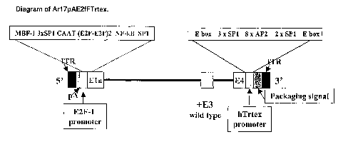
Figure 48: Diagram of Ar17pAE2fFTrtex: The diagram of the final vector is
depicted
schematically in this figure. The known transcription factor binding sites are
indicated above
each added promoter. Briefly, a E2F-1 promoter is driving the E1a
transcription unit and a
telomerase reverse transcriptase (Tent) promoter is driving the E4
transcription unit. The E3
region is completely wild type.
Figure 49: Adenoviral E4 expression measured by semi-quantitative RT-PCR. The
E4 region is
encoded on the opposite strand in the viral genome. Total RNA was isolated
from Hep3B cells
24 hours after infection with 10 ppc of Ar17pAE2fFTrtex. Depicted is a
schematic diagram of
the right end of the Ar17pAE2fFTrtex viral genome with relative positions of
primers used in RT-
PCR reactions along with the approximate size of the products. PCR 2.f paired
with PCR 3.r or
PCR 4.r were designed to detect all E4 transcripts. PCR 2.f paired with PCR
5.r was used to
detect transcripts that initiated from any cryptic start sites upstream of the
E4 region. +1,
indicates the approximate position of transcriptional initiation site of the
native hTERT promoter.
Figure 50: Sequence of a hTERT promoter and a portion of the E4 region of
Ar17pAE2fFTrtex
is shown (SEQ ID N0:21 ). In the Ad genome, the E4 genes are oriented in the
reverse
direction. A hTERT promoter sequence is indicated by the double underline. The
boxed
sequence labeled "ExtP1" indicates the antisense oligonucleotide primer used
in the primer
extension assay to map the transcriptional initiation sites for the E4 region.
Nucleotides
indicated by the gray boxes are the three transcription initiation sites we
identified. The start
sites previously identified by Horikawa I. Cable PL, Afshari C, Barrett JC.
Cloning and
characterization of the promoter region of human telomerase reverse
transcriptase gene
Cancer Res. 1999 Feb 15:59(4):826-30 and Takakura M, Kyo S, Kanaya T, Hirano H
Takeda Js
Yutsudo M, Inoue M. Cloning of human telomerase catalytic subunit (hTERT) Gene
promoter
and identification of proximal core promoter seguences essential for
transcriptional activation in
immortalized and cancer cells. Cancer Res. 1999 Feb 1'59(3):551-7 in the
endogenous hTERT
gene are indicated by bold solid underlines.
Figure 51: Tumors were established by injecting 1 x 10' Hep3B cells
subcutaneously into the
right flank of 6-8 week old female nude mice (Harlan). Two weeks after
implantation, mice with
tumors ranging from 91.6 - 218.5 mm3 were selected and randomly distributed
into groups
(n=17-18). Each mouse was weighed prior to intravenous injection. The control
groups
received HBSS or Add1312 at 4.5 x 10'2 vp/kg (n=18). Ar17pAE2fFTrtex treatment
groups
received 1.5x10' (n=18), 3.0x10'2 (n=17), or 4.5x10'2 (n=18) vp/kg. All dose
volumes were 10
ml/kg. Groups means + SEM are represented. *, p < 0.05 vs. HBSS controls
(Dunnett test).
CA 02439115 2003-08-22
WO 02/067861 PCT/US02/05300
11
Figure 52: Survival of tumor bearing animals after treatment with HBSS,
Add1312 (4.5 x 10'2
vp/kg), or Ar17pAE2fFTrtex (1.5 x 10'2 vp/kg, 3.0 x 10'2 vp/kg, or 4.5 x 10'2
vplkg). Animals
were observed until study day 42, n= 17-18 per group. The survival curves were
plotted using
GraphPad Prism, and analyzed by the Mantel-Haenszel logrank test (p<0.004 for
all treatment
doses compared to HBSS).
Figure 53: Group mean body weights are shown following a single intravenous
injection of the
indicated test article. The number of animals evaluated at each scheduled data
collection time
point was 18-33, except for SD29 when n = 9-22. Vector doses were adjusted on
the basis of
individual animal body weight on the day of dosing. Lo Dose: 1.5 x 10'2 vp/kg;
Mid Dose: 3.0 x
10'2 vp/kg; Hi Dose: 4.5 x 10'2 vp/kg. Group means + SD are represented, with
no statistically
significant differences between groups.
Figure 54: Comparison of in vivo growth of Hep3B tumors after a single iv
injection of
Ar17pAE2fFTrtex at 3x10'2 (n=16) or 4.5x10'2 (n=16) particles/kg. Control
groups were injected
with HBSS (n=16) or Add1312 (n=16) at 4.5x10'2 particles/kg. Data is expressed
as mean tumor
volume + SE. (*p < 0.05) For both Ar17pAE2fFTrtex treated groups compared to
HBSS treated
controls using one-way ANOVA with Student-Newman-Keuls test for multiple
comparison.
Figure 55: Survival of tumor bearing animals after treatment with
Ar17pAE2fFTrtex. Animals
were observed until study day 58. Numbers of animals in each treatment group
were as
follows: HBSS, n = 16; Ar17pAE2fFTrtex at 3x10'2, n = 16; and 4.5x10'2, n =
16; and Add1312
at 4.5x10'2 particles/kg, n =16. The survival of animals was analyzed by the
Mantel-Haenszel
logrank test.
Figure 56: Analysis of mean % body weight change from Hep3B tumor bearing
animals treated
with the oncolytic adenoviral vector Ar17pAE2fFTrtex, Add1312 or HBSS. Body
weights were
measured once per week. Data is expressed as mean tumor volume + SD.
Figure 57: Tumors were established by injecting 1 x 10' Hep3B cells
subcutaneously into the
right flank of 6-8 week old female nude mice (Harlan). Two weeks after
implantation, mice with
tumors ranging from 90 - 215 mm3 were selected and randomly distributed into
groups
(n=12/group). Each mouse was weighed prior to intravenous injection. The
control mice
received HBSS. Ar17pAE2fFTrtex treatment groups received 3x10" (n=12), 6x10"
(n=12),
CA 02439115 2003-08-22
WO 02/067861 PCT/US02/05300
12
1x10'2 (n=12), or 3x10'2 (n=12) vp/kg. All dose volumes were 10 ml/kg. Groups
means (+SEM)
are represented. *, p < 0.05 vs. HBSS controls (Dunnett's method).
Figure 58: Individual tumor volumes for study days 3 through 22 are presented.
All dose
volumes were 10 ml/kg. A) The control group treated with HBSS. Treatment
groups received
Ar17pAE2fFTrtex at B) 3x10" vp/kg, C) 6x10" vp/kg, D) 1x10'2 , or E) 3x10'2
vp/kg. (n=12 /
group).
Figure 59: Survival of tumor bearing animals after treatment with HBSS or
Arl7pAE2fFTrtex (3
x 10~~ vp/kg, 6 x 10'~ vp/kg, 1 x 10'2 vp/kg, or 3 x 10'2 vp/kg). Animals were
observed until
study day 39, n= 12 per group. The survival curves were plotted using GraphPad
Prism, and
analyzed by the Mantel-Haenszel logrank test.
Figure 60: Percent body weight change from study day 1 are shown following a
single
intravenous injection of the indicated dose (n=12). Vector doses were adjusted
on the basis of
individual animal body weight on the day of dosing. A single intravenous
injection of
Ar17pAE2fFhTrtex at 3x10" (n=12), 6x10" (n=12), 1x10'2 (n=12), or 3x10'2
(n=12) viral
particles/kg in a final volume of 10 ml/kg was administered on study day 1. *,
p < 0.05 vs study
day 1 percent body weight change, I<ruskal-Wallis One-Way ANOVA on Ranks.
Figure 61: Vector DNA copies per cell in tumors and livers collected from mice
prior to treatment
(n=3) and at indicated times and after intravenous injection of
Ar17pAE2fFTrtex at 3.0 x 10'2
vp/kg (n=5). Molecular analysis was done by PCR using primers specific for
adenoviral hexon
DNA. Results are expressed as hexon copy number per cell.
Figure 62: The mean body weight change as a percent of the SD1 body weight +st
dev was
followed for a cohort of five mice in each treatment group. Animals were
injected with a single
intravenous dose of the indicated vectors on SD1. *, p < 0.05 vs. HBSS (one-
way ANOVA).
Figure 63: Improved isobologram with additivity envelope for Ar17pAE2fFTrtex
and Taxol
against Hep3B and PC3M.2AC6 cells. In the table, EC5o of virus or chemotherapy
single
treatment was termed as 1. ECSO of virus or chemotherapy agents in the
combination were
divided by the EC5o of single treatments.
Figure 64: Improved isobologram with additivity envelope for Ar17pAE2fFTrtex
and Doxorubicin
against Hep3B and PC3M.2AC6 cells. In the table, ECSO of virus or chemotherapy
single
CA 02439115 2003-08-22
WO 02/067861 PCT/US02/05300
13
treatment was termed as 1. ECSO of virus or chemotherapy agents in the
combination were
divided by the EC5o of single treatments.
Figure 65: Improved isobologram with additivity envelope for Ar17pAE2fFTrtex
and Epothilone
B against Hep 3B cells. In the table, ECSO of virus or chemotherapy single
treatment was termed
as 1. EC5o of virus or chemotherapy agents in the combination were divided by
the EC5o of
single treatments.
Figure 66: Tumor growth curve in the doxorubicin combination study. Comparison
of in vivo
growth of Hep3B tumors after a single i.v. injection of Ar17pAE2fFTrtex at
3x10'2 particle/kg
(n=10) alone or in combination with doxorubicin given i.p. at 10 mg/kg. A
group was given
doxorubicin alone (n=10) i.p. at 10 mg/kg. A control group was injected with
HBSS (n=10).
Other details are described in the text. * means p < 0.001 by t-test compared
to all other groups
at study day 20. One mouse was found dead at study day 27 in combination
group, so the n=9
to end of study. Data is expressed as mean tumor volume + SEM.
Figure 67: Tumor growth curve in the Doxil~ combination study. Comparison of
in vivo growth of
Hep3B tumors after a single i.v. injection of Ar17pAE2fFTrtex at 1x10'2 (n=10)
vp/kg alone or at
1x10'2 or 6x10" vp/kg in combination with Doxil~ given i.v. at 9 mg/kg. A
group was given Doxil~
alone (n=10) i.v, at 9 mg/kg. Other details are described in the text. A
negative control group
was injected with HBSS (n=9). *means p < 0.01 by t-test compared to all other
groups at study
day 21. Data is expressed as mean tumor volume ~ SEM.
Figure 68: Toxicity of Ar17pAE2fFTrtex in primary human hepatocytes (PHH). PHH
were
transduced with indicated vectors at 1, 10 and 50 ppc. Panel A: Cytotoxicity
as measured by
LDH release was measured five days after transduction. Means ~ sd from
triplicate wells is
shown. * p<0.05 Ar17pAE2fFTrtex versus Ar13pAE2fF by t-test. Panel B:
Cytotoxicity measured
seven days after transduction. Means ~ range from 1-3 wells is shown.
Statistical comparisons
not possible for data in panel B due to low replicate number.
Figure 69: Ad35-based oncolytic vectors. Ar35OscE1A and Ar35E2FE1A both
contain the E1a
region under the control of a tumor-specific promoter, a osteocalcin or a E2F
promoter,
respectively. Ar35E2F+E1A contains in addition, the E4 region under the
control of a tumor-
specific promoter.
CA 02439115 2003-08-22
WO 02/067861 PCT/US02/05300
14
Figure 70: Effect of Ar35OscE1A on a subcutaneous PC3 tumor in nude mice.
Groups of 10
animals each were treated with vehicle (HBSS), Ad35v0.5 (an E1 a deficient
Ad35 based
vector), Ar6pAOscE3F (the Ad5-based oncolytic vector containing the
osteocalcin promoter
driving expression of E1a), Ad35 (wt virus), and Ar35OscE1a (the Ad35-based
oncolytic vector
containing the osteocalcin promoter driving expression of E1a). All vectors
were delivered
intratumorally (IT), using a single dose of 2 x 10" particles/mouse (1 x 10'3
particles/kg).
SUMMARY OF THE INVENTION
The present invention provides novel and improved oncolytic adenoviral vectors
and their uses
in methods of gene therapy. In a preferred embodiment, the oncolytic
adenoviral vector has an
E2F promoter operably linked to the E1a gene. In a particularly preferred
embodiment, the
oncolytic adenoviral vectors has an E2F promoter operably linked to the E1a
gene and the
human telomerase reverse transcriptase promoter operably linked to the E4
gene.
Accordingly, in one aspect, the present invention provides a recombinant viral
vector comprising
an adenoviral nucleic acid backbone, wherein said nucleic acid backbone
comprises in
sequential order: A left ITR, a termination signal sequence, an E2F responsive
promoter which
is operably linked to a first gene essential for replication of the
recombinant viral vector, an
adenoviral packaging signal and a right ITR.
In a second aspect, the invention provides a recombinant viral vector
comprising an adenoviral
nucleic acid backbone, wherein said nucleic acid backbone comprises in
sequential order: A left
ITR, a termination signal sequence, an E2F responsive promoter which is
operably linked to a
first gene essential replication of the recombinant viral vector, a telomerase
promoter operably
linked to a second gene essential for replication, an adenoviral packaging
signal and a right
ITR.
In another aspect, the invention provides adenoviral particles comprising
these vectors.
Preferably, the particles further comprise a targeting ligand included in a
capsid protein of the
particles.
In another aspect, the adenoviral particles carry at least one therapeutic
transgene. Preferably,
the particles further comprise a polynucleotide encoding a cytokine such as GM-
CSF that can
stimulate a systemic immune response against tumor cells.
CA 02439115 2003-08-22
WO 02/067861 PCT/US02/05300
In another aspect, there is provided a method of selectively killing a
neoplastic cell in a cell
population which comprises contacting a suitable amount of the recombinant
viral vector of the
invention with said cell population under conditions where the recombinant
viral vector can
transduce the cells of said cell population.
In a further aspect a pharmaceutical composition comprising the recombinant
viral vector of the
invention and a pharmaceutically acceptable carrier is provided.
In yet another aspect a method of treating a host organism having a neoplastic
condition is
provided, comprising administering a therapeutically effective amount of the
composition of the
invention to said host organism.
DETAILED DESCRIPTION OF THE INVENTION
The present invention provides novel viral vectors based on the oncolytic
adenoviral vector
strategy as described in US patent 5,998,205, issued December 7, 1999 to
Hallenbeck et al.,
the disclosure of which is hereby incorporated by reference in its entirety.
In particular, oncolytic
adenoviral vectors are disclosed in which expression of an adenoviral gene,
which is essential
for replication, is controlled by E2F-responsive promoters which are
selectively transactivated in
cancer cells. Examples of E2F-responsive promoters are disclosed in PCT
publication WO
98/13508, published April 2, 1998.
Without being bound by theory, the inventors believe that the mechanism of
action is as follows.
The selectivity of E2F-responsive promoters (hereinafter sometimes referred to
as E2F
promoters) is based on the derepression of the E2F promoter/transactivator in
Rb-pathway
defective tumor cells. In quiescent cells, E2F binds to the tumor suppressor
protein pRB in
ternary complexes. In its complexed form, E2F functions to repress
transcriptional activity from
promoters with E2F binding sites, including the E2F-1 promoter itself (Zwicker
J, and Muller R.
Cell cycle-regulated transcription in mammalian cells. Prog. Cell Cycle Res
1995' 1:91-99).
Thus the E2F-1 promoter is transcriptionally inactive in resting cells. In
normal cycling cells,
pRB-E2F complexes are dissociated in a regulated fashion, allowing for
controlled derepression
of E2F and subsequent cell cycling (Dyson, N. The regulation of E2F by pRB-
family proteins.
Genes and Development 1998; 12:2245-2262).
In the majority of tumor types, the Rb cell cycle regulatory pathway is
disrupted, suggesting that
Rb-pathway deregulation is obligatory for tumorigenesis (Strauss M, Lukass J
and Bartek J.
Unrestricted cell cycling and cancer. Nat ~Med 1995' 12:1245-1246). These
mutations can be in
CA 02439115 2003-08-22
WO 02/067861 PCT/US02/05300
16
Rb itself or in other factors that have an effect on upstream regulators of
pRB, such as the
cyclin-dependent kinase, p16 (Weinbera, RA. The retinoblastoma protein and
cell cycle control.
Cell 1995: 81:323-330). One consequence of these mutations is the disruption
of E2F-pRB
binding and an increase in free E2F in tumor cells. The abundance of free E2F
in turn results in
high level expression of E2F responsive genes in tumor cells, driving them
into S phase. The
E2F-1 promoter used here has been shown to up-regulate the expression of
marker genes in an
adenovirus vector in a rodent tumor model but not normal proliferating cells
in vivo (Part MJ et
al. Tumor-selective transaene expression in vivo mediated by an E2F-responsive
adenoviral
vector. Nature Med 1997; Oct;3(10):1145-1149).
In one aspect the present invention now provides recombinant viral vector
comprising an
adenoviral nucleic acid backbone, wherein said nucleic acid backbone comprises
in sequential
order: A left ITR, a termination signal sequence, an E2F responsive promoter
which is operably
linked to a first gene essential for replication of the recombinant viral
vector, an adenoviral
packaging signal, and a right ITR.
In another aspect, the present invention now provides recombinant viral vector
comprising an
adenoviral nucleic acid backbone, wherein said nucleic acid backbone comprises
in sequential
order: A left ITR, a termination signal sequence, an E2F responsive promoter
which is operably
linked to a first gene essential for replication of the recombinant viral
vector, a telomerase
promoter operably linked to a second gene essential for replication, an
adenoviral packaging
signal, and a right ITR.
The recombinant viral vectors of this invention are useful as therapeutics for
cancer therapy. In
particular, the vectors of the invention preferentially kill Rb-pathway
defective tumor cells as
compared to cells which are non-defective in the Rb-pathway. Furthermore, such
vectors exhibit
a favorable toxicity profile, which is clinically acceptable for the condition
to be treated. Without
wishing to be limited by theoretical considerations, the specific regulation
of viral replication by a
E2F promoter, which is preferably shielded from readthrough transcription by
the upstream
termination signal sequence, avoids toxicity that would occur if it replicated
in non-target
tissues, allowing for the favorable efficacy / toxicity profile. Preferably,
the specificity of the
regulation of viral replication by a E2F promoter may be further enhanced in
the vectors of the
invention because of the positioning of the packaging signal downstream of the
E2F-linked
gene essential for replication. This positioning provides for the possibility
to delete sequences of
the adenoviral backbone which are located upstream of the E2F-linked gene and
which would
encompass the packaging signal in its wild-type position. Such deletions
further improve the
CA 02439115 2003-08-22
WO 02/067861 PCT/US02/05300
17
specificity of regulation of viral replication by a E2F promoter. Thus, the
combination and the
sequential positioning of the genetic elements employed in the vectors of this
invention provide
for the vector's therapeutic efficacy, while at the same time synergistically
minimizing toxicity
and side effects in the patient. The recombinant viral vectors of the
invention may further
comprise a telomerase promoter operably linked to the E4 gene.
The present invention contemplates the use of all adenoviral serotypes. In a
preferred
embodiment, the adenoviral nucleic acid backbone is derived from adenovirus
serotype 2(Ad2),
(Ad5) or 35 (Ad35). A preferred vector comprises an Ad5 nucleic acid backbone,
wherein the
backbone comprises in sequential order a left ITR, an SV40 early polyA site, a
human E2F-1
promoter operably linked to the E1A gene, a telomerase promoter operably
linked to the E4
gene, an adenoviral packaging signal, and a right ITR.
A preferred vector is Ar6pAE2fF. The vector Ar6pAE2fF is an adenovirus vector
that uses a
fragment of the human E2F-1 promoter to selectively regulate E1A expression
and thus
adenoviral replication in tumor cells. Characterization of the Ar6pAE2fF
vector in vitro shows
that it selectively kills Rb-pathway defective tumor cells over normal primary
cells. Likewise, this
vector is shown to be preferentially replicated in human tumor cell lines
versus normal primary
cells. Studies in vivo show that this vector has a superior early toxicity
profile to the non-
selective replication competent virus, Add1327, when administered
intravenously in SCID mice.
Further in vivo studies in subcutaneous xenograft models in nude mice show
efficacy against
different tumors, in particular against tumors of the liver and lung.
Furthermore, fntra-tumoral
administration of Ar6pAE2fF in two independent human xenograft models elicited
dose-
dependent effects on tumor growth and progression. Ar6pAE2fF is shown to
provide
advantages in efficacy, selectivity, and safety as compared to the oncolytic
adenoviral vector
Ad d11520.
A particularly preferred vector is Ar17pAE2fFTrtex. Ar17pAE2fFTrtex is a tumor-
selective
oncolytic adenovirus designed for the treatment of a broad range of cancer
indications. Without
being bound by theory, the inventors engineered Ar17pAE2fFTrtex to be
dependent on the
presence of the two most common alterations in human cancer, namely defects in
the Rb-
pathway (~85% of all cancers) and over expression of telomerase (~85% of all
cancers). Like
the intratumoral oncolytic adenovirus Ar6pAE2fF, Ar17pAE2fFTrtex utilizes a
E2F-1 promoter to
control expression of the adenoviral E1a gene. To increase tumor selectivity
appropriate for
systemic delivery, the adenoviral E4 gene in Ar17pAE2fFTrtex is controlled by
a hTERT (human
telomerase reverse transcriptase) promoter. Ar17pAE2fFTrtex is expected to
replicate in the
CA 02439115 2003-08-22
WO 02/067861 PCT/US02/05300
18
majority of cancer cells, lead to tumor selective-expression of toxic viral
proteins, cytolysis, and
enhancement of sensitivity to chemotherapy, cytokines and cytotoxic T
lymphocytes.
As used herein, the term "viral vector" is used according to its art-
recognized meaning. It refers
to a nucleic acid vector construct which includes at least one element of
viral origin and may be
packaged into a viral vector particle. The viral vector particles may be
utilized for the purpose of
transferring DNA into cells either in vitro or in vivo.
A nucleic acid sequence is "operably linked" when it is placed into a
functional relationship with
another nucleic acid sequence. For instance, a promoter is operably linked to
a gene if it affects
the transcription of said gene. Operably linked DNA sequences are typically
contiguous.
A termination signal sequence within the meaning of the invention may be any
genetic element
that causes RNA polymerise to terminate transcription, such as for example a
polyadenylation
signal sequence. A polyadenylation signal sequence is a recognition region
necessary for
endonuclease cleavage of an RNA transcript that is followed by the
polyadenylation consensus
sequence AATAAA. A polyadenylation signal sequence provides a "polyA site",
i.e. a site on a
RNA transcript to which adenine residues will be added by post-transcriptional
polyadenylation.
Polyadenylation signal sequences are useful insulating sequences for
transcription units within
eukaryotic cells and eukaryotic viruses. Generally, the polyadenylation signal
sequence includes a
core poly(A) signal which consists of two recognition elements flanking a
cleavage-polyadenylation
site (Figure 1 ). Typically, an almost invariant AAUAAA hexamer lies 20 to 50
nucleotides
upstream of a more variable element rich in U or GU residues. Cleavage between
these two
elements is usually on the 3' side of an A residue and, in vitro, is mediated
by a large,
multicomponent protein complex. The choice of a suitable polyadenylation
signal sequence will
consider the strength of the polyadenylation signal sequence, as completion of
polyadenylation
process correlates with poly(A) site strength (Chao et al., Molecular and
Cellular Biology Aug_,
1999, pp5588-5600). For example, the strong SV40 late poly(A) site is
committed to cleavage
more rapidly than the weaker SV40 early poly(A) site. The person skilled in
the art will consider to
choose a stronger polyadenylation signal sequence if a more substantive
reduction of nonspecific
transcription is required in a particular vector construct. In principle, any
polyadenylation signal
sequence may be useful for the purposes of the present invention. However, in
preferred
embodiments of this invention the termination signal sequence is either the
SV40 late
polyadenylation signal sequence or the SV40 early polyadenylation signal
sequence. Preferably,
the termination signal sequence is isolated from its genetic source and
inserted into the viral
vector at a suitable position upstream of a E2F responsive promoter.
CA 02439115 2003-08-22
WO 02/067861 PCT/US02/05300
19
The termination signal sequence increases the therapeutic effect because it
will reduce
replication and toxicity of the oncolytic adenoviral vectors in non-target
cells. Oncolytic vectors
of the present invention with a polyadenylation signal inserted upstream of
the E1A coding
region are superior to their non-modified counterparts as they demonstrated
the lowest level of
E1A expression in nontarget cells. Thus, insertion of a polyadenylation signal
sequence to stop
nonspecific transcription from the left ITR will improve the specificity of
E1A expression from the
respective promoter. Insertion of the polyadenylation signal sequences will
reduce replication of
the oncolytic adenoviral vector in nontarget cells and therefore toxicity. A
termination signal
sequence could also be placed before (5') any promoter in the vector. In one
embodiment, the
terminal signal sequence is placed before a heterologous promoter operably
linked to the E4
gene.
A E2F-responsive promoter has at least one E2F binding site. Preferably, the
E2F-responsive
promoter is a mammalian E2F promoter, more preferred is a human E2F promoter.
In a
preferred embodiment of the invention, the E2F-responsive promoter is the
human E2F-1
promoter, particularly preferred is the human E2F-1 promoter having the
sequence as described
in Figure 3.
The E2F-responsive promoter does not have to be the full length wild type
promoter, but should
have a tumor-selectivity of at least 3-fold, preferably at least 10-fold, at
least 30-fold or even at
least 300-fold. Tumor-selectivity can be determined by a number of assays
using known
techniques, such as the techniques employed in example 4, for example RT-PCR.
Preferably
the tumor-selectivity of the adenoviral vectors is quantified by E1A RNA
levels, as further
described in example 4, and preferably the E1A RNA levels obtained in H460
cells are
compared to those in PrEC cells in order to determine tumor-selectivity for
the purposes of this
invention. The relevant conditions of the experiment should follow those
described in example 4.
For example, Ar6pAE2fF in example 4 displays a tumor-selectivity of 2665/8-
fold, i.e. about 332-
fold.
E2F responsive promoters typically share common features such as Sp I andlor
ATT7 sites in
proximity to their E2F site(s), which are frequently located near the
transcription start site, and
lack of a recognizable TATA box. E2F-responsive promoters include E2F
promoters such as the
E2F-1 promoter, dihydrofolate reductase (DHFR) promoter, DNA polymerise A
(DPA)
promoter, c-myc promoter and the B-myb promoter. The E2F-1 promoter contains
four E2F
CA 02439115 2003-08-22
WO 02/067861 PCT/US02/05300
sites that act as transcriptional repressor elements in serum-starved cells.
Preferably, an E2F-
responsive promoter has at least two E2F sites.
Without being bound by theory, the understanding of selective hTERT expression
in cancer is
based on the current knowledge of the molecular underpinnings involved in
tumorigenesis.
hTERT is the rate-limiting catalytic subunit of telomerase, a multicomponent
ribonucleoprotein
enzyme that has also been shown to be active in ~ 85 % of human cancers but
not normal
somatic cells (Kilian A et al. Hum Mol Genet. 1997 Nov;6(12):2011-9; Kim NW et
al. Science.
1994 Dec 23;266(5193):2011-5; Shay JW et al. European Journal of Cancer 1997'
5 787-791;
Stewart SA et al.. Semin Cancer Biol. 2000 Dec~10(6):399-406). Telomerase
synthesizes
telomeric DNA to enable cells to proliferate without senescence. In humans
this activity is
restricted to germ line cells, stem cells, and activated B and T cells, an
attribute necessary for
these cells to repopulate diminished cell populations or mediate an immune
response (Kim NW
et al. Science. 1994 Dec 23;266(5193):2011-5; Hiyama K et al. J Natl Cancer
Inst. 1995 Jun
21;87(12):895-902). However, most other normal human cells have a limited
lifespan due to
lack of telomerase (Poole JC et al. Gene. 2001 May 16;269(1-2):1-12; Shay JW
et al. Hum Mol
Genet. 2001 Apr;10(7):677-85). Cancer cells appear to require immortalization
for
tumorigenesis and telomerase activity is almost always necessary for
immortalization (Kim NW
et al. Science. 1994 Dec 23;266(5193):2011-5; Kiyono T et al. Nature
1998;396:84), although
there is an alternative pathway not involving telomerase that maintains
telomere length in a
small percentage of tumors (Bryan TM et al. Nat Med. 1997 Now3(11 ):1271-4).
Interestingly,
immortalization appears to require an Rb-pathway defect (Kiyono T et al.
Nature 1998;396:84).
Thus, the majority of tumors have both an Rb-pathway defect and disregulated
telomerase, two
pathways specifically targeted by Ar17pAE2fFTrtex.
The term TERT promoter refers to the native TERT promoter and functional
fragments,
mutations and derivatives thereof. The TERT promoter does not have to be the
full-length wild
type promoter. One skilled in the art knows how to derive fragments from a
TERT promoter and
test them for the desired specificity. Preferably, the TERT promoter of the
invention is a
mammalian TERT promoter, more preferred is a human TERT promoter (hTERT). In
one
embodiment of the invention, the TERT promoter consists essentially of SEQ ID
N0:93 which is
a 397 by fragment of the hTERT promoter. In a preferred embodiment of the
invention, the
TERT promoter consists essentially of SEQ ID N0:94, which is a 245 by fragment
of the hTERT
promoter. In a preferred embodiment, a TERT promoter is operably linked to the
adenovirus E4
region.
CA 02439115 2003-08-22
WO 02/067861 PCT/US02/05300
21
SEQ ID N0:93
ccctcgctggcgtccctgcaccctgggagcgcgagcggcgcgcgggcggggaagcgcggcccagacccccgggtccgcc
cgg
agcagctgcgctgtcggggccaggccgggctcccagtggattcgcgggcacagacgcccaggaccgcgcttcccacgtg
gcgg
agggactggggacccgggcacccgtcctgccccttcaccttccagctccgcctcctccgcgcggaccccgccccgtccc
gacccct
cccgggtccccggcccagccccctccgggccctcccagcccctccccttcctttccgcggccccgccctctcctcgcgg
cgcgagttt
caggcagcgctgcgtcctgctgcgcacgtgggaagccctggccccggccacccccgcg
SEQ ID N0:94
ccccacgtggcggagggactggggacccgggcacccgtcctgccccttcaccttccagctccgcctcctccgcgcggac
cccgcc
ccgtcccgacccctcccgggtccccggcccagccccctccgggccctcccagcccctccccttcctttccgcggccccg
ccctctcct
cgcggcgcgagtttcaggcagcgctgcgtcctgctgcgcacgtgggaagccctggccccggccacccccgcg
The recombinant viral vector comprises a gene essential for replication. The
term "gene
essential for replication" refers to a nucleic acid sequence whose
transcriptioh is required for the
vector to replicate in. the target cell. For example, if the vector construct
of the invention is an
adenoviral vector, the gene essential for replication may be selected from the
group consisting
of E1A, E1b, E2 and E4 coding sequences. Most preferably, the gene essential
for replication
is selected from the group consisting of the E1A, E1 b, and E4 coding
sequences. Particularly
preferred is the adenoviral E1A gene as the gene essential for replication.
In a preferred embodiment, the recombinant viral vector further comprises a
deletion upstream
of the termination signal sequence. Preferred are deletions between
nucleotides 103 and 551 of
the adenoviral type 5 backbone or corresponding positions in other serotypes.
In particular,
deletions between nucleotides 189 and 551 or corresponding positions in other
serotypes are
p refe rred .
A deletion in the packaging signal 5' to the termination signal sequence may
be such that the
packaging signal becomes non-functional. In one embodiment, the deletion
comprises a
deletion 5' to the termination signal sequence wherein the deletion spans at
least the
nucleotides 189 to 551. In another embodiment the deletion comprises a
deletion 5' to the
termination signal sequence wherein the deletion spans at least nucleotides
103 to 551 (Figure
2). In these situations, it is preferred that the packaging signal is located
(i.e. re-inserted) at a
position 3' to the termination signal sequence and downstream of the E2F-
linked gene essential
for replication.
CA 02439115 2003-08-22
WO 02/067861 PCT/US02/05300
22
In the context of adenoviral vectors, the term " 5' " is used interchangeably
with "upstream" and
means in the direction of the left ITR. In the context of adenoviral vectors,
the term "3"' is used
interchangeably with "downstream" and means in the direction of the right ITR.
In one embodiment, the invention further comprises a mutation or deletion in
the E3 region.
However, in an alternative, preferred embodiment, all or a part of the E3
region may be
preserved or re-inserted in the oncolytic adenoviral vector. Presence of all
or a part of the E3
region may decrease the immunogenicity of the adenoviral vector. It also
increases cytopathic
effect in tumor cells and decreases toxicity to normal cells. Preferably, the
vector expresses
more than half of the E3 proteins.
In an alternative embodiment, the invention further comprises' a mutation or
deletion in the E1 b
gene. Preferably the mutation or deletion in the E1 b gene is such that the E1
b-19kD protein
becomes non-functional. This modification of the E1b region may be combined
with vectors
where all or a part of the E3 region is present.
In a preferred embodiment, the oncolytic adenoviral vector further comprises
at least one
therapeutic gene. The therapeutic gene, preferably in the form of cDNA, can be
inserted in any
position that does not adversely affect the infectivity or replication of the
vector. Preferably, it is
inserted in the E3 region in place of at least one of the polynucleotide
sequences coding for the
E3 proteins. Most preferably, the therapeutic gene is inserted in place of the
19kD or 14.7 kD
E3 gene.
A therapeutic gene can be one that exerts its effect at the level of RNA or
protein. Therapeutic
genes that may be introduced into the adenovirus include a factor capable of
initiating
apoptosis, antisense or ribozymes, which among other capabilities may be
directed to mRNAs
encoding proteins essential for proliferation, such as structural proteins,
transcription factors,
polymerises, etc., genes encoding cytotoxic proteins, genes that encode an
engineered
cytoplasmic variant of a nuclease (e.g. RNase A) or protease (e.g. trypsin,
papain, proteinase K,
carboxypeptidase, etc.), or encode the Fas gene, and the like.
Other therapeutic genes of interest include, but are not limited to,
immunostimulatory, anti-
angiogenic, and suicide genes. Immunostimulatory genes include, but are not
limited to,
cytokines (GM-CSF, IL1, IL2, IL4, ILS, IFNa, IFNy, TNFa, IL12, IL18, and
flt3), proteins that
stimulate interactions with immune cells (B7, CD28, MHC class I, MHC class II,
TAPs), tumor-
associated antigens (immunogenic sequences from MART-1, gp100(pmel-17),
tyrosinase,
CA 02439115 2003-08-22
WO 02/067861 PCT/US02/05300
23
tyrosinase-related protein 1, tyrosinase-related protein 2, melanocyte-
stimulating hormone
receptor, MAGE1, MAGE2, MAGE3, MAGE12, BALE, GAGE, NY-ESO-1, (3-catenin, MUM-
1,
CDK-4, caspase 8, KIA 0205, HLA-A2R1701, a-fetoprotein, telomerase catalytic
protein, G-250,
MUC-1, carcinoembryonic protein, p53, Her2/neu, triosephosphate isomerase, CDC-
27, LDLR-
FUT, telomerase reverse transcriptase, and PSMA), cDNAs of antibodies that
block inhibitory
signals (CTLA4 blockade), chemokines (MIP1oc, MIP3a, CCR7 ligand, and
calreticulin), and
other proteins. Anti-angiogenic genes include, but are not limited to, METH-1,
METH -2, TrpRS
fragments, proliferin-related protein, prolactin fragment, PEDF, vasostatin,
various fragments of
extracellular matrix proteins and growth factor/cytokine inhibitors. Various
fragments of
extracellular matrix proteins include, but are not limited to, angiostatin,
endostatin, kininostatin,
fibrinogen-E fragment, thrombospondin, tumstatin, canstatin, and restin.
Growth factor/cytokine
inhibitors include, but are not limited to, VEGFNEGFR antagonist, sFlt-1,
sFlk, sNRP1,
angiopoietin/tie antagonist, sTie-2, chemokines (1P-10, PF-4, Gro-beta, IFN-
gamma (Mig),
IFNoc, FGF/FGFR antagonist (sFGFR), Ephrin/Eph antagonist (sEphB4 and
sephrinB2), PDGF,
TGF(3 and IGF-1.
A "suicide gene" encodes for a protein which itself can lead to cell death, as
with expression of
diphtheria toxin A, or the expression of the protein can render cells
selectively sensitive to
certain drugs, e.g., expression of the Herpes simplex thymidine kinase gene
(HSV-TK) renders
cells sensitive to antiviral compounds, such as acyclovir, gancyclovir and
FIAU (1-(2-deoxy-2-
fluoro-.beta.-D-arabinofuranosil)-5-iodouracil). Other suicide genes include,
but are not limited
to, genes that encode for carboxypeptidase G2 (CPG2), carboxylesterase (CA),
cytosine
deaminase (CD), cytochrome P450 (cyt-450), deoxycytidine kinase (dCK),
nitroreductase (NR),
purine nucleoside phosphorylase (PNP), thymidine phosphorylase (TP), varicella
zoster virus
thymidine kinase (VZV-TK), and xanthine-guanine phosphoribosyl transferase
(XGPRT).
Alternatively, the therapeutic gene can exert its effect at the level of RNA,
for instance, by
encoding an antisense message or ribozyme, a protein that affects splicing or
3' processing
(e.g., polyadenylation), or a protein that affects the level of expression of
another gene within
the cell, e.g. by mediating an altered rate of mRNA accumulation, an
alteration of mRNA
transport, and/or a change in post-transcriptional regulation. The addition of
a therapeuitc gene
to the virus would result in a virus with an additional antitumor mechanism of
action. Thus, a
single entity (i.e., the virus carrying a therapeutic transgene) would be
capable of inducing
multiple antitumor mechanisms.
CA 02439115 2003-08-22
WO 02/067861 PCT/US02/05300
24
The DNA sequence encoding the therapeutic gene may preferably be selected from
either GM-
CSF, thymidine kinase, Nos, Fast, or sFasR (soluble Fas receptor). In a
particularly preferred
embodiment, the therapeutic gene is GM-CSF.
Granulocyte macrophage colony stimulating factor (GM-CSF) is a multi-
functional glycoprotein
produced by T cells, macrophages, fibroblasts and endothelial cells. It
stimulates the
production of granulocytes (neutrophils, eosinophils & basophils) and cells of
the monocytic
lineage, including monocytes, macrophages and dendritic cells (reviewed in
Armitaae JO et al.
Blood 1998 Dec 15:92(12):4491-508). In addition, it activates the effector
functions of these
cells and also appears to stimulate the differentiation of B cells. Since the
early 1990's, a
number of groups have investigated the clinical use of recombinant human GM-
CSF for the
treatment of cancer.
Of central importance in the oncology setting is the ability of GM-CSF to
augment the antigen
presentation capability of the subclass of dendritic cells (DC) capable of
stimulating robust anti-
tumor responses (Gasson et al. Blood 1991 Mar 15:77(6):1131-45: Mach et al.
Cancer Res.
2000 Jun 15:60(12):3239-46; reviewed in Mach and Dranoff, Curr Opin Immunol.
2000
Oct;12(5):571-5). In the vaccine setting, DCs that are recruited by GM-CSF to
the vaccine site
are presumed to capture tumor proteins. Among the proteins captured by DCs
will be tumor
antigens (i.e., proteins expressed specifically by the tumor, Boon and Old.
Curr Opin Immunol.
1997 Oct 1;9(5):681-3). Presentation of tumor antigen epitopes to T cells in
the draining lymph
nodes is then expected to result in systemic immune responses to tumor
metastases. Also,
irradiated tumor cells expressing GM-CSF function as potent vaccines against
tumor challenge
(Dranoff, et al. Proc National Acad Sciences 1993' 90:3539-3543' Jaffee, et
al. J Clin Oncol
2001; 19:145-156: reviewed in Pardoll. Annu Rev Immunol 1995:13:399-415). Data
such as
these have stimulated a number of clinical trials, most notably in melanoma,
and prostate, renal
and pancreatic carcinoma (Simons JW et al. Cancer Res. 1999: 59:5160-5168'
Simons JW et
al. Cancer Res 1997: 57:1537-1546; Sniffer R et al. Proc. Natl. Acad. Sci USA
1998: 95:13141-
13146; Jaffee, et al. J Clin Oncol 2001; 19:145-156). In addition, GM-CSF
expression has been
shown preclinically to elicit a protease that cleaves plasminogen to produce
angiostatin, a
known anti-angiogenic protein (Dona Z et al, Cell. 1997 Mar 21;88(6):801-10;
Dona Z et al. J
Exp Med 1998: 188:755-763).
The DNA sequence encoding a therapeutic gene is under the control of a
suitable promoter.
Suitable promoters which may be employed include, but are not limited to,
adenoviral
promoters, such as the adenoviral major late promoter andlor the E3 promoter;
or hetorologous
CA 02439115 2003-08-22
WO 02/067861 PCT/US02/05300
promoters, such as the cytomegalovirus (CMV) promoter; the Rous Sarcoma Virus
(RSV)
promoter; inducible promoters, such as the MMT promoter, the metallothionein
promoter; heat
shock promoters; the albumin promoter; and the ApoAl promoter. In a preferred
embodiment,
the promoter is a tissue-specific promoter as disclosed in U.S. Patent No.
5,998,205, issued
December 7, 1999 to Hallenbeck, et al. An E2F-responsive promoter is
particularly preferred,
such as the human E2F-1 promoter.
The invention further comprises combinations of two or more transgenes with
synergistic,
complementary and/or nonoverlapping toxicities and methods of action. The
resulting oncolytic
adenovirus would retain the viral oncolytic functions and would, for example,
additionally be
endowed with the ability to induce immune and anti-angiogenic responses, etc.
The invention further comprises adenoviral vector particles, which comprise
the viral vectors of
the invention. Preferably, the viral particles further comprise a targeting
ligand included in a
capsid protein of the particle. Preferably, the capsid protein is a fiber
protein, and most
preferably, the ligand is in the HI loop of the fiber protein.
The adenoviral vectors of the invention are made by standard techniques known
to those skilled
in the art. The vectors are transferred into packaging cells by techniques
known to those skilled
in the art. Packaging cells provide complementing functions to the functions
provided by the
genes in the adenovirus genome that are to be packaged into the adenovirus
particle. The
production of such particles requires that the vector be replicated and that
those proteins
necessary for assembling an infectious virus be produced. The packaging cells
are cultured
under conditions that permit the production of the desired viral vector
particle. The particles are
recovered by standard techniques. The preferred packaging cells are those that
have been
designed to limit homologous recombination that could lead to wild-type
adenoviral particles.
Such cells are disclosed in U.S. Patent Nos. 5,994,128, issued November 30,
1999 to Fallaux,
et al., and 6,033,908, issued March 7, 2000 to Bout, et al. The packaging cell
known as
PER.C6, which is disclosed in these patents, is particularly preferred.
In a preferred embodiment of the invention, the recombinant viral vectors and
particles
selectively replicate in and lyse Rb-pathway defective cells. In the majority
of tumor types, the
Rb/cell cycle regulatory pathway is disrupted, suggesting that Rb-pathway
disregulation may be
obligatory for tumorgenesis (Strauss M, Lukass J and Bartek J. Unrestricted
cell cyclinc~and
cancer. Nat Med 1995: 12:1245-1246). Rb itself is mutated in some tumor types,
and in other
tumor types factors upstream of Rb are deregulated (Weinbera, RA. The
retinoblastoma
CA 02439115 2003-08-22
WO 02/067861 PCT/US02/05300
26
protein and cell cycle control. Cell 1995; 81:323-330). One effect of these Rb-
pathway changes
in tumors is the loss of pRB binding to E2F, and an apparent increase in free
E2F in tumor cells.
The abundance of free E2F in turn results in high level expression of E2F
responsive genes in
tumor cells, including the E2F-1 gene. Accordingly, the term "Rb-pathway
defective cells" may
be functionally defined as cells which display an abundance of "free" E2F, as
measured by gel
mobility shift assay or by chromatin immunoprecipitation (Takahashi Y, Ragman
JB, Dynlacht
BD. Analysis of promoter binding by the E2F and pRB families in vivo: distinct
E2F proteins
mediate activation and repression. Genes Dev. 2000 Apr 1;14(7):804-16).
In particular, cells which have mutations in genes encoding factors that
phosphorylate pRB may
be Rb-pathway defective cells within the meaning of the invention. pRB is
temporally regulated
by phosphorylation during the cell cycle. Among the factors that phosphorylate
pRB is the
complex of cyclin-dependent-kinase 4 (CDK4) and its regulatory subunit, D-type
cyclins (CycD).
CDK4 is in turn regulated by the p16 small molecular weight CDK inhibitor.
Phosphorylation by
CDKs reversibly inactivates pRB, resulting in transcriptional activation by
E2F-DP-1 dimers and
entry into S phase of the cell cycle. Dephosphorylation of pRB after mitosis
causes re-entry into
G, phase. In tumor cells, any one or several of the cell cycle checkpoint
proteins may be
modified, leading to cell cycle deregulation and unrestricted cell cycling.
Loss of the pRB-E2F-
DP-1 interaction, or abundance of "free E2F," results in
derepression/activation of promoters
having E2F sites. Although the inventors do not wish to be limited by these
theoretical
considerations, we believe that derepression of the E2F-1 promoter in
Ar6pAE2fF leads to
transcription of E1A, viral replication, and oncolysis.
Accordingly, in another aspect there is provided a method of selectively
killing a neoplastic cell
in a cell population which comprises contacting an effective amount of the
viral vectors or viral
particles of the invention with said cell population under conditions where
the viral vectors or
particles can transduce the neoplastic cells in the cell population,
replicate, and kill the
neoplastic cells. Preferably, the neoplastic cell has a defect in the Rb-
pathway.
The viral vectors of the invention are useful in studying methods of killing
neoplastic cells in vitro
or in animal models. Preferably, the cells are mammalian cells. More
preferably, the
mammalian cells are primate cells. Most preferably, the primate cells are
human cells.
In a further aspect of the invention, a pharmaceutical composition comprising
the recombinant
viral vectors and particles of the invention and a pharmaceutically acceptable
carrier is provided.
Such compositions, which can comprise an effective amount of adenoviral
vectors and particles
CA 02439115 2003-08-22
WO 02/067861 PCT/US02/05300
27
of this invention in a pharmaceutically acceptable carrier, are suitable for
local or systemic
administration to individuals in unit dosage forms, sterile parenteral
solutions or suspensions,
sterile non-parenteral solutions or oral solutions or suspensions, oil in
water or water in oil
emulsions and the like. Formulations for parenteral and non-parenteral drug
delivery are known
in the art. Compositions also include lyophilized and/or reconstituted forms
of the adenoviral
vectors and particles of the invention. Acceptable pharmaceutical carriers
are, for example,
saline solution, protamine sulfate (Elkins-Sinn, Inc., Cherry Hill, N.J.),
water, aqueous buffers,
such as phosphate buffers and Tris buffers, or Polybrene (Sigma Chemicel, St.
Louis MO) and
phosphate-buffered saline and sucrose. The selection of a suitable
pharmaceutical carrier is
deemed to be apparent to those skilled in the art from the teachings contained
herein. These
solutions are sterile and generally free of particulate matter other than the
desired adenoviral
virions. The compositions may contain pharmaceutically acceptable auxiliary
substances as
required to approximate physiological conditions such as pH adjusting and
buffering agents,
toxicity adjusting agents and the like, for example sodium acetate, sodium
chloride, potassium
chloride, calcium chloride, sodium lactate, etc. Excipients which enhance
infection of cells by
adenovirus may be included.
The viral vectors are administered to a host in an amount which is effective
to inhibit, prevent, or
destroy the growth of the tumor cells through replication of the viral vectors
in the tumor cells.
Such administration may be by systemic administration as hereinabove
described, or by direct
injection of the vectors in the tumor. In general, the vectors are
administered systemically in an
amount of at least 5 x 10⁹ particles per kilogram body weight and in general,
such an amount
does not exceed 2.5 x 10² particles per kilogram body weight. The vectors are
administered
intratumorally in an amount of at least 2 x 10'° particles and in
general such an amount does
not exceed 2 x 10'3 particles. The exact dosage to be administered is
dependent upon a variety
of factors including the age, weight, and sex of the patient, and the size and
severity of the
tumor being treated. The viruses may be administered one or more times,
depending upon the
immune response potential of the host. Single or multiple administrations of
the compositions
can be carried out with dose levels and pattern being selected by the treating
physician. If
necessary, the immune response may be diminished by employing a variety of
immunosuppressants, so as to permit repetitive administration, without a
strong immune
response. Antineoplastic adenoviral therapy of the present invention may be
combined with
other antineoplastic protocols.
Delivery can be achieved in a variety of ways, employing liposomes, direct
injection, catheters,
topical applications, etc.
CA 02439115 2003-08-22
WO 02/067861 PCT/US02/05300
28
In yet another aspect, a method of treating a host organism having a
neoplastic condition is
provided, comprising administering a therapeutically effective amount of the
composition of the
invention to said host organism.
In a preferred embodiment of the invention, the neoplastic tissue is
abnormally proliferating, and
preferably malignant tumor tissue. Preferably, the viral vector is distributed
essentially
throughout the tissue or tumor mass due to its capacity for selective
replication in the tumor
tissue.
All neoplastic conditions are potentially amenable to treatment with the
methods of the
invention. Tumor types include, but are not limited to hematopoietic,
pancreatic, neurologic,
hepatic, gastrointestinal tract, endocrine, biliary tract, sinopulmonary, head
and neck, soft tissue
sarcoma and carcinoma, dermatologic, reproductive tract, and the like.
Preferred tumors for
treatment are those with a high mitotic index relative to normal tissue.
Preferred tumors are
solid tumors.
In a preferred embodiment of the method of treatment, the neoplastic condition
is lung, colon,
breast, or prostate cancer.
In a preferred embodiment the host organism is a human patient. For human
patients, if a
therapeutic gene is included in the vector, the therapeutic gene will
generally be of human origin
although genes of closely related species that exhibit high homology and
biologically identical or
equivalent function in humans may be used if the gene does not produce an
adverse immune
reaction in the recipient. A therapeutic active amount of a nucleic acid
sequence or a
therapeutic gene is an amount effective at dosages and for a period of time
necessary to
achieve the desired result. This amount may vary according to various factors
including but not
limited to sex, age, weight of a subject, and the like.
EXAMPLES
The invention will now be described with respect to the following examples; it
is to be
understood, however, that the scope of the present invention is not intended
to be limited
thereby.
CA 02439115 2003-08-22
WO 02/067861 PCT/US02/05300
29
Example 1: Construction and molecular characterization of replication-
selective adenoviruses
Ar6F. Ar6pAF and Ar6pAE2fF
Two adenovirus backbones that were designed to minimize nonspecific activation
of the E1A
gene were developed. The Ar6F adenoviral vector contains the left side ITR
directly linked to
the E1A coding region (SEQ ID N0:5), with the intervening nucleotides deleted
(nucleotides
104-551 in the Ad5 sequence, GenBank accession number M73260) and replaced
with a
multiple cloning site (Figure 4). The Ar6pAF adenoviral vector is identical to
Ar6F except that it
contains the 145 nucleotide SV-40 early poly(A) signal inserted between the
left ITR and the
E1A coding region (SEQ ID NO: 6, Figure 5). In both of these vectors, the
packaging signal
normally present near the left ITR was moved to the right ITR (Figure 3, panel
B; Seq ID N0:4).
This was performed by replacing the right ITR with the reverse complementary
sequence of the
first 392bp of Ad5, which contains the left ITR and the packaging signal.
Finally, to generate
the adenoviral vector Ar6pAE2fF, the tumor selective promoter E2F-1 was
inserted between the
SV-40 early poly(A) signal and the E1A coding region present in Ar6pAF (Figure
3, panel A;
Seq ID N0:3).
The first 1802 nucleotides of the Ar6pAE2fF adenoviral vector, including the
ITR, poly(A), E2F-1
promoter and the E1A gene was confirmed by DNA sequencing (SEQ ID N0:3). In
addition, the
last 531 nucleotides at the right end of the vector, containing the packaging
signal and right ITR
was confirmed by sequencing (SEQ ID NO: 4, Figure 3).
Adenoviral genomes containing these modifications were cloned by standard
methods in
bacterial plasmids. Homologous recombination in E. coli was performed between
these bacterial
shuttle plasmids containing fragments of the Ad genome to generate plasmids
(pAr6F,
pAr6pAF, and pAr6pAE2fF) containing full-length infectious viral genomes (He
et al.. 1998. A
simplified system for Generating recombinant adenoviruses. PNAS 95 2509-2514).
These
plasmids containing full length adenovira! genomes were linearized with a
restriction enzyme to
release the adenoviral genome DNA from the bacterial plasmid sequences. The
adenoviral DNA
was then transfected into a complementing cell line AE1-2a (Gorzialia et
aI.L1996. Elimination
of both E1 and E2a from adenovirus vectors further improyes prospects for in
vivo humangiene
therapy. J. Virol. 6.4173-4178) using the LipofectaAMINE-PLUS reagent system
(Life
Technologies, Rockville, MD). The cells were incubated at 37°C for
approximately 5-7 days.
Adenovirus was amplified and purified by CsCI gradient as described (Jakubczak
et al., 2001.
Adenovirus type 5 viral particles pseudotyped with mutaaenized fiber proteins
show diminished
infectivity of coxsackie B-Adenovirus receptor-bearing cells. J. Virol.
75:2972-2981 ). Virus
particle concentrations were determined by spectrophotometric analysis
(Mittereder et al., 1996.
CA 02439115 2003-08-22
WO 02/067861 PCT/US02/05300
Evaluation of the concentration and bioactivity of adenovirus vectors for Gene
therapy. J Virol.
70, 7498-7509).
1.2 Viral DNA isolation and Southern analysis
DNA was isolated from CsCI-purified virus preparation as described (Puregene
Kit, Gentra).
Viral DNA was digested with the indicated restriction enzymes and analyzed on
1
agarose/TAE gels containing ethidium bromide. A total of 1 ug of each DNA
sample was
digested with Clal, Xbal, Hpal, Sall and BamHl and subjected to Southern
analysis according to
standard procedures. The probe was prepared by random oligonucleotide priming
and
contained the E2F-1 promoter.
Figure 6 summarizes the cloning and structures of Ar6pAF and Ar6pAE2fF
vectors. The DNA
structure of a research lot of Ar6pAE2fF vector was confirmed by Southern
analysis. The
expected left DNA region fragments were obtained using five independent
restriction
endonucleases. Southern blot analysis with an E2F promoter DNA probe
demonstrated the
expected hybridization pattern for all restriction endonucleases. Thus, these
results confirmed
the presence of the E2F-1 promoter in the correct position and verified the
integrity of the viral
D NA.
1.3 Limiting Dilution Cloning of Ar6pAE2fF vector in PER.C6 cells
A seed lot of Ar6pAE2fF vector was produced for further evaluations. To obtain
a pure seed lot
of a virus it is necessary to isolate a clone derived from a single virus
particle. The cloning of
Ar6pAE2fF virus was accomplished through viral limiting dilution as described
in below.
Ten 96 well plates of PER.C6 cells ~Fallaux et al., 1998. New helper cells and
matched early
region 1-deleted adenovirus vectors prevent Generation of replication-
competent adenoviruses.
Human. Gene Ther 9. 1909-1917) were plated at 5 x 10³ cells/well in 0.04 ml
volume /well.
PER.C6 cells were grown in DMEM with the addition of 10% FBS and 10 mM MgCl2.
10 u1 of
Ar6pAE2fF containing 1 x 10⁻² particles/ul was added to each well, giving a
final infection of 0.1
particle/well. Infected cells were incubated at 37 °C and 5% C02 for 4
hours, after which 150 u1
of media was added. The virus infected cells were incubated at 37 °C
and 5% C02 for 12
days followed by scoring for CPE. The 0.1 particle /cells clones 7-9 from
PER.C6 cells were
harvested on day 13.Three clones, 7-9 showed CPE and were freeze thawed 5
times and
amplified on PER.C6 cells plated in 6 well dishes. On day 3, CVL were prepared
from clones
7-9 and clone 7 was further amplified in a T150 of PER.C6 cells. Ar6pAE2fF
clone 7 T150
was harvested 2 days post-infection, a time at which the cells had reached
complete CPE.
CA 02439115 2003-08-22
WO 02/067861 PCT/US02/05300
31
The CVL was freeze thawed 5 times and cellular debris was spun out. A T75
flask of PER.C6
cells was plated and infected with 0.5 ml of the above CVL.
Of the 960 wells infected with 0.1 particle/cell, three wells showed CPE.
These 3 clones were in
the range of the theoretical numbers of clones expected. Statistically, only 4
wells out of the 10
plates should give CPE. This gives odds of 1:2500 that there will be more than
one infectious
particle/well when assuming a particle:pfu ratio of 25. The three clones were
amplified in
PER.C6 cells and the genome of clone 7 showed the expected size DNA fragments
when
analyzed with Hpal, Xhol and Xbal restriction endonuclease.
1.4 Seauence analysis.
The 5'-end first 1802 nucleotides and the last 3'-end nucleotides from by
33881-34412 of the
plasmids pDL6pAE2f and Ar6pAE2fF clone 7 were directly sequenced (SEQ ID N0:3,
Figure
3A).
Regions of Ar6pAE2fF were confirmed by DNA sequencing. Regions in first 1802
nucleotides
are ITR (nucleotides 1-103), poly-adenylation signal (nucleotides 116-261),
human E2F-1
promoter (nucleotides 283-555), E1A gene (nucleotides 574-1647) and a portion
of the E1b
gene (nucleotides 1648-1802) are indicated (SEQ ID N0:3, Figure 3A). Regions
in the last 531
nucleotides are the Pacl restriction site (nucleotides 33967-33974)
(underlined), the packaging
signal (nucleotides 34020-34217 and the ITR (34310-34412) (SEQ ID N0:4, Figure
3B).
Example 2: Characterization of E1 A expression by FACS
To determine if deletions of enhancer elements and insertion of a polyA signal
would be
sufficient for efficient transcription termination, a quantitative E1A FACS
assay was used to
evaluate E1A expression in a non-complementing A549 cell background (p16- p53+
Rb+). We
compared the E1A expression from cells infected with Add1327, Add1312, Ar6F,
Ar6pAF or
Ar6pAE2fF at doses of 10, 50, 250 and 1250 virus particles per cell (VPC)
(Table 1 ). The
highest level of E1A expression was observed with the Add1327 at all range of
doses. In
contrast, the E1A deleted mutant Add1312 showed no E1A expression. Under the
conditions
used in this experiment (10 to 1250 VPC) there was about 80% to 22% less E1A
detected in
cells transduced with Ar6F than in those transduced with Add1327. The E1A
expression in cells
transduced with Ar6pAF was significantly reduced about 100% to 96%, in all
doses, as
compared to the expression from cells infected with the Add1327. The
expression of E1A from
cells infected with the Ar6pAE2fF oncolytic vector was reduced 50% as compared
with the
Add1327 virus at a dose of 50VPC.
CA 02439115 2003-08-22
WO 02/067861 PCT/US02/05300
32
In conclusion, the insertion of a poly(A) signal in the Ar6pAF vector reduced
the E1A
expression in A549 cells. In contrast, insertion of the E2F-1 promoter
reestablished the E1A
expression, thus demonstrating that E1A expression was exclusively due to the
inserted
promoter.
Table 1. E1A expression in A549 noncomplementing cells
vpc 50 vpc 250 vpc 1250 vpc
Add1327 27.5~2.2 72.9~3.8 94.4~0.7 98.4~0.4
Add1312 0.0~0.0 0.0~0.0 0.0~0.0 0.0~0.0
Ar6F 5.6~0.8 28.3~1.1 59.4~4.7 76.9~3.6
Ar6pAF 0.0~0.0 0.1 ~0.1 0.3~0.1 3.8~2.4
Ar6pAE2fF ND 39.7~0.1 ND ND
Noncomplementing A549 cells were infected with either vector at 10, 50, 250
and 1250 VPC. E1A
expression was determined 24 hours postinfection by FRCS.
Protocol for E1A FACS Assays
Cells were plated the day before infection in 12-well plates. The next day,
media was aspirated
from cells, virus dose formulations in particles per cell (ppc) were added to
the wells and the
plates were rocked at 37°C for 4 hours. Virus/media was aspirated,
washed one time, then
replaced with complete growth media and incubated 20 hours at 37°C.
Cells were harvested by
trypsin-EDTA digestion, and fixed in 70% ethanol for 20 minutes at room
temperature. Then the
cells were washed one time and resuspended in FACS buffer (PBS, 3% FBS, 0.1 %
NaN3).
101 of a 1:10 dilution of unconjugated anti-E1A antibody (Calbiochem, Anti-
Adenovirus 2E1A,
Human (Ab-1 )) or mouse IgG~a isotype control (Sigma M-5409) was added and
incubated at
room temperature for 30 minutes. The cells were washed one time with FRCS
buffer. Then 501
of 1:40 dilution of GAM PE (Sigma P-9670) was added and incubated at room
temperature for
CA 02439115 2003-08-22
WO 02/067861 PCT/US02/05300
33
30 minutes. Then the cells were washed, resuspended in 2001 FACS buffer, and
20,000
events on FACSCAN were acquired.
Example 3: Toxicity of adenaviral vectors
Acute hepatic toxicity in Balb/c SCID male mice was used to distinguish
between adenoviral
vectors with different levels of E1A activity. A profound difference in serum
liver enzyme
elevations was observed between vectors with wild-type E1A expression and
those with minimal
or silent E1A expression.
Studies were designed with ten animals per group. Control groups were HBSS
vehicle alone,
the negative control E1A-deleted Add1312 and the E1A-containing positive
control Add1327.
Viruses were injected at a dose of 6.25 x 10" particles/kg intravenously into
the tail vein in a
volume of 10m1/kg; an equivalent dose volume of HBSS (10 mL/kg) was injected
in the vehicle
control group. Animals were injected on study day 1, with an interim sacrifice
of half of each
group on study day 4 and a terminal sacrifice of the remaining animals on
study day 15. On
study days 4 and 15, serum was collected from all mice, and the livers removed
from the
animals scheduled for sacrifice (5/group). In addition, body weights were
measured on all
surviving mice on study days -3, 1, 3, 4, 8 and 15.
Table 2. Acute toxicity of E1A containing adenoviral ectors
ALT AST D B
Vector mean sd Mean sd mean Sd
Ar6F 2213.40 1018.61 1500.40 922.53 0.19 0.33
Ar6pAF 57.6* 24.59 130.7* 40.33 0.01 * 0.03
*significant difference versus Ar6F (p<0.05) The acute toxicity of E1A-
containing adenoviral vectors in the
backbones Ar6F, Ar6PAF was compared. Viruses are prepared as described in
Example 1. Based on
body weight change (Figure 7, map of corresponding constructs see Figure 8)
and serum ALT and AST
levels (Table 2), the hepatotoxicity of Ar6F was higher than Ar6pAF.
Example 4: Selective regulation of E1A q_ene expression in tumor cells by the
onco~tic
adenoviral vector Ar6pAE2fF - g uantitative RT-PCR assay
In this study, E1A gene expression in Ar6pAE2fF in tumor cells versus non-
tumor cells was
compared using a quantitative RT-PCR assay. The E2F-1 promoter dependency of
E1A
expression was assessed by comparing the level of E1A RNA in Ar6pAE2fF with
the level in
Ar6pAF, an adenovirus identical to Ar6pAE2fF but lacking the E2F-1 promoter.
Adenoviral
transduction was determined by the viral DNA copy number. The impact of
selective E1A gene
expression on adenoviral replication was also examined in certain cell types.
E1A gene
CA 02439115 2003-08-22
WO 02/067861 PCT/US02/05300
34
expression was selective in tumor cells. This tumor selectivity was dependent
on E2F-1
promoter transactivation. In association with E1A gene expression, selective
adenoviral DNA
replication of Ar6pAE2fF was also observed in Hep3B versus primary
hepatocytes.
Viruses were prepared as described in Example 1.
4.1 Methods
4.1.1 Human tumor cell lines and normal cells
Selectivity of E1A gene expression by the adenoviral vectors was evaluated in
pRB/p16
pathway defective human tumor cell lines as well as in human normal non-tumor
cells. The
tumor cells were all obtained from American Type Culture Collection (ATCC,
Manasass VA),
including hepatocellular carcinoma (Hep 382.1-7; ATCC #HB-8064), non-small
cell lung cancer
(NCI-H460; ATCC #HTB-177 and NIH-H1299; ATCC #CRL-5803) and epithelioid
carcinoma of
pancreas (Panc-1; ATCC #CRL-1469). Hep3B cells were grown in EMEM with 10%
FBS, Panc-
1 in DMEM with 10% FBS, H460 in RPM11640 containing 10% FBS, 4.5g/L D-(+)-
glucose,
0.75g/L sodium bicarbonate, 10 mM HEPES and 2 mM L-glutamine, and H1299 in
RPM11640
with 10% FBS. Non-tumor human cells included small airway epithelial cells
(SAEC, Clonetics
Cat. #CC2547) and prostate epithelial cells (PrEC, Clonetics Cat. #CC2555).
These cells were
isolated from a single donor and passaged through early culture upon receipt
in cryopreserved
form. Since they are capable of proliferating up to 15 population doublings
when grown in Small
Airway Epithelial Cell Growth Medium (SAGM) and Prostate Epithelial Cell
Growth Medium
(PrEGM) respectively (Biowhittaker/Clonetics, Walkersville, MD), the SAEC and
PrEC are
considered "normal" cell lines to differentiate them from primary hepatocytes
that are not
subjected to subculture (Freshney RI. (Ed.) Bioloay of the cultured cell in
Culture of Animal
Cells, pp.9-19, 3~d Edition, 1994, Wiley-Liss New York). The primary human
hepatocytes were
purchased from In Vitro Technology (Baltimore, MD) and Dr. Steven Stom,
University of
Pittsburgh (Pittsburgh, PA) as primary cultures in HCGM
(Biowhittaker/Clonetics, Waldersville,
MD). Tested by the suppliers, these primary hepatocytes were negative in HIV,
hepatitis B virus
and hepatitis C virus. Experiments were performed on the hepatocytes after
less than one day
in culture.
4.1.2 Conditions for proliferating and auiescent culture and cell cycle
analysis
E2F-1 promoter is highly active in tumor cells but relatively inactive in
normal proliferating cells
(Parr MJ et al., Nat Med 1997 Oct; 3(10):1145- 9). As preliminary study, we
attempted to
address E2F-1 promoter driven E1A expression in Ar6pAE2fF in association with
cell cycle
CA 02439115 2003-08-22
WO 02/067861 PCT/US02/05300
status. Tumor cells were expected to be persistently proliferating in culture.
To obtain 50 to 60%
confluent cells upon viral infection, tumor cells were seeded in 6-well plates
at 4 x 10⁵/well one-
day prior to viral infection. Non-tumor cells require relatively lower seeding
density for growth
(Airway Epithelial Cell System Instruction Manual, Biowhittaker/Clonetics),
therefore, SAEC and
PrEC were plated at 2 x 10⁵/well one-day prior to infection. The cells were 40
to 50% confluent
upon viral infection. Same cell culture experiment was used for analyses of
E1A RT-PCR,
adenoviral DNA PCR and cell cycle status.
In order to test whether the non-tumor cell proliferation can be arrested,
quiescent cultures were
generated for SAEC and used for cell cycle analysis. Cells were seeded at 1 x
105/well in 6-well
plates and allowed to grow to 100% confluency in SAGM. Confluent SAECs were
then starved
by incubation in Small Airway Epithelial Cell Basal Medium (SABM,
BiowhittakerlClonetics) for
24 hours before infection with adenoviral vectors. SABM is SAGM but lacking
the following
growth nutrients: bovine pituitary extract, hydrocortisone, human epidermal
growth factor,
epinephrine, transferrin, insulin, retinoic acid, triiodothyronine, bovine
serum albumin-fatty acid
free, as well as gentamicin and amphotericin-B (Airway Epithelial Cell System
Instruction
Manual, Biowhittaker/Clonetics). Primary hepatocytes were a positive control
for quiescent
cultures in this study.
Cell cycling status in the above culture conditions was examined at the time
of infection. The
relative cell population undergoing DNA synthesis (S phase) in uninfected
cells was analyzed by
using a BrdU Flow I<it purchased from BD PharmMingen (San Diego, CA). Cells
were first
labeled with bromodeoxyuridine (BrdU), an analog of the DNA precursor
thymidine, as follows:
cells grown in 6-well plates were pulsed by 10 01/m1 1 mM BrdU (BD PharMingen)
for 4 hours at
37°C in a total of 3 ml culture media. Cells were then harvested with
trypsin EDTA, fixed,
permeablized and frozen at -70°C. Cells were then costained by
immunofluorescent antiBrdU
antibody and 7-amino-actinomycin D (7-AAD) for total DNA according to the
manufacturer's
manual (BD PharmMingen). The BrdU-tabled cell population was quantitated by
flow cytometric
analysis using FACSCalibur Manufactured by Becton Dickinson with CeIIQuest
software. The
cell cycle position was determined by analyzing the correlated expression of
total DNA and
incorporated BrdU levels. FRCS analysis of the cells costained with anti-BrdU
and 7-AAD
discriminates cell subsets that are in phases of GO/G1, S or G2 (BD
PharmMingen). Cell cycle
status was presented as % of cells in each cycle phase (Table 3). For each
sample, 8,000 to
19,000 events were collected. The BrdU pulsing and FACS analysis were
performed in two cell
samples and the average value is reported. Unstained cells were used for
background settings
in the Flow Cytometry.
CA 02439115 2003-08-22
WO 02/067861 PCT/US02/05300
36
Table 3. Cell cycling in uninfected tumor and normal cells
Cells S phase (%) GOlG1 phase G2 phase Rb/p16 & p53
(%) (%) status
Hep3B 38.5 40.7 16.4 Rb- p16+ p53-
H460 46.4 47.5 3.1 Rb+ p16- p53+
H 1299 58.6 37.8 1.7 Rb+ p 16- p53-
Panc-1 43.3 36.3 18.8 Rb+ p16- p53-
Primary hepatocytes 0.33 23.9 66.4 wild type
SAEC (Q) 0.78 90.6 7.8 wild type
SAEC (P) 22.2 68.6 7.8 wild type
Uninfected cells were stained by BrdU followed by co-staining of anti-BrdU and
7-AAD, and cell cycle
position was determined by analyzing the correlated expression of total DNA
and incorporated BrdU levels
as described in Methods. Tumor cell lines: Hep3B, H460, H1299 and Panc-1;
Normal non-tumor cells:
SAEC and primary hepatocytes. Q, quiescent cell culture conditions; P,
proliferating cell culture conditions.
Data represent mean of two cell samples. Rb/p16 and p53 status in tumor cells
was reported by Farshid
et al. J Viral Hepat 1994; 1 (1 ):45-53. Kaino M. J Gastroenterol 1997 Feb~
32(1 O40-6 Kataoka M et al
Oncoaene 2000 Mar 16; 19(12):1589-95. Spillare EA et al. Mol Carcinog 1996
May~16(1 O53-60.
4.1.3 Cell infection for E1A transcription study
E1A gene transcription occurs in both early and late stage of adenoviral life
cycle after infection
of host cells. Since the cell population in culture may not be infected
simultaneously by
adenoviruses, the E1A gene transcription may occur in each cell at different
time, therefore, the
level of early E1A gene transcription may be obscured. To control the time for
initiation of E1A
gene transcription, the infection was synchronized for viral internalization
into the cell. The
infections were carried out in 0.5 ml/well infectious media (basic media with
2% FBS) at 4°C,
rocking for one hour to allow attachment of the virus to the cytoplasmic
membrane. Viral
attachment is efficient at cold temperature but subsequent steps in the
infectious process
require energy. Thus, after viral attachment, the viral media were removed,
the cells were
washed with cold PBS, and incubated in growth media at 37°C for
variable times to allow viral
internalization and gene transcription. The time course for E1A gene
transcription was tested in
Hep3B cells infected with 1, 10, 100 particles/cell of Add1327 for 4, 8 and 16
hours. Based on
the results from the time course study, the infection for all the other
experiments was carried out
at a viral concentration of 10 and 100 ppc with one-hour incubation at
4°C for virus attachment
and four hours at 37 °C for virus internalization. The reason for
choosing 10 and 100 ppc is to
assure the level of E1A expression can be detected in all cell types infected
with different
vectors.
4.1.4 Real time E1A RT-PCR analysis
RNA samples were collected from approximate 4 x 10⁵ cells for E1A RT-PCR
analysis. The
cells were washed with PBS, lysed in 1 ml RNAzoI B and stored in -70°C
freezer until isolation
CA 02439115 2003-08-22
WO 02/067861 PCT/US02/05300
37
of the RNA. Total RNA from the cell lysates was isolated using the RNAzoI B
method (Tel-
TEST, Friendswood, TX). RNA concentration was determined
spectrophotometrically (Azso and
A28o). First strand cDNA was generated from 100 ng of RNA using Taqman Reverse
Transcription Reagents (Applied Biosystems). Primers specific for the
adenovirus E1A
sequences were:
E1A Forward primer: 5'-AGCTGTGACTCCGGTCCTTCT-3' (SEQ ID N0:22)
E1A Reverse primer: 3'-GCTCGTTAAGCAAGTCCTCGA-3' (SEQ ID N0:23)
E1A Probe: 5'-FAM-TGGTCCCGCTGTGCCCCATTAAA -TAMRA-3' (SEQ ID N0:24)
Amplification was performed in a reaction volume of 50,u1 under the following
conditions: 20,u1
of sample cDNA, 1X Taqman Universal PCR Master Mix (Applied Biosystems), 300
nM forward
primer, 900 nM reverse primer and 100 nM E1A probe. Thermal cycling conditions
were: a 2
minute incubation at 50°C, a 10 minute 95°C activation step for
the Amplitaq Gold, followed by
35 cycles of successive incubation at 95°C for 15 seconds and
60°C for 1 minute. Thermal
cycling was carried out with 7700 Sequence Detection System (Applied
Biosystems). To assess
RNA input (endogenous control), 10,u1 of a 1:1000 dilution of each cDNA was
amplified using a
Pre-Developed Taqman Assay Reagent 18S 4eit (Applied Biosystems) as per
manufacturer's
instruction. Data was collected and analyzed using the 7700 Sequence Detection
System
software v. 1.6.3 (Applied Biosystems). Relative levels of E1A were determined
based on an
E1A-plasmid curve with dilutions from 1,500,000 - 15 copies:
4.1.5 Hexon DNA PCR analysis
For viral DNA copy number analysis, cells were infected with 10 ppc adenoviral
vectors. The
cells were washed with PBS and harvested with trypsin EDTA after the
infection. Cell pellets
were frozen on dry ice and stored in -70°C freezer until isolation of
the DNA. DNA was isolated
from 3 x 1 O6 - 5 x 10⁶ cells using Qiagen Qiamp Mini Columns (Qiagen Inc.,
Chatsworth, CA),
according to the manufacturer's instructions. Elution of DNA was done in 275
~I of water and
concentrations were determined spectrophotometrically (A2so and Az$o). PCR
primers and a
Taqman probe specific to adenovirus hexon sequences were designed using Primer
Express
software v. 1.0 (Applied Biosystems, Foster City, CA). Primer and probe
sequences were:
Hexon Forward primer: 5'-CTTCGATGATGCCGCAGTG-3' (SEQ ID N0:25)
Hexon Reverse primer: 3'-GGGCTCAGGTACTCCGAGG-3' (SEQ ID N0:26)
Hexon Probe: 5'-FAM-TTACATGCACATCTCGGGCCAGGAC-TAMRA-3' (SEQ ID N0:27)
CA 02439115 2003-08-22
WO 02/067861 PCT/US02/05300
38
Amplification was performed in a reaction volume of 50 ~I under the following
conditions: 10 ng
of sample DNA, 1X Taqman Universal PCR Master Mix (Applied Biosystems), 600 nM
forward
primer, 900 nM reverse primer and 100 nM hexon probe. Thermal cycling
conditions were: 2
minute incubation at 50°C, 10 minutes at 95°C, followed by 35
cycles of successive incubation
at 95°C.for 15 seconds and 60°C for 1 minute. Data was collected
and analyzed using the 7700
Sequence Detection System software v. 1.6.3 (Applied Biosystems).
Quantification of
adenovirus copy number was performed using a standard curve consisting of
dilutions of
adenovirus DNA from 1,500,000 copies to 15 copies in 10ng human DNA. The
average number
of total copies was normalized to copies per cell based on the input DNA
weight amount and a
genome size of 6x10⁹ bp.
4.2 Results
4.2.1 Time course of E1A Gene expression
Our goal was to quantitate the level of E1A RNA for evaluation of the promoter
transcriptional
activity. E1A gene transcription occurs shortly after infection of host cells
and lasts
approximately 6-8 hours, after which viral DNA replication is first detected
(Shenk T.
Adenoviridae: The viruses and their replication. 1996' in Fields Viroloq_y
Fields BN Knipe DM
and Howley PM, eds. (Lippincott-Raven, Philadelphia) pp. 2111-2148' Russell WC
Update on
adenovirus and its vectors. J. General Virol 2000: 81:2573-2604). In order to
determine the
earliest time point at which E1A RNA could be detected by real time RT-PCR
assay after
adenoviral infection, we performed a time course for E1A RNA levels in Hep3B
cells infected
with 1, 10 and 100 ppc Add1327. The cellular E1A RNA was quantitated at 0, 4,
8, and 16 hours
post infection as described in Methods. E1A RNA was detected at 4 hours post
infection at all
three viral doses in a dose-dependent manner (Table 4). The level of E1A RNA
continued to
increase until the last time point (16 hours) post infection used in this
study. The E1A RNA
detected at 4-hour post infection is very likely the result of gene
transcription at early phase of
viral infection cycle since E1A transcription in the late phase is coupled
with viral DNA
replication and occurs 6-8 hours after infection (Shenk T. Adenoviridae: The
viruses and their
replication. 1996: in Fields Viroloay Fields BN Knipe DM and Howley PM eds
(Lippincott-
Raven. Philadelphia) pp. 2111-2148). The rapidly rising E1A RNA level after 4-
hour post
infection indicates that E1A gene expression is not limited by the host
cellular factors, indicating
the viral infection cycle is progressing to replication stage.
Table 4. Time and dose-dependence of E1A gene expression in Add1327
Time after E1A mRNA levels*
infection (hrs) 1 ppc l0ppc 100ppc
CA 02439115 2003-08-22
WO 02/067861 PCT/US02/05300
39
E1A mRNA levels*
0 1 0.2 0.1
4 1,200 8,800 110,000
8 6,600 55,000 570,000
16 46,000 390,000 2,000,000
Hep3B cells were infected and E1A RNA expression was measured by quantitative
RT-PCR. *E1A RNA
levels shown are units of RT-PCR product relative to an E1A plasmid standard
curve in a total of 28.5 ng
input RNA. ppc, particles of Addl327per cell.
4.2.2 Adenoviral DNA copy numbers
Adenoviral DNA copy number at 4-hours post infection
E1A RNA detected at 4-hour post infection is the result of gene transcription
at an early phase
of the viral infection cycle. Since viral DNA replication marks the late phase
of viral the infection
cycle, we examined the viral DNA copy number 4 hours post infection to confirm
the early phase
of the viral infection cycle. It has been reported that all adenoviral types
exhibit similar kinetics
for viral transduction (Kasamatsu H. Nakanishi A. Annu Rev Microbiol
1998'52:627-86).
Replication competent Ar6pAE2fF encodes identical capsid proteins as
replication defective
Add1312. Therefore, the Add1312 was used as a negative control for viral DNA
replication.
Hep3B cells were infected with 10 ppc of Add1312 or Ar6pAE2fF for 4 hour and
adenoviral DNA
copy number was analysed by PCR. We detected an average 0.5 Add1312 DNA
copy/cell as
compared with 0.4 Ar6pAE2fF DNA copy/cell in Hep3B cells (Table 5), indicating
that no
obvious adenoviral DNA replication was taking place at 4-hour post infection
in Ar6pAE2fF-
infected Hep3B cells. This observation is again consistent with adenoviral DNA
replication after
6-8 hour post infection. This result coupled with the ability to detect E1A
transcripts by the RT-
PCR assay lead us to assess the transcriptional control of the E1A gene at 4-
hour post
infection.
CA 02439115 2003-08-22
WO 02/067861 PCT/US02/05300
Table 5. Adenoviral vector transduction of cells
Adenoviral DNA copies / cell
Cells Vectors Exprmt.1 Exprmt.2 Mean
H460 Ar6pAE2fF 0.03 0.03 0.03
H1299 Ar6pAE2fF 0.1 NA 0.1
Panc-1 Ar6pAE2fF 0.1 NA 0.1
Hep3B Ar6pAE2fF 0.3 0.4 0.4
Hep3B Add1312 0.5 0.5 0.5
SAEC Ar6pAE2fF 0.01 0.01 0.01
Primary hepatocytes Ar6pAE2fF 0.11 0.1 0.1
Cells were infected with 10 ppc of the appropriate adenoviral vectors and the
adenoviral DNA copy number
was measured by hexon DNA PCR assay as described in Methods. Tumor cell lines:
H460, H1299,
Hep3B and Panc-1; Non-tumor cells: SAEC and primary hepatocytes. NA, not
available. Data for SAEC
and primary hepatocytes were from two separate experiments with different
donors. For all tumor cell
lines, data were from two experiments except H1299 and Panc-1.
Adenovirus transduction of cell lines
Since virus attachment, internalization, and nuclear transfer depend on host
cell factors, their
efficiency was expected to vary between different cell types. In order to
control for the effect of
transduction on the level of E1A gene expression in different cell types, we
determined the
adenoviral DNA copy number in four tumor cell lines and two normal cell types
following
infection with the same dose. Table 5 shows the virus DNA copy numbers
measured in cells
infected with 10 ppc. The data indicate that Hep3B had the highest, H1299,
Panc-1 and primary
hepatocytes intermediate, and H460 and SAEC the lowest amount of adenovirus
DNA,
indicating up to 50-fold variation among different cell types in viral
transduction. This finding is
generally consistent with the reported 20-fold differences in the uptake
efficiencies between
different tumor cell lines (Steinwaerder, 2000). The high transduction
variation among cell types
in our result may relate to broad cell types including normal cells covered in
the study. In
addition, the data in Table 5 is relatively preliminary since only two
experiments were performed
(except H1299 and Panc-1). However, since the variation in viral DNA copy
number between
the two experiments within the same cell type was small (Table 5), the values
were used for
normalization of E1A RNA level in different cell types.
4.2.3 Selective E1A giene expression in tumor cells
Selective E1A gene expression is critical to restrict viral DNA replication in
tumor cells. We
tested the ability of Ar6pAE2fF to mediate selective E1A gene expression
through the tumor-
selective E2F-1 promoter. Tumor and non-tumor cells were infected with 10 and
100 ppc
Ar6pAE2fF or Ar6pAF for 4 hours as described in Methods. To discern cell type
variation in
adenoviral genome uptake (Table 5), the E1A RNA levels were normalized to the
adenoviral
CA 02439115 2003-08-22
WO 02/067861 PCT/US02/05300
41
DNA copy number per cell and is presented as E1A RNA level per Ad genome
(Table 6). As
shown in Table 6, there is a clear trend of higher E1A RNA expressed from all
four Ar6pAE2fF-
infected tumor cells (381 - 2655 units/Ad genome) than the level expressed
from the two
Ar6pAE2fF-infected non-tumor cells (57-123 unitslAd genome), suggesting tumor
selective E1A
gene expression. Within each of the tumor cell types, Ar6pAE2fF tended to
mediate higher
levels of E1A RNA than that mediated by Ar6pAF (Table 6). The apparent
difference in E1A
levels between Ar6pAE2fF-infected cells versus Ar6pAF-infected cells at the
high dose was
approximately 30 to 100-fold, showing that the tumor selective E1A gene
expression is E2F-1
promoter-dependent.
Table 6. E1A gene expression in tumor and primary cells
Cell line Ad vector Dose (ppc)E1A RNA / Ad enome
Hep3B Ar6pAE2fF 100 2538
10 194
Ar6pAF 100 53
10 3.3
Add1327 100 26471
10 1929
Add1312 100 1.1
H1299 Ar6pAE2fF 100 381
10 38
Ar6pAF 100 8
10 2.9
Add1327 100 1464
10 165
Add1312 100 0
H460 Ar6pAE2fF 100 2655
10 38'5
Ar6pAF 100 0
10 37
Add1327 100 3145
10 162
Add1312 100 12
Panc-1 Ar6pAE2fF 100 1103
10 85
Ar6pAF 100 42
10 0
Add1327 100 4792
10 267
Add1312 100 2.2
SAEC Ar6pAE2fF 100 123
10 60
Ar6pAF 100 20
10 18
Add1327 100 17105
CA 02439115 2003-08-22
WO 02/067861 PCT/US02/05300
42
Cell line Ad vector Dose (ppc) E1A RNA / Ad enome
10 1165
Addl312 100 75
Hepatocytes Ar6pAE2fF100 57
10 6
Ar6pAF 100 3
10 5
Add1327 100 16475
10 1080
E1A RNA levels at 4 hours post-infection relative to an E1A plasmid standard
curve were quantitated by
RT-PCR. The E1 A RNA level was then normalized to the adenoviral DNA copy
number per cell
determined as in Table 8. Data represent mean of two experiments. ppc,
particles per cell. SAEC, small
airway epithelial cells.
Cell lines differ from one another in the ability to transactivate specific
promoters, including the
presence of cellular factors and efficiency of basal transcription machinery.
In order to estimate
the~E2F-1 promoter activity, the E1A RNA level expressed from 100 ppc
Ar6pAE2fF-infected
cells was analyzed relative to E1A RNA level from Add1327-infected cells. Both
Ar6pAE2fF and
Add1327 mediated E1A gene expression in tumor cells (Table 7); thus, both the
E2F-1 and the
E1A promoters are active in tumor cells tested. The E2F-1 promoter was 10-84%
as active as
the wild-type E1A promoter in the tumor cells (Table 7). In contrast, the E2F-
1 promoter was
0.2-0.7% as active as the wild-type promoter in the non-tumor cells (Table 7).
The low E2F-1
promoter activity from non-tumor cells is selective because the wild-type
promoter in these cells
(E1A RNA 4491-16475 units/Ad genome) was as active as in tumor cells (E1A RNA
1464-
26471 units/Ad genome, Table 7). These data showed that replacement of the
wild-type
promoter with the E2F-1 promoter introduces tumor selective E1A expression and
that the E2F-
1 promoter conveys substantial transcription activity in tumor cell but not in
non-tumor cells.
E1A gene expression in viral vectors lacking the E2F-1 promoter is a measure
of E1A gene
expression independent of the E2F-1 promoter. We observed 2 to 60 units/Ad
genome E1A
RNA expressed from high dose Ar6pAF-infected cells (Table 6) while little E1A
RNA was
detected in the negative control of Add1312-infected cells (Table 6). Compared
to 381 to 2655
units/Ad genome E1A RNA expressed by the selective E2F-1 promoter, the level
of E1A RNA
expressed by Ar6pAF lacking the E2F-1 promoter was very low. In a study of E1A
protein level
by FACS analysis, 0.1% E1A positive cell population was detected in 50 ppc
Ar6pAF-infected-
A549 cells. The E1A RNA analysis is consistent with the E1A protein analysis
and indicates
trace amount of E1A gene transcribed possibly by nonselective promoters in the
adenoviral
backbone.
CA 02439115 2003-08-22
WO 02/067861 PCT/US02/05300
43
Table 7. Relative E1A gene expression in Ar6pAE2fF-infected cells
E1A RNA level / Ad genome E1A RNA level
Cells Ar6pAE2fF Add1327 (% of Add1327)
H460 2655 3145 84
H1299 381 1464 26
Panc-1 1103 4792 23
Hep3B 2538 26471 10
Primary hepatocytes57 16475 0.3
SAEC (P) 123 17105 0.7
PrEC (P) 8 4491 0.2
Cells were infected with 100 ppc Ar6pAE2fF to compare relative E1A expression
in the tumor cells
(Hep3B, H460, H1299 and Panc-1 ) versus normal non-tumor cells (primary
hepatocyte, SAEC and PrEC).
Data represent mean of two experiments.
4.2.4 Cell cycling in tumor and normal cells
Deregulation of cell cycle control upregulates free E2F protein which is
required for E2F-1
promoter transactivation. In this study, cell cycle analysis showed that tumor
cells in culture
were highly proliferating (39-59% in S-phase), non-tumor SAECs were
proliferating (22% in S-
phase), and primary hepatocytes were quiescent (0.3% in S-phase, Table 3). The
E2F-1
promoter activity in Ar6pAE2fF-infected tumor cells (10-84% relative E1A RNA)
was higher than
in proliferating Ar6pAE2fF-infected SAECs (0.7% relative E1A RNA, Table 7),
suggesting that
E2F-1 promoter transactivation is more effective in tumor cells than in
proliferating non-tumor
cells although statistic significance remains to be determined.
4.2.5 Adenoviral replication in Hep3B and primary hepatocytes
The question remained as to whether the level of E1A expression had an impact
on late stages
in the viral life cycle. We measured Ar6pAE2fF DNA replication in comparison
with Add11520, a
prototype replication-based selective oncolytic adenoviral vector and
currently in clinic trial
(Heise CC, et al. Cancer Res 1999 Jun 1; 59(11 ):2623-8 Ganlv I, et al.
Clinical Cancer Res
2000 Mar: 6:798-806). Add1327 and Add1312 were included as positive and
negative replication
controls. As a preliminary study, a comparison was made in Hep3B and primary
hepatocytes.
Because of 5-fold difference in viral transduction between these two cell
types (Table 5), the
adenoviral DNA copy numbers in Table 8 cannot be directly compared to one
another.
Therefore, Ar6pAE2fF DNA copy number in the two cell types were compared to
Add1327 DNA
copy number. At 24 hours post-infection with Ar6pAE2fF, the relative
replication capability of
Ar6pAE2fF in Hep3B (44%) was approximately 10 fold higher than the relative
replication
capability in the primary hepatocytes (5%). This selective viral DNA
replication was also
observed with Add11520 in Hep3B (29%) and primary hepatocytes (5%). These data
show that
selective Ar6pAE2fF DNA replication in Hep3B is consistent with the selective
E1A gene
CA 02439115 2003-08-22
WO 02/067861 PCT/US02/05300
44
expression Hep3B (Table 6). The similar level of Ar6pAE2fF selective
replication compared to
the level of Add11520 selective replication shows that the E2F-1 promoter-
dependent E1A
expression is capable of mediating viral DNA replication that is potentially
sufficient for tumor
killing activity.
Table 8. Adenoviral DNA replication in Hep3B and primary hepatocytes
Ad Ad
Cell line vector copies/cell
Hep3B Ar6pAE 1290.0
2f F
Add1327 2943.0
Add1312 3.5
Hepatocy Ar6pAE 5.9
tes 2fF
Ad d1327 125.0
Add1312 0.6
Cells were infected with 10 particles per cell with the indicated vectors for
2 hours, washed and incubated
at 37°C for 24 hours. Adenoviral DNA copy was measured by a
quantitative PCR assay for adenoviral
hexon DNA. The numbers represent the mean of duplicate samples.
Example 5: Efficacy of intratumoral administration of the Ar6pAE2fF oncolytic
vector in a H460
model - xenoaraft model in nude mice
In this study, we tested the ability of the oncolytic vector Ar6pAE2fF to
inhibit the growth and
progression of pre-established human non-small cell lung carcinoma tumors in a
subcutaneous
xenograft model in nude mice. Results showed that five daily intratumoral
injections of
Ar6pAE2fF to H460 xenograft tumors in nude mice at doses of 5x10⁸, 5x10⁹ or
5x10'°
particlesldose/day lead to a dose-dependent inhibition in tumor growth. In
addition, Ar6pAE2fF
significantly increased the median survival time of animals in all treatment
groups when
compared to the HBSS treated control group. There were no significant
differences in mean
body weight change among any of the treatment or control groups and no
mortality as a result
of oncolytic vector treatment. Assessment of in vivo oncolytic vector function
revealed E1A to be
expressed intratumorally in all Ar6pAE2fF dose groups as measured by E1A-RNA-
specific RT-
PCR. Preliminary vector tissue distribution in the liver and lung was
determined by adenoviral
hexon gene specific PCR. Results indicated that animals treated intratumorally
with Ar6pAE2fF
at 5x10'° particles/dose/day had vector DNA present in both the liver
(1.73 ~ 0.77 copies/cell)
and lung (1.25 ~ 0.79 copies/cell). Nevertheless, E1A expression was minimal
in those same
tissues. These results show that intratumoral injections of Ar6pAE2fF lead to
significant dose-
dependent tumor growth inhibition and tumor-specific E1A expression.
CA 02439115 2003-08-22
WO 02/067861 PCT/US02/05300
5.1 Methods
5.1.1 Viruses and tumor cell line
The oncolytic vector Ar6pAE2fF is an E3-deleted adenoviral vector in which the
native E1A
promoter is replaced with the E2F-1 promoter, the packaging signal has been
moved from the
left ITR to the right ITR, and an SV40 polyadenylation signal has been
inserted between the left
ITR and the E2F-1 promoter. Add1327 is a replication competent serotype 5
adenovirus deleted
in the Xbal-D fragment of E3 (bp 28,592-30,470) resulting in the deletion of
all E3 genes except
the E3-12.5K gene (Tollefson AE, et al. J Virol. 1996 Apr;70(4):2296-306,
Younc~CSH, et al.
The Genetic system. In: Ginsberg HS ed. The adenoviruses comprehensive
virology Vol. 4
New York: Plenum Press. 1984:125-172). This virus is used as a replication
competent positive
control. Ar6pAE2fF and Add1327 were prepared using standard CsCI gradient
purification
methods. Vector concentration was determined by optical particle titer
(Mittereder N, Marck KL,
and Trapnell BC. J. Virology 1996; 70:7498-7509).
The human, non-small cell lung carcinoma line H460 (large cell lung cancer,
NCI-H460; ATCC
#HTB-177 lot number 945778) was obtained from American Type Culture Collection
(Manassas, VA) and found to be free of mycoplasma contamination. The H460
cells are
cultured in RPMI 1640 media containing 10% FBS, 4.5 glL glucose, 2 mM
glutamine, 10 mM
HEPES, 1 mM sodium pyruvate, and 1.5 g/L (w/v) sodium bicarbonate.
5.1.2 Mouse tumor model study
Female athymic outbred nu/nu mice (Harlan Sprague Dawley), 6-8 weeks of age,
were
implanted with 3x10⁶ H460 cells (resuspended in 0.1 ml of HBSS) subcutaneously
in the right
flank. Tumor measurements were recorded (in two dimensions) twice weekly using
calipers.
Tumor volume was calculated by the equation [(W x L)/4]~~. Body weights were
recorded once
per week for the duration of the study. When tumor volume reached 100 - 250mm3
animals
were randomly distributed into groups and injected intratumorally with
Ar6pAE2fF at 5x10⁸
(n=13), 5x10⁹ (n=13), or 5x10'° (n=12) particles/dose/day in a volume
of 50 p1 for 5 consecutive
days. A negative control group was injected with HBSS (n=13) and a positive
control group was
injected with Add1327 (n=12) at 5x10'° particles/dose/day also for 5
consecutive days. Four
days after the fast intratumoral injection (study day 9), 5 animals per group
were euthanized and
a gross necropsy performed. Separate samples of tumor, liver and lung were
collected for E1A
RT-PCR and hexon DNA PCR analysis. E1A RT-PCR samples were placed in
RNAIaterTM
(Ambion Inc.) and kept at 4°C for 24 hours, then stored at -20°C
until processed. Hexon DNA
PCR samples were.snap frozen and stored at -70°C until processed.
CA 02439115 2003-08-22
WO 02/067861 PCT/US02/05300
46
5.1.3 Molecular Analyses
Adenoviral DNA quantitation.
DNA from tissues was isolated using the Qiagen Blood and Cell Culture DNA Midi
or Mini Kits
(Qiagen Inc., Chatsworth, CA). Frozen tissues were partially thawed and minced
using sterile
disposable scalpels. Tissues were then lysed by incubation overnight at
55°C in Qiagen buffer
G2 containing 0.2mglml RNaseA and 0.1 mg/ml protease. Lysates were vortexed
briefly and
then applied to Qiagen-tip 100 or Qiagen-tip 25 columns. Columns were washed
and DNAs
were eluted as described in the manufacturer's instructions. After
precipitation, DNAs were
dissolved in water and the concentrations were spectrophotometrically
determined (Azso and
A2ao). Quantitation of adenoviral DNA copy number was determined by
quantitative PCR
detection of the hexon gene, as described in example 4.
Adenoviral E1A RNA quantitation
Tissue samples were collected, directly placed into RNAIaterTM (Ambion,
Auston, TX) and
stored at 4°C. For RNA isolation, tissues were cut into approximately
100-200 mm3 pieces using
sterile scalpels. Following disruption in RNAzoI B, samples were extracted 0.1
volume
chloroform. The RNA was precipitated with one volume isopropanol, washed with
75% ethanol
and resuspended in nuclease-free water. RNA samples were then treated with 10
Units DNase
I (Life Technologies, Rockville, MD) at room temperature and purified using
the RNeasy Mini Kit
(Qiagen Inc., Chatsworth, CA). RNA concentration was determined
photometrically (A2so and
A28o). Reverse transcription and PCR analysis was performed as described in
example 4.
5.1.4 Statistical Analyses
Statistical tests were done using the SigmaStat program. Mean tumor volume
values were
compared by one-way repeated measures analysis of variance with Tukey's test
used for
multiple comparison. Survival of animals was analyzed by the Mantel-Haenszel
logrank test
using the GraphPad Prism program.
5.2 Results
5.2.1 Tumor Volumes
A subcutaneous xenograft model of non-small cell lung carcinoma was used to
assess the
efficacy of the oncolytic adenovirus Ar6pAE2fF. H460 cells formed tumors of
100-250 mm3
between one and two weeks after subcutaneous injection into nude mice.
Intratumoral injection
CA 02439115 2003-08-22
WO 02/067861 PCT/US02/05300
47
of Ar6pAE2fF for 5 consecutive days at doses of 5x10$ (n=13), 5x1 O9 (n=13),
or 5x10'° (n=12)
particles/dose/day all showed a significant inhibition in tumor growth by
study day 16 (p<0.05 by
RM ANOVA) when compared to HBSS (n=13) injected controls (Figure 9, Table 9).
Tumor
volumes at day 16 in the high dose group (5 x 10'°) were significantly
different than tumor
volumes in the low dose group (5 x 10⁸) demonstrating a dose-dependent anti-
tumor response
(p=0.007 by t-test). Intratumoral injections of the positive control virus,
Add1327, (n=12) also
lead to significant tumor growth inhibition (p<0.05).
Table 9. T/C values
Treatment Group% TIC
HBSS 100
Ar6pAE2fF: 5 63.7
x 108
Ar6pAE2fF: 5 55.9
x 109
Ar6pAE2fF: 5 35.2
x 10'
Add1327: 5 x 10.2
10'
Percent T/C
= percent mean
tumor volume
for treatment
group over
HBSS control
group; determined
at
study day 16.
5.2.2 Survival
Comparison of long term survival (Figure 10) shows that treatment of tumors
with Ar6pAE2fF at
all doses significantly increased survival over negative control animals
(p=0.0002, p=0.009 and
p=Ø0111 for 5x10'°, 5x10⁹ and 5x10⁸ particles/dose/day,
respectively). Median survival times
increased from 16 days for the HBSS treated animals to 22 days for Ar6pAE2fF
treated at 5x10⁸
and 5x10⁹ particles/dose/day and to 29 days for animals treated with Ar6pAE2fF
at 5x10'°
particles/dose/day. Long term survival of mice treated with the positive
control virus, Add1327,
was significantly increased over negative control animals (p<0.001 ), and 80%
of Add1327-
treated mice survived to the end of the study.
5.2.3 Body Weights
Body weights increased for all groups for the duration of the study.
Comparison of mean body
weights showed no significant differences between all treatment and control
groups (Table 10).
Table 10. Mean body weights from H460 tumor bearing mice
Virus Study
dose Day
Treatment(Particles/_g 4 11 18 25 32
Group injection)
HBSS 0 21.7 22.4 22.6 23.5 26.1 25.8
0.4 0.4 0.4 0.1
Ar6pAE2fF5 x 108 22.3 22.9 23.8 24.3 24.7 23.9
t0.5 t0.5 t0.5 0.4 0.4 0.3
Ar6pAE2fF5 x 109 22.1 22.7 23.5 24.2 24.7 23.7
0.5 t0.5 t0.4 0.5 0.3 t0.3
Ar6pAE2fF5 x 10' 21.9 22.1 22.7 23.6 24.6 24.8
0.5 0.4 0.4 0.5 0.4 0.5
Add1327 5 x 10' 21.8 22.4 23.2 24.0 24.1 24.1
t0.3 t0.3 f0.4 0.3 0.3 0.4
CA 02439115 2003-08-22
WO 02/067861 PCT/US02/05300
48
Body weights were measured on indicated study days. Mice were treated on study
day 1. Values represent
mean ~ SE.
5.2.4 E1A expression in tumors
Assessment of in vivo oncolytic vector function was determined by analyzing
tumor samples
isolated on study day 9 from 5 animals from each dose group for expression of
E1A. Detection
and relative quantitation of E1A expression was carried out by reverse
transcription of E1A RNA
followed by Taqman real-time PCR. Data were reported as mean E1A RNA units per
ng total
RNA ~ standard error (Table 11 ). Tumor samples treated with the positive
control virus, Add1327
(5x10'° particles/dose/day) were found to have the highest relative E1A
at 6351 ~ 871 E1A RNA
units/ng total RNA. All Ar6pAE2fF treated tumor samples expressed E1A message
with relative
E1A RNA values of 5064 ~ 1969, 3291 ~ 147 and 3993 ~ 1951 E1A RNA units/ng
total RNA for
dose groups of 5x10'°, 5x10⁹ and 5x10⁸ particles/dose/day respectively.
Tumor samples
treated with HBSS were found to have barely detectable levels of E1A RNA
present.
Table 11. E1A expression in tumors and distal tissues
TreatmentVirus dose E1A RNA units/ng
total RNA
Group (particles/injection)Tumor Liver Lung
HBSS 0 0.0413 0.0410 0
Ar6pAE2fF5 x 108 3993 1951 ND ND
Ar6pAE2fF5 x 109 3291 147 ND ND
Ar6pAE2fF5 x 10~ 5064 1969 0.029 0.014 0.209 0.077
Add1327 5 x 10~ 6351.6 871 2.49 1.57 0.32 0.21
Average E1A RNA unitslng total RNA~ SE from tissue samples isolated on study
day 9. RNA units are
determined relative to an E1A-plasmid copy number control. ND, not determined.
5.2.5 Vector tissue distribution and E1A expression
Preliminary vector tissue distribution was assessed by analyzing the liver and
lung from animals
treated intratumorally with Ar6pAE2fF at 5x10'° particlesldose/day for
expression of E1A by
E1 A RNA RT-PCR assay (Table 11 ). In addition, vector genome copy number was
assessed by
hexon gene DNA PCR assay in the same liver and lungs to see if Ar6pAE2fF
vector DNA was
present even in the absense of E1A expression (Table 12). Hexon DNA copy
number indicated
that Ar6pAE2fF vector DNA was present in both the liver (1.73 ~ 0.77
copies/cell) and lung
(1.25 ~ 0.79 copies/cell). Add1327 vector DNA was also defected in liver and
lung (0.62 ~ 0.51
and 0.06 ~ 0.07, respectively).
For the Ar6pAE2fF-treated mice, E1A expression was barely detectable in the
liver
(0.029~0.014 E1A RNA units/ng total RNA) and lung (0.209~0.077 E1A RNA
units/ng total
CA 02439115 2003-08-22
WO 02/067861 PCT/US02/05300
49
RNA). E1A expression was higher in liver of Add1327 treated mice (2.49 ~ 1.57)
and similar in
lung (0.32 ~ 0.21 ). (Table 11.)
Table 12. Hexon DNA PCR Results
Treatment Vector genome (copies/cell)
Group Tumor Liver Lung
HBSS ND 0.0 0.0
Ar6pAE2fF ND 1.73 ~ 0.77 1.25 ~ 0.79
Add1327 ND 0.62 ~ 0.51 0.06 ~ 0.07
Viral genome copies determined by hexon gene specific PCR assay from samples
isolated on study day 9,
Mean Hexon DNA copies/cell ~ SE are listed. ND - not determined.
Example 6: Efficacy of intratumoral administration of the Ar6pAE2fF oncolytic
vector in a Hep3B
model - xenoaraft model in nude mice
In this study, we tested the ability of the oncolytic vector Ar6pAE2fF to
inhibit the growth and
progression of pre-established human hepatocellular carcinoma (Hep3B) tumors
in a
subcutaneous xenograft model in nude mice. Results showed that five daily
intratumoral
injections of Ar6pAE2fF to Hep3B xenograft tumors in nude mice at doses of
5x10⁸, 5x10⁹ or
5x10'° particles/dose/day lead to a dose-dependent inhibition in tumor
growth. In addition,
Ar6pAE2fF significantly increased the median survival time of animals in all
treatment groups
when compared to the HBSS treated control group. There were no significant
differences in
mean body weight change among any of the treatment or control groups and no
mortality as a
result of oncolytic vector treatment. Assessment of in vivo oncolytic vector
function revealed
E1A to be expressed intratumorally in the Ar6pAE2fF (5x10'°
particles/dose/day) dose group as
measured by E1A-RNA-specific RT-PCR. Preliminary vector tissue distribution in
the liver and
lung was determined by adenoviral hexon gene specific PCR. Results indicated
that animals
treated intratumorally with Ar6pAE2fF at 5x10'° particles/dose/day had
vector DNA present in
both the liver (0.50 ~ 0.62 copies/ceil) and lung (0.80 ~ 0.93 copieslcelf).
Nevertheless, E1A
expression was minimal in those same tissues. These results show that
intratumoral injections
of Ar6pAE2fF lead to significant dose-dependent tumor growth inhibition and
tumor-specific E1A
expression.
The human, liver hepatocellular carcinoma line Hep3B (Hep 382.1-7; ATCC #HB-
8064, batch
number F-9462) was obtained from American Type Culture Collection (Mantissas,
VA) and
found to be free of mycoplasma contamination. The Hep3B cells are cultured in
Eagle minimal
essential media (EMEM) containing 10% fetal bovine serum (FBS).
CA 02439115 2003-08-22
WO 02/067861 PCT/US02/05300
6.1 Methods
6.1.1 Mouse tumor model study
Female athymic outbred nu/nu mice (Harlan Sprague Dawley), 6-8 weeks of age,
were
implanted with 1 x 10' Hep3B cells (resuspended in 0.1 ml of HBSS)
subcutaneously in the right
flank. Tumor measurements were recorded (in two dimensions) twice weekly using
calipers.
Tumor volume was calculated by the equation [(W x L)/4]3~. Body weights were
recorded once
per week for the duration of the study. When tumor volume reached 100 - 250mm3
animals
were randomly distributed into groups and injected intratumorally with
Ar6pAE2fF at 5x10$
(n=11 ), 5x1 O9 (n=11 ), or 5x10'° (n=10) particles/doselday in a
volume of 50 p1 for 5 consecutive
days. A negative control group was injected with HBSS (n=10) and a positive
control group was
injected with Add1327 (n=11 ) at 5x10'° particles/doselday also for 5
consecutive days. Five
additional animals per group were treated as above for molecular analysis of
vector distribution
and E1A expression. Four days after the last intratumoral injection (study day
9), 5 animals per
group were euthanized and a gross necropsy performed. Separate samples of
tumor, liver and
lung were collected for E1A RT-PCR and hexon DNA PCR analysis. E1A RT-PCR
samples
were placed in RNAIaterTM (Ambion Inc.) and kept at 4°C for 24 hours,
then stored at -20°C until
processed. Hexon DNA PCR samples were snap frozen and stored at -70°C
until processed.
6.1.2 Molecular Analyses
Molecular Analyses; DNA isolation from tissue; Hexon Taqman real-time PCR
assay;
RNA isolation from tissue; cDNA synthesis; Detection and relative quantitation
of E1A
expression by Taqman real-time PCR and Statistical Analyses were performed as
described in
Example 5.
6.2 Results
6.2.1 Tumor Volumes
A subcutaneous xenograft model of hepatocellular carcinoma was used to assess
the efficacy
of the oncolytic adenovirus Ar6pAE2fF. H460 cells formed tumors of 100-250 mm3
between
one and two weeks after subcutaneous injection into nude mice. Intratumoral
injection of
Ar6pAE2fF for 5 consecutive days at doses of 5x1 O8 (n=11 ), 5x10⁹ (n=11 ), or
5x10'° (n=10)
particles/dose/day all showed a significant inhibition in tumor growth by
study day 25 (p<0.05 by
RM ANOVA) when compared to HBSS (n=10) injected controls (Figure 11 ). Tumor
volumes at
day 32 in the high and medium dose group (5 x 10'° and 5x10⁹
particles/dose/day) were
significantly different than tumor volumes in the low dose group (5 x 10⁸)
demonstrating a dose-
dependent anti-tumor response (p<0.05 by t-test). Intratumoral injections of
the positive control
CA 02439115 2003-08-22
WO 02/067861 PCT/US02/05300
51
virus, Add1327, (n=11 ) also lead to significant tumor growth inhibition
compared to HBSS
injected controls (p<0.05).
Tumor volume data on study day 25 expressed as percent treatment/control (T/C)
is shown in
Table 13. These results show a dose-dependent anti-tumor response with T/C
equal to 29.5 for
the low dose and 12.8 for the high dose groups.
Table 13. T/C values
Treatment Group% T/C
HBSS 100.0
Ar6pAE2fF: 5 29.5
x 108
Ar6pAE2fF: 5 12.7
x 109
Ar6pAE2fF: 5 12.8
x 10~
Add1327: 5 x 4.4
10~
Percent T/C
= percent mean
tumor volume
for treatment
group over
HBSS control
group; determined
at
study day 25.
6.2.2 Survival
Comparison of survival (Figure 12) shows that treatment of tumors with
Ar6pAE2fF or Add1327
at all doses significantly increased survival over HBSS treated control
animals (p<0.0001 by
Mantel-Haenszel logrank test, for all groups). Median survival time was 32
days for the HBSS
treated animals and undefined for all the other treatment groups since
100°I° of the Ar6pAE2fF
treated animals survived to the end of the study, day 32. Long term survival
of mice treated
with the positive control virus, Add1327, was also significantly increased
over negative control
animals (p<0.0001 ), and 100% of Add1327-treated mice survived to the end of
the study.
6.2.3 Body Weights
Comparison of mean body weights showed no significant differences between all
treatment and
control groups (Table 14).
Table 14. Mean body weights from Hep3B tumor bearing mice
TreatmentVirus dose Week
Group (particles/injection)0 1 2 3 4
HBSS 0 24.5 24.2 24.4 24.3 23.9
0.5 0.4 0.5 0.6 0.7
Ar6pAE2fF5 x 10$ 24.9 24.4 25.1 25.1 24.9
0.6 0.4 0.6 0.5 0.5
Ar6pAE2fF5 x 109 24.2 23.7 24.0 25.0 24.7
0.5 0.5 0.6 0.7 0.6
Ar6pAE2fF5 x 10~ 24.2 23.6 24.4 25.0 25.1
0.3 0.4 0.4 0.5 0.5
Add1327 5 x 10' 23.2 23.0 24.2 24.8 25.0
0.5 0.5 0.7 0.6 0.6
Body weights were measured on indicated study weeks. Values represent mean ~
SE.
CA 02439115 2003-08-22
WO 02/067861 PCT/US02/05300
52
6.2.4 Vector tissue distribution and E1A expression
Preliminary vector tissue distribution was assessed by analyzing the tumor,
liver and lung from
animals treated intratumorally with HBSS, Ar6pAE2fF (5x10'°
particles/dose/day) and Add1327
(5x10'° particles/dose/day) for expression of E1A by E1A RNA RT-PCR
assay (Table 15).
Detection and relative quantitation of E1A expression was carried out by
reverse transcription of
E1A RNA followed by Taqman real-time PCR. Data were reported as mean E1A RNA
units per
ng total RNA ~ standard error. Tumor samples treated with the positive control
virus, Add1327
(5x10° particles/dose/day) were found to have relative E1A values of
723 ~ 353 (E1A RNA
units/ng total RNA). The Ar6pAE2fF (5x10'° particlesldose/day) treated
tumor samples also
expressed E1A message with relative E1A RNA value of 869 ~ 235 (E1A RNA
units/ng total
RNA). Tumor samples treated with HBSS were found to have little or no E1A RNA
present (0.01
~ 0.01 units/ng total RNA). For distal tissues E1A expression of Ar6pAE2fF-
treated mice was
barely detectable in the liver (0.18 ~ 0.14 E1A RNA units/ng total RNA) and
lung (0.45 ~ 0.40
E1A RNA units/ng total RNA). E1A expression was similar in the liver (0.07 ~
p,04) and lung
(0.14 ~ 0.07) of Add1327 treated mice.
Table 15. E1A expression in tumors and distal tissues
Treatment Virus dose E1A RNA units/ng total RNA
Group (particles/injection) Tumor Liver Lung
HBSS 0 0.01 ~ 0.01 0 0
Ar6pAE2fF 5 x 10~° 869.3 ~ 234.8 0.18 ~ 0.14 0.45 ~ 0.40
Add1327 5 x 10~° 723.5 ~ 352.8 0.07 ~ 0.04 0.14 ~ 0.07
Average E1A RNA units/ng total RNA~ SE from samples isolated on study day 9.
RNA units are
determined relative to an E1A-plasmid copy number control.
Vector genome copy number was assessed by hexon gene DNA PCR assay in the same
tumor,
liver and lungs to see if Ar6pAE2fF vector DNA was present (Table 16). Hexon
DNA copy
number indicated that Ar6pAE2fF vector DNA was present in the tumor (311 ~
150), the liver
(0.50 ~ 0.62 copies/cell) and lung (0.80 ~ 0.93 copies/cell). Add1327 vector
DNA was also
detected in the tumor (291.40 ~ 89.36), liver (0.34 ~ 0.15), and lung (0.17 ~
0.14).
Table 16. Hexon DNA PCR Results
Treatment Vector genome (copies/cell)
Group Tumor Liver Lung
HBSS 0.10 ~ 0.00 0.01 ~ 0.02 0.01 ~ 0.01
Ar6pAE2fF 311.5 ~ 150.3 0.50~ 0.62 0.80~ 0.93
Add1327 291.4 ~ 89.4 0.34 ~ 0.15 0.17 ~ 0.14
Viral genome copies determined by hexon gene specific PCR assay from samples
isolated on study day 9.
Mean Hexon DNA copies/cell ~ SE is shown.
CA 02439115 2003-08-22
WO 02/067861 PCT/US02/05300
53
Example 7: Reconstitution of the E3 region, deletion of the E1 B-19kD Gene and
combination of
E3-reconstitution and E1 B-19K deletion in the aenome of the Ar6pAE2fF vector
7.1 Vector construction
Modified oncolytic vectors were generated in the Ar6pAE2fF backbone to
reconstitute the wildtype
E3 region, delete the E1 B-19KD gene, or combine of the E3 and E1 B-19kD
modifications.
7.1.1 Construction of Ar6pAE2fE3F
Ar6pAE2fF is based on the Add1327 backbone which is deleted in the 1878
nucleotide Xbal
fragment in the E3 region, leaving a unique Xbal site in E3 (nucleotide 28592
in AdS, GenBank
accession number M73260). To regenerate the entire E3 region in the Ar6pAE2fF
vector, an 1878
nucleotide Xbal fragment from adenovirus type 5 (nucleotides 28593-30470)
containing the
missing portion of the E3 region was inserted into the single Xbal site in an
Ar6pAE2fF right arm
shuttle. An infectious adenoviral plasmid was generated in bacteria by
homologous recombination
as described (He et al., 1998. A simplified system for aeneratingi recombinant
adenoviruses.
PNAS 95, 2509-2514). The viruses were generated as described above in example
1. This vector
was named Ar6pAE2fE3F.
7.1.2 Construction of Ar6pAE2fd119K
To remove the E1 b 19K protein from the Ar6pAE2fF backbone, nucleotides 1716
to 2010 (as
numbered in AdS, GenBank accession number M73260) were deleted from a left end
Ar6pAE2fF shuttle plasmid. An infectious adenoviral plasmid was generated in
bacteria by
homologous recombination as described (He et al.. 1998. A simplified system
for Generating
recombinant adenoviruses. PNAS 95. 2509-2514). The viruses were generated as
described
above in example 1. This vector was named Ar6pAE2fFd119K.
7.1.3 Construction of Ar6pAE2fE3d119K
An infectious adenoviral plasmid containing both the E3 addition (described in
section 7.1.1 )
and the E1b 19K deletion (described in section 7.1.2) was generated in
bacteria by homologous
recombination as described (He et al.. 1998. A simplified system for
aeneratina recombinant
adenoviruses. PNAS 95. 2509-2514). The viruses were generated as described
above in
example 1. This vector was named Ar6pAE2fd119KE3F.
CA 02439115 2003-08-22
WO 02/067861 PCT/US02/05300
54
7.2 In vitro cytotoxicity
In order to compare the cytotoxicity of Ar6pAE2fF versus Ar6pAE2fE3F,
Ar6pAE2fdl19k and
Ar6pAE2fE3d119K oncolytic vectors in tumor and primary non-tumor cells, a
quantitative method
for cell killing was utilized (CeIITiter 96~ AQueous Assay by Promega,
Madison, WI, or "MTS
assay").
The tumor cells tested were H460 (ATCC#HTB-177), H1299 (ATCC#CRL-5803), and
PANC-I
(ATCC#CRL#1469) available from American Type Culture Collection (ATCC,
Manassas, VA)
and primary non-tumor cell PREC (Clonetics #CC2555).
One day prior to viral infection, cells were plated in 96-well dishes. The
number of cells per well
was determined empirically for each cell type such that they were 60-80%
confluent at the time
of viral infection.
The next day, cells were infected with the adenoviral vectors. The
adenoviruses to be tested
were four-fold serially diluted in growth media over a dose-range that yielded
a sigmoidal dose
response curve. Nine serial dilutions of each virus are added in a 10 p1
volume across the plate,
starting with the highest dose. To the blank wells and the control wells, 10
p1 of media without
virus was added at the time of infection to bring the total volume of all
wells to 100 NI. The plate
was then incubated at 37°C in a humidified 5°lo COZ incubator
for seven days.
The MTS assay was performed according to the manufacturer's instructions
(CeIITiter 96~
AQueous Assay by Promega, Madison, WI). The CeIITiter 96~ AQueous Assay uses
the novel
tetrazolium compound (3-(4,5-dimethylthiazol-2-yl)-5-(3-carboxymethoxyphenyl)-
2-(4-
sulfophenyl)-2H-tetrazolium, inner salt; MTS) and the electron coupling
reagent, phenazine
methosulfate (PMS).
7.2.1. Results
In the tumor cell lines, all the modified Ar6pAE2fF oncolytic vectors tested
(Ar6pAE2fFd119K,
Ar6pAE2fE3F and Ar6pAE2fFE3d119K) showed better killing activity than the
parental
Ar6pAE2fF virus, as demonstrated by the mean LDSO values (Table 17). In H460
cells, the mean
LD5o values calculated for the three modified Ar6pAE2fF oncolytic vectors
ranged from 14.7 -
34.2. For Ar6pAE2fF, the mean LD5o value was 159, showing a 10 to 4.6- fold
increase in cell
killing activity for the modified oncolytic vectors. In the PANC1 cells the
mean LDSO values for the
three modified oncolytic vectors ranged from 9.6-20.7 , while the mean LD5o
value for
Ar6pAE2fF was 87. This represents between 9- 4.2-fold increase in cell killing
activity for the
modified oncolytic vectors. In H1299 cells, the mean LDSOvalues for the
modified vectors ranged
CA 02439115 2003-08-22
WO 02/067861 PCT/US02/05300
from 23-53, while for Ar6pAE2fF the LDSO was 79, an increase between 3.4-1.4-
fold in cell killing
activity for the modified oncolytic vectors.
In the normal PREC ceH, in contrast, the mean LD5o values calculated for the
three modified
oncolytic vectors ranged from 98.4 to 158, while for Ar6pAE2fF the mean LDSO
value was 39,
representing a 2.5-4.1 fold-reduction in killing activity for the three
modified oncolytic vectors.
Table 17. Cytotoxicity of oncolytic vectors on tumor cell lines and normal
cells
Tumor cell lines Normal
Vector H460 (n) H1299 Panc-1 PrEC (n)
(n) (n)
Ar6pAE2fF 159 94 (5) 79 78 87 80 (14)39 32 (8)
(8)
Ar6pAE2fE3F 15 11* (4) 5452 2124* (11)9846* (8)
(9)
Ar6pAE2fd119K 34 f8* (4) 23 20 1717* (10)106 52* (8)
(7)
Ar6pAE2fE3d119kF28 29 (3) 29 28 10 14* 158 13* (3)
(9) (9)
Shown are LDSO values calculated in an MTS assay. Absorbance units were
converted to percent
uninfected control values and plotted versus vector dose on a logarithmic
scale. A sigmoidal dose-
response curve was fit to the data and an LDSO value determined for each
vector. *p<0.05 versus
Ar6pAE2fF by t-test. n=number of replicates.
7.2.2 Summary
The cell cytopathic effect of the three modified oncolytic vectors was more
closely related to one
another than to the Ar6pAE2fF vector. The insertion of the adenovirus E3
region or the deletion
of the E1b-19k gene in the parental Ar6pAE2fF genome effectively increases the
killing of tumor
cell lines while decreases the killing of normal primary cell cultures. The
combination of the E3
insertion and E1 b-19k deletion in the Ar6pAE2fF genome appeared to provide no
synergistic
effects in cell killing activity.
7.3 In vitro vector replication
The selectivity of the oncolytic vector production in tumor versus primary
cell cultures was
determined. Briefly, the tumor cell line H-460 and primary non-tumor cell PREC
were infected
with identical doses of the adenovirus vectors. After three and six days,
cells were harvested
and crude lysate were titered by a TCIDSO assay on AE12a indicator cells
(Gorzialia et al., 1996.
Elimination of both E1 and E2a from adenovirus vectors further improves
prosaects for in vivo
human Gene therapy. J. Virol 6,4173-4178).
CA 02439115 2003-08-22
WO 02/067861 PCT/US02/05300
56
7.3.1 Results
The vector titers by Ar6pAE2fE3F, Ar6pAE2fd119kE3F and Ar6pAE2fd119kF were 10,
3 and 2.4-
fold higher respectively than the level observed for Ar6pAE2fF at day six post-
infection (Table
18). For the oncolytic vector containing the entire E3 region or E1 B-19k
deletion or both
modifications, the titers of day 6 increased >250-fold as compared with the
titers of day three.
For the Ar6pAE2fF vector the difference in titer between day three and six was
only 25-fold. For
the primary non-tumor cell, the three new oncolytic vectors produced slighty
less virus than
Ar6pAE2fF vector.
Table 18. Vector production on tumor cell lines and normal cells
H460 SAEC
Vector Day 3 Day 6 Day 3 Day 6
Ar6pAE2fF 1.2x10⁶ 2.9x10' 5.2x10⁶ 4.0x10⁶
9.1 x10⁵ 1.2x10' 4.0x1 O6 1.9x10⁶
Ar6pAE2fE3F 9.6x10⁵ 2.6x10⁸ 1.1x10⁶ 2.5x10⁶
2.8x10⁵ 1.1 x10⁸ 1.6x10⁵ 1.7x10a
Ar6pAE2fd119K 1.6x10⁵ 7x10' 4.1x10⁵ 6.3x10⁵
3.65x10⁴ 8.8x10' 6.7x10⁴ 1.9x10⁵
Ar6pAE2fE3d119kF2x105 8.7x10' 7.3x10⁵ 1.2x10⁶
1.3x10⁵ 1.1x10' 2.7x10⁴ 1.0x10⁵
Indicated cells were infected at a dose of 1 particle/cell. Data shown are
plaque-forming units (pfu)/ml, as
determined by TCIDSO assay at the indicated days post-infection (O'Reilly DR,
et al., 1994 Ap~~endix 6 In:
Baculovirus Expression Vectors: A Laboratory Manual. Oxford: Oxford University
press pp132-134).
7.3.2 Summary
In the tumor cell lines examined, the reconstitution of the E3 region or the
deletion of the E1 B-
19k gene or the combination of both resulted in production of higher amounts
of virus than the
parental Ar6pAE2fF vector. In primary non-tumor cell the Ar6pAE2fF-derivative
vectors
replicated less efficiently than the parental vector.
Example 8: A tumor selective adenoviral vector (Ar6pAE2fmGmF)
expressinc~murine
g-ranulocyte-macrophage colony stimulatingi factor (mGM-CSF)
8.1.1 Construction ofpAr6pAE2fmGmF
The mGM-CSF cDNA has been cloned into an Xba1 site located at the site of a
major deletion in
the E3 region, the result of which is that the only remaining E3 product is
the 12.5kDa protein.
CA 02439115 2003-08-22
WO 02/067861 PCT/US02/05300
57
Since it has been demonstrated that GM-CSF exhibits species specificity, the
mouse GM-CSF
vector was prepared in order to facilitate in vivo mouse modeling studies.
The pDRF2 plasmid (Figure 13) was used to construct an adenovirus right donor
plasmid,
pDR2mGmF (Figure 14) containing an insertion of the mGM-CSF cDNA in the Xbal
site of the
pDR2F plasmid. Mouse GM-CSF cDNA was obtained from Gerald McMahon in 1992
(Sandoz,
East Hanover, NJ) in plasmid pXMT2-muCSF. The plasmid pXMT2-muCSF is described
in
PCT publication WO 96/33746, published October 31, 1996. The mGM-CSF cDNA was
excised
from pXMT2-muCSF and used to create the pG1 mGmSvNa plasmid (Figure 15).
Plasmid
pG1mGm was constructed from the directed ligation of Notl/Sall fragments of
pG1 (4640bp,
additionally treated with CIP) and pG1mGmSvNa (634bp carrying mGM-CSF cDNA).
The pG1
plasmid is described in U.S. Patent No. 5,672,510. The pG1 mGm plasmid was
used to
generate plasmid pKSmGm as follows:
Vector pBCKS+ (Stratagene Inc., La Jolla, CA) was digested with EcoRV, treated
with
CIP and gel purified.
I I. Mouse GM-CSF (634bp fragment) was liberated from plasmid pG1 mGm by
digestion
with Notl and Sall, filled in with Klenow Fragment DNA Polymerase I in the
presence of
dNTP and gel purified.
III. The insert and vector DNA were blunt ligated and transformed into E. coli
HB101
competent cells to generate pKSmGm.
The pDR2F plasmid contains the adenoviral packaging signal at the right ITR
followed by a
Swal site. The packaging signal was moved from the left ITR to the right ITR
using PCR primers
(ITRF-2/PkgR3) designed to amplify the left ITR and the packaging signal
through base pair
393 of the native Ad5 sequence. The PCR product was flanked with Clal-Swal
restriction sites
at the 5'-end and with a Pacl restriction site at the 3'-end. This PCR product
was digested with
Pacl/Clal and cloned into pSFIoxHRL, which is a derivative prePac (Gorzigilia
et al. "Generation
of an adenovirus vector lacking E1, E2a, E3, and all of E4 except open
readings frame 3 " J
Virol, 73 (7):6048-6055 (1999). pDR2F is the right end donor plasmid utilized
to generate a
plasmid that contains the entire adenovirus genome. pDR2F has a lox P site
within the Xbal
site that partially alters the 3' E3 14.7 kDa protein amino acid sequence.
Plasmid pDR2mGmF was constructed as follows:
I. Plasmid pDR2F was prepared by digestion with Xbal, filled with Klenow
Fragment DNA
Polymerase I in the presence of dNTP and treated with CIP to remove 5'
phosphate
CA 02439115 2003-08-22
WO 02/067861 PCT/US02/05300
58
groups. The digest was electrophoresed in a 0.8% agarose gel and the 10877bp
fragment was purified using GeneClean Il (B10101, Inc., CA).
I I. Mouse GM-CSF insert cDNA was isolated from pKSmGm by digestion with Aval
and
filled with Klenow Fragment DNA Polymerise I in the presence of dNTP. The 641
by
fragment was recovered from an agarose gel and purified with GeneCiean II.
III. The insert and vector DNA were ligated and transformed into E. coli HB101
competent
cells to generate pDR2mGmF.
Plasmid clones were screened using restriction enzyme digestion. The resulting
sequence
(Figure 17) for by 7878 through 8826, containing the mGM-CSF insert in
pDR2mGmF was as
predicted.
The pAr6pAE2fmGmF plasmid (Figure 18) was generated as follows. Plasmid
pDR2mGmF was
digested with Fspl and Spel; the 9284bp fragment containing the mGM-CSF
insertion was
recovered from an agarose gel and purified using a GeneClean II kit. Fifty to
100ng of the DNA
fragment was co-transformed into E. coli BJ5183 competent cells with 100ng of
Srfl/Pacl
digested pAr6pAE2fF plasmid DNA (Figure 18). Transformed BJ5183 cells were
plated onto LB
agar plates containing 100~g/ml ampicillin and allowed to grow at 37°C
overnight. Colonies
were inoculated into 2mi LB medium containing 100p,g/ml ampicillin and
incubated at 30°C for
4-5 hours at 250 rpm. The plasmid DNA was then isolated from the BJ5183
culture using the
alkali-lysis method described by Sambrook, et al. (Molecular cloning: A
laboratory manual,
Second Editions, pp 9.31-9.52 1989) with minor modifications. Briefly, 1.5m1
of BJ5183 culture
was transferred into a microfuge tube, after centrifugation at 14,000 rpm for
1 minute at room
temperature, the medium was aspirated and the bacterial pellet was resuspended
in 100p1 of
ice-cold Solution I provided in the Plasmid DNA Mini-prep Kit from Qiagen. One
hundred p1 of
Solution 1l (Qiagen) was added into each tube and mixed by inverting 5 times.
After incubation
at room temperature for 3 minutes, 100.1 of N3 buffer (Qiagen) was added into
each tube.
Sample was mixed by inverting 5 times and centrifuged immediately at 14,000
rpm for 10
minutes at room temperature. The supernatant was transferred to a fresh tube
and 1 p.1 of
glycogen (1 ~,g/wl, Boehringer Mannheim) and 600.1 of ethanol were added to
precipitate DNA.
Sample was kept at -20°C for at least 1 hour, and plasmid DNA was
recovered by
centrifugation at 14,000 rpm for 10 minutes at room temperature. Plasmid DNA
was
resuspended in 15p,1 of dH20 and 10p,1 was applied to a 0.8% agarose gel
containing ethidium
bromide. One microliter of mini-preps that contained large plasmids (i.e.,
>30kbp) were used to
transform 100p,1 of E. coli HB101 competent cells (Life Technologies lnc.).
The efficiency of
homologous recombination was observed to be higher when the transformation was
carried out
immediately after isolation of the mini-prep. The plasmid DNA obtained from
the second
CA 02439115 2003-08-22
WO 02/067861 PCT/US02/05300
59
transformation was analyzed by restriction enzyme digestion and plasmids
containing the
correct restriction fragments were selected for production of the viral
vector.
8.1.2 Viral Vector Generation of Ar6pAE2fmGmF
The AE1-2a clone S8 cells (S8 cells) (Gorzialia et al., 1996. Elimination of
both E1 and E2a
from adenovirus vectors further improves prospects for in vivo human Gene
therapy J Virol
6,4173-4178) were cultured in IMEM containing 10% heat inactivated FBS. Two Dg
of Swal-
digested plasmid pAr6pAE2fmGmF (Figure 18) was transfected into S8 cells and
cultured in a
6-well plate using the LipofectAMINE-PLUS reagent system (Life Technologies,
Rockville, MD).
After 7 days incubation at 37°C, 5% C02, humidified, the viral vector
was further amplified and
purified by CsCI gradient. Viral vector concentrations were determined by
spectrophotometric
analysis (Mittereder, et al., "Evaluation of the Concentration and Bioactivity
of Adenovirus
Vectors for Gene Therapy," J. Virol., 70:7498-7509 (1996)).
The viral genome DNA of Ar6pAE2fmGmF was isolated by incubation of 1001 viral
vector
solution (4x10'2 viral particles/ml) with 501 of 10% SDS and 201 of Proteinase
K (10mg/ml) at
37°C overnight, followed by phenol/chloroform extraction and ethanol
precipitation. The viral
genome DNA was digested with restriction enzymes EcoRV, Sall, Xbal plus BamHl
or Bspel
and loaded to a 0.8% agarose gel. Southern blot analysis of the viral genome
was then carried
out by transferring the DNA fragments from the restriction endonuclease
digestions to a
nitrocellulose membrane (VWR, #28151-113) and hybridizing with a 32P-labeled
mGM-CSF
probe using standard procedures (Sambrook, et al., Molecular cloning: A
laboratory manual
Second Editions, pp 9.31-9.52. 1989). To generate the DNA probe, a 509 by DNA
fragment
was PCR amplified using a pair of primers,
5'-CACCCTTGCGTCAGCCCACGGTACCATGGCCCACGAGAGAAAGGC-3' (SEQ ID N0:25)
and 5'-CCTTAAAATCCACCTTTTGGGTTCATTTTTGGACTGGTTTTTTGC-3' (SEQ ID N0:26),
using the pDR2mGmF plasmid DNA as the template. The resulting 509bp DNA
fragment
contains the 461 by coding sequence for mGM-CSF, 26bp of adenoviral E3
sequence at the 5'
end and 22bp of adenoviral E2 sequence at its 3' terminus. The 32P-labeled DNA
probe was
synthesized using the random primer extension method and 32P-dCTP. The
specific activity of
the probe was about 100~Ci/50 ng template DNA.
8.2 In yitro cytotoxicity (MTS Assay)
To evaluate the ability of the Ar6pAE2fmGmF vector to kill cells, MTS assays
were performed.
CA 02439115 2003-08-22
WO 02/067861 PCT/US02/05300
8.2.1 Day -1 Plate set-up
One day prior to viral infection, cells are plated in 96-well dishes in a 90
NI volume of growth
media using a multichannel pipettor. The number of cells per well is
determined empirically for
each cell type such that they are 60-80% confluent at the time of viral
infection. This is typically
5,000 to 10,000 cells per well. For H460 cells, 10,000 cells per well were
seeded. The outer
perimeter wells are filled with 200 p1 of dPBS to prevent media evaporation
leading to edge
effects; only the inner 60 wells are used in the assay. Three wells are filled
with 90 p1 of media
only (blank) and three wells are used as uninfected controls.
8.2.2. Day 0 Virus infection
The next day, cells are infected with adenoviral vectors. The adenoviruses to
be tested are four-
fold serially diluted in growth media over a dose-range that yields a
sigmoidal dose response
curve. This dose-range is cell- and vector-specific and is usually between 100
PPC to 10,000
PPC. Nine serial dilutions of each virus are added in a 10 p1 volume across
the plate, starting
with the highest dose in row B, column 3. One virus is added per row, in some
cases in
duplicate rows, such that three to six viruses are tested on one plate. The
positive control virus
Add1327 is tested in every experiment. To the blank wells and the control
wells, 10 p1 of media
without virus is added at the time of infection to bring the total volume of
all wells to 100 p1. The
plate is then incubated at 37°C in a humidified 5% CO~ incubator for
seven days.
8.2.3. Day 7 MTS assay
The MTS assay is performed according to the manufacturer's instructions. The
CeIITiter 96~
Aqueous Assay uses the tetrazolium compound (3-(4,5-dimethylthiazol-2-yl)-5-(3-
carboxymethoxyphenyl)-2-(4-sulfophenyl)-2H-tetrazolium, inner salt; MTS) and
the electron
coupling reagent, phenazine methosulfate (PMS). These reagents are provided by
the supplier
in 100 ml and 5 ml solutions respectively (Catalog #G5430). The MTS Solution
and the PMS
Solution are thawed at room temperature (about 90 minutes) or in a 37°C
waterbath (about 10
minutes). For each 96-well dish to be tested, 2 ml of MTS Solution is
transferred to a disposable
test tube. To this tube, 100 p1 of PMS Solution per 2 ml of MTS Solution is
added immediately
before addition to the culture plate containing cells. The tube is gently
swirled to ensure
complete mixing of the combined MTS/PMS solution. A 20 p1 aliquot of the
combined MTS/PMS
solution is pipetted into the medium in each well of the 96-well assay plate
using a repeating
pipettor. The plates) are incubated for 1-4 hours at 37°C in a
humidified 5% C02 incubator. The
total time required is determined by the absorbance levels measured and
differs depending on
cell type and seeding density. Absorbance units between approximately 0.5 and
1.5 are
CA 02439115 2003-08-22
WO 02/067861 PCT/US02/05300
61
desirable with background levels of approximately 0.1 to 0.2 absorbance units.
The absorbance
is recorded at 490nm using a microtiter plate reader (Spectra Max Plus Model
from Molecular
Devices Corp., Sunnyvale, CA). Since color continues to develop throughout the
time period, all
plates should be read in rapid succession and should not be incubated longer
than 4 hours.
8.2.4 Data analysis
The raw absorbance unit (AU) data from the plate reader is exported as a tab-
delimited text file
and imported into Microsoft Excel as a 6 row by 10 column spreadsheet for
further
manipulation. The Background is determined by calculating the average of the
absorbance units
(AU) from the three blank wells (media only). The average AU of the three
"cells only" wells is
determined and the Background subtracted from this to generate the Control
value. The
Background value is subtracted from each sample AU. The net sample AU value is
then divided
by the Control value and multiplied by 100 to generate Percent Control values
for each sample
well. If duplicates are done, mean Percent Control values are calculated.
Background = Average (blank AU)
Control = Average (cells only AU) - Background
Percent Control = [(Sample AU - Background)/Control] x 100
The dose of virus in particles per cell (X value) is plotted versus the
corresponding Percent
Control (Y value) in a semi-log scale. If the data forms a sigmoidal dose-
response curve, the
data can be used to calculate LD50. The LD50 was calculated using the GraphPad
Prism 3.0
program.
LD50 is defined as the dose of vector in particles per cell which corresponds
to one half the
difference between the maximal (LD100 plateau) and minimal (LDO plateau)
response. Vector
dose versus Percent Control values are imported into a template in the
GraphPad Prism 3.0
program. In this template, a sigmoidal dose-response (variable slope) equation
is used to fit a
curve to the data points.
Y = Bottom + (Top - Bottom)/(1 + 10 IogEC50-X~HiIISlope )
In this equation, the variable Bottom is the Y value at the bottom plateau and
the variable Top is
the Y value at the top plateau. The LogEC50 (effective concentration, 50%) is
the X value when
the response is halfway between.Bottom and Top (LogLD50). The variable
HiIISlope describes
the steepness of the curve. This variable, called the Hill slope, the slope
factor, or the Hill
CA 02439115 2003-08-22
WO 02/067861 PCT/US02/05300
62
coefficient, is negative when the curve decreases as X increases. A standard
sigmoidal dose-
response curve has a Hill Slope of 1Ø When the HiIISlope is less than 1.0,
the curve is more
shallow. When HiIISlope is greater than 1.0, the curve is steeper. The Hill
slope has no units.
Once a curve has been fit to the input data, the LD50 value for each dose-
response curve is
provided as output data from the Prism program. MTS assay results are reported
as mean
LD50 values ~ standard deviation of three or more replicates. Each replicate
represents a
separate 96-well dish.
8.2.5 Cell lines and viruses tested
Several human and rodent tumor cell lines were tested, including human tumor
cell lines H460
(non-small cell lung carcinoma, NCI-H460, ATCC #HTB-177) and Hep3B
(hepatocellular
carcinoma, Hep3B2.1-7, ATCC #HB-8064), a mouse tumor cell line RM-1 (prostate
carcinoma,
Nasu et al., 1999) and a rat tumor cell line McA-RH-7777 (hepatoma, ATCC #CRL-
1601 ). The
mGM-CSF armed vector Ar6pAE2fmGmF (5.2x10'2 viral particles/ml), an E1A-
positive control
vector Add1327 (4.7x10'2 viral particles/ml), an E1A-negative control vector
Add1312 (5.6x10'2
viral particles/ml lot), and an unarmed oncolytic adenoviral vector (OAV)
Ar6pAE2fF (5.0x10'2
viral particles/ml, in which E1A expression is driven by the E2F-1 promoter as
in the mGM-CSF
armed vector) were included in the MTS assays.
8.3 Results
A tumor replication selective adenoviral vector, Ar6pAE2fmGmF, expressing
murine
granulocyte-macrophage colony stimulating factor (mGM-CSF) was constructed for
preclinical
efficacy and toxicity modeling and its cytotoxic properties were characterized
in vitro. The
vector harbors a major E3 deletion such that the only E3 gene remaining codes
for the E3
12.5kDa protein. The Ar6pAE2fmGmF oncolytic vector, "armed" with the ability
to produce GM-
CSF in tumors, was generated by cloning the mGM-CSF cDNA into the region of E3
deleted in
Ar6pAE2fF from example #1. In Ar6pAE2fmGmF, the expression of mGM-CSF is
driven by the
E3 promoter that is in turn controlled by the adenoviral E1A protein. E1A
expression in both
Ar6pAE2fmGmF and Ar6pAE2fF is driven by the tumor selective E2F-1 promoter
active in Rb-
pathway disregulated cells. MTS cytotoxicity assay data demonstrated that the
mGM-CSF
armed Ar6pAE2fmGmF viral vector retains the in vitro oncolytic properties of
the parental
Ar6pAE2fF vector. The Ar6pAE2fmGmF viral vector was shown to kill human and
murine cell
lines at LD50 values very close to those of the parental Ar6pAE2fF vector
(Figure 19). The
mouse and rat tumor cells are less sensitive to killing by the human Ad5
derived oncolytic
vectors than human tumor cell lines.
CA 02439115 2003-08-22
WO 02/067861 PCT/US02/05300
63
8.4 Summary
An oncolytic adenovirus, Ar6pAE2fmGmF that expresses mouse GM-CSF has been
constructed. The mGM-CSF cDNA was inserted into an Xbal sequence located at
the site of a
major deletion of the adenovira! E3 region. The resulting vector lacks the E3-
6.7kDa, gp19kDa,
11.6kDa (ADP), 10.4kDa (RIDoc), 14.5kDa (RID(3) and 14.7kDa proteins (E3
region reviewed in
Wold, et al., E3 transcription unit of adenovirus. in "The Molecular
Repertoire of Adenoviruses
[," ed. by W Doerfler and P Bohm pp 237-274 Sprinaer-Verlaa Berlin 1995) The
pDR2mGmF
plasmid also has a lox P site within the Xbal site that partially alters the
3' E3 14.7 KDa protein
amino acid sequence. Only the 12.5kDa open reading frame is retained in the
vector.
Restriction digests and sequencing confirmed the integrity and orientation of
the mGM-CSF
cDNA insert contained in the adenoviral right end pDR2mGmF plasmid. This
plasmid was co-
transformed into the recombinase positive E. coli strain BJ5183 to generate
the
pAr6pAE2fmGmF 37.8kb plasmid that was then transfected into AE1-2a clone S8
cells to
generate the Ar6pAE2fmGmF oncolytic adenoviral vector. Restriction digestion
of this vector
gave the predicted pattern. The Ar6pAE2fmGmF vector retained the in vitro
oncolytic properties
of the parental Ar6pAE2fF vector. Further studies have confirmed the
production of substantial
quantities of biologically active mGM-CSF following infection of tumor cells
(Table 19).
Table 19. Comparison of mouse GM-CSF biological activity (proliferation) to
mouse GM-CSF total protein
(ELISA).
Ar6pAE2fmGmF, mGM-CSF activity,mGM-CSF total protein,
Particles/cell ng/106 cells/24 ng/106 cells/24
hours, hours,
detected by proliferationdetected by ELISA
of MC/9 cells
1000 803 420
250 219 160
50 98 40
20 15
2 22 <7.8
Serum-free supernatants from H460 cells infected with Ar6pAE2fmGmF at the
indicated particles per cell
were assayed for their ability to induce the proliferation of mouse MC/9
cells, or in an ELISA to detect
mouse GM-CSF. In each assay, standard curves were generated using recombinant
mouse GM-CSF and
values of unknowns were calculated from the standard curves. Data are reported
with identical units to
facilitate a direct comparison.
Example 9: A tumor selective adenoviral vector (Ar6pAE2fhGmF) expressing human
granulocyte-macrophage colony stimulatingi factor (hGM-CSF)
9.1.1 Construction of pAr6pAE2fhGmF
An adenovirus right donor plasmid, pDR2hGmF containing the human GM-CSF cDNA
in the
Xbal site of the pDR2F plasmid was constructed.
CA 02439115 2003-08-22
WO 02/067861 PCT/US02/05300
64
Plasmid pDR2hGmF was constructed as follows:
Plasmid pG1 NaSvGm was constructed as follows: The 466 base pair human GM-CSF
cDNA was derived by Bgl II/Hind III digestion of the PCR amplified DNA
fragment using
plasmid pGMCSF as template. pGMCSF is a pBR322 based plasmid, which contains
human GM-CSF. Plasmid pG1NaSvGm was isolated following the directed ligation
of
Bgl II/Hind III fragments of hGM-CSF cDNA (466bp) and pG1 NaSvBg (Figure 16,
CIP
treated, 5844 bp). The plasmid pG1 NaSvBg is described in U.S. Patent No.
5,672,510.
II. Human GM-CSF (466 by fragment) (Figure 20) was liberated from plasmid
pG1 NaSvGm by digestion with Bgl II and Hind III, filled in with Klenow Large
Fragment
,of DNA Polymerase I in the presence of dNTP. The 466bp fragment was recovered
from
an agarose gel and purified with a GeneClean II kit (B10101, Inc., CA), and
was used as
the insert DNA in the ligation reaction in step IV.
III. Plasmid pDR2F (as described in example #8 (Section 8.1.1 )) was prepared
by digestion
with Xbal, filled with Klenow Large Fragment of DNA Polymerase I in the
presence of
dNTP and treated with CIP to remove 5' phosphate groups. The digest was
electrophoresed in a 0.8% agarose gel and the 10877bp fragment was purified
using a
GeneClean II kit. The purified DNA fragment was used as the vector DNA in
ligation
reaction in step IV.
IV. The insert and vector DNA were ligated and transformed into E. coli HB101
competent
cells to generate pDR2hGmF. (Figure 21 )
The pAr6pAE2fhGmF plasmid was generated as follows. Plasmid pDR2hGmF was
digested
with Fspl and Spel; the 9109 by fragment containing the hGM-CSF cDNA was
recovered from
an agarose gel and purified using a GeneClean II kit. Fifty to 100ng of the
DNA fragment was
co-transformed into E. coli BJ5183 competent cells with 100ng of Srfl/Pacl
digested
pAr6pAE2fF plasmid DNA (Figure 21 ). Transformed BJ5183 cells were plated onto
LB agar
plates containing 100~g/ml ampicillin and allowed to grow at 37°C
overnight. Colonies were
screened and the correct plasmid was isolated as described in example #8
(Section 8.1.1 ).
9.1.2 Viral Vector Generation of Ar6pAE2fhGmF
Viral vector was produced by transfecting 2wg of Swa I-digested plasmid
pAr6pAE2fhGmF
using the lipofectamine-plus reagent system (Life Technologies, Rockville, MD)
into S8 cells
and cultured in a 6-well plate. Virus was propagated and viral genomic DNA of
Ar6pAE2fhGmF
was analyzed as described in example #8 (Section 8.1.2). The viral genome DNA
was digested
with restriction enzymes EcoRV, Sall, Xbal plus BamHl or Bspel and loaded to a
0.8% agarose
CA 02439115 2003-08-22
WO 02/067861 PCT/US02/05300
gel. Southern blot analysis of the viral genome was performed by transferring
the DNA
fragments from the restriction endonuclease digestions to a nitrocellulose
membrane (VWR,
#28151-113) and hybridizing with a 32P-labeled hGM-CSF probe using standard
procedures
(Sambrook, et al., Molecular cloninq: A laboratory manual Second Editions pp 9
31-9 52
1989). To generate the DNA probe, a 482 by DNA fragment was PCR amplified with
a pair of
primers,
5'-CACCCTTGCGTCAGCCCACGGTACCATGTGGCTGCAGAGCCTGCTGC-3'(SEQ ID
N0:27)
5'-CCTTAAAATCCACCTTTTGGGTTCACTCCTGGACTGGCTCCCAGC-3' (SEQ ID N0:28),
using the pDR2hGmF plasmid DNA as the template. The resulting 482bp DNA
fragment
contains the 434bp coding sequence for human GM-CSF, 26bp of adenoviral E3
sequence at
the 5' end and 22bp of adenoviral E2 sequence at its 3' terminus. The 32P-
labeled DNA probe
was synthesized using the random primer extension method and 32P-dCTP. The
specific
activity of the probe was approximately 100p,Ci/50ng template DNA.
9.1.3 Results
Following cloning, orientation of pDR2hGmF plasmids was screened with Sal I
digestion. The
structure was confirmed by further restriction enzyme analysis which showed
the expected
patterns. The cloning scheme and the structure of the pAr6pAE2fhGmF large
plasmid are
summarized in Figure 21. ; The integrity of pAr6pAE2fhGmF was confirmed by Sal
I digestion.
After transfection of S8 cells with Swa I digested pAr6pAE2fhGmF plasmid DNA,
the
Ar6pAE2fhGmF viral vector was isolated and amplified. Viral genomic DNA of
Ar6pAE2fmGmF
was isolated and digested with restriction enzymes. The restriction enzyme
digests showed the
expected pattern. The integrity of the human GM-CSF cDNA insert was confirmed
by
sequencing by 28536 to 29273, as shown in Figure 20 (SEQ ID N0:19).
9.2 In Vitro Cytotoxicity
To evaluate the ability of the Ar6pAE2fhGmF vector to kill cells, MTS assays
were performed as
described in example 8. Several human and rodent tumor cell lines were tested,
including
human tumor cell lines H460 (non-small cell lung carcinoma, NCI-H460, ATCC
#HTB-177) and
Hep3B (hepatocellular carcinoma, Hep3B2.1-7, ATCC #HB-8064), a mouse tumor
cell line
CMT-93 (rectal carcinoma, ATCC #CCL-223) and KLN 205 (mouse squamous cell
carcinoma,
ATCC #CRL-1453). Ar6pAE2fhGmF (the human GM-CSF armed vector, 2.4x10' viral
particles/ml), Add1327 (an E1A-positive control vector, 4.7x10' viral
particles/ml,), Add1312 (an
E1A-negative control vector, 5.6x10' viral particles/ml), Ar6pAE2fF (an
unarmed OAV, 5.0x10'2
CA 02439115 2003-08-22
WO 02/067861 PCT/US02/05300
66
viral particles/ml), and Ar6pAE2fmGmF (the mouse GM-CSF armed vector, 5.2
x10'2 viral
particles/ml) were included in the MTS assays. The results obtained from the
MTS assays were
subjected to sigmoidal dose-response curve fit analysis using the Prism
GraphPad software.
The program was used to calculate LD50; the effective concentration at which
50% of the cells
were dead compared to untreated control cells 7 days after infection, as
described in Example
8.
9.2.1 Results
The Ar6pAE2fhGmF viral vector was shown to kill human and murine cell lines at
LD50 values
very close to those of the parental Ar6pAE2fF vector (Figure 22). The mouse
and rat tumor
cells are less sensitive to killing by the human Ad5 derived oncolytic vectors
than human tumor
cell lines. Further studies have confirmed the production of substantial
quantities of biologically
active hGM-CSF following infection of tumor cells (Table 20).
Table 20. Comparison of human GM-CSF biological activity (proliferation) to
human GM-CSF total protein
(ELISA).
Ar6pAE2fhGmF, hGM-CSF activity,HGM-CSF total protein,
Particles/cell ng/106 cells/24 Ng/106 cells/24
hours, hours,
detected by proliferationdetected by ELISA
- of TF-1 cells
500 2536 1500
250 846 500
100 472 344
50 149 70
34 20
Serum-free supernatants from H460 cells infected with Ar6pAE2fhGmF at the
indicated particles per cell
were assayed for their ability to induce the proliferation of human TF-1
cells, or in an ELISA to detect
human GM-CSF. In each assay, standard curves were generated using recombinant
human GM-CSF and
values of unknowns were calculated from the standard curves. Data are reported
with identical units to
facilitate a direct comparison.
9.2.2 Summary
An oncolytic adenovirus, Ar6pAE2fhGmF that expresses human GM-CSF has been
constructed. The hGM-CSF cDNA was inserted into an Xba I site located at the
site of a major
deletion of the adenoviral E3 region. The resulting vector lacks the E3-
6.7kDa, gp19kDa,
11.6kDa (ADP), 10.4kDa (RIDa), 14.5kDa (RID(i) and 14.7kDa proteins (E3 region
reviewed in
Wold, et al., 1995). Ar6pA2fhGmF also has a lox P site within the Xbal site
that partially alters
the 3' E3 14.7 kDa protein amino acid sequence. Only the 12.5kDa open reading
frame is
retained in the vector. Restriction enzyme digestions confirmed the integrity
and orientation of
the hGM-CSF cDNA insert contained in the adenoviral right end pDR2hGmF
plasmid. This
CA 02439115 2003-08-22
WO 02/067861 PCT/US02/05300
67
plasmid was co-transformed into the recombinase positive E. coli strain BJ5183
to generate the
pAr6pAE2fhGmF 37.6kb plasmid that was then transfected into AE1-2a clone S8
cells to
generate the Ar6pAE2fhGmF oncolytic adenoviral vector. Restriction digestion
and partial
sequencing of this vector gave the predicted pattern. The Ar6pAE2fhGmF vector
retained the
in vitro oncolytic properties of the parental Ar6pAE2fF vector and induced the
production of
substantial quantities of biologically active GM-CSF.
9.3 In Vivo Anti-tumor Efficay of Ar6pAE2fhGmF and Ar6pAE2fmGmF
9.3.1 Studies using H460 non-small cell lung carcinoma xenoaraft model
The H460 (Rb+, p16-, p53+, Kataoka et al., "Downregulation of bcl-2 is
associated with p161NK4
mediated apoptosis in non-small cell lung cancer cells " Oncogene 19(12):1589-
1595 (2000))
human non-small cell lung carcinoma cell line provides a rigorous in vivo test
of oncolytic vector
function. These tumor cells have one of the highest LD50 values (370~108ppc)
in the MTS
assay of any human tumor cell line tested. This cell line was used to examine
whether the
addition of mouse GM-CSF in Ar6pAE2fmGmF (mouse version of NVP-OGM527) confers
an
added benefit to the treatment of pre-existing tumors. These studies were
performed in T cell
deficient nude mice. Thus, any added benefit would be due to the stimulation
of non-immune
anti-tumor mechanisms (e.g., anti-angiogenesis and/or inflammation). Pre-
established H460
tumors (average 92~50mm3) growing on the flanks of nude mice were injected
with 2x10'° or
1x10' of the indicated viral particles or PBS (n = 10/group) five times on
days 10, 12, 14, 17
and 19. Tumors were measured twice weekly for 49 days and group tumor volumes
were
compared by repeated-measures one way analysis of variance (RM-OW-ANOVA) using
Tukey's test.
9.3.1.1 Results
Both doses of the Ar6pAE2fF and Ar6pAE2fmGmF vectors significantly inhibited
the growth of
H460 tumors compared to saline-treated tumors, and the vectors significantly
inhibited tumor
growth compared to the E1A-negative control vector Add1312 at the lower
2x10'° particles dose.
At the higher dose (1x10" particles/injection), only the Ar6pAE2fmGmF vector
significantly
inhibited tumor growth, as compared to the Add1312 vector, evidence of added
benefit due to
species-relevant GM-CSF expression by this vector (Figure 23).
The anti-tumor efficacy of vectors was evaluated by calculating T/C
(treated/control) ratios as
shown in Table 21. The data show that all adenoviral vectors at both doses
elicited significant
anti-tumor activity compared to saline-treated tumors. In addition, the
Ar6pAE2fmGmF vector
CA 02439115 2003-08-22
WO 02/067861 PCT/US02/05300
68
elicited significant anti-tumor activity at both low and high doses using the
Add1312 control
vector as the denominator value, whereas the Ar6pAE2fF vector treatment was
significant only
at the low dose. Unexpectedly in the nude immunodeficient mice, the data
demonstrate that the
Ar6pAE2fmGmF vector that expresses mouse GM-CSF induced more efficacious anti-
tumor
activity in vivo compared to the Ar6pAE2fF vector. In addition, tumor free
survival was also
enhanced such that 50% of Ar6pAE2fmGmF treated animals were free of tumor at
day 49.
Table 21. T/C values and % tumor free survival of mice bearing H460 tumors
Treatment Group TlC Fraction T/C Fraction% Tumor free at
(particles) vs PBS vs Add1312 day 49
Add1312: 2x10' 0.39*
na 0
Add1312: 1 x10" 0.13*
na 0
Add1327: 2x10' 0.12* 0.23* 10 ns
Add1327: 1 x10" 0.08* 0.39 0
Ar6pAE2fF: 2x10' 0.14* 0.3* 20 ns
Ar6pAE2fF: 1 x10" 0.11 * 0.67 10
Ar6pAE2fmGmF:2x10' 0.29* 0.25* 40 p<.001
Ar6pAE2fmGmF:1x10" 0.04* 0.15* 60
PBS Na na 0
T/C -- mean of treatment tumor volume divided by tumor volume of control group
at study day 21 for PBS,
or over the Add1312-treated group on study day 31 (columns 2 and 3,
respectively). Asterisks indicate
significant differences (p<0.05) compared to the respective control by RM-OW-
ANOVA. na; not applicable.
Percent tumor free animals at day 49 were compared in the last column.
Significantly increased tumor
free survival was observed in mice treated with Ar6pAE2fmGmF (armed with mouse
GM-CSF) as
compared to the Add1312 treated group (p<.001, Fisher's Exact Test). In
contrast, tumor free survival in
the Ar6pAE2fF or Add1327 groups was not statistically greater than the Add1312
group. ns; not significant.
9.3.2 Studies using Hep3B hepatocellular carcinoma xenoaraft model
The Hep3B (Rb-, p16+, p53-, Spillare et al., "Suppression of Growth in vitro
and tumorigenesis in
vivo of human carcinoma cell lines by transfected p161NK4 " Mol
Carcinog~enesis 16(1)'53-60
1996 ; Farshid et al., "Alterations of the RB tumor suppressor Gene in
hepatocellular carcinoma
and hepatoblastoma cell lines in association with abnormal p53 expression " J
Viral Hepat
1 (1 ):45-53 (2000)) human hepatocellular carcinoma cell line was also used to
test oncolytic
vector function in vivo. Hep3B tumor cells are much more sensitive than H460
to the in vitro
cytolytic effects of oncolytic adenoviruses (LD50 value 0.0134~0.0199ppc). As
with H460
tumors, pre-existing xenografted Hep3B tumors were analyzed to determine
whether the
addition of mouse GM-CSF in Ar6pAE2fmGmF confers an added benefit in vivo. The
slower
growth of the Hep3B tumor afforded the opportunity to perform a dose response
study
beginning with a very low dose of virus. Pre-established Hep3B tumors (average
135~51 mm3)
growing on the flanks of nude mice were injected with 2x10', 2x1 O$ or 2x10⁹
particles of the
CA 02439115 2003-08-22
WO 02/067861 PCT/US02/05300
69
indicated viral vectors or PBS (n = 10/group) five times on days 15, 18, 20,
22 and 25. Tumors
were measured twice weekly for 47 days and group volumes were compared by
repeated-
measures one way analysis of variance (RM-OW-ANOVA) using Tukey's test.
9.3.2.1 Results
The unarmed Ar6pAE2fF vector and the human GM-CSF-armed Ar6pAE2fhGmF vector
displayed similar anti-tumor effects at each vector dose tested, indicating
that the human GM-
CSF is not bioactive in the mouse, as expected, but the vector retains the
selective oncolytic
activity of the parental vector. No significant differences between these two
vectors were
observed at any dose or timepoint.
Unexpectedly, there was significant enhancement of anti-tumor activity with
the mouse GM-CSF
secreting Ar6pAE2fmGmF vector compared to the unarmed Ar6pAE2fF vector at the
low 2x10'
vector particle dose beginning on day 34, indicating that intratumoral
expression of mouse GM-
CSF in Hep3B tumor-bearing mice induced a more potent anti-tumor effect than
the unarmed
oncolytic vector, thus presenting the possibility that a GM-CSF armed OAV can
be administered
to patients at a lower dose than an unarmed vector. In addition, because the
nude mice are
immunodeficient, an oncolytic adenovirus expressing human GM-CSF may have
activity due to
the GM-CSF in cancer patients whose immune systems are compromised by
chemotherapy or
radiotherapy. These data show that Ar6pAE2fhGmF retains the oncolytic capacity
of the
parental Ar6pAE2fF vector and that, when measured in the appropriate species,
GM-CSF
confers a significant benefit to the oncolytic vector, even in the
immunodeficient nude mouse
(Figure 24).
As was done in the H460 model, anti-tumor efficacy of vectors .in the Hep3B
model was
evaluated by calculating T/C (treated/control) ratios (Table 22). Treatment of
Hep3B tumors with
each dose of the Ar6pAE2fF, Ar6pAE2fhGmF, or Ar6pAE2fmGmF vectors resulted in
T/C ratios
less than 0.5 using either PBS or the Add1312 control adenovirus as the
comparator, except for
mice treated with 2x10' particles/injection of Ar6pAE2fF (T/C=0.53). The data
demonstrate a
therapeutic benefit following treatment with oncolytic vectors. When the
unarmed vector
Ar6pAE2fF is used as the comparator, the data showed T/C values >0.5 for all
doses of the
Ar6pAE2fhGmF vector, whereas all doses of the Ar6pAE2fmGmF vector yielded T/C
values
<0.5. This evaluation demonstrates a superior therapeutic benefit from
treatment with the GM-
CSF-armed oncolytic vector compared to the unarmed oncolytic vector.
CA 02439115 2003-08-22
WO 02/067861 PCT/US02/05300
Table 22. T/C values and % tumor free survival of mice bearing Hep3B tumors
Treatment Group TIC Fraction TIC Fraction TIC Fraction % Tumor free
(particles) vs PBS vs Add1312 vs Ar6pAE2fF at day 47
Add1312, 2x10' 0.79 Na Na 0
Add1312, 2x10⁸ 0.83 Na Na 0
Add1312, 2x10⁹ 0.48* Na Na 0
Ar6pAE2fF, 2x10' 0.42* 0.53* Na 0 ns
Ar6pAE2fF, 2x10⁸ 0.25* 0.30* Na 20
Ar6pAE2fF, 2x10⁹ 0.19* 0.40* Na 0
Ar6pAE2fhGmF, 2x10' 0.27* 0.34* 0.74 0
Ar6pAE2fhGmF, 2x10⁸ 0.17* 0.20* 0.61 0 ns
Ar6pAE2fhGmF, 2x10⁹ 0.18* 0.36* 0.95 0
Ar6pAE2fmGmF, 2x10' 0.16* 0.20* 0.32* 10
p<.0;
Ar6pAE2fmGmF, 2x10$ 0.10* 0.11* 0.34 10
Ar6pAE2fmGmF, 2x10⁹ 0.10* 0.21* 0.46 40
T/C fraction, mean tumor volume for treatment group compared to the PBS
control group at study day 27;
or the Add1312 vector control group determined at study day 31; or the
Ar6pAE2fF vector-treated group
determined at study day 34 (columns 2, 3 and 4, respectively). Asterisks
indicate p<0.05 compared to the
respective control group by RM-OW-ANOVA. na; not applicable.
Percent tumor free animals at day 47 were compared in column 5. Significantly
increased tumor free
survival was observed in mice treated with Ar6pAE2fmGmF (armed with mouse GM-
CSF) as compared to
the Add1312 treated group (p<.05, Fisher's Exact Test). In contrast, tumor
free survival in the Ar6pAE2fF
or Ar6pAE2fhGmF groups was not statistically greater than in the Add1312
group. ns; not significant.
Example 10: Construction and Characterization of E3 Containing Oncolytic
Adenoviruses with
GM-CSF cDNA Inserted in the Position of the Adenoviral E3-gip19 Gene
10.1.1 Construction of pAr6pAE2f(E3+,mGm,Dg19b)F and pAr6pAE2f(E3+ hGm Dg19b)F
PCR amplifications were performed in order to precisely replace the adenoviral
E3-gp19 gene
with the GM-CSF cDNA from the start codon to the stop codon. Three DNA
fragments were
generated for each vector and 3-fragment overlap PCR was employed to SOE (Site
Overlap
Extension) these together, thus generating a single DNA fragment. The DNA
fragments were
then digested with BsiWl and Notl and used to replace the BsiWl/Notl region
containing the E3-
gp19 gene of the Ar6pAE2fE3F vector (Figure 25).
In the native adenovirus, the reading-frame of the E3-6.7 gene overlaps the E3-
gpl9 gene
(Figure 26a). To optimize GM-CSF expression from the position of the E3-gp19
gene, viral
vectors with alternative junctions (Figure 26b) between the E3-6.7 gene and
the GM-CSF cDNA
were generated and tested for GM-CSF expression. These alternatives include:
Ogp19a: The GM-CSF cDNA (ATG to TGA) was directly swapped for the E3-gp19
coding sequence (ATG to TGA) without regard to the effect on the E3-6.7 stop
codon.
As a result of the cloning, fusion proteins between E3-6.7 and GM-CSF can be
CA 02439115 2003-08-22
WO 02/067861 PCT/US02/05300
71
predicted. For the ~gp19a human GM-CSF vector, the fusion protein is likely to
contain
the full-length E3-6.7kDa protein with an additional 33 as at the carboxyl-
terminal end.
For the Ogp19a mouse GM-CSF vector, the fusion protein would include an
additional
nine as at its carboxyl-terminal end.
Ogp19b: A stop codon (TAA) for the E3-6.7 gene was inserted 5' to the start
codon
(ATG) of the GM-CSF cDNA to ensure the correct termination of the E3-6.7kDa
protein.
A Kozak sequence was also included between the end of the E3-6.7 gene and the
start
of the GM-CSF cDNA in order to enhance the expression of GM-CSF.
III. ~gp19c: The E3-6.7 stop codon (TGA) was restored by mutating the 4t"
nucleotide of
GM-CSF to an A, thus resembling the native overlapping ATG and TGA codons.
This
single nucleotide mutation results in a single as mutation in the signal
peptides of both
hGM-CSF and mGM-CSF. For hGM-CSF, the 2~d as is mutated from tryptophan to
arginine. For mGM-CSF, the 2"d as is mutated from alanine to threonine. If GM-
CSF
can be secreted, it is unlikely that the mutation will affect the biological
activity of either
hGM-CSF or mGM-CSF.
IV. L1gp19d: The E3-6.7 stop codon (TGA) was restored by overlapping with an
ATGACC
sequence added to the 5' end of GM-CSF. This construct will likely add two
amino acids
(a methionine and a threonine) to the amino terminus of the signal sequence of
both
mouse and human GM-CSF.
V. ~gp191RES: The GM-CSF cDNA was attached to the ATG of the 573bp EMC IRES
(Ghattas, et al. The encephalomyocarditis virus ribosomal entry site allows
efficient
coexaression of two Genes from a recombinant provirus in cultured cells and
embryos
Mol Cell Biol 11:5848-5859. 1991 ) using overlap PCR and the IRES/GM-CSF was
inserted 3' to the native stop codon (TGA) of E3-6.7.
An adenovirus right donor plasmid was constructed for each ~gp19 vector
(Tables 23 & 24).
These donor plasmids contain hGM-CSF, mGM-CSF cDNA or IRES/mGM-CSF in the
position
of the E3-gp19 gene of the pDR2FE3 plasmid. Table 23 also shows the primer
pairs used in
generating each PCR product. The exact sequences of the primers are shown in
Table 25.
Table 23. Primers for construction of ~gpl9 viral vectors
Viral Vector Primer 1 / PrimerPrimer 3 / PrimerPrimer 5 / Primer
2 4 6
descri tion Tem late DNA Tem late DNA Tem late DNA
Ar6pAE2f(E3+,Dg19a)FE3.1/E3.7 E3.8/E3a.4
D 19a control DR2FE3 PDR2FE3
Ar6pAE2f(E3+,Dg19b)E3.1/E3.7b E3.8b/E3a.4
D 19b control DR2 E3+,D 19a PDR2 E3+,D 19a
F F
CA 02439115 2003-08-22
WO 02/067861 PCT/US02/05300
72
Ar6pAE2f(E3+,mGm,Dg19a)FE3.1/E3.5 E3m.1/E3m.2 E3.6/E3a.4
D 19a, mGM-CSF DR2FE3 DR2mGmF PDR2FE3
Ar6pAE2f(E3+,mGm,Dg19b)FE3.1/E3.5b E3m.b/E3m.2 E3.6/E3a.4
D 19b, mGM-CSF DR2 E3+,mGm,D19a DR2 E3+,mGm,D PDR2 E3+,mGm,D19a
F 19a F F
Ar6pAE2f(E3+,mGm,Dg19c)FE3.1/E3.5c E3m.c/E3m.2 E3.6/E3a.4
D 19c, mGM-CSF DR2 E3+,mGm,D19a DR2 E3+,mGm,D PDR2 E3+;mGm,D19a
F 19a F F
Ar6pAE2f(E3+,mGm,Dg19d)FE3.1/E3.5d E3m.d/E3m.2 E3.6/E3a.4
D 19d, mGM-CSF DR2 E3+,mGm,D19a DR2 E3+,mGm,D PDR2 E3+,mGm,D19a
F 19a F F
Ar6pAE2f(E3,ImGm,Dg19b)FE3.1/E31M-2 E31M-1/E3m.2 E3.6/E3a.4
D 19 IRES, mGM-CSF*DR2 E3+,mGm,D19a IRESmGm fra mentPDR2 E3+,mGm,D19a
F F
Ar6pAE2f(E3+,hGm,Dg19a)FE3.1/E3.2 E3h.1/E3h.2 E3a.3/E3a.4
D 19a, hGM-CSF DR2FE3 DR2hGmTK3 PDR2FE3
Ar6pAE2f(E3+,hGm,Dg19b)FE3.1/E3.2b E3h.b/E3h.2 E3a.3/E3a.4
D 19b, hGM-CSF DR2 E3+,hGm,D19a DR2 E3+,hGm,D PDR2 E3+,hGm,D19a
F 19a F F
Ar6pAE2f(E3+,hGm,Dg19c)FE3.1/E3.2c E3h.c/E3h.2 E3a.3/E3a.4
D 19c, hGM-CSF DR2 E3+,hGm,D19a DR2 E3+,hGm,D PDR2 E3+,hGm,D19a
F 19a F F
(IRESmGm fragment)IRES1/IRES3 mGm1lmGm2
DR2mGmiresTK3 DR2mGmF
* The IRES and DNA fraament
mGM-CSF were first (as
attached by PCR
to aenerate an
IRESmGm
shown in the final row above) which was then used as the template for PCR
amplification using primers
E31M-1 and E3m.2
Table 24. Viral vectors, donor plasri~ids and large plasmids for ~gp19
constructs
Viral Vector Donor Plasmid Large Plasmid
Descri tion
Ar6pAE2f(E3+,Dg19a)F pDR2(E3+,Dg19a)F PAr6pAE2f(E3+,Dg19a)F
D 19a control
Ar6pAE2f(E3+,Dg19b) pDR2(E3+,Dg19b)F PAr6pAE2f(E3+,Dg19b)F
D 19b control
Ar6pAE2f(E3+,mGm,Dg19a)FpDR2(E3+,mGm,Dg19a)FPAr6pAE2f(E3+,mGm,Dg19a)F
D 19a, mGM-CSF
Ar6pAE2f(E3+,mGm,Dg19b)FpDR2(E3+,mGm,Dg19b)FPAr6pAE2f(E3+,mGm,Dg19b)F
0 19b, mGM-CSF
Ar6pAE2f(E3+,mGm,Dg19c)FpDR2(E3+,mGm,Dg19c)FPAr6pAE2f(E3+,mGm,Dg19c)F
a 19c, mGM-CSF
Ar6pAE2f(E3+,mGm,Dg19d)FpDR2(E3+,mGm,Dg19d)FPAr6pAE2f(E3+,mGm,Dg19d)F
0 19d, mGM-CSF
Ar6pAE2f(E3,ImGm,Dg19b)FpDR2(E3+,ImGm,Dg19b)FPAr6pAE2f(E3,ImGm,Dg19b)F
0 19 IRES, mGM-CSF
Ar6pAE2f(E3+,hGm,Dg19a)FpDR2(E3+,hGm,Dgl9a)FPAr6pAE2f(E3+,hGm,Dg19a)F
D 19a, hGM-CSF
Ar6pAE2f(E3+,hGm,Dg19b)FpDR2(E3+,hGm,Dg19b)FPAr6pAE2f(E3+,hGm,Dg19b)F
0 19b, hGM-CSF
Ar6pAE2f(E3+,hGm,Dg19c)FpDR2(E3+,hGm,Dg19c)FPAr6pAE2f(E3+,hGm,Dg19c)F
0 19c, hGM-CSF
CA 02439115 2003-08-22
WO 02/067861 PCT/US02/05300
73
Table 25. Sequences of PCR primers
Primer Sequence SEQ
ID
NO:
E3.1 5'-CCTGCCGGGAACGTACGAGTGC-3' 29
E3.2 5'-CTGCAGCCACATCTTGGGTGGCGACCCCAGC-3' 30
E3.2b 5'-CTGCAGCCACATGGTTATCTTGGGTGGCGACCCCAGC-3' 31
E3.2c 5'-CTGCAGCCTCATCTTGGGTGGCGACCCAGC-3' 32
E3.5 5'-CTCTCGTGGGCCATCTTGGGTGGCGACCCCAG-3' 33
E3.5b 5'-CTCTCGTGGGCCATGGTTATCTTGGGTGGCGACCCCAG-3' 34
E3.5c 5'-CTCTCGTGGGTCATCTTGGGTGGCGACCCCAG-3' 35
E3.5d 5'-CTCTCGTGGGCCATGGTCATCTTGGGTGGCGACCCCAG-3' 36
E3.6 5'-CCAAAAATAATTTACTAAGTTACAAAGCTAAT-3' 37
E3.7 5'-GTAACTTAGTAAATTACTTGGGTGGCGACCCCAGCG-3' 38
E3.7b 5'-GTAACTTAGTAAATTATCTTGGGTGGCGACCCCAGCG-3' 39
E3.8 5'-CGCCACCCAAGTAATTTACTAAGTTACAAAGC-3' 40
E3.8b 5'-CGCCACCCAAGATAATTTACTAAGTTACAAAGC-3' 41
E3a.3 5'-CCAGGAGTAATTTACTAAGTTACAAAGC-3' 42
E3a.4 5'-GTCCGGTAGCGGCGGCCGCG-3' 43
E3m.1 5'-CGCCACCCAAGATGGCCCACGAGAGAAAGGC-3' 44
E3m.b 5'-CGCCACCCAAGATAACCATGGCCCACGAGAGAAAGGC-3' 45
E3m.c 5'-CGCCACCCAAGATGACCCACGAGAGAAAGGC-3' 46
E3m.d 5'-CGCCACCCAAGATGACCATGGCCCACGAGAGAAAGGC-3' 47
E3h.1 5'-CGCCACCCAAGATGTGGCTGCAGAGCCTGCTG-3' 48
E3h.b 5'-CGCCACCCAAGATAACCATGTGGCTGCAGAGCCTGCTGC-3' 49
E3h.c 5'-CGCCACCCAAGATGAGGCTGCAGAGCCTGCTGC-3' 50
E31M-1 5'-GGGGTCGCCACCCAAGATGACAATTCCGCCCCCCCCCTAACG-3'51
E31M-2 5'-GTCATCTTGGGTGGCGACCCCAGC-3' 52
IRES1 5'-TCCCCCCGGGCAATTCCGCCCCCCCCCTAA-3' 53
IRES3 5'-CTCTCGTGGGCCATGGTATTATCGTGTTTTTC-3' 54
MGm 1 5'-GAAAAACACGATAATACCATG GCCCACGAGAGAAAGG-3' 55
MGm2 5'-GCATGTTAACTTCCTCATTTTTGGACTGG-3' 56
The plasmid pDr2FE3 was cloned by ligating the 1.9kb Xbal fragment, containing
the majority of
the E3 genes, from the genomic DNA of wildtype Ad5 virus into the Xbal site of
pDr2F. The
plasmid pDR2F was previously described in example #8 (Section 8.1.1 ). pDr2F
has a IoxP site
within the Xbal site that partially alters the 3' E3 14.7 kDa protein amino
acid sequence.
The donor plasmids were constructed as follows:
CA 02439115 2003-08-22
WO 02/067861 PCT/US02/05300
74
I. PCR amplification of DNA fragments: As shown in Figure 25, three DNA
fragments were
PCR amplified for each donor plasmid using the primer pairs shown in Table 23
(only
two fragments were generated for the control donor plasmids Ogp19a and ~gp19b
that
lack the GM-CSF transgene). The IRES containing pDR2(E3+,ImGm,Dg19b)F plasmid
required two PCR amplifications followed by a two-fragment overlap PCR
amplification
in order to generate the IRESmGm fragment. All PCR amplifications were carried
out
using Platinum Taq DNA Polymerise High Fidelity (Invitrogen Inc., Carlsbad,
CA, CAT#
11304-011 ) and conditions were as suggested by the manufacturer.
II. Overlap PCR: The PCR products from step I were purified using a StrataPrep
PCR
Purification Kit (Stratagene Inc., La Jolla, CA, CAT# 400771 ). An overlap PCR
amplification was then performed for each vector using the mixture of the 3
purified DNA
fragments (2 for control plasmids) as template and Primer 1 (primer E3.1 ) and
Primer 6
(primer E3a.4) as primers. The overlap PCR generates a single DNA fragment in
which
the hGM-CSF, mGM-CSF or IRES/mGM-CSF cDNA is embedded within the E3 region.
For the control plasmid, the fragment contains the E3 region with a deletion
of the E3-
gp19 gene. As in step I, all PCR amplifications were carried out using
Platinum Taq DNA
Polymerise High Fidelity and conditions as suggested by the manufacturer.
III. Preparation of Insert: The PCR products from step II were purified using
a StrataPrep
PCR Purification Kit and subjected to BsiWl/Notl double digestion. The digest
was
recovered from an agarose gel, purified with a GeneClean II kit (B10101, Inc.,
CA), and
used as the insert DNA in the ligation reaction of step V.
IV. Plasmid pDR2FE3 was digested with BsiWl/Notl and treated with CIP to
remove 5'
phosphate groups. The digest was electrophoresed in a ,0.8% agarose gel and
the
11635 by fragment was purified using a GeneClean II kit. The purified DNA
fragment
was used as the vector DNA in ligation reaction of step V.
V. The insert and vector DNAs were ligated and transformed into E. coli HB101
competent
cells to generate donor plasmids. Plasmid clones were screened using
restriction
enzyme digestion .
The donor plasmids were digested with Fspl and Spel; the large fragment
containing the GM-
CSF cDNA was recovered from an agarose gel and purified using a GeneClean II
kit. Fifty to
100ng of the DNA fragment was co-transformed into E. coli BJ5183 competent
cells with 100ng
of Srfl/Pacl digested pAr6pAE2fF plasmid DNA (generation of
pAr6pAE2f(E3+,mGm,Dg19b)F
and pAr6pAE2f(E3+,hGm,Dg19b)F shown in Figures 27a & b, respectively).
Transformed
BJ5183 cells were plated onto LB agar plates containing 100p,g/ml ampicillin
and allowed to
grow at 37°C overnight. Colonies were screened and the correct plasmid
was isolated as
CA 02439115 2003-08-22
WO 02/067861 PCT/US02/05300
described in example #8 (Section 8.1.1 ). Table 24 shows the plasmids (donor
and large) used in
generating each viral vector.
10.1.2 Generation of Ar6pAE2f(E3+,mGm,Dgi19b)F & Ar6pAE2f(E3+ hGm Dg19b)F
Viral
Vectors
Viral vector was produced by transfecting 2 pg of Swal-digested large plasmid
using the
LipofectAMINE-PLUS reagent system (Life Technologies, Rockville, MD) into S8
cells and
cultured in a 6-well plate. Virus was propagated and viral genomic DNA was
isolated as
described in example #8 (Section 8.1.2). The viral genomic DNA was digested
with restriction
enzymes EcoRV, Sall, Xbal plus BamHl or Bspel and loaded to a 0.8% agarose
gel. The
restriction patterns were as expected and the sequences of the GM-CSF insert
and the cloning
junctions were confirmed.
10.2 In vitro cytotoxicity
To evaluate the ability of ~gp19 viral vectors to kill cells, MTS assays were
performed as
described in example #8 (Section 8.2). Human tumor cell lines H460 (non-small
cell lung
carcinoma, NCI-H460, ATCC #HTB-177) and Hep3B (hepatocellular carcinoma,
Hep3B2.1-7,
ATCC #HB-8064) were included in the MTS assay. All ~gp19 viral vector
backbones were
included in the MTS assay. Experimental ~gp19 viruses used in the assay
included
Ar6pAE2f(E3+,mGm,Dg19a)F, Ar6pAE2f(E3+,mGm,Dg19b)F, Ar6pAE2f(E3+,mGm,Dg19c)F,
Ar6pAE2f(E3+,mGm,Dg19d)F, Ar6pAE2f(E3+,ImGm,Dg19b)F, Ar6pAE2f(E3+,hGm,Dg19a)F,
Ar6pAE2f(E3+,hGm,Dg19b)F and Ar6pAE2f(E3+,hGm,Dg19c)F. Control viruses
included
Add1312 (E1A negative control), Add1327 (E1A positive control), Ar6pAE2fF (E2F-
1 promoted or
E3 deleted backbone vector) and Ar6pAE2fmGmF (E2F-1 promoted, E3 deleted
backbone
vector with mGM-CSF).
The results (Figure 28) obtained from the MTS assays were subjected to
sigmoidal dose-
response curve fit analysis using Prism GraphPad software. The program was
used to calculate
LD50; the effective concentration at which 50% of the cells were dead compared
to untreated
control cells 7 days after infection. The dgpl9 vectors retained the oncolytic
capacity of the
Ar6pAE2fF vector in all cell lines tested.
10.3 GM-CSF Transaene Expression
Mouse and human GM-CSF ELISAs were used as a screen to confirm expression of
the
mGM-CSF and hGM-CSF transgenes following infection of H460 cells with the
ogp19 viral
CA 02439115 2003-08-22
WO 02/067861 PCT/US02/05300
76
vectors using the same lots as in the MTS assays. H460 cells were plated in 6-
well plates using
2 ml/well of serum-free culture media at a density of 2.5x10⁵ cells/well. The
next day, the media
was removed and the cultured cells were transduced in duplicate with the viral
vector at 10, 100,
and 1000 particles per cell in 500 p1 serum-free medium. After two hours of
incubation at 37°C
in a 5% C02 incubator, virus was aspirated and 2 ml of fresh complete culture
medium was
added to each well. At 24 or 48 hours post infection, the supernatants were
collected for ELISA.
The results are shown if Figure 29.
10.4 H460 cell/nude mice xenoaraft tumor model
The H460 xenograft nude mouse tumor model was utilized to evaluate the in vivo
anti-tumor
efficacy of the Ar6pAE2f(E3+,mGm,Dg19b)F vector. In the H460 xenograft model,
2x10⁶ H460
cells were injected subcutaneously into the right flanks of female nude mice.
Approximately 8
days after tumor inoculation, tumors were measurable in two dimensions using
an electronic
caliper and ranged from 60-150 mm3 (using the formula V = (W2xL)~/6; V,
volume; W, width; L,
length). Mice with tumors were randomly divided into treatment groups and
received 5
intratumoral injections of Add1312, Ar6pAE2fF, Ar6pAE2fmGmF or
Ar6pAE2f(E3+,mGm,Dg19b)F or PBS on a Monday, Wednesday, Friday, Monday and
Wednesday schedule. Tumors were treated with 2x10'° or 1x10" viral
particles/injection.
Tumor volumes were measured twice weekly for 34 days, group means were
calculated and
data analyzed by repeat-measures one way ANOVA (Figure 30). Mice were
sacrificed when
they became moribund or when the tumor volume exceeded 2000 mm3.
10.5 Results and Discussion
The structure and cloning of pDR2(E3+,hGm,Dg19b)F and pDR2(E3+,mGm,Dg19b)F was
described. Following cloning, orientation of donor plasmids was screened with
Sall digestion
and the structure was further confirmed by digestion with Spel and Fspl or
Spel and Clal
digestions (data not shown). The cloning scheme and the structure of the large
plasmids are
summarized in Figure 27 using pAr6pAE2f(E3+,hGm,Dg19b)F and
pAr6pAE2f(E3+,mGm,Dg19b)F as examples. The integrity of
pAr6pAE2f(E3+,hGm,Dg19b)F
and pAr6pAE2f(E3+,mGm,Dg19b)F was confirmed by EcoRV, BsrGl, Notl and Mlul
digestion.
Following transfection of S8 cells with Swal digested large plasmid DNA, the
~gp19 viral vectors
were isolated and amplified. Genomic DNA from Ar6pAE2f(E3+,hGm,Dg19b)F and
Ar6pAE2f(E3+,mGm,Dg19b)F was isolated and digested with restriction enzymes
EcoRV, Sal I,
BsrGl and Notl. The restriction enzyme digests showed the expected patterns.
The integrity of
the mouse GM-CSF cDNA insert and viral flanking sequences was confirmed by
sequencing by
28381 to 29550 of Ar6pAE2f(E3+,mGm,Dgl9b)F. Similarly, the
Ar6pAE2f(E3+,hGm,Dg19b)F
CA 02439115 2003-08-22
WO 02/067861 PCT/US02/05300
77
vector was sequenced from by 28381 to 29540. In the human GM-CSF vector, a
single T to C
mutation at by position 29240 in the 3' untranslated sequence of GM-CSF was
detected. This
mutation does not affect the GM-CSF open reading frame or any viral coding
sequence
The ~gp19 viral vectors were shown to kill human tumor cell lines in vitro at
LD50 values very
close to those of the parental Ar6pAE2fF vector (Figure 28). Thus, the
oncolytic function of the
vectors was maintained following the modifications at the gp19 gene. In
contrast, clear
differences were evident in the ability of the various constructs to induce
the secretion of GM-
CSF by H460 cells. The differences were a function of the vector modifications
and not the
transgene such that for a given modification, similar levels of human and
mouse GM-CSF were
detected in the supernatants. Though the ~gpl9a vectors were oncolytic as
measured in the
MTS assay, surprisingly, neither the human nor the mouse GM-CSF ~gp19a vectors
produced
detectable levels of GM-CSF in the culture supernatants. In contrast, the
Ogp19b and ~gp19c
vectors produced GM-CSF at levels that were comparable to the GM-CSF produced
by the
Ar6pAE2fGmF vector. The mouse GM-CSF secreting Ogp19d vector appeared to
produce
about half as much GM-CSF as Ar6pAE2fGmF (Figure 29).
The MTS cytotoxicity and GM-CSF production assays narrowed the viruses under
consideration
for further study to the ~gp19b and ~gp19c constructs. As depicted in Figure
26b, the Ogp19c
vector restores the overlap between the E3-6.7 stop codon and the initial
methionine of the
immediate downstream gene. In order to accomplish this, it was necessary to
mutate the 4th
nucleotide of both human and mouse GM-CSF with subsequent mutations at the
amino acid
level as well. In the ~gp19b constructs, the E3-6.7 stop codon was altered to
a TAA from the
native TGA. The result of this change is that the final four nucleotides of
the E3-6.7 gene are
ATAA rather than the native ATGA and gene downstream of E3-6.7 (i.e., GM-CSF)
fails to
initiate translation at this site. To optimize GM-CSF expression, a concensus
Kozak sequence
was placed immediately upstream of the native GM-CSF sequence. Consequently,
the GM-
CSF produced by the Ogp19b viruses, in contrast to the Ogp19c viruses, is
expected to have the
native amino acid sequence. Thus, ~gp19b vectors (i.e.,
Ar6pAE2f(E3+,Gm,Dg19b)F) were
chosen for further analysis.
The in vivo anti-tumor effects of the Ar6pAE2f(E3+,mGm,Dg19b)F vector were
evaluated in pre-
existing tumors using the H460 and Hep3B xenograft nude mouse models. Figure
30 shows
the data from one Hep3B xenograft anti-tumor experiment. Significant anti-
tumor activities of
both Ar6pAE2fmGmF and Ar6pAE2f(E3+,mGm,Dg19b)F oncolytic vectors compared to
PBS-
treated tumors and the Add1312 control vector-treated tumors following five
intratumoral vector
CA 02439115 2003-08-22
WO 02/067861 PCT/US02/05300
78
injections were observed. At this vector dose (2x10⁹ particles/injection)
using the Hep3B tumor
model, the Ar6pAE2fF unarmed vector control also demonstrated significant anti-
tumor activity.
However, it is important to note that in this study, the Hep3B tumors treated
with the
Ar6pAE2f(E3+,mGm,Dg19b)F vector were 290 mm3 when the injections began,
whereas the
tumor treated with the other vectors averaged 175 mm3 when vector injections
began (or 65%
larger). Thus, the Ar6pAE2f(E3+,mGm,Dg19b)F vector displayed anti-tumor
activity that could
inhibit the growth of relatively large tumors. Hep3B cells in this experiment
were contaminated
with a very low amount of E1A expressing cells. Though appropriate caution
should be
exercised in the interpretation of these data, it is apparent that the effects
of the E1A deleted
Add1312 control vector could be readily distinguished from the effects of the
experimental
vectors. Figure 31 shows data from one H460 xenograft anti-tumor experiment in
which two
vector doses were used to treat H460 tumors that were approximately equal in
volume when the
vector injections began (120-160 mm3). The data in Figure 31, panel A show
that H460 tumors
treated with 1x10'° particles/injection of Ar6pAE2f(E3+,mGm,Dg19b)F
significantly slowed the
growth of this'aggressive tumor compared to the PBS-treated tumors or control
Add1312 treated
tumors treated with 2x10'° particles/injection. H460 tumors treated
with a five-fold higher dose
of vectors demonstrated significant growth inhibition compared to PBS-treated
tumors. As
observed at the lower vector dose, significant differences were noted for the
Ar6pAE2f(E3+,mGm,Dg19b)F vector compared to Add1312 control vector-treated
tumors on day
2g. ,
Example 11: Construction and Characterization of E3 Containing Oncolytic
Adenoviruses with
GM-CSF cDNA Inserted in the Position of the Adenoviral E3-ap19 Gene and with
a restored E3-14.7 Gene
The construction of the Ar15pAE2fhGmF and Ar15pAE2fmGmF viral vectors is
described.
These viral vectors closely resemble Ar6pAE2f(E3+,hGm,Dg19b)F and
Ar6pAE2f(E3+,mGm,Dg19b)F but have the E3-14.7 gene restored and the IoxP site
removed.
11.1.1 Construction of pArl5pAE2fhGmF and pAr15pAE2fmGmF
Generation of donor plasmids pDrShGmF and pDrSmGmF
Donor plasmids pDrShGmF and pDrSmGmF were generated from plasmids
pDR2(E3+,hGm,Dg19b)F and pDR2(E3+,mGm,Dg19b)F, respectively, by removing the
IoxP
site from the pDR2 plasmids. Figure 32 shows the structure of donor plasmids
pDrShGmF and
pDrSmGmF.
CA 02439115 2003-08-22
WO 02/067861 PCT/US02/05300
79
The donor plasmids pDrShGmF and pDrSmGmF were constructed as follows:
Preparation of Insert: The Ad5 genome DNA was digested with Notl/Sphl,
electrophoresed
in a 0.8% agarose gel and the 1714 by fragment was recovered and purified with
a
GeneClean II kit (B10101, Inc., CA). The 1714 by fragment was used as the
insert DNA in
the ligation reactions of step III.
Plasmids pDR2(E3+,hGm,Dg19b)F and pDR2(E3+,mGm,Dg19b)F were digested with
Notl/Sphl. The digests were electrophoresed in a 0.8% agarose gel and the
10960 by
fragment (for hGM-CSF) and 10987 by fragment (for mGM-CSF) were isolated from
the
gel and purified using a GeneClean II kit. The purified DNA fragments were
used as the
vector DNAs in the ligation reactions of step III.
III. The insert and vector DNA were ligated and transformed into E. coli HB101
competent
cells to generate donor plasmids pDrSmGmF and pDrShGmF. Plasmid clones were
screened using restriction enzyme digestion and plasmids demonstrating the
correct
patterns were use in the generation of large plasmids.
Generation of large plasmids pArl5pAE2fhGmF and pAr15pAE2fmGmF
The donor plasmids pDrShGmF (Figure 32A) and pDr5mGmF (Figure 32B) were
digested with
Fspl and Spel. The large fragments containing the hGM-CSF or mGM-CSF cDNA were
recovered from agarose gels and purified using a GeneClean II kit. Fifty to
100ng of the DNA
fragments were co-transformed into E. coli BJ5183 competent cells with 100ng
of SrfI/Pacl
digested pAr6pAE2fF plasmid DNA (Figures 33 & 34). Transformed BJ5183 cells
were plated onto
LB agar plates containing 100~g/ml ampicillin and allowed to grow at
37°C overnight. Colonies
were screened and the correct plasmid was isolated as described in Example 8
(Section 8.1.1 ).
11.1.2 Generation of Ar15pAE2fhGmF & Ar15pAE2fmGmF Viral Vectors
Viral vector was produced by transfecting 2 ~g of Swal-digested large plasmid
using the
LipofectAMINE-PLUS reagent system (Life Technologies, Rockville, MD) into S8
cells and
cultured in a 6-well plate. Virus was propagated and viral genomic DNA was
isolated as
described in example #8 (Section 8.1.2). The viral genomic DNA was digested
with restriction
enzymes EcoRV, BsrGl, Notl, or Mlul and loaded on a 0.8% agarose gel. The
restriction
patterns were as expected and the sequences of the GM-CSF insert and the
cloning junctions
were confirmed.
CA 02439115 2003-08-22
WO 02/067861 PCT/US02/05300
11.2 In vitro c to~ity
To evaluate the ability of the Ar15pAE2fhGmF and Ar15pAE2fmGmF viral vectors
to kill cells,
MTS assays were performed as described in example #8 (Section 8.2). Human
tumor cell lines
H460 (non-small cell lung carcinoma, NCI-H460, ATCC #HTB-177) and Hep3B
(hepatocellular
carcinoma, Hep3B2.1-7, ATCC #HB-8064) were included in the MTS assay. Control
viruses
included Add1312 (E1A negative control), Add1327 (E1A positive control),
Ar6pAE2fmGmF (E2F-
1 promoted, E3 deleted backbone vector with mGM-CSF), Ar6pAE2f(E3+,
mGm,Dg19b)F
(described in example 10), and Ar15pAE2fF (Ar15 backbone lacking a GM-CSF
transgene with
E3-14.7 IoxP correction and E3+ except for gp19).
The results (Figure 35) obtained from the MTS assays were subjected to
sigmoidal dose-
response curve fit analysis using Prism GraphPad software. The program was
used to calculate
LD50; the effective concentration at which 50% of the cells were dead compared
to untreated
control cells 7 days after infection. The Ar15 vectors retained the oncolytic
capacity of the
Ar6pAE2fF vector in all cell lines tested.
11.3 GM-CSF Transaene Expression
Mouse and human GM-CSF ELISAs were used as a screen to confirm expression of
the human
and mouse GM-CSF transgenes following infection of H460 and Hep3B cells with
the
Ar15pAE2fhGmF and Ar15pAE2fmGmF viral vectors. H460 and Hep3B cells were
plated in 6-
well plates using 2 ml/well of serum-free culture media at a density of
2.5x10⁵ cells/well. The
next day, the media was removed and the cultured cells were transduced in
duplicate with the
viral vectors at 10, 100, and 1000 particles per cell in 500 p1 serum-free
medium. After two
hours of incubation at 37°C in a 5% COz incubator, virus was aspirated
and 2 ml of fresh
complete culture ,medium was added to each well. At 24 or 48 hours post
infection, the
supernatants were collected for ELISA. The results are shown if Figure 36.
11.4 Results and Discussion
Arl5pAE2fhGmF and Arl5pAE2fmGmF, GM-CSF armed oncolytic adenoviruses were
constructed. The vectors have the same gp19 deletion as the
Ar6pAE2f(E3+,Gm,Dgp19)F
series of example 10, but with the IoxP site removed and the E3-14.7 gene
restored. The
Ar15pAE2fhGmF and Ar15pAE2fmGmF vectors lack only the E3-gp19 gene and retain
all the
other E3 proteins, including E3-12.5, E3-6.7, E3-11.6 (ADP), E3-10.4 (RIDS),
E3-14.5 (RIDS)
and E3-14.7 proteins (E3 region reviewed in Wold et al., 1995). Restriction
digestion and partial
sequencing of the vectors confirmed the structure of the viruses. As shown by
MTS assays, the
Ar15pAE2fhGmF and Ar15pAE2fmGmF vectors retained the in vitro oncolytic
properties of the
CA 02439115 2003-08-22
WO 02/067861 PCT/US02/05300
81
parental vectors. Human and mouse GM-CSF specific ELISAs confirmed the
production of
substantial quantities of hGM-CSF or mGM-CSF following infection of tumor
cells.
Example 12: Construction and Characterization of E3 Containing Oncolytic
Adenoviruses with
GM-CSF cDNA Inserted in the Position of the Adenoviral E3-14.7 Gene
12.1 Construction of pAr16pAE2fhGmF
PCR amplifications were performed in order to precisely replace the adenoviral
E3-14.7 open
reading frame with the human GM-CSF cDNA from the start to the stop codon
while keeping the
stop codon for the upstream E3-14.5 gene intact. Three DNA fragments were
generated and 3-
fragment overlap PCR was employed to attach these fragments by site overlap
extension, thus
generating a single DNA fragment (Figure 37). The DNA fragment was digested
with Xhol and
Sphl and used to replace the Xhol/Sphl region containing the E3-14.7 gene of
the pDR4F
plasmid (the pDR4F plasmid closely resembles the pDR2F plasmid except that the
entire wild-
type Ad5 E3 region has been included).
In the native adenovirus, the reading-frame of the E3-14.5 gene overlaps the
E3-14.7 gene
(Figure 38a). The junction between the E3-14.5 gene and the inserted GM-CSF
was designed
to optimize GM-CSF expression while maintaining the integrity of the E3-14.5
gene (Figure
38b). A single T to C mutation in the original ATG start codon for the E3-14.7
gene was made
such that the carboxy terminal amino acid sequence of the E3-14.5kDa protein
was not altered
and a Kozak sequence was inserted between the end of the E3-14.5 gene and the
start of the
GM-CSF cDNA in order to enhance the expression of GM-CSF. This strategy was
previously
successful in the insertion of GM-CSF into the E3-gp19 position, which also
has an overlapping
start/stop codon with the upstream E3-6.7 gene (see Example 10).
An adenovirus right donor plasmid, pDR6hGmF (Figure 39), was constructed for
the
Ar16pAE2fGmF vector (Tables 26 & 28). This donor plasmid contains the hGM-CSF
cDNA in
the position of the E3-14.7 gene of the pDR4F. Table 26 also shows the primer
pairs used in
generating each PCR product and the exact sequences of the primers are shown
in Table 27.
The donor plasmids were constructed as follows:
I. PCR amplification of DNA fragments: As shown in Figure 37, three DNA
fragments were
PCR amplified for each donor plasmid using the primer pairs shown in Table 26
(only two
fragments were generated for the control donor plasmid pDR6F that lacks the GM-
CSF
transgene). All PCR amplifications were carried out using Platinum Taq DNA
Polymerise
CA 02439115 2003-08-22
WO 02/067861 PCT/US02/05300
82
High Fidelity (Invitrogen Inc., Carlsbad, CA, CAT #11304-011 ) and conditions
as
suggested by the manufacturer.
Overlap PCR: The PCR products from step I were purified using a StrataPrep PCR
Purification Kit (Stratagene Inc., La Jolla, CA, CAT #400771 ). An overlap PCR
amplification was performed for each vector using the mixture of the 3
purified DNA
fragments (2 for pDR6F plasmids) as template and Primer 1 (primer 147A) and
Primer 6
(primer 147F) as primers. The overlap PCR generated a single DNA fragment in
which the
hGM-CSF cDNA is embedded within the E3-14.7 region. For the control plasmid,
the
fragment contains the E3 region with a deletion of the E3-14.7 gene. As in
step I, all PCR
amplifications were carried out using Platinum Taq DNA Polymerise High
Fidelity and
conditions as suggested by the manufacturer.
III. Preparation of Insert: The PCR product from step II was purified using a
StrataPrep PCR
Purification Kit and subjected to Xhol/Sphl double digestion. The digest was
recovered
from an agarose gel and purified with a GeneClean II kit (B10101, Inc., CA),
and was used
as the insert DNA in the ligation reaction of step V.
IV. Plasmid pDR4F was digested with Xhol/Sphl and treated with CIP to remove
5' phosphate
groups. The digest was electrophoresed in a 0.8% agarose gel and the 11635 by
fragment was purified using a GeneClean II kit. The purified DNA fragment was
used as
the vector DNA in ligation reaction of step V.
V. The insert and vector DNAs were ligated and transformed into E. coli HB101
competent
cells to generate donor plasmid pDR6hGmF. Plasmid clones were screened using
restriction enzyme digestion.
Figure 39 shows the structure of donor plasmid pDR6hGmF.
Table 26. Primers for construction of Ar16pAE2fGm viral vector
Viral Vector Primer 1 / PrimerPrimer 3 l PrimerPrimer 5 /
2 4 Primer
descri tion Tem late DNA Tem late DNA 6 Tem late
DNA
Ar16pAE2fF 147A/Ar16.2 Ar16.1 /147F
DR4F DR4F
Ar16pAE2fhGmF 147A/147BH 147CH/147DH 147EH/147F
DR4F DR2hGmF DR4F
CA 02439115 2003-08-22
WO 02/067861 PCT/US02/05300
83
Table 27. Sequences of PCR primers
PrimerSequence sEQ.
ID
NO:
147A 5'-CGGGTTCTATGTAAACTCCTTCATG-3' 57
147BH 5'-GCAGCCACATGGTCAGTCGTCTCCTCCTGTTAGATTAAAGTAGC-3' 58
147CH 5'-CTAACAGGAGGAGACGACTGACCATGTGGCTGCAGAGCCTGCTGC-3' 59
147DH 5'-GCTTTATTATTTTTTTTTATTACTCCTGGACTGGCTCCCAGCAG-3' 60
147EH 5'-CCAGGAGTAAT TAATAAAGCATCAC-3' 61
147F 5'-GGCCGTTGCCCATTTTGAGCGCAAGC-3' 62
Ar16.15'-CTAACAGGAGGAGACGACTGAT TAATAAAGCATCAC-3' 63
Ar16.25'-GCTTTATTATTTTTTTTTATCAGTCGTCTCCTCCTGTTAGATTAAAG-3' 64
Table 28. Viral vectors, donor plasmids and large plasmids for Ar16pAE2fGmF
constructs
Viral Vector Descri Donor Plasmid Lar a Plasmid
tion
Ar16 AE2fF DR6F PAr16 AE2fF
Ar16 AE2fhGmF DR6hGmF PAr16 AE2fhGmF
The donor plasmid was digested with Fspl and Spel; the large fragment
containing the GM-CSF
cDNA was recovered from an agarose gel and purified using a GeneClean II kit.
Fifty to 100ng
of the DNA fragment was co-transformed into E. coli BJ5183 competent cells
with 1 OOng of
Srfl/Pacl digested pAr6pAE2fF plasmid DNA (generation of pAr16pAE2fhGmF shown
in Figure
40). Transformed BJ5183 cells were plated onto LB agar plates containing
1000g/ml ampicillin
and allowed to grow at 37°C overnight. Colonies were screened and the
correct plasmid was
isolated as described in Example 8 (Section 8.1.1 ). Table 28 shows the
plasmids (donor and
large) used in generating the Ar16pAE2fGmF viral vector.
12.2 Generation of Viral vector Ar16pAE2fhGmF
Generation of the adenoviral vector was performed as described in Example 10
using the
plasmids detailed in Table 28. The AE1-2a clone S8 cells (S8 cells) were
cultured in IMEM
containing 10% heat inactivated FBS. Two Og of Swal-digested large plasmid was
transfected
into S8 cells and cultured in a 6-well plate using the LipofectAMINE-PLUS
reagent system (Life
Technologies, Rockville, MD). After 7 days incubation at 37°C, 5% CO~,
humidified, the viral
vector was amplified and purified by CsCI gradient. Viral vector
concentrations were
determined by spectrophotometric analysis (Mittereder, et al., "Evaluation of
the concentration
and bioactivity of adenovirus vectors for Gene therapy," J Virol 70:7498-7509
(1996)). To
confirm the structure of Ar16pAE2fhGmF, viral genomic DNA was isolated with a
Puregene
CA 02439115 2003-08-22
WO 02/067861 PCT/US02/05300
84
DNA Isolation Kit from Gentra Systems as follows: 15001 viral vector solution
was mixed with
3001 Cell Lysis Solution (supplied with Puregene kit) and 201 of Proteinase K
(10mg/ml,
Promega, Inc.) at 56°C for 4 hours to overnight, with agitation. At the
end of incubation the
samples were cooled to room temperature by placing the tubes on ice for 1
minute. One
hundred fifty ~I of Protein Precipitation Solution (Puregene kit) was added to
the samples,
vortexed vigorously at high speed for 20 seconds and centrifuged at 14,OOOrpm
for 1 minute.
The supernatants containing the viral DNAs (the precipitated proteins form
tight pellets at the
bottom of tubes) were transferred into fresh 1.5m1 microfuge tubes containing
45001 100%
Isopropanol (2-propanol). The samples were mixed by inverting gently 50 times,
centrifuged at
14,000 rpm for 1 minute, the supernatant was discarded and the DNA pellets
washed with
4500! of 70% ethanol followed by centrifugation at 14,OOOrpm for 1 minute. The
supernatants
were removed and the DNA pellets were air dried for 10 minutes. Fifty DI of
DNA Hydration
Solution (supplied with Puregene kit) was added to each tube and the DNAs were
rehydrated at
65 °C for 1 hour. The viral genomic DNAs were digested with restriction
enzymes (RE) EcoRV,
BsrGl, Notl, and Mlul, and electrophoresed on a 0.8% agarose gel. The
restriction enzyme
patterns of the Arl6pAE2fhGmF viral vector observed in the agarose gels
matched the
predicted patterns.
12.3 In vitro cytotoxicity assay (MTS assay)
To evaluate the ability of Ar16pAE2fhGmF to kill tumor cells, MTS assays were
performed as
described in Example 10 using human H460 tumor cells. Cytotoxicity due to
Ar16pAE2fhGmF
was compared to control viruses Add1312, Add1327, Ar6pAE2fF, Ar6pAE2fhGmF and
Ar6pAE2f(E3+,hGm,~gp19)F. The results (Figure 41) obtained from the MTS assay
screens
were subjected to sigmoidal dose-response curve fit analysis using Prism
GraphPad software.
The program was used to calculate LD50; the effective concentration at which
50% of the cells
were dead compared to untreated control cells 7 days after infection. The
Ar16pAE2fhGmF
vector retained the oncolytic capacity of the Ar6pAE2fF vector.
12.4 In vitro GM-CSF production induced by Ar16pAE2fhGmF
An ELISA for human GM-CSF was used to confirm expression of the hGM-CSF
transgene
following infection of H460 cells with the Ar16pAE2fhGmF viral vector using
the same lots as in
the MTS assay. The infection, media collection, ELISA and analysis were
performed as
described in Example 10 and the results are plotted in Figure 42. Human GM-CSF
production
following infection of H460 cells with Ar16pAE2fhGmF was very similar to GM-
CSF production
following infection with Ar6pAE2fhGmF and Ar6pAE2f(E3+,hGm,~g19)F.
CA 02439115 2003-08-22
WO 02/067861 PCT/US02/05300
12.5 Summary
In the Ar16pAE2fhGmF vector, the GM-CSF transgene was cloned using a scheme
that was
successful in the cloning of Ar6pAE2f(E3+,hGm,Og19)F. The vector produced GM-
CSF at
levels similar to Ar6pAE2f(E3+,hGm, ag19)F and retained the oncolytic capacity
of the
adenoviral backbone, thus illustrating the generality of the cloning scheme.
The combined Ar6pAE2f(E3+,hGm, Og19)F and Arl6pAE2fhGmF data illustrate the
concept
that trangene expression levels can be fine-tuned according to the
requirements of a particular
application. Two different transgenes could be inserted into two different
regions (e.g., into the
E3-gp19 position and the E3-14.7 position) to create a vector that
simultaneously expresses two
different transgenes. This could be useful for the simultaneous production of
immune activating
cytokines or the production of an immune activating cytokine together with a
suicide gene (eg,
thymidine kinase or cytosine deaminase). With the appropriate prodrug, given
following the
stimulation of the immune system and production of the suicide gene, the
infected cells could be
ablated in an immunologically activated millieau. The resulting debris from
ablated tumor cells
would serve as an excellent source of antigen for the generation of anti-tumor
immune
responses that are additionally boosted by the presence of a cytokine (Albert
ML, et als
"Dendritic Cells Acauire Antigen from Apoptotic Cells and Induce Class I-
Restricted CTLs "
Nature 392:86-89 (1998)). Additionally, it is likely that the placement of
transgenes in other
areas of the E3 region will result in a broader range of expression levels
with varying kinetics
(Wold et al., "E3 transcription unit of adenovirus," Curr Top Microbiol
Immunol 199:237-274
1995 ). In particular, the addition of a transgene in the position of the
adenoviral death protein
(ADP) is expected to result in a vector that would express the transgene late
in the viral cycle.
This could be combined with the insertion of the same or a different transgene
in the E3-gp19 or
E3-14.7 position to create a vector with prolonged expression of a transgene
or a vector that
expresses two transgenes sequentially during the viral life cycle. If the
transgenes are the
same, the ORF may need to be recoded to minimize or eliminate homologous
recombinations
between the inserts. The recoded ORF would produce the same protein as the
original ORF.
Early expression of a cytokine (e.g., GM-CSF, flt-3, MIP1-a) that stimulates
early phases of an
immune response from the E3-gp19 or E3-14.7 position could be followed by
delayed
expression of a cytokine from the ADP position that stimulates later phases
(e.g., IL-2 or IL-5) of
immunity. Finally, vectors capable of the expression of three or more
transgenes from the
position of any of the E3 open reading frames can also be envisioned.
CA 02439115 2003-08-22
WO 02/067861 PCT/US02/05300
86
Example 13: Selective production of human GM-CSF in tumor cells by oncolytic
vectors
controlled by the E2F-1 promoter
13.1. Adenoyiral vectors
Ar6pAE2fF is an oncolytic adenovirus vector in which the E1A promoter is
replaced with the
human E2F-1 promoter. Ar6pAE2fhGmF is an oncolytic vector generated with the
human GM-
CSF cDNA cloned into the deleted E3 region of Ar6pAE2fF.
Ar6pAE2f(E3+,hGm,Dg19b)F is an
oncolytic adenovirus vector that expresses most of the genes in the E3 region
with the
exception that the human GM-CSF gene has been cloned into the gp19 region and
it contains a
IoxP site that disrupts the E3-14.7 gene (see example #10). Ar15pAE2fF and
Arl5pAE2fhGmF
are similar to Ar6pAE2f(E3+,hGm,Dg19)F except that they lack the IoxP site and
the E3-14.7
region has been restored.
13.2 Cell lines
Human embyro lung fibroblast cell line Wi38 (ATCC #CCL-75), and its SV40
transformed
variant, Wi38-VA13 (ATCC #CCL-75.1 ) were cultured in EMEM containing 10% FBS,
supplemented with 1 mM sodium pyruvate, 2mM L-glutamine, 0.1 mM NEAA and
1.5g/L sodium
bicarbonate.
13.3 E2F-1 Northern analysis
Northern analysis was performed with 20p,g of total RNA resolved on 6%
formaldehyde-0.8%
agarose gels along with RNA size markers, in 1x NorthernmaxTM Running Buffer
(Ambion, Inc.,
Austin, TX; Sambrook et al., Molecular cloning: A laboratory manual Second
Editions pp 9.31-
9.52. 1989). The RNA was transferred to a HybondTM-N+ nylon membrane (Amersham
Life
Science, Amersham UK) as specified by the manufacturer. To generate a DNA
probe specific
for the E2F-1 transcript, full-length cDNA sequences of six members of the E2F
gene family
were retrieved from the GenBank database and aligned using Multiple Alignment
software. The
3'-untranslated region (UTR) of the E2F-1 gene contains sequences that are not
shared with
other E2F gene family members. A pair of oligonucleotide primers forward:
5'-GTCCCTGAGCTGTTCTTCTGCCCCATAC-3' (SEQ ID N0:65) and reverse:
5'-AGCAGGAGGGAACAGAGCTGTTAGGAAGC-3') (SEQ ID N0:66) was designed. PCR was
performed using human genomic DNA (Clontech, Palo Alto, CA) as template primed
with the
oligo primers. The PCR conditions were 1 minute at 95°C, 60°C
and 72°C, for 35 thermal
cycles to isolate a 350 by DNA fragment from the E2F-1 3'-UTR located in exon
6. A total of
100ng probe DNA was labeled through random incorporation of 32P-dCTP. To
hybridize the
RNA with the specific DNA probes, the filter was prehybridized with
NorthernmaxTM
CA 02439115 2003-08-22
WO 02/067861 PCT/US02/05300
87
Prehybridization/hybridization Buffer (Ambion, Inc., Austin, TX) at
42°C for at least 2 hours. The
radiolabeled probe was added and hybridization continued for approximately 16
hours. Blots
were washed in 2x SSC/0.01 % SDS at room temperature for 30 min, 0.1x SSC/0.01
% SDS at
55°C for 20 min and then 68°C for 30 min. The signals were
visualized by autoradiography. As
reported in the literature (Slansky et al. A protein synthesis-dependent
increase in E2F1 mRNA
correlates with Growth regulation of the dihydrofolate reductase promoter. Mol
Ce118io1. 1993
Mar;13(3):1610-8; Neuman et al. Structure and partial aenomic seguence of the
human E2F1
gene. Gene. 1996 Sep 16;173(2):163-9), the E2F-1 message was visualized as a
2.5kb
transcript. A probe for the the housekeeping GAPDH gene was purchased from
Ambion, Inc
(Austin, TX), radiolabeled and used as an internal control for the amount of
mRNA loaded in
each lane.
13.3.1 Results: E2F-1 expression in RB-pathway defective Wi38-VA13 and Wi38
cells
Wi38-VA13 is the SV40 transformed variant of normal human fibroblast Wi38. The
T-Ag from
SV40 binds to and disrupts the normal pRb pathway and results in a higher
level of free E2F,
which then activates its own promoter. E2F expression in the cell lines was
examined by
Northern blot analysis. Upregulation of E2F-1 expression in Wi38-VA13 cells
compared to the
normal parental Wi38 cells was confirmed (data not shown).
13.4 Infection of cells
Wi38 or Wi38-VA13 cells were seeded in 6-well plates at 4x10⁵ per well one day
prior to virus
infection to obtain 50% to 60% confluence upon viral infection. To control the
time for the
initiation of E1A gene transcription, the infection was synchronized for viral
internalization into
the cells. Cells were infected with adenovirus vectors at 100 and 1000 ppc in
0.5 ml/well media
with 2% FBS at 4°C for 1 hour with gentle rocking. The cold temperature
allows the viruses to
attach to the cytoplasmic membrane without entering the cells (Shenk T.
Adenoviridae: The
viruses and their replication. 1996; in Fields Virology . Fields BN, Knipe DM
and Howley PM
eds. (Lippincott-Raven. Philadelphia) pp. 2111-2148). Virus containing media
was removed and
cells were washed with cold PBS then incubated at 37°C in growth media
(basic media with
10% FBS) for variable times.
13.5 Determination of E1A transcript levels by reartime guantitative PCR
After infection with the viruses for one hour at 4°C, cells were
cultured for 4 hours at 37°C in
complete media to allow for viral internalization and gene transcription. At
the end of the
incubation, cells were washed with cold PBS, lysed in 1 ml RNAzoI B (Tel-Test
Inc, Friendwood,
CA 02439115 2003-08-22
WO 02/067861 PCT/US02/05300
88
TX) and stored at -70°C until the isolation of RNA. The cell lysates
were extracted with 0.1
volume chloroform. RNA was precipitated with one volume isopropanol, washed
with 75%
ethanol, and resuspended in nuclease-free water. Each sample was treated with
5 Units DNase
I (Life Technologies, Rockville, MD) for 15 minutes at room temperature. The
DNase I was
inactivated by the addition of EDTA (to a final concentration of 2.5 mM) and
incubated at 65°C
for 10 minutes. The RNA samples were re-precipitated with 2 volumes of
Ethanol/0.1 volume
3M sodium acetate, washed in 75% ethanol and resuspended in nuclease-free
water. RNA
concentration was determined spectrophotometrically (A260 and A280) using the
SPECTRAmax PLUS (Molecular Devices, Sunnyvale, CA). First strand cDNA was
generated
from 100ng of test sample RNA using Taqman Reverse Transcription Reagents
(Applied
Biosystems). The reverse transcription was performed in a 70p1 reaction volume
under the
following conditions: 1X TaqMan RT Buffer, 5.5mM MgCl2, 3.8mM deoxyNTP mixture
(0.96 mM
of each deoxyNTP), 2.5pM random Hexamer, 1 Unit RNase Inhibitor and 2.5 Units
of
Multiscribe Reverse Transcriptase. The reactions were incubated for 10 minutes
at 25°C, 30
minutes at 48°C, 5 minutes at 95°C and were held at 4°C.
Primers specific for the adenoviral
E1A sequences were designed using the Primer Express software v. 1.0 (Applied
Biosystems,
Foster City, CA). Primer and probe sequences were:
E1A Forward primer: 5'-AGCTGTGACTCCGGTCCTTCT-3' (SEQ ID N0:67)
E1A Reverse primer: 3'-GCTCGTTAAGCAAGTCCTCGA-3' (SEQ ID N0:68)
E1A Probe: 5'-FAM-TGGTCCCGCTGTGCCCCATTAAA -TAMRA-3' (SEQ ID N0:69)
Amplification was performed in a reaction volume of 50,u1 under the following
conditions: 20,u1 of
sample cDNA, 1X Taqman Universal PCR Master Mix (Applied Biosystems), 300nM
forward
primer, 900nM reverse primer and 100nM E1A probe. Thermal cycling conditions
were: a 2
minute incubation at 50°C, a 10 minute 95°C activation step for
the Amplitaq Gold, followed by
35 cycles of successive incubations at 95°C for 15 seconds and
60°C for 1 minute. Thermal
cycling was carried out with the 7700 Sequence Detection System (Applied
Biosystems). Data
was collected and analyzed using the 7700 Sequence Detection System software
v1.6.3
(Applied Biosystems). Relative levels of E1A were determined based on an E1A
plasmid curve;
with dilutions from 1,500,000 to 15 copies placed in a background RT reaction.
Endogenous
control: Input RNA was assessed by using a calibrator RNA sample on each
plate. The
threshold cycle (Ct) for each sample was then compared to the threshold cycle
of the calibrator
using the following equation: 2-°~t where ~Ct = sample 18S Ct value
minus calibrator Ct value.
Reverse transcription reactions for E1A and 18S RNA were performed in
duplicate and the
average value for each was reported.
CA 02439115 2003-08-22
WO 02/067861 PCT/US02/05300
89
13.5.1 Results: E1A Gene expression in Wi38-VA13 cells 4-hour post infection
E1A gene transcription begins shortly after host cell infection. A time course
of E1A gene
expression indicates that the E1A transcripts had reached a significant level
4-hours post
infection (data not shown). Accordingly, Wi38-VA13 and Wi38 cells were
infected with 1000
ppc Ar6pAE2fhGmF, Ar6pAE2f(E3+,hGm,Dg19b)F or Ar15pAE2fhGmF for 4 hours as
described. The results were normalized to hexon DNA copy number 4 hours post-
infection.
The E1A RNA copies in SV40 transformed Wi38-VA13 cells were higher than in
normal Wi38
cells infected with different adenoviruses, indicating that E1A is selectively
expressed in T-Ag
transformed cells that have increased E2F levels.
13.6 Hexon DNA PCR
Adenovirus copies in the cells were measured by hexon DNA PCR. Multiple wells
for each
infection were plated to achieve the 3x10⁶ cells required for the DNA PCR.
Cells were washed
and trypsinized after infection, centrifuged and the cell pellets were quickly
frozen on dry ice and
stored at -70°C for later extraction of DNA. DNA was isolated from
approximately 3x10⁶ cells
using Qiagen Qiamp Mini Columns (Qiagen Inc., Chatsworth, CA), according to
the
manufacturer's instructions. Elution of DNA was done in 250 to 300,u1 of water
and
concentrations were determined spectrophotometrically (A260 and A280) on the
SPECTRAmax
PLUS (Molecular Devices, Sunnyvale, CA). PCR primers and a Taqman probe
specific to
adenovirus hexon sequences were designed using Primer Express software v1.0
(Applied
Biosystems, Foster City, CA). Primer and probe sequences were:
Hexon Forward primer: 5'-CTTCGATGATGCCGCAGTG-3' (SEQ ID N0:70)
Hexon Reverse primer: 3'-GGGCTCAGGTACTCCGAGG-3' (SEQ ID N0:71 )
Hexon Probe: 5'-FAM-TTACATGCACATCTCGGGCCAGGAC-TAMRA-3' (SEQ ID N0:72)
Amplification was performed in a 501 reaction volume under the following
conditions: 10ng of
sample DNA, 1X Taqman Universal PCR Master Mix (Applied Biosystems), 600nM
forward
primer, 900nM reverse primer and 100nM hexon probe. Thermal cycling conditions
were: 2
minute incubation at 50°C, 10 minutes at 95°C, followed by 35
cycles of successive incubations
at 95°C for 15 seconds and 60°C for 1 minute. Data was collected
and analyzed using the 7700
Sequence Detection System software v.1.6.3 (Applied Biosystems).
Quantification of
adenovirus copy number was performed using a standard curve consisting of
dilutions of
adenovirus DNA from 1,500,000 to 15 copies in 10ng of cellular genomic DNA.
The average
number of total copies was normalized to copies per cell based on the input
DNA amount and a
genome size of 6x10⁹ bp.
CA 02439115 2003-08-22
WO 02/067861 PCT/US02/05300
13.6.1 Results: Adenoviral hexon DNA copy number 4 hours post infection
E1A gene transcription occurs following adenoviral entry into the host cells
and lasts
approximately 6-8 hours, after which viral DNA replication is first detected
(Russell, et al.,
Update on adenovirus and its vectors, Gen.Virol., 81 (pt. 11 ):2573-2604
(2000)). Thus, four
hours following infection, adenoviral DNA replication has yet to begin in the
host cells such that
detection of DNA copies per cell at this time point should reflect the
adenovirus particles that
entered the cells during the infection period (ie, the transduction
efficiency). Hexon DNA PCR
was performed 4 hours post infection to determine the number of adenoviral
particles that had
been internalized by Wi38 and Wi38-VA13 cells. As all known components of the
adenoviral
transduction mechanism are integral components of the vectors used here,
transduction
efficiency was not expected to vary between these vectors and an average
transduction
efficiency was determined for each cell type and ppc used. At 100 ppc, there
were an average
of 0.13 adenoviral DNA copies/cell in Wi38 cells and 0.2 DNA copies/cell in
Wi38-VA13 cells.
At 1000 ppc, an average of 0.8 DNA copies/cell in Wi38 cells and 1.9 DNA
copies/cell in Wi38-
VA13 cells were detected (Table 29). These averages were used as transduction
efficiencies to
normalize the differences observed in hGM-CSF production between Wi38 and Wi38-
VA13.
Table 29. Hexon DNA copy per cell in Wi38-VA13 and Wi38 at 4-hour post
infection
Vectors W i38-VA13 W i38
Ppc=100 ppc=1000 ppc=100 Ppc=1000
Ar6pAE2fhGmF 0.22 2.03 0.11 0.48
Ar6pAE2f(E3+,hGm,Dgp19b)F0.14 1.91 0.17 1.17
Ar15pAE2fhGmF 0.26 1.86 0.10 0.77
Averages: 0.2 + 0.061.9 + 0.090.13 + 0.04 0.8 +
0.35
Cells were infected with 100 or 1000ppc adenoviral vectors at 4°C for 1
hour, moved to a 37°C incubator,
5% CO2, humidified incubator and the hexon DNA copy number was measured by
PCR. Averages are
means + SD of the three determinations shown.
13.7 Human GM-CSF ELISA
Conditioned media from Wi38 and Wi38-VA13 cells were collected following 24
hours of
infection with adenoviruses. The amount of human GM-CSF in the supernatant was
determined
by ELISA, using kits purchased from R&D Systems (Quantikine HS Human GM-CSF
Immunoassay Kit, Catalog #HSGMO and Quantikine Human GM-CSF Immunoassay Kit,
Catalog #DGM00). Briefly, 100p1 of Assay Diluent was loaded into each well of
the hGM-CSF
96-well microplates using a multichannel pipette. One hundred fifty p1 of the
samples or hGM-
CSF standards was added to each well, which had previously been coated with
anti-human GM-
CSF monoclonal antibody. The plates were incubated at room temperature for 3
hours, the
solutions were aspirated and the wells were washed five times with washing
buffer. Two
hundred p1 of hGM-CSF conjugate solution was added to each well and incubated
at room
CA 02439115 2003-08-22
WO 02/067861 PCT/US02/05300
91
temperature for 2 hours. The plates were washed 5 times, 50p1 of Substrate
Solution was
added to each well and the plates were incubated at room temperature for 1
hour. Fifty pi of the
amplifier solution was added to each well and further incubated for 30
minutes. At the end of the
incubation, 50p1 of Stop Solution was added and the optical density was
measured using a
Molecular Devices SpectraMAX 190 microplate reader with the wavelength set to
read the OD
at 490nm. The concentrations of hGM-CSF in the diluted unknown samples were
calculated
from the standard curve based on the OD490 values. The concentrations of hGM-
CSF in the
original (neat) test samples were calculated by multiplying the hGM-CSF
concentrations
obtained as the read-out from the plate reader by the sample dilutions.
13.7.1 Results: Human GM-CSF levels in culture supernatant
The oncolytic vector Ar6pAE2fhGmF and its derivatives utilize the E2F-1
promoter to control the
expression of E1A. E1A proteins then transactivate the E3 promoter to induce
the expression
of hGM-CSF. Wi38-VA13 cells are an SV40 transformed derivative of Wi38. In
Wi38-VA13
cells, the T-Ag binds to the Rb/E2F complex and releases E2F protein. These
free E2F
proteins then bind the E2F binding site of the E2F promoter in Ar6pAE2fhGmF,
leading to the
transcription of E1A and in turn, hGM-CSF. Wi38-VA13 and Wi38 cells were
infected with the
oncolytic adenovirus vectors Ar6pAE2fhGmF, Ar6pAE2f(E3+,hGm,Dg19b)F or
Ar15pAE2fhGmF at 100 and 1000 ppc. Culture supernatants collected 24 hours
after infection
were analyzed by ELISA for hGM-CSF expression. Since the number of viruses
transducing
the two cell lines could vary, differences in GM-CSF production could be a
reflection of different
transduction efficiencies. To exclude this possibility, hGM-CSF production
detected by ELISA
was normalized to the number of virus copies in the cells. A 4-hour post-
infection hexon DNA
PCR served this purpose. The normalized levels of hGM-CSF in Wi38-VA13 cells
infected with
all three adenovirus vectors were substantially higher compared to the levels
produced by
normal Wi38 cells. In addition, the amounts of hGM-CSF produced in Wi38-VA13
cells by the
three vectors are at comparable levels, indicating that the structural
differences between the
vectors do not have obvious effects on the levels of GM-CSF transcription in
vitro. These
results suggest that the E2F promoter is substantially more active in Wi38-
VA13 cells than in
Wi38 cells, and the promoter is regulated by the level of E2F in the cells.
13.8 Human GM-CSF FACS analysis
13.8.1 Monoclonal Antibodies (mAbs):
GM-CSF specific monoclonal antibodies were purchased from BD/PharMingen (San
Diego,
CA). Phycoerythrin (PE) conjugated rat anti-human GM-CSF monoclonal antibody
(IgG2a,
CA 02439115 2003-08-22
WO 02/067861 PCT/US02/05300
92
catalogue #18595A, clone BVD2-21 C11 ) was used to stain cells infected with
adenovirus
vectors. PE conjugated rat IgG2a, K monoclonal antibody (catalogue #20625A,
clone R35-95)
was used as an isotype control.
13.8.2 Intracellular stainingi of human GM-CSF and FACS analysis
Cells were fixed and permeabilized in 250p1/tube of cytofix/cytoperm solution
(BD/PharMingen,
catalogue #554722) at 4°C for 20 minutes. Cells were washed and
incubated with 2p1 of anti-
human GM-CSF monoclonal antibody at 4°C for 30 minutes in the dark.
Cells were washed
twice in 1x perm/wash solution (BD/PharMingen, catalogue #552723) and
resuspended in PBS
prior to flow cytometric analysis.
13.8.3 Data acauisition and analysis
Cells were acquired and analyzed with the use of CeIIQuest software on a
Becton Dickinson
FACSCalibur System. The photomultiplier tube (PMT) voltage was set using the
isotype control
stained cells. The homogeneous cell population was gated on the forward
scatter (FSC) and
side scatter (SSC) dot plot and the marker was set to the position where fewer
than 2% of cells
fall in the range of positive using the isotype control antibody stained
cells. In most cases,
10,000 gated events were collected and analyzed. Data were displayed on
histograms to
determine the percentage of cells that express GM-CSF.
13.8.4 Results: Intracellular detection of human GM-CSF in Wi38-VA13 cells.
In addition to measuring bulk GM-CSF production in the supernatants of cells
infected with
hGM-CSF armed oncolytic adenoviral vectors, GM-CSF production was assessed at
the single-
cell level by flow cytometry. This provides information on the percentage of
cells in the culture
that are producing hGM-CSF. At 24 hours following infection, Wi38-VA13 cells
infected with
1000 ppc hGM-CSF armed vectors were up to 40% positive for GM-CSF expression,
while only
about 2% (background levels) of Wi38 cells were positive. These data further
demonstrate the
correlation between cellular E2F-1 levels and regulation of GM-CSF expression.
13.9 Summary
In cancer therapy, replication competent adenoviruses have a major advantage
over replication
defective viruses because the therapeutic effect of the injected virus is
expected to be
augmented by viral replication within the tumor. The tumor selective E2F-1
promoter is used in
the oncolytic adenovirus vector Ar6pAE2fhGmF to replace the wild-type
adenoviral E1
CA 02439115 2003-08-22
WO 02/067861 PCT/US02/05300
93
promoter. The selectivity of the E2F-1 promoter is most likely based on the
derepression of the
E2F-1 promoter transactivator in pRb-pathway disrupted tumor cells.
The isogeneic Wi38-VA13 and Wi38 cell system was used in this study. Wi38 is a
normal
fibroblast cell line derived from embryonic lung tissue that has an intact Rb
pathway. The Wi38-
VA13 cell line is an SV40 transformed cell derived from Wi38 in which the SV40
T-Ag has
disrupted the pRb-E2F pathway, resulting in high levels of free E2F
transcription factor. This
model was used to evaluate the influence of cellular E2F levels on E3 promoter
driven GM-CSF
production in oncolytic adenoviruses in which E1A expression is under the
control of the E2F-1
promoter.
High hGM-CSF production was observed in infected Wi38-VA13 cells and minimal
production
was observed in Wi38 cells. These data suggest that the E2F-1 promoter is
selectively
activated in cells with abundant E2F levels, resulting in tumor selective
production of GM-CSF.
Limiting the production of GM-CSF to tumors should reduce possible toxicities
as a result of
systemic expression of GM-CSF (Emenens et al. "Chemotherapy: friend or foe to
cancer
yaccines?" Curr Opin Mol Ther, 3(1 ):77-84 (2001 )). At the 24 hour time point
analyzed,
differences in GM-CSF production between Wi38 and Wi38-VA13 cells is the sum
total of a
cascade of molecular events initiated by differential activation of the E2F-1
promoter, resulting
in transcription/translation of E1A, initiation of viral replication and
activation of the E3 promoter.
These events are selectively amplified in Wi38-VA13 cells such that GM-CSF
production by the
three viral vectors was 13 to 60 fold higher than in the E2F-1 low Wi38 cells.
In summary, we have provided evidence using the Wi38-VA13 model system that
the human
E2F-1 promoter in Ar6pAE2fhGmF and related vectors are capable of selectively
regulating
adenoviral E1A gene transcription and downstream E3 promoter regulated hGM-CSF
expression in pRb-pathway defective cells.
Example 14: In Vivo Spread of Oncolytic Adenoviruses Through Tumors
Ar6pAE2fF is a replication-competent oncolytic adenovirus in which the E2F-1
tumor selective
promoter regulates the expression of adenoviral E1A. To improve efficacy,
another adenovirus,
Ar6pAE2fE3F was constructed which contains the entire adenoviral E3 region.
The E3 region
has long been considered unnecessary for replication of adenovirus in vitro
and has been
frequently deleted from adenoviral gene therapy constructs. The E3 region is
known to encode
seven proteins designed to counteract host antiviral responses (Wold et aL,
1995; 1999). In
addition to encoding immunoregulatory genes, the E3 region also contains the
11.6kDa
CA 02439115 2003-08-22
WO 02/067861 PCT/US02/05300
94
adenovirus death protein (ADP), which mediates efficient lysis of infected
cells and the release
of the infectious virions. Similarly, Ar6pAE2fGmF and its E3-containing
variant
Ar6pAE2f(E3+,Gm,Dg19)F (lacking only the gp19 region), were constructed and
have been
tested. In this example, the spread of virus in solid tumors was estimated by
detecting changes
in the percentage of hexon positive cells following in vivo administration of
viral vectors to H460
and Hep3B tumor xenografts.
14.1 Adenoviral Vectors
The oncolytic vector Ar6pAE2fF is an E3-deleted adenovirus vector in which the
E1A promoter
is replaced with the tumor selective human E2F-1 promoter to achieve selective
viral replication
in tumor cells. Ar6pAE2fE3F is an oncolytic adenoviral vector similar to
Ar6pAE2fF except that
it contains the majority of E3 genes. Add1312 is a replication-defective
adenovirus that has a
deletion of the E1A region and was used as a negative control vector.
Ar6pAE2fhGmF is an
oncolytic vector with the same E3 deletion as Ar6pAE2fF and carries the human
GM-CSF cDNA
under control of the E3 promoter. Ar6pAE2f(E3+,Gm,Dg19)F is an oncolytic
adenoviral vector
that carries the human GM-CSF cDNA in the E3-gp19 position and expresses most
of the
remaining E3 genes.
14.2 Tumor Cell Lines
Two tumor cell lines were used in nude mouse xenograft models. Human non-small
cell lung
carcinoma cell line H460 (NCI-H460, ATCC HTB-177) and human hepatocellular
carcinoma cell
line Hep3B (Hep3B2.1-7, ATCC HB-8064) were obtained from American Type Culture
Collection (ATCC, Mantissas, VA). H460 cells were cultured in RPM11640 medium
containing
10% fetal bovine serum (FBS) and 2mM L-glutamine. Hep3B cells were cultured in
Earl's
modified eagle media (EMEM) containing 10% FBS.
14.3 Xenoaraft Nude Mouse Tumor Models
Six to 8 week old female athymic outbred nulnu mice (Harlan Sprague Dawley)
were used in
both models. All studies were conducted according to Genetic Therapy, Inc.
animal facility
regulations (AAALAC-accredited).
H460 tumor model:
Two million H460 tumor cells, resuspended in 0.1 ml of HBSS were implanted
subcutaneously
in the right flanks of the mice. When tumor volume reached 200 mm3 (calculated
by
V=(W~xL)~ /6; V, volume; W, width; L, length), 7-9 days after tumor
inoculation, the animals
CA 02439115 2003-08-22
WO 02/067861 PCT/US02/05300
were distributed into groups of similar tumor sizes and intratumoral
administration of
Ar6pAE2fF, Ar6pAE2fE3F, Add1312 adenoviruses or HBSS was initiated. A single
injection of
2x10'° viral particles in a volume of 30p1 per mouse was administered.
Tumors were excised at
1, 4 or 7 days after the injection for hexon FACS analyses.
Hep3B tumor model:
Ten million Hep3B tumor cells were resuspended in 0.1 ml of HBSS and implanted
subcutaneously in the right flanks of the mice. When tumor volume reached ~
200 mm3, 12-14
days after tumor inoculation, intratumoral administration of Ar6pAE2fGmF,
Ar6pAE2f(E3+,Gm,Dg19)F, Add1312 adenoviruses or HBSS was initiated. A single
injection of
2x10⁸ viral particles per mouse was given. Tumors were excised at 1, 4 or 7
days after the
injection for hexon FACS analyses.
14.4 Preparation of Single Cell Suspensions from Tumors
One, 4 or 7 days post intratumoral viral injection, 10 mice from each group
were sacrificed and
the subcutaneous tumors were excised. Tumors were cut into small pieces and
dissociated
with collagenase (Sigma, Catalog C5138) treatment at 37°C for 1 hour at
2 mg/ml/100mg
tissue. Cells were then washed with PBS and passed through a 100p.m nylon cell
strainer
(Becton Dickinson Labware, Catalog 352369), washed and resuspended in PBS.
14.5 Immunofluorescent Staining of Tumor Cells
Tumor cell surface MHC class I staining
The tumor cells from each mouse were spun down and incubated with purified rat
anti-mouse
CD16/CD32 (Fcy III/II receptor) mAb (BD PharMingen 01240D, clone 2.462) to
reduce the non-
specific background cause by Fc receptor binding. Cells were then stained with
cy-chrome
conjugated human HLA-A,B,C mAb (BD PharMingen 32298X, IgG1, K, clone G46-2.6),
a pan
human MHC class I antibody, for 30 minutes at 4°C in the dark. A cy-
chrome labeled mouse
IgG1 mAb (BD PharMingen 33818X, IgG1, K clone MOPC-21) was used as an isotype
control
mAb. Cells were washed with PBS prior to fixation and permeabilization.
Fixation and permeabilization of cells
Cells were resuspended in 250p,1/tube of cytofix/cytoperm solution (BD
PharMingen 554722, Lot
M065585) and incubated at 4°C for 20 minutes. Cells were washed in 1 ml
of 1 x perm/wash
buffer (BD PharMingen 552723, Lot M065585) and spun down.
Intracellular staining of adenoviral hexon
CA 02439115 2003-08-22
WO 02/067861 PCT/US02/05300
96
Cells were resuspended in 50p,1 of perm/wash solution. A FITC labeled anti-
hexon mAb
(0.5~.g/106 cells, Chemicon International Inc., Mab8052, custom conjugated
with FITC, clone
20/11, IgG1,K) was added and staining was carried out for 30 minutes at
4°C in the dark. A
FITC labeled mouse IgG1, KmAb (BD Pharmingen cat #20604A, clone MOPC-21) was
used as
an isotype control.
14.6 Flow cytometric data acguisition and statistical analysis
Cells were acquired and analyzed with the use of CeIIQuest software on a
Becton Dickinson
FACSCalibur System equipped with dual laser and four-color fluorescence
capability. The
photomultiplier tube (PMT) voltage was set using the unstained or isotype
control cells such that
fewer than 2% of the cells were positive. The gate was set to the large
(tumor) cell population
on the SSC vs FSC dot plot and the compensation was set using the single cy-
chrome or single
FITC positive cells. Usually 10,000 gated events were collected and analyzed.
The data were
displayed on cy-chrome/FITC two-color dot plots to determine hexon positive
human tumor
cells. Statistical analysis was performed using SigmaStat Software. The
percentages of hexon
positive cells between groups were compared with a one-way repeated measure
analysis of
variance (ANOVA) using the Student-Newman-Keuls (SNK) test.
14.7 Results and Discussion
14.7.1 Viral Spread in H460 Tumors
The oncolytic adenovirus Ar6pAE2fF, which lacks the E3 region, has been shown
to selectively
replicate in and lyse tumor cells in vitro and in vivo due to its E2F-1 tumor
selective promoter.
Ar6pAE2fE3F, a vector otherwise identical to Ar6pAE2fF, contains the majority
of the E3 region,
including the ADP protein for efficient host cell lysis. In this example, the
spread of Ar6pAE2fF
and Ar6pAE2fE3F in xenografted °H460 tumors was compared. The
subcutaneously grown
human H460 tumors in nude mice were given one dose of the viruses
(2x10'° particles/mouse)
by intratumoral injection. Mice (n=10) were sacrificed 1, 4 or 7 days after
the viral injection.
Tumors from each mouse were analyzed for intracellular hexon expression by
FACS using
hexon specific. Anti-human HLA-A,B,C mAbs were used to differentiate human
tumor cells
from mouse cells. Over 90% of the cells from the tumors were HLA-A,B,C
positive. One day
after the virus injection, hexon expression was detectable in tumors injected
with Ar6pAE2fF
and Ar6pAE2fE3F (Figure 43A). Four days after the injection, the percentage of
hexon positive
cells were significantly higher in Ar6pAE2fF and Ar6pAE2fE3F injected tumors
than in the
negative control groups (Figure 43B). In addition, there was a significantly
higher percentage of
hexon positive cells in the E3-containing vector Ar6pAE2fE3F infected tumors
than in the
CA 02439115 2003-08-22
WO 02/067861 PCT/US02/05300
97
Ar6pAE2fF infected tumors. Similarly, at day 7, hexon positive cells were
significantly higher
than control groups in both Ar6pAE2fF and Ar6pAE2fE3F injected tumors and
there were
significantly more hexon positive cells in Ar6pAE2fE3F infected tumors than in
Ar6pAE2fF
infected tumors (Figure 43C). These results suggest that the oncolytic
adenoviruses effectively
spread in the tumor, regardless of the E3 status of the vector. In addition,
despite similar initial
day 1 levels, the E3-containing adenovirus spread significantly more rapidly
in the H460 solid
tumor than the E3-deleted viruses. As the Ar6pAE2fE3F vector lacked the native
E3-14.7 gene,
this gene was not required for efficient spread through a tumor in vivo.
14.7.2 Viral Spread in Hep3B Tumors
Ar6pAE2fhGmF is an oncolytic adenovirus with the same E3 deletion as Ar6pAE2fF
that carries
the human GM-CSF cDNA under control of the E3 promoter.
Ar6pAE2f(E3+,hGm,Dg19b)F is
an oncolytic adenovirus vector that also carries human GM-CSF and expresses
most of the
genes of the E3 region with the exception of gp19 and E3-14.7. The addition of
GM-CSF into
the adenovirus vector has the potential of inducing specific antitumor immune
responses (e.g.,
tumor-specific CTL) in tumor-bearing hosts. Human Hep3B tumors in nude mice
were given a
single viral dose (2x10$ particles/mouse) by intratumoral injection. Mice
(n=10) were sacrificed
one day, four days and seven days after the injection and tumor cells from
each mouse were
analyzed for intracellular hexon expression by flow cytometry. Contrary to
H460 tumors, Hep3B
xenograft tumors taken from nude mice expressed low levels of MHC-Class I
antigen although
Hep3B expressed high levels of MHC-Class I antigens in vitro (data not shown).
Tumor cells
were discriminated from mouse cells by the unusually large size of the tumor
cells. As in the
H460 studies, the oncolytic adenoviruses effectively spread in the tumor,
regardless of the E3
status of the vector and the E3-containing adenovirus spread significantly
more rapidly in the
Hep3B solid tumor than the E3-deleted viruses (Figure 44). Hexon staining in
tumors treated
with Add1312 failed to rise above the background staining following PBS
treatment. As the
Ar6pAE2f(E3+,hGm,Dg19b)F vector lacked the native E3-gp19 and E3-14.7 genes,
these
genes were not required for efficient oncolysis and spread through a tumor in
vivo.
One limitation of nearly all current cancer gene therapy approaches is the low
efficiency of
therapeutic gene delivery in vivo to the tumor site. One approach to overcome
this problem is
to use replication competent adenoviruses. First, this approach takes
advantage of the virus'
lytic nature of infection to lyse the tumor cells and contribute to anti-tumor
efficacy. Second, the
therapeutic gene carried in the virus can be replicated and delivered to more
tumor cells
through the local spread of the virus. The oncolytic adenovirus Ar6pAE2fF uses
a tumor
selective promoter E2F-1 to achieve tumor selective replication. Since the E3
region is
CA 02439115 2003-08-22
WO 02/067861 PCT/US02/05300
98
considered unnecessary for replication of adenovirus, it was deleted from
Ar6pAE2fF to
accommodate potential therapeutic genes. However, many genes encoded in the E3
region are
immunoregulatory genes that assist immune evasion by the virus. With this
feature, it may be
possible to express therapeutic genes carried in the virus for a prolonged
period of time. In
addition, ADP encoded by the E3 region has been shown to facilitate the lysis
of the infected
cells and viral spread from cell to cell. The studies reported in this example
demonstrate a
surprisingly high level of transduction in vivo following a single
administration of oncolytic
adenoviruses that was not fully dependent on an intact E3 region such that
most (as in
Ar6pAE2fF and Ar6pAE2fhGmF) or specific parts (as in Ar6pAE2fE3F and
Ar6pAE2f(E3+,hGm,Dg19b)F) of the region could be deleted.
Nevertheless, the E3 containing viruses Ar6pAE2fE3F and Ar6pAE2f(E3,Gm,Dg19)F
spread
more efficiently through tumors than their E3 deleted counterparts, Ar6pAE2fF
and
Ar6pAE2fGmF (Figures 43 & 44). The E3 region encodes several proteins that
have
immunoregulatory functions. The E3-gpl9kDa protein has been shown to
downregulate
surface MHC Class I molecules and RIDoc and ~ have been shown to downregulate
TNF family
receptors on the surfaces of infected cells (reviewed in Horwitz. MS.
Adenovirus
immunoregulatory genes and their celluiar targets Virology 279' 1-8 2001 ).
However, how
these immunoregulatory E3 functions relate to the tumor regressions seen in
the xenograft
models can not be discerned in the immunocompromised nude mice. The effects
reported here
in xenograft tumor models could best be explained by the effect of the
addition of the ADP
function to the vectors.
In summary, with the detection of intracellular hexon expression by flow
cytometry, oncolytic
adenoviruses effectively spread in the tumor, regardless of the E3 status of
the vector.
Nevertheless, the E3-containing adenoviruses spread significantly more rapidly
in both the
H460 and Hep3B solid tumors than the E3-deleted viruses. In the nude mice used
in these
studies, the increased spread of E3 containing adenoviruses may be due to the
lytic function of
the ADP protein.
Example 15: Construction of the dual promoter-controlled vector
15.1 Plasmid generation:
The entire cloning scheme is described below and is outlined in Figure 45. The
diagrams of the
shuttle plasmid, pDr17TrtexF, and the large plasmid containing the entire
viral genome,
pAr17pAE2fFTrtex, are depicted in Figure 46. Confirmatory restriction digests
and sequencing
were performed at each step.
CA 02439115 2003-08-22
WO 02/067861 PCT/US02/05300
99
15.1.1 Removal of E4 promoter seauences:
The deletion of the E4 promoter encompasses the Ad5 wild type sequence from
35575 to
35786 (all numbers refer to the wild type Ad5 sequences as assigned in the
G~nbank sequence
accession number M73260; Fang B, Koch P, and Roth JA. Diminishing adenovirus
gene
expression and viral replication by promoter replacement. J. Virol 1997;
71:4798-4803). The
starting plasmid for creating this was pDr2FE3 (equivalent region in the
plasmid map at position
2286 to 2496). The plasmid pDr2FE3 is described in Example 10 (Section 10.1.1
). There is a
Pacl site inserted at Ad by 35575, that is not in the wild type Ad sequence
but engineered into
this plasmid previously. Using the primers OV20 and OV21 (Table 30), a
fragment of DNA was
PCR-amplified which corresponds to the Ad sequence from by 35213 to 35574,
plus the
additional restriction sites BamHl and Pacl to the end at 35574. The plasmid
pDr2FE3 and the
PCR-amplified fragment were digested with the enzymes BstEll and Pacl. The
large fragment
of the plasmid and the expected PCR fragment were gel-purified and ligated
together. The
ligation was transformed and minipreps were grown and screened by restriction
digests. The
correct clone, pDr12FE3, has a deletion from Ad by 35574 to 35786, deleting
the E4 promoter,
and adding a newly engineered BamHl site juxtaposed to a Pacl site. Either or
both of these
sites can be used for inserting exogenous promoters and/or enhancers.
Table 30. Primers used to make E4 promoter deletion.
Primer Sequence 5' to 3' Corresponding
name Ad bases
OV20 GGCGTGACCGTAAAAAAACTGGTCACCGTGAT 35213 to35244
SEQ ID N0:73
OV21 CGCCTTAATTAAGGATCCGAGTGGTGTTTTTTTAATAGG Pacl+BamHl+
(SEQ ID N0:74) '
35574 to 35554
15.1.2 Cloning Trtex into the E4 promoter:
The Trtex promoter fragment was cut from the Geron plasmid, pGRN316 (received
from Geron
by MTA), using the enzymes Nhel and EcoRl which released a fragment of 252 bp.
This
fragment corresponds to 252 bases upstream of the translational start site for
a Tert coding
sequence (Takakura M, Kyo S, Kanaya T, Hirano H, Takeda J, Yutsudo M, and
Inoue M. 1999;
Cancer Research 59: 551-557). This was purified and the overhanging ends were
filled in with
Klenow. The plasmid generated above, pDr12FE3, was digested with BamHl, the
ends were
filled in and dephosphorylated, and then used for ligation with the promoter
fragment. Colonies
CA 02439115 2003-08-22
WO 02/067861 PCT/US02/05300
100
were generated and screened for insertion and orientation by restriction
digestion and
sequencing. This plasmid was called pDr12TrtexFE3.
15.1.3 Reconstitution of E3 in pDr12TrtexF:
While the plasmid pDr2FE3 does contain an E3 region, there is additional
foreign sequence, a
IoxP site, inserted in frame in the N-terminus of the 14.7K gene that creates
a change in the first
six amino acids of the 14.7K protein. To create a vector with a completely
wild type version of
the E3 region, Ad5 viral DNA was digested with Notl and Sphl (29510 to 31225)
and the 1715
by fragment was isolated. The plasmid pDr2FE3 was digested with Sphl and Notl
and the viral
fragment was inserted into this region. The resulting plasmid, pDr4F, was
screened by
sequencing. PDr4F now contains the completely wild type E3 region and the
packaging signal
at the viral right end upstream of the ITR.
The next cloning step was performed to add the wild type E3 region into the
plasmid
pDr12TrtexFE3. PDr4F was digested with Clal and Stul and an 8.4-kb fragment
isolated. The
pDr12TrtexFE3 was digested with Clal and Stul and a 4.3-kb fragment was
isolated. These
fragments were ligated together and plasmids were generated and screened. The
final
° construct shown in Figure 46 was sequenced in the E3 region and
designated pDr17TrtexF; it
contains the Trtex promoter driving the E4 transcription unit, the packaging
signal at the right
1TR, and a fully wild type E3 region.
15.1.4 Large plasmid Generation:
The final plasmid used to make the virus was generated from plasmids
pAr6pAE2fF and
pDr17TrtexF by homologous recombination in bacteria as described above. The
final large
plasmid, designated pDr17pAE2fFTrtex (Figure 46) was confirmed by restriction
digests and by
sequencing relevant regions.
15.2 Oncolytic vector Generation:
The oncolytic vector was generated as described above using the plasmid
pAr17pAE2fFTrtex.
15.2.1 Restriction digests and seauence confirmation:
At each step in the cloning process, the DNA was digested and sequenced to
confirm its
structure. The plasmid used to make the virus, pAr17pAE2fFTrtex, was digested
with the
restriction enzymes EcoRl, Xhol, Swal, and EcoRV. After generation of the
virus, the viral DNA
from Ar17pAE2fFTrtex was extracted and digested with the indicated enzymes.
All digestions
CA 02439115 2003-08-22
WO 02/067861 PCT/US02/05300
101
resulted in the predicted fragments. The virus was also sequenced for the
critical regions,
namely E3, Trtex~promoter, and packaging signal at the right end, and the E2F-
1 promoter at
the left end. Sequence of the right end is shown in Figure 47.
15.3 Summary:
The oncolytic vector, Ar17E2fFTrtex was designed and constructed as detailed
above and is
diagrammed in Figure 48.
This vector has the same left end as described for Ar6pAE2fF (see Figure 6).
Briefly, the
packaging signal has been relocated to the right end of the virus. An SV40
polyadenylation
signal has been inserted just downstream of the left ITR. The E2F-1 promoter
has been
inserted upstream of the E1a transcription unit. In this vector, the E3 region
has been
reconstituted to be completely wild type. The endogenous E4 promoter and
transcription start
site has been deleted and Trtex (hTERT promoter fragment from -252 to f1,
relative the
hTERT translation start site) has been inserted. Remaining are 74 by of
endogenous Ad5
sequences upstream of the first E4 translational start site (E4 orf1 ) and now
downstream of the
Trtex promoter insertion. All plasmids and the final vector were confirmed to
be correct by
restriction digests and sequencing.
Example 16. In vitro activity of Ar17pAE2fFTrtex on relevant biological
targets
Adenovirus infection is a complex biological process involving the interplay
between host and
viral processes (e.g. virus-cell binding, internalization, release from the
endosome, trafficking to
the nucleus, DNA replication, viral packaging, and lysis) (Russell WC. Update
on adenovirus
and its vectors. J, of Gen. Virol. 2000: 81:2573-2604). Cell lines can differ
dramatically from one
another in their permissiveness to Ad infection. The in vitro studies
described here use a
diverse panel of tumor cell lines with Rb-pathway defects and hTERT activation
(Table 31 ). In
addition, primary human cell cultures were used as normal cell controls. In
order to focus on the
relevant selective function of Ar17pAE2fFTrtex in each cell type,
Arl7pAE2fFTrtex was directly
compared to the Ad5 wildtype virus or normalized to the number of adenoviral
genomes per cell.
The key difference between Ad5 and Ar17pAE2fFTrtex is that the wildtype E1a
and E4
promoters have been replaced with tumor-selective E2F-1 and hTERT promoters,
respectively.
Thus, Ad5 was used as a non-selective positive control to determine whether
the replacement
of the wildtype E1a and E4 promoters with E2F-1 and hTERT promoters was
sufficient to confer
tumor cell line-selective early gene expression, virus production, and cell
killing.
CA 02439115 2003-08-22
WO 02/067861 PCT/US02/05300
102
Table 31. Rb-pathway and hTERT status of cell lines
Tissue origin hTERT Rb pathway
Hep3B Hepatocellular carcinoma -* -
LNCaP Prostate tumor - -
PC-3M2AC6Prostate tumor - -
SW480 Colon tumor - **
HCT116 Colon tumor - -
W I-38 Normal lung fibroblast + +
PHH Normal hepatocytes + +
The hTERT status was determined by RNA expression. +, indicates wildtype
status. -, indicates defect in
Rb-pathway or that hTERT is expressed. *, indicates that while the hTERT
status of Hep3B is unknown it
is likely to be expressed because c-myc, a known transcriptional activator of
hTERT (Oh S, Song YH, Kim
UJ. Yim J. Kim TK. In vivo and in vitro analyses of Myc for differential
promoter activities of the human
telomerase (hTERT) Gene in normal and tumor cells. Biochem Biophys Res Commun.
1999 Sep
24;263(2):361-5), is upregulated. **SW480 cells have been reported to have
higher phosphorylated Rb
and less DNA binding, indicative of a potential p16 defect (Gope R, Gope ML.
Abundance and state of
phosphorvlation of the retinoblastoma susceptibility gene product in human
colon cancer. Mol Cell
Biochem. 1992 Mar 25:110(2):123-33).
16.1 Transduction of tumor and normal cells in vitro
16.1.1 Rationale
The ability to transduce human cells by oncolytic adenoviruses is required for
oncolysis. We
evaluated the transducibility of the cell lines used in our in vitro systems
by measuring the viral
copy number per cell following transduction.
16.1.2 Methods
When measured before the onset of replication, adenoviral DNA copy number per
cell can be
used as a measure of viral transduction. Average adenoviral hexon gene DNA
copy numbers
per cell four hours post-infection were calculated by real-time quantitative
PCR, as described in
Examples 4 and 13.
16.1.3 Results and conclusions
Efficient infection of cell lines by oncolytic vectors such as Ar17pAE2fFTrtex
depends on levels
of adenoviral receptors. Because these levels vary between cell types, the
transduction
efficiency of cell lines can differ. The cell cultures used here were all
transducible by
Ar17pAE2fFTrtex (Table 32). Hep3B, LNCaP and PC-3M2AC6 cells were the most
susceptible
to adenoviral infection, followed by SW480 and primary human hepatocytes. In
this panel of cell
lines, HCT116 and WI-38 cells were the least sensitive cells to infection.
CA 02439115 2003-08-22
WO 02/067861 PCT/US02/05300
103
Table 32. Viral DNA copy number per cell of Ar17pAE2fFTrtex
Human cell cultures Hexon copies per
cell
Hep3B 114.3
LNCaP 20.7
PC3M2AC6 14.6
SW480 8.1
HCT116 2.7
W i38 2.9
Primary hepatocytes 9.3
(PHH)
Transduction of each cell line was measured using real-time PCR for detection
of the Ad hexon gene, as
described in Examples 4 and 13. Cells were infected in duplicate at 100
particles per cell and DNA was
isolated at four hours post-infection. Hexon copies per cell values represent
the mean of duplicate
samples.
16.2 Selective E1 a and E4 expression
16.2.1 Rationale
The ability of oncolytic vectors to replicate in tumor cells is essential for
their therapeutic effect.
One of the parameters useful for measuring efficiency of viral replication is
transcription of viral
genes. Expression of E1a and E4 RNA levels were quantitated by real time
reverse
transcriptase PCR (real time RT-PCR) to determine selectivity of E2F-1 and
hTERT promoter-
mediated expression.
16.2.2 Methods
Selective E1a and E4 expression, parameters of oncolytic potential and
selectivity, were
measured by infecting human tumor cell lines and normal non-tumor cell
cultures with
Ar17pAE2fFTrtex. Briefly, a panel of tumor cell lines and normal cell cultures
were infected with
100 particles per cell (ppc) of Ar17pAE2fFTrtex, Ad5 as a wildtype positive
control, and
Add1312, as a replication-defective negative control. RNA and DNA were
isolated 4 hours after
infection. The 4 hour time point allows sufficient time for detecting
expression of viral E1 a and
E4 mRNA without viral DNA replication. A quantitative RT-PCR assay for E1 a
and E4
expression levels and a quantitative DNA PCR assay for adenoviral DNA copy
number were
used, as described in Examples 4 and 13.
CA 02439115 2003-08-22
WO 02/067861 PCT/US02/05300
104
16.2.3 Results
The mean values for E1 a and E4 expression levels at 4 hours post-transduction
are shown in
Table 33.
Table 33. E1 a and E4 levels per viral genome
E1a RNA levels per Ad genome E4 RNA levels per Ad genome '
Cell line Ar17pAE2fFTrtex Ad5 Ar17pAE2fFTrtex Ad5
PHH 0.9 282.7 4.5 1137.3
Hep3B 46.1 4652.9 8.8 12341.7
HCT116 166.1 1134.7 77.2 5293.7
SW480 44.6 83.2 21.3 51.0
LNCaP 151.8 744.7 133.3 563.0
Mean E1 a and E4 transcript units per Ad genome were determined at four hours
post-infection from
duplicate samples, as described in Example 13. Wi38, not detectable.
In order to better quantify whether there was tumor cell line-selectivity, we
calculated a
"selectivity index" for early gene expression by Ar17pAE2fFTrtex. In
evaluating the selectivity of
an oncolytic vector in vitro, comparison with a wildtype control such as Ad5
can help control for
differences in transduction efficiency between cell lines. Selectivity for
tumor cell lines can be
represented mathematically by a "selectivity index" value. A selectivity index
value for a vector is
simply expression of the particular early gene by an oncolytic vector relative
to Ad5 on primary
cells and tumor cells. Selectivity index values above "1" indicate tumor cell
selectivity. For
example, the selectivity index for E1 a expression per viral genome (SIEIa)
can be calculated by
the following equation:
Ela per Arl7pAE2fFTrtex genome tumor
SIE1a -
Ela per Arl7pAE2~ Trtex genome prima
The mean values for E1 a and E4 expression levels and hexon DNA copy number
were used to
calculate the selectivity index (Table 34). In this case, the normal cell
values used were from
PHH.
Table 34. Selective expression of E1a and E4 in cells infected with
Ar17pAE2fFTrtex.
Selectivity
Index
Cell E1a E4
line
Hep3B 51.2 1.96
HCT116 184.6 17.16
SW480 49.6 4.73
LNCaP 168.7 29.62
CA 02439115 2003-08-22
WO 02/067861 PCT/US02/05300
105
Cells were infected with Ar17pAE2fFTrtex at 100 ppc and the RNA and DNA was
harvested at four hr
post-infection. The amount of E1 a and E4 RNA was determined by RT-PCR and
values were normalized
to vector copy numbers per cell and then to Ad5 levels. This value was divided
by the same value obtained
for primary human hepatocytes (PHH) to give the selectivity index (S1). The
vector is determined to be
selective if the value is over one.
For Ar17pAE2fFTrtex, the selectivity in E1a expression for tumor cell lines
was E2F-1 promoter-
dependent since relative E1a expression was greater in tumor cells than in
normal cells. The
selectivity index ranged from 49.6 for SW480 cells to 184.6 for HCT116 cells.
Selectivity was
also seen with E4 expression. In all cases, the selectivity was above 1,
ranging from 1.96 for
Hep3B to 29.62 for LNCaP cells. The replacement of a tumor selective promoter
for
transcription of E4 is desirable since E4 can be expressed in the absence of
E1a. We showed
that in Add1312-infected cells, where the E1a transcription unit is deleted,
there was still some
expression of E4 RNA. Although this was at relatively low levels, this could
result in unwanted
toxicities in vivo.
Although there is selectivity for expression of the transcripts in tumor
cells, the level of RNA
from Ar17pAE2fFTrtex was still much lower than the level from Ad5 and its
native promoters.
These differences range from several fold to several logs difference between
expression
obtained from the oncolytic vector to that obtained by AdS. This could reflect
the relative
strength of the promoters and may impact downstream viral processes such as
production of
progeny.
16.3 Selective production of virus
16.3.1 Rationale
The purpose of this assay is to measure the production of viral progeny using
Ar17pAE2fFTrtex
on various tumor and normal cells. The amount of vector produced will reflect
the ability of a
particular cell type to allow replication of the vector and is influenced by
the expression of
proteins encoded in regions E1a and E4. Since Arl7pAE2fFTrtex has tumor
selective
promoters driving the transcription of these genes, it is important to
determine both the
selectivity and the effect of these alterations on the normal viral life
cycle.
16.3.2 Methods
Vector production was determined on five human tumor cell lines (Hep3B,
HCT116, LNCaP,
SW480, and PC-3M2AC6), one normal cell line (WI-38), and one primary cell
culture (PHH).
Initial infections were performed at a dose of 1 or 10 ppc with AdS,
Arl7pAE2fFTrtex, or
CA 02439115 2003-08-22
WO 02/067861 PCT/US02/05300
106
Add1312 and were harvested on days three or six post-infection. The titer was
then determined
by limiting dilution. The procedure is described as follows:
Day -1: Plate set-up.
Cells in which adenovirus production was to be measured were counted and
plated in 96 well
plates at 1 x 10⁴ cells per well in a volume of 190p1 of the appropriate cell
culture media 24 hr
prior to infection.
Day 0: Primary infection
Each adenovirus vector was diluted in the appropriate media to achieve the
desired dose range
in particles per cell (ppc) in a 10p1 volume. For each vector on each cell
line, the appropriate
dose was applied in a 10p1 volume to triplicate wells containing cells and
190p1 of media for a
total infection volume of 200p1. The infected cells were incubated at
37°C in a humidified 5%
CO2 incubator.
Day 3: Harvest of crude viral Iysates and titer plate preparation
At day 3 or 6, the virus infection media was aspirated by using a multichannel
pipette and 250y~1
of 1X THP buffer (200mM Tris, 50mM HEPES) was placed on the cells. To generate
crude viral
lysates (CVL), the 96-well plates were subjected to five freeze-thaw cycles by
alternating
between incubation of the plates on dry ice and at 37°C.
AE1-2a cells were used as the indicator cell line for adenovirus infection.
They are derived from
A549 cells and express the E1a and E2a gene products under the control of
glucocorticoid-
responsive promoters (Gorzialia et al., 1996. Elimination of both E1 and E2a
from adenovirus
vectors further improves prospects for in vivo human gene therapy. J. Virol. 6
4173-4178). The
expression of E1a and E2a is induced in the presence of 0.3pM dexamethasone.
AE1-2A cells
were plated in 96 well plates at 1 x 10⁴ cells per well in a volume of 200p1
of Richter's, 5% FBS
and 0.3pM dexamethosone 24 hr prior to the secondary infection with CVL. At
least one 96 well
plate is needed per virus replicate for the TCIDSO assay. The plates were
incubated overnight at
37°C and 5% COz until secondary infection.
Day 4: Secondary infection
The CVL was diluted either 1:10 or 1:100 in Richter's media supplemented with
5% FBS. A 20N1
volume of diluted CVL was added to each well in the top row across the plate
of AE1-2A cells. A
multichannel pipette was used to do 10-fold serial dilutions down the 96 well
plate by removing
22u1 from each, well in a row and adding it to the 200p1 of volume in the row
below. This was
CA 02439115 2003-08-22
WO 02/067861 PCT/US02/05300
107
repeated for each row through Row G so that each row represents 12 cultures at
each dilution.
Row H was left uninfected as a control. The multichannel pipette tips were
changed between
each row of dilutions. The infected cells were incubated at 37°C and 5%
COZfor 10 to 14 days.
Day 10 to 14: Scoring for cytopathic effect
Between days 10 and 14, the titer plates of AE1-2A cells were examined
visually for cytopathic
effect (cpe) caused by productive virus infections. A well is scored positive
for cpe if any of the
cells have rounded up or if a plaque/foci has formed in any part of the well.
The number of wells
in a row that were positive for cpe was recorded.
TCIDSO calculation
The number of wells in a row that were positive for cpe was then entered into
the TCIDSo
calculation. A detailed description of the mathematics behind the TCIDSO
calculation is given in
O'Reillv DR. Muller LK. Luckow VA. Aaaendix 6. In: Baculovirus Expression
Vectors: A
Laboratory Manual. Oxford: Oxford University Press. 1994:132-134.
A virus production Selectivity index (Slpr°) was determined by the
formula:
Titer Arl7pAE2fFTrtex tumor = titer Ad5 tumor
Slpro -
Titer Arl7pAE2fFTrtex primary = tlteT Ad5 primary
Values above one indicate that vector is selectively produced in tumor cells.
16.3.3 Results
An objective of this analysis was to assess vector production of the oncolytic
vector on tumor
and normal cells compared to that of AdS. Virus production selectivity was
determined and is
represented in Table 36. This number is referred to as the virus production
selectivity index
(Slp~°) and the vector is considered selective if the number is above
one. Since this normalizes
the transduction efficiency of the vectors on each cell line or culture,
SIP~° can be compared
among various cell lines, potentially reflecting the ability of that cancer
type to support
replication of the oncolytic vector. Slp~° was determined relative to
the normal cell cultures PHH
or W I-38. Data shown was calculated from cells infected at 10 ppc and
harvested at six days
post-infection. Comparing these tumor cells to either PHH or WI-38 produced
qualitatively
similar results. In both cases, the SI was highest for LNCaP and Hep3B cells,
with SW480
showing some selectivity when compared to WI-38. PC3M.2AC6 and HCT116 showed
no
selectivity.
CA 02439115 2003-08-22
WO 02/067861 PCT/US02/05300
108
Table 36. Selectivity index for vector production.
Selectivity Index
Cell line relative to relative to
PHH WI-38
Hep3B 2.42 134.97
PC-3M2AC6 0.05 2.52
SW480 0.67 37.26
LNCaP 3.99 222.3
HCT116 0.01 0.29
Cell lines were infected with Ad5 and Ar17pAE2fFTrtex at an MOI of 10 and
harvested at six days post-
infection and vector production (pfu/ml) was determined. Selectivity index
(S1) was calculated as described
in the text. The normal cell was either primary human hepatocytes (PHH) or W I-
38, Values above one
indicate selectivity for tumor cells.
16.4 Selective in vitro killing
16.4.1 Rationale
The cytotoxicity of Ar17pAE2fFTrtex in tumor cell lines versus primary cell
cultures in vitro was
evaluated to determine the tumor-selectivity of cell killing.
16.4.2 Methods
The cytotoxicity assay was based on the Promega CeIITiter 96~ AQueaus Non-
Radioactive Cell
Proliferation Assay and was performed as previously described in Example 8.
16.4.3 Results
Cytotoxicity is another measure of the potency of oncolytic vectors. A
quantitative evaluation of
the relative cytotoxicity of the adenoviral vectors can be performed by
calculating the LDSo
values from the dose-response curves. The average LDSO values for the dual
promoter-
controlled Ar17pAE2fFTrtex, the single promoter-controlled vector Ar13pAE2fF,
and a positive
control replication-competent virus Ad5 on each cell culture are reported in
Table 37.
Ar13pAE2fF is identical to Ar6pAE2fE3F described in Example 7 except that a
IoxP site present
in the E3 region of Ar6pAE2fE3F has been removed. The LDSO values for
Ar17pAE2fFTrtex
were similar to those of Ar13pAE2fF on Hep3B, LNCaP and SW480 tumor cell
lines. On PC-
3M2AC6 and HCT116 cells, the LDSO values for Arl7pAE2fFTrtex were
significantly higher than
those of Ar13pAE2fF. In the primary cell cultures, the LDSO value for
Ar17pAE2fFTrtex was
significantly higher on WI-38 cells. These results suggest that while the dual
promoter-
controlled Ar17pAE2fFTrtex has a potency equal to or less than that of the
single promoter
CA 02439115 2003-08-22
WO 02/067861 PCT/US02/05300
109
controlled Ar13pAE2fF on tumor cell lines, it also has equal or lower potency
in the primary cell
culture.
Table 37. LDSO values on primary and tumor cell cultures: Ar17pAE2fFTrtex
versus Ar13pAE2fF
Cell line Ar17pAE2fFTrtex Ar13pAE2fF Ad5
(no. experiments) mean sd n mean sd n mean n
sd
Hep3B (4) 0.06 0. 12 0.01 0.0713 0.01 0. 12
05 * * 07
LNCaP (3) 14.0 77.69 4.4 3.58 0.3* 0.4 8
PC-3M2AC6 (3) 4.2 1.5 8 1.5* 0.68 0.1* 0.048
SW 480 (4) 33.3 15.115 21.6 14 15 5.5* 4.4 15
HCT-116 (3) 43.4 98.311 8.4* 3.612 1.9* 1.3 12
Wi38 (4) 2987 632 15 213* 67 15 23* 77 13
Abbreviations: n, number of replicates averaged. *, p<0.05 vs. Ar17pAE2fFTrtex
(one-way ANOVA on
logo transformed data),
The LDSO data indicate that although the Ar17pAE2fFTrtex and Ar13pAE2fF LDSO
values were
higher than Ad5 for both tumor cell lines and primary cell cultures, the LDSO
values were closer
to wildtype levels on tumor cell lines. In order to more easily compare tumor
cell line-selectivity,
a "selectivity index" was calculated for both Ar17pAE2fFTrtex and Ar13pAE2fF.
In evaluating
the selectivity of an oncolytic vector in vitro, comparison with a wildtype
control such as Ad5 can
be used to normalize for transduction efficiency differences between cell
lines. Cytotoxicity
selectivity for tumor cell lines can be represented mathematically by a
"selectivity index" (Sl~o)
value. An SI~p value for a vector is simply the LDSO of an oncolytic vector
relative to Ad5 on
primary cells and tumor cells. The selectivity index has two components.
First, the LDSO ratio
between the oncolytic vector and the wildtype virus on tumor cells gives a
measure of the
"potency" on tumor cells. Second, the LDSO ratio between the oncolytic vector
and the wildtype
virus on primary cell cultures gives a measure of the "safety" on normal
cells. For a tumor cell-
selective vector, the ratio between the vector and the wildtype virus will be
greater on the
primary cell cultures than on the tumor cell lines. Dividing the LDSO
"potency" ratio by the "safety"
ratio yields the SI~p value. SI~p values above "1" indicate tumor cell
selectivity. The selectivity of
a vector can be quantified by the following equation:
Sl~p = LDsoAdS c,u"or = LDsoArl7pAE2fFTrtex tumor
LDsoAdS prima, = LDSOArI7pAE2fF'I'rteX primary
The selectivity index for Ar17pAE2fFTrtex and Ar13pAE2fF on the tumor cell
lines versus the
WI-38 primary cell cultures was calculated and is presented in Tabhe 38. In
all five tumor cell
CA 02439115 2003-08-22
WO 02/067861 PCT/US02/05300
110
lines, the selectivity index for Ar17pAE2fFTrtex was higher than that of the
single promoter-
controlled Ar13pAE2fF. This suggests that the decrease in "potency" on tumor
cells between
Ar17pAE2fFTrtex and Ar13pAE2fF, was offset by an even greater increase in
"safety".
Table 38. Cytotoxicity selectivity index (Sl~o) versus Wl-38
Cell line Ar13pAE2fF Ar17pAE2fFTrtex
Hep3B 9.3 21.6
LNCaP 0.6 2.8
PC-3M2AC6 0.6 3.1
SW480 2.4 21.4
HCT 116 2.1 5.7
The Shp is calculated for the indicated tumor cell lines versus WI-38 primary
cells, according to the
following formula: LDSO Ad5 / LD5° test vector on tumor cell line /
LD5° Ad5 / LDSO test vector on Wi-38
primary cells.
16.5 Dependence of E4 expression on hTERT~romoter
16.5.1 Rationale
E4 transcripts in Ar17pAE2fFTrtex-infected cells were characterized to
determine whether
replacement of the native E4 promoter with a hTERT promoter maintained the
ability to express
the E4 region in an hTERT promoter-dependent fashion.
16.5.2 Methods: Semi-duantitative RT-PCR
Semi-quantitative RT-PCR analysis was done on Hep3B cells infected with
Ar17pAE2fFTrtex or
wildtype AdS. Primer sets were designed to distinguish transcripts initiating
in the hTERT
promoter of Ar17pAE2fFTrtex versus initiation from upstream cryptic start
sites. The E4
transcriptional initiation sites for Ar17pAE2fFTertex were mapped by primer
extension analysis.
Total RNA and gene specific oligonucleotide primers were used for E4 cDNA
production in the
SuperScriptTM One-step RT-PCR with Platinum Taq system (Invitrogen, Carlsbad,
CA. Cat. #
10928-034). RT-PCR was performed using the manufacturer's method with minor
modification.
The reaction was done using 0.4 to 0.6 ug of total RNA isolated from 10 ppc
vector-infected
Hep3B cells. In the presence of 0.2 uM specific primers and
SuperScript/Platinum Taq mix in 1
x Reaction Buffer, reverse transcription and PCR was carried out at 50
°C for 30 min followed
by 35 cycles of 94°C, 15 s; 60°C, 30 s; and 70°C, 30 s.
Amplification products were analyzed on
ethidium bromide-stained 1.2% agarose gels along with 100-by DNA ladder
(Invitrogen,
Carlsbad, CA).
CA 02439115 2003-08-22
WO 02/067861 PCT/US02/05300
111
Oligonucleotide primers were synthesized by Sigma-Genosys (Woodlands, TX).
Based upon
Ar17pAE2fFTrtex and Ad5 vector genomes, the primers were designed and their
sequences are
listed in Table 39. The relative positions of each primer in the genomes are
shown schematically
in Fig. 49. The forward primers (PCR1.f and PCR2.f) contained different
regions of E4 coding
sequences and were used for both RT and PCR analysis. Reverse primer PCR 3.r
contained 5'
non-coding sequence of the E4 gene. PCR 4.r included 5' non-coding sequence of
the hTERT
transcript. This fragment is linked to the E4 transcript by cloning as
schematically indicated in
Fig. 49, and thus was specific for E4 transcription in Ar17pAE2fFTrtex. PCR
5.r contains non-
coding sequence at 5' of hTERT or E4 promoter and 3' of right ITR sequences
(and packaging
signal if exists, Fig 49. Variable combinations of forward and reverse primers
were used in RT-
PCR analysis (Fig. 49), and the PCR products were used to evaluate specific E4
gene
transcription and the origin of transcripts.
Table 39. Primer sequences
Primer PropertySequence 5' to 3' SEQ ID
name NO:
PCR 1.f Forward GGAATACATACCCGCAGGCGTAGAGACAAC 75
PCR 2.f Forward CACATAAACACCTGAAAAACCCTCCTGCC 76
PCR 3.r Reverse TTTACTGGTAAGGCTGACTGTTAGGCTGC 77
PCR 4.r Reverse AGTTTCAGGCAGCGCTGCGTCCTGCTGC 78
PCR 5.r Reverse GGGCGGAGTAACTTGTATGTGTTGGGAATTG 79
ExtP 1 Forward ACAGCGCTTCACAGCGGCAGCCTAACAGTC 80
16.5.3 Methods: Primer extension
Primer extension analysis was based on a method in Current Protocols in
Molecular Biology
(Ausubel et al., ed., Primer Extension Assay in Current Protocols in Molecular
Biology John
Wiley & Sons, Inc., pp. 4.8.1 - 4.9.1, 1995) with modification to accommodate
buffer
components for Superscript II reverse transcriptase (Invitrogen, Carlsbad,
CA). ExtP1
contained 5'-noncoding sequence of the E4 gene and was used as a primer to map
the 5' end
of E4 transcript in the Ar17pAE2fFTrtex and Ad5-infected Hep3B RNA templates
(Table 39, Fig.
51 ). The primers were labeled at their 5'-ends with [~ ~ZP] dATP to allow the
detection of
extension products. Thirty ug of total RNA isolated from 1 and 10 ppc viral-
infected Hep3B cells
was hybridized with 2 x 10⁵ cpm radiolabeled ExtP1 in hybridization buffer
(150 mM KCI; 10 mM
Tris-HCI, pH 8.0; 1 mM EDTA) at 65 °C for 90 min. Primer annealing was
carried out by
gradually cooling the samples down to room temperature in a heating block.
Primer extension
was performed by the Superscript II reverse transcriptase in RT buffer (50 mM
Tris-HCI, pH8.0;
CA 02439115 2003-08-22
WO 02/067861 PCT/US02/05300
112
75 mM I<CI; 13 mM MgCl2; 10 mM DTT; 0.5 mM dNTP) at 42 °C for 2 hrs.
Samples were then
purified with phenol-chloroform extraction and ethanol precipitation. The
primer extension
products were isolated on a 7 M urea-6% polyacrylamide sequencing gel. The
position of the 5'-
end of the E4 transcript was determined by sequence analysis along with the
vector sequence
and was then defined as E4 transcription start site. As a negative control,
the primer extension
reaction was carried out in the absence of Superscript II reverse
transcriptase in the same
condition and the reaction was analyzed along with the experiments as
described above.
16.5.4 Results
E4 transcripts were readily detected in Arl7pAE2fFTrtex-infected Hep3B cells.
PCR primers
that were designed to detect read-through from cryptic transcriptional start
sites failed to detect
expression initiating upstream of this hTERT promoter. This suggests that E4
expression in
Ar17pAE2fFTrtex is dependent on this hTERT promoter. To further verify this,
the E4
transcriptional start sites in Ar17pAE2fFTrtex-infected cells were mapped by
primer extension
analysis. Three major transcription initiation sites were identified, a(1 of
which mapped to the
hTERT promoter (Fig. 50).
Example 17. Efficacy of Ar17pAE2fFTrtex in vivo
A mouse xenograft model was used to evaluate both efficacy and selected
toxicological
endpoints following a single intravenous injection of Ar17pAE2fFTrtex at three
doses, 1.5 x 10'2,
3.0 x 10'2 and 4.5 x 10'2 particlesikg. Efficacy was assessed by measurements
of tumor
volume and survival. Analyses of vector content by hexon gene-specific
quantitative PCR and
gene expression by E1 a and E4 region RT-PCR in tumor and normal tissues were
made 3 days
after vector administration. Clinical and anatomic pathology endpoints were
evaluated one
week and one month after vector administration.
17.1 Methods
17.1.1 Viruses and tumor cell line
Ar17pAE2fFTrtex, an oncolytic adenoviral vector with the native E1 a promoter
replaced with the
E2F-1 promoter and the native E4 promoter replaced with the human telomerase
promoter
(hTrt), was prepared using standard cesium chloride gradient purification
methods. Vector
concentration was determined by optical particle titer. A non-replicating, E1a-
deleted
adenovirus, Add1312, was included in this study as a negative control vector
(Young CSH,
Shenk T, Ginsberg HS. The genetic system. In: Ginsberg HS, ed. The
adenoviruses
comprehensive virology, Vol. 4, New York: Plenum Press. 1984:125-172).
CA 02439115 2003-08-22
WO 02/067861 PCT/US02/05300
113
The human liver hepatocellular carcinoma line Hep3B (Hep 382.1-7; ATCC #HB-
8064, batch
number F-9462) was obtained from American Type Culture Collection (Mantissas,
VA) and
found to be free of mycoplasma contamination. The Hep3B cells are cultured in
Eagle's minimal
essential media (EMEM) containing 10% fetal bovine serum (FBS).
17.1.2 Mouse tumor model study design
A Hep3B xenograft mouse tumor model was used to assess the effects of
Ar17pAE2fFTrtex
following a single intravenous injection at 3 doses (Table 40). Hep3B cells (1
x 10' cells / 100
p1 of HBSS) were, implanted subcutaneously on the right flank of female
athymic outbred nu/nu
mice (Harlan, 6-8 weeks old). Tumor measurements were recorded twice weekly in
two
dimensions using calipers. Tumor volume was calculated using the formula
Length x Width2 x
~/6. Body weights were recorded twice per week for the initial two weeks, then
once per week
for the duration of the study. An efficacy cohort of 89 mice (n/group = 17 -
18; group mean
tumor volume 147.73 - 151.93 mm3) and a preliminary toxicity cohort of 60 mice
(n/group = 15;
group mean tumor volumes 147.45 - 148.19 mm3) were selected and evenly
distributed by
tumor volume into five dose groups. Mice were dosed according to individual
body weights
collected on the day of vector administration. Mice were injected
intravenously via the tail vein
with Ar17pAE2fFhTrtex at 1.5x10'2 (n=33), 3.0x10'2 (n=32), or 4.5x10'2 (n=33)
viral particles/kg
in a final volume of 10 ml/kg. A replication deficient vector control group
was injected with
Add1312 at 4.5x10'2 (n=18, no preliminary toxicity) viral particles/kg in a
final volume of 10
ml/kg, and a diluent control group was injected with HBSS (n=33), 10 ml/kg.
Sub-sets of mice
(n=3) were sacrificed three days after vector injection for preliminary
analysis of vector
distribution and expression of E1 a and E4 genes. At one week and one month
after vector
injection, the remaining sub-sets of mice (n=5-6) were sacrificed for clinical
and anatomic
pathology evaluations.
Table 40. Study design
Group Dose Dose Volume No. per
Scheduled
Sacrifice
Test Material
No. (vp/kg)(mL/kg) D4 D8 D29 Efficacy
1 HBSS - 10 3 6 6 18
2 Add1312 4.5e12 10 - - - 18
3 Ar17pAE2fFTrtex1.5e12 10 3 6 6 18
4 Ar17pAE2fFTrtex3.0e12 10 3 6 6 18
Ar17pAE2fFTrtex4.5e12 10 3 g 6 18
CA 02439115 2003-08-22
WO 02/067861 PCT/US02/05300
114
17.1.3. Survival criteria and analysis
Mice with tumors exceeding 2000 mm3, mice that were found dead, and moribund
mice that
were humanely sacrificed were scored as study deaths. Deaths occurring less
than 12 hours
after intravenous injection were considered to be injection related and were
excluded from the
study (n=1 ). A Mantel-Haenszel logrank test of the data, which included both
tumor-related and
vector-related deaths, was performed.
17.1.4. Preliminary vector distribution and expression
Tumor and normal tissues, including liver, kidney, lung, bone marrow, brain,
spleen, and ovary,
were collected three days after vector administrations from 3 mice/group with
the exception of
Group 2. DNA was extracted from each tissue and quantitative PCR performed for
the hexon
gene. Tissues that were positive for hexon DNA were evaluated for E1 a and E4
expression
using RT-PCR methods.
17.1.5 DNA isolation from tissues
DNA from tissues was isolated using the Qiagen Blood and Cell Culture DNA Midi
or Mini Kits
(Qiagen Inc., Chatsworth, CA). Frozen tissues were partially thawed and minced
using sterile
disposable scalpels. Tissues were then lysed by incubation overnight at
55°C in Qiagen buffer
G2 containing 0.2 mg/ml RNaseA and 0.1 mg/ml protease. Lysates were vortexed
briefly and
then applied to Qiagen-tip 100 or Qiagen-tip 25 columns. Columns were washed
and DNAs
were eluted as described in the manufacturer's instructions. After
precipitation, DNAs were
dissolved in water and the concentrations were spectrophotometrically
determined (A26o and
A28o) on a DU-600 (Beckman Coulter, Inc.; Fullerton, CA) or a SPECTRAmax PLUS
(Molecular
Devices, Inc.; Sunnyvale, CA) spectrophotometer.
17.1.6 Hexon Taxman real-time PCR assay
PCR primers and a Taqman probe specific to adenovirus hexon sequences were
designed
using Primer Express software v. 1.0 (Applied Biosystems, Foster City, CA).
Primer and probe
sequences were:
Hexon Forward primer: 5'-CTTCGATGATGCCGCAGTG-3' (SEQ ID N0:81 )
Hexon Reverse primer: 5'-GGGCTCAGGTACTCCGAGG-3' (SEQ ID N0:82)
Hexon Probe: 5'-FAM-TTACATGCACATCTCGGGCCAGGAC-TAMRA-3' (SEQ ID N0:83)
Amplification was performed in a reaction volume of 50,u1 under the following
conditions: 10 ng
(tumor) or 1 ,ug (liver and lung) of sample DNA, 1X Taqman Universal PCR
Master Mix (Applied
CA 02439115 2003-08-22
WO 02/067861 PCT/US02/05300
115
Biosystems), 600 nM forward primer, 900 nM reverse primer and 100 nM hexon
probe.
Thermal cycling conditions were: 2 minute incubation at 50°C, 10
minutes at 95°C, followed by
35 cycles of successive incubation at 95°C for 15 seconds and
60°C for 1 minute.
Data was collected and analyzed using the 7700 Sequence. Detection System
software v. 1.6.3
(Applied Biosystems). Quantification of adenovirus copy number was performed
using a
standard curve consisting of dilutions of adenovirus DNA from 1,500,000 copies
to 15 copies in
the appropriate background of cellular genomic DNA. For analysis of tumor
tissues, a standard
curve in a background of 10 ng human DNA was generated. For analysis of mouse
liver and
lung tissues, a standard curve consisting of the same adenovirus DNA dilutions
in a background
of 1 ,ug CD-1 mouse genomic DNA was generated. Samples were amplified in
triplicate, and
the average number of total copies was normalized to copies per cell based on
the input DNA
weight amount and a genome size of 6x10⁹ bp.
17.1.7 RNA isolation from tissue
Tissue samples were collected, directly placed into RNAIaterTM (Ambion,
Austin, TX) and stored
at 4°C. Tissues stored for more that one week were placed at -
20°C following an initial
incubation at 4°C as per manufacturer's instructions. Tissues were cut
into approximately 100-
200mm3 pieces using sterile scalpels. Each piece of tissue was placed in a
BioPulverizer Green
Tube (Bio101, Carlsbad, CA). One mi of RNAzoI B (Tel-TEST, Friendswood, TX)
was added
and the tissue was immediately disrupted using the Fast Prep FP120 instrument
(Bio101 )
according to manufacturer's instructions. Following disruption in RNAzoI B,
samples were
extracted in 0.1 volume chloroform. The RNA was precipitated with one volume
isopropanol,
washed with 75% ethanol and resuspended in nuclease-free water. RNA samples
were then
treated with 10 Units DNase I (Life Technologies, Rockville, MD) at room
temperature and
purified using the RNeasy Mini Kit (Qiagen Inc., Chatsworth, CA). RNA
concentration was
determined spectrophotometrically (Also and A~BO) on a DU-600 (Beckman
Coulter, Inc.;
Fullerton, CA) or the SPECTRAmax PLUS (Molecular Devices, Inc.; Sunnyvale, CA)
spectrophotometer.
17.1.8 cDNA synthesis
First strand cDNA was generated from 100 ng of test sample RNA using Taqman
Reverse
Transcription Reagents (Applied Biosystems). The reverse transcription was
performed in a 70
p1 reaction volume at the following conditions: 1X TaqMan RT Buffer, 5.5 mM
MgCh, 3.8 mM
deoxyNTP mixture (0.96 mM of each deoxyNTP), 2.5 pM random Hexamer, 1 Unit
Rnase
Inhibitor and 2.5 Units of Multiscribe Reverse Transcriptase. The reactions
incubated for 10
CA 02439115 2003-08-22
WO 02/067861 PCT/US02/05300
116
minutes at 25°C, 30 minutes at 48°C, 5 minutes at 95°C,
and were then held at 4°C. A no-RT
control reaction was performed for each test sample. The no-RT reaction mix
was prepared by
omitting the Multiscribe Reverse Transcriptase from the components listed
above.
Primers specific for the adenovirus E1 a and E4 sequences were designed using
the Primer
Express software v. 1.0 (Applied Biosystems, Foster City, CA). Primer and
probe sequences are
shown below. The numbers at the end of each oligonucleotide indicate the
nucleotide positions
found in plasmid pAr17pAE2fFTrtex (Fig. 46):
E1a Forward primer: 5'-AGCTGTGACTCCGGTCCTTCT-3' (1388-1408) (SEQ ID N0:84)
E1a Reverse primer: 5'-GCTCGTTAAGCAAGTCCTCGA-3' (1523-1503) (SEQ ID N0:85)
E1a Probe: 5'-FAM-TGGTCCCGCTGTGCCCCATTAAA-TAMRA-3' (1434-1456) (SEQ ID
NO:86)
E4 orf63 Forward primer: 5'-TCTGTCTCAAAAGGAGGTAGACGA-3'(33993-34016) (SEQ ID
N0:87)
E4 orf63 Reverse primer: 5'-GACCAACACGATCTCGGTTTGT-3' (34062-34042) (SEQ ID
N0:88)
E4 orf63 Probe: 5'-FAM-CCCTACTGTACGGAGTGCGCCGA-TAMRA-3' (34018-34040) (SEQ
ID N0:89)
The E4 probe and primer set is targeted to the E4 orf6 region.
Amplification was performed' in a reaction volume of 50,u1 under the following
conditions: 20,u1
of sample cDNA, 1X Taqman Universal PCR Master Mix (Applied Biosystems), 300
nM forward
primer, 900 nM reverse primer and 100 nM E1 a or E4 probe. Thermal cycling
conditions were: a
2 minute incubation at 50°C, a 10 minute 95°C activation step
for the Amplitaq Gold, followed by
35 cycles of successive incubation at 95°C for 15 seconds and
60°C for 1 minute. Thermal
cycling was carried out with 7700 Sequence Detection System (Applied
Biosystems). To assess
RNA input (endogenous control), 10,u1 of a 1:1000 dilution of each cDNA was
amplified using a
Pre-Developed Taqman Assay Reagent 18S kit (Applied Biosystems) as per
manufacturer's
instruction.
Data was collected and analyzed using the 7700 Sequence Detection System
software v. 1.6.3
(Applied Biosystems). Relative levels of E1 a and E4 were determined based on
plasmid curve
generated with dilutions from 1,500,000 -15 copies. For endogenous control,
input RNA was
assessed by using an RNA standard curve. A standard curve consisting of
tenfold dilutions from
CA 02439115 2003-08-22
WO 02/067861 PCT/US02/05300
117
1 pg to 1 ng of cellular RNA (H460 was selected in this study) was reverse
transcribed along with
each set of test samples. Ten p1 of a 1:1000 dilution of each standard curve
point cDNA was
amplified using Applied Biosystems Pre-Developed Taqman Assay Reagent 18S kit
according
to manufacturer's instructions. Amplifications of E1a, E4 and 18S RNA were
performed in
duplicate and the average value for each was reported.
17.1.9 Preliminary toxicity evaluations
17.1.10 In-life observations and measurements
Daily observations for morbidity and moribundity were performed. Body weights
were collected
one and three days after dosing (SD2 and SD4, respectively) to assess acute
effects, then
weekly for the duration of the study. Clinical pathology analyses were
performed by AniLytics,
Inc. (Gaithersburg, MD). Blood was collected by orbital bleed from 5
mice/group at SD2, 4, 8,
15 and 29 and processed to serum for measurement of ALT, AST, ALP, CPK, and
Creatinine.
A complete blood cell count (CBC) was performed on whole, unclotted blood
collected from 5
mice/group at SD4, 8, 15 and 29.
17.1.11 Post-mortem observations and measurements
Necropsies were performed on 5-6 mice/group on SD8 and SD29. Tissues were
collected and
placed in 10% neutral-buffered formalin, embedded in paraffin and processed to
slides. Slides
were stained with H&E and delivered to EPL (Herndon, VA) for microscopic
evaluation by a
pathologist. Tissues evaluated were tumor, liver, kidney, lung, brain, and
spleen.
17.1.12 Statistical analyses
All quantitative data were tested for normality and variance. Using SigmaStat
2.03, one way
analysis of variance (ANOVA) was used to determine whether statistically
significant differences
between treatment groups were present. If statistically significant
differences were indicated,
the Dunnett's t-test (parametric data) or Dunn's test (non-parametric data)
was performed to
distinguish significant differences from control groups. The level of
significance was set at
p<0.05 for all tests. For survival curves, a Mantel-Haenszel logrank test was
performed using
GraphPad Prism version 3Ø
CA 02439115 2003-08-22
WO 02/067861 PCT/US02/05300
118
17.2 Results
17.2.1 Tumor Volume
A xenograft model of hepatocellular carcinoma was used to assess the efficacy
of
Ar17pAE2fFhTrtex following systemic administration. A cohort of female nude
mice formed
tumors (91.6 - 218.5 mm3) two weeks after subcutaneous injection of Hep3B
cells into the right
flank. A single intravenous injection of Ar17pAE2fFhTrtex at 1.5x10'2 (n=18),
3.0x10'2 (n=17),
or 4.5x10'2 (n=18) viral particles/kg in a final volume of 10 mUkg showed a
significant inhibition
in tumor growth starting on study day 18 (p < 0.05) when compared to HBSS
(n=18) injected
controls (Figure 51 ). All three doses also showed a significant inhibition in
tumor growth on
study day 25 (p < 0.05) when compared to Add1312 (n=18) injected controls
(Figure 51 ).
However, no dose response was observed for Ar17pAE2fFTrtex over this
relatively narrow dose
range.
Tumor volume data expressed as the percent ratio of treated/control (%TlC) is
shown in Table
49 for study days 18, 21, and 25. The lower tumor growth rate of the treated
groups versus the
HBSS control is reflected in the decreasing trend in %T/C values for all three
treatment groups.
Ar17pAE2fFhTrtex at 1.5x10'2, 3.0x10'2, and 4.5x10'2 vp/kg have %T/C of about
55, 56, and
55, respectively, on SD25, the last day the HBSS group is intact.
Table 49. % T/C Values
Treatment SD SD 21 SD 25
18
Add1312 93 95 87
Ar17pAE2fFTrtex 69 66 55
1.5e12
Ar17pAE2fFTrtex 72 67 56
3.0e12
Ar17pAE2fFTrtex 70 65 55
4.5e12
T/C = mean tumor volume for treatment group divided by mean tumor volume for
HBSS control group x
100.
17.2.2 Survival
The survival curves demonstrate significant survival enhancement for mice
treated at each
vector dose compared to HBSS control (Figure 52). Whereas median survival for
the HBSS
control group was 28 days, median survival for Ar17pAE2fFhTrtex at 1.5x10'2
vp/kg was 42
days, median survival for Ar17pAE2fFhTrtex at 3.0x10'2 vp/kg was 42 days, and
median
survival for Ar17pAE2fFhTrtex 4.5x10'2 vp/kg was 35 days (p<0.0001, p<0.0001,
and p<0.0004,
respectively, versus HBSS control).
CA 02439115 2003-08-22
WO 02/067861 PCT/US02/05300
119
17.2.3 Preliminary vector distribution and expression
Vector was quantitated by PCR for the adenoviral hexon gene in selected
tissues 3 days after
intravenous dosing (SD4). Tissues from HBSS-treated control animals were
negative for vector
DNA. At all vector doses, tumors contained high levels of vector copies per
cell (Table 50).
Vector copy number in the tumors did not appear to be dose-dependent which is
consistent with
the observed lack of dose-dependence in the anti-tumor efficacy (Figure 51 ).
Of the normal
tissues tested, liver had the highest level of vector copies with a marked
increase at the highest
dose (Table 50). No statistical difference between vector copy number in the
tumors and in the
livers could be determined at any vector dose, possibly due to small sample
size and high
variability within groups. All other tissues analyzed contained low but
measurable levels of
vector DNA (Table 50).
Table 17-3. Vector copies per cell in selected tissues (Hexon PCR)
Vector Copies per Cell (~SD)
Treatment Bone
Tumor Liver Lung Spleen Kidney Brain Ovary
Marrow
HBSS 0.00 0.00 0.00 0.00 0.00 0.00 0.00 0.00
(0.00) (0.00) (0.00)(0.00) (0.00)(0.00) (0.00)(0.00)
Low 43.68 1.85 0.33 1.01 0.04 0.17 0.05 0.04
Dose
(34.15)(0.29) (0.11 (0.22) (0.01 (0.04) (0.05)(0.02)
) )
Mid 45.75 9.09 0.80 1.64 0.08 0.46 0.01 0.05
Dose
(57.59)(2.27) (0.07)(0.33) (0.02)(0.08) (0.01 (0.03)
)
High 28.02 91.64 1.92 1.71 0.17 1.16 0.01 0.19
Dose
(21.9) (75.54)(0.68)(0.28) (0.02)(0.41 (na) (0.10)
, )
Vector DNA copies per cell in tissues collected from mice (n=3) after
treatment with HBSS or
Ar17pAE2fFTrtex at a Low Dose (1.5 x 10'z vp/kg), Mid Dose (3.0 x 10'z vp/kg),
or High Dose (4.5 x 10'z
vp/kg). HBSS treated mice were used as negative controls. Molecular analysis
was done by PCR using
primers specific for adenoviral Hexon DNA. Results are mean hexon copy number
per cell (~ SD). (na =
not applicable).
Expression of vector E1a and E4 genes was analyzed in tumors and livers
collected at SD4
(Table 51 ). E1 a and E4 expression in livers was highest at the high dose.
There were no
statistical differences in either E1a or E4 levels in tumors and livers, at
any vector dose because
of the high variability within groups.
CA 02439115 2003-08-22
WO 02/067861 PCT/US02/05300
120
Table 51. Mean E1a and E4 expression levels in tumor and liver
RNA Levels (Normalized to 18S)
Treatment E1 a RT PCR E4 RT PCR
Tumor Liver Tumor Liver
HBSS 00 00 31 0na
Low dose 472 na 75 90 4465 na 85 91
Mid dose 634 820 198 7 5333 6698 224 56
High dose 885 899 9722 10938 7298 6709 30259 34925
Results are mean RNA level ~ SD. (na = not applicable).
17.2.4 Preliminary toxicity assessments
17.2.5 Body weights
There was a general trend of body weight loss one-day after injection in all
treatment groups,
but this was not statistically significant (Figure 53). At the highest dose of
Ar17pAE2fFTrtex
there was a 6% decrease in body weight. At the equivalent dose of a non-
replicating control
virus, Add1312, there was a 3% decrease in body weight. Body weights recovered
in all groups
after one week; after two weeks, all groups showed an increase from pre-
treatment weights
except the highest dose of Ar17pAE2fFTrtex. As the study progressed, there
appeared to be a
generalized body weight loss in all groups, possibly as a result of a general
decline in condition
due to tumor progression.
17.3 Repeat efficacy Efficacy of Ar17pAE2fFTrtex in vivo
In this study, we tested the ability of the oncolytic vector Arl7pAE2fFTrtex,
delivered by
systemic administration, to inhibit the growth and progression of pre-
established human
hepatocellular carcinoma (Hep3B) tumors in a subcutaneous xenograft model in
nude mice.
Results showed that a single intravenous injection of Ar17pAE2fFTrtex to nude
mice bearing
Hep3B xenograft tumors at doses of 3x10'2 or 4.5x10'2 particles/kg lead to a
significant
inhibition in tumor growth by study day 12. Tumor volume data on study day 19
expressed as
percent treatment/control (T/C) show an anti-tumor response equal to 55% for
the low dose and
46% for the high dose groups. In addition, Ar17pAE2fFTrtex at both doses
significantly
increased the median survival time of animals in both treatment groups when
compared to the
HBSS or Ad1312-treated control groups. There were no significant differences
in mean body
weight change among any of the treatment or control groups and no mortality as
a result of
oncolytic vector treatment. However, there was a slight decrease in mean body
weights at study
day 16 for the dose group treated with 4.5x10'2 particles/kg of
Ar17pAE2fFTrtex. These results
CA 02439115 2003-08-22
WO 02/067861 PCT/US02/05300
121
indicate that a single intravenous injection of Ar17pAE2fFTrtex leads to a
significant inhibition in
tumor growth.
17.3.1 Methods
17.3.1.2 Viruses and tumor cell line
Add1312 is a replication-defective adenovirus that has a deletion of the E1a
region (bp 448-
1349) and is used as a negative control (Young CSH, Shenk T, Ginsberg HS. The
genetic
system. In: Ginsberg HS, ed. The adenoviruses comprehensive virology. New York
Plenum
Press 1984; 4:125-172.). Add1312 was produced by the GTI Tissue Culture Core
(4.0 x 10'2
particles /ml). Ar17pAE2fFTrtex is an oncolytic adenoviral vector in which the
E1a promoter is
replaced with a human E2F-1 promoter and the E4 promoter is replaced with a
human
telomerase catalytic subunit promoter (hTERT) Ar17pAE2fFTrtex was produced
(4.7 x 10'2
particles/ml). Vector concentration was determined by optical particle titer
(Mittereder et al., J.
Virology 1996; 70:7498-7509). The human, hepatocellular carcinoma line Hep3B
(Hep3B2.1-7;
ATCC #HB-8064, batch number F-9462) was obtained from American Type Culture
Collection
(Manassas, VA) and found to be free of mycoplasma contamination. The Hep3B
cells are
cultured in Eagle minimal essential media (EMEM) containing 10% fetal bovine
serum (FBS).
17.3.1.3 Mouse tumor model study
Female athymic outbred nu/nu mice (Harlan Sprague Dawley), 6-8 weeks of age,
were
implanted with 1 x 10' Hep3B cells (resuspended in 0.1 ml of HBSS)
subcutaneously in the right
flank. Tumor measurements were recorded (in two dimensions) twice weekly using
calipers.
Tumor volume was calculated by the equation L x W2 x ~/6. Body weights were
recorded once
per week for the duration of the study. When tumor volume reached 100 - 200
mm3, animals
were randomly distributed into groups and intravenously injected with
Ar17pAE2fFTrtex at
3x10'2 (n=16) or 4.5x10'2 (n=16) particles/kg. Dose volumes were adjusted to
10 ml/kg of body
weight. Control groups were injected with HBSS (n=16) or Add1312 (n=16) at
4.5x10'2
particles/kg. Animals were sacrificed when tumor volumes reached 2000 mm3.
17.3.2 Results
17.3.2.1 Tumor Volumes
A subcutaneous xenograft model of hepatocellular carcinoma was used to assess
the efficacy
of the oncolytic adenovirus Ar17pAE2fFTrtex. Hep3B cells formed tumors of 100-
200 mm3
approximately two weeks after subcutaneous injection into nude mice.
Intravenous injection of
Ar17pAE2fFTrtex at doses of 3x10'2 (n=16), or 4.5x10'2 (n=16) particles/kg
both showed a
CA 02439115 2003-08-22
WO 02/067861 PCT/US02/05300
122
significant inhibition in tumor growth starting at study day 12 (p<0.05 by
ANOVA) when
compared to HBSS (n=16) or Add1312 (n=16) injected controls (Figure 54). There
appeared to
be a slight increase in efficacy with Ar17pAE2fFTrtex at a dose of 4.5x10'2
particleslkg when
compared to the 3x10' particles/kg dose group, however the difference was not
statistically
significant.
Tumor volume data on study day 19 expressed as percent treatment/control (T/C)
is shown in
Table 52. These results show an anti-tumor response with T/C equal to 55 for
the low dose and
46 for the high dose groups.
Table 52. T/C values
Treatment Group % TIC
HBSS 100
Add1312: 4.5 x 10'z 99
Ar17pAE2fFTrtex: 55
3 x 10'z
Ar17pAE2fFTrtex: 46
4.5 x
10' z
Percent TlC = percent
mean tumor volume
for treatment group
over HBSS control
group; determined
at
study day 19.
17.3.2.2 Survival
Comparison of survival curves (Figure 55) indicates that treatment of tumors
with
Ar17pAE2fFTrtex at all doses significantly increased survival over HBSS and
Add1312 treated
control animals (p<0.01 by Mantel-Haenszel logrank test, for all groups).
Median survival time
was 26 days for the HBSS and Add1312 Treated animals. Median survival time for
Ar17pAE2fFTrtex treated animals was 43 days for the 3x10' particle/kg dose
group and 56
days for the 4.5x10' particles/kg dose group. Surviving animals were observed
until study day
58. There was no significant difference in survival between the
Ar17pAE2fFTrtex treated
groups. There was no significant difference in survival between the HBSS and
AddI312 groups.
17.3.2.3 Body Weights
Although comparison of mean percent body weight change indicated no
significant differences
between all treatment and control groups (Figure 56), there was a transient
decrease in the
Ar17pAE2fFTrtex 4.5x10' part/kg dose group at day 16 when compared to the
other groups.
17.4 Repeat Efficacy of Ar17pAE2fFTrtex in vivo-Dose Response
CA 02439115 2003-08-22
WO 02/067861 PCT/US02/05300
123
A subcutaneous xenograft model of hepatocellular carcinoma was used to assess
the dose
dependence of efficacy of four different doses of a single intravenous
administration of the
oncolytic adenovirus Ar17pAE2fFTrtex.
17.4.1 Methods
Human hepatoceilular carcinoma Hep3B cells were inoculated into the flanks of
female nude
mice. When tumor volume reached 90 - 215 mm3, animals were randomly
distributed into
groups (n=12 per group) and intraveneously injected with Ar17pAE2fFTrtex at
3x10", 6x10",
1x10'2, or 3x10'2 (n=12) particles/kg. Dose volumes were adjusted to 10 ml/kg.
A control group
was injected with 10 ml/kg HBSS (n=12). Tumor volumes were measure twice
weekly.
17.4.2 Statistical analysis
Tumor volumes were analyzed for statistical significance by computer-based
statistical
programs, Excel with the XLfit module and SigmaStat, 2.03. A natural log
transformation of
tumor volumes for study days 3 to 22 resulted in a linear curve when plotted
against time.
Linear regression analysis was used to calculate slopes for individual mice
and then group
slopes were compared by Student's t-test (unpaired, 2 tail analysis). For
comparison of survival
curves, a Mantel-Haenszel logrank test was performed in GraphPad Prism 3Ø
Kruskal-Wallis
One-way analysis of variance on ranks was performed for body weight analysis.
The level of
significance was set at p<0.05 for all tests.
17.4.3 Results
17.4.3.1 Tumor volume
The dose response for Ar17pAE2fFhTrtex following systemic administration was
assessed in a
xenograft model of hepatocellular carcinoma. A cohort of 60 female nude mice
formed tumors
(90-215 mm3) two weeks after subcutaneous injection of Hep3B cells into the
right flank. A
single intravenous injection of Ar17pAE2fFhTrtex at 3x10", 6x10", 1x10'2, or
3x10'2 viral
particles/kg in a final volume of 10 ml/kg was administered on study day 1.
The groups
receiving the highest two doses showed significant inhibition in tumor
progression on study day
22 (p < 0.05) when compared to HBSS (n=12) injected vehicle controls (Figure
57). The trend
for the two lower doses was toward slower tumor progression, but the
differences were not
statistically significant.
Tumor volume data was expressed as the percent ratio of treated/control (%T/C)
and is shown
in Table 53 for study days 16 and 22. Ar17pAE2fFhTrtex at 3x10", 6x10",
1x10'2, and 3x10'2
CA 02439115 2003-08-22
WO 02/067861 PCT/US02/05300
124
vp/kg have %T/C of about 85, 68, 60, and 55, respectively, on SD22, the last
day the HBSS
group was intact. The trend toward a dose response was reflected in the rank
order of the
T/C values for the four treatment groups.
Table 53. % T/C Values
Ar17pAE2fFTrtex Dose SD16 SD22
(vp/kg )
3 x 10" 90 85
6 x 10" 80 68
1 x 10'2 82 60
3 x 10'2 ~ 78 55
T/C = mean tumor volume for treatment group divided by mean tumor volume for
HBSS control group x
100.
The rank order of the efficacy response to vector dose observed at day 22
(Figure 57, Table 53)
was consistent with a dose response since increased dose correlated with
increased efficacy.
While statistical analysis of mean tumor volumes at study day 22 was unable to
identify a
statistical difference between doses, plotting tumor volume progression for
the individual mice
contributing to the tumor volume means in Figure 58 strongly suggested a dose
response was
occurring. Therefore, the kinetics of tumor progression were analyzed. Tumor
volumes
underwent natural log transformation and then were plotted as a function of
study day to
produce tumor progression curves that more closely fit a linear equation than
the untransformed
data (Figure 58). HBSS (Fig. 58A) and Ar17pAE2fFTrtex at 3x10' vp/kg (Fig.
58B) groups
have individual tumor progression curves that show steady increases in tumor
volume over
time. In contrast, the three higher doses of Ar17pAE2fFTrtex, 6x10" vp/kg
(Fig. 58C), 1 x 10'2
vp/kg (Fig. 58D), and 3 x 10'2 vp/kg (Fig. 58E), have greater variability in
tumor progression and
multiple tumors with a decreasing rate of progression. Linear regression
analysis was used to
determine the slope for tumor growth between study day 3 and study day 22
(Table 54). Slopes
were compared by Student's t-test and a statistical difference was observed
for
Ar17pAE2fFTrtex at 3x10" vp/kg (Fig. 58B) versus 1x10'2 (Fig. 58D) vp/kg.
CA 02439115 2003-08-22
WO 02/067861 PCT/US02/05300
125
Table 54.
Average
tumor volume
slopes
Treatment HBSS Ar17pAE2fFTrt Ar17pAE2fF Ar17pAE2fF Ar17pAE2fF
(Dose vp/kg)(3 x 10") Trt (6 x Trt (1 x Trt (3 x
10") 10'2) 10'2)
Average
7.955 x 10⁻² 7.871 6.812 x 5.776 x 5.177 x
x 10-2 10-2 10-2 * 10-2
slope
Std Dev 2.587 x 10⁻² 1.465x 2.851 x 2.165 x 4.604 x
10-2 10-2 10-~ 10-2
Natural log (Ln) transformed tumor volumes were plotted as a function of time.
Linear regression analysis
in Excel was used to determine the slope of the line for individual mice
between SD3 and SD22. Mean
slopes for each group were compared by Student's t-test (*p=0.044 vs HBSS,
0.014 vs Ar17pAE2fFTrtex
3x10~~ vp/kg).
17.4.3.2 Survival
There was a significant enhancement of survival for all vector doses compared
to the HBSS
control (Figure 59). Whereas median survival for the HBSS control group was 25
days, median
survival for Ar17pAE2fFhTrtex at 3x10'2 vp/kg was 28 days, median survival for
Ar17pAE2fFhTrtex at 6x10'2 vp/kg was 29.5 days, median survival for
Ar17pAE2fFhTrtex 1x10'2
vp/kg was 41 days, and median survival for Ar17pAE2fFhTrtex 3x10'2 vp/kg was
37.5 days (p=
0.010, 0.016, 0.002, and 0.001, respectively). Although there was a trend
toward increased
survival with increased dose, a statistically significant dose response
relationship was not
demonstrated.
17.4.3.3 Body Weir
Comparison of the percent body weight change among the treatment groups
indicated a
significant decline for HBSS and Ar17pAE2fFTrtex 3 x 1 O" vp/kg treated mice
on study day 15,
18 and 22 (Figure 60). None of the other Ar17pAE2fFTrtex treatment groups had
a statistically
significant change in body weight following vector administration. The body
weights of mice
treated with Ar17pAE2fFTrtex at 6 x 10" vp/kg were relatively unchanged from
study 1 to 22.
Mice treated with Ar17pAE2fFTrtex at 1 x 10'2 vplkg lost a small amount of
weight by study day
4, maintained that weight up to study day 18, and then experienced another
weight loss by
study day 22. Finally, mice treated with Ar17pAE2fFTrtex at 3 x 10² vp/kg lost
a small amount
of weight by study day 4, consistently gained weight up to study day 18, and
then experienced
another weight loss by study day 22. In previous studies using SCID mice,
dramatic declines in
body weight have been observed when mice are given an oncolytic vector at or
above the acute
tolerated dose. In this study, the rate and scale of body weight loss are very
different from the
observations in the SCID studies, indicating no overt toxicity is associated
with any of these
vector doses. In fact, there is an inverse correlation between dose and body
weight because
CA 02439115 2003-08-22
WO 02/067861 PCT/US02/05300
126
the highest dose maintained weight best while the HBSS control experienced the
greatest
weight loss.
Example 18: Level and duration of Ar17pAE2fFTrtex DNA replication following a
single
intravenous infection
This study evaluated both the level and duration of Ar17pAE2fFTrtex DNA in
xenograft tumors
and mouse livers over a 28 day time course. Animals bearing human xenograft
tumors
received a single intravenous injection of Ar17pAE3fFTrtex. At various times
after vector
administration, animals were sacrificed and tumors and livers collected for
quantitative
molecular analysis of vector DNA. Molecular evaluation indicated that
significant vector DNA
replication occurs in tumors as the level of vector increased from an average
of 0.25 vector
copies per cell 4 hrs after injection to an average of 666.8 vector copies per
cell 14 days after
injection. An elevated level of vector DNA persisted at 28 days after
injection. The kinetics of
Ar17pAE2fFTrtex level and duration in tumors suggest that significant DNA
replication is
occurring, consistent with previous investigations demonstrating both efficacy
and tolerability of
a single intravenous injection of Ar17pAE2fFTrtex at a dose of 3 x 10'2 vp/kg.
Molecular
evaluation also indicated that there was a significant loss of vector DNA in
livers from an initial
level of 73.6 vector copies per liver cell 4 hrs after vector administration
with a decline to
approximately 10 vector copies per cell 24 hours after injection. This level
of vector DNA
persisted in livers for the remainder of the study. The kinetics of
Ar17pAE2fFTrtex level and
duration in livers suggest that vector DNA replication is not occurring at a
significant level,
consistent with the tolerability previous observed at this same dose.
18.1 Methods
18.1.1 Virus and tumor cell line
Ar17pAE2fFTrtex, an oncolytic adenoviral vector with the native E1 a promoter
replaced with
the E2F-1 promoter and the native E4 promoter replaced with the human
telomerase promoter
(hTrt) was prepared using standard cesium chloride gradient purification
methods. Vector
concentration was determined by optical particle titer (Mittereder et al., J.
Virology 1996;
70:7498-7509).
The human hepatocellular carcinoma line Hep3B (Hep 382.1-7; ATCC #HB-8064,
batch
number F-9462) was obtained from American Type Culture Collection (Manassas,
VA) and
found to be free of pathogens (IMPACT II PCR Profile, Missouri University
Research Animal
CA 02439115 2003-08-22
WO 02/067861 PCT/US02/05300
127
Diagnostic and Investigative Laboratory, Accession Number 2845-2001 ). The
Hep3B cells are
cultured in Eagle's minimal essential media (EMEM) containing 10% fetal bovine
serum (FBS).
18.1.2. Stu~~ Design
Hep3B cells are a human hepatocellular carcinoma tumor cell line that can be
killed in vitro and
in vivo by administration of Ar17pAE2fFTrtex. Hep3B cells (1 x 10' cells l 100
p1 of HBSS) were
implanted subcutaneously on the right flank of female athymic outbred nulnu
mice (Harlan, 6-8
weeks old). Study design is shown in Table 55. Thirty-three mice with
subcutaneous Hep3B
tumors (mean tumor volume of 149.1 mm3; range 89.6 - 212.5 mm3) were selected
and
distributed to sample collection timepoints on the basis of tumor volume to
eliminate bias.
Vector was diluted to the appropriate concentration with HBSS immediately
prior to dosing and
then administered intravenously via lateral tail vein.
Table 55. Study Design
Dose Dose Volume No. per Sample Collection Timepoint
Test Material
(partieles/kg) (mL/kg) Pre 4hr 24hr D4 D8 D15 D29
Test
Ar17pAE2fFTrtex 3 x 10~~ 10 3 5 5 5 5 5 5
18.1.3. Vector level and duration
Tumor and liver were collected from 3 mice without vector injection (pretest).
Tumor and liver
were collected from 5 mice at 4 hours, 24 hours, 3 days (D4), 7 days (D8), 14
days (D15), and
28 days (D29) after vector administration. DNA was extracted from each tissue
and quantitative
PCR performed for the vector hexon gene.
18.1.3.1 DNA isolation from tissues
DNA from tissues was isolated using the Qiagen Blood and Cell Culture DNA Midi
or Mini Kits
(Qiagen inc., Chatsworth, CA). Frozen tissues were partially thawed and minced
using sterile
disposable scalpels. Tissues were then lysed by incubation overnight at
55°C in Qiagen buffer
G2 containing 0.2 mg/ml RNaseA and 0.1 mg/ml protease. Lysates were vortexed
briefly and
then applied to Qiagen-tip 100 or Qiagen-tip 25 columns. Columns were washed
and DNAs
were eluted as described in the manufacturer's instructions. After
precipitation, DNAs were
dissolved in water and the concentrations were spectrophotometrically
determined (A26o and
AZSO) on a DU-600 (Beckman Coulter, Inc.; Fullerton, CA) or a SPECTRAmax PLUS
(Molecular
Devices, Inc.; Sunnyvale, CA) spectrophotometer.
CA 02439115 2003-08-22
WO 02/067861 PCT/US02/05300
128
18.1.3.2 Hexon Taxman real-time PCR assay
PCR primers and a Taqman probe specific to adenovirus hexon sequences were
designed
using Primer Express software v. 1.0 (Applied Biosystems, Foster City, CA).
Primer and probe
sequences were:
Hexon Forward primer: 5'-CTTCGATGATGCCGCAGTG-3' (SEQ ID N0:90)
Hexon Reverse primer: 5'-GGGCTCAGGTACTCCGAGG-3' (SEQ ID N0:91 )
Hexon Probe: 5'-FAM-TTACATGCACATCTCGGGCCAGGAC-TAMRA-3' (SEQ ID N0:92)
Amplification was performed in a reaction volume of 50 ~l under the following
conditions: 10 ng
(tumor) or 1,ug (liver and lung) of sample DNA, 1X Taqman Universal PCR Master
Mix (Applied
Biosystems), 600 nM forward primer, 900 nM reverse primer and 100 nM hexon
probe.
Thermal cycling conditions were: 2 minute incubation at 50°C, 10
minutes at 95°C, followed by
35 cycles of successive incubation at 95°C for 15 seconds and
60°C for 1 minute.
Data was collected and analyzed using the 7700 Sequence Detection System
software v. 1.6.3
(Applied Biosystems). Quantification of adenovirus copy number was performed
using a
standard curve consisting of dilutions of adenovirus DNA from 1,500,000 copies
to 15 copies in
the appropriate background of cellular genomic DNA. For analysis of tumor
tissues, a standard
curve in a background of 10 ng human DNA was generated. For analysis of mouse
liver and
lung tissues, a standard curve consisting of the same adenovirus DNA dilutions
in a background
of 1 ,ug CD-1 mouse genomic DNA was generated. Samples were amplified in
triplicate, and
the average number of total copies was normalized to copies per cell based on
the input DNA
weight amount and a genome size of 6x10⁹ bp.
18.1.4 Statistical analyses
All quantitative data were tested for normality and variance. Using SigmaStat
2.03, Student's t-
test was used to assess the statistical significance of treatment group
differences. The level of
significance was set at p<0.05.
18.2 Results
18.2.1 Vector Copy Number
Ar17pAE2fFTrtex vector copy number in tumors and livers was quantitated by
hexon DNA PCR
afi various timepoints after intravenous dosing. Pretest samples from
untreated animals were
negative for vector DNA as expected. In tumors the number of vector copies was
low but
detectable at the early timepoints and then increases significantly after the
24 hour timepoint,
CA 02439115 2003-08-22
WO 02/067861 PCT/US02/05300
129
suggesting vector DNA replication in the tumor (Table 56, Figure 61). While
the increase in
vector DNA from 4 hours (0.25 ~ 0.07 vector copies per cell) to 24 hours (1.31
~ 1.01 vector
copies per cell) was statistically significant (p=0.032), the dramatic
differences between 24
hours and either D4 (118.5 ~ 118.0 vector copies per cell) or D8 (168.5 ~
181.6 vector copies
per cell) were not statistically different due to the high individual
variability at these timepoints.
The peak observed at D15 (666.8 ~ 199.3 vector copies per cell) was
statistically different from
DS (p= 0.006), but the slight decline to D29 (429.9 ~ 127.4 vector copies per
cell) from the D15
peak was not significant.
In livers the highest vector copy number is detected at 4 hours (73.6 ~ 17.7
vector copies per
cell), the earliest timepoint assessed. Vector copy number in liver declined 7
fold by 24 hours
(10.7 ~ vector copies per cell, p= <0.001 ). After 24 hours, vector copy
number in liver at D4,
D8, D15, and D29 persists at levels that were statistically indistinguishable
from the 24 hour
level.
The relative levels of vector in tumor and liver changed dramatically over the
course of the
study. There was higher vector copy number in liver versus tumor at 4 and 24
hours (p=
<0.001 ), >10 fold higher levels in tumor versus liver at D4 and D8 that were
not statistically
different, and >25 fold higher levels in tumor at D15 and D29 that were
statistically significant
(p= 0.015 and <0.001, respectively).
Table 56. Mean vector copies in tumor and liver
Vector Copies / Cell
(hexon PCR)
PreTest 4 hr 24 hr D4 D8 D15 D29
Tumor 0.00 0.25* 1.31 * 118.5 168.5 666.8* 429.9
0.00 0.07 1.01 118.0 181.6 199.3 127.4
Liver 0.00 73.6* 10.7 10.6 10.5 8.2 16.0
0.00 17.7 0.81 4,7 4.7 4.2 19.3
Vector DNA copies per cell in tumors and livers collected from mice prior to
treatment (n=3) and at
indicated times and after intravenous injection of Ar17pAE2fFTrtex at 3.0 x
10'Z vp/kg (n=5). Molecular
analysis was done by PCR using primers specific for adenoviral hexon DNA.
Results are mean hexon
copy number per cell ~ SD. *p< 0.05 compared to matched tissue at previous
timepoint by Student's t-test.
Example 19: In vivo SCID model for evaluation of Ar17pAE2fFTrtex
19.1 Rationale.
The SCID mouse model has been described previously as a screen for oncolytic
vectors (see
example 3). The rationale for choosing this model is based on the toxicity
associated with
expression of the viral E1a region. The SCID screening model was originally
developed to
distinguish between oncolytic vectors in which only E1a expression was
controlled and used
CA 02439115 2003-08-22
WO 02/067861 PCT/US02/05300
130
vector doses of 6.25x10" particles/kg, as in Example 3. As oncolytic vectors
were constructed
with improved tumor-selective promoters, candidates could no longer be
distinguished from
each other or negative controls since they produced no overt signs of
toxicity. Therefore, the
model was modified for this example by administering higher vector doses such
that effects on
selected parameters, such as body weight and liver enzyme levels, could be
used to distinguish
between more selective vectors. In addition, E1a and E4 mRNA levels and
adenoviral DNA
copy number can be used as endpoints for replication of oncolytic vectors in
non-tumor cells in
vivo. We compared the dual promoter-controlled Ar17pAE2fFTrtex, the single
promoter- ,
controlled Ar6pAE2fE3F (described in Example 7) and the E1a-deleted Add1312 to
determine
the effect of E1 a and E4 transcription on control of DNA replication in a
normal tissue in vivo.
19.2 Methods
Each animal was weighed on the morning of dosing, study day SD1. The dose
volume was
adjusted based on individual body weight to achieve a dose of 4x10'2
particles/kg of each test
vector in a dose volume of 10 ml/kg. The dose was administered intravenously
into the lateral
tail vein (n=10/group). A control group of animals (n=10) was injected with an
equivalent dose
volume (10 ml/kg) of HBSS. Clinical observations and evaluations of selected
clinical pathology
parameters were performed. In addition, livers were collected at SD4 and SD15
scheduled
necropsies and portions were frozen on dry ice for subsequent adenoviral hexon
DNA PCR,
E1a RT-PCR, and E4 RT-PCR, as described in Examples 4 and 13.
19.3 Results and conclusions
The effect on body weight after a single administration of the indicated
vector at the dose of 4 x
10'~ particles/kg is shown in Fig. 62. After an initial loss of body weight at
SD2 in all treatment
groups, the HBSS and Add1312-treated animals gained weight over the course of
the study.
Ar6pAEF2fE3F-treated animals lost weight steadily between SD1 and SD4, at
which time they
became moribund and were sacrificed. The mean body weight change in this group
was
significantly different than that of the HBSS groups at SD2, SD3 and SD4
(p<0.05, one-way
ANOVA). In contrast, the Ar17pAE2fFTrtex treatment group maintained mean body
weight over
the course of the study. The difference in body weight change between the
Ar17pAE2fFTrtex
and the HBSS-treated groups was not significant at any point during the study.
Serum levels of ALT, AST, CPK and CREAT were measured on SD4 and SD15. CPK and
CREAT levels were not different between any treatment group at SD4 or SD15,
indicating that
there was little toxicity in skeletal muscle, cardiac muscle, brain or kidney.
While the
CA 02439115 2003-08-22
WO 02/067861 PCT/US02/05300
131
Ar17pAE2fFTrtex treated group had elevated levels of AST and ALT on SD4
relative to HBSS-
treated mice (p<0.05, t-test), the Ar17pAE2fFTrtex treated group had
significantly lower levels
than the single promoter controlled vector Ar6pAE2fE3F (p < 0.05, one-way
ANOVA). Table 57
shows the data for AST levels; results for ALT levels were similar. Thus,
relative to the single
promoter-controlled Ar6pAE2fE3F, additional control of E4 expression in the
dual promoter-
controlled Ar17pAE2fFTrtex was associated with decreased toxicity.
Table 57. Serum AST levels in SCID mice
Treatment SD4 SD15
HBSS 103 126
Add1312 114 172
Ar6pAE2fE3F 14604 ND
Ar17pAE2fFTrtex1383 1331
Animals were dosed on SD1 with HBSS or with 4x10² particles/kg of the
indicated vector. Serum was
collected on SD4 (n=10) and SD15 (n=5). The mean AST values in each treatment
group (+SD) are
shown. SD15 for Ar6pAE2fE3F not determined since animals in this group were
sacrificed before SD15.
*p<0.05 versus NBSS (t-test on logo-transformed data). tp<0.05 versus
Ar6pAE2fE3F (t-test on loglo-
transformed data). ND, not done.
Mice treated with the replication-defective vector Add1312 averaged 19 copies
of vector DNA
per liver cell 3 days after a single intravenous injection. In contrast, mice
treated with the single
promoter-controlled vector Ar6pAE2fE3F averaged 1522 copies per cell (Table
58). This 80-fold
difference in vector copy number suggests that vector replication can occur in
the livers of mice
treated with this dose of Ar6pAE2fE3F. However, the Ar17pAE2fFTrtex treatment
group had a
significantly lower mean of 232 copies per cell (p<0.05, one-way ANOVA). Thus,
despite similar
input doses, there was a 7-fold drop in viral DNA levels between the vector
with E1 a control
versus the vector with both E1a and E4 transcriptionai control.
To determine whether the differences that we saw in the DNA copy number
between
Ar6pAE2fE3F and Ar17pAE2fFTrtex were associated with hTERT promoter control in
the E4
region, we evaluated the early gene expression levels in these same treatment
groups (Table
58). The levels of E1a on a per Ad genome basis were similar in both
Ar6pAE2fE3F and
Ar17pAE2fFTrtex-treated mice, within 1.5-fold. Since the E1a region in both
vectors is under the
control of the E2F-1 promoter, this result was expected.
This similarity in E1 a RNA levels between Ar17pAE2fFTrtex and Ar6pAE2fE3F-
treated mice
suggested that the differences that we saw in the DNA copy number may be
associated with
hTERT promoter control in the E4 region. Therefore, E4 expression levels were
measured in
CA 02439115 2003-08-22
WO 02/067861 PCT/US02/05300
132
mice treated with Ar17pAE2fFTrtex, Ar6pAE2fE3F and Add1312 (Table 58). Even in
the
absence of E1a, like in Add1312, there is residual activation of the E4
promoter jGorziglia MI,
Lapceyich C, Roy S, Kana Q, Kadan M, Wu V, Pechan P, Kaleko M. Generation of
an
adenovirus vector lacking E1, e2a, E3, and all of E4 except open reading frame
3. J Virol. 1999
Ju1:7~7):6048-55). Ar6pAE2fE3F and Add1312 both have wildtype E4 promoters;
mice treated
with these vectors showed no difference in E4 expression levels per genome.
However, mice
treated with Ar17pAE2fFTrtex in which E4 is under the control of a hTERT
promoter showed a
significant reduction in E4 RNA levels. This reduction is associated with the
decrease in viral
DNA copy number. These data suggest that placing the E4 region under the
control of a second
tumor-selective promoter has led to a decrease in DNA replication in a normal
tissue relative to
the single promoter controlled Ar6pAE2fE3F.
Table 58. Levels of Ad DNA, and E1a/E4 RNA in SCID livers following iv
administration, SD4
Treatment No. Ad genomes E1a RNA levels E4 RNA levels
per cell per Ad genome per Ad genome
Add1312 19 ~2 3 ~2t 2169 ~548t
Ar6pAE2fE3F 1522 ~244 106 ~32 1787 ~443t
Ar17pAE2fFTrtex 232 ~244* 155 ~142 272 ~246
Liver was collected from five mice per group on SD4 for extraction of total
RNA. Molecular analysis was
done to determine adenoviral genome copy number per cell by quantitative DNA
PCR for the hexon gene.
Expression of E1 a and E4 genes was measured by quantitative RT-PCR and
normalized on a per
adenoviral genome basis. Results are expressed as mean values ~ SD. *, p<0.05
versus Add1312 and
Ar6pAE2fE3F (one-way ANOVA). t, p<0.05 versus Ar17pAE2fFTrtex (one-way ANOVA).
These investigations showed that Ar17pAE2fFTrtex is associated with
significantly lower levels
of E1 a-related hepatoxicity, E4 expression and hexon DNA copy number relative
to the single
promoter-controlled Ar6pAE2fE3F. The operative difference between these two
vectors is the
transcriptional control of the E4 region. These results suggest that the
attenuation of replication
in normal liver achieved with single promoter control of E1 a has been
enhanced by additional
control of the E4 region. While E1a RNA levels in the mouse livers remained
similar between
Ar17pAE2fFTrtex and Ar6pAE2fE3F, we observed lower levels of E4 expression
when the
wildtype E4 promoter was replaced with a hTERT promoter in Ar17pAE2fFTrtex.
This was
associated with lower Ad DNA copy number and reduced hepatotoxicity. The low
levels of E4
expression by a hTERT promoter in Ar17pAE2fFTrtex-treated mice was not
attributable to the
inability of a hTERT promoter to use the mouse transcriptional machinery since
expression of
an hTERT promoter LacZ reporter construct has been seen in the mouse lung
carcinoma cell
line M109 (Gu J, Kaaawa S, Takakura M, Kyo S Inoue M Roth JA. Fang 8. Tumor-
specific
CA 02439115 2003-08-22
WO 02/067861 PCT/US02/05300
133
Transaene Expression from the Human Telomerase Reverse Transcriptase Promoter
Enables
Tarqetina of the Therapeutic Effects of the Bax Gene to Cancers. Cancer Res.
2000 60:
5359-5364).
Example 20: Synerctistic effect of combination oncolytic adenovirus with
chemotherapy
The synergistic effect of combining the oncolytic adenoviral vector
Ar17pAE2fFTrtex with
chemotherapeutics was examined in vitro and in vivo. Chemotherapy agents with
different
functions, doxorubicin, paclitaxel and Epothilone B, were tested in
combination with
Ar17pAE2fFTrtex against liver and prostate carcinoma cells by cell killing
assay. The
combination effect is analyzed by isobologram. Strong synergistic effects are
shown in vitro at
certain combinations. In vivo, two formulations of doxorubicin, free
doxorubicin and doxorubicin
entrapped in long-circulation liposomes (Doxil~), were tested in combination
with
Ar17pAE2fFTrtex in a hepatocellular carcinoma xenograft model by assessing
tumor growth.
Both combinations had higher efficacy to regress tumor growth than oncolytic
vector or
chemotherapy alone. The Doxil~ combination showed stronger synergism than
the.doxorubicin
combination in vivo.
20.1 Combination with chemotherapy in vitro
20.1.1 Methods
20.1.1.1 Cell culture and virus
Prostate cancer cell line PC3M.2AC6 (also known as PC-3M2AC6) was obtained
from the PC-
3M human prostatic cancer cell line by transfection with a firefly luciferase
expression vector,
pGL-3 (Promega, Madison, WI). A clone, designated PC-3M2AC6 was selected based
on high
light output and retention of in vitro sensitivity to antiproliferative
activity of cytotoxic drugs. The
PC-3M2AC6 cell line was generated at Xenogen Corporation (Alameda, CA). The
cell line is
maintained in RPMI 1640 media containing 10% FBS. Hepatocellular carcinoma
cell line Hep3B
(Hep3B2.1-7 ATCC #HB-8064) is cultured in EMEM containing 10% FBS.
Virus Ar17pAE2fFTrtex was prepared as described in example 15.
20.1.1.2 Chemotherapy drugs
Taxol~ (Paclitaxel) and doxorubicin were purchased from Washington Wholesale
Drug (Savage,
MD). Taxol is a product of Mead Johnson Oncology (a Bristol-Myers Squibb Co.
Princeton, NJ
08543). Each ml contains 6 mg paclitaxel, 527 mg purified Cremophor~ EL
(polyoxyethylated
castor oil) and 49.7% (V/V) dehydrated alcohol, USP.
CA 02439115 2003-08-22
WO 02/067861 PCT/US02/05300
134
Doxorubicin is a product of Gensia Sicor Pharmaceuticals, Inc. (Irvine, CA).
Each ml contains
Doxorubicin HCI 2 mg, Sodium chloride 0.9%, pH is 2.5-4.5.
Epothilone B (WO 99/43320), also called CGP 47906 or EP0906, was from Novartis
AG (Basel,
Switzerland). Its molecular structure and physical characteristics are
described by Altmann K,
Wartmann M and O'Reilly T, 1998, Biochimica et Biophysics Acta, 1470: M79-M91.
20.1.1.3 Combination cell killings assay in vitro
The isobologram method used to evaluate synergy between combinations of
chemotherapy
drugs is described in detail (Tallarida RJ. 1992; Pain, 49:93-97). This method
was adapted to
evaluate in vitro synergy between combinations of oncolytic vectors and
chemotherapy with the
following modifications. Briefly, 5000 cells per well are plated into 96-well
plate one day before
virus infection. One day after virus infection, chemotherapy drugs are added
to the cells. Each
96-well plate can be used to test dose-response curves of two single drugs and
four
combinations at a fixed mixture ratio of virus to chemotherapy drug per row.
Seven days after
virus infection (6 days after the treatment with chemotherapy drugs), MTS
assay is used to
determine percent cell death. Sigmoidal dose-response curves are fitted by
GraphPad Prism
program and effective concentration (EC) at different percent effect or
response levels are
calculated with output parameters from the Prism program. Combination index
CI=dA/DA+dB/DB.
DA is ECSO of virus alone treatment; DB is EC5o of chemotherapy drug alone
treatment; dA is ECSo
of virus in combination treatment; dB is ECSO of chemotherapy drug in
combination treatment.
Similarly, CI at 30%, 70%, and 90% cell death (Cl3o, Cl~o and Cl9o) are
calculated with
appropriate EC3o, EC~o, and EC9o, respectively.
Improved isobologram with envelope of additivity was constructed at 50% cell
death to better
distinguish synergism and antagonism from additivity (Steel GG and Peckham
MJ.1979; Int. J.
Radiation Oncology Biol. Phys., 5:85-91.). Mode I represents heteroaddition,
in which the two
agents act additively by independent mechanisms. Mode II represents
isoaddition, in which the
two agents act additively by similar mechanisms. Mode Ila indicates that agent
A acts first and
Mode Ilb indicates that agent B acts first.
If the CI is less than 1 or the isoeffective points of combination fall to the
left of envelope of
additivity, the combination is synergistic. If the CI is equal to 1 or the
isoeffective points of the
combination fall inside the envelope of additivity, the combination is
additive. If the CI is greater
than 1 or the isoeffective points of combination fall to the right of the
envelope of additivity, the
combination is antagonistic.
CA 02439115 2003-08-22
WO 02/067861 PCT/US02/05300
135
20.1.2 Results
20.1.2.1 Combination of Ar17pAE2fFTrtex and Taxol
Results of combinations of Ar17pAE2fFTrtex and Taxol tested in Hep3B cells and
PC3M.2AC6
cells are shown in Tables 64 and 65, respectively. For Hep3B cells, ClSO-so
were less than 1 at all
tested mixture ratios. Only Cl3o at mixture ratios of 3.33x10⁻⁴ ppc/nM and
5.33x10⁻³ ppc/nM were
greater than 1. When Ar17pAE2fFTrtex and Taxol were mixed at a ratio of
3.33x10⁻⁴ ppc/nM,
the Clso was as low as 0.08 and the ClSO was as low as 0.238. At all tested
ratios, CI decreased
with increase in response level (% cell death) (Table 59). This indicates that
the synergy is
stronger at higher response levels than at lower levels. For PC3M.2AC6 cells,
when
Arl7pAE2fFTrtex and Taxol were mixed at ratios of 0.2 and 2 ppc/nM, Cl3o-so
were less than 1
and again decreased with increase in response level. When mixture ratios were
0.02 or 20, Cl3o-
~o and Cl~o_sowere greater than 1 indicating antagonism (Table 60).
An improved isobologram method using envelope of additivity confirmed the
synergistic
combinations of Ar17pAE2fFTrtex and Taxol at all tested mixture ratios against
Hep3B cells,
and of Ar17pAE2fFTrtex and Taxol at mixture ratios of 0.2 and 2 against
PC3M.2AC6 cells
(Figure 63). All the points of combinations were closer to the X-axis than to
the Y-axis in Figure
63, which indicates chemosensitization by the virus is predominant. In the
synergistic
combinations, Ar17pAE2fFTrtex had 1.1 to 4.3-fold reduction in EC5o and Taxol
had 232 to
3753-fold reduction in ECSO against Hep3B cells. Similarly, when PC3M.2AC6
cells were
treated, Ar17pAE2fFTrtex had 1.3 to 1.4-fold reduction and Taxol had 35 to
2592-fold reduction
in ECSO. These data show that the synergistic effect leads to greater
reduction in Taxol dosage
than in Ar17pAE2fFTrtex dosage. This implicates the oncolytic vector in
sensitizing both cell
lines to Taxol at the tested mixture ratios.
Table 59. Combination of Ar17pAE2fFTrtex and Taxol on Hep3B cells.
MR (ppc/nM) Cl3o ClSO Cl~o Clso
8.33E-05 0.454 0.238 0.131 0.080
3.33E-04 1.010 0.532 0.284 0.121
1.33E-03 0.605 0.400 0.266 0.144
5.33E-03 1.395 0.927 0.617 0.326
CA 02439115 2003-08-22
WO 02/067861 PCT/US02/05300
136
Table 60. Combination of Ar17pAE2fFTrtex and Taxol on PG3M.2AC6 cells.
MR (ppc/nM) Cl3o ClSO Cho Cl9o
0.02 11.115 4.728 2.195 0.802
0.2 0.930 0.739 0.597 0.454
2 0.834 0.753 0.681 0.585
20 0.797 0.973 1.188 1.633
20.1.2.2 Combination of Ar17pAE2fFTrtex and Doxorubicin
Results of combinations of Ar17pAE2fFTrtex and Doxorubicin tested in Hep3B
cells are shown
in Table 61. Cl3o-so were less than 1 when mixture ratios were 1.25x10⁻⁵ to
8x10⁻⁴ ppc/nM. Again,
the CI decreased with increase in effect at the tested mixture ratios except
at 1.25x10⁻⁵ ppc/nM.
In the other words, the synergy is stronger at higher level of cell killing
than at lower levels.
Furthermore, the improved isobologram (Figure 64) confirmed that the
combinations at mixture
ratios of 1.25x10⁻⁵ to 8x10 were synergistic. The reduction of virus ECSO (9
to 358-fold) were
much greater than the reduction in doxorubicin ECSO (1.7 to 2.8-fold) in the
tested combinations.
This suggests that, unlike Taxol, doxorubicin sensitizes Hep3B cells to
oncolytic vectors at the
tested mixture ratios.
When PC3M.2AC6 cells were treated, Cl3oaowere less than 1 and Cl9o were
greater than 1 for
Ar17pAE2fFTrtex and doxorubicin mixed at 10, 100, and 1000 ppc/nM (Table 62).
For the
mixture ratio of 1 ppc/nM, Cl3o-so were greater than 1 and Cl~o_9owere less
than 1. Improved
isobologram agreed that the combinations at mixture ratios of 10-1000 were
synergistic and the
mixture ratio of 1 was antagonistic (Figure 64). The 1.1 to 3.2-fold reduction
in EC5ofor
Ar17pAE2fFTrtex and the 68 to 470,000-fold reduction in EC5ofor doxorubicin
indicate that
Arl7pAE2fFTrtex sensitizes PC3M.2AC6 cells to doxorubicin at the tested
mixture ratios.
Table 61. Combination of Ar17pAE2fFTrtex and Doxorubicin on Hep3B cells.
MR (ppc/nM) Cl3o ClSO Cho Cl9o
7.81 E-07 2.426 1.469 0.890 0.401
1.25E-05 0.579 0.600 0.622 0.658
5.00E-05 0.473 0.426 0.383 0.324
2.00E-04 0.732 0.419 0.240 0.099
8.00E-04 0.930 0.464 0.232 0.078
CA 02439115 2003-08-22
WO 02/067861 PCT/US02/05300
137
Table 62. Combination of Ar17pAE2fFTrtex and Doxorubicin on PC3M.2AC6 cells.
MR (ppc/nM) Cl3o ClSO Cl~o Cl9o
1 5.246 2.232 0.974 0.378
0.854 0.920 0,995 1.180
100 0.138 0.336 0.820 3.402
1000 0.107 0.317 0.944 5.368
20.1.2.3 Combination of Ar17pAE2fFTrtex and Epothilone B
Epothilone B has a function similar to Taxol, i.e. inhibiting the assembly of
microtubules, but it is
more potent. When Arl7pAE2fFTrtex and Epothifone B were mixed at ratios of
3.13x10⁻⁶, or
1.25x10⁻⁵ ppc/nM, the combinations were synergistic at all levels of effect
(Table 63). The C130-
50 were less than 1 at mixture ratios of 5x10⁻⁵ and 1.28x10⁻² ppc/nM. Improved
isobologram
analysis confirmed that the combinations at mixture ratio of 3.13x10⁻⁶,
1.25x10⁻⁵ and 5x10⁻⁵
ppc/nM were synergistic at the 50% response level, but the combination at a
mixture ratio of
1.28x10⁻² ppc/nM was only additive (Figure 65). The positions of the
synergistic combinations
were closer to the Y-axis than to the X-axis suggesting Epothilone B
sensitizes the cells to the
virus. The reduction of Ar17pAE2fFTrtex EC50 in the synergistic combinations
was 119 to
2851-fold while the reduction of Epothilone B EC50 was only 1.1 to 1.6-fold.
This is consistent
with the conclusion that Epothilone B sensitizes Hep3B cells to
Ar17pAE2fFTrtex at the tested
mixture ratios.
Table 63. Combination of Ar17pAE2fFTrtex and Epothilone B on Hep3B cells.
MR (ppc/nM) Cl3o Cl5o Cl~o Cl9o
3.13E-06 0.665 0.632 0.601 0.555
1.25E-05 0.749 0.802 0.859 0.961
5.00E-05 0.840 0.955 1.088 1.346
2.00E-04 1.036 1.219 1.455 1.955
8.00E-04 1.227 1.819 2.847 6.121
3.20E-03 1.128 1.675 2.817 7.550
1.28E-02 0.766 0.881 1.158 2.388
5.12E-02 1.080 1.237 1.513 2.641
20.2 Combination with chemotherapy in yivo
Two in vivo combination studies are presented here. Both showed that
chemotherapy enhanced
efficacy of Ar17pAE2fFTrtex. The first study was Ar17pAE2fFTrtex (3X10'2
particle/kg)
combined with doxorubicin. The second study was Ar17pAE2fFTrtex (1X10'2 and
6X10"
particle/kg) combined with Doxil~. Doxil~ is doxorubicin HCI encapsulated in
long-circulating
CA 02439115 2003-08-22
WO 02/067861 PCT/US02/05300
138
STEALTH~ liposomes. The stealth liposome contains polyethylene glycol (PEG) on
the surface
that can shield liposomes from fast take-up by macrophages in the circulation.
Hence, liposome
formulations stay in the circulation for a relatively long period of time.
More importantly, these
long-circulating liposomes show significantly greater accumulation in solid
tumors compared
with conventional liposomes (no PEG) and free drug.
20.2.1 Methods
20.2.1.1 Materials
Doxil~ (doxorubicin HCI liposome injection) is product of ALZA Pharmaceuticals
(Mountain View,
CA). Each ml contains 2 mg of doxorubicin HCI, 3.19 mg of N-(carbonyl-
methoxypolyethyleneglycol 2000)-1,2,distearoyl-sn-glycero-3-
phosphoethanolamine sodium
salt, 9.58 mg of fully hydrogenated soy phosphatidylcholine, 3.19 mg of
cholesterol, 2 mg of
ammonium sulfate, sucrose, histidine, hydrochloric acid and/or sodium
hydroxide. Doxil~ was
diluted appropriately in 5% Glucose before administration to mice.
20.2.1.2 Combination of Ar17pAE2fFTrtex with Doxorubicin
Hep3B cells (1 x 10' cells / 100 p1 of HBSS) were implanted subcutaneously on
the right flank of
female athymic outbred nu/nu mice (Harlan Sprague Dawley, 6-8 weeks old).
Tumor
measurements were recorded twice weekly in two dimensions using calipers.
Tumor volume
was calculated using the formula Length x Width2 x ~/6. When tumor volumes
reached between
89 and 280 mm3, mice were selected and evenly distributed into four groups
(n=10),
Ar17pAE2fFTrtex only, doxorubicin only, Ar17pAE2fFTrtex combined with
doxorubicin, and
negative control HBSS. Mean tumor volume on study day 1 was 183 ~18.3 mm3.
On study day 1, mice were injected intravenously (i.v.) via the tail vein with
Ar17pAE2fFTrtex
alone at 3.Ox10'~ particle/kg, intraperitoneal (i.p.) with doxorubicin alone
at 10 mg/kg, or with
both Ar17pAE2fFTrtex, 3.0x10'2 particle/kg i.v. and doxorubicin, 10 mg/kg,
i.p. A negative
control group was injected intravenously with HBSS, 10 ml/kg.
20.2.1.3 Combination of Ar17pAE2fFTrtex with Doxif~ (ON1-029y~
When subcutaneously implanted Hep3B tumors reached a volume between 173 and
321 mm3,
mice were selected and evenly distributed into four treatment groups (n=10)
and one vehicle
control group (HBSS, n=9). The four treatment groups were Ar17pAE2fFTrtex only
(3x10²
particle/kg), Doxil~ only, Arl7pAE2fFTrtex (1x10'2 particle/kg) combined with
Doxil~,
Ar17pAE2fFTrtex (6x10" particles/kg) combined with Doxil~. Mean tumor volume
on study day
0 was 250 -_r13 mm3.
CA 02439115 2003-08-22
WO 02/067861 PCT/US02/05300
139
On study day 1, mice were injected intravenously (i.v.) via the tail vein with
Doxil~ at 9 mg/kg.
On study day 2, mice were injected i.v. with Ar17pAE2fFTrtex at 1x10'2
particle/kg or at 6x10"
particle/kg. Injection volume was 10 mUkg. The control group was injected i.v.
with HBSS, 10
ml/kg, on study day 1.
20.2.1.4 Statistical Analyses
Differences between treatment groups were analysed for statistical
significance by computer-
based statistical programs, SigmaStat, version 2.0 or GraphPad Prism version
3Ø For tumor
volumes, t-tests were performed by Student t-test using Microsoft Excel.
20.2.2 Results
20.2.2.1 Efficacy of Doxorubicin Combination
A xenograft model of hepatocellular carcinoma was used to assess the efficacy
of
Ar17pAE2fFTrtex in combination with the chemotherapeutic compound doxorubicin
following
systemic administration. A single intravenous injection of Ar17pAE2fFTrtex at
3.0x10'2 viral
particles/kg in a final volume of 10 ml/kg (n=10) showed a significant
inhibition in tumor growth
starting on study day 16 (p < 0.001, Student t-test) when compared to HBSS
injected controls
(Figure 66). Doxorubicin treatment alone showed significant tumor inhibition
compared to HBSS
on study day 13 (p< 0.001 by Student t-test). Combining doxorubicin with
oncolytic vector
treatment improved efficacy significantly over treatment with either agent
alone (p < 0.001 by
Student t-test on study day 20). In this study, one complete tumor regression
was noted in the
vector only treated group.
Tumor volume data expressed as the percent ratio of treated / control (T/C) is
shown in Table
64 for study days 13, 16, and 20. The lower tumor growth rate of the treated
groups versus the
HBSS control is reflected in the decreasing trend in T/C values for all three
treatment groups.
The combination group has the lowest value, 20% on study day 20; the last day
all groups were
intact.
Table 64. Doxorubicin Combination Study % T/C Values
Treatment Day 13 Day 16 Day 20
Ar17pAE2fFTrtex 3.0e12 vp/kg 68 55 42
Doxorubicin 10 mg/kg 55 55 50
Ar17pAE2fFTrtex 3.0e12 vp/kg & Doxorubicin 10 mg/kg 41 28 20
T/C = mean tumor volume for treatment group divided by mean tumor volume for
HBSS control group x
100.
CA 02439115 2003-08-22
WO 02/067861 PCT/US02/05300
140
20.2.2.2 Efficacy of Doxil~ Combination
Treatment with Doxil~ alone lead to significant efficacy (p< 0.001 on study
day 21 ) with a T/C
value of 37% on study day 21 (Figure 69, Table 70). Treatment with vector only
at 1 x 10'2
vp/kg also lead to significant tumor inhibition compared to HBSS control
(Figure 20.5, p<0.005
on study day 21 ). Vector only was less efficacious in this study with T/C =
65% on study day 21
(Figure 67, Table 65). The starting average tumor volumes were large (250 ~ 13
mm3) which
may account for the more moderate effect following vector treatment only.
Efficacy in
combination with Doxil~ was significantly improved compared to either agent
alone (p<0.01 by t-
test on study day 21 ). The low dose of vector, 6 x 10" vp/kg, in combination
with Doxil~ gave
similar efficacy as the high dose, 1 x 10'2 vp/kg, in combination with Doxil~
with T/C values on
study day 21 of 17% and 18% respectively (Table 65). In the high dose vector
plus Doxil~
combination treated group, 5 of 10 mice had complete tumor regressions by
study day 28. In
the low dose plus Doxil~ combination group, 3 of 10 had complete regressions
by study day 28.
No other treated or control groups had complete tumor regressions in this
study.
Table 65. Doxil~ Combination Study % T/C Values
Treatment Day l4 Day Day 21
16
Ar17pAE2fFTrtex 1 e12 vp/kg 80 77 65
Doxil 9 mg/kg (Doxil only) 40 35 37
Ar17pAE2fFTrtex 1 e12 vp/kg, Doxil 32 26 18
9 mg/kg
Ar17pAE2fFTrtex 6e11 vp/kg, Doxil 26 26 17
9 mg/kg
T/C = mean tumor volume for treatment group divided by mean tumor volume for
HBSS control group x
100.
20.2.2.3 Body weights
Tolerability of the doxorubicin combination treatment was monitored by weekly
body weight
measurements (Table 66). Vector treatment alone did not result in decreased
body weight.
Doxorubicin treatment lead to a maximal body weight loss of 4.8% on study day
27 and the
combination treatment group lead to 8.4% maximal body weight loss on study day
13. There
was one death noted on day 27 in the doxorubicin combination .treatment group.
In the second study, Doxil~ treatment alone lead to a maximal body weights
loss of 7.5% on
study day 7. Vector treatment alone at the high dose (1 x 10'z vp/kg) resulted
in a 4.2%
decrease in body weight on study day 7. The combination treatment of
Doxil° with high dose
vector caused a 6.3% body weight loss on study day 7 and the low dose vector
combination a
6.4% decrease in body weight (Table 67).
CA 02439115 2003-08-22
WO 02/067861 PCT/US02/05300
141
Table 66. Effect of Doxorubicin Combination on Mean Body Weight Change (%)
Study
day
3 6 13 20 27
HBSS 1012.5 1012.6 1012.3 98.91.699.32.8
Ar17pAE2fFTrtex 3e12P/Kg1012.4 1022.2 1051.9 1061.8 1061.6
Doxorubicin 10 mg/Kg96.82.497.12.396.72.3 96.12.595.25.4
Ar17pAE2fFTrtex+ 92.41.995.32.391.62.7 93.33.6nd
Doxorubicin
Analysis of mean % body weight change after treatment. The body weight of each
mouse at indicated
study day was compared to its weight at study day 1 that was termed as 100%.
Body weights were
measured once per week. Data is expressed as mean percent of body weight on
SD1 ~ SEM. The nd
means mean body weight not determined because one mouse died before weighing.
Table 67. Effect of Doxorubicin Combination on Mean Body Weight Change (%)
Studv d
2 4 7 14 21 28 35
HBSS nd 1021.41001.598.62 nd nd nd
Ar17pAE2fFTrtex 1 nd 981.6 95.82 96.10 nd nd nd
el2P/Kg
Doxil 9 mg/Kg 1013.197.13 92.53 953.3 1013.495.63 95.33
Ar17pAE2fFTrtex 1e12+Doxil1022.896.93 93.73 982.6 1052.799.53 1013.2
Ar17pAE2fFTrtex 6e111015.396.95 93.65 96.3 1046 1015.51036
+Doxil
Analysis of mean % body weight change after treatment. The body weight of each
mouse at indicated
study day was compared to its weight of study day 0 that was termed as 100%.
Body weights were
measured once per week. Data is expressed as mean percent of body weight on
SD1 ~ SEM. The nd
means no measurement.
20.2.2.4. In vivo combination effect
Our studies in vitro have measured a strong synergistic relationship between
Ar17pAE2fFhTrtex
and paclitaxel, Epothilone B, and doxorubicin against the prostate cancer cell
line PC3M.2AC6
and the hepatocellular carcinoma cell line Hep3B. This in vitro method uses
isobolographic
analysis to quantitate the extent of synergy. Quantitation of the in vivo
synergistic effect is more
problematic but one method is to compare the expected and the observed TlC
values in the
combinations. The expected TlC value is determined by multiplying the T/C
value observed for
each agent alone. The ratio of expected / observed TlC indicates synergy if it
is greater than 1
and antagonism if it is less than 1. Based on this type of analysis, as shown
in Table 68, the
anti-tumor effect of combining doxorubicin with Ar17pAE2fFhTrtex is marginally
synergistic. In
the second study, the evidence for synergy is stronger with a ratio of
expected/observed equal
to 1.34 on study day 21 (Table 74). This analysis was done for the combination
of Doxil~ with
the high dose of vector because the low dose of vector only was not tested in
this experiment.
Nevertheless, the effect of combining a low dose of vector, 6 x 10~' vp/kg,
with Doxil~ was just
as efficacious as combination with the high dose of vector, 1 x 10'2 vp/kg. We
can only
speculate that the synergistic effect calculated for the low vector dose would
be even greater.
CA 02439115 2003-08-22
WO 02/067861 PCT/US02/05300
142
Table 68. Expected and Observed T/C values for Doxorubicin Combination
Treatment
Study Combination treatmentRatio6 of
Da Ar17pAE2fFTrtexDox a ex ected/observedElect
Y only Expected ObservedP
16 0.55 0.55 0.31 0.28 1.08 synergistic
20 0.42 Ø50 0.21 0.20 1.03 synergistic
T/C is mean tumor size of treated group divided by HESS control group.
aExpected T/C obtained by
multiplying the mean TlC of Ar17pAE2fFTrtex by mean T/C of Doxorubicin. bRatio
determined by dividing
the expected T/C by the observed T/C. A ratio of >1 indicates a synergistic
effect; a ratio of <1 indicates a
antagonistic effect; and a ratio of =1 indicates an additive effect.
Table 69. Expected and Observed T/C values for Doxil~ Combination Treatment
Study o Combination treatmentRatiob of
Da Ar17pAE2fFTrtexDoxil a ex ected/observedElect
Y Expected Observedp
14 0.80 0.40 0.32 0.32 1.00 additive
16 0.77 0.35 0.27 0.26 1.04 synergistic
21 0.65 0.37 0.24 0.18 1.34 synergistic
T/C is mean tumor size of treated group divided by HBSS control group.
aExpected T/C obtained by
multiplying the mean T/C of Ar17pAE2fFTrtex (1 x 10² vp/kg) by mean T/C of
Doxil~. bRatio determined
by dividing the expected T/C by the observed TIC. A ratio of >1 indicates a
synergistic effect; a ratio of <1
indicates a antagonistic effect; and a ratio of =1 indicates an additive
effect. Combination group analyzed
is Ar17pAE2fFTrtex, 1 x 10² vp/kg with Doxil°, 9 mg/kg.
Example 21: In vitro toxicity assessment of Ar17pAE2fFTrtex using primar)i
human hepatocytes
The current study was to determine the in vitro cytotoxicity of the oncolytic
vector
Ar17pAE2fFTrtex (E2F-1 promoter control of E1a and hTert promoter control of
E4
transcription) in the primary human hepatocyte (PHH) system. The reduction in
the cytotoxicity
by this vector was evaluated by comparing to the cytotoxicity from a single
promoter-controlled
vector Ar13pAE2fF (E2F-1 promoter control of E1 a transcription). Our results
showed that no
obvious cytotoxicity was detected from Ar17pAE2fFTrtex and Ar13pAE2fF at 5
days post
transduction of PHH. However, at 7 days post transduction, Ar13pAE2fF had
twice the
cytotoxicity as that of Ar17pAE2fFTrtex and Add1312 vectors. These results
suggest that the
E2F-1 single promoter control of E1a gene has effectively reduced the
cytotoxicity and the E2F-
1/hTERT dual promoter control of E1a and E4 genes further reduced this
cytotoxicity.
21.1 Methods
21.1.1 Primary human hepatocyte culture
Primary human hepatocytes were purchased from In Vitro Technologies
(Baltimore, MD). The
growth media was Hepatocyte Culture Media (HCMTM, BioWhittaker/Clonetics Inc.,
San Diego,
CA). Amphotericin B and Penicillin/Streptomycin were added to the HCM at final
concentrations
of 250 ng/ml, 10 unit/ml & 10 ~g/ml respectively to prevent contamination.
Upon arrival of cells,
CA 02439115 2003-08-22
WO 02/067861 PCT/US02/05300
143
media was aspirated from the wells and replaced with fresh HCM. Cells were
maintained at
37°C with 5% C02 in a humidified incubator, and media was replaced
every 2 days.
21.1.2 Cell infection and LDH analysis
Primary human hepatocytes in 12-well collagen-coated plates at a concentration
of
approximately 1.5-1.8 x 10⁵ cells/well were infected with Ar17pAE2fFTrtex,
Ar13pAE2fF, Ad5 or
Add1312 adenoviral vectors. Cells were treated with adenoviral doses of 0.1,
1.0, 10, and 50 ppc
in 200 ~I infection media (HCM containing 2% FBS). For infection, culture
plates were rocked at
37°C for 1 hour. Then 2 ml growth media was added to each well and the
cells were incubated
under 37°C with 5% C02 in a humidified incubator. On days 5 and 7 post
infection, growth
medium was collected and LDH in the supernatant was measured to determine the
amount of
LDH released from the cells. Cell lysates for determination of maximal
cellular LDH were
simultaneously collected by freeze-thaw for three cycles.
The CytoTox~ 96 Non-Radioactive Cytotoxicity Assay (Promega, Cat No. 61780)
was used for
the quantitation of LDH released by cells. Cytotoxicity was defined by the
level of LDH released
from the cells and was calculated by the following formula:
Cytotoxicity = LDH units in culture supernatant x100
Sum of LDH units in supernatant and cell lysate
The cytotoxicity at each dose and time point was determined in triplicate
wells except where
indicated. Statistical analysis was performed using t test.
21.2 Results and Discussion
PHH were transduced with 1, 10 and 50 ppc of the indicated vectors and
cytotoxicity was
measured at five and seven days post-infection by an LDH release assay (Figure
70). At the 1
and 10 ppc doses, the level of cytotoxicity associated with Ar17pAE2fFTrtex or
Ar13pAE2fF
transduction remained low and comparable to the level in Add1312-transduced
PHH. In contrast,
at a dose of 50 ppc, the single promoter-controlled vector, Ar13pAE2fF,
elicited 14%
cytotoxicity, which is significantly higher than the 8% cytotoxicity measured
in Ar17pAE2fFTrtex
transduced cells.
The difference in vector-mediated cytotoxicity between the single and dual
promoter-controlled
oncolytic vectors was more pronounced at 7 days post infection. At lower
doses, the level of
cytotoxicity with Ar13pAE2fF, the single-promoter-controlled vector, increased
to 16-25%. This
greater degree of cytotoxicity at 7 versus 5 days post-infection may be the
result of vector
CA 02439115 2003-08-22
WO 02/067861 PCT/US02/05300
144
replication. In contrast, cytotoxicity with Ar17pAE2fTrtex (8-11 %) remained
at levels seen 5
days post-infection and was comparable to the level in Add1312-transduced PHH.
Interestingly,
at the high dose of 50 ppc, cytotoxicity associated with Add1312 was 30% and
with
Ar17pAE2fFTrtex was 16%. That an E1a-deleted vector such as Add1312 can have
lower
toxicity than Ar17pAE2fFTrtex indicates that other viral genes, such as E4,
can contribute to
cytotoxicity in this system in vitro. By controlling expression of both E1 a
and E4 genes, as in the
case of Ar17pAE2fFTrtex, in vitro cytotoxicity can be significantly reduced.
Example 22: Construction and Generation of the Ad35 oncolytic vectors
Ar35OscE1A and
Ar35E2FE1A
Ad5 genomic DNA was isolated by standard methods from Ad35 virus (ATCC:Catalog
#VR-718,
Designation: Holden, Stalder H., et al., 1977, J. Clin. Microbiol., 6:257-
265). The left (nt 1-985)
and the right (nt 29387-34794) terminal restriction enzyme fragments generated
by Pstl
digestion of Ad35 genomic DNA were first cloned into a modified pGEM-3Z
(Promega) to
generate pGEM3S-Ad35LTF and pGEM3S-Ad35RTF respectively. The modified pGEM-3Z
was
generated from pGEM-3Z by insertion of a restriction enzyme recognition site
for I-Scel
between the Smal and Kpnl recognition sites. Next; the cloned left and the
right terminal
fragments were combined into a single plasmid, pBluescript (Stratagene) to
generate
pBSMAd35L&RTF. The terminal fragments were combined such that the left and the
right ITRs
were separated from each other by plasmid sequences. Cotransformation of the
Pstl-linearized
pBSMAd35L&RTF and Ad35 genomic DNA into E.coli BJ5183 rec8C sbc8C resulted in
a
plasmid, pFLAd35, containing the total Ad35 genome. In pFLAd35, there are two
unique I-Scel
sites immediately upstream of the left ITR and downstream of the right ITR.
Since the I-Scel
recognition site is absent in Ad35 genomic DNA, I-Scel digestion allowed the
liberation of the
full-length Ad35 genome from the pFLAd35. The infectivity of pFLAd35-derived
Ad35 genome
was demonstrated by LipofectAMINE (Invitrogen-Life Technologies) transfection
of PER.C6
cells. The resulting recovered virus was amplified on PER.C6 cells and the
genomic DNA was
analyzed by restriction enzyme analyses. By restriction enzyme analysis,
pFLAd35 derived
Ad35 was indistinguishable from wild-type Ad35.
The left (nt 1-3098) and the right (nt 32683-34794) terminal restriction
enzyme fragments
generated by Sphl digestion were subcloned from pFLAd35 to generate the
plasmid
pAd35L&RSph. The E1A promoter region between nts 339 and 542 was deleted using
a PCR
approach and a unique Pmel site was engineered into the region to create
pAd35L&RSphdelE1 P(L). A 872 by fragment containing SV40 poly (A) signal and
the mouse
osteocalcin promoter obtained from pDL6pAOsc was inserted into the Pmel site
of
CA 02439115 2003-08-22
WO 02/067861 PCT/US02/05300
145
pAd35L&RsphdelE1 P(L) to generate pAd350SOCP(L). The SV40 poly (A) signal was
inserted
upstream of the osteocalcin promoter to prevent potential transcriptional read
through from
promoter-like elements present in the left ITR and packaging signal and thus
allowing tight
regulation of osteocalcin promoter activity in specific tissue.
Cotransformation of the Sphl-
linearized pAd350SOCP(L) and Ad35 genomic DNA into E.coli BJ5183 resulted in
the plasmid,
pFLAd35OstpE1 A(L).
The desired Ad35 recombinant virus, Ar35OscE1A was generated following
transfection of I-
Scel digested pFLAD35OstpE1A(L) into PER.C6 cells. The recovered progeny virus
was
amplified and viral DNA analyzed by restriction enzyme digestions and sequence
determination
of the left end of the viral DNA.
The Ad35 oncolytic vector, Ad35E2FE1A, was generated in a similar manner to
that of
Ar35OscElA. However, the osteocalcin promoter was replaced with the E2F
promoter. The
viral DNA of Ar35OscE1 a was analyzed by restriction digest and the digestion
pattern was as
expected. A schematic diagram of Ad35OscE1A and Ad35E2FE1A is displayed in
Figure 69.
Example23: Eyaluation of oncolytic potential of Ar35OscE1a in a PC3 xenoaraft
tumor model in
nude mice
The objective of this study was to evaluate the oncolytic activity of
Ar35OscElA, compared to
the Ad5-based vector, Ar6pAOscE3F, in a PC3 xenograft model. Groups of 10
animals each
were treated with vehicle (HBSS), Ad350.5 (an E1a deficient Ad35 based
vector),
Ar6pAOscE3F (the Ad5- based oncolytic vector containing the osteocalcin
promoter driving
expression of E1a), Ad35 (wt virus), and Ar35OscE1a (the Ad35-based oncolytic
vector
containing the osteocalcin promoter driving expression of E1a). All vectors
were delivered
intratumorally (IT), using a single dose of 2 x 10" particles/mouse (1 x 10'3
particles/kg).
Animals with tumors ranging in size from 100-200 mm3 at study day 0, were
treated by IT
delivery of the indicated vector, or HBSS. Following vector delivery, tumor
size was monitored
twice weekly, up to day 56, at which time the study was terminated. Tumor
measurements were
taken in two dimensions and tumor volume calculated as W x (L)2~/6. Animals
with tumors
larger than 2000 mm3 and moribund animals were sacrificed prior to study
termination.
The mean tumor sizes for each group are displayed in Figure 70. These data
demonstrate that
the Ad35OscE1A vector, as well as the Ar6pAOscE3F vector displayed a
significant inhibition of
tumor growth compared to the HBSS-treated cohorts at time points between day
10 and day 38.
CA 02439115 2003-08-22
WO 02/067861 PCT/US02/05300
146
However, the effect of Ar35OscE1A was apparent from days 10- 38, while the
effect of
Ar6pAOscE3F was apparent from days 17 to 31. These data demonstrate the
oncolytic
potential of the Ad35-based vector system.
Statistical analyses (ANOVA, and Newman-Keuls Multiple Comparison Test)
revealed at day 10
a difference between the HBSS and Ar35OscE1A cohorts (P<0.001), and between
Ar35OscE1A and Ad350.5 (P<0.001 ), and Ad35 wt and HBSS (P<0.05), and Ad35 wt
and
Ad350.5 (P<0.05). At day 14 a difference was detected between HBSS and
Ar35OscE1A
cohorts (P<0.01 ), and between Ar35OscE1A and Ad350.5 (P<0.05), and Ad35 wt
and
Ar35OscE1A (P<0.05). At day 17 a difference was detected between HBSS and
Ar35OscE1A
cohorts (P<0.001 ), and between Ar35OscE1A and Ad350.5 (P<0.01 ), and HBSS and
Ar6pAOscE3F (P<0.01 ), and Ar6pAOscE3F and Ad350.5 (P<0.05), and HBSS and Ad35
wt
(P<0.01). At day 24 a difference was detected between HBSS and Ar35OscE1A
cohorts
(P<0.0001 ), and HBSS and Ar6pAOscE3F (P<0.01 ), and Ar6pAOscE3F and Ad350.5
(P<0.05),
and HBSS and Ad35 wt (P<0.05). At day 28 a difference was detected between
HBSS and
Ar35OscE1A cohorts (P<0.001), and between Ar35OscE1A and Ad350.5 (P<0.01), and
HBSS
and Ar6pAOscE3F (P<0.05), and HBSS and Ad35 wt (P<0.01 ). At day 31 a
difference was
detected between HBSS and Ar35OscE1A cohorts (P<0.01), and between Ar35OscE1A
and
Ad350.5 (P<0.01), and HBSS and Ar6pAOscE3F (P<0.05). At day 35 a difference
was
detected between HBSS and Ar35OscE1A cohorts (P<0.01), and between Ar35OscE1A
and
Ad350.5 (P<0.01 ). At day 38 a difference was detected between HBSS and
Ar35OscE1A
cohorts (P<0.01 ), and HBSS and Ar6pAOscE3F (P<0.05), and HBSS and Ad35 wt
(P<0.05).
The disclosures of all patents, patent applications, publications (including
published patent
applications), and database accession numbers referred to in this
specification are specifically
incorporated herein by reference in their entirety to the same extent as if
each such individual
patent, patent application, publication, and database number were specifically
and individually
indicated to be incorporated by reference in its entirety.