Note: Descriptions are shown in the official language in which they were submitted.
CA 02439509 2003-08-27
Deoxyribonucleic acids which encode the constant region
of the heavy chain of an eguine IgE allotype,
recombinant immunoglobulins obtained using them, and
corresponding isotype-specific monoclonal antibodies
and their use
CA 02439509 2003-08-27
- 2 -
Description:
The invention relates to DNA molecules for the constant
region of the heavy chains of different equine IgE
allotypes comprising newly found CEa and CEb genes, and
recombinant class IgE immunoglobulins generated with
the aid of these DNA sequences, which are valuable aids
for equine diagnostics, in particular the diagnostics
of equine allergies. The invention also relates to
monoclonal anti-IgE antibodies which are raised with
the aid of the recombinant immunoglobulins, and to
their use in diagnostics and therapy.
Antibody responses in both desired reactions
(protective antibodies owing to natural infection or
inoculation) and undesired reactions, such as
autoaggression reactions and allergies, play an
important role in the organism.
Antibodies or immunoglobulins occur in the form of
different classes and subclasses, hereinbelow referred
to as isotypes. The isotype of the immunoglobulin
decisively affects the functional properties which can
be exerted by this molecule. In order to allow an
assessment of immunological reactions to specific
antigens, findings regarding an existing total antibody
titer are not sufficient in a number of cases (for
example in allergy diagnostics). Rather, a particular,
antigen-specific isotype diagnosis is required in order
to be able to assess the protective or else undesired
functional properties of the antibodies which are
generated in the context of an immune response.
In horses, for example, the isotypes IgM, IgGa, IgGb,
IgGc, IgG(T), IgG(B), IgE and IgA were identified
serologically (Lunn D.P., Holmes M.A., Schram B.,
Duffus W.F.H., (1995). Vet. Immunol. Immun.opathol.
47:239-251; Butler J.E., (1998). Rev. Sci. Tech. Off.
Int. Epiz. 17:4x-70). However, studies into the equine
CA 02439509 2003-08-27
_ 't _
genome have shown that a sixth IgG isotype exists in
horses, since, correspondingly, six genes for these
proteins exist (Wagner B., Overesch G., Sheoran A.S.,
Holmes M.A., Richards C., Leibold W., Radbruch A.,
(1998). Immunobiol. 199: 105-119).
The different function and pathogenetic importance of
some equine isctypes is also mentioned in some studies:
horses infected naturally with influenza viruses
produced antigen-specific IgA, IgGa and IgGb in the
serum and the nasal secretions. These horses were
protected from reinfection with influenza virus three
months later. In contrast, inoculated horses reacted
with the formation of IgG(T) and succumbed to the
subsequent infection with the corresponding virus.
While IgG(T) plays an important role in the
neutralization of toxins (for example tetanus toxin),
it is unable in the case of the abovementioned viral
infection to bring about the decisive protective
effector functions, such as complement activation or
increasing phagocytotic activity (opsonizing effect).
As regards other undesired immune reactions, such as,
for example, type I hypersensitivity, it is probable
that, in horses as in other mammals, allergen-specific
IgE antibodies play a decisive role for triggering the
allergic reaction. As in humans, it is possible that in
horses, too, an as yet,uncharacterized IgG isotype is
involved.
These few known examples demonstrate clearly the
importance of isotype-specific diagnostics for a
qualitative assessment of the antibody response.
Compared with the determination of the overall antibody
content, an isotype-specific diagnosis promises
improved, clinically relevant findings regarding the
protective, insufficient or pathogenic effects of the
antibodies produced in the course of a particular
immune response, not only for desired reactions of the
CA 02439509 2003-08-27
- 4 -
' immune system, such as protection from reinfection
owing to natural infections, antibody responses
triggered by inoculation, but also in the case of
undesired immune reactions.
One of the aims of isotype-specific diagnostics - in
horses and in other animals or indeed humans - consists
in elucidating the protective or pathogenetic effects
of the different immunoglobulin isotypes in relevant
diseases or immune reactions and harnessing them for
therapeutic purposes.
There is currently still a lack of reliable detection
reagents, in particular for equine IgE, with the aid of
which informative, isotype-specific diagnostic methods
can be developed. Monoclonal antibodies with high
specificity for the isotype to be recognised while
lacking cross-reactivity with other isotypes have
proved particularly suitable. These properties of
isotype-specific monoclonal antibodies are
indispensable, in particular for detecting those
immunoglobulin isotypes which occur in low
concentrations only. Isotype-specific monoclonal
antibodies can be employed in a large number of assay
systems such as ELISA, flow-cytometric analyses,
biochemical studies, cellular assays for the
differentiation of B cells, functional assays
(complement activation, phagocytotoxic activity, the
release of mediators) and the like. Isotype-specific
monoclonal antibodies already exist for some isotypes
which are found in the serum in higher concentrations
(IgM (Wagner B, Irienbusch S., Paetkau H., Sheoran A.,
Holmes M.A., Radbruch A., Leibold W., (1998).
Immunobiol. 199:679), IgGa, b, c, (T) (Sheoran A.S.,
Lunn D.F., Holmes M.A., (1998). Vet. Immunol.
Immunopathol. 62: 153-165), IgA), but no specific
monoclonal detection reagents exist as yet for equines
for the remaining IgG isotype~ and also for IgE. The
starting substance used for the production of the
CA 02439509 2003-08-27
- 5 -
' existing monoclonal antibodies was purified isotypes,
which are found in the serum in sufficiently high
concentrations. However, this method is difficult or
indeed hopeless for physiologically rare isotypes, such
as IgE.
A feasible route for the generation of monoclonal
antibodies which are specific for equine IgE involves
the production of recombinant reference substances
which are very similar to, or identical with, natural
equine IgE. To generate the equine recombinant IgE, it
is first necessary to know the complete gene which
encodes the equine IgE. However, chimeric
immunoglobulins of, for example, murine light chains
and equine heavy chains are also suitable for the
intended end since the immunodominant epitopes, which
are recognised specifically by antibodies, and the
functional regions of the immunoglobulin are generally
located on the constant domains of the heavy chains.
Such chimeric constructs are known, for example, from
"Generation of a recombinant mouse-human chimeric
monoclonal antibody directed against human
carcinoembryonic antigen", Hardman et al., Int.J.Cancer
1989, 44 424-433, and "Expression of a recombinant
sheep IgE gene" Clarke et al. in Immunological
Investigations 23, 25-37 (1994).
Even though the complete mRNA, cDNA and corresponding
amino acid sequences have been known for a number of
years (NCBI sequence "U17041"-"Equus caballus Ig
epsilon heavy chain mRNA, partial cds" (1994); NCBI
sequence "U15150"-"Equus caballus IgE heavy chain mRNA,
partial cds"(1996); "The complete cDNA and deduced
amino acid sequence of equine IgE", Navarro et al. in:
Molecular Immunology 32, 1-8 (1995)), the production of
satisfactory monoclonal antibodies which are specific
for equine IgG has been unsuccessful up to the present
invention.
CA 02439509 2003-08-27
- 6 -
Polyclonal antibodies directed against an equine IgE
heavy-chain fragment expressed in E. coli are already
known from "Chicken antibodies to a recombinant
fragment of the equine immunoglobulin epsilon heavy-
chain recognising native horse IgE", Marti et al. in:
Veterinary Immunology and Immunopathology 59 (1997),
253-270. This fragment comprises part of the CH3 and
the CH4 domain of the heavy chain of an IgE allotype,
that is to say it does not constitute a complete
functional IgE molecule. It corresponds to natural IgE
in the above-described region only with regard to the
primary structure, that is to say the amino acid
sequence. In contrast, complete immunoglobulins
expressed in mammalian cells, such as the recombinant
equine IgE described herein, have a high degree of
homology with natural equine IgE even with regard to
their tertiary structure. Furthermore, the high degree
of glycosylation of natural equine IgE, which involves
six N-glycosylation sites, is not found in bacterial
expression systems, but has a pronounced effect on IgE
structure and function. Thus, the N-glycosylation site
at position N269 of the equine CH3 domain of the IgEa
and IgEb sequences is involved in the binding to the
equine FEE receptor (F~ERI) and is thus functionally
important. In contrast, the recombinant IgE described
by us in the present context, which is identical with
or very similar to natural IgE in terms of structure
and function, enables the development of highly-
specific monoclonal anti-IgE antibodies.
These antibodies have a large number of advantages over
polyclonal anti-IgE antibodies: monoclonal anti-IgE
antibodies recognise a defined epitope in the region of
the constant domains of the heavy chains of the IgE. As
a rule, they have higher specificity and affinity for
the corresponding epitope of the equine IgE, i.e. show
no cross-reactivity with other proteins, in particular
other equine immunoglobulin isotypes (IgM, IgG and the
like; see 3.3 and 3.4) and show a higher sensitivity to
CA 02439509 2003-08-27
equine IgE in diagnostic assay systems, which is
approximately 1 ng IgE/ml serum in the case of the
monoclonal anti-IgE antibodies described herein when
detecting IgE by means of ELISA. The reason why the
higher specificity and sensitivity is particularly
important for the use of such monoclonal anti-IgE
antibodies in diagnostics is that IgE, and in
particular antigen-/allergen-specific IgE, is usually
found only in very small amounts in the sample material
(for example equine serum).
Owing to their epitope specificity, monoclonal anti-IgE
antibodies additionally open up possibilities for
therapy, for example of type I allergies in horses, in
particular when these antibodies are capable of binding
free IgE without simultaneously reactivating mast cells
and/or basophile granulocytes via their receptor-bound
IgE (see 3.5 and 3.6).
The object of the invention is therefore to provide
recombinant immunoglobulins and specifically equine or
equi-chimeric recombinant immunoglobulins which can act
as reference substances in diagnostics. The invention
furthermore encompasses the production of valuable
isotype-specific monoclonal antibodies, in particular
IgEs, for diagnostics and therapy with the aid of the
IgE reference substance.
To achieve this aim, it is first necessary to identify
and clone equine DNA regions which encode the constant
domains of the equine IgE heavy chain and which are
then fused in combination with complementing DNA
segments for the variable domain of the heavy chain.
Such an equine or chimeric DNA construct encodes, after
it has been inserted into a suitable expression vector,
a complete heavy chain of the equine or chimeric IgE.
Complete immunoglobulins are then obtained by
transfecting a cell line which secretes light
immunoglobulir~ chains with such an expression vector.
CA 02439509 2003-08-27
The resulting recombinant IgE was used for raising
monoclonal IgE-specific detection antibodies, and thus
constitutes the basis for informative IgE diagnostics
in horses. Moreover, the monoclonal anti-IgE antibodies
may also be exploited for therapeutic approaches.
In accordance with the invention, a CE gene is used for
preparing the recombinant IgEs, i.e. an mRNA which
encodes the constant region of the heavy chain of an
equine IgE allotype is obtained from peripheral horse
blood and transcribed into a CE-cDNA as described in the
examples. Two allelic forms which encode two different
equine IgE allotypes were found and referred to as C~°
and CEb. The sequences of these novel, allotype-specific
CE genes, together with the corresponding amino acid
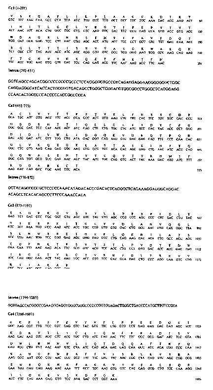
sequence, are indicated in Fig. 1. The sequence of the
CEa gene, the CEb gene and the corresponding amino acids
are also mentioned in the sequence listing.
The equine CE° and CEb sequences which encode the
constant region of the heavy chain of an equine IgE
allotype and which can be used for the purposes of the
invention are distinguished by the fact that they agree
at least in the region from T569 to C630 with the
sequences as shown in Seq.ID 1 and Seq.ID 3 indicated
in Fig. 1, as is stated in Figure 2. Functionally
important regions are shown against a dark background
in Fig. 2. As has been shown experimentally, and in
particular when using these genes, it is possible to
obtain recombinant IgE which is identical structurally
and functionally with natural IgE and with which, irl
turn, antibodies can be developed which are specific
for equine IgE and thus valuable in the diagnostics of
equine allergies. The functionality of the IgEs
generated with the CEa and CEO genes according to the
invention, which has been found in this context, is not
matter of course since mutations in functional
regions may lead inter alia to modifications in the
tertiary structure or binding capacity of the IgE so
CA 02439509 2003-08-27
- 9 -
that antibodies generated with recombinant IgE which is
not largely identical with natural IgE as regards its
tertiary structure, are not, or not optimally, specific
for natural equine IgE. In accordance with the
invention, in contrast, antibodies are obtained which
bind to natural equine IgE with high specificity, as
will be demonstrated hereinbelow. Thus, monoclonal
antibodies which are specific for natural equine IgE
have been produced successfully for the first time.
Thus, it is possible for the first time, with the aid
of the novel CEa and CEb genes, to generate functional
recombinant immunoglobulins with the aid of which
antibodies which are specific for equine IgE can be
obtained.
Instead of the abovementioned CEa and CEb genes, it is
also possible to use equivalent homologous sequences
which lead to corresponding functional immunoglobulins,
but with the exception of the nucleotide sequence for
the CE' and CEa gene (NCBI sequences U15150 and U17041).
The newly found CE genes agree in that they have at
least 55~ homology with a corresponding human sequence.
The homology of the equine CE sequences to the human one
is relatively low. The homology levels found for the
novel allotypes were: .CE°:56.4 0; CEb: 56. 0~, while the
level of homology of the CE sequence NCBI U15150 (see
above, CE' , Navarro P., Barbis D.P., Antczak D., Butler
J.E., 1995, Mol. Immunol. 32:1-87) is only 54$. The
percentage of conserved amino acid sequence regions in
comparison with human IgE is thus relatively high in
the case of the newly found equine IgE allotypes. The
sequence differences between the equine CE alleles can
also be demonstrated with the aid of restriction
fragment length polymorphisms (RFLPs). The following
holds true for the restriction enzymes StuI and Smal:
CE°: one SmaI site (position 751)
CA 02439509 2003-08-27
- 10 -
CEb: two SmaI sites (positions 12i and 751)
CEO: (published in "Navarro"): one StuI site (position
493), two SmaI sites (positions 107 and 790; according
to published sequence)
CEd(U17041): one SmaI site (position 808)
Chimeric recombinant immunoglobulins are prepared in a
manner known per se. One possible method is described
by Clarke et al. (loc.cit.). To carry out this method,
CEa or CEb DNA can be cloned into a cloning cassette of G
eukaryotic expression vector. The VH gene, for example
the VH186.2 cDNA (GenBank Acc.No. ,700529; Bothwell
A.L.M., Paskind M., Reth M., Imanishi-Kari T., Rajewsky
K., Baltimore D., (1981). Cell 24: 625-637) can
subsequently be cloned into the expression vector 5' of
the Cs gene. The C~-cDNA can be excised whenever desired
from an expression vector thus obtained and can be
replaced by any desired heavy-chain gene. In this
manner, further recombinant immunoglobulins can be
obtained with the aid of this construct according to
the invention by exchange of the C~ gene or gene
fragments.
Monoclonal isotype-specific anti-equine-IgE antibodies
are raised by standard methods via the immunization of
experimental animals with the recombinant equi-murine
IgE. The result is monoclonal antibodies which are
specific for equine IgE which is characterized by the
respective CE allele used for producing the recombinant
protein. Such monoclonal antibodies, which in
exceptional cases can recognise the murine components
of the recombinant chimeric IgE, can be eliminated for
example using the assay described in the examples ir.
Section 3.1. In most cases, however, epitopes on the
equine heavy-chain region (which is the rule in the
isotypes studied in the present context) serve for the
recognition of these IgEs by monoclonal anti-IgE
antibodies.
CA 02439509 2003-08-27
- 11 -
- The recombinant chimeric DNA used for the purposes of
the present invention gives rise to immunoglobulin
molecules which correspond largely to natural ones,
since the monoclonal antibodies obtained with the aid
of the reference substance according to the invention
(IgE) show a high degree of isotype specificity for
both the reference substance and natural equine IgE
(see hereinbelow, Table 1). Thus, the method according
to the invention provides a good yield of very
advantageous Ig products which resemble natural ones,
and these Ig products can be used as reference
substances and for the production of highly specific
monoclonal antibodies.
The sequences according to the invention, which are
shown in the sequence listing, are represented in
relation to each other in Figure 1. Figure 2 shows the
sequences of the CE°-cDNA (Cea-cDNA) and of the CEb-cDNA
(Ceb-cDNA) in comparison with the known sequences
U15150 (NCBI, Equus caballus Ig epsilon heavy chain
mRNA; partial cds, Navarro, P., Barbis, D.P., Antczak,
D. and Butler, J.E.) and U17041 (NCBI, Equus caballus
IgE heavy chain mRNA, Watson, J.L., Wilson, L.K. and
Gershwin, L.J.):
Fig. l: Equine genomic Csa nucleotide and amino acid
sequence
The two nucleotide substitutions in C~1 (121 T--~C) and
the Cs3 exon (972 C-~A) of the Cs° and C~b alleles are
shown against a gray background. Both base
substitutions bring about modifications in the amino
acid sequence in the CH1 (41 W--~R) and CH3 domains (239
L--~M) of the resulting IgE allotypes.
Fig.2: Sequence alignment between Cea-cDNA (Seq.ID1),
Ceb-cDNA (Seq.ID3) and NCBI sequence U15150 and NCBI
sequence U17041.
CA 02439509 2003-08-27
- 12 -
The invention is described in more detail hereinbelow
with reference to a practical example:
CA 02439509 2003-08-27
- 13 -
Description of the isolation of novel equine CE
sequences (Csa and Csb), their use for producing a
functional, recombinant equine IgE, and the first
development of monoclonal antibodies directed against
equine IgE:
1. Isolation of equine Cs cDNA and its use for
expressing recombinant equine IgE
1.1. Obtaining the cDNA for the constant region of the
equine IgE heavy chain (Cs)
DNA primers were synthesized according to the published
Cs sequence (Navarro P., Barbis D.P., Antczak D.,
Butler J.E., (1995). Mol. Immunol. 32: 1-87):
5' GTCTCCAAGCAAGCCCCATTA 3' - corresponds to the 5' end
of the equine C~1 exon and 5'
TCGCAAGCTTTACCAGGGTCTTTGGACACCTC 3' - corresponds to
the antisense sequence of the 3' end of the CE4 exon
and contains a Hind III cleavage site.
Following standard methods, mononuclear cells of the
peripheral blood of a horse were used for obtaining the
total RNA (RNeasy-Kit, Quiagen, Hilden, FRG). The
equine RNA was transcribed into cDNA by means of a
reverse-transcriptase reaction using an oligo(dT)is
primer (Promega, Mannheim, FRG) and Superscript II
reverse transcriptase (Life Technologies, Karlsruhe,
FRG). Using this cDNA and the above-described primers,
an equine Cs cDNA sequence was amplified by means of
polymerase chain reaction. To this end, 1 ~1 of cDNA
was mixed with a reaction mixture consisting of 4 mmol
MgCl~, 200 Eunol of each dNTP (dATP, dTTP, dCTP, dGTP;
Promega, Mannheim, FRG), 0.2 pmol of each primer (Life
Technologies, Karlsruhe, FRG) and 1.25 U Pfu DNA
polymerase in 1x Pfu DNA polymerase buffer (Promega,
Mannheim, FRG) and amplified in a thermocycler
(Biometra, Gottingen, FRG). In addition, a genomic C~
gene which we had isolated from are equine genomic gene
library and cloned (Wagner B., Siebenkotten G.,
CA 02439509 2003-08-27
- 14 -
Leibold W., Radbruch A, (1997). Vet. Immunol.
Immunopathol. 60: 1-13) was used as template and
likewise amplified in this polymerase chain reaction.
The two Cs sequences were sequenced (SEQ LAB,
Gottingen, FRG) and, even though they are derived from
different, non-related horses, show 100 nucleotide
sequence homology within the coding regions. However,
the sequence homology with the Cs sequences which have
already been published amounts to only 96~ (GenBank
Acc.No. U15150; Navarro P., Barbis D.P., Antczak D.,
Butler J.E., (1995). Mol. Immunol. 32: 1-87) and 98~
(GenBank Acc.No. U17041; Watson J.L., Pettigrew H.D.,
Wilson L.K., Gershwin L.J., (1997). J. Vet. Allergy
Clin. Immunol. 5: 135-142). These differences between
the Cs sequences determined by ourselves (Fig. l, Csa)
and those which have been published earlier allow the
conclusion that different Cs alleles exist in horses. CE
alleles were identified in a substantial number of non-
related horses by means of restriction analysis with
the restriction endonucleases Sma I and Stu I, which,
owing to the sequence differences, have different
cleavage sites within the C~ cDNA. In this process, a
further Cs allele (Csb) was identified, and this allele
deviates from the CEa, which had been sequenced by
ourselves, in two bases. Both base substitutions in the
Csb allele also result in amino acid substitutions at
the corresponding positions (Fig.1), i.e. the two new
alleles C~a and CEb, like the Cs alleles which are known
to date (U15150, referred to as CE~, and U17041,
referred to as CE°), encode different IgE allotypes. The
resulting modifications in the derived amino acid
sequences of the four IgE allotypes which have been
identified to date may also result in functional
modifications, such as, for example, in a different
binding behavior at FCC receptors and/or modifications
ir~ the ability of bringing about the release of
inflammatory mediators from mast cells. These
functional differences may play a role in particular in
the development of type I allergies.
CA 02439509 2003-08-27
- 15 -
1.2. Expression of equine recombinant IgEs
The method used in this context for expressing
recombinant immunoglobulins is known (0i V.T., Morrison
S.L., Herzenberg L.A., Berg P., (1983), Proc. Natl.
Acad. Sci. USA 80: 825-829; Knight K.L., Suter M.,
Becker R.S., (1988). J. Immunol. 140: 3654-3659; Clarke
R.A., Beh K.J., (1994). Immunol. Invest. 23: 25-37).
The principle of the procedure for expressing the
complete equine recombinant IgE which has been
generated for the first time will be summarized
hereinbelow:
To produce a recombinant equine IgE, the above
described equine CEb cDNA and the murine VH186.2 cDNA
(GenBank Acc.No. J00529; Bothwell A.L.M., Paskind M.,
Reth M., Imanishi-Kari T., Rajewsky K., Baltimore, D.,
(1981). Cell 24: 625-637), which together encode the
chimeric heavy immunoglobulin chain of IgE, were cloned
into a eukaryotic expression vector. This construct was
used to transfect the murine myeloma cell line J558L
which produces murine light n, chains (0i V.T., Morrison
S.L., Herzenberg L.A., Berg P., (1983). Proc. Natl.
Acad. Sci. USA 80: 825-829). The cells which secreted
complete IgE immunoglobulins were subsequently
selected. Light chains from the J558L cell line
together with heavy chains containing the VH186.2 gene
product form antibodies with a defined antigen
specificity for 4-(hydroxy-3-nitrophenyl)acetyl (NP),
in this case NP-specific equine IgE. Protein-
biochemical analyses of the expressed protein have
demonstrated that this recombinant IgE has high
structural similarity with native equine IgE. Moreover,
the recombinant protein binds to the FCsRI of mast
cells and basophile granulocytes and is capable of
mediating a release of inflammatory mediators from
these cells in vitro and in vivo, i.e. it also
corresponds to native equine IgE with regard to the
functional properties.
CA 02439509 2003-08-27
- 16 -
2. Raising IgE-specific monoclonal antibodies (anti-
equine IgE)
2.1. Immunization of mice
Female BALB/C mice were immunized with recombinant IgE.
The purified equine NP-specific IgE (NP-IgE) was
employed in a total amount of 2.5 ~g at the first
immunization and 1.25 ~g for all further immunizations.
For the first (day 0), the second (day 14) and the
third immunization (day 21), the protein was mixed with
Gerbu Adjuvanz MM (Gerbu Biotechnik, Gaiberg, FRG)
following the manufacturer's instructions. For the
further immunizations on days 28, 29 and 30, the NP-IgE
was applied in PBS without added adjuvant. All
injections were given intraperitoneally. Cell fusion
was performed on day 31.
2.2. Raising monoclonal antibodies
On day 31, one mouse whose NP-IgE serum titer had
previously been studied (ELISA see 2.3.1.) was
sacrificed, the spleen was removed under sterile
conditions, and the spleen cells were plated out
carefully. The spleen cells were taken up in Hybridoma
SFM medium (Life Technologies, Karlsruhe, FRG), counted
and mixed 1:2 with murine X63-Ag8.653 myeloma cells
(Kearney J.F., Radbruch A., Liesegang B., Rajewsky K.,
(1979). J. Immunol. 123: 1548-1550). Following
centrifugation and removal of the supernatant, the
cells were resuspended carefully and treated slowly
with 1.5 ml polyethylene glycol 1500 (Boehringer,
Mannheim, FRG) which had been warmed to 37°C. After
incubation for 1 minute at 37°C, 20 ml of Hybridoma SFM
medium were slowly added dropwise tc dilute the
polyethylene glycol (1 m1 over 1 minute, 3 ml over 1
minute, 16 mi over 1 minute). Following centrifugation
and removal of the supernatant, the cell pellet was
resuspended carefully in 200 ml of Hybridoma SFM
CA 02439509 2003-08-27
- 17 -
supplemented with HAT media supplement (Sigma,
Steinheim; FRG), 10~ (v/v) Myclone FKS (Life
Technologies, Karlsruhe, FRG), 100 IU/ml penicillin,
100 ~g/ml streptomycin (PAN Biotech, Aidenbach, FRG)
and 4U/ml human recombinant IL6. This cell suspension
was plated into 24-well cell culture plates. After 7-10
days, individual clones were visible. They were picked
from the 24-well plates and transferred into 96-well
plates. After a further 2-3 days, the supernatants of
these 96-well plates were tested in an ELISA assay for
anti-IgE-specific antibodies. Positive clones were
characterized further (see 3.) and recloned once or
twice. The HAT supplement in the medium was replaced
after two weeks by HT supplement (Sigma, Steinheim,
FRG). After a further 3-4 weeks, the cells were weaned
onto Hybridoma SFM medium without further selection
additives and without human recombinant IL6.
3. Detecting the IgE specificity of the monoclonal
antibodies
The IgE specificity of the (in total) 18 monoclonal
antibodies (Table 1) was detected by standard methods
which were modified in a suitable manner for this
purpose. Monoclonal antibodies which specifically
recognised the heavy chain of the recombinant equi-
murine NP-IgE were detected in ELISA assays (see 3.1.).
The ability of the monoclonal antibodies to recognise
not only the recombinant protein, but also native
equine IgE, was verified by SDS-PAGE (see 3.2.) and
membrane immunofluorescence (see 3.3.). The specificity
of the monoclonal anti-IgE antibodies for native equine
IgE, and the lack of reaction with all other equine
immunoglobulins available, were verified in an isotype-
specific ELISA (see 3.4.). The specificity of the anti-
IgE antibodies for various epitopes of the equine IgE
was detected in an inhibition ELISA (see 3.5.).
CA 02439509 2003-08-27
- 18 -
3.1. Detection of NP-IgE-specific monoclonal antibodies
Following cell fusion, the supernatants of the clones
which had grown were first analyzed for the presence of
specific antibodies which react with the recombinant
IgE. An ELISA was used for detecting monoclonal
antibodies which recognize the NP-IgE heavy chain. The
ELISA plates (Nunc, Wiesbaden, FRG) were coated with NP
derivative 4-(hydroxy-3-indo-5-nitrophenyl)acetyl (NIP)
conjugated with bovine serum albumin (BSA) (NIP15-BSA;
Biosearch Technologies, Navato, CA, USA) in a
concentration of 5 ~g/ml in carbonate buffer (15 mmol
Na2C03, 35 mmol NaHC03, pH 9.6) . After the plates had
been washed with phosphate buffer (2.5 mmol NaH2P04,
7.5 mmol Na2HP04, 145 mmol NaCl, 0.1~ (v/v) Tween 20, pH
7.2), they were incubated with NP-IgE, which binds to
the NIP-BSA-coated plate. After a further washing step,
the supernatants from the 96-well plates of the cell
fusion were then applied to the plate thus prepared. If
NP-IgE-specific monoclonal antibodies were present,
they were bound in this step to the ELISA plate and,
after a further washing step, detected using a
peroxidase-conjugated polyclonal anti-mouse-IgG
antibody (Dianova, Hamburg, FRG). After addition of
substrate solution (33.3 mmol citric acid, 66.7 mmol
NaH2P04, pH 5.0), freshly treated with 130 ~g/ml
3,3',5,5'-tetramethylbenzidine (TMB, Sigma, Steinheim,
FRG) and 0.010 (v/v) hydrogen peroxide (Sigma,
Steinheim, FRG)), the anti-IgE-antibody producing
clones were identified by the color reaction which had
taken place.
As a distinction from monoclonal antibodies which
recognize the murine portions of the NP-IgE, the
supernatants which were positive in the first assay
were additionally checked on G plate which was coated
with NIF-BSA and subsequently incubated with murine
NF-IgD. Monoclonal antibodies which specifically
recognized the equine IgE heavy chairi reacted only with
CA 02439509 2003-08-27
- 19 -
NP-IgE, but not with NP-IgD.
3.2. Biochemical detection of IgE in equine serum
IgE is found in the serum in small concentrations only
and has a short half-life. However, in particular in
allergic patients, serum IgE levels may rise
drastically. IgE was detected using the monoclonal
anti-IgE antibodies after the equine serum had been
separated by sodium dodecyl sulfate/polyacrylamide gel
electrophoresis (SDS-PAGE). To this end, the equine
sera were treated with SDS sample buffer (62.5 mmol
Tris, 10~ (v/v) glycerol, 2~ (w/v) SDS, 0.1~ (w/v)
bromphenol Blue, pH 6.8), pH 6.8) and separated under
nonreducing conditions -in a 7.5~ SDS gel in a Mini
Protean II chamber (Bio-Rad Laboratories, Munich, FRG).
These proteins were transferred from the gel to a
polyvinylidene difluoride membrane by Western Blotting
which was incubated with the monoclonal anti-IgE
antibodies after the free binding sites had been
blocked with 1$ (w/v) gelatin. The monoclonal anti-IgE
antibodies identified an approx. 220 000 Dalton protein
in the equine serum, which corresponds to the molecular
weight of equine IgE. The protein identified by the
monoclonal antibodies was not detectable in the serum
of all of the horses studied; however, in particular
horses with clinical allergic symptoms usually also
showed a pronounced IgE band in the serum.
The immunoglobulins in the equine serum were separated
into their light and heavy chains under reducing
conditions by treating the SDS sample buffer with
5~ (v/v) 2-mercaptoethanol. One monoclonal anti-IgE
antibody (aIgE-176) also recognized the isolated equine
IgE heavy chain with G relative molecular weight of
76 000 Daltons. Accordingly, the aIgE-176 antibody
recognizes a different epitope of the equine IgE than
the remaining monoclonal antibodies, which only
recognize the unreduced IgE (see 3.5.). In contrast,
CA 02439509 2003-08-27
- 20 -
the IgE epitope recognized by aIgE-176 is also present
on the isolated IgE heavy chain.
3.3. Labeling IgE on equine blood leukocytes
IgE can be bound at the surface of certain blood
leukocytes by what are known as Fc receptors (in the
present case FcERI or FesRII). FcsRI can be expressed by
basophile and eosinophile granulocytes, while FcsRII
can be expressed by some of the monocytes, B cells and
eosinophile granulocytes. Free serum IgE can be bound
to the cells via these receptors, so that it can be
detected at the cell surface by fluorochrome-coupled
antibodies (membrane immunofluorescence).
The equine blood leukocytes were obtained from
anticoagulant-treated whole blood of various horses. To
this end, the leukocyte-rich plasma above the
erythrocyte sediment was obtained after approximately
30 minutes of spontaneous sedimentation and then
centrifuged; thereafter, the leukocytes were washed 2x
with PBS in order to remove the thrombocytes (80 x g,
5 min). Thereafter, the leukocytes were taken up in
PBS/BSA (PBS supplemented with 0.5% (w/v) bovine serum
albumin and 0.02% (w/v) sodium azide) and placed on
ice. 5 x 10~ aliquots of equine leukocytes were
incubated on ice for 10 minutes in 10 u1 of PBS/BSA in
a tube containing 30 u1 of the monoclonal anti-IgE
antibodies (1:2 in PBS/BSA). As a control, 5 x 10⁶
aliquots of equine leukocytes were incubated under
identical conditions with an irrelevant murine
monoclonal IgG1 antibody (isotype control). After the
cell samples had been washed once with cold PBS/BSA,
they were all incubated for 5 minutes on ice together
with a phycoerythrin-conjugated anti-mouse IgG antibody
(Dianova, Hamburg, FRG), washed again and, after
addition of propidium-iodide-containing PBS, measured
ire a flow cytometer. The amount of surface-IgE-positive
cells was 1.28 y 0.52% irl the case of the adult horses
CA 02439509 2003-08-27
- 21 -
studied and differed highly significantly (p < 0.001)
from the isotype control 0.02 ~ 0.02.
The IgE-positive cells of adult horses were isolated by
magnetic cell sorting and studied under the microscope
and by flow cytometry. The cell fraction which can be
isolated by the monoclonal anti-IgE antibodies which
were developed consists to approximately 30~ of
basophile granulocytes which have bound IgE via their
FcsRI, to approx. 68~ of mononuclear cells
(lymphocytes, lymphoblasts and monocytes) which are
capable of binding IgE complexes with their FcsRII, and
to a minor extent of approx. 2~ of other cells (for
example eosinophile granulocytes). These studies
demonstrate that the monoclonal anti-IgE antibodies are
capable of recognizing equine IgE not only in its
native form (see 3.2.), but also when bound to FcE
receptors.
3.4. Isotype-specific ELISA
To detect the IgE specificity of monoclonal antibodies,
' the ELISA plates were coated with a polyclonal anti-
horse IgG(H+L) antibody (Dianova, Hamburg, FRG) and
subsequently incubated with various equine reference
immunoglobulins (IgM, IgGa, IgGb, IgG(T) light chains,
purified serum IgE). In the next step, the monoclonal
anti-IgE antibodies were incubated with in each case
all of these reference proteins, and the anti-IgE
binding was then visualized by means of a peroxidase-
conjugated anti-mouse IgG antibody and the subsequent
substrate reaction. Binding of the anti-IgE antibodies
to the purified serum IgE was detected, but not to the
other equine immunoglobulins.
3.5. Inhibition ELISA for identifying different epitope
specificities of the anti-IgE antibodies.
The inhibition ELISA made possible the identification
CA 02439509 2003-08-27
- 22 -
of different IgE epitopes which are recognized and
bound by the various monoclonal anti-IgE antibodies. To
this end, the ELISA plates were coated with NIP15-BSA
and subsequently incubated with recombinant IgE. Then,
the 18 different monoclonal anti-IgE antibodies were
applied to the plates thus coated with recombinant IgE.
During the incubation time, the antibodies had a chance
to bind to their respective specific epitopes of the
equine recombinant IgE. In the next step, the
biotinylated aIgE-134 antibody, which was only capable
of binding with the recombinant IgE if the epitopes
which this aIgE-134 antibody recognizes on the
recombinant IgE were still freely accessible, i.e. not
blocked by one of the anti-IgE antibodies in the
previous step, was added. Binding of the biotinylated
aIgE-134 antibody was then visualized using
streptavidine-peroxidase and a final substrate
reaction. The epitopes of the recombinant IgE which are
recognized by the monoclonal antibodies aIgE-22,
algE-41, aIgE-132 and aIgE-176 did not inhibit the
binding of the biotinylated aIgE-134 antibody, i.e.
these anti-IgE antibodies recognize different epitopes
of the recombinant IgE.
3.6. Capability of basophile granulocytes of being
activated by the monoclonal anti-IgE antibodies
The anti-IgE antibodies aIgE-41, aIgE-132, aIgE-134 and
aIgE-176 were studied for their ability to release
mediators from equine basophile granulocytes. To this
end, these monoclonal antibodies were employed in a
histamine release. assay which has already been
described (Kaul, S., 1998. Typ I Allergien beim Pferd:
Prinzipielle Entwicklung eines funktionellen in vitro
Nachweises (Type I Allergies in horses: principle of
the development of a functional in-vitro assay] PhD
thesis, Veterinary School Hanover]), in which the
ability of the vGrious monoclonal antibodies to release
histamine from equine blood basophiles is measured in
CA 02439509 2003-08-27
- 23 -
relation to the maximum and spontaneous release of
histamine from these cells. The induction of histamine
release was only achieved with the antibody aIgE-134.
Data on the characterization of monoclonal anti-IgE
antibodies are compiled in table 1.
Table 1
CA 02439509 2003-08-27
d~
M
r1
M I
W + + + + + + + + + + + + +
+
!v
~1
H
N
N
~
O
1~
H '1
N
W
W H
'~ + + + +
W
1~
-''i
H
.:a
-
W
t77 H
H
23
~ N
1~
-rl ~
M
r~
0 0 o W
v
.,..I-rt I I I I I I I II I I I t I I H
U7 I I I
v y
1~
G
N W
H ~ b
I
-~ O
1~ U
I ~ ~ 1~
v
'-1 f~ ~ M 1
J
N O -
p ~'~'~ ++ + ++ + + + + ++ + + + + + + -3
I I
E-i ~ U N
U v >.,
~
O
O
N
4-1 'J
O W -rl
p ~
I I I 1 I I I I+ I I I I 1 I (J]
I I I
ri
N
t71
H
~1 O M I I
-rl ~".,
~ W +
O
W _
H ~
O + + + + t + + ++ + + + + + +
+ + t
1
,, H ~ r1
U .i.~
ro
p
W'
r
t I I
v P.
N Z
I
cn + + + + + + + ++ + + + + + +
~ + + +
W
H I -r-I
z ''
.r.,
0
W
N O~O V~O r1t0l0~-iN O d~N
>31 N '-IM H N M ~ L!1I~M (VN ODO1I'~II
00 H N
M
N d~Cfr r1v-i .-i~-iv-ir1M tnInlf~tf1~D
N ~ r-1
+
CA 02439509 2003-08-27
- 1 -
SEQUENCE LISTING
<11C> ~'agne= Dr., 3ettina
~ebcld Prof., Wolfgang
Radbruch Prod., Andrews
<1,20> eqiine C-epsilon constant heavy chain gene region
<'130> 3064-1 DE-1
<1!0>
<im>
<160> 5
<170> PatentT_n Ver. 2.Z
<210> 3
<211> 1272
<212> ~7NA
<213> Eguns callus
<220>
<221> source
<222> (1)..(1272y
<223> m..~NA from ecuine peripheral blood mononuclear
cells
<220>
<G21> C~r9g::~n
<222> (')..(291)
<223> C-epsilon I exan, allele a
<220>
<221> C region
<222> (292)..(615)
<223> C-epsilcn 2 axon, allele a
<220~
<221> C~region
<222> (616) . . (936)
<223> C-epsilon 3 exoa, allele a
<220>
<221> C~region
<222> (93?)..(1272)
<223> C-epsilon 4 axon, allele a
CA 02439509 2003-08-27
- G -
<4vD>
gtctccaagc aagccccatt aatcttgccc ttggctgcct gctgcaaaga caccaagact 60
actaacatca c~ctgggctg cctggtcaag ggctacttcc cggagccagt gaccgtgacc 120
tgggatgcag ggtccctta2 ccggagcacc atgaccttcc ctgccgtctt tgaccanacc 180
tctggcctct acaccaccat cagcagggtg gtcgcctcgg ggaagtgggc caagcagaag 240
ttcacctgca acgtggtgca ctcccaggag accttcaaca agaccttcaa cgcatgcatc 300
gtgaccttca ccccacccac cgtgaagctc ttccactcct cctgcgaccc cggcggcgac 360
tcccatacca ccatccagct cctgtgcctc atctccgact acacccctgg cgacatcgac 420
atcgtttggc tgatagacgg gcagaaggtc gscgagcagt tccctcaaca cggcctcgtg 980
aagcaggagg gcaagctggc ctccacacac agcgagctca acatcaccca gggccagtgg 540
gcgtccgaaa aca~ctacac ctgccaggtc acttacaaag acatgttctt taaggaccag 600
gcccgcaagt gcacagagtc tgacccccgc ggtgtgagcg tctacc~gag cccgcccagc 66D
cccctcgacc tgtacgtctc taaatcgccc aagatcacct gcctggtggt ggacctggcc 720
aacgtgcagg gcttaagcct gaactggtcc cgggagagcg gggagcccct gcagaagcac 780
acactggcca ccagcgaaca atttaacaag acattctcgg tcacgtccac cctgcctgtg 940
gacaccaccg actggatcga gggcgagact tacaagtgca ccgtgtccca cccagacctg 900
cccagggaag tcgtgcgctc catcgccsag gcccctggca agcgtttgtc ccccgaggtc 960
tacgtgttcc tgccgcctga ggaggaccag agctccaagg acaaggtcac cctcacctgc 1020
ctgatccaga acttcttccc cgcggacatc tccgtacagt ggctgcgtaa caatgtocta 1080
atccagacag accagcaagc caccacaegg ccccaaaagg ccaatggccc caacccagcc 1140
ttcttcgtct tcagccgcct agaggtcagc cgggcggaat gggagcagaa gaacaaattt 1200
gcctq~aagg tggtccacga ggcgctgtcc caaaggaccc 'tccagaaaga ggtgtccaaa 1260
gaccctggta as 1272
<210> 2
<211> 42.4
<212> PRT
<213> Equus caballus
<220>
<22i> MN4.iIIN
<222> {1j . . {97)
<223> Cfil' domain, IgE allowpe a
<220>
<221 > DOMA.FN
<222> 198)..(205)
<223> GH2 domain, Ig~ allotype a
<220>
<22I> DC3MAIN
<222> {206j..{312)
<223> CH3 domain, IgE allotype a
<220>
<221> DOt4Ft_N
<222> (313)..{424)
CA 02439509 2003-08-27
- 3 -
<223> CSC dosain, Tg~ allotyge a
c4 GO> 2
Val Ser Lys Gln Ala Pro ireu Zle heu Prc L~eu Ala Ala Cys Cya Lys
a 5 10 i5
Asp Thr Lys Thr Thr Asn Ile Thr Len Gly Cys heu Vsl Lys 61y Tyr
20 25 30
Fhe Pro Glu Pro Va3 Thr Val Thr Trp Asp Ala Gly 5er Leu Asn Arg
- 35 90 ' 95
Ser 2hr Met Thr Phe Pro Rla Val Phe Asp Gln mhY Ser Gly Lau Tyr
50 55 60
Thr Tht IIe Ser Arg Val Vsl A?a Ser Gly Lys Trp Ala Lys GZn Lys
65 '?0 75 80
Phe Thr Cys Asn Val Val His Ser Gln Glu Thr Phe Asn Trys Thr Phe
85 90 95
Asn Ala Cys Ile Val ?'hr Phe T.zr Pro Pro Thx Val ~ys heu Phe His
100 105 I10
Ser Ser Cys Asp Pro Gl.y Gly P.Bp Ser Ais Thr Thr i1 a Gln heu Leu
115 120 - 125
Cys Leu 21e Ser Asp Tyr ?'hr Prb Gly Asp Ile Asp Ile Val Trp Leu
130 135 140
I1e Asp Gly G3.n Lxs Val Asp Glu Glr~ Phe Pra Gln Ais Gly yeu Va_7.
345 150 3.55 160
~ys Gln Glu Gly hys 1eu Ala Ser Thr His Sex Glu hea Asn Ile Trr
165 1.70 175
Gln Gly 61n Trp ~3a Ser Glu Asn Thr .yr 'i'hx Cys Gln Val Thr Tyr
- 184 - ' 185 I9a
=ys Asp Met Ile Phe Lys fisp G.ln Ala Arg hys Cya Thr Glu Ser Asp
1°5 200 205
Pro Arg Gly Val Ser Val fi~rr Leu 3ez~ Pxo Pro Ser Pro heu Asp Len
-210 215 220
Tyr Va3. Sex vys Ser Fro Lys rle Thr Cys Leu Val Val Asp Leu Ala
2i5 230 235 240
CA 02439509 2003-08-27
- 4 -
Ass 'val G1:~ Gly Leu Ser Leu Asn TrF Ser A=g Glt Ser Gly Giu Prc
295 250 255
Leu Gln L~ya His Thr Leu A?a Thr Ser Glu Gln Phe Pin Lys Thr Fhe
264 265 270
Ser Va1 T hr Ser T!-.r Leu ?ro Val Asp Thr Thr Asp T~-p Iie Glu Gly
275 2B0 285
Gle Thz Tyr Lys Cys Thr Val Ser His Fro Asp heu Pro Arg Giu Val
290 295 ' ~ 300
Val Azg Ssz Zle A1a hys Ala Pro Gly Lye Arg Leu Ser Pro Glu Va1
305 310 3I5 320
Tyr val Phe Leu Fro Bro~Glu Glu Asp 61n Ser Ser T~ys Asp Lys val
325 330 335
Thr Leu Thr Cys Leu Ile Gla Asn Phe P:~e Pro Als Asp Ile Ser Val
390 345 350
Gin Trp Leu Arg Asn Asn Val 3~eu rle Gln ~_'hr Asp Glri Gin Ala Thr
355 360 365
Thr Arg Pro Gln hys Ala Asn Glp Pro Asn Pra Ala Phe the Val Phe
370 375 380
Ser Arg Leu GIu Val Ser Arg P.la Giu Trp GZwGZa Lys Asn hys Phe
385 990 395 900
Ala Cys Lys vat val His Glu F.ia Leu Ser Gln Arg 2hr T~eu Gln Lys
4D5 ~ 43.0 97.5
G1u Val Ser hys Asp Pxo Gly Lys
420
<230> 3
<2ia 1272
<2i2> DNA
<213> Equus caballus
<220>
<221> source
<222> :1)..(1272)
<223> mRi~A frog e~~lTe peripheral blood mononuclear
cells
CA 02439509 2003-08-27
- 5 -
<220>
<221> C region
<222> !1)..!291}
<223> C-epsilon ? exon, allele b
<220>
<221> C region
<222> 1292}..1615)
<223> C-eps~.lon 2 excn, allele H
<220> .
<?21> C region
<222> t616)..~936)
<223> C-epsi3o.~. 3 excz, allele b
<220>
<221> C region
<222> (937}..!1272}
<223> C-epsilon 6 exon, alieie b
<900> 3
gtctccaagC aagccccatt aatcttgccc ttggctgcct gctgcaaaga caccaagact 60
actsacatca caetgggctg cctggtcaag ggctacttcc cggagccagt gacegtgacc 120
cgggatgcag gatcccttaa ccggagcacc atgaccttcc ctgccgtctt tgaccaaacc 180
sctggcctct acaccaccat cagcagggtg gtcgcctcgg ggaagtgggc caagcagaag 2!0
t=cacctgca~acgtagtgca ctcccaggag accttcaaca agaccttcaa cgcatgcatc 300
gtgaccttca ccccacccac cgtgaagctc ttccactcct cctgcgaccc cggcggcgac 360
tcccatacca ccatccagct cctgtgcctc atctccgact acacccctgg cgacatcgsc 420
atcgtttggc tgatagacgg gcagaaggtc gacgagcagt tccctcaaca cggcctcy~tg 480
aagcaggagg gcaagctgac ctccacacac agcgagctca acatcaccca gggccagzgg 540
gcgtccgaaa acacctacac ctgccaggtc acttaca.aeg acatgatctt taaggaccag 500
gcccgcaagt gcacagagtc tgacccccgc ggtgtgagcg tctacctgag cccgcccagc 660
cecctcgacc tgtacgtctc taaatcgccc aagatcacct gcctggtggt ggacatggcc 720
aacgtgcagg gcttmagcct gaactggtcc cgggagagcg gggagcccct gcagaagcac 780
acactggcca ccagcgaaca atttaacaag acattctcgg tcacgtccac cctgcctgtg BEO
gacaccaccg actggatcga gggcgaga~t tacaagtgca ccgtgtccca cccagacctg 900
cccagggaag tcgtgcgctc catcgccaaq gcccctggca agcgtttgtc ccccgaggtc 960
tacgtgttcc tgccgcctga ggaggaccag agctccaagg acaaggteae cctcacctgc 1020
ctgatccaga acttcttccc cgcggacatc tccgtacagt ggctgcgtaa caatgtccta 1080
atccagacag accagcaagc caCCac~cgg ccccasaagg ccaatggccc caaccccgcc 1190
ttcttcgtct tcagccgcct agaggtGagc cgggcggsat gggagcagaa gaacaaattt 1200
gcctgcaagg tggtccacga ggcgctgtcc caaaggaccc tccagaaaga ggtgtccaea 1260
gaccctggta as
'272
<210>
<23.~.> 929
<212> PBT
CA 02439509 2003-08-27
- 6 -
<213> ~~,»us caballus
<220>
<221> DOMA.r.N
<222> (1)..(97)
<223> CH1 donain, IgE aliotype b
<220>
<221 > DOM.~.IN
<222> (98)..1205)
<223> CH2 damair., IgE allotyne b
<22~i
<221> DOMAIN
<222> (206)..(3"s2)
<223> CH3 domain, IgE a2lo~ype b
<220>
<22I> N
<222> (313)..(924)
<223> Cg9 domain, IgP. a~atyrpe b
<900> 4
Val Ser Lye Gln Ala Pro ?.eu Ile i~eu Fro ~eu Ala A.3.a Cys Cys i,ys
3. ~ 10 15
Asp Thr Toys Th_r ?'hr Asn Ile Thr Leu GIy Cys hev Val Lys Gly Tyz
20 25 30
Phe ?ro Glu Pro Val Thr Val Thr Arg Asp Tea Gly Ser i~eu Asn Arg
35 !fl 43
Ser Thr Met '"hr Phe Pra Ala Vat Phe Asp Gln Thr Ser Gly Leu Tyr
S~J 55 50
Thx Thx Ile Ser Arg Val Val Ala Ser Gly Lys Trp Ala hys Gln Lys
65 ?0 ' 75 80
Fhe Thr Cys Asn Vsl Val His Ser G3.n G3u Tizr ?he As. iys Thz Phe
85 90 9S
Asr. A1a Cys Ile Va? ?hr Phe Thr Pro pro Thz.Vzl hys l~ev Pha Fiis
100 3.05 110
Ser Sex Cys Asp Pro Gly Gly Asp Ser ~lis Thr Thr T_le Gln Leu T~eu
115 ?20 125
Cys Len Ile Ser Asp Tyr Trr Pro Gly Asp Ile Asp Ile Vsi Trp Leu
CA 02439509 2003-08-27
_ 7
130 135 ia0
Ile Fssp GZy Gin T~ys Vai Asp Glu Gln Phe Pro Gln His Gly Leu Val
iQ5 ?50 1S5 ?60
i~ys Gl:~ Glr~ Gly hys Leu Ala 5er Thr His Ser Glu ~eu Asn Ile Thr
365 .70 175
Gla Gly Gln Trp A1a 5er Glu Asn Thr Tyr T:~r Cys 67.n Val Thr Tyr
180 185 1s0
hys Asp Met Ile Phe ~ys Asp Gln Ala ?rg Lys Cys Thr Glu Ser Asp
3.95 200 205
Pro Arg Gly Val Ser Val :'yr 5eu Ber Pro Pxo Se. Pro heu Aso Leu
210 2l5 220
Tyx Val Ser ~ys Ser Pro Lys ale Thr Cys Leu Val Vai Asp Met Ala
225 230 235 240
Asn Val Gin Gly heu Ser ~eu Asn Trp Sez Arg Giu Ser Gly Glu Pro
245 250 255
Zeu Gln Lys His Thr beu Ala Thr Ser G1u Gl:~ Phe P.,sx~ Lys Thr Phe
2s0 265 270
Se_- val Thr Ser Thr heu Pro Val Asn Thr Thr Asp Trp Zle Glu Gly
275 2B0 285
Clu Thz Tyr yyQ Cys Thr Val Se= H;s iro Asp veu Pro Arg GI:~ Val
290 - 295 300
Val Arg Sex Ile Ala hys Ala Pro G?y i~ys Arg Leu Ser Pro G3u Val
305 310 313 320
Tyr Val Phe ~eu Pro Pro Gln Glu Asp Gln Ser Ser hys Asp hys Val
325 330 335
~hr Le;~ Thr Cys T,eu Ile Gln Asa Phe Phe ?ro Ala Asp Zle Ser Val
340 395 350
Gln Txp ~eu Arg Asn Asg Val reu _Tle Gln Thr Asp Gln Gln Ala Thz
355 360 ~ 365
Thr Arg P.o Gln ~ys Ala Asn Gly Pro Asn Pro Ala Phe Phe Val Phe
370 375 380
Ser Arg veu Glu VaI Ser Arg Ala Glu Trp Gln Gln Lys Asn Lys Fhe
CA 02439509 2003-08-27
385 390 395 t00
Al.a Cys Lys Val Val Hs.s Glu Rla Leu Bar Gl.n Arg Thr :~eu Gig vys
a05 910 415
roe Val Ser Lys psp Pro Gly i~ys
920
<220> 5
<211> ?603
<212> DNA
<213> Ec_r,~us caballus
<220>
<221> source
<222> (1)..(1601}
1223? equsze genosaic DIvA
<220>
<221> C region
<222> (1}..(29x}
<223~ C-eps3loa 1 axon, a17.e3e a
<220>
<221> irstzox~
<222> (292)..(A51}
<220>
<221> C region
<222> (452)..(775) .
<223> C-epsi3on 2 axon, allele a
<220>
<22i> intros
<222> 1776)..;872)
<220>
<221> C region
<2a2> (873}..(1193)
<223> C-epsilfln 3 axon, a?lele a
<220>
<221> inzron
<222> (119d)..(3265}
<220>
<221> C_~egio:~
CA 02439509 2003-08-27
_ g _
~~2a> c12s6a..c~scn
<223> C~egsi,ion ~ exo_~., allele a
«00> 5
gtctccaagc aagccccatt aatcttuccc ttggotgcct gctgcaaaga caccaagact 60
actaacatca cactgggctg cctggtcaag ggctacttcc cggagccagt gaccgtgacc 120
=gggatgcag ggtcccttaa ccggagcacc atgaccttcc ctgccgtctt tgaccaaacc ?80
tctggcctct acaccaccst cagcagggtg gtcgcctcgg ggaagtgggc caagcagsag 240
ttcacctgca a~gtggtgcz ctcceaggag accttcaaca zgaccttcaa cggtgagcca 300
ggacggcccc gcccgccctc cagggggtgc cgtcagagga ggasgggggg gctggccagg 360
agggcatcac cactgccggt gacagcctgg gctgggacgt ggcggcctgg gctcagggag 420
gccaacactg cgcccacccc caccgccccc agcatgcatc gtgaccttca ccccacccac 980
cgtgaagctc ttccactcct cctgcgaccc cggcggcgac tcccatacca ccatccagct 540
cctgtgcctc atctccgact aascccctgg cgacatcgac atcgtttggc tgatagacgg 600
gcagaaggtc gacgagcagt tccctcaaca cggcctcgtg aagcaggagg gcaagctggc 660
ctccacscac agcgrgctca acatcaccca gggccagtgg gcgtccgaaa acacctacac 720
ctgccaggtc acttacaaag acatgatctt taaggaccag gcccgcaagt gcacaggtac 780
agccccogct cccccaaaca tagacacccg acactcaggg ctcagaaagg agggcaggac 840
acagcctcac acagccctct tcccaaacca cagagtctaa cccccgcggt gtgagogtct 90D
acctgagccc gcccagcccc ctcgacctgt acgtctctaa atcgcccs~g atcacctgcc 960
tggtggtcga cctggccaac gtgcagggct taagcctgaa ctggtcccgg gagagcgggg 1020
agcccctgca gaagcacaca ctggccacca gcgaaceatt taacaagaca ttctcggtca 1080
cgtccaacct gactgtggac acceccgact ggatcgaggg cgagacttac aagtgcaccc 1140
tct cccaccc agacctaccc aggg2agtcg tgcgctccat cgccaaggcc cctggtgagc 1200
cacgggccga agggaggtgg gcgggccccc cggtggagac tgggc:gscc ccatgcttgt 1260
ccgtaggcaa gcgtttgtcc cccgaggtct acgtgttcct gccgcctgag gaggaccaga 1320
gctccaagga caaggtcacc ctcacctgcc tgatccagaa cttcttcccc gcggacatct 1380
ccgtacagtg gctgcgtaac aatgtcctaa tccagacaga ccagcaagcc accacacggc 1440
cccaaaaggc caatggcccc aaccccgcct tcttcgtctt cagccgccta gaggtcagcc 1500
gggcggaatg ggagcag2ag aacaaatttg cctgcaagg'v ggtccacgag gcgctgtccc 156D
aaaggaccct ccagaaagag gtgtccaaag accctggtaa a isD1