Note: Descriptions are shown in the official language in which they were submitted.
CA 02441021 2011-05-31
METHOD FOR ALTERATION DETECTION
FIELD OF THE INVENTION
[0002] The invention relates generally to methods for detecting an
alteration in a
target nucleic acid.
BACKGROUND OF THE INVENTION
[0003] Many diseases are associated with gnomic instability. As such,
instability
markers have been proposed as diagnostics. For example, mutations are
considered
valuable markers for a variety of diseases, and have formed the basis for
screening
assays. Specific mutations might be a basis for molecular screening assays for
the
early stages of certain types of cancer. See, e.g., Sidransky, et al..
Science, 256: 102-
105 (1992). For example, mutations in the BRCA genes have been proposed as
markers for breast cancer, and mutations in the p53 cell cycle regulator gene
have
been associated with the development of numerous types of cancers.
[0004] Early alteration detection allows early disease diagnosis, and
thus also
provides an avenue for intervention prior to the presentation of disease
symptoms that
often occurs after metastasis when a cure is less readily attainable. However,
the
detection of genetic mutations or other alterations is difficult, or
impossible, in certain
sample types. For example, the difficulty of isolating DNA from complex,
heterogeneous samples makes identification of early-stage mutation difficult.
1
CA 02441021 2003-09-15
WO 02/074995
PCT/US02/07926
[0005] Therefore, there is a need in the art for efficient methods for
determining
the presence or absence of certain genetic mutations or other alterations in a
target
nucleic acid in a biological sample.
SUMMARY OF THE INVENTION
[0006] The invention provides methods for detecting an alteration in a
target
nucleic acid in a biological sample. According to the invention, a series of
nucleic
acid probes complementary to a contiguous region of wild type target DNA are
exposed to a sample suspected to contain the target. Probes are designed to
hybridize
to the target in a contiguous manner to form a duplex comprising the target
and the
contiguous probes "tiled" along the target. An example of this duplex is shown
in
Figure 1. If a mutation or other alteration exists in the target, contiguous
tiling will be
interrupted, producing regions of single-stranded target in which no duplex
exists.
This is shown in Figure 2. Identification of one or more single-stranded
regions in the
target is indicative of a mutation or other alteration in the target that
prevented probe
hybridization in that region. For purposes of the present invention, a "tiled
sequence"
or "tiling" refers to the contiguous hybridization of probes to a target
region, whether
separated by single-stranded sequence or not.
[0007] Accordingly, in methods of the invention, a sample comprising a
single-
stranded target nucleic acid is exposed to a plurality of nucleic acid probes.
In a
preferred embodiment, the plurality of probes comprises probes that are
complementary to different positions of the target such that hybridization of
members
of the plurality with a wild-type target results in a contiguous series of
probes along at
least a portion of the target sequence when the target is a wild-type target.
Probes
including RNA, DNA and/or Peptide Nucleic Acid (PNA) may be employed to
hybridize to the target nucleic acid. It is not necessary to ligate the series
of probes to
2
CA 02441021 2013-03-14
form a continuous strand, although ligation may be performed at the discretion
of the
user.
[0008] When the target is a wild-type sequence, there will be no
single-stranded
portion in the region in which the probes are tiled. However, when a mutation
or
other alteration exists in the region of the target to which probes are
directed, one or
more of the probes will fail to hybridize, resulting in one or more single-
stranded
portions of the target region. Identification of this single-stranded region
is, according
to the invention, a positive assay for a mutation or other alteration in the
target.
In another embodiment, the invention provides a method for detecting an
alteration
in a target nucleic acid suspected to be in a biological sample, the method
comprising the steps of:
a) adding, to a biological sample suspected to contain a target nucleic
acid, a plurality of single-stranded peptide nucleic acids that hybridize
contiguously to a region of said target nucleic acid if said region. is
unaltered;
b) adding to said biological sample an agent that degrades single-
stranded nucleic acids; and,
c) detecting an alteration in said target nucleic acid as the presence of a
degradation product from steps a) and b) resulting from degradation within
said region of said target nucleic acid.
In another embodiment, the invention provides a method for detecting an
alteration
in a polymorphic target nucleic acid suspected to be in a biological sample,
the
method comprising the steps of:
3
CA 02441021 2012-07-11
a) adding to a biological sample suspected to contain a polymorphic
target nucleic acid, a plurality of probes, said plurality comprising a probe
complementary to each polymorphic variant of said target nucleic acid, such
that said probes hybridize contiguously to a region of said target nucleic
acid
if said region is unaltered;
b) adding to said biological sample an agent that degrades single-
stranded nucleic acids; and,
c) detecting an alteration in said target nucleic acid as the presence of a
degradation product from steps a) and b) resulting from degradation within
said region of said target nucleic acid.
[0009] In a preferred embodiment, a single-stranded region indicative of a
mutation in the target is detected by exposing the target, subsequent to probe
hybridization, to an agent that selectively cleaves single-stranded nucleic
acid. hi a
mutated target, methods of the invention produce more than one "tiled" duplex
in the
target region. Multiple double-stranded tiled duplexes result from cleavage of
the
target in the single-stranded region to which any probe failed to hybridize.
Numerous
cleavage enzymes are known which selectively cleave or degrade single-stranded
nucleic acids (e.g., Si, MutY, MutS and Mungbean nuclease). Identification of
a
single contiguous duplex comprising the target and the contiguous tiled probes
upon
exposure to the selective cleavage or degradation agent is indicative of a
wild-type
3a
CA 02441021 2012-07-11
a
(non-mutated) target region. Alternatively, the products of cleavage are
measured to
determine, for example, whether the molecular weight of the products is
different than
would be expected from a single contiguous duplex.
[0010]
According to the invention, single-stranded nucleic acids can be
generated
to form the target by standard methods known in the art. Such methods include
denaturing double stranded nucleic acids and/or generating single stranded
nucleic
acids in an in-vitro enzyme catalyzed reaction wherein an excess of one strand
is
3b
CA 02441021 2003-09-15
WO 02/074995
PCT/US02/07926
generated. Preferred methods include capturing a single stranded target
nucleic acid
on a solid or semi-solid support.
[0011] Also in a preferred embodiment, the assay described above is
multiplexed
in order to interrogate multiple targets simultaneously. As such, one can look
for
specific double-stranded cleavage products in order to identify the specific
mutated
target(s) or one can simply identify multiple cleavage products (resulting, as
described
above, from intervening single-stranded regions in the "tiled target") as
evidence of a
mutation at one of the interrogated targets. For example, multiple targets,
each
containing a so-called "hot spot" for mutation in cancer are interrogated, the
production of a single-stranded target region after tiling being sufficient to
result in a
positive screen for cancer or pre-cancer.
[0012] Also, in yet another preferred embodiment, a heterogeneous sample
is
diluted by adding buffer, for example, sample or assay buffer, and the diluted
sample
is then separated into fractions. Preferred fractions contain on average 1-5
nucleic
acid molecules from the original heterogeneous sample. The separate sample
fractions are preferably amplified by, for example, PCR, which concentrates
each
separate sample fraction of target nucleic acid. As a result, each separate
fraction is
enriched for a subset of nucleic acid molecules that were present in the
original
heterogeneous sample. Accordingly, when probes complementary to the wild-type
sequence are added to each separate sample fraction, a mutation that is
enriched in
one of the separate fractions may be detected as an enhanced signal. Such
methods
may be employed, for example, to detect rare events in the sample.
[0013] Methods of the invention are also useful for detecting non-
hybridized
regions at the termini of a target. When a mutation occurs in a region of
target to
which a terminal tile would hybridize if the target is a wild-type target, the
resulting
4
CA 02441021 2003-09-15
WO 02/074995
PCT/US02/07926
degradation of the single-stranded terminus will not, as described above,
produce
multiple duplex products indicative of an intervening single-stranded region.
Instead,
the terminal single-stranded region will be cleaved or degraded, leaving the
tiled
portion of the target intact. In that case, the terminal mutation is
identified in by the
reduced expected molecular weight of the tiled target or by the activity of
the
degrading agent (e.g., an exonuclease).
[0014] Alternatively, a mutation or other alteration in the termini of a
target may
also be detected by evaluating both the sense strand and antisense strand of
the target.
According to methods of the invention, both the sense and antisense strands of
the
target are bound to a solid support by the same respective terminus; for
example, both
the sense and the antisense strands of the target are bound to a solid support
by their
respective 5' ends. Thereafter, the bound sense and antisense strands of the
target are
interrogated in solution. A terminal mutation on, for example, the unbound 3'
end of
the sense strand would go undetected, however, the mutation presents a duplex
cleaved from the mutation site near the bound 5' end of the antisense strand.
The
mutation is detected when the solid support is removed and the duplex cleaved
off of
the antisense strand remains in solution. If only the sense strand were
tested, then the
mutation would go undetected, thus testing both the sense and the antisense
strands
avoids a false negative caused by a terminal mutation on one of the strands.
[0015] In another embodiment, the invention provides methods to avoid
detecting
a known polymorphism as a false positive for an alteration. One or more
polymorphisms may be present in a region of the target nucleic acid. In that
case,
multiple probes are designed to hybridize to each of the polymorphic variants
known
to be present in the target region. The probe complimentary to the
polymorphism
variant that is present in the target will hybridize to the region of the
polymorphism.
5
CA 02441021 2003-09-15
WO 02/074995
PCT/US02/07926
Providing the range of variants complementary to the associated polymorphisms
ensures that the polymorphism region is "tiled" and any positive signal
detected
indicates the presence of an alteration other than the associated polymorphism
on the
target nucleic acid.
[0016] Methods of the invention are also useful for detecting the presence,
in a
population, of one or more polymophisms on a target nucleic acid. A plurality
of
nucleic acid probes is complementary to a first variant of a target nucleic
acid
comprising a polymorphism such that hybridization of members of the plurality
with
the target comprising the polymorphism variant results in a contiguous series
of
probes along at least a portion of the target. When one or more other
polymorphic
variants are present in the region of the target to which the probes are
directed, at least
one of the probes will fail to hybridize at the site of the other polymorphic
variant.
After hybridization, a single-stranded portion of the target region is exposed
at the site
of, for example, a second polymorphic variant. Upon exposure to an agent that
selectively degrades single-stranded nucleic acid, the polymorphic variant
presents a
duplex cleaved from, for example, the site of the second polymorphic variant.
Accordingly, detection of a cleavage product is a positive assay for an
alteration, in
this case the one or more other polymorphic variants, in the target.
[0017] In another aspect, probes are designed to hybridize to the target
such that
gaps separate one or more of the hybridized probes from each other. According
to
this aspect of the invention, a sample comprising single-stranded target
nucleic acid is
exposed to a plurality of nucleic acid probes. The plurality comprises probes
that are
complementary to different positions on the target nucleic acid such that
hybridization
of members of the plurality with a wild-type target results in a series of
probes, along
at least a portion of the target sequence, some of which may be separated a
gap.
6
CA 02441021 2003-09-15
WO 02/074995
PCT/US02/07926
Preferably, the gaps are small enough to prevent the degradation agent from
cutting at
the gap. Accordingly, preferred gap separations are shorter than the probes,
i.e., the
probes have a higher number of base pairs then the gaps. In preferred
embodiments,
the gap separations range between about 1 base pair and about 3 base pairs in
length.
Alternatively, gaps can be between about 3 base pairs and 15 base pairs in
length.
According to the invention, a detection assay can be performed using a
plurality of
probes some of which are separated by gaps and some of which are not when
hybridized to the target nucleic acid.
[0018]
According to this aspect of the invention, the degradation agent that that is
selected, or alternatively, the conditions employed with the agent, do not
cleave the
target nucleic acid at the single-stranded gaps. In some embodiments, milder
degradation conditions that avoid degrading any single-stranded gap
separations are
employed. For example, the number of degradation agent units used are held at
a
temperature and for a time period appropriate to maintain the single-stranded
gap
separations between tiles. The degradation agent conditions simultaneously
degrade
larger single-stranded regions where one or more probes failed to hybridize
due to an
alteration in the target. Because the gap separations are not degraded, false
positives
created by gap separations between the probes are avoided. However, the
conditions
enable degradation of the target region where the probe failed to hybridize,
avoiding a
false negative. Thus, according to the invention, a positive assay for an
alteration in a
target nucleic acid may comprise gaps separating complementary probes
hybridized to
the target. Where gap separations are present between probes, the selected
degradation agent, units of agent used, temperature and exposure time are
selected to
provide conditions that maintain single stranded gap separations and that
7
CA 02441021 2003-09-15
WO 02/074995
PCT/US02/07926
simultaneously cleave a single stranded alteration region where a probe failed
to
hybridize. Steric hindrance can also be used to prevent cutting at the gaps.
[0019] In another embodiment of the invention, the probes may be
designed such
that they prevent adjacent hybridized probes from creating a stabilizing
effect that
results in a probe hybridizing at the site of an alteration in a region of the
target
nucleic acid. Accordingly, the probes themselves are altered to include
between about
1 and about 10 Abasic sites on the 5' and/or the 3' end of one or more of the
probes.
Similarly, the probes may be altered to put between about 1 to about 10 DNA,
RNA,
or Peptide Nucleic Acids that do not match the target nucleic acid on the 5'
and/or the
3' end, and that do not prevent the probe from hybridizing to its
complementary
target.
[0020] In a preferred embodiment, a target nucleic acid is bound to a
solid-
support at either its 3' or 5' terminus. Complementary probes are tiled along
the
length of the target as described above. A mutation is indicated when double-
stranded
hybridization products are detected in solution after the sample is treated
with a
degradation agent indicating that one or more tiling probes failed to
hybridize to the
target due to the mutation. More than one target nucleic acid from more than
one
source can be simultaneously screened by binding multiple target nucleic acids
to
solid supports. Also, double-stranded nucleic acid according to the invention
can be
melted by, for example, heating.
[0021] In the event that a mutation is detected on a target nucleic
acid, the identity
of the mutation is determined by any method known in the art, such as
sequencing,
mass spectroscopy, and others.
8
CA 02441021 2011-05-31
[0022] In a preferred embodiment, a biological sample is exposed to
probes
complementary to a target DNA under stringent hybridization conditions so that
each
probe will hybridize only to the wild-type target nucleic acid. Such
conditions are
well-known in the art. See, e.g., 2 Joseph Sambrook, Peter MacCallum, & David
Russel, Molecular Cloning: A Laboratory Manual ch., 10 (3d ed. 2001). In one
embodiment, the hybridization melting temperature of each probe is about the
same. In another embodiment, the probes are between about 8 and about 30
nucleotides long. In one preferred embodiment, each probe is the same length
i.e
composed of the same number of nucleotides.
lo [0023] Preferred biological samples are sputum, pancreatic fluid,
bile, lymph,
plasma, urine, cerebrospinal fluid, seminal fluid, saliva, breast nipple
aspirate, pus,
biopsy tissue, fetal cells, amniotic fluid, and stool.
[0024] In another embodiment, at least one of the tiling probes
comprises a
detectable label. Each probe may comprise a different detectable label,
permitting the
differential detection of the probes (i.e., for example, the different probes
may
comprise a nucleotide with a different radioactive isotope, a fluorescent tag,
or a
molecular weight modifying entity). Differential probe labeling allows the
identification of the probe that did not anneal to its target in the case of a
mutation.
Each probe or a subset of probes may be labeled and/or all or a portion of the
target
20 nucleic acid may be labeled. In a preferred embodiment, all probes are
labeled. In a
9
CA 02441021 2012-07-11
-
more preferred embodiment the target nucleic acid is bound to a solid support,
and a
label, disposed on either the target, the probes, or both, is distal
preferably most distal,
i.e., at the unbound end, to the position of an alteration.
[0025] In another embodiment, the target nucleic acid comprises a
detectable
label in the region at which a mutation is suspected. When the suspected
mutation is
9a
CA 02441021 2003-09-15
WO 02/074995
PCT/US02/07926
present in the target, no probe will hybridize to the target and the region of
the
mutation comprising the detectable label will remain single stranded. Upon
exposure
to an agent that cleaves single-stranded nucleic acid, the single-stranded
mutation
region comprising the detectable label is degraded from the target. The
absence of the
label in the degradation products is indicative of the presence of a mutation
in the
region of the detectable label.
[0026] In another embodiment, a quencher is placed at one end and a
reporter is
placed at the other end of each of the probes such that the absence of
hybridization of
one probe results in the reporter of an adjacent probe failing to be quenched.
Accordingly, an alteration is detected by the reporter signal. The reporter
may be
detected via a fluorescent plate reader or alternatively with a gel apparatus
equipped
with fluorescent detection.
[0027] In yet another embodiment, a catalyst is placed at one end and an
inhibitor
is placed at the other end of each of the probes such that the absence of
hybridization
of one probe results in the exposure of an uninhibited catalyst. Accordingly,
an
alteration is detected by a signal (e.g., fluorescence) generated by addition
of an
enzyme catalyzed by the uninhibited catalyst. The enzyme may catalyze an
enzymatic amplification of the signal.
[0028] In still yet another embodiment, Fluorescence Resonance Energy
Transfer
(FRET) may be employed in accordance with the invention. A donor dye molecule
and an acceptor dye molecule may be attached at different points along a
target
nucleic acid and/or at different points along one or more probes complementary
to the
target sample. In accordance with FRET, when the donor dye molecule is photo-
excited at distances from the acceptor dye molecule, fluorescence comes from
the
donor. As the distance is increased, the fluorescence comes exclusively from
the
CA 02441021 2003-09-15
WO 02/074995
PCT/US02/07926
donor. As the space between the donor dye molecule and the acceptor dye
molecule
is lessened, fluorescence is transferred to the acceptor dye molecule; the
fluorescence
transferred to the acceptor dye molecule increases as the distance decreases.
The
transfer in the source of fluorescence from donor dye molecule to acceptor dye
molecule provides a detection signal due to a change in, for example,
intensity or
wavelength of the fluorescence.
[0029] The means of alteration identification, including, for example,
probe
labeling, probe preparation, for example, to a solid support, the disease
associated
mutations, sample sources, degradation agents, and other embodiments and
illustrative examples may apply to all of the alteration detection methods
disclosed
herein. For example, fluorescently labeled probes may be hybridized to the
target in a
contiguous manner, the fluorescently labeled probes may be separated by gaps,
or the
fluorescently labeled probes may complimentary to polymorphic variants present
in
the target. In another embodiment, a target that is bound to a solid support
may be
exposed to probes: that hybridize to the target in a contiguous manner, that
hybridize
to the target with gap separations, or that hybridize to polymorphic variants
present in
the target.
[0030] In preferred embodiments of the invention, different patient
samples,
different fractions of a patient sample, or a combination thereof, are
analyzed
simultaneously, for example, in a two-dimensional array such as on a multiwell
plate,
using methods of the invention.
[0031] In one embodiment, methods of the invention comprise detecting a
mutation at a genetic locus that is associated with a disease, such as K-RAS,
p53,
APC, DCC, or BAT26. In a preferred embodiment, that mutation is associated
with
11
CA 02441021 2003-09-15
WO 02/074995
PCT/US02/07926
cancer, such as colon cancer, lung cancer, esophageal cancer, prostate cancer,
breast
cancer, pancreatic cancer, stomach cancer, liver cancer, or lymphoma.
[0032] A detailed description of certain embodiments of the invention is
provided
below. Further aspects and advantages of the invention are apparent upon
consideration of the following drawings, description and claims.
DESCRIPTION OF THE DRAWINGS
[0033] Figure 1 shows a flow chart diagram that illustrates an
embodiment of a
method of the invention of detecting the absence of mutation in a target
nucleic acid
sample.
[0034] Figure 2A shows a flow chart diagram that illustrates an embodiment
of a
method of the invention for detecting the presence of mutation in a target
nucleic acid
sample.
[0035] Figure 2B illustrates another embodiment of the method of the
invention
for detecting the absence of mutation in a target nucleic acid sample.
[0036] Figure 2C shows a flow chart diagram that illustrates another
embodiment
of a method of the invention for detecting the presence of mutation in a
target nucleic
acid sample.
[0037] Figure 3 shows results from a gel where polymorphic variant
probes are
selectively employed to block polymorphic variants of the target nucleic acid
sample.
[0038] Figure 4A illustrates a probe having a reporting moiety and a
quenching
moiety.
[0039] Figure 4B illustrates the reporting moiety of a first probe of
Figure 4A
annealed to the quenching moiety of a second probe.
12
CA 02441021 2003-09-15
WO 02/074995
PCT/US02/07926
[0040] Figure 4C shows a flow chart diagram that illustrates another
embodiment
of a method of the invention for detecting the absence of mutation in a target
nucleic
acid sample.
[0041] Figure 5 shows a flow chart diagram that illustrates another
embodiment
of a method of the invention for detecting the presence of mutation in a
target nucleic
acid sample.
DETAILED DESCRIPTION OF THE INVENTION
[0042] The present invention provides methods for detecting a genetic
alteration
in target nucleic acids indicative of genomic instability. For example,
methods of the
present invention are useful to detect and/or to identify mutations or other
alterations
associated with diseases, such as cancer and other pathological genetic
conditions,
disorders or syndromes. Such mutations include nucleotide insertions,
deletions,
rearrangements, transitions, translations, tranversions, polymorphisms, and
substitutions. The present invention may be used to identify inherited
mutations or
other alterations, such as induced or spontaneous sporadic mutations.
Generally,
however, alterations include any change in the target nucleic acid, such as a
mutation,
loss of heterozygosity, or other indicia of genomic instability.
[0043] Methods of the invention rely upon the use of a plurality of
probes, each
probe comprises single-stranded nucleic acids and each probe is complementary
to a
different portion of a contiguous region of the target nucleic acid. According
to the
invention, each probe hybridizes to its complementary region on the target
nucleic
acid. When no mutation or other alteration is present in the target, the
plurality of
probes form a contiguous "tile" along the length of the target region. In the
event that
a portion of the target contains a mutation or other alteration, the target
remains
13
CA 02441021 2003-09-15
WO 02/074995
PCT/US02/07926
single-stranded in that region because the otherwise complementary probe will
fail to
hybridize in the presence of the mutation. Identification of the single-
stranded region
is indicative of a mutation or other alteration.
[0044] In a preferred embodiment, a single-stranded region indicative of
a
mutation or other alteration is detected by exposing the tiled target to an
agent that
preferentially degrades or cleaves single-stranded nucleic acid, and analyzing
the
degradation product(s). Exemplary degradation agents include chemical agents
and
enzymes, such as Si, MutY, MutS, and Mungbean nuclease. The presence of a
singular intact double-stranded nucleic acid product is indicative of the
absence of a
mutation in any of the regions of the target nucleic acid (i.e., no cleavage
of the target
due to the absence of a single-stranded portion). The presence of two or more
double-
stranded products is indicative of the presence of a mutation or other
alteration in one
or more of the regions of the target nucleic acid (evidencing cleavage of the
target at
the single-stranded region(s) containing the mutation).
[0045] Biological samples that are useful in the present invention include
any
sample from a patient in which a target nucleic acid is present. Such samples
are
prepared from any tissue, cell, or body fluid. Examples of biological cell
sources
include blood cells, colon cells, buccal cells, cervicovaginal cells,
epithelial cells from
urine, fetal cells or cells present in tissue obtained by biopsy. Exemplary
tissues or
body fluids include sputum, pancreatic fluid, bile, lymph, plasma, urine,
cerebrospinal
fluid, seminal fluid, saliva, breast nipple aspirate, pus, amniotic fluid and
stool.
Useful biological samples also include isolated nucleic acid from a patient.
Nucleic
acid can be isolated from any tissue, cell, or body fluid using any of
numerous
methods that are standard in the art. The particular nucleic acid isolation
method will
depend on the source of the patient sample.
14
CA 02441021 2003-09-15
WO 02/074995
PCT/US02/07926
[0046] The biological sample comprising a target nucleic acid may be
analyzed
by methods of the present invention without further preparation or
purification. In a
preferred embodiment, one or more specific regions present in the target
nucleic acid
may be amplified by, for example, PCR. Concentrating the target nucleic acid
by
amplification improves accuracy by reducing background noise in the sample.
[0047] In one embodiment, the target nucleic acid is bound to a solid
phase or
semi-solid phase matrix. Support binding allows the simultaneous processing
and
screening of a plurality of nucleic acid samples from different sources, and
allows
degradation products to be compared in the liquid phase. Exemplary matrices
suitable
for use in the present invention include nitrocellulose or nylon filters,
glass beads,
magnetic beads coated with agents for affinity capture, treated or untreated
microtiter
plates, polymer gels, agarose and the like. It will be understood by a skilled
practitioner that the method by which the target nucleic acid is bound to the
matrix
will depend on the particular matrix used. For example, binding to
nitrocellulose can
be achieved by simple absorption of nucleic acid to the filter followed by
baking the
filter at 750-800 C under vacuum for 25 minutes to 2 hours. Alternatively,
charged
nylon membranes that do not require any further treatment of the bound nucleic
acid
can be used. Beads and microtiter plates that are coated with avidin can be
used to
bind target nucleic acid to which biotin is attached (by, for example, the use
of biotin-
conjugated PCR primers). In addition, antibodies can be used to attach target
nucleic
acid to any of the above solid supports by coating the surfaces with an
antibody and
incorporating an antibody-specific hapten into the target nucleic acid. Excess
binding
agents are removed from the bound target nucleic acid by washing with
appropriate
buffers.
CA 02441021 2011-05-31
[0048] In practicing the present invention, the target nucleic acid,
preferably
bound to a solid phase or semi-solid phase matrix, is incubated with a
plurality of
nucleic acid probes. The length of individual probes may be 8-100 nucleotides.
In a
preferred embodiment, individual probes are 8-30 nucleotides in length. In a
more
preferred embodiment, probes are about 17 nucleotides in length. Probes
comprising
RNA, DNA, and/or Peptide Nucleic Acid (PNA) may be employed to hybridize to
the
target nucleic acid. The probes may be synthesized chemically by methods that
are
standard in the art, e.g., using commercially-available automated
synthesizers. One or
more of the probes may be labeled. For example, fiuorochromes (such as F1TC or
rhodamine), enzymes (such as alkaline phosphatase), biotin; or other well-
known
labeling compounds may be attached directly or indirectly. Alternatively, the
probes
may be radioactively labeled (e.g., end-labeled with 32P using polynucleotide
kinase)
or conjugated to other commonly used labels or reporter molecules. Further,
these
oligonucleotides can be marked with a molecular weight modifying entity (MWME)
that uniquely identifies each of the probes.
[0049] As described in Shuber et at., Human Molecular Genetics, 2:153-
158,
(1993), the hybridization reaction can be performed under conditions in which
probes having different nucleic acid sequences hybridize to their
complementary
DNA with equivalent strength. This is achieved by: 1) employing probes of
equivalent length; and 2) including in the hybridization mixture appropriate
concentrations of one or more agents that eliminate the disparity in melting
temperatures (Tm) among probes of identical length but different
guanosine+cytosine (G+C) content. Thus, under these conditions, the
hybridization
16
CA 02441021 2012-07-11
..
-
melting temperatures (Tm) of each member of the plurality of single-stranded
nucleic
16a
CA 02441021 2003-09-15
WO 02/074995
PCT/US02/07926
acids is approximately equivalent. Agents that may be used for this purpose
include
quaternary ammonium compounds such as tetramethylammonium chloride (TMAC).
[0050] TMAC reduces hydrogen-bonding energy between G-C pairs. At the
same
time, TMAC increases the thermal stability of hydrogen bonds between A-T
pairs.
Those opposing influences reduce the difference in normal bond strength
between the
triple-hydrogen bonded G-C based pair and the double-hydrogen bonded A-T pair.
TMAC also increases the slope of the melting curve for each probe. Together,
those
effects allow the stringency of hybridization to be increased to the point
that single-
base differences can be resolved, and non-specific hybridization minimized.
See, e.g.,
Wood et al., Proc. Natl. Acad. Sci., U.S.A. 82:1585, (1985), incorporated by
reference
herein. Any agent that exhibits those properties can be employed in practicing
the
present invention. Such agents are easily identified by determining melting
curves for
different test probes in the presence and absence of increasing concentrations
of the
agent. This can be achieved by attaching a target nucleic acid to a solid
matrix such
as a nylon filter, individually hybridizing radiolabeled probes of identical
lengths but
different G+C content to the filter, washing the filter at increasing
temperatures, and
measuring the relative amount of radiolabeled probe bound to the filter at
each
temperature. Any agent that, when present in the hybridization and washing
steps
described above, results in approximately superimposable and steep melting
curves
for the different oligonucleotides may be used.
[0051] In practicing the present invention, the target nucleic acid and
probes are
incubated for sufficient time and under appropriate conditions to maximize
specific
hybridization and minimize non-specific hybridization. The conditions to be
considered include the concentration of each probe, the temperature of
hybridization,
the salt concentration, and the presence or absence of unrelated nucleic acid.
17
CA 02441021 2003-09-15
WO 02/074995
PCT/US02/07926
[0052] The concentration of each probe generally ranges from about 0.025
to
about 0.2 pmol per ml of hybridization solution. In one embodiment, each of
the
probes comprises an equal number of nucleotides. The probe sequences are
designed
to hybridize to consecutive, adjacent regions of the target nucleic acid. The
optimal
concentration for each probe can be determined by test hybridizations in which
the
signal-to-noise ratio (i.e., specific versus non-specific binding) of each
probe is
measured at increasing concentrations of labeled probes.
[0053] The temperature for hybridization can be optimized for the length
of the
probes being used. This can be determined empirically, using the melting curve
determination procedure described above. It will be understood by skilled
practitioners that hybridization condition determination of optimal time,
temperature,
probe concentration, salt type, and salt concentration should be done in
concert.
[0054] According to the methods of the present invention, tiling probes
hybridize
only to their complementary region on the target nucleic acid. Thus, the
target nucleic
acid will remain single-stranded at any locus at which a mutation is present
because
no probe will hybridize at that locus. An exemplary alteration includes a
single
nucleotide polymorphism. Following hybridization, unbound probes are, if
necessary,
removed by washing under conditions that preserve perfectly matched target
nucleic
acid:probe hybridization products. Washing conditions such as temperature,
time of
washing, salt types and salt concentrations are determined empirically as
described
above.
[0055] Methods of the invention also avoid known polymorphisms being
detected
as a false positive for an alteration. Where one or more polymorphisms are
associated
with a region of the target nucleic acid, multiple probes, each designed to
hybridize to
one of the polymorphic variants are provided. A probe complimentary to a
18
CA 02441021 2003-09-15
WO 02/074995
PCT/US02/07926
polymorphic variant on the target will hybridize to the region of the
polymorphism.
Thus, according to the method, probes are designed to block the polymorphic
variants
such that where the target is otherwise unaltered, the probes hybridize
contiguously to
a region of the target nucleic acid. Thus, providing probes complementary to
each
polymorphic variant ensures that the polymorphic region is "tiled" and any
single-
stranded regions detected according to the method indicate the presence on the
target
nucleic acid of an alteration other than an associated polymorphic variant.
[0056] In another aspect, probes are designed to hybridize to the target
such that
gaps separate one or more of the hybridized probes. According to this aspect
of the
invention, a sample comprising single-stranded target nucleic acid is exposed
to a
plurality of nucleic acid probes. The plurality comprise probes that are
complementary to different positions of the target nucleic acid such that
hybridization
of members of the plurality with a wild-type target results in a series of
probes, along
at least a portion of the target sequence, that are separated by a gap of
single-stranded
nucleic acid. The gaps are sized such that the probes are longer then the gap
separations, i.e., the probes have a higher number of base pairs then the
gaps. In some
embodiments, the gaps range between about 1 base pair and about 3 base pairs
in
length. Alternatively, gaps can range between about 3 base pairs and about 15
base
pairs in length.
[0057] According to this aspect of the invention, the agent that that is
selected, or
alternatively, the conditions employed with the agent, do not cleave the
target nucleic
acid at the single-stranded gap separations. In some embodiments, a milder
degradation agent is employed under conditions selected to avoid degrading
single-
stranded gap separations, such as, for example, the agent Mungbean nuclease is
exposed at a temperature of about 37 C for a period of about 10 minutes. Where
gap
19
CA 02441021 2003-09-15
WO 02/074995
PCT/US02/07926
separations are present between probes, the selected degradation agent, units
of agent
used, temperature and exposure time are selected to provide conditions that
maintain
(i.e., do not cut) single-stranded gap separations in the nucleic acid.
However, the
selected conditions degrade the single-stranded region of the nucleic acid
where one
or more probes failed to hybridize due to an alteration in the target. Because
the gap
separations do not degrade, false positives created by gap separations between
the
probes are avoided. However, the conditions enable degradation of the region
of
single-stranded nucleic acid on the target where the probe failed to
hybridize,
avoiding a false negative. Thus, according to the invention, a positive assay
for an
alteration in a target nucleic acid may comprise single-stranded gaps on the
target
separating complementary probes hybridized to the target.
[0058] In one embodiment, the target nucleic acid is present at a higher
concentration than each individual probe, at least one of which is labeled
with, for
example, a fluorescent label that can be detected by excitation at the
specific
absorption wavelength from a light source in a spectrophotometer (fluorescent
reporter). The hybridization products are removed from the solution, and the
solution
is evaluated for fluorescence. If no mutation is present in the target nucleic
acid, no
labeled probe should remain in the solution as all of the labeled probes will
be bound
to the target nucleic acid. Thus, the absence of mutation in the target
nucleic acid is
indicated if the solution does not fluoresce at an appreciable level.
Alternatively, if
the target nucleic acid is solid-support bound, the fluorescence of hybridized
nucleic
acid in solution after exposure to a degradation agent is indicative of the
presence of a
mutation in the target nucleic acid.
[0059] In another embodiment, the probe is radioactively labeled or
chemiluminescent probes are employed and the presence of a mutation in the
target
CA 02441021 2003-09-15
WO 02/074995
PCT/US02/07926
nucleic acid is determined by exposure to X-ray film. Alternatively, or in
addition,
probes may carry a molecular weight modifying entity (MWME) that is unique for
each probe. Such an entity allows direct identification of the separated probe
by
determination of the relative molecular weight by any number of methods.
[0060] While immobilization of the target nucleic acid is generally
preferred, in
some embodiments it may be desirable to hybridize the tiling probes to the
target
nucleic acid in solution. After exposing the hybridization product in solution
to a
degradation agent that preferentially degrades single-stranded nucleic acid,
the
degradation product(s) is analyzed by methods of the art that include SDS
polyacrylamide gel electrophoresis, mass spectrophotometer, chromatography,
hybridization capture and others. See, Ausubel et al., Short Protocols in
Molecular
Biology, 3rd ed. (John Wiley & Sons, Inc., 1995); Wu Recombinant DNA
Methodology II, (Academic Press, 1995).
[0061] After detection of a mutation, the region, or genetic locus in
the target
nucleic acid where the mutation is present may be determined by identification
of
specific probes that failed to hybridize to the target nucleic acid. For
example, in one
embodiment, the hybridization product is cleaved into two separate double-
stranded
nucleic acids upon treatment with a degradation agent that preferentially
degrades
single-stranded nucleic acid. The two nucleic acids are separated and
sequenced
according to methods known in the art. The relative location and identity of
the
probes that successfully hybridize to the target nucleic acid can then be
determined.
Through the process of elimination, the one or more probes that failed to
hybridize
can be identified, as well as their relative position on the target nucleic
acid. The
genetic locus having a mutation will have a corresponding wild-type that is
complementary to the probe that failed to hybridize.
21
CA 02441021 2003-09-15
WO 02/074995
PCT/US02/07926
[0062] Figure 1 shows a flowchart diagram illustrating an embodiment of
the
present invention. As shown in Figure 1, the absence of a mutation in a target
nucleic
acid is determined when the target nucleic acid is not cleaved into two or
more double
stranded fragments. In general overview the method comprises the steps of:
exposing
a bound target nucleic acid to a plurality of probes; exposing the target
nucleic acid
and probe mixture to an agent that preferentially degrades single-stranded
nucleic
acids; and determining that there is an absence of a mutation in the target
nucleic acid
if a singular intact double-stranded nucleic acid product is present in the
sample after
exposure to the degradation agent.
[0063] More specifically, the target nucleic acid (6) is bound to a solid
phase or
semi-solid phase matrix (10). The target nucleic acid is exposed to a
plurality of
probes (2) that are labeled with, for example, a fluorescent molecule. The
target
nucleic acid (6) and the plurality of probes (2) are incubated under optimal
time,
temperature, probe concentration, salt type, and salt concentration
conditions.
Stringent hybridization conditions that maximize specific hybridization by
improving
bonding energy symmetry and providing similar melting temperatures for each
probe
are employed. Those hybridization conditions enable only complementary probes
to
hybridize to the target nucleic acid. The target nucleic acid (6) and probe
(2) mixture
is then exposed to a degradation agent that preferentially degrades single-
strand
nucleic acid. The agent may be, for example, Si nuclease.
[0064] The hybridization product comprising the target nucleic acid and
probes
(18) is then removed by its bound end from solution. The use of bound target
nucleic
acid enables a number of samples to be screened simultaneously by removing the
bound portion from solution (for example by removing the supernatant from a
22
CA 02441021 2003-09-15
WO 02/074995
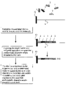
PCT/US02/07926
reaction mixture containing bound target) then analyzing the solution phase
for
degradation product indicative of a mutation.
[0065] Figure 2A shows a flowchart diagram illustrating an embodiment of
the
present invention. In general overview, the method comprises the steps of:
exposing a
bound target nucleic acid having a region at which a mutation is present to a
plurality
of probes; exposing the hybridized target nucleic acid and probe mixture to a
degradation agent that preferentially degrades single-stranded nucleic acids;
and
detecting the presence of mutation in the target nucleic acid when a single-
stranded
region is degraded.
[0066] More specifically, the target nucleic acid (8) having a region with
a
mutation (22) is bound to a solid phase or semi-solid phase matrix (10). The
target
nucleic acid (8) is exposed to a plurality of probes (2) that are labeled by,
for example,
fluorescence. The target nucleic acid (8) and the plurality of probes (2) are
incubated
under optimal time, temperature, oligonucleotide concentration, salt type, and
salt
concentration conditions. Stringent hybridization conditions that maximize
specific
hybridization by improving bonding energy symmetry and providing similar
melting
temperature for each probe are employed. The hybridization conditions enable
only
complementary probes to hybridize to the target nucleic acid. Because no probe
will
be complementary to the region having a mutation (22), hybridization will not
occur
at that region, and the region will remain single-stranded.
[0067] After exposure to a degradation agent that preferentially
degrades single-
strand nucleic acid, the hybridization product is removed from solution by its
bound
end. The use of bound target nucleic acid enables a number of samples to be
screened
simultaneously by removing the bound portion from solution and then analyzing
the
solution phase for segments of hybridized (i.e., double-stranded) degradation
product
23
CA 02441021 2003-09-15
WO 02/074995
PCT/US02/07926
(26) indicative of the presence of a mutation in the target nucleic acid. The
presence
of one or more segments of hybridized degradation product (26) in solution is
indicative that the target nucleic acid comprises a region having mutation
(22) that
was degraded by the degradation agent. The mutation is detected by exposing
the
solution to a light source in a spectrophotometer at the specific absorption
wavelength, which reveals the appreciable quantities of fluorescing
degradation
product (26) indicative of a mutation (22).
[00681 Figure 2B illustrates an embodiment of the method of the
invention
wherein a mutation is not present in a target nucleic acid sample. In this
embodiment,
the target nucleic acid is bound to a solid phase or semi-solid phase matrix
(10). The
target nucleic acid is exposed to a plurality of probes (4) that are designed
to
hybridize to the target such that gaps separate one or more of the hybridized
probes.
The probe (4) designed to anneal to the area of the target nucleic acid most
distal to
the matrix (10) is labeled with a reporter (12), for example, a
radionucleotide or a
fluorophore.
[0069] The target nucleic acid and the plurality of probes (4) are
incubated under
optimal time, temperature, probe concentration, salt type, and salt
concentration
conditions. The plurality of probes (4) are complementary to different
positions of the
target nucleic acid such that hybridization of members of the plurality with a
wild-
type target results in a series of probes (4), along at least a portion of the
target
sequence, that are separated by a gap of single-stranded nucleic acid.
Stringent
hybridization conditions that maximize specific hybridization by improving
bonding
energy symmetry and providing similar melting temperatures for each probe are
preferably employed. Those hybridization conditions enable only complementary
probes to hybridize to the target nucleic acid.
24
CA 02441021 2003-09-15
WO 02/074995
PCT/US02/07926
[0070] The target nucleic acid and probe (4) hybridization product (18)
is then
exposed to an agent, or alternatively conditions employed with the agent, that
do not
cleave the target nucleic acid at the single-stranded gap separations, but
that degrades
a single stranded gap region where one or more probes failed to hybridize. The
hybridization product (18) comprising the target nucleic acid and probes is
then
removed by its bound end from solution. The use of bound target nucleic acid
enables
a number of samples to be screened simultaneously by removing the bound
portion
from solution then analyzing the solution phase for degradation product
indicative of
a mutation. The absence of the reporter (12) remaining in solution indicates
the
absence of mutation in the target nucleic acid.
[0071] Figure 2C shows a flow chart diagram that illustrates an
embodiment of
the invention wherein a mutation is present in a target nucleic acid in the
sample.
According to this embodiment, the target nucleic acid (8) having a mutation
(22) is
bound to a solid phase or semi-solid phase matrix (10). The target nucleic
acid (8) is
exposed to a plurality of probes (4) designed to hybridize to the target (8)
such that
gaps separate one or more of the hybridized probes and the probe (4) designed
to
complement the area of the target nucleic acid (8) most distal to the matrix
(10) is
labeled with a reporter (12).
[0072] The target nucleic acid (8) and the plurality of probes (4) are
incubated
under the above-described stringent hybridization conditions. The conditions
enable
the plurality of probes (4) complementary to different positions of the target
nucleic
acid such that hybridization of members of the plurality with a wild-type
target results
in a series of probes (4), along at least a portion of the target sequence
(8), that are
separated by a gap of single-stranded nucleic acid. The stringent
hybridization
conditions enable only complementary probes to hybridize to the target nucleic
acid.
CA 02441021 2011-05-31
[0073] The target nucleic acid (8) and probe (4) hybridization is
then exposed to
an agent, or alternatively conditions employed with the agent, that do not
cleave the
target nucleic acid at the single-stranded gap separations, but that degrade a
single
stranded gap region where one or more probes failed to hybridize. The
hybridization
product comprising the target nucleic acid and probes is then removed by its
bound
end from solution. The presence of one or more segments of hybridized
degradation
product (28) including the labeled reporter (12) remaining in solution
indicates the
presence of mutation (22) in the target nucleic acid (8), because the single
stranded
region of mutation (22) was degraded by the degradation agent.
[0074] The following example illustrates methods of the invention useful to
detect
a mutation in the mutation cluster region of the APC in samples prepared from
stool.
Example 1: Mutation detection in the APC Mutation Cluster Region
[0075] Methods of the invention are used to detect the C -0 T point
mutation at
codon 1450 in the APC mutation cluster region, at
http://perso.curie.fr/Thierry.Soussi/APC.html (last visited Feb. 20, 2001).
Any
biological sample that comprises APC may be used, including, for example, a
stool
sample. For the analysis of stool samples, preferred methods of the invention
comprise obtaining at least a cross-section or circumferential portion of a
voided stool
as taught in U.S. patent numbers 5,741,650, and 5,952,178. While a cross-
sectional or circumferential portion of stool is desirable, methods provided
herein
are conducted on random samples obtained from voided stool, which include
26
CA 02441021 2011-05-31
smears or scrapings. Once obtained, the stool specimen is homogenized. A
preferable buffer for homogenization is one that contains at least 16 mM
ethylenediaminetetraacetic acid (EDTA), as taught in co-pending, co-owned U.S.
patent application serial number 09/491,093. It has been discovered that the
use of
at least 16mM EDTA, and preferably 100 mM EDTA or greater improves the yield
of
nucleic acid from stool. Thus, a preferred buffer for stool homogenization
comprises
phosphate buffered saline, 20-100 mM NaCI or KCI, at least 16 mM EDTA, and
optionally a detergent (such as SDS) and a proteinase (e.g., proteinase K).
[0076] After homogenization, nucleic acid is preferably isolated from
the stool
13 sample. Isolation or extraction of nucleic acid is not required in all
methods of the
invention, as methods of the invention can be adequately performed in
homogenized
stool without isolation of nucleic acids. In a preferred embodiment, however,
homogenized stool is spun to create a supernatant containing nucleic acids,
proteins,
lipids, and other cellular debris. The supernatant is treated with a detergent
and
proteinase to degrade protein, and the nucleic acid is phenol-chloroform
extracted.
The extracted nucleic acids are then precipitated with alcohol. Other
techniques can
be used to isolate nucleic acid from the sample. Such techniques include
hybrid
capture, and amplification directly from the homogenized stool. Nucleic acids
can be
purified and/or isolated to the extent required by the screening assay to be
employed.
20 [0077] The nucleic acid is then mixed with steptavidin coated Dynal
beads, which
provides a solid phase matrix. The nucleic acid and bead mixture is vortexed
and
incubated which binds the beads to the nucleic acid. The nucleic acid can be
27
CA 02441021 2012-07-11
amplified by PCR, which requires the nucleic acid template to be mixed with
binding
and wash buffers. The nucleic acid mixture is vortexed. The supernatant is
removed,
and buffer is added. These steps are then repeated a number of times.
[0078] Nucleic acid probes designed to complement consecutive regions of
the
known APC mutation cluster region are employed. The probes are uniform in
length
and are fluorescently labeled. The probe and the target nucleic acid
comprising a
27a
CA 02441021 2003-09-15
WO 02/074995
PCT/US02/07926
point mutation in codon 1450 are incubated under conditions that maximize
hybridization selectivity. Probe melting temperature disparities are
eliminated,
improving selectivity, when a suitable combination of hybridization
temperature,
time, probe concentration, salt type and salt concentration conditions are
employed.
TMAC is the agent selected to improve hybridization selectivity.
[0079] The probes are designed to detect mutations at codon 1450 in the
APC
mutation cluster region. When hybridizing under these selective hybridization
conditions, the presence of a single mutation in the mutation cluster region
will
prevent the complementary probe from hybridizing, such that a portion of the
region
remains single stranded.
[0080] Consecutive complementary probes are designed to hybridize to the
wild
type APC mutation cluster region where the 5' end of that region is (5'-
CTCCACCACCTCCTCAA ACAGCTCAAACCAAGCG
AGAAGTACCTAAAAATA -3', SEQ ID NO:1).
[0081] In the experiment, each probe comprises 17 nucleotides, and the 5'
end of
the complementary probe designed for the region of codon 1450 is
(5'- CGCTTGGTTTGAGCTGT -3', SEQ ID NO: 2). The complimentary probe
upstream of the codon 1450 point mutation region is (5'-
TTGAGGAGGTGGTGGAG ¨3', SEQ ID NO: 3). The complimentary probe
downstream of the 1450 point mutation region is (5'- TATTTTTAGGTACTTCT -
3', SEQ ID NO: 4). The probes and the target nucleic acid sample comprising
the
point mutation at codon 1450 in the mutation cluster region are incubated
under
conditions that maximize hybridization selectivity. The probe complimentary to
the
wild type region, SEQ ID No. 2, will not hybridize to the sequence comprising
the
28
CA 02441021 2003-09-15
WO 02/074995
PCT/US02/07926
point mutation at codon 1450 (C ¨> T at the codon 1450 point mutation), (5'-
ACAGCTCAAACCAAGTG -3', SEQ ID NO:5). The point mutation at codon 1450
prevents hybridization and the portion of the APC region containing the
mutation will
remain single stranded.
[0082] After hybridization, unhybridized probes are removed by washing the
nucleic acid mixture under time, temperature, salt type and salt concentration
conditions that preserve the nucleic acid:probe hybrids. Exposure to the
enzyme Si
cleaves the target nucleic acid at the single-stranded region comprising the
point
mutation at codon 1450, where the complimentary probe failed to hybridize.
[0083] The degradation products are separated by gel electrophoresis and
analyzed using a spectrophotometer. The presence of mutation is detected by
the
presence of one or more degradation products, each comprising double-stranded
nucleic acids which fluoresce upon excitation at the appropriate
spectrophotometer
wavelength.
Example 2: Probes Blocking Polymorphic Variants at Codon 1493
[0084] Generally, a single nucleotide polymorphism is associated with
each
region having 1,000 nucleotide base pairs. According to methods of the
invention,
associated polymorphic variants are factored into probe design such that
associated
polymorphisms are "blocked" and other alterations may be detected.
[0085] It is possible to avoid detecting a polymorphic variant as a
false positive
on a target nucleic acid. This provides an opportunity for de novo alteration
detection
on such targets. Figure 3 illustrates methods of the invention employed to
block the G
---> A polymorphism at codon 1493 of the APC mutation cluster region, to
enable de
novo detection.
29
CA 02441021 2003-09-15
WO 02/074995
PCT/US02/07926
[0086] Any biological sample that comprises an associated polymorphism
may be
used, for example, a tissue, stool or blood sample. For this analysis, blood
samples
from six individuals were separately tested. The sample set included a
polymorphic
variant base at codon 1493. For the purposes of a control in this experiment,
genotype sequencing was performed on the six individual samples prior to
performing
the analysis illustrated in Figure 3. The control sequence confirmed that the
sample
set being tested included two homozygous individuals having two G bases, two
homozygous individuals having an A base and a G base, and two homozygous
individuals having two A bases.
[0087] Each individual sample was analyzed as follows. Nucleic acid was
extracted from each individual blood sample. The nucleic acid was then mixed
with
magnetic streptavidin coated Dynal beads, which provided a solid phase matrix.
The
nucleic acid and bead mixture was vortexed and incubated to bind the beads to
the
nucleic acid. The nucleic acid was thereafter amplified by PCR, which required
the
nucleic acid template to be mixed with binding and wash buffers. During the
PCR
process, the reverse PCR primer was biotinylated. The PCR product was
denatured
and made single stranded. The nucleic acid mixture was vortexed. The
supernatant
was removed, and buffer was added.
[0088] Nucleic acid probes were designed to compliment the target
nucleic acid
including the polymorphic variants in codon 1493 of the APC mutation cluster
region.
The probes are uniform in length are 17 base pairs long. The probes are
designed so
that after hybridization the probe positioned second in from the free end of
the target
nucleic acid (i.e., the end of the target nucleic acid that is not bound to
the bead) has a
P32 reporter on its 5' end. The probes and the target nucleic acid are
hybridized at
59 C for one hour. The hybridization solution was a mixture of 3 molar
tetramethyl
CA 02441021 2003-09-15
WO 02/074995
PCT/US02/07926
ammonium chloride (TMA); 1mM EDTA; 10 mM phosphate buffer at a pH of 6.8;
5X of Denhardts solution; 0.04 mg/ml of yeast RNA; SDS at 0.1% of the mixture
and
between about 0.04 micromolar and about 0.64 micromolar of the probe mixture.
The
tube is exposed to a magnet that attracts the magnetic beads and retains the
target
nucleic acid that is bound to the beads and the supernatant is removed.
[0089] After hybridization, the unhybridized probes were removed by
washing the
nucleic acid mixture under conditions that preserve the nucleic acid:probe
hybrids.
The hybridization solution was washed a series of times under varying time and
temperature conditions. The wash solution contained a mixture of 3 molar
tetramethyl amonium chloride (TMA); 1mM EDTA; 10 mM phosphate buffer at a pH
of 6.8; and SDS at 0.1% of the mixture. After each wash, the tube is exposed
to a
magnet that attracts the magnetic beads and retains the target nucleic acid
that is
bound to the beads and the supernatant is removed. In the first wash, the
hybridization solution was mixed with the wash solution at 59 C for 15
minutes.
Thereafter, in the second wash, the hybridization solution was mixed with the
wash
solution at room temperature, about 22 C, for 1 minute. In the third wash, the
hybridization solution was mixed with a the wash solution at room temperature,
about
22 C, for about 1 minute.
[0090] In a test tube, the target nucleic acid was exposed to 0.015 unit
per
microliter of the enzyme Si, in the buffer containing sodium acetate at 50 mM,
NaC1
at 280 mM, and ZnSO4 at 4.5 mM, per microliter of the cutting reaction for
thirty
minutes at room temperature, about 22 C. The reaction is a 100 microliter
reaction.
Exposure to the enzyme Si cleaves any single stranded regions of the target
nucleic
acid. The tube is exposed to a magnet to attract the magnetic beads and retain
the
31
CA 02441021 2003-09-15
WO 02/074995
PCT/US02/07926
target nucleic acid that is bound to the beads. The supernatant is pipetted
out of the
tube and mixed with a load dye.
[0091] The supernatant is run on a 6% non-denaturing acrylamide gel at a
rate of
1,200 Volt hours. The gel is exposed to an instant imager, which picks up
radioactivity. The gel is analyzed for fragment size and any cuts in the
target nucleic
acid can be determined by the size of the product on the gel.
[0092] Referring to Figure 3, Run 1 shows experimental results where the
six
samples being tested were exposed to probes designed to complement the
polymorphic variant where codon 1493 is a G. The six samples include two
individuals having two G bases, two individuals having an A base and a G base,
and
two individuals having two A bases. Referring to the sample from the two
individuals
having two G bases, the product on the gel shows that exposure to the agent,
Si, did
not cut the target. The target nucleic acid was not cut because the probes
designed to
complement the polymorphic variant where codon 1493 is a G hybridized to the
target
nucleic acid of the two individuals having two G bases. Referring again to Run
1,
samples from two individuals having an A base and a G base were exposed to the
probes designed to complement the polymorphic variant where codon 1493 is a G.
The probes hybridized to the G variant of the two individuals having an A base
and a
G base. However, no probe hybridized to the polymorphic variant of the target
sample where codon 1493 is an A and upon exposure to the agent Si, the single
stranded region at codon 1493 was cut generating product on the gel. Finally,
in Run
1, samples from two individuals having two A bases were exposed to the probes
designed to complement the polymorphic variant where codon 1493 is a G. No
probe
hybridized to the polymorphic variant of the two individuals having two A
bases, and
32
CA 02441021 2003-09-15
WO 02/074995
PCT/US02/07926
exposure to the agent, Si, cut the target at the region of the polymorphic
variant
generating product on the gel.
[0093] Referring to Figure 3, Run 2 shows experimental results where the
six
samples being tested were exposed to probes designed to complement the
polymorphic variant where codon 1493 is an A. Again, the six samples include
two
individuals having two G bases, two individuals having an A base and a G base,
and
two individuals having two A bases. Referring to the sample from the two
individuals
having two G bases, the product on the gel shows exposure to the agent, Si,
cut the
target at the region of the polymorphic variant leaving product on the gel.
The target
nucleic acid was cut because the probes including the polymorphic variant
where
codon 1493 is A failed to hybridize to the target nucleic acid of the two
individuals
having two G bases. Referring again to Run 2, samples from two individuals
having
an A base and a G base were exposed to the probes designed to complement the
polymorphic variant where codon 1493 is an A. The probes hybridized to the A
variant of the two individuals having an A base and a G base. However, no
probe
hybridized to the polymorphic variant of the target sample where codon 1493 is
G and
upon exposure to the agent Si, the single stranded region at codon 1493 was
cut
generating product on the gel. Finally, in Run 2 samples from two individuals
having
two A bases were exposed to the probes designed to complement the polymorphic
variant where codon 1493 is a A. The product on the gel shows exposure to the
agent,
Si, did not cut the target. The target nucleic acid was not cut because the
probes
designed to complement the polymorphic variant where codon 1493 is an A
hybridized to the target nucleic acid of the two individuals having two A
bases.
[0094] Referring to Figure 3, Run 3 shows experimental results where the
six
samples being tested were exposed to two different sets of probes: one
designed to
33
CA 02441021 2003-09-15
WO 02/074995
PCT/US02/07926
complement the polymorphic variant where codon 1493 is an A and one designed
to
complement the polymorphic variant where codon 1493 is a G. Again, the six
samples include two individuals having two G bases, two individuals having an
A
base and a G base, and two individuals having two A bases. Referring again to
Run 3,
exposure to sets of probes designed to complement both the polymorphic variant
A
and the polymorphic variant G succeeds in blocking the codon 1493 region in
all six
samples. Thus, both polymophic variant probes successfully block the
polymorphic
variants on the target nucleic acids tested and, according to methods of the
invention,
any other alterations on the target may be detected.
[0095] Finally, referring to Figure 3, Run 4 shows experimental results
where,
again, six samples were tested, the samples from two individuals having two G
bases,
two individuals having an A base and a G base, and two individuals having two
A
bases. The six samples being tested were exposed to probes where no probe was
designed to complement the polymorphic variants present in the six samples.
Referring again to Run 4, upon exposure to the agent Si, the single stranded
region at
codon 1493 for both the polymorphic variant where codon 1493 is an A and the
polymorphic variant where codon 1493 is a G was cut generating product on the
gel.
Thus, according to methods of the invention, probes may be designed to block
the
G ¨> A polymorphism at codon 1493 of the APC to enable detection of other
alterations in the target nucleic acid.
Example 3: Mutation detection by probe reporting moiety signal
[0096] In another embodiment, referring to Figure 4A, a quenching moiety
(34) is
placed at one end and a reporting moiety (32) is placed at the other end of
each of the
probes (30) such that the absence of hybridization of one probe results in the
reporter
of an adjacent probe failing to be quenched resulting in a reporter signal
(35).
34
CA 02441021 2003-09-15
WO 02/074995
PCT/US02/07926
Accordingly, an alteration is detected by the reporting moiety (32) signal
(35) that
results when a probe (30) fails to hybridize to the target at the site of the
alteration.
The reporting moiety (32) or reporter may be detected via a fluorescent plate
reader or
alternatively with a gel apparatus equipped with fluorescent detection. A
degradation
agent is not required to detect the presence or absence of mutation in the
target
nucleic acid in this method of the invention. Accordingly, serial analysis may
be
avoided by employing this method of the invention.
[0097] Figure 4B illustrates the reporting moiety (32) of a first probe
(30)
annealed to the quenching moiety (34) of a second probe (30). The quenching
moiety
(34) quenches or suppresses the reporting moiety (32) signal (35). The
quenching
moiety (34) is conjugated to an arm on one end of probe (30) and the reporting
moiety
(32) is conjugated to an arm on the other end of probe (30), and each arm may
include
a sequence. In one embodiment, the sequences on the arm conjugated to the
quenching moiety (34) are designed to anneal to the arm conjugated to any
reporting
moiety (32). In one embodiment, the sequences on the arms are not specific to
the
sequence complementary to the target nucleic acid. The sequences on each arm
may
range between about I base pair and about 7 base pairs.
[0098] Figure 4C shows a flow chart diagram that illustrates a method of
the
invention using probes having a reporting moiety (32) and a quenching moiety
(34)
and wherein no mutation is present in a target nucleic acid. According to this
method,
when a series of probes (30), as illustrated in Figures 4A and 4B, are mixed
together,
for example in solution, the arm conjugated to each quenching moiety (34) will
anneal
to the arm conjugated to each reporting moiety (32). Accordingly, each
reporting
moiety (32) is quenched by each adjacent quenching moiety (34) and the probe
(30)
mixture in solution presents no reporting signal.
CA 02441021 2003-09-15
WO 02/074995
PCT/US02/07926
[0099] Thereafter, a target nucleic acid (6) is exposed to the probe
(30) mixture to
form a hybridization product (20). According to the method, the hybridization
conditions enable only complementary probes to hybridize to the target nucleic
acid
(6). When the hybridization product (20) fails to present any signal, the
absence of
mutation in the target nucleic acid (6) is detected.
[0100] Figure 5 shows a flow chart diagram that illustrates a method of
the
invention using probes having a reporting moiety (32) and a quenching moiety
(34)
and wherein a mutation (22) is present in a target nucleic acid (8). According
to this
method, when a mixture of probes (30), as described above with reference to
Figures
4A-4C, is prepared, the probe (30) mixture presents no reporting signal (35).
[0101] Thereafter, a target nucleic acid (8) is exposed to the probe
(30) mixture to
form a hybridization product (20). The hybridization conditions enable only
complementary probes (30) to hybridize to the target nucleic acid (8). Because
no
probe (30) will be complementary to the region having a mutation (22),
hybridization
will not occur in the mutation (22) region and the mutation region will remain
single-
stranded. When a probe (30) fails to hybridize in the region of the mutation
(22) then
the reporting moiety (32) adjacent the region having the mutation (22) will
present a
signal (35), the signal (35) indicating the presence of mutation (22).
[0102] In the examples described above, embodiments of the invention
wherein a
target nucleic acid contains no mutation are described separately from
embodiments
of the invention wherein a target nucleic acid contains a mutation. However,
according to the invention, a sample may be heterogeneous and contain target
nucleic
acid molecules that are mutant or altered in addition to target nucleic acid
molecules
that are non-mutant or non-altered.
36
CA 02441021 2012-07-11
[0103] Methods of the invention are useful to detect a mutant or altered
target
nucleic acid in a heterogeneous mixture containing non-mutant or non-altered
target
nucleic acid, because the non-mutant or non-altered nucleic acid does not
interfere
with the generation of a signal due to the presence of the mutant or altered
nucleic
acid. In preferred embodiments, a detection assay of the invention is
performed on a
sample or sample fraction that has been enriched for mutant or altered target
nucleic
acid using methods described herein or known in the art.
EQUIVALENTS
[0104] The scope of the claims should not be limited by the preferred
embodiments set forth in the examples, but should be given the broadest
interpretation consistent with the description as a whole.
37
CA 02441021 2004-03-11
2004-03-11 revised sequence listing.txt
SEQUENCE LISTING
<110> Exact Sciences Corporation
<120> Method For Alteration Detection
<130> 011141-0019
<140> CA 2,441,021
<141> 2002-03-15
<150> US 09/809,713
<151> 2001-03-15
<150> US 09/988,491
<151> 2001-11-20
<160> 5
<170> PatentIn version 3.0
<210> 1
<211> 51
<212> DNA
<213> HOMO sapiens
<400> 1
ctccaccacc tcctcaaaca gctcaaacca agcgagaagt acctaaaaat a 51
<210> 2
<211> 17
<212> DNA
<213> Artificial Sequence
<220>
<223> Probe designed for the region of codon 1450
<400> 2
cgcttggttt gagctgt 17
<210> 3
<211> 17
<212> DNA
<213> Artificial Sequence
<220>
<223> probe upstream of the 1450 point mutation region
<400> 3
ttgaggaggt ggtggag 17
<210> 4
<211> 17
<212> DNA
<213> Artificial Sequence
<220>
<223> probe downstream of the 1450 point mutation
<400> 4
tatttttagg tacttct 17
<210> 5
<211> 17
Page 1
CA 02441021 2004-03-11
2004-03-11 revised sequence listing.txt
<212> DNA
<213> HOMO sapiens
<400> 5
acagctcaaa ccaagtg 17
Page 2