Note: Descriptions are shown in the official language in which they were submitted.
CA 02442057 2003-09-22
WO 02/076525 PCT/US02/07841
MEDICAL DEVICE HAVING RADIO-OPACIFICATION
AND BARRIER LAYERS
Field of the Invention
The present invention relates generally to devices for preventing vascular
diseases, and more specifically to in-vivo stents used in medical procedures.
Background of the Invention
As an alternative to vascular surgery, percutaneous transluminal angioplasty
(PTA) and percutaneous transluminal coronary angioplasty (PTCA) procedures are
being
widely used for treating stenotic atherosclerotic regions of a patient's
vasculature to
restore adequate blood flow. Catheters having an expansible distal end,
typically in the
form of an inflatable balloon, are positioned in a vessel, such as a coronary
artery, at a
stenotic site. The expansible end is then expanded to dilate the vessel in
order to restore
adequate blood flow to regions beyond the stenosis. While PTA and PTCA have
gained
wide acceptance, these angioplasty procedures suffer from two major problems:
abrupt
closure and restenosis.
Abrupt closure refers to rapid re-occlusion of the vessel immediately after or
within hours of the initial treatment, and often can result in myocardial
infarction if blood
flow is not restored in a timely manner. Abrupt closure often results from
either an
intimal dissection or from rapid thrombus formation which occurs in response
to injury of
the vascular wall from the initial angioplasty procedure. Restenosis refers to
a re-
narrowing of the artery over the weeks or months following an initially
apparently
successful angioplasty procedure. Restenosis occurs in a significant amount of
all
angioplasty patients and results, at least in part, from smooth muscle cell
proliferation and
migration.
Many different strategies have been proposed to diminish the likelihood of
abrupt
closure and reduce the rate of restenosis. One such method involves the
implantation of a
vascular stent following angioplasty. Stents are thin-walled tubular
scaffolds, which are
expanded in the arterial lumen following the angioplasty procedure. Most
commonly, the
stents are formed f om a malleable material, such as stainless steel, and are
expanded in-
situ using a balloon. Alternatively, the stents may be formed from a shape
memory alloy
or other elastic material, in which case they are allowed to self-expand at
the angioplasty
CA 02442057 2009-09-11
60412-3210
treatment site. In either case, the stent acts as a mechanical support for the
artery wall,
thereby inhibiting abrupt closure and reducing the restenosis rate as compared
to PTCA.
Recent developments in medical devices have stressed the importance of
visually
perceiving the stent in-vivo as it is being placed within the vasculature of
the patient.
Additionally, it is advantageous and sometimes necessary to visually locate
and inspect a
previously deployed stent or to treat restenosis occurring at the location of
the stent.
Fluoroscopy is one technique that allows visualization of a stent in-vivo. To
visualize the
stent in-vivo using fluoroscopy, the stent must be made from a material that
is highly
radio-opaque or must use a delivery catheter that provides radio-opaque
markers.
However, the preferred structural material, stainless steel, used in stents is
not highly
radio-opaque. Thus, several solutions have been proposed such as coating a
conventional
stainless steel stent with a radio-opaque material such as gold.
While coated and non-coated stents have been successful in inhibiting abrupt
closure and reasonably successful in inhibiting restenosis, a significant
portion of the
treated patient population still experiences restenosis over time. It is
possible for the
alloying metals of the stent material (e.g. stainless steel ) or the gold
alloy coating to be
leached by the body fluids resulting in the activation of platelets and cells,
the possible
precursor to thrombus formation, on a localized level. Additionally, most
stent structures
comprise an open lattice, typically in a diamond or spiral pattern, and cell
proliferation
(also referred to as intimal hyperplasia) can intrude through the interstices
between the
support elements of the lattice and the treatment site once again becomes
occluded.
Therefore, there is a need for an improved medical device that can be
visualized
in-vivo while further aiding in the prevention of restenosis.
-2-
CA 02442057 2009-09-11
60412-3210
Summary of the Invention
Some embodiments of the present invention address the need for an
improved medical device that can be visualized in-vivo while further aiding in
the prevention
of restenosis by providing a medical device having radio-opacification and at
least one
barrier layer.
In accordance with a first aspect of the present invention, there is provided
a
laminate structure for making a medical device comprising: a core having an
outer surface;
a first radio-opaque layer disposed on at least a portion of the outer surface
of the core, the
first radio-opaque layer having an outer surface; and a second layer disposed
on at least a
portion of the outer surface of the first radio-opaque layer, the second layer
comprising an
oxide of Ti, an oxide of Cr, an oxide of Ta, an oxide of Al, a nitride of Cr,
a nitride of Ta, a
nitride of Al, a carbide of Ti, a carbide of Cr, a carbide of Ta, or a carbide
of V; wherein the
second layer isolates the first radio-opaque layer from blood within a
patient's vessel.
In accordance with another aspect of the present invention, the outer
surface of the second layer has micro-pores or other structures to receive
therapeutic drugs
and deliver them to the vessel in the area of the medical device.
There is also provided in a medical device implantable within a patient's
vessel, the medical device comprising: a core having an outer surface, the
outer surface
having a layered structure thereon, the layered structure comprising: a radio-
opaque inner
layer disposed onto the outer surface of the core, and an outer bio-compatible
layer
surrounding the radio-opaque inner layer, the outer bio-compatible layer
comprising an
oxide of Ti, an oxide of Cr, an oxide of Ta, an oxide of Al, a nitride of Cr,
a nitride of Ta, a
nitride of Al, a carbide of Ti, a carbide of Cr, a carbide of Ta, or a carbide
of V; wherein the
outer layer isolates the radio-opaque inner layer from blood or tissue within
the patient's
vessel.
Another aspect of the invention provides a medical device comprising: a
core having an outer surface; a radio-opaque inner layer disposed onto at
least a portion of
the outer surface of the core, and a bio-compatible outer layer, the outer
layer covering at
least a portion of the radio-opaque inner layer to reduce contact between the
radio-opaque
material and blood within a patient's vessel, wherein the outer layer
comprises an oxide of
Ti, an oxide of Cr, an oxide of Ta, an oxide of Al, a nitride of Cr, a nitride
of Ta, a nitride of
Al, a carbide of Ti, a carbide of Cr, a carbide of Ta, or a carbide of V.
-3-
CA 02442057 2009-09-11
60412-3210
Brief Description of the Drawings
The foregoing aspects and many of the attendant advantages of embodiments of
this
invention will become more readily appreciated as the same become better
understood by
reference to the following detailed description, when taken in conjunction
with the
accompanying drawings, wherein:
FIGURE 1 illustrates a side view of a conventional medical device; .
FIGURE 2 illustrates a side view of a medical device in accordance with an
embodiment of the present invention;
FIGURE 3 illustrates a cross-sectional view taken along lines A-A of the
medical
device shown in FIGURE 2;
FIGURE 4 illustrates a magnified portion of the cross-sectional view taken
along
lines A-A of the medical device shown in FIGURE 2;
FIGURE 5 illustrates a cross-sectional view of a portion of a medical device
according to a second embodiment of the present invention;
FIGURE 6 illustrates a cross-sectional view of a portion of a medical device
according to a third embodiment of the present invention;
FIGURE 7 illustrates a cross-sectional view of a medical device in-situ in a
patient's vessel according to a fourth embodiment of the present invention;
FIGURE 8 illustrates a cross-sectional view of a medical device in-situ in a
patient's vessel according to a fifth embodiment of the present invention;
FIGURE 9 illustrates a cross-sectional view of a medical device in-situ in a
patient's vessel according to a sixth embodiment of the present invention; and
FIGURE 10 illustrates a cross-sectional view of a portion of a medical device
having a circular cross-section.
Detailed Description
While, as will be better understood from the following description, the
present
invention was developed for coronary stents and, thus, is expected to find its
primary use
-3a-
CA 02442057 2003-09-22
WO 02/076525 PCT/US02/07841
with such coronary stents, it is to be understood that the invention can be
used with other
medical devices such as vena cava filters, aneurysm coils or other implantable
devices
that require the ability to be visualized in-vivo and to have a bio-compatible
barrier layer.
Thus, it is to be understood that the disclosed embodiment is only by way of
example and
should not be construed as limiting.
Prior to describing an illustrative embodiment of the invention, a brief
discussion
of the structure of one type of medial device is set forth. In this regard,
attention is
directed to FIGURE 1, which illustrates a conventional medical device known in
the art
as a coronary stent 10. The coronary stent 10 is deployed in-vivo at a
stenosed vessel
following a PTCA procedure. The stent 10 is deployed from a delivery catheter
just
proximal to the diseased section of the vessel and is expanded into abutment
against the
interior lining of the vessel wall. Once in-situ, the stent 10 acts as a
mechanical support
for the vessel wall, inhibiting abrupt closure.
Referring again to FIGURE 1, the skeletal frame of the stent 10 preferably
includes wire or bar-like members 12, each forming a distinct, repetitive
zigzag pattern.
This repetitive zigzag pattern consists of multiple V-shaped curves 14. The
areas 16
within the V-shaped curves 14 are open. With no recognizable beginning or end
to this
zigzag pattern, the bar-like member 12 forms expandable zigzag segment 18. A
plurality
of zigzag segments 18 are arranged along the longitudinal axis of the stent 10
so that the
V-shaped curves 14 of abutting zigzag segments 18 may be joined through an
interconnecting element 20. Through the interconnecting elements 20, a
continuous wire-
like framework is created between the multiple zigzag elements 18 forming the
stent 10.
The coronary stent illustrated in FIGURE 1 is only exemplary of many of the
various medical devices which may incorporate the benefits of the present
invention. The
present invention could also be used with devices such as vena cava filters or
aneurysm
coils and other small implanted devices that need to be fluoroscopically
visible. For
clarity, the remaining detailed description refers only to a stent. However,
it will be
appreciated that any medical device can incorporate the aspects of the present
invention.
The method of making and using the stents described above and used in
conjunction with
PTCA procedures are well known in the art and are not described in detail
here.
The present invention is directed to an improved coronary stent that provides
in-
vivo visualization and a bio-compatible barrier layer that may reduce the
possibility of
-4-
CA 02442057 2003-09-22
WO 02/076525 PCT/US02/07841
restenosis. These characteristics are attributable to constructing the
coronary stent with a
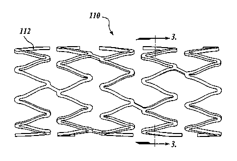
laminate or composite structure. FIGURES 2-3 illustrates an exemplary
embodiment of
the improved stent 110 constructed in accordance with the aspects of the
present
invention. The stent 110 is comprised of many bar-like members 112. As best
shown in
FIGURE 4, the members 112 when viewed in cross-section include a core or body
130,
and a first or inner layer 132 disposed directly adjacent to and preferably
surrounding the
core 130. However, it will be appreciated that other configurations of the
inner layer may
be utilized. For example, as best shown in FIGURE 6, the inner layer 132 may
be
disposed on one side of the core 130.
The core 130 is constructed from a material that provides the stent with the
necessary strength and flexibility to support the diseased vessel. The core
130 is
preferably made from 316 stainless steel; however, other materials may be used
such as
titanium, nickel titanium, or tantalum or their alloys. In an alternative
embodiment, the
core 130 can include a centrally located lumen extending longitudinally
therethrough,
instead of being of a solid construction, as shown in FIGURE 4. The inner
layer 132
disposed over the core is constructed from a radio-opaque material that
permits
fluoroscopic imaging and is magnetic resonance imaging (MRI) distortion free
such as
gold or a gold alloy of nickel, chromium, copper, or iron. It will be
understood that the
thickness of the inner layer is such (preferably 3-12 microns) that it can be
viewable
during fluoroscopy.
Disposed over the inner layer 132 is an outer layer 134 that forms the
outermost
surface of the stent. The outer layer 134 overlays the inner layer 132 to form
a barrier
between the inner layer and the blood and/or tissue of the patient's vessel.
Additionally,
the outer layer 134 provides a dielectric barrier that inhibits charge
transfer to and from
the inner layer 132. Through the multiple layers of the core 130, inner layer
132, and
outer layer 134, a laminate or composite structure 136 is constructed to form
the
members 112. The members 112 may be arranged in a variety of configurations to
form
the stent 110.
The outer layer 134 is made from a bio-compatible or "bio-friendly" material
that
is chemically inert with human blood and tissue and preferably has a thickness
of
approximately one micron. The outer layer is chemically inert from its
inherent ability to
form a stable oxide or nitride. The oxide or nitride forms a thin film on the
outer surface
-5-
CA 02442057 2003-09-22
WO 02/076525 PCT/US02/07841
of the outer layer to form a protective barrier. Some examples of suitable
materials that
may be used for the outer layer include, but are not limited to stainless
steel, titanium
(Ti), chromium (Cr), tantalum (Ta), aluminum (Al), and vanadium (V), all of
which form
stable oxides in the native form or are induced by thermal oxidation.
Stainless steel may
also be suitably passivated to form a robust oxide. Likewise, nitrides of the
same
materials can be used as the outer layer and are formed in a plasma reactor.
Other
suitable complexes such as carbides, oxy-nitrides, and silicides may be also
used based
on their relative compatibility with blood and tissue. Further, any bio-
compatible
polymer may be used. The outer layer 134 may also include platinum, irridium
and their
alloys. Regardless of the material used, it is preferable to use one that is
MRI distortion
free.
FIGURE 5 illustrates another exemplary embodiment of the stent according to
the
present invention. The stent comprises a core 230 having an outer layer 234
disposed
thereon. The core 230 is preferably comprised of an alloy of gold and titanium
or
tantalum or combinations thereof. Other materials having the necessary
requirements of
strength and radio-opacity may also be utilized to form the core 230. For
example, the
core can be composed of an alloy consisting of 70% gold and 30% titanium. The
outer
layer 234, made from any suitable bio-compatible material described above, is
then plated
onto the core 230 to provide a barrier between the alloy and the patient's
blood and/or
tissue. Alternatively, the core and outer layer may be bonded together by co-
extrusion or
rolling and the stent is fabricated from this laminate composite.
FIGURE 7 illustrates a cross-sectional view of a stent in-situ in a patient's
vessel
according to yet another exemplary embodiment of the present invention. The
stent 310
is comprised of multiple bar-like members 312. The members 312 include a
rectangular
shaped core or body 330, a radio-opaque inner layer 332 disposed on a portion
of the
core 330, and an outer layer 334 that overlays the radio-opaque inner layer
332 to form a
laminate or composite structure. The bottom surface 340 of the core 330, which
is left
uncovered by the inner layer 332, engages the vessel wall 342 when the stent
is in-situ.
The outside layer 334 provides a barrier between the radio-opaque inner layer
332 and the
blood within the patient's vessel. Any suitable material, as discussed above
with
reference to FIGURE 4, may be used for each layer of the laminate structure.
-6-
CA 02442057 2003-09-22
WO 02/076525 PCT/US02/07841
FIGURE 8 illustrates a cross-sectional view of a stent in-situ in a patient's
vessel
according to yet another exemplary embodiment of the present invention. The
stent 410
is comprised of multiple bar-like members 412. The members 412 include a
rectangular
shaped core or body 430, a radio-opaque inner layer 432 disposed on the top
surface 438
of the core 430, and an outer layer 434 disposed over the inner layer 432 and
a portion of
the core 430 to form a laminate or composite structure. The bottom surface 440
of the
core 430, which is left uncovered by the inner layer 432, engages the vessel
wall 442
when the stent is in-situ. The outside layer 434 provides a barrier between
the radio-
opaque inner layer 432 and the blood within the patient's vessel.
Additionally, the
core 430 provides a barrier between the radio-opaque inner layer 432 and the
vessel wall.
Any suitable material, as discussed above with reference to FIGURE 4, may be
used for
each layer of the laminate structure.
FIGURE 9 illustrates a cross-sectional view of a stent in-situ in a patient's
vessel
according to still yet another exemplary embodiment of the present invention.
The
stent 510 is comprised of multiple bar-like members 512. The members 512
include a
rectangular shaped core or body 530, a radio-opaque inner layer 532, and an
outer
layer 534 to form a laminate or composite structure. The inner layer 532 is
disposed over
the top surface 538 of the core and a portion 544 of the side surfaces of the
core 530. The
outer layer 534 overlays the inner layer 532 and the remaining portion of the
side surfaces
of the core 530. The bottom surface 540 of the core 530, which is left
uncovered by the
inner layer 532, engages the vessel wall 552 when the stent is in-situ. The
outside
layer 534, in conjunction with the core 530, provides a barrier between the
radio-opaque
inner layer 532 and the blood and/or tissue within the patient's vessel. Any
suitable
material, as discussed above with reference to FIGURE 4, may be used for each
layer of
the laminate structure.
It will be appreciated by those skilled in the art that the laminate or
composite
structure that forms the stent illustrated in FIGURES 3-9 can be fabricated by
various
methods know in the art. For example, the inner layer may be disposed onto the
core
using conventional plating methods such as electro and/or electroless plating.
Likewise,
the outer layer may be disposed onto the inner layer by conventional plating
methods.
Other methods of disposing or bonding the layers onto the core can be used
such as
chemical vapor deposition and physical deposition in conjunction with
selective masking,
-7-
CA 02442057 2003-09-22
WO 02/076525 PCT/US02/07841
wet-chemical processing, and sol gel processing. Alternatively, separate
sheets or tubes
of material corresponding to the core and the inner and outer layers,
respectively, can be
fabricated into the laminate or composite structure by rolling (roll bonding)
or co-
extruding, or a combination of co-extruding, rolling, and plating. Those
skilled in the art
will appreciate that additional manufacturing processes such as annealing or
electro-
polishing may be administered during the fabrication of the composite
structure to control
the microstructure, internal stresses, composition and surface finish.
Additionally, it will
,be appreciated by those skilled in the art that the outer layer can be
fabricated to have a
crystallographic structure that minimizes surface energy to reduce chemical
and
biochemical reactions at the surface of the outer layer.
Often it is beneficial to treat the localized area of the diseased vessel that
is
stented. The outer layer may include a textured surface of micro-pores,
grooves, cross-
hatched lines or the like to receive a therapeutic agent. Drugs and treatments
which
utilize anti-thrombogenic agents, and anti-proliferation agents may be readily
deployed
from the textured outer surface of the outer layer of the stent. Specific
examples of
preferred therapeutic agents include Taxol and Heparin. However, it is to be
understood
that other agents may be deployed. Additionally, the cellular response can be
regulated
with a suitable textured surface even in the absence of drugs. To this end,
the textured
surface of the outer layer of the stent may induce favorable biological
reactions within the
patient's vessel.
In conjunction with the various embodiments of the present invention, it will
be
appreciated by those skilled in the art that the gold alloy composition used
for the inner
layer can be varied throughout the thickness of the deposit to achieve
specific mechanical
properties such as flexibility, strength, and weight. For example, the density
of the gold
layer may fluctuate as it extends circumferentially around the core and as it
extends
outwardly from the core.
While the preferred embodiment of the invention has been illustrated and
described, it will be appreciated that various changes can be made therein
without
departing from the spirit and scope of the invention. For example, it is
contemplated to
be within the scope of the invention to have a stent provided that already has
been coated
with a gold layer. The gold coated stent may then be plated with any suitable
bio-
compatible material discussed above to form a barrier between the gold plating
and the
-8-
CA 02442057 2003-09-22
WO 02/076525 PCT/US02/07841
blood and tissue within the patient's vessel. Additionally, the stent members
are shown in
FIGURES 2-9 as having a rectangular cross-section. However, it will be
appreciated by
those skilled in the art that other cross-sectional shapes may be utilized to
provide the
desired mechanical characteristics to the stent, such as a circular core,
which is shown in
FIGURE 10, or elliptical. The stent members formed by these other cross-
sectional
shapes may also include a centrally located lumen extending longitudinally
therethough,
as described above with the exemplary embodiment shown in FIGURE 4.
-9-