Note: Descriptions are shown in the official language in which they were submitted.
CA 02442275 2010-04-06
23940-1482
1
QUANTITATIVE ANALYSIS OF A TURBID PHARMACEUTICAL SAMPLE BY
IRRADIATION OF THE SAMPLE
Field of the invention
The present invention relates to apparatuses for analysing a turbid
pharmaceutical
s sample, e.g. a tablet, a capsule - especially a multiple unit pellet system
(MUPS) - or a
similar sample forming a pharmaceutical dose.
Background of the invention
Non-invasive, non-destructible analysis of whole tablets can be carried out by
io means of near-infrared (NIR) or Raman spectrometry. Today, NIR spectroscopy
is a
recognised technique for perforating a fast analysis of compounds. The common
feature of
both these techniques is that they utilise light in the NIR wavelength region
(700-2500 nm,
specifically 700-1500 nm) where pharmaceutical tablets are relatively
transparent (low
molar absorptivity). That is, light can in this region penetrate compressed
powders several
15 mm:s why information in the content can be obtained emanating from the bulk
of the tablet
and not only from the surface. A practical advantage of using NIR radiation is
that diode
lasers can be used.
One-example of such an analysis is described in US 5,760,399, assigned to Foss
NiRsystems Inc. This document discloses an instrument for performing a NIR
20 spectrographic transmission measurement of a pharmaceutical tablet. This
instrument is,
however, capable of providing only limited information as to the content of a
sample,
typically the quantity of a particular component in a sample. This prior-art
instrument
cannot provide detailed information of, for example, the three-dimensional
distribution of
one or more components in a sample. The technical background on which this
limitation is
25 based will be further discussed in connection. with the description of the
present invention.
The prior art also includes a significant amount of methods for optical
imaging of
human tissues, in particular for detecting disturbances of homogeneity, such
as the
presence of a tumour in human tissue. These methods are generally qualitative
measurements, not quantitative, in the sense that they primarily focus on
determining the
30 presence and the location of an inhomogeneity. These prior-art methods are
not suitable for
performing a quantitative analysis on pharmaceutical, turbid samples, such as
tablets and
capsules, in order to determine e.g. content and stru ctural parameters.
CA 02442275 2010-04-06
23940-1482
2
Summary of the invention
According to a first aspect of the invention there is provided
apparatuses for use in quantitative analysis of a turbid, pharmaceutical
sample, in
particular a pharmaceutical tablet or capsule of an equivalent pharmaceutical
dose.
According to the invention, the apparatuses comprises:
- means for generating an excitation beam of radiation; and
- means for focusing said excitation beam onto said sample.
According to one embodiment the apparatus also comprises:
- means for intensity modulating said excitation beam; and
- means for detecting all wavelengths simultaneously.
According to another embodiment the apparatus also comprises:
- means for splitting said excitation beam into two beams; and
- means for detecting transmitted light and non-transmitted light
respectively.
According to another embodiment, there is provided an apparatus
for use in quantitative analysis of a turbid pharmaceutical sample,
comprising:
- means for generating an excitation beam of radiation;
- means for intensity modulating said excitation beam;
- means for focusing said excitation beam onto said sample, wherein
said means for focusing said excitation beam onto said sample are parts of a
Fourier spectrometer; and
- means for detecting all wavelengths of radiation transmitted
through the sample simultaneously.
CA 02442275 2010-04-06
23940-1482
2a
According to another embodiment, there is provided an apparatus
for use in quantitative analysis of a turbid pharmaceutical sample,
comprising:
- means for generating an excitation beam of radiation;
- means for focusing said excitation beam onto said sample;
- means for splitting said excitation beam into two beams; and
- means for detecting transmitted light and non-transmitted light
respectively, wherein said means for detecting comprises a time-resolved or
phase-resolved detection unit.
The invention is based on the following principles. A sample to be
analysed by a spectrometric transmission and/or reflection measurement
presents
a number of so called optical properties. These optical properties are (i) the
absorption coefficient, (ii) the scattering coefficient and (iii) the
scattering
anisotropy. Thus, when the photons of the excitation beam propagate through
the
turbid sample - in transmission and/or reflective mode - they are influenced
by
these optical properties and, as a result, subjected to both absorption and
scattering. Photons that by coincidence travel along an essentially straight
path
through the sample and thus do not experience any appreciable scattering will
exit
the sample with a relatively short time delay. Photons that are directly
reflected on
the irradiated surface will also present a relatively short time delay, in the
case of
measurements on reflected light. On the other hand, highly scattered photons
(transmitted and/or reflected) exit with a substantial time delay or phase
difference. This means that all these emitted photons - presenting different
propagation times - mediate complementary information about the sample.
In a conventional steady state (no time-resolution) measurement,
some of that complementary information is added together since the emitted
light
is captured by a time-integrated detection. Accordingly, the complementary
information is lost in a conventional technique. For instance, a decrease in
the
registered light intensity may be caused by an increase in the sample
scattering
coefficient. However, the information about the actual cause is hidden, since
all
the emitted light has been time-integrated.
CA 02442275 2010-04-06
23940-1482
2b
According to the invention and in contrast to such prior-art
NIR spectroscopy with time-integrated intensity detection, the intensity of
the
emitted radiation from the sample is
CA 02442275 2003-09-17
WO 02/075286 PCT/SE02/00510
3
measured both as a function of the wavelength and as a function of the photon
propagation
time through said sample. Thus, the inventive method can be said to be both
wavelength-
resolved and time-resolved. It is important to note that the method is time-
resolved in the
sense that it provides information about the kinetics of the radiation
interaction with the
sample. Thus, in this context, the term "time resolved" means "photon
propagation time
resolved". In other words, the time resolution used in the invention is in a
time scale which
corresponds to the photon propagation time in the sample (i.e. the photon
transit time from
the source to the detector) and which, as a consequence, makes it possible to
avoid time-
integrating the information relating to different photon propagation times. As
an illustrative
example, the transit time for the photons may be in the order of 0,1-2 ns.
Especially, the
term "time resolved" is not related to a time period necessary for performing
a spatial
scanning, which is the case in some prior-art NIR-techniques where "time
resolution" is
used.
As a result of not time-integrating the radiation (and thereby "hiding" a lot
of
information) as done in the prior art, but instead time resolving the
information from the
excitation of the sample in combination with wavelength resolving the
information, the
invention makes it possible to establish quantitative analytical parameters of
the sample,
such as content, concentration, structure, homogeneity, etc.
Both the transmitted radiation and the reflected radiation from the irradiated
sample
comprise photons with different time delay. Accordingly, the time-resolved and
wavelength resolved detection may be performed on transmitted radiation only,
reflected
radiation only, as well as a combination of transmitted and reflected
radiation.
The excitation beam of radiation used in the present invention may include
infrared
radiation, especially near infrared radiation (NIR) in the range corresponding
to
wavelengths of from about 700 to about 1700 nm, particularly form 700 to 1300
nm.
However, the excitation beam of radiation may also include visible light (400
to 700 nm)
and UV radiation. In this connection, it should also be stated that the term
"excitation"
should be interpreted as "illumination", i.e. no chemical excitation of the
sample is
necessary. .
Preferably, the step of measuring the intensity as a function of photon
propagation
time is performed in time-synchronism with the excitation of the sample. In a
first
preferred embodiment, this time synchronism is implemented by using a pulsed
excitation
beam, presenting a pulse train of short excitation pulses, wherein each
excitation pulse
triggers the intensity measurement. To this end, a pulsed laser system or
laser diodes can
be used. This technique makes it possible to perform a photon propagation time-
resolved
CA 02442275 2003-09-17
WO 02/075286 PCT/SE02/00510
4
measurement of the emitted intensity (transmitted and/or reflected) for each
given
excitation pulse, during the time period up to the subsequent excitation
pulse.
In order to avoid any undesired interference between the intensity
measurements
relating to two subsequent pulses,.such excitation pulses should have a pulse
length short
enough in relation to the photon propagation time in the sample and,
preferably, much
shorter than the photon propagation time.
To summarise, in this embodiment of the invention the intensity detection of
the
emitted radiation associated with a given excitation pulse is time-
synchronised with this
pulse, and the detection of the emitted light from one pulse is completed
before next pulse.
The data evaluation can be done in different ways. By defining the boundary
conditions and the optical geometry of the .set-up, iterative methods such as
Monte Carlo
simulations can be utilised to calculate the optical properties of the sample
and indirectly
content and structural parameters. Alternatively, a multivariate calibration
can be used for
a direct extraction of such parameters. In multivariate calibration, measured
data is utilised
to establish an empirical mathematical relationship to the analytical
parameter of interest,
such-as the content or structure of a pharmaceutical substance. When new
measurements
are performed, the model can be used to predict the analytical parameters of
the unknown
sample.
In an alternative embodiment the radiation source is intensity modulated in
time.
Then, frequency, domain spectroscopy can be used for determining phase shift
and/or
modulation depth of the emitted radiation from the sample. Thus, the phase
and/or
modulation depth of the emitted sample radiation is compared with those of the
excitation
radiation. That information can be used to extract information about the time
delay of the
radiation in the sample. Moreover, the emitted radiation can be measured for a
multitude of
wavelengths to obtain spectral information. It should be noted that the above
mentioned
frequency domain spectroscopy is also a "time-resolved" technique according to
the
invention, since it also provides information about the kinetics of the photon
interaction
with the sample. With similar mathematical procedures as above, the same
quantitative
analytical information can be extracted.
A pulsed excitation beam according to the first embodiment, and an intensity
modulated excitation beam according to the second embodiment, share the common
feature that they make it possible to identify - in said excitation beam - a
specific
"excitation time point" which can be used to trigger the detection of the
emitted radiation
from the sample, i.e. to time-synchronise the time-resolved detection with the
excitation of
the sample. This can be performed by letting the pulsed or modulated beam
trigger a
CA 02442275 2003-09-17
WO 02/075286 PCT/SE02/00510
photodetector or the equivalent, which in its turn triggers the detection unit
via suitable
time-control circuitry.
The time detection may be implemented by the use of a time-resolved detector,
such as a streak camera. It may also be implemented by the use of a time-gated
system, by
5 which the detection of emitted radiation is performed during a limited
number of very short
time slices instead of the full time course. The time length of each such time
slice is only a
fraction of the detection time period during which the time resolved detection
is performed
for each excitation. By measuring several such "time slices" a coarse time
resolution is
achieved: An attractive alternative is to measure wavelength resolved spectra
at two such
io time gates, prompt light and delayed light. Furthermore, time-resolved data
may be
recorded by means of other time-resolved equipment, transient digitizers or
equivalent.
In a further embodiment a Fourier transform detector is used, whereby a mirror
is
scanned back and forth producing an interferogram. The interferogram will
contain
information about the light transmitted through the sample at all wavelengths.
Since an
interferogram is used, all wavelengths are monitored simultaneously. The
result will be a
spectrum of the transmitted light. The light source is intensity modulated
with a
modulation driver at high frequency (MHz-GHz). The phase and the modulation
depth of
the detected signal and the modulation driver are compared and used as output
signals.
These will provide information about the time behaviour of the photon
propagation
through the sample. If the scanning speed of the moving mirror of the Fourier
spectrometer
is much, slower than the light modulation frequency, a value for the phase
difference, and
the modulation depth is obtained for each position of the moving mirror. Thus,
the phase
difference and the modulation depth are measured by a scan in the Fourier
space and not a
scan in the wavelength domain. Information about physically relevant
parameters,' such as
contents or particle size, of the sample can be extracted by deconvolution
techniques and
chemometric models. A multitude of modulation frequencies can be utilised for
more
accurate analysis.
In yet a further embodiment intensity modulated light is directed onto a
sample.
The transmitted or diffusely reflected light is detected by a fast detector
and a second
detector detects the light before irradiating the sample. The signals from the
two detectors
are compared regarding the phase difference and modulation depth. These two
values are
registered for each wavelength in sequence and from these values information
about, for
example, contents can be extracted with deconvolution techniques and
chemometric
models.
The wavelength resolved detection may be implemented in many different,
conventional ways. It may be implemented by the use of a multi-channel
detector, such as
CA 02442275 2010-12-13
23940-1482
6
microchannel plate or a streak camera. Use can be made of light dispersive
systems, such as (i) a spectrometer, (ii) a wavelength dependent beam
splitter,
(iii) a non-wavelength dependent beam splitter in combination with a plurality
of
filters for filtering each of respective components for providing radiation of
different
wavelength or wavelength band, (iv) a prism array or a lens system separating
the
emitted radiation from the sample into a plurality of components in
combination
with a plurality of filters, etc.
In accordance with another aspect of the invention, there is provided
an apparatus for use in quantitative analysis of a turbid pharmaceutical
sample,
comprising: means for generating an excitation beam of radiation; means for
intensity modulating said excitation beam; means for focusing said excitation
beam onto said sample, wherein said means for focusing said excitation beam
onto said sample are parts of a Fourier spectrometer which comprises a mirror
for
scanning back and forth for producing an interferogram; and means for
detecting
all wavelengths of radiation transmitted through the sample simultaneously.
In accordance with another aspect of the invention, there is provided
an apparatus for use in quantitative analysis of a turbid pharmaceutical
sample,
comprising: means for generating an excitation beam of radiation comprising an
array of diode lasers and a multiplexer; means for focusing said excitation
beam
onto said sample; means for splitting said excitation beam into two beams; and
means for detecting transmitted light and non-transmitted light respectively,
wherein a phase comparator is arranged to compare the signals from said means
for detecting transmitted light and non-transmitted light.
Description of the drawings
The above and other features and advantages of the invention are
defined in the claims and described in greater detail below with reference to
the
accompanying drawings, which illustrate preferred embodiments.
CA 02442275 2010-12-13
23940-1482
6a
Figure 1 a illustrates a set-up for performing a time-resolved and
wavelength resolved analysis.
Figure 1 b illustrates an embodiment where the excitation and the
collection of emitted light are performed at the irradiation side only of the
sample.
Figure 2 illustrates functional components for implementing the
present invention.
Figure 3a is a streak camera image, illustrating an experimental
result of a wavelength-resolved and time-resolved tablet transmission
measurement according to the invention.
Figure 3b is a 3D plot of the streak camera image in figure 3a.
Figure 4a is a streak camera image, illustrating an experimental
result of a time-resolved tablet transmission measurement according to the
invention, in combination with spatial resolution.
Figure 4b is a 3D plot of the streak camera image in figure 4a.
Figure 5 is a diagram illustrating experimental results from
transmission measurements on two different tablet samples.
Figure 6 illustrates an alternative set-up for performing a time-
resolved and wavelength resolved analysis.
Figure 7 illustrates yet another alternative set-up for performing a
time-resolved and wavelength resolved analysis.
Figure 8 is a diagram illustrating experimental results from
measurements made with the set-up in figure 7.
Description of preferred embodiments
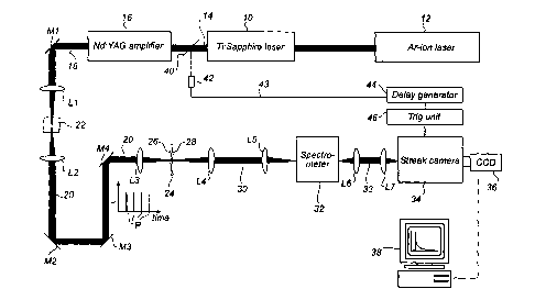
Referring now to figure 1 a, an apparatus according to a first
embodiment for performing a time-resolved analysis according to the invention
comprises a Ti:sapphire
CA 02442275 2003-09-17
WO 02/075286 PCT/SE02/00510
7
laser 10 pumped by an argon laser 12. The laser beam 14 thereby generated is
amplified by
a neodymium YAG amplifier stage 16 into an amplified laser beam 18. In order
to create
an excitation beam 20 of "white" light, the laser beam 18 is passed through a
water filled
cuvette 22 via a mirror Ml and a first lens system L1.
A sample to be analysed is schematically illustrated at reference numeral 24
and
comprises a front surface 26 and a back surface 28. The sample 24 is
temporarily fixed in a
sample-positioning unit (not shown). The excitation laser beam 20 is focused
onto the front
surface 26 of sample 24 via a lens system L2/L3 and mirrors M2-M4. On the
opposite side
of sample 24, the transmitted laser beam 30 is collected from the backside by
lens system
L4/L5 and focused into spectrometer 32. In the illustrated set-up, the sample
24 may be a
pharmaceutical, solid tablet having a diameter of e.g. 9 mm. The excitation
beam 20 may
be focused in a spot of about 1 mm. In other embodiments, the excitation beam
may be
focused on the whole sample, or scanned over the sample.
In an alternative embodiment the apparatus is attached to for example a
fluidised
bed for remote sampling of a selected part of the contents in the bed.
As schematically illustrated in figure 1 a, the excitation beam 20 in this
embodiment
is time-pulsed into a pulse train of short, repetitive excitation pulses P.
The pulse length of
each excitation pulse P is short enough and the time spacing between two
consecutive
excitation pulses P is long enough in relation to the transit time of the beam
(i.e. in relation
to the time taken for each pulse to be completely measured in time), such that
any
interference is avoided between the detected light from one given excitation
pulse Pn and
the detected light from the next excitation pulse Pn+l. Thereby, it is
possible to perform a
time-resolved measurement on the radiation from one excitation pulse P at a
time.
From the spectrometer 32, the detected light beam 33 is passed via lens system
L6/L7 to a time-resolved detection unit, which in this embodiment is
implemented as a
streak camera 34. The streak camera 34 used in an experimental set-up
according to figure
la was a Hamamutsu Streak Camera Model C5680. Specifically, the streak camera
34 has
an entrance slit (not shown) onto which the detected light beam 33 from the
spectrometer
32 is focused. It should be noted that only a fraction of the light emitted
from the sample is
actually collected in the spectrometer 32 and, thereby, in the detection unit
34. As a result
of passing through the spectrometer 32, the emitted radiation 30 from the
sample 24 is
spectrally divided in space, such that radiation received by the streak camera
34 presents a
wavelength distribution along the entrance slit.
The incident photons at the slit are converted by the streak camera into
photoelectrons and accelerated in a path between pairs of deflection plates
(not shown).
Thereby, the photoelectrons are swept along an axis onto a microchannel plate
inside the
CA 02442275 2003-09-17
WO 02/075286 PCT/SE02/00510
8
camera, such that the time axis of the incident photons is converted into a
spatial axis on
said microchannel plate. Thereby, the time in which the photons reached the
streak camera
and the intensity can be determined by the position and the luminance of the
streak image.
The wavelength-resolution is obtained along the other axis. The photoelectron
image is
read out by a CCD device 36, which is optically coupled to the streak camera
34. The data
collected by the CCD device 36 is coupled to an analysing unit 38,
schematically
illustrated as a computer and a monitor.
In the set-up in figure 1 a, the intensity of the emitted light is measured as
a function
of time in time-synchronism with each excitation of the sample. This means
that the
io detection unit comprising the streak camera 34 and the associated CCD
device 36 is time-
synchronised with the repetitive excitation pulses P. This time-synchronism is
accomplished as follows: each excitation pulse P of the laser beam 14 triggers
a
photodetector 42 or the equivalent via an optical element 40. An output signal
43 from the
photodetector 42 is passed via a delay generator 44 to a trig unit 46,
providing trig pulses
to the streak camera 34. In this manner, the photon detection operation of the
streak camera
is activated and de-activated at exact predetermined points in time after the
generation of
each excitation pulse P.
As mentioned above, the evaluation and analysis of the collected, time-
resolved
information can be done in different ways. As schematically illustrated in
figure la, the
collected data information from each excitation is transferred from the streak
camera 34
and the CCD device 36 to a computer 38 for evaluation of the information.
Monte Carlo
simulations, multivariate calibrations, etc as mentioned in the introductory
part of this
application can be utilised in order to calculate the optical properties of
the sample and,
indirectly, content and structural parameters of the sample 24.
In the embodiment shown in figure Ib, it is the transmitted radiation - the
beam 30
- which is detected in a time-resolved manner. However, the invention can also
be
implemented by detecting the radiation reflected from the sample. Figure lb
schematically
illustrates how an excitation beam 20' corresponding to excitation beam 20 in
figure 1 a is
focused via a lens L3' onto the front surface 26 of a sample 24. The photons
of each
excitation pulse will be reflected both as directly reflected photons from the
front surface
26 as well as diffusely backscattered photons with more or less time delay.
This directly
reflected radiation as well as the diffusely backscattered radiation is
collected by a lens L4'
into a detection beam 30', corresponding to detection beam 30 in figure 1 a.
As stated above, it is possible to combine the embodiments illustrated in
figures 1 a
and lb into one single embodiment, where both transmitted and backscattered
light is
CA 02442275 2003-09-17
WO 02/075286 PCT/SE02/00510
9
detected and analysed in a time-resolved and wavelength-resolved manner
according to the
invention.
Figure 2 schematically discloses the main functional components in an
embodiment
for implementing the inventive method, including a radiation generation unit
100
(components 10, 12 and 16 in figure la), a sample positioning unit 102, one or
more
wavelength dispersive/selective elements 104 (component 32 in figure la), one
or more
detector units 106 (components 34 and 36 in figure la) and an analysing unit
108
(component 38 in figure la).
The, water filled cuvette 22 producing white laser light in combination with
the
io spectrometer 32 acting as a wavelength-dispersive element makes it possible
to collect data
that is both wavelength-resolved and time-resolved. Figures 3a and 3b
illustrate the
experimental result of such a detection. It should be noted that the time
scale in both figure
3a and figure 3b illustrate the intensity variation over time for one pulse
only, although the
actual data used for producing these figures is based in accumulated data from
many
readings. The time axis in figures 3a and 3b is in nano second scale.
Figure 3a illustrates a streak camera image pasted into a time-wavelength
diagram,
the light portions correspond to high intensity values. The left part of the
image .
corresponds to detected photons having a relatively short time delay, whereas
the right part
of the image corresponds to photons with a relatively long delay time.
The 3D plot in figure 3b corresponds to the image in figure 3a. This 3D plot
clearly
illustrates how the time-resolved spectroscopy according to the invention
results in an
intensity measurement as a function of both wavelength and photon propagation
time. This
3D plot also clearly illustrate that the total information content as obtained
by the present
invention is significantly greater than the information obtainable with a
conventional time-
integrated detection.
In figure 3b, for each wavelength (such as for the wavelengths k I and X2 as
identified in figure 3b) there is a multitude of timely spaced intensity
readings. Thus, for
each wavelength it is possible to obtain a full curve of emitted (transmitted
and/or
reflected) intensity vs. propagation time. The form of these "time profiles"
shown in figure
3b is dependent on the relation between the optical properties of the analysed
sample. With
such a time-resolved and wavelength-resolved spectroscopy, it is possible to
obtain
information for describing the light interaction with the sample. As an
example, this
provides the basis-for determining an analytical concentration in a sample
that is
proportional to the absorption coefficient but not related to the scattering.
As another
example, one might want to measure an analytical quantity that correlates to
the scattering
properties of the sample instead.
CA 02442275 2003-09-17
WO 02/075286 PCT/SE02/00510
As illustrated by the dashed lines ti and t2 in figure 3b, it is also possible
to
evaluate the emitted light by detecting the intensity during fixed time
slices. This would
give a more coarse time resolution. In one embodiment, wavelength-resolved
spectra are
measured at two time gates only - one for "prompt" light and one for "delayed"
light.
5 The intensity-time diagram in figure 5 illustrates two experimental, time-
resolved
results from measurements on two different tablets. By selecting suitable time
gates where
the difference is substantial, one can easily distinguish different tablets
from each other.
As an alternative to the set-ups illustrated in figures la and lb, instead of
using the
water cuvette 20 in combination with the spectrometer 32, it is possible to
use wavelength
io selective light sources, such as diode lasers. On the detector side,
wavelength selective
detectors, such combinations of filters and detector diodes, can be used for
each
wavelength.
It is possible to combine the invention with spatial-resolved intensity
detection on
the emitted light from the sample. In this context, the term "spatial
resolved" refers to a
is spatial resolution obtained for each excitation pulse. Especially, "spatial
resolved" does not
refer to a spatial resolution based on a scanning in time of the excitation
beam in relation to
the sample. As an illustrative example, by removing the water cuvette22 and
the
spectrometer 32 in the figure la set-up, the light focused on the entrance
slit of the streak
camera would be spatial resolved along the slit, corresponding to a "slit"
across the sample.
A streak camera image obtained by such a set-up is illustrated in figure 4a,
and a
corresponding 3D plot is illustrated in figure 4b. In accordance with figures
3a and 3b
discussed above, figures 4a and 4b represent one pulse only; i.e. the spatial
resolution
illustrated does not correspond to any scanning of the excitation beam over
the sample.
A further alternative set-up is illustrated in figure 6. A modulation driver
50
intensity modulates 51 a light source 52. The light source is intensity
modulated with a
high frequency (MHz-GHz). The light source 52, preferably a light emitting
diode (LED),
emits an excitation beam 53 in broad range of wavelengths. The excitation beam
53
reaches a beam splitter 54 where the excitation beam 53 is divided. One part
of the
excitation beam 53 continues towards a mirror 56 where it is reflected back to
the beam
splitter 54. The other part of the excitation beam 53 continues towards a
moving mirror 55
where it is reflected back to the beam splitter 54. The two parts of the split
excitation beam
53 are brought together again at the beam splitter 54 where they continue
towards the
sample 57. The sample 57 is thus irradiated and the transmitted light detected
by a detector
58. By scanning the moving mirror 55 back and forth, an interferogram is
produced. This
interferogram contains information about the light transmitted through the
sample at all
wavelengths. By using an interferogram all wavelengths are monitored
simultaneously and
CA 02442275 2003-09-17
WO 02/075286 PCT/SE02/00510
11
the result will be a spectrum of the transmitted light intensity. The signal
60 from the
modulation driver 50 is compared to the signal 59 from the detector 58 by a
phase
comparator 61. From the comparison in the comparator 61, information can be
extracted
with deconvolution techniques and chemometric models.
A further alternative set-up of the present invention is.illustrated in figure
7. In this
embodiment the light source producing intensity modulated light is made up of
an array of
diode lasers 62. The array of diode lasers 62 covers a wide range of
wavelengths and a
multiplexer 63 is used to scan the various diode lasers 62 in the array, i.e.
the multiplexer
63 executes the scan through the different wavelengths. The produced
excitation beam
travels through a set of mirrors, illustrated in figure 7 with one mirror 65,
until it reaches a
beam splitter 66 where the excitation beam 64 is divided up into two beams 70
and 74. One
beam 74 irradiates the sample 67 and the transmitted light is detected by a
photomultiplier
68. The other beam 70 is directed directly to a photomultiplier 71 without
irradiating the
sample 67. The two signals 69 and 72 produced by the photomultipliers 68 and
71 due to
the incident beams are compared in a phase comparator 73. These two signals 69
and 72
are recorded for each wavelength in sequence according to the scanning of the
diode laser
array 62 by the multiplexer 63. The diagram in figure 8 shows an example of
the two .
signals 69 and 72 where the excitation sinus curve corresponds to the beam 70
detected by
photomultiplier 71 in figure 7, i.e. the beam unaffected by the sample 67. The
beam 74,
after irradiating the sample, is the detection sinus curve in figure 8.
Information about
physical parameters of the sample can be extracted from the type of diagram
illustrated in
figure 8 by comparing the two sinus shapes.
In either of the above embodiments the measurements can be carried out by
remote
sampling, i.e. the sample does not have to be positioned in specific means.
Therefore, the
apparatuses can be placed to measure the contents in a turbid, pharmaceutical
sample flow
and not only in a specifically selected sample, e.g. a tablet or a capsule.
The foregoing is a disclosure of preferred embodiments for practicing the
present
invention. However, it is apparent that device incorporating modifications and
variations
will be obvious to one skilled in the art. Inasmuch as the foregoing
disclosure is intended
to enable one skilled in the art to practice the instant invention, it should
not be construed
to be limited thereby, but should be construed to include such modifications
and variations
as -fall within its true spirit and scope.