Note: Descriptions are shown in the official language in which they were submitted.
CA 02444383 2003-10-16
WO 02/087497 PCT/US02/12502
THERAPEUTIC AGENT/LIGAND CONJUGATE COMPOSITIONS,
THEIR METHODS OF SYNTHESIS AND USE
CROSS REFERENCE TO RELATED APPLICATIONS
s This application claims the benefit of: U.S. Provisional Patent Application
No.
60/286,453, entitled "Methods for Visualizing Tumors Using a Radioisotope
Conjugate" filed April 26, 2001; U.S. Provisional Patent Application No.
60/334,969,
entitled "Therapeutic Agent/Ligand Conjugate Compositions and Methods of Use"
filed December 4, 2001; and U.S. Provisional Patent Application No.
60/343,147,
io entitled "Diagnostic Imaging Compositions, Their Methods of Synthesis and
Use"
filed December 20, 2001, all three of which are hereby incorporated herein by
reference in their entirety. This application is related to U.S. Patent
Application Ser.
No. , entitled "Diagnostic Imaging Compositions, Their Methods of
Synthesis and Use," f led April 19, 2002, inventors Chun Li, et al., which is
hereby
is incorporated herein by reference,in its entirety.
RIGHTS IN THE INVENTION
This invention was made, in part, with United States Government support
under grant CA 74819 from the NCI, and the United States Government may
therefore have certain rights in the invention.
2o BACKGROUND OF THE INVENTION
1. Field of the Invention
This invention relates to compositions useful in the treatment of cancer and
other diseases, and, more specifically, to compositions comprising therapeutic
agents
(e.g., chemotherapeutic drugs) and other compounds conjugated to ligands,
useful for
as the selective delivery of the agent or compound to tumors and other target
tissues.
The invention also relates to methods for synthesizing and using such
compositions.
2. Description of the Background
Cancer chemotherapy is ultimately limited by the toxicity of drugs to normal
tissues. Selective delivery of drugs to target cells theoretically allows the
use of a
so reduced dose to achieve the same therapeutic response, with a consequent
decrease in
systemic toxicity. A number of methods have been used to selectively target
tumors
with therapeutic agents to treat cancers in humans and other animals.
Targeting
moieties such as monoclonal antibodies (mAb) or their fragments have been
conjugated to linear polymers via their side chain functional groups. However,
this
CA 02444383 2003-10-16
WO 02/087497 PCT/US02/12502
2
approach usually results in reduced receptor binding affinity either due to
changes in
the chemical properties of the antibodies or due to folded configuration of
polymers
that imbed the targeting moiety in the random coiled structure. Moreover,
crosslinks
and aggregates of polymers may form as a result of side-chain coupling
procedures.
s Immunoconjugates have been synthesized by employing intermediate carriers
such as
dextran, serum albumin, and synthetic polymers to increase the amount of drugs
attached to the antibody without significantly impairing its antigen binding
activity.
However, in these cases, the antibodies were attached to the side chains of
the
polymer, which is believed to adversely affect the binding affinity of the
antibody and
io the ih vivo behavior of the immunoconjugates.
Thus, there exists a need for new and improved compositions and methods for
the treatment of tumors and other diseases.
SUMMARY OF THE INVENTION
The present invention overcomes problems and disadvantages associated with
is current therapeutic agents, and provides novel compositions for treatment
of tumors
and other diseases. Preferred embodiments allow for the selective delivery of
a
therapeutic agent (e.g., a chemotherapeutic agent) or another compound or
agent to
the target tumor or tissue. Compositions according the invention include
conjugates of
a ligand, a polymer spacer, a polymer carrier, and a therapeutic agent or
another
zo compound or agent. A preferred composition of the invention comprises a
conjugate
of an antibody, a polyethylene glycol (PEG) spacer, a polymer earner, and a
therapeutic agent. In a particularly preferred embodiment, the ligand is a
monoclonal
antibody, the polymer spacer is a PEG spacer, the polymer carrier is poly(1-
glutamic
acid) (PG), and the therapeutic agent is a chemotherapeutic agent such as
Adriamycin
is or paclitaxel.
Accordingly, one embodiment of the invention is directed to a conjugate
molecule comprising: a ligand; a polymer spacer; a polymer carrier; and a
therapeutic
agent. The ligand is bonded to the polymer spacer, the polymer spacer is
bonded to
the polymer carrier, and the polymer carrier is bonded to the therapeutic
agent. The
so polymer carrier may be bonded to the therapeutic agent with or without the
assistance
of a linker molecule.
CA 02444383 2003-10-16
WO 02/087497 PCT/US02/12502
3
Another embodiment of the invention is directed to a composition comprising
any of the conjugate molecules described herein and a pharmaceutically
acceptable
Garner.
Still another embodiment is directed to a method for selectively delivering a
s therapeutic agent to a target tissue in a patient comprising: administering
a conjugate
molecule to the patient having said target tissue, wherein the conjugate
molecule
comprises: a ligand with affinity for the target tissue; a polymer spacer; a
polymer
carrier; and a therapeutic agent. The ligand is bonded to the polymer spacer,
the
polymer spacer is bonded to the polymer carrier, and the polymer carrier is
bonded to
io the therapeutic agent.
A further embodiment is directed to a method of treating a patient having a
diseased tissue, the method comprising administering a therapeutically
effective
amount of a conjugate molecule to the patient, wherein the conjugate molecule
comprises: a Iigand with affinity for the diseased tissue; a polymer spacer; a
polymer
is carrier; and a therapeutic agent. The ligand is bonded to the polymer
spacer, the
polymer spacer is bonded to the polymer carrier, and the polymer Garner is
bonded to
the therapeutic agent.
The invention also includes different methods for synthesizing conjugate
molecules of the invention. One such method comprises the steps of: providing
a
ao polymer spacer-polymer carrier construct having a sulthydryl-reactive vinyl
sulfone
group at one end of the polymer spacer; conjugating the therapeutic agent to
the
polymer carrier to form a vinyl sulfone-polymer spacer-polymer carrier-
therapeutic
agent construct; pretreating the ligand to introduce a sulffiydryl group on
the ligand;
and combining the ligand with the vinyl sulfone-polymer spacer-polymer carrier-
zs therapeutic agent construct, wherein the vinyl sulfone group reacts with
the sulfhydryl
group to form a conjugate molecule comprising the ligand, the polymer spacer,
the
polymer carrier, and the therapeutic agent, and wherein the ligand is bonded
to the
polymer spacer, the polymer spacer is bonded to the polymer Garner, and the
polymer
carrier is bonded to the therapeutic agent.
so Other obj ects and advantages of the invention are set forth in part in the
description which follows, and, in part, will be obvious from this
description, or may
be learned from the practice of the invention.
CA 02444383 2003-10-16
WO 02/087497 PCT/US02/12502
4
DESCRIPTION OF THE FIGURES
The following figures form part of the present specification and are included
to further demonstrate certain aspects of the present invention. The invention
may be
better understood by reference to one or more of these drawings in combination
with
s the detailed description of the specific embodiments presented herein.
Figure 1. Schematics of conjugate molecules depicting site-specific attachment
of
homing ligand to one terminus of PEG molecules for targeted delivery of
diagnostic and therapeutic agents.
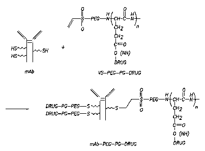
Figure 2. Synthetic scheme for the synthesis of mAb-PEG-PG-Drug conjugates.
Figure 3. Comparison of GPC chromatograms of VS-PEG-PG conjugate (B) and
VS-PEG (A).
Figure 4. 1H-NMR of VS-PEG-PG.
Figure 5. Structure of Adriamycin.
Figure 6. GPC elution profile of Herceptin (A), VS-PEG-PG-TXL (B), and
purified
Herceptin-PEG-PG-TXL conjugate (C) using a Superdex 200 column (1.0
x 30 cm).
Figure 7. Purification of C225-PEG-PG-Adr by FPLC using a Resource Q anion-
exchange column. Fractions 3-5 correspond to C225; fractions 14-21
correspond to C225-PEG-PG-Adr conjugate.
Figure 8. Gel permeation chromatography of C225 (A), PEG-PG-Adr (B), and
purified C225-PEG-PG-Adr conjugate (C) using a Superdex 200 column
(1.0 x 30 cm).
Figure 9. Volume-weighted Gaussian Analysis showing particle size and size
distribution of C225-PEG-PG-Adr.
Figure 10. Hypothetical structure of polymeric nanoparticles (A) and
targetable
polymeric nanopaxticles (B) from amphiphilic block copolymer PEG-PG-
Adr.
Figure 11. Graphs showing cytotoxicity of Herceptin-PEG-PG-TXL in MDA-MB-
468 (Her 2/neu-) cells (A); and SKOVipl (Her 2/neu+) cells (B).
Figure 12. Graphs showing cytotoxicity of Herceptin-PEG-PG-TXL in MDA 435/neo
cells (A) and MDA 435/e B2 cells (B).
Figure 13. Graph showing cytotoxicity of C225-PEG-PG-Adr in A431 Cells: 6
hours
exposure followed by washing, and additional 72 hours incubation.
CA 02444383 2003-10-16
WO 02/087497 PCT/US02/12502
DESCRIPTION OF THE INVENTION
The present invention is directed to novel conjugates useful for the selective
delivery of therapeutic agents (e.g., chemotherapeutic drugs, hormonal agents
and
diagnostic agents) and other compounds and agents to tumors or another target
tissue.
s The invention is also directed to novel methods of synthesizing and using
such
conjugates. Preferred embodiments of the invention comprise a ligand, such as
a
monoclonal antibody (e.g., C225 or Herceptin), indirectly coupled to a
therapeutic
agent, such as a chemotherapeutic drug. The coupling is achieved by
conjugating the
ligand site-specifically to the termini of a polymer-therapeutic agent
conjugate using a
1o polymer spacer or linker (e.g., a PEG spacer).
Copending provisional patent application 60/286,453, incorporated herein by
reference in its entirety, describes the coupling of a radionuclide 111In to a
terminus of
polyethylene glycol (PEG) chain which was in turn attached to 0225, a mAb
directed
against EGF receptor. Specifically, as shown in Figure 1A, a polyethylene
glycol
is (PEG) conjugated monoclonal antibody (mAb) with a radionuclide attached to
one
terminus of the PEG chain and the antibody to the another terminus of PEG
chain was
designed and synthesized (See also, X-X. Wen et al, Polyethylene glycol)
conjugated
anti-EGF receptor antibody C225 with radiometal chelator attached to the
termini of
polymer chains. Bioconjug. Chem. 12:545-553, 2001). The conjugate exhibited
ao significantly reduced nonspecific interaction and improved nuclear imaging
property
(X-X. Wen et al, Improved imaging of 111In-DTPA-polyethylene glycol)
conjugated
anti-EGF receptor antibody C225. J. Nucl. Med., 42:1530-1537, 2001).
It has been discovered that conjugation of a receptor-homing ligand to the end
of a polymer chain through a PEG linker enhances the targeted delivery of
therapeutic
as agents. As shown in the Examples, mAbs were coupled site-specifically to
the termini
of PG-drug conjugates via a PEG linker. Specifically, 0225 (an anti-EGF
receptor
mAb), and Herceptin (an anti-Her2/neu mAb), were site-specifically conjugated
to the
termini of poly(1-glutamic acid)-drug conjugates through a PEG spacer. A
schematic
of the construct is shown in Figure 1B.
so The novel conjugates of the invention demonstrated enhanced cellular uptake
of the polymeric construct into tumor cells overexpressing EGF receptors and
for
Her2/neu receptors. The polymeric immunoconjugates maintained the binding
affinity of the corresponding mAbs. Specifically, C225 and Herceptin
conjugates
CA 02444383 2003-10-16
WO 02/087497 PCT/US02/12502
6
bound to target cell surfaces. In addition, the 0225 conjugate appeared to be
internalized. As shown in the biologic assays, the attachment of drugs to the
polymeric carrier through hydrolytically stable amide linkage and the
efficient cellular
internalization yielded significantly increased selective cytotoxicity against
target
s cells. Further, targetable polymeric nanoparticles formed when Adriamycin
was used
as the drug to conjugate to mAb-PEG-PG carrier.
Accordingly, one embodiment of the invention is directed to a conjugate
molecule comprising: a ligand; a polymer spacer; a polymer carrier; and a
therapeutic
agent. The conjugate molecule is useful for the selective delivery of the
therapeutic
io agent to tumors or other tissues with biological receptors. Preferably, the
ligand is
bonded to the polymer spacer, the polymer spacer is bonded to the polymer
carrier,
and the polymer carrier is bonded to the therapeutic agent. As used herein,
"bonded"
refers to any physical or chemical attachment, including, but not limited to,
covalent
bonding, ionic or chelating interactions.
is More preferably, the ligand is bonded to the polymer spacer via a covalent
bond, the polymer spacer is bonded to the polymer carrier via a covalent bond,
and
the polymer carrier is bonded to the therapeutic agent directly via a covalent
bond, or
indirectly using a linker. For example, the ligand and polymer spacer may be
joined
by an amide bond, a thioether (S-C) bond, a disulfide (S-S) bond, or a
thiourethane
ao bond, more preferably, an amide, thioether or disulfide bond, and, most
preferably, a
thioether bond. The polymer spacer and polymer carrier may be joined, for
example,
by an amide bond, a thioether (S-C) bond, a disulfide (S-S) bond, a
thiourethane bond,
a carbonate bond or a urethane bond, more preferably, an amide or a urethane
bond,
and, most preferably, an amide bond. The polymer carrier and therapeutic agent
may
zs be bonded to each other, for example, by an amide, thioether, disulfide,
thiourethane,
hydrazone or ester bond, and more preferably, by an amide or ester bond.
Alternately,
the polymer carrier and therapeutic agent can be bonded or joined using a
linker.
Useful linkers include, but are not limited to, aliphatic chains, lipids,
amino acids or
peptides. In the latter embodiments, the polymer carrier is preferably
covalently
so bonded to the linker, and the linker is preferably covalently bonded to the
therapeutic
agent.
Further, as shown in Figure 2, more than one polymer spacer-polymer carrier-
therapeutic agent construct may be bonded to a single ligand or antibody.
Multiple
CA 02444383 2003-10-16
WO 02/087497 PCT/US02/12502
7
therapeutic agents may be bonded to the polymer carrier. Ligands different
from
those attached to the PEG chain terminus may be bonded to the side chains of
the
polymer carrier.
The polymer carrier to which the therapeutic agent or other compound is
s attached is preferably poly(1-glutamic acid). However, other polymers,
particularly
those which are biocompatible, water-soluble, biodegradable, and have multiple
side-
chain functional groups that allow attachment of multiple drug molecules, may
be
used without departing from the scope of the invention. These polymers
include, but
are not limited to, poly(d-glutamic acid), poly(dl-glutamic acid), poly(1-
aspartic acid),
io poly(d-aspartic acid), poly(dl-aspartic acid), polylysine, polysaccharides,
polyhydroxypropylinethacryamide (HPMA), dextran, poly(hydroxypropylglutamine),
poly(hydroxyethylglutamine), hyaluronic acid, carboxymethyl dextran,
polyacrylic
acid and chitosan, and copolymers between two or more of them.
The polymer carrier can generally have any number average molecular weight,
is and preferably has a number average molecular weight of at least about
1,000 daltons.
The poly(1-glutamic acid) preferably has a number average molecular weight of
about
1,000 daltons to about 100,000 daltons. The other polymers listed above as
Garners
preferably have a number average molecular weight of about 1,000 daltons to
about
150,000 daltons.
ao The polymer spacer between the ligand and the polymer is preferably PEG.
However, other linear polymers, particularly those which are biocompatible and
uncharged, may be used without departing from the scope of the invention.
These
polymers include, but are not limited to, a polyamino acid, such as
polyglycine,
polytyrosine, polyphenylalanine, dextran, polysaccharides, polypropylene oxide
as (PPO), a copolymer of polyethylene glycol (PEG) with PPO, polyglycolic
acid,
polyvinyl pyrolidone, polylactic acid and polyvinyl alcohol.
The polymer spacer can generally have any number average molecular weight,
and preferably has a number average molecular weight of at least about 1,000
daltons.
The polyethylene glycol preferably has a number average molecular weight of
about
so 1,000 daltons to about 100,000 daltons. The other polymers listed above as
spacers
preferably have a number average molecular weight of about 1,000 daltons to
about
100,000 daltons.
CA 02444383 2003-10-16
WO 02/087497 PCT/US02/12502
8
The ligand (or targeting moiety) can generally be any ligand, and preferably
is
an antibody or its fragments, a peptide or a protein. The antibody can
generally be a
monoclonal antibody, or a polyclonal antibody. For example, useful antibodies
include, but are not limited to, C225, Herceptin, Rituxan, phage Iibxary
antibodies,
s anti-CD, DC141, antibodies to the integrins alpha v-beta 3 (such as LM609),
antibodies to VEGF receptors, antibodies to VEGF, or any other suitable
antibody.
The antibody can be an antibody fragment such as F(ab')2, Fab', or ScFv
fragment or
an antibody fragment such as chimeric (c) 7E3Fab (c7E3Fab) that binds to
integrin
receptors. The antibody can be a humanized antibody. The peptide can generally
be
io any peptide, such as a cell surface targeting peptide, and preferably is a
growth factor,
such as VEGF (Vascular Endothelial Growth Factor)-A, -B, -C or -D, PDGF
(Platelet-Derived Growth Factor), Angiopoietin-1 or -2, HGF (Hepatocyte Growth
Factor), EGF (Epidermal Growth Factor), bFGF (Basic Fibroblast Growth Factor),
cyclic CTTHWGFTLC, cyclic CNGRC, or cyclic RGD-4C. The protein can
is generally be any protein, such as annexin V, interferons (e.g., interferon
a,, interferon
(3), tumor necrosis factors, endostatin, angiostatin, or thrombospondin, and
preferably
is annexin V, endostatin, angiostatin, interferon-a, or interferon-[3. More
preferably,
the ligand is a monoclonal antibody, such as a C225, Herceptin or c7E3Fab
antibody,
or a protein, such as annexin V. Preferably, the ligand has affinity for a
target tissue.
zo Preferred ligands bind specifically to receptors or other binding paxtners
on the target
tissue.
As used herein "therapeutic agent" broadly includes, but is not limited to,
drugs, chemotherapeutic drugs/agents, diagnostic agents, hormonal
drugs/agents, and
other compounds and compositions useful in the treatment, diagnosis and
monitoring
as of disease. The invention is particularly useful for the delivery of
chemotherapeutic
agents. Chemotherapeutic agents useful in the practice of the invention
include, but
are not limited to, Adriamycin (Adr or doxorubicin), daunorubicin, paclitaxel
(Taxol),
docetaxel (taxotere), epothilone, camptothecin, cisplatin, carboplatin,
etoposide,
tenoposide, geldanamycin, methotrexate, maytansinoid DMI or 5-FU. Preferably,
the
3o chemotherapeutic agent is Adriamycin or paclitaxel, and, more preferably,
is
Adriamycin. Other therapeutic agents that can be used include, but are not
limited to,
magnetic resonance imaging contrast agents such as gadolinium-DTPA (Gd-DTPA),
and near-infrared optical imaging agents such as Cy 5.5, indocyanine green
(ICG) and
CA 02444383 2003-10-16
WO 02/087497 PCT/US02/12502
9
its derivatives, and Alexa fluor. However, the invention is not limited to the
foregoing, and other compounds and agents can be used without departing from
the
scope of the invention.
Another embodiment of the invention is directed to a composition comprising
s a plurality of nanoparticles. The nanoparticles comprise a plurality of the
conjugate
molecules described herein. Preferably, the therapeutic agent in the
nanoparticles is
Adriamycin. In this embodiment, the polymer spacer and polymer carrier have
hydrophilic/hydrophobic characters or hydrophobic/hydrophilic characters. For
example, as shown in Example 2, the PEG block in PEG-PG-Adr is hydrophilic,
and
io the PG-Adr block in the copolymer is hydrophobic.
Still another embodiment is directed to compositions comprising any of the
conjugate molecules described herein and a pharmaceutically acceptable
carrier. As
used herein, "pharmaceutically acceptable Garner" includes any and all
solvents,
dispersion media, coatings, antibacterial and antifungal agents and isotonic
agents and
is the like. The use of such media and agents for pharmaceutically active
substances is
well known in the art. For example, the carrier may comprise water, alcohol,
saccharides, polysaccharides, drugs, sorbitol, stabilizers, colorants,
antioxidants,
buffers, or other materials commonly used in pharmaceutical compositions.
Except
insofar as any conventional media or agent is incompatible with the active
ingredient,
zo its use in the therapeutic compositions is contemplated. Supplementary
active
ingredients can also be incorporated into the compositions.
The phrase "pharmaceutically acceptable" also refers to molecular entities and
compositions that do not produce an allergic or similar untoward reaction when
administered to an animal or a human.
as A preferred composition is a pharmaceutical preparation suitable for
injectable
use. Pharmaceutical preparations of the invention suitable for injectable use
include
sterile aqueous solutions or dispersions and sterile powders fox the
preparation of
sterile injectable solutions or dispersions. Preferably, the preparations are
stable under
the conditions of manufacture and storage and are preserved against the
3o contaminating action of microorganisms, such as bacteria and fungi. The
carrier may
be a solvent or dispersion medium containing, for example, water, ethanol,
polyol (for
example, glycerol, propylene glycol, and liquid polyethylene glycol, and the
like),
suitable mixtures thereof, and vegetable oils. The prevention of the action of
CA 02444383 2003-10-16
WO 02/087497 PCT/US02/12502
microorganisms may be brought about by various antibacterial and antifungal
agents,
for example, parabens, chlorobutanol, phenol, sorbic acid, thimerosal, and the
like. In
many cases, it will be preferable to include isotonic agents, for example,
sugars or
sodium chloride.
s Sterile injectable solutions may be prepared 'by incorporating the active
compounds in the required amount in the appropriate solvent with various of
the other
ingredients enumerated above, as required, followed by filtered sterilization.
Generally, dispersions may be prepared by incorporating the various sterilized
active
ingredients into a sterile vehicle which contains the basic dispersion medium
and the
io required other ingredients from those enumerated above. In the case of
sterile
powders fox the preparation of sterile injectable solutions, the preferred
methods of
preparation include vacuum-drying and freeze-drying techniques which yield a
powder of the active ingredient plus any additional desired ingredient from a
previously sterile-filtered solution thereof.
is For parenteral administration in an aqueous solution, the solution is
preferably
suitably buffered, if necessary, and the liquid diluent first rendered
isotonic with
sufficient saline or glucose. These particular aqueous solutions are
especially suitable
for intravenous and intraperitoneal administration.
A further embodiment of the invention is directed towards methods for
zo selectively targeting tumors or other target tissues with biological
receptors using any
of the herein described conjugate molecules and compositions. For example, one
such embodiment is directed to a method for selectively delivering a
therapeutic agent
to a target tissue in a patient comprising: administering a conjugate molecule
to a
patient having the target tissue, wherein the conjugate molecule comprises: a
ligand
zs with affinity for the target tissue; a polymer spacer; a polymer Garner;
and a
therapeutic agent. Preferably, the ligand is bonded to the polymer spacer, the
polymer
spacer is bonded to the polymer Garner, and the polymer carrier is bonded to
the
therapeutic agent. Preferably, the ligand is an antibody, the polymer spacer
is
polyethylene glycol, and the polymer carrier is poly(1-glutamic acid).
3o Because of the affinity of the ligand for the target tissue, the
therapeutic agent
is selectively delivered to the tissue, where it exerts its therapeutic
effect. For
example, in a preferred embodiment, the therapeutic agent is a cytotoxic agent
which
exerts a cytotoxic effect on the target tissue.
CA 02444383 2003-10-16
WO 02/087497 PCT/US02/12502
11
The administering step may be performed parenterally, e.g., by intravascular,
intraperitoneal, intramuscular or intratumoral injection. The conjugate
molecule may
be administered by inhalation or another suitable route. Preferably,
administration is
by intravascular injection.
s The target tissue may be any desired tissue, including, but not limited to,
a
tumor or other neoplasm, inflammatory, infectious, reparative or regenerative
tissue
(including post trauma and post surgery tissues). As used herein, "tumor"
includes
benign and malignant tumors or neoplasia. In one embodiment, the target tissue
is a
solid tumor, such as breast cancer, ovarian cancer, colon cancer, lung cancer,
head
io and neck cancer, a brain tumor, liver cancer, a pancreatic tumor, bone
cancer, or
prostate cancer. Alternately, the target tumor may be a malignancy such as
leukemia
or lymphoma. The patient can be any animal. Preferably the patient is a
mammal.
The mammal can be a human, a dog, a cat, a horse, a cow, a pig, a rat, a mouse
or
other mammal. More preferably, the patient is a human. As used herein,
"patient"
is broadly includes, but is not limited to, a human or any animal being
treated, tested or
monitored in any kind of therapeutic, diagnostic, research, development or
other
application.
Additional embodiments of the invention are directed towards other
therapeutic applications using the herein described conjugate molecules. One
such
zo embodiment is directed to a method of treating a patient having or
suspected of
having a diseased tissue, the method comprising administering a
therapeutically
effective amount of a conjugate molecule to the patient, wherein the conjugate
molecule comprises: a ligand with affinity for the diseased tissue; a polymer
spacer; a
polymer carrier; and a therapeutic agent. Preferably, the ligand is bonded to
the
zs polymer spacer, the polymer spacer is bonded to the polymer carrier, and
the polymer
Garner is bonded to the therapeutic agent. Any of the conjugates described
herein can
be used.
Because of the affinity of the ligand for the diseased tissue, the therapeutic
agent is selectively delivered to the tissue, where it exerts its therapeutic
effect. For
3o example, when the therapeutic agent is a chemotherapeutic or cytotoxic
agent and the
diseased tissue is a tumor, the therapeutic effect may include inhibition or
killing of
the tumor cells.
CA 02444383 2003-10-16
WO 02/087497 PCT/US02/12502
12
The administering step may be performed parenterally, e.g., by intravascular,
intraperitoneal, intramuscular or intratumoral injection. The conjugate
molecule may
be administered by inhalation or another suitable route. Preferably,
administration is
by intravascular injection. The dosage of the conjugate molecule can be
increased or
s decreased to modulate the therapeutic effect on the targeted diseased
tissue.
The patient can generally be any animal. Preferably the patient is a mammal.
The mammal can be a human, a dog, a cat, a horse, a cow, a pig, a rat, a mouse
or
other mammal. More preferably, the patient is a human. The diseased tissue may
be
any type of tissue, including, but not limited to, a tumor or other neoplasm,
io inflammatory, infectious, reparative or regenerative tissue. In one
embodiment, the
diseased tissue is a tumor, and, more preferably, is a solid tumor such as
breast
cancer, ovarian cancer, colon cancer, lung cancer, head and neck cancer, a
brain
tumor, liver cancer, a pancreatic tumor, bone cancer, or prostate cancer.
Alternately,
the target tumor may be a malignancy such as leukemia or lymphoma.
is As used herein the term "treating" a tumor is understood as including any
medical management of a subject having a tumor. The term would encompass any
inhibition of tumor growth or metastasis, or any attempt to visualize,
inhibit, slow or
abrogate tumor growth or metastasis. The method includes killing a cancer cell
by
non-apoptotic as well as apoptotic mechanisms of cell death.
zo In the foregoing methods, a therapeutically effective amount of the
conjugate
molecules of the invention is preferably administered to achieve the desired
effect.
The actual dosage amount of a composition comprising the conjugate molecule of
the
present invention administered to the patient to achieve the desired effect
(e.g.,
delivery to or treatment of the target or diseased tissue) can be determined
by physical
zs and physiological factors such as body weight, severity of condition, the
type of
disease being treated, previous or concurrent therapeutic interventions,
idiopathy of
the patient and route of administration, as well as other factors known to
those of skill
in the art. The practitioner responsible for administration will, in any
event,
determine the concentration of active ingredients) in a composition and
appropriate
so doses) for the individual subject.
The invention also includes methods for synthesizing the novel conjugates and
compositions of the invention. One such method for synthesizing a conjugate
molecule comprising a therapeutic agent and ligand comprises the steps of
providing
CA 02444383 2003-10-16
WO 02/087497 PCT/US02/12502
13
a polymer spacer-polymer carrier construct having a sulfl~ydryl-reactive vinyl
sulfone
group at one end of the polymer spacer; conjugating the therapeutic agent to
the
polymer carrier to form a vinyl sulfone-polymer spacer-polymer carrier-
therapeutic
agent construct; pretreating the ligand to introduce a sulfhydryl group on the
ligand;
s and combining the ligand with the vinyl sulfone-polymer spacer-polymer
carrier-
therapeutic agent construct, wherein the vinyl sulfone group reacts with the
sulfhydryl
group to form a conjugate molecule comprising the ligand, the polymer spacer,
the
polymer carrier, and the therapeutic agent, and wherein the ligand is bonded
to the
polymer spacer, the polymer spacer is bonded to the polymer carrier, and the
polymer
io carrier is bonded to the therapeutic agent.
Another method for synthesizing a conjugate molecule of the invention
comprises the steps of introducing at least one protected sulfhydryl group
(SH) to an
end of a polymer spacer; conjugating the polymer spacer to a polymer carrier
to form
a protected SH-polymer spacer-polymer carrier construct; conjugating a
therapeutic
is agent to the polymer tamer to form a protected SH-polymer spacer-polymer
carrier-
therapeutic agent construct; pretreating a ligand to introduce a sulfhydryl
reactive
functional group on said ligand; deprotecting the protected SH group to obtain
a free
SH group; and combining the pretreated ligand with the SH-polymer spacer-
polymer
carrier-therapeutic agent construct, wherein the SH group reacts with the
sulfliydryl
zo reactive functional group to form a conjugate molecule comprising the
ligand, the
polymer spacer, the polymer carrier, and the therapeutic agent. In the
resulting
conjugate, the ligand is bonded to the polymer spacer, the polymer spacer is
bonded to
the polymer carrier, and the polymer carrier is bonded to the therapeutic
agent.
Tn this method, the ligand is preferably pretreated with a suitable agent,
such
zs as vinyl sulfone or maleimide to introduce the sulfhydryl reactive
functional group.
Preferably, the SH group is deprotected to obtain a free SH group before
combining
with the ligand.
Still another method for synthesizing a conjugate molecule comprises the steps
of: providing a polymer-spacer-polymer carrier-therapeutic agent construct;
so introducing a protected amine to an end of the polymer spacer to form a
protected
amine-polymer spacer-polymer carrier-therapeutic agent construct; deprotecting
the
protected amine-polymer spacer-polymer tamer-therapeutic agent construct to
obtain
a free amine-polymer spacer-polymer tamer-therapeutic agent construct; and
CA 02444383 2003-10-16
WO 02/087497 PCT/US02/12502
14
combining the free amine-polymer spacer-polymer carrier-therapeutic agent
construct
with a ligand having a carboxylic acid group. The carboxylic acid in the
ligand
conjugates with the free amine to form an amide bond, thereby forming a
conjugate
molecule comprising the ligand, the polymer spacer, the polymer carrier and
the
s therapeutic agent. In the resulting conjugate molecule, the ligand is bonded
to the
polymer spacer, the polymer spacer is bonded to the polymer carrier, and the
polymer
carrier is bonded to the therapeutic agent.
Ligands, polymer spacers, polymer carriers and therapeutic agents useful in
the practice of the foregoing synthetic methods include, but are not limited
to, any of
io the Iigands, polymer spacers, polymer carriers and therapeutic agents
disclosed
herein. For example, the therapeutic agent rnay comprise a contrast agent or a
chemotherapeutic drug. In the resulting conjugates, the ligand is preferably
bonded to
the polymer spacer via a covalent bond, the polymer spacer is bonded to the
polymer
carrier via a covalent bond, and the polymer carrier is bonded to the
therapeutic agent
is directly via a covalent bond or indirectly using a linker.
The use of the above described conjugate molecules is advantageous over
those previously described in the art. Preferred embodiments of the conjugate
molecules are useful for the targeted treatment of tumors and other diseased
tissue.
Preferred embodiments have improved irc vivo half lives and exhibit reduced or
ao eliminated accumulation in the liver. The use of polymers reduces non-
specific
interaction with non-target tissues and reduces background activity.
Attachment of
the therapeutic agent and polymer carrier to the ligand with a polymer spacer
instead
of to the Iigand directly improves retention of the ligand's receptor binding
affinity.
The conjugate molecule design strategy is flexible, and allows for the
preparation of a
zs wide array of molecules for different diagnostic and clinical uses. It
allows both
passive targeting (when ligand is not attached) and active targeting (when
ligand is
attached).
The following examples are included to demonstrate preferred embodiments
of the invention. It should be appreciated by those of skill in the art that
the
so techniques disclosed in the examples which follow represent techniques
discovered by
the inventors to function well in the practice of the invention, and thus can
be
considered to constitute preferred modes for its practice. However, those of
skill in
the art should, in light of the present disclosure, appreciate that many
changes can be
CA 02444383 2003-10-16
WO 02/087497 PCT/US02/12502
made in the specific embodiments which are disclosed and still obtain a like
or similar
result without departing from the spirit and scope of the invention.
EXAMPLES
EXAMPLE 1- MATERIALS AND METHODS
s a. Materials
Diisopropylcarbodiimide (DIC), dimethylformamide (DMF), poly(1-glutamic
acid) (PG, MW 31I~), p-nitrophenol, p-nitrophenyl chloroformate (PNP),
dimehtylaminopyridine (DMAP), 2-isobutoxy-1-isobutoxycarbonyl-1,2-
dihydroquinoline) (IIDQ) were purchased from Sigma-Aldrich (Milwaukee, WI).
io Paclitaxel and Adriamycin-hydrochloride (AdnHCI) were obtained from Hande
Tech.
(Houston, TX). BODIPY FL hydrazide dye was obtained from Molecular Probes
(Eugene, OR). Vinylsulfonyl N hydroxysuccinimidyl PEG (VS-PEG-NHS, MW
3400) and NH2-PEG-OH were obtained from Shearwater (Huntsville, AL). N
succinimidyl S-acetylthioacetate (SATA), y-maleimidobutyric acid N-
is hydroxysuccinimide ester (GMBS), N-succinimidyl 3-[2-
pyridyldithio]propionate
(SPDP), dithiothreitol (DTT), and hydroxyamine were obtained from Pierce
Chemical
Co. (Rockford, IL). C225 is a human-mouse chimeric monoclonal antibody that
taxgets epidermal growth factor receptor (EGFR or EGF receptor) and was kindly
provided by ImClone Systems Inc. (New York, NY)., Herceptin (Trastuzumab) was
ao obtained from Genentech (San Francisco, CA).
UV measurements were recorded using a Beckman DU640 spectrophotometer
(Fullerton, CA). 1H-NMR spectra were obtained with a Broker 300 MHz instrument
(Billerica, MA).
b. Gel Permeation Chromato -~raphy (GPC)
zs GPC was performed with a Waters HPLC system (Waters Corporation,
Milford, MA) consisting of a 717 plus autosampler, a 2410 refractive index
detector,
and a 2487 dual ~, UV detector. Samples were eluted with 0.1 M phosphate
buffer
(pH 7.4) containing 0.1 % Liar at a flow rate of 1 ml/minute through a
Superdex 200
column (Amersham Pharmacia Biotech, Piscataway, NJ) (system 1). Alternatively,
3o the mobile phase was run at a rate of 0.5 ml/minute through the same column
(system
2).
CA 02444383 2003-10-16
WO 02/087497 PCT/US02/12502
16
c. Ion-Exchange Chromatography
The system consisted of an AKTA fast protein liquid chromatography (FPLC)
(Amersham Pharmacia Biotech) and a Resource Q anion exchange column
(Amersham Pharmacia). The mobile phase was run from Buffer A (20 mM Tris
s buffer, pH 7.5) to Buffer B (20 mM Tris buffer containing 0.15 or 1.0 N
NaCI, pH
7.5) in a linear fashion at a flow rate of 3 ml/minute for 20 ml (6.67
minutes). The
column was eluted with 100% Buffer B for the rest of the chromatographic
period.
d. Analytical Assays Used to Determine the Degree of mAb Modification
The degree of substitution of 0225 by SATA was estimated by measuring the
io changes in the concentrations of free amino groups using 2,4,6-
trinitrobenzenesulfonic acid (TNBS) assay, and by monitoring the presence of
sulfhydryl groups using Ellman's test (GT Hermanson, ed., Amine detection
reagents.
Bioconjugate Techniques. San Diego, Academic Press. pp. 112-114 and pp. 88-90,
1996).
is e. Determination of the Molar Ratio of Components in hnmunoconj~a, tes
The concentration of each component of the conjugate was determined and the
molar ratio was calculated. The concentration of the antibody was measured by
UV
at 650 nm using the Bio-Rad Laboratory protein assay kits (Hucoles, CA). In
these
measurements, known concentration of C225 or Herceptin was used as a reference
ao standard and PEG-PG-BODIPY FL was used as the background. Taxol, Adr, and
BODIl'Y FL concentrations were quantified by determining the absorbance at 230
nm, 480 nm, or 503 nm. The concentration of paclitaxel in Her-PEG-PG-TXL or
0225-PEG-PG-TXL was further determined by a hydrolysis/HPLC method.
Alternatively, the concentration of TXL in the immunoconjugates was estimated
by
as assuming a molar ratio of 1:1 between mAb and PEG-PG-TXL. The assumption is
based on UV absorbance of mAb-PEG-PG-BODIPY FL where the concentration of
BODIPY FL, and hence the molar ratio between mAb and PEG-PG, could be
conveniently determined by UV measurements.
f. Quantification of TXL concentration in mAb-PEG-PG-TXL by Hydrol.
3o and HPLC Analysis
Ten mg of PEG-PG-TXL dissolved in 200 ,u1 of 1 N NaHC03, or 1.0 ml of
mAb-PEG-PG-TXL solution containing 50 mg of NaHC03 with known concentration
of mAb was charged into a 5-ml vial with a septum, a needle for gas release,
and a
CA 02444383 2003-10-16
WO 02/087497 PCT/US02/12502
17
stirring bar. Into the vial was added 600 ,u1 of a 50% HZOz solution and 1 ml
of
CHZCIz. The mixture was stirred vigorously overnight at room temperature. The
aqueous portion was extracted with rnethylene chloride twice and the organic
portions
were combined. Taxol concentration was obtained from HPLC analysis with the
s following conditions: 1 mL/minute flow rate, gradient of water/CH3CN
(changing
acetonitrile from 0% to 40%), column: Nova Pak (3.9 x 150 mm) and UV detector
at
228 nm. A standard curve was constructed using a range of Taxol solution in
methylene chloride with concentrations ranging between 0.5 and 6 mg/ml. Ten
,u1
aliquot was inj ected from each standard and each extracted solution.
io EXAMPLE 2- SYNTHESIS OF CONJUGATES
To conjugate homing ligand to the end of a PG chain through a PEG spacer, a
linear PEG-PG conjugate that contains a sulfhydryl reactive vinyl sulfone (VS)
group
at the end of the PEG block of the copolymer was synthesized. The anticancer
agent
Adriamycin (Adr) or paclitaxel (Taxol, TXL) was conjugated to the side chain
is carboxyl groups in the PG block of VS-PEG-PG via p-nitrophenol activated
esters,
IIDQ, or carbodiimide-mediated reaction. Subsequent coupling of mAb, which was
pre-treated with N succinimidyl S-acetylthioacetate (SATA) and hydroxyamine to
introduce sulfliydryl groups, yielded the final conjugates mAb-PEG-PG-drug.
The
synthetic scheme is shown in Figure 2. The mAb used in this study included
C225, a
zo mAb directed against epidermal growth factor receptor (EGFR) and Herceptin,
a mAb
directed against Her-2/neu receptor. Both receptors are overexpressed in a
variety of
solid tumors. For example, EGFR is overexpressed on the cells of over one-
third of
alI solid tumors, including bladder, breast, colon, ovarian, prostate, renal
cell,
squamous cell, non-small cell lung, and head and neck carcinomas.
zs a. Synthesis of VS-PEG-PG
Into 500 mg of PG in 1 M phosphate buffer (pH=8) was added 200 mg of VS-
PEG-NHS in five fractions in a course of 2 hours. The reaction mixture was
stirred
for an additional 5 hours at room temperature. Ninhydrin spray was used to
monitor
the consumption of unreacted NHz at the terminal of PG polymer. To stop the
so reaction, the reaction mixture was acidified with 1 N HCl to pH 3.0 and the
precipitate was recovered by centrifugation at 3,000 rpm for 10 minutes. The
solid
was washed two times with distilled water to remove free PEG and lyophilized
to
yield 360 mg of the conjugate product in acid form.
CA 02444383 2003-10-16
WO 02/087497 PCT/US02/12502
18
The simple purification scheme removed most of the unreacted PEG as
revealed by GPC analysis (system 1) (Figure 3). Specifically, Figure 3 is a
comparison of GPC chromatograms of VS-PEG-PG conjugate (B) and VS-PEG (A).
The concentrations of the compounds were monitored by RI detector.
(Conditions:
s Flow Rate - 1 mL/minute; Column - Superdex 200; Buffer - PBS with 0.1%
LiBr).
Since both unconjugated PG and VS-PEG-PG had the same retention times of
9.0 minutes, fixrther study was performed to verify that the isolated product
was
indeed VS-PEG-PG conjugate. An 1H NMR spectrum of the isolated product, VS-
PEG-PG, is shown in Figure 4. The spectrum revealed the presence of
characteristic
io peaks attributable to both PEG (b 3.72 ppm, s) and PG (8 4.29-4.34, 2.19-
2.34 ppm,
1.89-2.04 ppm for a-CH, y-CH2, and (3-CHZ, respectively). Furthermore, the
molar
ratio between PEG and PG was 0.96 based on the integrals between CHZ of PEG
and
a-CH of PG (Figure 4). These data confirmed that the peak at 9.0 minutes in
GPC
chromatogram of the isolated product is attributed to VS-PEG-PG rather than
that of
is the free PG.
b. Synthesis of VS-PEG-PG-TXL and VS-PEG-PG-Adr
Into a solution of 250 mg VS-PEG-PG in 10 ml DMF was dissolved 150 mg
(176 ,umol) of paclitaxel, 30 mg of DIC (238 ,umol), 75 p.L of pyridine and a
trace
amount of DMAP. The reaction mixture was stirred overnight at room
temperature.
ao After evaporation of the solvent under vacuum, the residual was dissolved
in O.1N
NaHC03. The aqueous solution was filtered through a 0.22-,um filter and
dialyzed
against distilled water overnight using membrane with molecular weight cut-off
(MWCO) of lOK (Spectrum Laboratories, Rancho Dominguez, CA). The product
was recovered as lyophilized powder. Yield: 379 mg polymer conjugate.
Paclitaxel
as content: 21.6% (w/w) based on UV measurement at 230 nm. Each polymer chain
contained about 11 TXL molecules. TXL yield: 54.6%. No free paclitaxel was
detected by silica gel thin layer chromatography (MeCl2/methanol, 4/1, v/v)
and by
GPC (system 1).
Adr was conjugated to VS-PEG-PG via the DIC-mediated coupling reaction
3o using similar procedures. (The structure of Adr is shown in Figure 5; the
drug was
conjugated to VS-PEG-PG polymer through its amino groups on the sugar moiety.)
Thus, into a solution of 100 mg VS-PEG-PG in 5 ml DMF was added Adr free amine
(40 mg, 74 ,umol), 30 ,u1 DIC (24.3 mg, 192 ,umol), 100 ,u1 pyridine, and
trace amount
CA 02444383 2003-10-16
WO 02/087497 PCT/US02/12502
19
of DMAP. Adr free amine was obtained by extracting an aqueous solution of
Adr~HCl and triethylamine (molar ratio 1:3) with chloroform. The reaction
mixture
was worked up as follows: The aqueous solution of polymer conjugate was
acidified
with 1.0 HCI. The precipitate was collected by centrifugation, washed with
water, re-
s dissolved in 0.1 N NaHC03, and dialyzed. GPC (system 2) revealed the absence
of
free Adr in the isolated product. The amount of Adr in the polymer was
estimated to
be 15% (w/w) as measured by UV at 480 nm. Each polymer chain contained about
11 Adr molecules. Yield: 120 mg polymer conjugate, Yield of Adr, 45%.
The fluorescent dye BODIPY was conjugated to VS-PEG-PG to facilitate
io confocal fluorescent microscopic study. Briefly, 5 mg BODIPY-hydrazide
(16.3
,umol) was conjugated to 120 mg of VS-PEG-PG to yield 150 mg of VS-PEG-PG
sodium salt. Approximately 5 dye molecules were attached to each polymer
chain.
c. S'mthesis of Herceptin-PEG-PG-TXL, Herceptin-PEG-PG-BODIPY, and
C225-PEG-PG-Adr
is Into a solution of 50 mg C225 or Herceptin (0.33 ,umol) in 5 ml PBS
(pH=7.2)
was added an aliquot of SATA in DMF (190 ,u1, 8 mg/ml, molar ratio: 1:20).
After
being stirred for 1 hour at room temperature, 0.5 ml of 50 M hydroxylamine
aqueous
solution was added into the solution. The reaction mixture was stirred for an
additional 2 hours, then concentrated to 1-2 ml by ultracentrifugation (MWCO,
10K;
ao Millipore Corp., Bedford, MA). The resulting SH-containing mAb was purified
with
a PD-10 column to remove small molecular weight contaminants. Finally, mAb was
mixed with VS-PEG-PG-TXL, VS-PEG-PG-Adr, or VS-PEG-PG-BODIPY with a
molar ratio of mAb to polymer of 1:8-1:10. After being stirred at 4 °C
overnight, the
solution was passed through a nickel affinity column (FreeZyme conjugate
as purification kit, Pierce Chemical Co., Rockford, IL) to remove unreacted
polymer,
followed by purification with an anion exchange chromatography to remove free
mAb
from polymer bound mAb. The yield of mAb was calculated to be 8-10%. The molar
ratios of Herceptin to PEG-PG polymer and C225 to PEG-PG were 1 based on the
measurements of protein and BODIPY FL concentrations. Using the ratio of
3o Herceptin to PEG-PG of 1, the calculated TXL content in the conjugate was
4.3%.
TXL content obtained from hydrolysis/HPLC assay was 6.65%, which suggests a
molar ratio of Herceptin to PEG-PG of I.B. Thus, the molar ratios of mAb to
PEG-
PG in immunoconjugates varied between 1.0 to 1.8.
CA 02444383 2003-10-16
WO 02/087497 PCT/US02/12502
When GPC Superdex 200 chromatography was applied to the affinity purified
and ion-exchange purified Her-PEG-PG-TXL conjugate, a single peak at 8.15
minutes
was found Specifically, Figure 6 shows the GPC elution profile of Herceptin
(A), VS-
PEG-PG-TXL (B), and purified Herceptin-PEG-PG-TXL conjugate (C) using a
s Superdex 200 column (2.4 x 20 cm). The concentrations of the compounds were
monitored by RI detector. Herceptin and PEG-PG-TXL appeared almost in the same
position (retention time 12.12-12.39 minutes) (Figure 6), although their
molecular
weights are approximately 150,000 and 41,000, suggesting that the hydrodynamic
volume of PEG-PG-TXL is similar to that of the globular protein IgG.
io C225-PEG-PG-Adr was purified following a similar protocol. After removal
of unconjugated PEG-PG-Adr polymer from the C225-PEG-PG-Adr conjugate by
affinity chromatography, the immunoconjugate was further purified by anion
exchange chromatography to remove unconjugated C225. Specifically, Figure 7
shows the purification of C225-PEG-PG-Adr by FPLC using a Resource Q anion-
ic exchange column. Each fraction was 0.5 ml. From the FPLC elution profile,
the
fractions (fractions 3-5) corresponding to the first peak were free C225, and
the
fractions corresponding to the second peak (fractions 14-21) were the desired
conjugate (C225-PEG-PG-Adr), which was pooled, concentrated and stored at 4
°C.
As confirmed by GPC (system 2) analysis of purified C225-PEG-PG-Adr, the
ao immunoconjugate was free of unconjugated C225 (Figure 8). Specifically,
Figure 8
shows results of gel permeation chromatography of C225 (A), PEG-PG-Adr (B),
and
purified C225-PEG-PG-Adr conjugate (C) using a Superdex 200 column (1.0 x 30
cm). The compounds were monitored by measuring absorbance at 254 nm. Although
C225-PEG-PG-Adr and PEG-PG-Adr was not completely resolved by GPC, the lack
zs of tailing and the absence of a peak at 23.81 minutes in the chromatogram
of C225
PEG-PG-Adr suggests that the product was free of unconjugated PEG-PG-Adr.
The elution curve of VS-PEG-PG-Adr had two peaks with retention times of
15.81 and 23.81 minutes, respectively. The first peak at 15.81 minutes in the
GPC
chromatogram of VS-PEG-PG-Adr appeared at the dead volume of the column, which
3o may be attributed to the formation of polymer aggregates. The second peak
may be
attributed to the soluble form of the PEG-PG-Adr.
The nature of the polymeric aggregates may be attributed to the formation of
nanoparticles with a hydrophobic core stabilized by outer hydrophilic PEG
chains.
CA 02444383 2003-10-16
WO 02/087497 PCT/US02/12502
21
Several lines of evidence support this conclusion. 1). PEG-poly(L-aspartic
acid)
(PEG-PAA) block copolymer with Adr coupled to the PAA has been shown to form
micelles with average diameter of 40-60 nm (M. Yokoyama, et al, Preparation of
micelle-forming polymer-drug conjugates. Bioconjugate Chem. 3:295-101, 1992).
s 2). The formation of particles with volume-average diameter of 207 nm was
detected
by light scattering. The size of the particles was decreased from 207 nm to 16
nm
upon conjugation with 0225 because of the increase in the hydrophilic segment
of the
amphiphilic block copolymer (Table 1, Figure 9).
Table 1
Compounds dvolume (nm)
PEG-PG(31K)-Adr 207
PEG-PG(7.7K)-Adr 121
C225-PEG-PG(31K)-Adr 16
io On the other hand, decreasing the contribution of the hydrophobic block PG-
Adr in PEG-PG-Adr by reducing the molecular weight of PG from 31K to 7.7K also
resulted in reduction in particle size to 121 nm (Table 1). 3). PEG-PG-Adr did
not
form particles when it was dissolved in DMF.
As shown in Figure 10A, a block copolymer 10 (e.g., PEG-PG-Adr) composed
is of hydrophobic components 12 (e.g., PG-Adr) and hydrophilic components 14
(e.g.,
PEG) can form a nanoparticle structure 20 as a result of its amphiphilic
character.
The evidence indicates that PEG-PG-Adr adapted this structural feature,
forming a
plurality of nanoparticles 20. As shown in Figure 10A, such nanoparticles 20
would
consist of a hydrophobic PG-Adr core 22 surrounded by a hydrophilic outer PEG
ao shell 24. As shown in Figure 10B, one or more ligands 26 (e.g., mAb) may be
attached to one or more of hydrophilic components 14, respectively, (e.g.,
PEG) to
form targeted nanoparticle 2~.
Attaching 0225 to VS-PEG-PG-Adr affected the balance between hydrophilic
and hydrophobic segments, resulting in decrease in particle size. Thus, the
present
as invention describes a method to prepare targetable polymeric nanoparticles.
Polymeric nanoparticles were obtained from a VS-PEG-PG-Adr copolymer. The VS
functional groups residing on the surface of the nanoparticles provided a
handle to
CA 02444383 2003-10-16
WO 02/087497 PCT/US02/12502
22
further introduce homing moieties to the surface of the nanoparticles, whereas
Adr
attached to the core facilitated hydrophobic interactions to stabilize the
nanoparticle
structure.
EXAMPLE 3- BIOLOGICAL ASSAYS
s Human vulvar squamous carcinoma A431 cells, human ovarian carcinoma
SKOV-3 cells, or human breast cancer MDA-MB-468 cells were grown in DMEM-
F12 medium containing 10% fetal bovine serum at 37 °C.
a. Tmmunoprecipitation and Western Blottin~a~sis
Cell pellets were treated with cold lysis buffer containing lx protease
inhibitor
io cocktails (Sigma, St Louis, MO) on soft ice for 30 minutes, followed by
centrifugation to remove cell debris. Each test drug was added into 200 ,u1 of
supernatant in 0.5-ml microcentrifuge tubes. Two microliters of protein A
beads
(Sigma) were then added into each tube. The microcentrifuge tubes were
incubated at
4 °C for 1 hour, centrifuged, and the beads washed 3 times with 0.5 ml
lysis buffer.
is The beads were heated at 95 °C in 20 ,u1 of lx SDS-PAGE laemmli
sample buffer
(Bio-Rad, Hercules, CA) for 5 minutes, centrifuged, and analyzed by 7% SDS
polyacrylamide gel electrophoresis (PAGE). Western blot was carried out by
electronically transfernng the samples into a nitrocellulose membrane and
incubation
of the membrane for 1 hour with an anti-EGF receptor antibody (Sigma) or anti-
zo Her2/neu receptor antibody (Oncogen, Boston, MA). The receptor signals in
the
membrane were developed by the ECL chemoluminescence detection kit (Amersham
Pharmacia Biotech Inc., Piscataway, NJ).
Immunoprecipitation and western blot analysis were used to investigate the
ability of Her-PEG-PG-Adr to bind to SKOV3 cells, which express a high level
of
zs Her2/neu receptors, as well as the ability of C225-PEG-PG-Adr to bind to
A431 cells,
which express a high level of EGF receptors. Both conjugates bound to their
corresponding receptors in a dose-dependent manner with affinity similar to
that of
their parent antibodies. With respect to the A431 cells, a control of PEG-PG-
Adr was
also used; this control polymer without antibody did not bind to the
receptors.
so b. Intracellular Localization by Confocal Laser Microsc~y
Confocal fluorescent microscope was used to investigate the binding of Her-
PEG-PG-BOD1PY to SKOV3 cells and C225-PEG-PG-Adr to A431 cells and their
subsequent internalization. BODIPY (excitation/emission: 503/SII nm) and Adr
CA 02444383 2003-10-16
WO 02/087497 PCT/US02/12502
23
(excitation/emission: 480/540-nm) were used to facilitate confocal fluorescent
microscopic studies. Cells were grown on Lab-Tek II Chamber Slide (Nalge Nunc
International, Naperville, IL) to SO% of confluence and incubated with
immunoconjugates at 37 °C for various times. PEG-PG-Adr and PEG-PG-
BODIPY
s were used as no-antibody polymer controls. MDA-MB-468 cells that do not
express
Her2/neu receptors were used as negative control when studying the binding of
Her-
PEG-PG-BODIPY to Her2/neu receptors. Cells were washed three times with PBS,
fixed in 95% ethanol, and then treated with 1 ,uM TO-PRO-3 Iodide (Molecular
Probes, Eugene, OR) for 15 minutes for nuclei staining. Fluorescent images of
cells
io were analyzed using LMS-510 confocal microscopy (Zeiss, Thornwood, NY).
At one hour incubation confocal fluorescent microscopy images demonstrated
that Her-PEG-PG-BODIPY, but not PEG-PG-BODIPY, selectively bound to SKOV3
cells overexpressing Her2/neu receptors. Furthermore, Her-PEG-PG-BODIPY did
not bind to MDA-MB-468 cells that do not express Her2/neu receptors. It was
not
is clear, however, whether the conjugates were internalized.
Similarly, confocal fluorescent microscopy images (at 15 minutes)
demonstrated that C225-PEG-PG-Adr, but not PEG-PG-Adr, selectively bound to
A431 cells. Unlike Herceptin conjugate, however, internalization of the C225-
polymer conjugate was clearly visualized. C225-PEG-PG-Adr co-localized with
zo nuclei, whereas PEG-PG-Adr without antibody was not localized to the
nuclei. The
process was very rapid, internalization was observed as early as 5 minutes
after drug
exposure. These results demonstrate that conjugation of a receptor-homing
Iigand to
the end of a polymer chain through a PEG linker enhances the targeted delivery
of
therapeutic agents.
zs c. C otoxicitx
One hundred microliter of growth medium suspending 1000-2000 cells per
well was plated out in 96-well plates and incubated for 2 days to allow the
cells to
attach. Various dilutions of the drug or conjugates were added to each well
and the
plates were incubated for 72 hours at 37 °C. Alternatively, a 6-hour
pretreatment
so protocol was used. Cells were exposed to various concentrations of the drug
or
conjugate for 6 hours at 37 °C, and then washed twice with the fresh
culture medium.
The cells were incubated for additional 72 hours. At the end of the incubation
period,
twenty microliters of MTT solution from Promega Cell Proliferation Assay kit
CA 02444383 2003-10-16
WO 02/087497 PCT/US02/12502
24
(Madison, Wn were added to the wells. The microplates were then incubated for
1
hour at 37 °C. Absorbance was measured at 490 nm using a microplate
reader
(Molecular Devices Corp, Sunnyvale, CA). The data reported represent the means
of
quadruplicate measurement and the standard errors of the mean were less than
15%.
s The ICso, concentration exhibiting 50% growth inhibition were calculated
from the
growth-inhibition curve.
The cytotoxicities of Herceptin-conjugated PEG-PG-TXL and PEG-PG-TXL
after 72 hours of continuous exposure were tested in two pairs of cells lines.
The first pair of cell lines included SKOV3ipl, a human ovarian cancer cell
io variant that overexpress Her2/neu, and MDA-MB-468, which does not express
Her2/neu receptors. Results are shown in Figures 11A and 11B. The graph in
Figure
11A represents the MDA-MB-468 cells, and the graph in Figure 11B represents
the
SKOV3ip1 cells. The y axis in each graph represents the % viability and the x
axis
represents the dose in nM. The data for PEG-PGT is represented by squares. The
is data for Her-PEG-PGT is represented by triangles.
The second pair included MDA-MB-435 transfected with neo only
(MDA435/neo), which does not express the receptor, and the stable Her2/neu
transfectant MDA-MB435/eB2 (MDA 435/eB2). Results are shown in Figures I2A
and 12B. The graph in Figure 12A represents the MDA 435/neo cells; the graph
in
zo Figure 12B represents MDA 435/eB2 cells. The y axis in each graph
represents the
viability and the x axis represents the dose in nM. The data for PEG-PGT is
represented by diamonds. The data for Her-PEG-PGT is represented by squares.
ICso values thus obtained were used to calculate targeting index, defined as
the
ratio of ICSO values obtained with no-mAb drug conjugate PEG-PG-TXL in target
as cells and in non-target cells, times the ratio of ICso values obtained with
mAb
conjugated PEG-PG-TXL in non-target cells and in target cells. The targeting
index
for the first pair and second pair of cell lines were 3.95 and 1.75,
respectively. Since
TXL is releasable from the immunoconjugates, one would expect that after 72
hours
of incubation, a fraction of free TXL released from the conjugates could also
so contribute to the cytotoxic effect, resulting in reduced targeting index or
selective
cytotoxicity. It is anticipated that with a shorter incubation time, greater
difference in
potency between immunoconjugate and no-mAb polymer-drug conjugate would be
observed.
CA 02444383 2003-10-16
WO 02/087497 PCT/US02/12502
2$
To assess the specific cytotoxicity of the conjugate under conditions somewhat
comparable to the ih vivo situation, the cytotoxic activity was measured using
a 6-
hour pretreatment system. Confocal microscopic studies revealed that the
binding of
conjugate to cells reached maximum within 6 hours at 37 °C. Under these
conditions,
s the immunoconjugate C225-PEG-PG-Adr (ICso 1.69 ,uM) was 10-fold more potent
than free Adr (ICso 17.7 ,uM). Results are shown in Figure 13. In Figure 13,
data for
0225-PEG-PG-Adr is represented by diamonds, and data for Adr is represented by
squares. The y axis represents the % viability and the x axis represents the
concentration in ,ug/ml. These data suggest that the binding and subsequent
io internalization of immunoconjugates significantly enhanced the cytotoxic
activity of
Adr.
d. Statistical Methods
Differences in cell growth inhibition were compared between different drugs
using Student's t-test at the 0.05 significant level. Fluorescent intensity
across the
is cells between different treatments were compared by repeat general linear
model
(p<0.05).
EXAMPLE 4 - ALTERNATIVE SYNTHESIS METHOD
As shown in this example, the horning ligand may also be introduced to the
end of a PEG-PG block copolymer that contains a sulfhydryl group. The ligand
is
ao pretreated with y-maleimidobutyric acid N-hydroxysuccinimide ester (GMBS)
to
introduce thin-reactive maleimide groups to the ligand.
a. Synthesis of SPDP-PEG-PNP
Into 245 mg NHa-PEG-OH (MW 3,400, 0.072 mmol) in 3 ml of CH2C12 was
added 10 ~.l triethylamine and 45 mg of N-succinimidyl 3-[2-
pyridyldithio]propionate
zs (SPDP) (0.144 rninol). The extent of the reaction was followed by ninhydrin
spray
test. To stop the reaction, the polymer was precipitated from CHZC12 with
ethyl ether
to give 233 mg of SPDP-PEG-OH (95%). The product was subsequently redissolved
in 2 ml of CHZCl2, and 17.5 mg (0.087 mmol) of p-nitrophenyl chloroformate
(PNP)
was added. The mixture was stirred at room temperature for 2 hours and the
solvent
3o was evaporated under vacuum. The residual material was precipitated and
washed
with ether to remove unreacted p-nitrophenyl chloroformate. Obtained 210 mg
(86%
from NHZ-PEG-OH).
CA 02444383 2003-10-16
WO 02/087497 PCT/US02/12502
26
b. Synthesis of SPDP-PEG-PG
The reaction followed procedures similar to those used for the synthesis of
VS-PEG-PG. Briefly, PDP-PEG-pNP (100 mg, 0.029 mmol) was added into a
solution of PG (200 mg, 0.0064 mmol) in 1 ml PBS (pH 8) in small portions over
a
s period of 2 hours. The occurrence of the coupling reaction was evidenced by
the
release of yellowish p-nitrophenol into the reaction medium. The reaction was
complete in 5 hours as revealed by ninhydrin test. The product was
precipitated with
1 N HCI, dialyzed, and dried to afford 170 mg (86%).
c. Synthesis of SPDP-PEG-PG-Dox
io Into a solution of 100 mg of SPDP-PEG-PG in 5 ml of DMF was added 2S mg
of doxorubicin hydrochloride (Dox~HCl, 0.043 mmol) in lml of DMF containing 20
~,1 of triethylamine. After being stirred for 15 min, 13 mg of coupling agent
ITDQ
(0.043 mmol) was added into the reaction mixture. The reaction was allowed to
proceed at room temperature overnight. The solvent was evaporated under vacuum
is and the remaining solid was washed with ether, redissolved in 1 N NaHC03,
dialyzed
sequentially against PBS buffer (pH 7.2) and water, and lyophilized. Thin
layer
chromatography on silica using n-butanol:acetic acid:water (volume ratio
4:1:1) as
mobile phase showed the absence of free Dox in the purified product. The
conjugate
contained 20% Dox (w/w) as determined by UV at 480 nm. Obtained conjugate 119
zo mg, the yield of Dox was 95%.
d. Synthesis of C225-PEG-PG-Dox
Four milligrams of dithiothreitol (DTT) was added into a solution of 10 mg
SPDP-PEG-PG-Dox in 600 ~,1 of PBS (pH 7.4) to obtain SH-PEG-PG Dox. The
reaction was allowed to proceed at room temperature for 30 minutes followed by
as passing through a PD-10 column to remove unreacted DTT. In a separate
reaction
vessel, 50 mg of C225 was treated with y-maleimidobutyric acid N-
hydroxysuccinimide ester (GMBS) at a C225-to-GMBS molar ratio of 1:20. The
reaction was stirred for 45 min and purified with a PD-10 column to remove
small
molecular weight contaminants. The solutions containing both SH-PEG-PG-Dox and
so maleimide-treated C225 were then mixed and stirred at room temperature for
2 hours.
Unconjugated C225 was removed using a Resource Q anionic exchange column from
a FPLC system as described above and concentrated with a Biomax-10000
Millipore
ultracentrifuge with molecular-weight-cut-off of 10,000. The solution was
desalted
CA 02444383 2003-10-16
WO 02/087497 PCT/US02/12502
27
with a PD-10 column. One milliliter of C225-PEG-PG-Dox with a doxorubicin
concentration of 0.8 mg/ml was recovered (40%). Blue precipitate (positive
reaction)
was observed when the Bio-Rad protein assay reagent was added into the
conjugate
solution, suggesting successful conjugation of C225 to PEG-PG polymer.
s All of the compositions and/or methods disclosed and claimed herein can be
made and executed without undue experimentation iri light of the present
disclosure.
While the compositions and methods of this invention have been described in
terms of
preferred embodiments, it will be apparent to those of skill in the art that
variations
may be applied to the compositions and/or methods and in the steps or in the
sequence
io of steps of the methods described herein without departing from the
concept, spirit
and scope of the invention. More specifically, it will be apparent that
certain agents
which are both chemically and physiologically related may be substituted for
the
agents described herein while the same or similar results would be achieved.
All such
similar substitutes and modifications apparent to those skilled in the art are
deemed to
is be within the spirit, scope and concept of the invention. Not all
embodiments of the
invention will include all the specified advantages. The specification and
examples
should be considered exemplary only with the true scope and spirit of the
invention
indicated by the following claims.