Note: Descriptions are shown in the official language in which they were submitted.
CA 02444706 2010-04-09
ISOLATION OF NEURAL STEM CELLS USING GANGLIOSIDES AND OTHER
SURFACE MARKERS
FIELD OF THE INVENTION
The present invention relates generally to the isolation of neural stem cells.
More
specifically, the present invention relates to a method of using gangliosides
and other
markers to isolate neural stem cells from the central nervous system,
including brain, spinal
cord, and retina.
BACKGROUND OF THE INVENTION
Cell transplantation has over the last two decades emerged as a promising
approach
for restoration of function in neurodegenerative diseases, in particular
Parkinson's and
Huntington's disease. Clinical trials have so far focused on the use of
implants of
embryonic mesencephalic tissue containing already fate-committed dopatninergic
neuroblasts with the capacity to develop into fully mature dopamine neurons in
their new
location in the host brain. A major limitation of the fetal cell
transplantation procedure is
the low survival rate of the grafted neurons (in the range of 5-20%) which
makes it difficult
to obtain sufficient cells for grafting in patients. Currently, mesencephalic
fragments from
at least 6-8 embryos are needed for transplantation in one Parkinson's disease
patient.
Moreover, the ethical, practical and safety issues associated with the use of
tissue from
aborted human fetuses are problematic, and severely restrict the possibility
for applying the
procedure outside highly specialized centers.
It was recently demonstrated that immature neural progenitor cells with
multipotent
properties, called neural stem cells (NSCs), can be isolated from both the
developing and
adult CNS. Neural stem cells are multipotent cells which can be differentiated
into any type
of neural cell, including neurons, astrocytes, glia and oligodendrocytes. The
successful
propagation of mammalian neural stem cells (NSC) in culture, first reported by
Reynolds,
BA and Weiss, S. 1992 "Generation of neurons and astrocytes from isolated
cells of the
adult mammalian central nervous system" Science 255:1707-1710, has opened up
hitherto
unforeseen opportunities in the field of neural transplantation and,
therefore, harvesting
these cells from donated adult human tissue is of great interest.
However, it would be advantageous to isolate the NSCs from the other cells in
the
brain or enrich them such that the purest population of multipotent cells
possible can be
obtained. One favored strategy for cell isolation is the identification of
target epitopes on
the surface of NSCs accessible to monoclonal antibodies. These antibodies can
then be
selectively tagged, e. g., with a fluorescent label, whereupon selecting for
the tagged
1
CA 02444706 2010-04-09
antibody results in selection of the cell on which it is bound. If the target
molecule is
expressed only by the desired cell type, very high levels of enrichment are
possible. The
problem with this strategy is that of identifying the target epitopes which
will isolate the
NSCs, allow for specificity of attachment and will not activate cellular
processes.
The identification and enrichment using specific cell surface markers has
been used previously in the isolation of another type of stem cell, neural
crest stem
cells (NCSC's) (Anderson and Stemple, 1998, United States Patent 5,824,489).
However, the use of the method in the isolation of neural stem cells (NSCs)
has
been slow to develop, possibly due to the difficulty in identifying NSC-
specific
markers. Uchida, N. et al. 2000 "Direct isolation of human central nervous
system
stem cells" Proc Nall Acad Sc! USA 97:14720-14725, describes a method for the
isolation of NSCs using a specific epitope. In this work, the authors restrict
their
definition of human neural stem cells to those cells within the human brain
which
are CD133+/CD34- (and CD24-/lo) and describe the use of this marker profile to
isolate NSCs. They also state that the CD133+/CD34- (and CD24-/lo) fraction
alone
contains neural stem cells because neurospheres could not be generated from
the
CD133-fraction. However, there are a number of problems and inconsistencies
with
this method. For example, the authors state that neurospheres could not be
generated from the CD133-population. However, the population and method of
enriching may have produced variability in frequency of neurosphere initiating
cells
resulting from such manipulation. It is likely that the process of mincing,
enzymatically-dissociating, and sorting the cells twice, increased levels of
damage
to constituent cells, leaving a number of the cells non-viable.
Embryonic stem cells have been shown to be useful for transplantation
treatment of
a number of diseases. Since 1987, about 250 patients with advanced Parkinson's
Disease
have received transplants of mesencephalic dopamine neurons, obtained from 6-9
week old
cadaver embryos at several centers in Europe and America. There is now
convincing data
to show that embryonic human nigral neurons, taken at a stage of development
when they
have started to express their dopaminergic phenotype, can survive, integrate
and function
over a long time in the human brain (i. e. in a tissue environment with an
ongoing disease
process). Embryonic stem cells are very primitive, non-neuronal cells which
can be
induced to differentiate into neural progenitor cells by the treatment with
specific
morphogens. Thus, there is reason to believe that neural progenitors or neural
stem cells
2
CA 02444706 2003-10-17
WO 02/086082 PCT/US02/12689
(NSCs) could be used for the same purpose. For example, neural stem cells were
shown to
be useful for the treatment of hypoxic-ischemic (HI) brain injury (stroke).
When NSCs
were injected into mice brains subjected to focal HI injury, they appeared to
integrate
appropriately into the degenerating central nervous system (CNS), and showed
robust
engraftment and foreign gene expression within the region of HI injury. They
also appeared
to have migrated preferentially to the site of ischemia, experienced limited
proliferation,
and differentiated into the neural cells that were lost to injury, trying to
repopulate the
damaged brain area. Therefore, the transplantation of exogenous NSCs may, in
fact,
augment a natural self repair process in which the damaged CNS "attempts" to
mobilize its
own pool of stem cells. Providing additional NSCs and trophic factors may
optimize this
response (Park, 1(1; 2000, Yonsei Med J, Dec;41(6):825-35). Therefore, NSCs
may
provide a novel approach to reconstituting brains damaged by HI brain injury
as well as
Parkinson's disease and other neurodegenerative disorders.
Because NSCs appear to be excellent candidates for restorative cell
replacement and
gene transfer therapies, and could eventually offer a powerful alternative to
primary fetal
CNS tissue in clinical transplantation protocols, methods for the successful
isolation from
adult brain is needed.
SUMMARY OF THE INVENTION
Previous methods for the isolation of neural stem cells (NSCs) using the cell
marker
CD133 have proved problematic, therefore, a method was developed which allows
neural
stem cells, human or otherwise, to be enriched without reference to CD133. In
fact,
completely different marker molecules were identified and used. Furthermore,
in contrast
to previous studies (Uchida, et al, 2000) it was shown that these neural
progenitors are
CD34+ and CD133-, suggesting that the previous method of identifying NSCs was
flawed.
One embodiment is a method for enriching for neural stem cells or a more
restricted
subset of progenitors, by, identifying cells with at least one positive or
negative neural stem
cell-specific markers from a population of cells; and enriching for said cells
with the at least
one positive or negative neural stem cell-specific markers. In one embodiment,
the
positive or negative neural stem cell markers are selected from the group
consisting of:
proteinaceous or nonproteinaceous markers. In a further embodment, the
positive neural
stem cell markers are proteinaceous and are selected from the group consisting
of: CD9,
CD15, CD95, CD3, MHC 1 and 132 microglobulin.
-3-
CA 02444706 2014-01-16
CA2444706
Various embodiments of this invention relate to a method for enriching for
neural stem
cells (NSCs) or a more restricted subset of progenitors, comprising:
identifying cells with a
positive neural stem cell marker from a population of cells; and enriching
said population of
cells for cells having said positive neural stem cell marker, wherein said
positive neural stem
cell marker is CD15, thereby obtaining a population of cells enriched for NSCs
or a subset of
progenitors of neural cells more restricted than NSCs.
Various embodiments of this invention relate to a method for enriching for
neural stem
cells (NSCs) or a more restricted subset of progenitors, comprising:
identifying cells with at
least one positive neural stem cell marker from a population of cells from a
tissue selected
from the group consisting of brain and retina; and enriching said population
of cells for cells
having said positive neural stem cell marker, wherein said positive neural
stem cell marker is
CD95, thereby obtaining a population of cells enriched for NSCs or a subset of
progenitors of
neural cells more restricted than NSCs.
In one embodiment, the negative neural stem cell markers are proteinaceous and
are
selected from the group consisting of: MHC class II, HLA-DR, Glycophorin-A,
CD3, CD5,
CD7, CD10, CD11b, CD13, CD14, CD16, CD19, CD20, CD22, CD23, CD25, CD31, CD33,
CD41, CD45, CD54, CD80, CD83, CD86, TAPA-1, CD15, CD95, CD9, MHC classI, 132
microglobulin, CD8, CD34, CD38, CD56, CD81, and CD152, CD133, CD117, CD154.
Various embodiments of this invention relate to a method for enriching for
neural stem
cells (NSCs) or a more restricted subset of progenitors, comprising:
identifying cells lacking
at least one negative neural stem cell marker and/or having at least one
positive neural stem
cell marker from a population of cells from a tissue selected from the group
consisting of
brain and retina; and enriching said population of cells for cells lacking
said at least one
negative neural stem cell marker and/or having said at least one positive
neural stem cell
marker, wherein said negative neural stem cell marker is selected from the
group consisting of
HLA-DR, CD7, CD22, CD23, CD31, CD54, CD80, CD83, CD86, CD117, and CD154, and
wherein said positive stem cell marker is selected from the group consisting
of TAPA-1 and
CD8, thereby obtaining a population of cells enriched for NSCs or a subset of
progenitors of
neural cells more restricted than NSCs.
-4-
CA 02444706 2014-01-16
CA2444706
Various embodiments of this invention relate to a method for enriching for
neural stem
cells (NSCs) or a more restricted subset of progenitors, comprising:
identifying cells lacking
at least one negative neural stem cell marker and having at least one positive
neural stem cell
marker from a population of cells from a tissue selected from the group
consisting of brain
and retina; and enriching said population of cells for cells lacking said at
least one negative
neural stem cell marker and having said at least one positive neural stem cell
marker, wherein
said negative neural stem cell marker is selected from the group consisting of
HLA-DR, CD7,
CD22, CD23, CD31, CD54, CD80, CD83, CD86, CD117, and CD154, and wherein said
positive stem cell marker is selected from the group consisting of ganglioside
GD2, TAPA-1
and CD8, thereby obtaining a population of cells enriched for neural stem
cells or a subset of
progenitors of neural cells more restricted than NSCs.
In a further embodiment, the positive neural stem cell markers are
nonproteinaceous
and are selected from the group consisting of: ganglioside GD2.
In one embodiment, the enriching for cells with neural stem cell-specific
markers is by
cell sorting. In a further embodiment, the enriching for cells with neural
stem cell-specific
markers by at least one affinity column.
In one embodiment, the population of cells is from a tissue selected from the
group
consisting of: the brain, the spinal cord, the retina, and fetal tissue.
Preferably, the brain and
retina are adult brain and retina.
One embodiment it an enriched neuronal stem cell population enriched by the
method
above or by use of GD2 alone.
A further embodiment is a method of enriching for retinal stem cells, by:
identifying
cells which express the GD2 gangliosidic marker from a population of cells;
and enriching for
said cells which express the GD2 gangliosidic marker. In one embodiment, the
population of
cells is retinal tissue and fetal tissue.
A further embodiment is a method of testing for drugs which are agonists or
antagonists of neural stem cells.
A further embodiment is a method for diagnosing and identifying neural tumors,
by:
identifying whether cells from said neural tumors express positive or negative
neural stem cell
markers. In one embodiment, the positive neural stem cell markers are selected
from the
-4a-
CA 02444706 2014-01-16
CA2444706
group consisting of; Ganglioside GD2, TAPA-1, CD15, CD95, CD9, MHC classI, 132
microglobulin, CD8, CD34, CD38, CD56, CD81, and CD152.
In a further embodiment, the negative neural stem cell markers are selected
from the
group consisting of; MHC class II, HLA-DR, Glycophorin-A, CD3, CD5, CD7, CD10,
CD11b, CD13, CD14, CD16, CD19, CD20, CD22, CD23, CD25, CD31, CD33, CD41,
CD45, CD54, CD80, CD83, CD86, CD133, CD117, CD154.
-4b-
CA 02444706 2003-10-17
WO 02/086082 PCT/US02/12689
A further embodiment is a method for controlling excessive proliferation of
neural
transplants, by: administering an agent which reduces the proliferation of
neural transplant
cells selected from the group consisting of: antibodies to neural stem cell
markers,
antisense oligonucleotides for neural stem cell markers, and antagonists of
neural stem cell
markers. In one embodiment, the neural stem cell markers are selected from the
group
consisting of: GD2, TAPA-1, CD15, CD95, CD9, and CD15.
A further embodiment is a method for treating neural tumors, by: administering
an
agent which reduces the proliferation of neural transplant cells selected from
the group
consisting of: antibodies to neural stem cell markers, antisense
oligonucleotides for neural
stem cell markers, and antagonists of neural stem cell markers. In one
embodiment, the
neural stem cell markers are selected from the group consisting of: GD2, TAPA-
1, and
CD1 5.
A farther embodiment is a method for the isolation of NSCs by: isolating
tissue
from a mammalian subject, treating said cells with a differentiation agent,
and identifying
cells which express MHC class I markers and/or peptides of internal cellular
markers of
NSCs. In one embodiment, the internal cellular markers of NSCs are selected
from the
group consisting of: Nestin, MASH I, and MSH I.
A further embodiment is a method for the isolation of RSCs by: isolating
tissue
from a mammalian subject, treating said cells with a differentiation agent;
and identifying
cells which express MHC class I markers and/or peptides of internal cellular
markers of
RSCs.
BRIEF DESCRIPTION OF THE DRAWINGS
Figures la-k depicts flow cytometric evidence for the presence of specific
target
molecules on the surface of NSCs. Figures la-i show data obtained using human
cells
(human neuronal progenitor from Clonetics), Figures lj-k using mouse cells.
The mouse
cells used were mouse brain and retina from a transgenic GFP mouse or mouse
brain from a
transgenic pNestin-GFP mouse. The GFP mouse expresses GFP in all cells, and
the
pNestin-GFP mouse expresses GFP only in brain. In each case the target
molecule is
shown as solid gray, the isotype control with a fine black outline. The
disparity between
target and isotype along the X-axis defines the intensity of the target
signal. Figure la
shows high intensity labeling for GD2 ganglioside (gray solid line) - isotype
control (fine
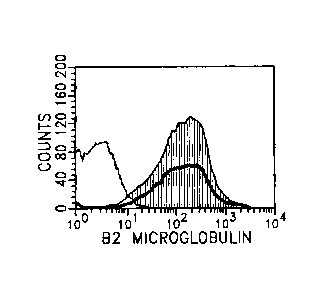
outline), Figure lb shows equally intense labeling for MHC class I (bold
outline) and 13-2
-5-
CA 02444706 2003-10-17
WO 02/086082 PCT/US02/12689
microglobulin (gray solid line) - isotype control (fine outline), Figure lc
shows high
intensity labeling for CD81 (gray solid line) - isotype control (fine
outline), Figure id
shows high intensity labeling for CD56 (gray solid line) - isotype control
(fine outline),
Figure 1 e shows moderately high intensity labeling for CD15 (Gray solid line)
- isotype
control (fine outline), Figure If shows moderately high intensity labeling for
CD95 (Gray
solid line) - isotype control (fine outline), Figure I g shows moderately high
intensity
labeling for CD95 (Gray solid line) - isotype control (fine outline), Figure
lg shows
moderately high intensity labeling for CD9 (Gray solid line) - isotype control
(fine outline),
Figure lh shows moderate labeling for CD34 (Gray solid line) - isotype control
(fine
outline). Figure li shows a small subpopulation of hNSCs labeling for GD3
ganglioside
(Gray solid line) - isotype control (fine outline) over a broad range of
intensities. Figures lj
shows GD2 ganglioside labeling (Gray solid line) - isotype control (fine
outline) on mouse
brain-derived neural stem cells obtained from GFP-transgenic mice. Figure lk
shows GD2
ganglioside labeling (Gray solid line) - isotype control (fine outline) on
retinal stem cells
also obtained from GFP-transgenic mice.
Figures 2a-e shows stem cells from the neural retina of GFP-transgenic mice
which
express the markers previously shown for brain-derived stem cells. 2a is the
isotype
control, 2b is for GD2 ganglioside, 2c is for CD9 (tetraspanin), 2d is for
CD15 (Lewis X,
lacto-N-fu.copentose III), 2e is for CD81 (tetraspanin).
Figures 3a-d depicts the influence of differentiating conditions on the
expression of
target molecules by hNSCs. In each case the target molecule is shown as solid
gray, the
isotype control with a fine black outline. The bold outline indicates the
profile of the target
molecule after hNSCs were cultured in fetal bovine serum (FBS). Figure 3a
shows that
CD34 expression increases under these conditions (Gray solid line = CD34, Bold
outline =
CD34 expression following fetal calf serum exposure, Fine outline = isotype
control),
Figure 3b shows that CD15 expression falls to control levels (Gray solid line
= CD15,
Bold outline = CD15 expression following fetal calf serum exposure, Fine
outline = isotype
control), Figure 3c shows that GD2 ganglioside expression decreases by an
order of
magnitude (Gray solid line = GD2 ganglioside, Bold outline = GD2 expression
following
fetal calf serum exposure, Fine outline = isotype control), Figure 3d shows
that CD9
expression falls to a lesser degree (Gray solid line = CD9, Bold outline = CD9
expression
following fetal calf serum exposure, Fine outline = isotype control).
-6-
CA 02444706 2003-10-17
WO 02/086082 PCT/US02/12689
Figure 4 depicts the induced expression of MHC surface markers in stem cells
from
the brain of GFP-transgenic mice after treatment with interferon gamma (IFNI)
for the
number of days shown as measured by flow cytometry.
Figures 5a-e show flow cytometric documentation of specific markers on
conditionally green stem cells derived from the brain of neonatal pNestin-GFP
mice.
Signal from marker antibody is the shaded curve, from isotype control is open.
5a is CD9,
5b is CD81, 5c is CD15, 5d is GD2 ganglioside, Figure 5e is 13-2
microglobulin.
Figures 6a-c are flow cytometric evaluations of pNestin-GFP neural stem cells,
before and after exposure to differentiation conditions (20 ng,/m1 CNTF).
Figure 6a is a
histogram showing the bright endogenous FITC + fluorescence emitted by pNestin-
GFP
neural stem cells when cultured under standard proliferation conditions (20
ng/ml EGF).
Figure 6b illustrates a modest decrease in endogenous fluorescence (left
shift) induced by 3
days of culture under differentiation conditions. Figure 6c is the marked
decrease in
endogenous fluorescence induced by 7 days of differentiation.
Figures 7a-c are an evaluation of selected marker expression by whole brain
homogenates. Mouse brain (pNestin-GFP transgenic) was removed from adult mice,
dissociated, and analyzed by flow cytometry. Figure 7a shows the profile for
GD2
ganglioside, Figure 7b shows the profile for MHC antigen IA-d, Figure 7c shows
the profile
for MHC antigen H2Kb.
Figures 8a-f depict the use of anti-Gm ganglioside antibody during
fluorescence-
activated cell sorting (FACS) to effectively enrich for neural stem cells.
Cultured hNSCs
were combined with human apheresis product and mixture was labeled with anti-
GD2-FITC,
CD56-PE and CD45 Pe-Cy5. Figure 8a depicts the light scatter gate (R1)
employed to
eliminate possible red blood cells and debris. Figure 8b depicts how gates
were then drawn
to encompass the CD45 positive (R3) and CD45 negative (R2) populations.
Figures 8c-d
depict how logical gating was used to sort hNSC (R2 and R4) from apheresis
product cells
(R3 and R5). Figures 8e-f depict the two resulting sorted populations and
demonstrate the
efficiency of the sorting procedure.
Figures 9a-c depicts the isolation of GD2+ stem cells from whole brain
homogenate from adult mice. Figure 9a shows whole brain homogenate which was
incubated with anti-GD2 primary antibody and PE conjugated secondary antibody,
then
sorted by FACS to select for GD2+ cells. Figure 9b depicts the initial GD2
population
-7-
CA 02444706 2003-10-17
WO 02/086082 PCT/US02/12689
labeled R1 which was 10.9%. Figure 9c depicts the resulting sorted population
which was
71% GD2+ representing an enrichment of approximately 700%.
DETAILED DESCRIPTION OF THE PREFERRED EMBODIMENT
The work herein identifies a range of molecules consistently present on or
absent
from the surface of multipotent neural stem and progenitor cells (NSCs) and in
different
subsets of NSCs and retinal stem cells RSCs. These molecules or markers were
identified
using a number of human and murine neural stem cell lines, including retinal
stem cells
(RSCs). The NSC-specific markers identified include gene products as well as
non-protein
molecules and sugar epitopes not directly coded in the genome. Together with
surface
markers which were determined to be absent from the surface of hNSCs, the
molecules
described herein provide a means to enrich for neural stem cells, or neural
progenitor
subpopulations, particularly using combinatorial cell sorting strategies.
These same
molecules also represent targets for pharmacological manipulation of NSC
populations and
subpopulations, both in vivo and ex vivo. Furthermore, these molecules provide
potential
targets for therapeutic manipulation of other neural precursor-related cell
types including
those that can be found in malignant conditions as well as other diseases
originating from,
or preferentially affecting, various uncommitted or replication-competent cell
types.
Definitions
A "neural stem cell" as used herein is a neural progenitor cell which is proto-
neuronal/proto glial. During development, embryonic stem cells which are very
primitive
totipotent cells are thought to pass through a neural stem cell stage as they
are developing
into neural cells. Neural stem cells can be induced to differentiate into any
neural cells
including glia, oligodendrocytes, neurons, or astrocytes. Cells were
characterized as
multipotent neural progenitor cells based on the ability to propagate over
many passages,
expression of nestin and Ki-67, proto-neuronal morphology, as well as the
ability to
differentiate into neurons and glia.
As used herein "embryonic stem cells" are totipotent cells isolated from
embryonic
or fetal tissue which may be treated with morphogens to differentiate into
neural stem cells.
Neural Stem Cells (NSCs)
Neural stem cells are multipotent progenitor cells which can be found in adult
brain
and related tissue as well as embryonic tissue. When neural stem cells are
contacted with
certain factors permissive for neuronal and glial cell differentiation, such
cells will
-8-
CA 02444706 2003-10-17
WO 02/086082 PCT/US02/12689
differentiate into neurons, glia, oligodendrocytes and astrocytes. When NSCs
are grown in
the presence of fetal calf serum, or other morphogenic agents, they can be
differentiated
into these various cell types or less primitive stem cells.
Sources of NSCs may be any tissue known to one of skill in the art, including
but
not limited to: brain, spinal cord, fetal tissue, retina, and embryo.
NSC-specific markers
Because previous methods for the isolation of neural stem cells (NSCs) using
the
cellular marker CD133, proved problematic, a method was developed herein which
allows
neural stem cells, human or otherwise, to be enriched without reference to
CD133. In fact,
completely different marker molecules were identified and used herein.
Furthermore, in
contrast to previous studies (Uchida, et al, 2000) it was shown that some
neural progenitors
are CD133- and CD34+, suggesting that the previous method of identifying NSCs
as
CD133+CD34- was flawed.
For example, the method of Uchida et al., 2000, allows for the isolation of
NSCs
using the specific epitope CD133. In this work, the authors restricted their
definition of
human neural stem cells to those cells within the human brain which were
CD133+/CD34-
(and CD24-/lo) and described the use of this marker profile to isolate NSCs.
They also
stated that the CD133+/CD34- (and CD24-/lo) fraction alone contains neural
stem cells
because neurospheres could not be generated from the CD133- fraction. CD133
was
previously thought to be a definitive marker of neural stem cells. However,
the results
herein show that it is in fact a negative marker.
Although the method of Uchida et al. appears to isolate a population NSCs
there are
a number of problems with the method as well as inconsistencies in the data.
For example,
there is no data presented that the subset of cells that the authors describe
as CD34-
/CD133+ are in fact CD34 negative, suggesting that the NSCs isolated may be a
mixed
population. Also, CD133 (5F3) cells were artificially separated from a single
population by
sorting the cells at the CD133 expressing-end of the population on two
separate occasions,
then claimed, upon reanalysis, that two separate populations existed (their
Fig. 1c). They
used two CD133 clones (5F3), commercially available from Miltenyi Biotec, and
infer that
an antibody they developed against the same immunogen, CD133 (5E12) clone, has
similar
properties. In a similar manner, they gated cells extending from a single
population of cells
in a two fluorescence dot plot, comparing the 5F3 CD133 and the 5E12 CD133
clones,
-9-
CA 02444706 2003-10-17
WO 02/086082 PCT/US02/12689
which extended outward from the population at a 45 degree angle toward the
upper right
area of the plot. This type of pattern is a hallmark of non-specific binding,
thus also
consistent with dead or dying cells (the authors mention that a viability dye,
propidium
iodide, was used but no evidence is presented). Comparison of single
populations to a
matched isotype control is not shown or mentioned. An isotype control is
mentioned with
regards to sort regions used to separate CD133+/CD24+ and CD133+/CD24- cells.
However, these cells represent a small percentage of the CD133-expressing
extreme of
single CD24+ and CD24- fetal brain cells and thus could result from non-
specific binding.
When tissue is minced, enzymatically-dissociated, and sorted twice as is done
to the NSC
population from Uchida et al 2000, there is likely to be increased levels of
damage to
constituent cells. This fact may have contributed to the variability in
frequency of
neurosphere initiating cells resulting from such manipulation. No mention of
statistical
significance of neurosphere initiating cell frequency in the experiments
relating to Fig. 2c,
(n=8) is mentioned.
Thus, it is clear that a convincing method for isolating NSCs has not yet been
developed, Uchida et al and other previous studies have focused on the neural
stem cell
markers which are protein in origin. However, it has long been appreciated
that the cellular
membranes of CNS neurons are a rich source of gangliosides. More recently it
has been
shown that these molecules are present during neural development in the
membranes of
cells of various lineage's. These studies clearly demonstrated that
gangliosides can rarely if
ever be used as lineage-specific markers. Although less obvious, these complex
ganglioside patterns are consistent with the behavior of neural
stem/progenitor cells, which
retain multipotency much further into the differentiation process than had
hitherto been
appreciated. Furthermore, there has been no prior evaluation of gangliosides
in a neural
stein/progenitor cell line, despite general appreciation of the abundance of
gangliosides in a
variety of neoplasms, particularly those of neuroepithelial origin. The
studies herein
provide a number of useful positive and negative NSC markers which are protein
as well as
ganglioside in origin.
Differentiation of NSCs
Many differentiation agents are known to one of skill in the art which can
differentiate stem cells, retinal stem cells, or neural stem cells into
specific types of nerve
cells, retina cells or types of progenitors. Therefore, it is envisioned that
the stem cells
-10-
CA 02444706 2003-10-17
WO 02/086082 PCT/US02/12689
isolated herein may be differentiated by any means known to one of skill in
the art. Some
examples of differentiation agents, include, but are not limited to Interferon
gamma, fetal
calf serum, nerve growth factor, removal of EGF, removal of bFGF (or both),
neurogenin,
BDNF, thyroid hormone, BMPs, LIF, sonic hedgehog, GDNFs, VEGFs, interleukins,
interferons, SCF, activins, inhibins, chemokines, retinoic acid and CNTF. The
cells may be
differentiated permanently or temporarily. For example, cells may be
differentiated
temporarily to express a specific marker, for example, in order to use that
marker for
identification. Then, the differentiation agent may be removed and the marker
may no
longer be expressed. However, it is to be understood that within the context
of
differentiation, agents such as interferon gamma, though inducing the
expression of
different markers, may not be classified as classical differentiation agents.
It is also to be understood that any anti-differentiation agents known to one
of skill
in the art may be used, including but not limited to:TGF-p, TGF-a, EGF, FGFs,
and delta
(notch ligand).
Uses of NSC specific markers
Although there are an extensive number of uses for NSC-specific markers, a few
of
the more common ones will be presented in more detail below. A major role of
such
markers involves enrichment of NSCs from a mixed population.
It is envisioned that the target molecules described herein are useful for the
enrichment of neural stem cells when isolating these cells from any source,
embryonic
(fetal) brain or neural tissue or post-embryonic brain or spinal' cord tissue.
Preferably, a
tissue homogenate derived from either surgical specimen or post-mortem
donation is used
as the source for isolating NSCs. In this way a large easily obtainable source
of NSCs can
be produced for use in further research or treatment of brain pathology.
In one embodiment, a method is used to identify neural stem cells wherein said
method identifies said NSCs using at least one positive neural stem cell
marker. In a
further embodiment one positive and one negative marker is used. In another
embodiment,
more than one positive marker and more than one negative marker is used.
In a further embodiment, the positive and negative markers can be a
"fingerprint"
for identification of the NSCs. The positive and negative markers can be any
identifiable
marker. In one embodiment, the positive and negative markers are protein
and/or
=
carbohydrate (glycosidic).
-11..
CA 02444706 2003-10-17
WO 02/086082 PCT/US02/12689
In one embodiment, the positive markers are any one or more of the following:
MHC I, P-microglobulin, GD2 ganglioside, GD3 ganglioside (in some cases), CD8,
CD9,
CD15, CD34, CD38, CD56, CD81, CD95, and CD152. In a further embodiment, the
positive markers are any one or more of the following: CD9, CD15, CD81, CD95,
GD2,
GD3, and CD34. In a further embodiment, the positive markers are selected from
the group
consisting of: CD15, CD81, CD95, and GD2. In a further embodiment, the
positive
markers are MHC I, MASH 1, MSI 1, and Nestin (MHC I or MHC 11 when induced).
In a
further embodiment, the positive markers are any one or more of MHCI and a
peptidic
fragment of MASH 1, MSI 1, or Nestin. In one embodiment, the antigen CD54 is
tested
before or after differentiation and cells which express it only after
differentiation are
isolated. In a further embodiment, Cells which express CD15 only before
differentiation
but not after are identified. In a further embodiment, cells which express
CD34 before
differentiation and more highly after differentiation are isolated.
In a further embodiment, the positive markers may be used alone and include,
but
are not limited to, GD2 and CD15. The tetraspanins (CD9 and CD81) may not work
as
well alone as a single marker, however, they may be very useful in combination
with other
positive or negative markers and may be useful for pharmaceutical intervention
or to
manipulate the cells which have already been isolated.
In a further embodiment, the MHC class I markers may be used for isolation
alone
or in combination with other markers. However, it is envisioned that the MHC
class I
markers may be usefull as a potential marker to induce rejection of cells
which are not
behaving appropriately. For example, transplanted cells which are over-growing
may be
destroyed. The MHC class I markers are variably expressed on different
subsets. However,
in a subset which does not express them, they may be induced with agents such
as
interferon when necessary.
In one embodiment, the negative markers are any one or more of the following:
MHC II, HLA-DR, Glycophorin-A, GD3 (positive in a very few cases), CD1a, CD3,
CD5,
CD7, CD10, CD11, CD13, CD14, CD16, CD19, CD20, CD22, CD23, CD25, CD31,
CD33, CD41, CD45, CD54, CD80, CD83, CD86, CD133, CD117, and CD154. In a
further embodiment, the negative markers are any one or more of the following:
MHC
class IT, CD3 (TCRa13-1), CD7, CD10, CD16, CD54. In a further embodimnt, the
negative
markers may be MHC class II and/or CD133.
-12-
CA 02444706 2003-10-17
WO 02/086082 PCT/US02/12689
The method of enrichment can be any method known to one of skill in the art
which
enriches for a population of cells using specific cell surface markers. For
example, the
method can be fluorescence-activated cell sorting (FACS), affinity columns,
affinity beads,
or any method which selectively binds the specific cell surface molecules.
Alternatively,
the method may use the cell surface molecules which are not expressed by NSCs
to
selectively remove or kill the undesirable cells, and, in this way, enrich for
the desirable
cells. Alternatively, the method can be with the use of magnetic beads which
selectively
bind the NSCs.
A further embodiment of the invention is the use of these specific cell
surface
markers/molecules to enrich for particular subpopulations of neural progenitor
cells. For
example, it is thought that the population of neural progenitor cells contains
some totipotent
cells which can differentiate into any neural cell type, some multipotent
cells which can
only differentiate into certain cell types, and some cells which have advanced
further along
the path of differentiation and may only be able to differentiate into one
cell type.
Therefore, it may be advantageous to enrich for a population of cells which is
no longer .
able to differentiate into a particular type (i.e. glial cell) or which is
only able to
differentiate into one specific cell type (e.g. photoreceptors or dopaminergic
neuron).
A further embodiment is the use of these positive and negative neural stem
cell
specific markers for identification of neural stem cells or of a subpopulation
of neural stem
cells which are associated with a disease or may be identified post-
operatively during a cell
transplantation.
A further embodiment is the use of the cell specific neural stem cell markers
to
identify and diagnose any cell, but particularly, cancer cells from a tumor or
metastasis,
which has a neural origin. This may have a relation to the course of treatment
for the
cancer. For example, typically, the less differentiated the cancer, the more
invasive. Thus,
a tumor which is composed of less differentiated cells may need to be treated
more
aggressively then one which is composed of more differentiated neural cells.
Since neural
stem cells are thought to represent one of the earliest cells in development,
the more the
presence or absence of specific neural stem cell markers (or "fingerprint")
matches the
cancer cell's "fingerprint", the more likely it is that the cancer cells are
undifferentiated. In
other words, if the cancer cell possesses the positive markers identified
herein and does not
have the negative markers, then it is a very undifferentiated cancer. The
specific neural
-13-
CA 02444706 2003-10-17
WO 02/086082 PCT/US02/12689
stem cell markers or proteins and enzymes important to their expression, can
be identified
in any way known to one of skill in the art, including FACS, cytometry,
enzymatically, by
Western blot and by PCR.
Research into neuronal cell development:
A further embodiment is the use of the NSC cell surface markers, both positive
and
negative, to study stein and progenitor cell behavior during development and
in maturity.
For example, it is unclear whether cell development proceeds along a linear,
temporal or
branched progression. It is also unclear how important the effect of
neighboring cells are to
development and differentiation of the neural stem cells.
Use of antisense oligonucleotides and/or antibodies to NSC-specific markers as
therapeutics:
One embodiment is the use of antisense oligonucleotides which specifically
inhibit
the expression of positive NSC-specific protein markers. The antisense
oligonucleotide can
be identified and synthesized using techniques known to one of skill in the
art. In addition,
variants may be produced using any bases known to one of skill in the art,
including various
well-known modified bases. It is envisioned that the antisense when acting on
a positive
NSC marker will inhibit the growth of NSCs or NSC-related cancer cells.
Alternatively,
the antisense oligonucleotides may specifically act on a negative NSC marker
in an NSC or
NSC-like cell. It is envisioned that when acting on a negative NSC marker in
the right
environment, the antisense oligonucleotide would increase the growth of an NSC
or a less
differentiated cell.
A further embodiment is the use of antibodies which specifically bind to
positive
NSC markers. It is envisioned that the antibodies would target, identify, or
bind to the
NSCs for treatment, enrichment or diagnosis. For example, antibodies to the
NSC-specific
markers could be used to target a therapeutic agent to the NSCs specifically.
Alternatively,
antibodies to negative NSC markers may be used to weed non-NSC cells from a
population.
Alternatively, markers may be induced by the addition of cytokines or other
agents
before the application of antibodies which are specific to the induced
markers.
Identification of therapeutics:
A further embodiment of the invention is the use of the NSC-specific molecules
as
targets for pharmacological manipulation of NSCs, neural progenitors, and more
differentiated neural cell types, both in vivo and following isolation.
Desirable interventions
-14-
CA 02444706 2003-10-17
WO 02/086082 PCT/US02/12689
include positive and negative modulation of proliferation and differentiation
by
identification of agonists and antagonists of NSCs. For example, the agonist
may act on a
neural stem cell specific marker, thus increasing the growth of the cells. Of
particular
interest are markers which are either down-regulated or not up-regulated
during
differentiation of neural stem cells. The method of testing for agonist of
NSCs could
involve any method known to one of skill in the art, but essentially, the
method may
involve treating NSCs in vitro or in vivo with a pharmaceutical or other
chemical and
looking for an increase in number of the NSCs. Antagonists are also of
interest for
decreasing the growth of NSC or NSC-like cells. These antagonists may act on
the cell-
specific markers to antagonize growth of NSCs. Of particular interest are
markers which
are upregulated during the differentiation of NSCs. The method of testing for
antagonists
would be by treating NSCs in vitro or in vivo with the pharmaceutical or other
chemical,
and identifying a decrease in the number of NSCs which would identify the
chemical as an
antagonist.
Examples of methods for the identification of agonists or antagonists are as
follows:
NSCs are grown in vitro, in tissue culture, to about 50-80% confluency, the
chemical or
pharmaceutical is then be added at various concentrations in the presence of a
marker for
cell division and the increase or decrease in cell cycling measured relative
to control.
Alternatively, the NSCs could be gown in vivo and treated before or after in
vivo
implantation with the chemical or pharmaceutical in the presence of a marker
for cell
division or cell cycling and the increase or decrease in cell cycling measured
relative to
control.
A further embodiment of the invention is the use of the NSC-specific molecules
before, during, and after isolating undifferentiated neural cell types for use
in drug
development. Such drugs may have utility as treatments for conditions
involving neural
stem cells both directly and indirectly, although not always recognized as
such. Here we "
include malignant neoplasms such as glioblastoma multiforme, astrocytoma, and
retinoblastoma; infectious diseases such as CMV, rubella, and HIV;
inflammatory diseases
such as trauma, multiple sclerosis, diabetes, and SLE, as well as
neurodestructive and
degenerative diseases such as stroke, Parkinson's Disease, Alzheimer's
Disease,
Huntington's Disease, ALS, retinitis pigmentosa, and macular degeneration. In
addition,
such drugs may have utility in reducing the overmultiplication of transplanted
cells.
-15-
CA 02444706 2003-10-17
WO 02/086082 PCT/US02/12689
Transplantation of NSCs:
A further embodiment of the present invention is the use of NSCs for
transplantation into damaged areas of the brain to repopulate the area.
Preferably, the NSCs
differentiate into the damaged cell type. The cells may also be treated before
or after
transplantation to be more likely to differentiate into the missing cell type.
Such
differentiation factors include but are not limited to: Some examples of
differentiation
agents, include, but are not limited to Interferon gamma, fetal calf serum,
nerve growth
factor, removal of EGF, removal of bFGF (or both), neurogenin, BDNF, thyroid
hormone,
BMPs, LW, sonic hedgehog, GDNFs, VEGFs, interleukins, interferons, SCF,
activins,
inhibins, chemoldnes, retinoic acid and CNTF.
Cells having the characteristics of multipotent neural stem cells, neuronal
progenitors, and/or glial progenitors of the CNS (identified by in vitro
assays) are
introduced into a mammal exhibiting a neurological disorder to examine the
therapeutic
potential of these cells. The cells are preferably isolated from a mammal
having similar
MHC genotypes or the host mammal could be immunosuppressed using drugs such as
cyclosporin A. The cells are injected into the spinal cord, retina or brain.
The cells are
injected at a range of concentrations to determine the optimal concentration
into the desired
site. Alternatively, the cells are introduced in a plasma clot or collagen gel
to prevent rapid
dispersal of cells from the site of injection. The effect of this treatment on
the neurological
status of the model animal is noted. Desired therapeutic effects in the above
mutant mice
include the reduction or cessation of seizures or improved movement of lower
motor
extremities. The cells may be administered using any method known to one of
skill in the
art. In addition, it is envisioned the new methods will be developed which
provide
advantages for the various therapeutic treatments or uses of NSCs and RSCs.
The NSCs
and/or RSCs herein may be administered using the new methods.
Having now generally described the invention, the following examples are
offered
to illustrate, but not to limit the claimed invention.
EXAMPLES
Example 1 presents that data which was obtained when human NSCs were analyzed
for the presence and absence of cell surface markers using a panel of
antibodies known in
the art (See Table 1).
-16-
CA 02444706 2010-04-09
. .
. . .
EXAMPLE 1
Identification of Positive and Negative Neural Stem Cell Markers in human
neural stem cells
Initially, a human neuronal progenitor cell line was obtained from Michael
Young,
Ph. D. who obtained it from Clonetics, (Walkersville, Maryland, catalog number
CC-2599).
These cells had been obtained from donated prenatal tissue and tested negative
for a range of
infectious pathogens. Cells were obtained frozen and subsequently grown in
tissue culture
using a defined medium consisting of DMEM/F12 high glucose, N2 Supplement
(Life
Technologies), bFGF (20-25 ng/ml) and EGF (20-25 ng/ml), and L-glutamine.
Cells were
characterized as multipotent neural progenitor cells based on the ability to
propagate over
many passages, expression of nestin and Ki-67, proto-neuronal morphology, as
well as the
ability to differentiate into neurons and glia (Miziunoto, H. et al. 2001
"Transplantation of
human neural progenitor cells to the vitreous cavity of the Royal College of
Surgeons rat"
Cell Transplant 10:223-233).
For the present study, cells were initially grown as neurospheres until
plentiful, then
spheres were broken up and seeded into flasks coated with polyornithine and
Jaminin where
the cells grew as an adherent monolayer using the same defined medium
described above.
After reaching confluence, cells were harvested using Custom ATV (Irvine
Scientific),
washed in PBS (Ca/M? free; Dulbecces Phosphate Buffered Saline, (3ibco BRL,
Grand
Island, N.Y.) and centrifuged at 400 x g for 4 minutes. The resulting pellet
was
resuspended in PBS using a flame-polished glass Pasteur pipette with a narrow
bore. 100
III of cell suspension, containing approximately 5 x 10⁵ cells was distributed
among 12 x 75
polystyrene tubes containing appropriate quantities of listed antibodies.
Manufacturers
suggested concentrations were observed, with the exception of GD2-11.1C in
which case
15141 neat (0.25merni) was used according to previous experience and
titration. Cells were
incubated with antibody for 20 minutes at room temperature, protected from.
light. The
cells stained with directly conjugated antibodies were then washed with 2 ml
PBS
(Dulbecco's Phosphate Buffered Saline, Gibco BRIõ Grand Island, N.Y.) and spun
at 400 x
g for 4 minutes, decanted and resuspended in 200)21 of PBS containing 7-amino
Actinomycin D (7-AAD) in PBS (11.1g/ro1). Following initial incubation and
wash, cells
incubated with unconjugated antibodies were then stained with F1TC goat anti-
mouse or
PE-conjugated sheep anti-mouse antibody. Unbound antibodies were then removed
by
washing with 2 ml PBS, as previously described, and resuspended in PBS
containing 7-
-17-
CA 02444706 2003-10-17
WO 02/086082 PCT/US02/12689
AAD (114m1). FAGS Lysing Soln, (Ammonium Chloride, Tetra Sodium EDTA,
Potassium phosphate) was used to ready cells for FAGS analysis.
-18-
CA 02444706 2003-10-17
WO 02/086082
PCT/US02/12689
Table 1: Antibodies used in the cell surface identification studies
Simultest Control (MsIgGl-FITC/MsIgG2a-PE), BD Biosciences, San Jose,CA.
Ms IgM,Ic-FITC Isotype control, BD Biosciences, San Jose, CA
Ms IgGi¨PE Isotype control, BD Biosciences, San Jose, CA
Anti-MHC Class I, BD Biosciences, San Jose, CA
132 microglobulin, BD Biosciences, San Jose, CA
Anti GD2 ganglioside, US Biological, Swampscott, MA
Anti GD3 ganglioside, US Biological, Swampscott, MA
Anti CD9-FITC, BD Biosciences, San Jose, CA
Anti CD15-FITC, BD Biosciences, San Jose, CA
Anti CD34-PE (8G12), BD Biosciences, San Jose, CA
Anti CD56-PE, BD Biosciences, San Jose, CA
Anti CD81, BD Biosciences, San Jose, CA
Anti CD95-PE, BD Biosciences, San Jose, CA
Anti MHC Class II, BD Biosciences, San Jose, CA
Anti HLA-DR PE, BD Biosciences, San Jose, CA
Anti Glycophorin-A, Pharmingen, LaJolla, CA
Anti CD1a, BD Biosciences, San Jose, CA
Anti CD3-FITC, BD Biosciences, San Jose, CA
Anti Zeta-FITC, BD Biosciences, San Jose, CA
Anti CD5-FITC, BD Biosciences, San Jose, CA
Anti CD7-FITC, BD Biosciences, San Jose, CA
Anti CD8-
Anti CD1O-FITC, BD Biosciences, San Jose, CA
Anti CD11b-FITC, BD Biosciences, San Jose, CA
Anti CD13-PE, BD Biosciences, San Jose, CA
Anti CD14-FITC, BD Biosciences, San Jose, CA
Anti CD16-FITC, BD Biosciences, San Jose, CA
Anti CD19-PE, BD Biosciences, San Jose, CA
Anti CD2O-PE, BD Biosciences, San Jose, CA
Anti CD22-FITC, BD Biosciences, San Jose, CA
Anti CD23-PE, BD Biosciences, San Jose, CA
Anti CD25-PE, BD Biosciences, San Jose, CA
Anti CD31-FITC, BD Biosciences, San Jose, CA
Anti CD33-FITC, BD Biosciences, San Jose, CA
Anti CD34-(epitope 561) and/or Anti CD34 (epitope 8G12)
Anti CD38
Anti CD45-FITC, BD Biosciences, San Jose, CA
Anti CD54-PE, BD Biosciences, San Jose, CA
Anti CD8O-FITC, BD Biosciences, San Jose, CA
Anti CD83-PE, BD Biosciences, San Jose, CA
Anti CD86-PE, BD Biosciences, San Jose, CA
Anti CD117-FITC, BD Biosciences, San Jose, CA
Anti CD133
Anit CD152
Anti CD154, BD Biosciences, San Jose, CA
Cytometric Evaluation:
-19-
CA 02444706 2003-10-17
WO 02/086082 PCT/US02/12689
The FACS Vantage, equipped with an Enterprise 488 nm argon laser (FACS
Vantage cell sorter, BD Biosciences, San Jose, CA), was calibrated and aligned
using
chicken RBC, according to manufacturers directions. Color
compensation was
preliminarily set using calibrite beads, BD Biosciences, San Jose, CA.
Individual samples
were optimized using single positive (CD56) antibody labeling, compared to
negative
matched isotype controls, for each fluorochrome used. Two color live gating
acquisition
was used to optimize settings and acquire data. Optimally, 30,000 events were
collected
and stored electronically for subsequent analysis.
Fluorescence Activated Cell Sorting (FACS):
Cultured hNSCs were combined with human apheresis product and the mixture was
labeled with anti-GD2-FITC, CD56-PE and CD45 Pe-Cy5. A light scatter gate (R1)
was
employed to eliminate any possible red blood cells and debris. Gates were then
drawn to
encompass the CD45 positive (R3) and CD45 negative (R2) populations. Logical
gating
was used to sort hNSC (R2 and R4) from apheresis product cells (R3 and R5).
The two
resulting sorted populations were reanalyzed flow-cytometfically to evaluate
the efficiency
of the sorting procedure.
-20-
CA 02444706 2003-10-17
WO 02/086082
PCT/US02/12689
TABLE 2: Summary of Target Molecules Identified and Eliminated
Positive markers: Effect of differentiating conditions:
MHC class I and 132-microglobulin (variable) No change (except
induced by IFN-y)
GD2 ganglioside Decreases
CD8
CD9 (TM4 superfamily) Slight decrease
CD15 (SLex) Disappears
CD34 (hematopoietic stem cell antigen)
(8G12 epitope) Increases
CD34 (561 epitope)
CD38
CD56 (NCAM)
CD81 (TAPA-1)
CD95 (Fas)
CD152
Negative markers
MHC class II (DR DQ DP) No change
HLA-DR
Glycophorin-A
GD3 ganglioside (only expressed by small subpopulation)
CD1a
CD3 (TCRa13-1)
CD3 (TCR chain) No change
CD5
CD7 No change
CD10 No change
CD1lb
CD13
CD14
CD16 No change
CD19
CD20
CD22
CD23
CD25
CD31
CD33
CD41
CD45
CD54 (ICAM) Becomes positive with FCS
CD80
CD83
CD86
CD133
CD117
CD154
-21-
CA 02444706 2003-10-17
WO 02/086082 PCT/US02/12689
A large number of positive and negative markers were identified using this
procedure (shown in Table 2). Of special interest was GD2 ganglioside because
it was
highly expressed by the majority of NSCs (especially as compared to
ganglioside GD3
which was only expressed by very few) and because it is a sugar-related marker
rather than
a protein. In addition, other positive markers of NSCs identified were CD9
(TM4
superfamily) CD15 (LeX), CD34 (hematopoietic stem cell antigen) as tested with
antibodies to two epitopes, CD56 (NCAM), CD81 (TAPA-1)CD95 (Fas) and MHC class
I
and f32-microglobulin.
In direct contrast to the results of Uchida et al. the human neural stem cells
which
were tested in the present study were CD133-/CD34+. These cells clearly
possessed the
ability to differentiate into neurons and glia, can be grown as neurospheres,
express nestin
and Ki-67, and have a proto-neuronal morphology. Thus, the results herein
suggest that the
method of Uchida et al does not identify the cells known as neural stem cells.
It is envisioned that certain markers would be lost during differentiation of
the
NSCs and if they are down-regulated during differentiation of the NSCs, they
are likely to
be very specific NSC markers. In Example 3, the neural stem cells were grown
under
conditions which induced differentiation and retested for expression of the
above markers.
The results now provide supporting evidence that additional positive markers
originally identified on human brain derived stem cells (Fig. 1 a-i) are
consistently
expressed by similar cells obtained from a variety of central nervous system
(CNS) sources,
in this case the brain (Fig. 5) and retina (Fig. 2) of the mouse. In both
instances, the
additional markers are CD9, CD15, and CD81.
EXAMPLE 2
Identification of Neural Retinal Cell Markers
Stem cells from the neural retina of GFP-transgenic mice were found to express
the
markers previously shown for brain-derived stem cells. In Figure 2 the
expression of GD2
ganglioside, CD15, and the tetraspanins CD9 and CD81, are shown using flow
cytometry.
-22-
CA 02444706 2003-10-17
WO 02/086082 PCT/US02/12689
EXAMPLE 3
Identification of Neural Cell Markers Associated with Differentiation
The human neuronal progenitor cells (hNSCs) in Example 1 were treated with
Fetal
calf serum (FCS) to induce differentiation and some of the markers were
reexamined.
Figure 3 depicts the influence of these differentiating conditions on the
expression of target
molecules by hNSCs. In each case the target molecule is shown as solid gray,
the isotype
control with a fine black outline. The bold outline indicates the profile of
the target
molecule after hNSCs were cultured in fetal bovine serum (FBS). Figure 3a
shows that
CD34 expression increased under these conditions, Figure 3b shows that CD15
expression
fell to control levels, Figure 3c shows that GD2 ganglioside expression
decreased by an
order of magnitude, Figure 3d shows that CD9 expression fell to a lesser
degree.
The expression of certain NSC-specific markers decreased during
differentiation,
suggesting that these markers are strong markers of "true" neural stem cells.
Thus, CD15
and GD2 are important markers for identifying "true" neural stem cells and CD9
to a lesser
degree.
In addition, as the cells differentiated, the level of CD34 actually
increased. This
was a surprising result and in direct contrast to the work by Uchida et al.
which specifically
isolated a population of cells which were CD34- as neural stem cells. Thus,
the results
herein suggest that the population isolated by Uchida et al were either a
different population
and not "true" neural stem cells, or were a more differentiated version of the
neural stem
cells identified herein.
EXAMPLE 4
Expression of Cell Surface Markers During Treatment with IFN-y
Stem cells from the brain of GFP-transgenic mice did not express class I or
class II '
MHC antigens at baseline or under differentiation conditions. These antigens
could,
however, be induced by the addition of the cytokine interferon-gamma (IFN-y),
shown in
Figure 4 using flow cytometry. MHC induction by IFN-y was reversible.
In addition, evidence is provided that expression of some markers differs
among
neural stem cell populations. Whereas the initial brain-derived line (of human
origin)
expressed MHC class I surface molecules (Fig. lb), the subsequent brain and
retina-derived
lines (from mice) did not. Stem cells from the brain (Fig. 4) and retina of
GFP-transgenic
mice did not express MHC class I antigens at baseline or under differentiation
conditions,
-23-
CA 02444706 2003-10-17
WO 02/086082 PCT/US02/12689
although both class I and class II MHC could be induced by IFN gamma. However,
many
of the same markers are identified on NSCs from mouse brain and retina.
EXAMPLE 5
Expression of Cell Surface Markers by pNestin-GFP Transgenic Neural Stem Cells
Having documented the presence, or absence, of multiple surface epitopes on
human and then mouse brained-derived stem/progenitor cell lines, the analogous
cells
derived from the brain of neonatal mice transgenic for GFP under the control
of the nestin
promoter (pNestin-GFP) were analyzed. Analysis was by flow cytometric
documentation of
specific markers on conditionally green stem cells derived from the brain of
neonatal
pNestin-GFP mice. In Figure 5, the signal from the marker antibody is the
Shaded curve,
from isotype control is open. In this figure there was a high expression of
the tetraspanin
CD9, as well as that of CD81, another tetraspanin (TM4 protein). The Lewis
antigen, CD15
was also clearly expressed, as was GD2 ganglioside. In contrast, signal from
the MHC class
I-associated marker beta-2 microglobulin was indistinguishable from isotype
and therefore
not expressed.
These results confirmed that the surface marker profile of pNestin-GFPgmBSCs
(brain-derived neural stem cells) is quite comparable to that of human neural
stem cells, as
shown in Fig. 1. These data establish the basis for additional work utilizing
these markers
in a variety of flow cytometric analyses, including cell sorting.
EXAMPLE 6
Expression of Cell Surface Markers by pNestin-GFP Transgenic Neural Stem Cells
When flow cytometric evaluation of pNestin-GFP neural stem cells was performed
before and after exposure to differentiation conditions, the following was
concluded.
In Figure 6, the top histogram shows the bright endogenous FITC+ fluorescence
emitted by pNestin-GFP neural stem cells when cultured under standard
proliferation
conditions (20 ng/ml EGF). The middle histogram illustrates that there was a
modest
decrease in endogenous fluorescence (left shift) induced by 3 days of culture
under
differentiation conditions. The apparent magnitude of this shift is lessened
by the log scale
of the X-axis, but confirmed by the statistics provided. At bottom can be seen
the marked
decrease in endogenous fluorescence induced by 7 days of differentiation. In
additional
work, the changes in GFP expression induced by culturing pNestin-GFPstem cells
under
differentiation conditions was assessed. In this case, withdrawal from mitogen
was used
-24-
CA 02444706 2003-10-17
WO 02/086082 PCT/US02/12689
combined with concomitant exposure to CNTF, a factor known to promote
astrocytic
differentiation. Using unstained pNestin-GFP stem cells and flow cytometry,
the fact that
these cells exhibit a high level of endogenous FITC+ fluorescence under
proliferation
conditions (EGF, 20 ng/ml) was confirmed. When removed from EGF and cultured
in
CNTF (20 ng/ml) the level of GFP-associated fluorescence progressively
decreased (Fig.
D), consistent with down-regulation of nestin expression during
differentiation.
Thus, additional data is provided relating the identified surface markers to
CNS-
derived stem cells. For instance, stem cells were derived from the brains of
mice transgenic
for GFP under the control of the Nestin promoter. These cells not only
expressed GD2
ganglioside, CD9, CD15, and CD81 (new Fig. 5), but also emitted high levels of
baseline
FITC+ fluorescence due to conditional expression of the GFP reporter gene
(Fig. 6).
Endogenous GFP expression by these cells was down-regulated by the addition of
the
cytokine CNTF, which is known to promote astrocytic differentiation in these
cells (Fig. 6).
The regulation of GFP expression by these cells is therefore consistent with
expression of
Nestin, and hence with neural stem cell phenotype. The simultaneous expression
of both
GFP and the markers GD2 ganglioside, CD9, CD15, and CD81 further supports the
idea
herein the latter markers are expressed by neural stem cells.
EXAMPLE 7
Selected Marker Expression Analysis of Whole Brain Homogenates
Evaluation of selected marker expression by whole brain homogenate was as
follows: Mouse brain (pNestin-GFP transgenic) was removed from adult mice,
dissociated,
and analyzed by flow cytometry. The tetraspanins CD9 and CD81 were found to be
highly
expressed by a majority of brain cells. CD15 was expressed by many, but not
all, brain
cells. MHC antigens (Figure 7b IA-d, Figure 7c H2Kb) were not widely
expressed. Of
particular note, the GD2 ganglioside (Figure 7a) was not heavily expressed in
the brain,
despite being prominently expressed by CNS stem cells. This indicates that GD2
ganglioside can be used to prospectively identify and isolate CNS stem cells
with good
efficiency.
By examining a whole-brain homogenate from pNestin-GFP transgenic mice, a
comparison can be made of the expression of these markers between a stem cell
population
and a whole-brain population (Fig. 7). These data show that whereas GD2
ganglioside is
highly expressed on the stem cell population, it is rare on cells of the
brain, consistent with
-25-
=
CA 02444706 2003-10-17
WO 02/086082 PCT/US02/12689
the interpretation that GD2 ganglioside provides a selective neural stem cell
marker.
Expression of CD15 is more widespread in brain, and CD9 and CD81 are both
heavily
expressed in brain as well as on neural stem cells. Class I and class II MHC
antigens are
poorly expressed by either.
EXAMPLE 8
Identification of Positive and Negative Neural Stem Cell Markers in Neural
Stem
Cells Isolated from Mouse brain and retinal cells.
The mouse neural stem cell lines were generated in the lab of Michael Young,
Ph.D.
Mouse brain stem cells expressed CD15 and did not express detectable GD3
ganglioside,
MHC class I or MHC class II at baseline. Furthermore, the addition of
interferon-gamma
induced the expression of MHC I and beta-2 microglobulin by the mouse brain
stem cell
line.
In Example 9, a method for enriching for or isolating NSCs is presented. The
method uses the newly identified neural stem cell-specific markers from
Examples 1 and 2.
EXAMPLE 9
Identification and Method of Isolating/Enriching for Neural Stem Cells with
Anti-
Ganglioside Antibodies
The present studies show that the ganglioside GD2 is present at high abundance
in
the cell membrane of the majority of cells comprising the neural progenitor
population (See
Table 2). This is true across species lines. The presence of GD2 has herein
been confirmed in
a human brain-derived stem cell line (see Example 1) and a mouse brain-derived
neural
stem cell line (see Example 1), as well as a mouse neural retina-derived stem
cell line (see
Example 2) . In contrast, the ganglioside GD3 is expressed, by a small
fraction of the human
neural progenitor population.
Thus, because neural stem cells express abundant levels of gangliosides, and
because ganglioside subtypes are not uniformly distributed across neural cell
types during
development, anti-ganglioside antibodies (to GD2) can be used to generate NSC-
enriched
fractions from a CNS homogenate or other sample containing mixed cell types.
The use of non-proteinaceous external epitopes for purposes of stem cell
isolation
represents a novel concept. The fact that gangliosides are not gene products
may have
contributed to their being overlooked as potential candidates for specific
stem cell markers.
However one major advantage of using non-proteinaceous markers for isolation
and
-26-
CA 02444706 2003-10-17
WO 02/086082 PCT/US02/12689
identification of NSCs is that such non-proteinaceous markers have a lack of
susceptibility
to proteolytic enzymes routinely used during the preparation of tissue for
cell harvest. This
is important because normal, non-immortalized, neural stem cells require
intact growth
factor receptors in order to proliferate. Also, because ganglioside molecules
are so plentiful
within the membrane, antibodies of lower affinity might still be adequate to
selectively
enrich for NSCs.
Figures 8a-f depict the use of anti-GD2 ganglioside antibody during
fluorescence-
activated cell sorting (FACS) to effectively enrich for neural stem cells.
Cultured hNSCs
were combined with human apheresis product and the mixture was labeled with
anti-GD2-
FITC, CD56-PE and CD45 Pe-Cy5. Figure 8a depicts the light scatter gate (R1)
employed
to eliminate possible red blood cells and debris. Figure 8b depicts how gates
were then
drawn to encompass the CD45 positive (R3) and CD45 negative (R2) populations.
Figures
8c-d depict how logical gating was used to sort hNSC (R2 and R4) from
apheresis product
cells (R3 and R5). Figures 8e-f depict the two resulting sorted populations
and demonstrate
the efficiency of the sorting procedure.
For isolating NSCs from fetal/embryonic or adult brain tissue, the following
method
is used. Fetal/embryonic or adult brain tissue from surgical specimen or post-
mortem
donation is homogenized and labeled with anti-GD2-FITC. The cells are then
sorted using
FACS. The cells which are GD2 positive are collected and further grown in
tissue culture or
treated and transplanted.
In Example 10, Retinal Stem cells are isolated using essentially the same
method.
EXAMPLE 10
Identification and Method of Isolating/Enriching for Retinal Stein Cells Using
Anti-
GD2 Ganglioside Antibodies on a Retinal Cells Population
The present studies are the first to show that the ganglioside GD2 is present
at high
abundance in the cell membrane of the majority of cells comprising the retinal
progenitor
population (See Table 2). Therefore GD2 is used to isolate retinal progenitor
cells (RSCs)
as follows: retinal tissue from a transgenic GFP-mouse, which were propagated
and
obtained from the lab of Michael Young were labeled with anti-GD2-FITC. The
cells are
then sorted using FACS. The cells which are GD2 positive are collected and
further grown
in tissue culture or treated and transplanted.
-27-
CA 02444706 2003-10-17
WO 02/086082 PCT/US02/12689
=
RSCs are isolated from fetal/embryonic or adult brain tissue using the
following
method. Fetal/embryonic or adult retinal tissue from surgical specimen or post-
mortem
donation is homogenized and labeled with anti-GD2-FITC. The cells are then
sorted using
FACS. The cells which are GD2 positive are collected and further grown in
tissue culture or
treated and transplanted.
EXAMPLE 11
Method of Isolating/Enriching for Neural Stem Cells Using Positive and
Negative
Cellular Markers
In identifying and isolating neural stem cells, it is advantageous to use a
number of
different positive and negative markers. Table 2 shows the identification of a
large number
of positive and negative markers for NSCs, which can be thought of as the
"fingerprint" of
NCSA's. The following method can be performed using antibodies to one positive
neural
stem cell marker as set out in Table 2. Alternatively, the method can be
performed using
two neural stem cell markers, one marker may be positive and one negative or
both may be
positive or negative markers. It can be envisioned that the more markers that
are used, the
more likely it is that the desired neural stem cell is isolated.
In one embodiment, GD2 is used as follows: Antibodies to GD2 are used to treat
a
population of neural stem cells from donated tissue from an adult brain. The
antibodies are
goat anti-human antibodies. After binding to the cells, the cells are treated
with a FITC
labeled rabbit anti-goat antibody. Subsequently or concurrently, Antibodies to
CD15 are
used to treat the same population of neural stem cells. The antibodies are
goat anti-human
antibodies. After binding to the cells, the cells are treated with a different
FITC labeled
rabbit anti-goat antibody. The cells which bound both antibodies are
identified as cells
having both Fluorescence associated with them by a FACS analyzer. These cells
are then
combined and grown in tissue culture.
Alternatively, NSCs are isolated using a positive and a negative marker as
follows:
NSCs are isolated from fetal/embryonic or adult brain tissue using the
following method.
Fetal/embryonic or adult retinal tissue from surgical specimen or post-mortem
donation is
homogenized and labeled with anti-GD2-FITC, and CD54-PE . The cells are then
sorted
using FACS. The cells which are GD2 positive and CD54 negative are collected
and further
grown in tissue culture or treated and transplanted.
Alternatively, a plurality of positive and negative markers can be used.
-28-
CA 02444706 2003-10-17
WO 02/086082 PCT/US02/12689
In Example 12, a similar method is used to isolate NSCs from treated or
untreated
embryonic stem cells.
EXAMPLE 12
Method of Selecting/Generating/Directing Neural Stem Cells from Embryonic Stem
Cells
Embryonic stem cells (ES) are non-neuronal, primitive cells which can be
induced
to form neural stem cells (NSC) by adding specific morphogens. The markers
herein can
be used to select for NSCs that are a part of the ES population before or
after the treatment
with a morphogen. Alternatively, the ES can be treated with substances that
induce the
expression of NSC positive markers or substances that decrease the expression
of negative
markers to produce NSCs from ES cells. This is because it is believed that ES
cells may
have to "pass through" a neural stem cell stage to become useful for the
treatment of
neurological conditions.
Alternatively, other cells may be de-differentiated or trans-differentiated to
produce
NSCs. For example, recent reports that cells in the skin can become NSC-like,
and that fat
can become bone, cartilage, or muscle (presumably via conversion to a
mesenchymal stem
cell-like intermediary) suggest that the neural stem cell specific markers
identified herein
can be used for directing such transitions.
In Example 13, Antisense oligonucleotides which down-regulate Neural stem cell
specific markers are used as a therapeutic or research tool.
EXAMPLE 13
Method of Treating Neural Tumors or Over-proliferation of NSCs with Antisense
to
Neural Stem Cell Specific Markers
Antisense oligonucleotides are designed which are complementary to the mRNA of
positive neural stem cell markers, GD2 and CD15. The antisense
oligonucleotides are
enclosed into vesicles and administered to the patient with a neural stem cell-
derived tumor
by injection into the spinal cord. The tumor or overproliferating NSCs are
monitored for
reduction in the size. Further treatments are administered as needed.
Alternatively, Neural Stem Cells are treated in vitro or in vivo and the
effect on
multiplication, differentiation and expression of neural stem cell specific
genes is
monitored.
Example 14 identifies a method of treating neural tumors with antibodies.
-29-
CA 02444706 2003-10-17
WO 02/086082 PCT/US02/12689
EXAMPLE 14
Method of Treating Neural Tumors or Over-proliferation of NSCs with Antibodies
to
Neural cell Specific Markers
Antibodies to the positive neural stem cell markers, GD2 and CD15 are produced
by
recombinant means, hybridoma technology or in an animal model. The antibodies
are
humanized or used as fragments. The antibodies are fused with a therapeutic
molecule such
as a chemotherapeutic agent or a toxin. The antibodies are then administered
to the patient
with a neural stem cell-derived tumor by injection into the spinal cord. The
tumor and/or
over-proliferating NSCs are monitored for reduction in the size. Further
treatments are
administered as needed.
EXAMPLE 15
Method of Blocking MHC Before Transplantation
Even when not expressed at baseline, class I and class II MHC antigens could
be
induced on neural stem cells by treatment with the cytokine interferon-gamma
(IFN-y) and
this induction was reversible by cytokine withdrawal (Fig. 4). These
observations are of
importance, both therapeutically and as a means of prospective identification.
Therapeutically, there could be benefits to blocking certain cytokine
receptors on
neural stem cells to protect them under pro-inflammatory conditions,
particularly as part of
stem cell transplantation. Such targets include the IFN-y receptor, as well as
the receptor
for the cytokine TNF-a. The use of pharmacological, genetic, or immunological
antagonists
to these receptors or their expression, or their underlying signaling
pathways, could impede
induction of MHC expression by stem cells and thus help to protect
transplanted stem cells
from immunological rejection or apoptosis.
EXAMPLE 16
Use of MHC for Identification and Selection
The reversible induction of MHC expression by neural stem cells may be used as
a
means of prospective identification and selection of these cells within a
mixed CNS
population as follows:
The method takes advantage of the fact that MHC molecules contain bound
peptide
fragments of intracellular proteins. These fragments serve as epitopes as part
of normal
immune function. Whereas it has been known that neural stem cells contain
relatively
specific intracellular markers, these have not been useful for prospective
identification and
-30-
CA 02444706 2003-10-17
WO 02/086082 PCT/US02/12689
selection because these epitopes are sequestered internally and not accessible
for antibody
binding without first killing the cells. In addition, although MHC-bound
fragments of
intracellular proteins represent external epitopes on the surface of MHC-
bearing cells,
MHC expression tends to be low-to-absent on resident cells of the CNS,
including neurons,
astrocytes, and oligodendrocytes of the brain, retina, and spinal cord.
Furthermore, the data
presented in the current application indicates that MHC expression by neural
stem cells is
frequently quite low, although this expression can vary considerably (Figures
4 and lb).
The method uses the MHC-bound fragments of intracellular stem cell markers as
epitopes for purposes of prospective identification and selection of these
cells. An example
of such .a marker is the cytoskeletal protein Nestin. Examples of other
markers include
MASH 1 and Musashi 1 (MSI 1). When MHC molecules are not present on a stem
cell
population, they are transiently induced by the addition of MHC-inducing
agents such as
the cytokines IFN-7 or TNF-a. These are added ex vivo to a CNS tissue
homogenate or,
where appropriate, to the intact organism prior to harvesting.
Following induction, the CNS tissue homogenate is exposed to ligands which
specifically bind to epitopes formed by fragments of intracellular stem cell
markers.
Examples of these ligands include, but are not limited to, immunological
molecules such as
monoclonal or polyclonal antibodies, or T cell receptors, or modified versions
of such
molecules. Following selection, the bound cells are eluted and cultured in
growth medium
in the absence of MHC-inducing agents. The MHC expression is temporary, so
that after
culture, the cells may be transplanted into a mammal.
In Example 17, a method of transplanting the NSCs of the preferred embodiment
is
presented.
EXAMPLE 17
Method of Transplanting Neural Stem Cells of the Preferred Embodiment
Cells having the characteristics of multipotent neural stem cells, neuronal
progenitors, or glial progenitors of the CNS (identified by in vitro assays)
are introduced
into a mammal exhibiting a neurological disorder to examine the therapeutic
potential of
these cells. The cells are preferably isolated from a mammal having similar
MHC
genotypes or the host mammal is immunosuppressed using drugs such as
cyclosporin A.
The cells are injected into the spinal cord or brain. The cells are injected
at a range of
concentrations to determine the optimal concentration into the desired site.
Alternatively,
-31-
CA 02444706 2003-10-17
WO 02/086082 PCT/US02/12689
the cells are introduced in a plasma clot or collagen gel to prevent rapid
dispersal of cells
from the site of injection. The effect of this treatment on the neurological
status of the
model animal is noted. Desired therapeutic effects in the above mutant mice
include the
reduction or cessation of seizures or improved movement of lower motor
extremities.
In Example 18, a method for treating the excessive proliferation of neural
transplants is presented.
EXAMPLE 18
Method of Treating Excessive Proliferation of Neural Transplants
A patient who has received a neural stem cell transplant which is over-
proliferating is identified. The antisense oligonucleotide of Example 13 or
the antibodies of
Example 14 are administered to the patient who has received a neural stem cell
transplant
in an amount effective to reduce the overgrowth of the transplant, but not so
much as to kill
the transplanted cells. The patient is monitored and further treatments are
administered as
needed.
In Example 19, a method for the treatment of Parkinson's Disease using the
NSCs of
the preferred embodiment is presented.
EXAMPLE 19
Method of Treating Parkinson's Disease
Sufficient cells for grafting (assuming a 20% viability) are isolated using
the method
of Example 9 or 11. The cells are then transplanted into the striatum or the
substantia nigra
using the method of Example 17. The transplant is monitored for viability and
differentiation of the cells. Further treatments are included as needed.
Although any of the positive markers for brain-derived stem cells (Fig 1 a-j)
and
could be used for isolating these cells, the following example uses GD2
ganglioside which
is also present on (mouse) retinal stem cells (RSCs).
EXAMPLE 20
A Method for Sorting of Cells Using the GD2 Cell Surface Marker
Whole brain homogenate from adult mice was incubated with anti-GD2 primary
antibody and PE-conjugated secondary antibody and then sorted by FACS to
select for
GD2+ cells. Figures 9a-c depict the isolation of GD2+ stem cells from whole
brain
homogenate from adult mice. Figure 9a shows whole brain homogenate which was
incubated with anti-GD2 primary antibody and PE conjugated secondary antibody,
then
-32-
CA 02444706 2012-12-10
=
sorted by FACS to select for GD2+ cells. Figure 9b depicts the initial GD2
population
labeled R1 which was 10.9%. Figure 9c depicts the resulting sorted population
which was
71% GD2+ representing an enrichment of approximately 700%.
These results demonstrate that GM+ cells, a relatively small subpopulation of
brain
cells, can be effectively identified and selected using fluorescence activated
cell sorting
(FACS). By eliminating the majority of GD2- brain cells, the resulting sorted
population is
enriched for neural stem cells.
The various methods and techniques described above provide a number of ways to
carry out the invention. Of course, it is to be understood that not
necessarily all objectives
or advantages described may be achieved in accordance with any particular
embodiment
described herein.
-33-