Note: Descriptions are shown in the official language in which they were submitted.
CA 02445737 2003-10-20
PROSTHETIC IMPLANT AND METHOD OF USE
FIELD OF THE INVENTION
The present invention relates to implants for replacing the articulating
portion of a
bone.
SUMMARY OF INVENTION
In one aspect the invention provides a femoral implant for replacing the
articular head
of a femur, the implant comprising:
a neck having a ball receiving portion; and
a stem extending opposite the neck to a distal end, the stem including an open
helix
able to penetrate and engage the femoral bone in corkscrew fashion.
In another aspect there is provided an implant comprising:
means for spacing adjacent bones in articulating relation; and
means for threadably anchoring the implant to a bone, the means for threadably
anchoring the implant permitting bone growth axially and transversely
therethrough.
In another aspect there is provided an implant for replacing the articulating
portion of
a bone, the implant comprising a body, the body having an articulating portion
and an
anchoring portion, the anchoring portion including an open helical anchor
element
engageable with said bone.
~ CA 02445737 2003-10-20
~ '0102-OQ09
Customer No. 34086
Albertorio
BRIEF DESCRIPTION OF THE DRAWINGS
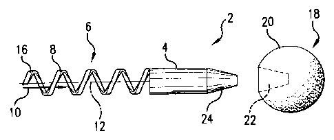
FIG. 1 is an exploded front plan view of an embodiment of the present
invention.
FIG. 2 is a side view of the embodiment of FIG. 1.
FIG. 3 is a front plan sectional view of an embodiment of the present
invention.
FIG. 4 is a front plan view of an embodiment of the present invention.
FIG. 5 is a front plan view of an embodiment of the present invention.
FIG. 6 is a partial sectional view of an embodiment of the present invention.
FIG. 7 is a front plan view of an embodiment of the present invention.
15
2
CA 02445737 2003-10-20
0102-Op09
Customer No. 34086
Albertorio
DESCRIPTION OF THE ILLUSTRATED EMBODIMENTS
FIGS. 1-7 show several illustrative embodiments of an implant for replacing
the
articulating portion of a bone. The implant includes means for spacing adj
scent bones in
articulating relationship and means for anchoring the implant to a bone. The
means for
spacing adjacent bones may include an articular surface component, e.g. a neck
and head
or an acetabular socket as in a ball type joint such as a hip or shoulder, a
tibial plateau or
femoral condylar surface in a knee joint, or other spacing component for an
articulating
joint. The means for anchoring the implant may include a shaft extending from
the
spacing component such as a femoral stem component for insertion into a
femoral bone, a
stem for a tibial tray component for insertion into a tibial bone, or other
anchoring
element for another articulating joint. The implant could be made of a variety
of
biocompatible materials including a variety of known metals, ceramics,
polymers, or
other suitable materials.
Referring now to FIGS. l and 2, an embodiment of the invention is shown
comprising a femoral implant for replacing the articular head of a femur. The
implant 2
includes a neck 4 and a stem 6. The neck 4 functions to space adjacent bones
in
articulating relationship. The stem 6 functions to anchor the neck 4 securely
to a bone.
In this embodiment, the stem 6 includes a threaded portion 8 attached to the
neck 4 for
threadably anchoring the implant to a bone. The threaded portion 8 includes an
axial
bore or cannula 10 having a longitudinal axis 12. It further includes
transverse openings
14. Thus the threaded portion 8 is open both axially and transversely. In
particular, the
threaded portion 8 of this embodiment includes an open helix. Such a stem may
be
formed by bending a rod around a cylindrical core, by casting, by machining
from bulk
3
"0102-0009 CA 02445737 2003-10-20
Customer No. 34086
Albertorio
material, or by using other material forming methods. The tip 16 of the
threaded portion
8 may form a sharp point to ease insertion into a bone. In the particular
helical
embodiment shown, the helix and sharp tip have the form of a corkscrew. A
modular
head 18 is shown for use with the implant 2. The head 18 includes an
articulating surface
S 20 shaped to articulate within a receiving socket of a natural or prosthetic
acetabulum.
The head 18 includes a neck receiving portion 22 and the neck includes a head
engaging
portion 24. The particular embodiment shown includes a locking taper such as a
6° taper
or a 12/14 style taper as is known in the art of locking tapers. Heads 18
having varying
diameters, offset distances, and offset angles may be provided. The
illustrative
embodiment depicts a male neck taper and a female head taper. This arrangement
could
be reversed within the scope of this invention. FIG. 3 shows a monoblock
implant 30
similar to that of FIGS. 1 and 2 but including a head 32 that is permanently
connected to
the neck 34.
FIG. 4 shows another embodiment of the invention. This implant 40 is similar
to
the implant 2 of FIGS. 1 and 2. However, the implant 40 further includes a
collar 42
intermediate the neck 44 and stem 46. The collar 42 extends outwardly beyond
the stem
46 to form a bone seating surface 48 to provide further support and resistance
to
subsidence to the implant 40. The illustrative collar 42 shown in this
embodiment
projects radially to form a bone seating surface 48 completely around the neck
44 and
stem 46. A partial collar is also contemplated that would only provide support
fox a
portion of the area around the neck 44 and stem 46; e.g. a narrow collar
projecting
medially relative to the femoral bone. The collar in this embodiment includes
a bone
growth receptive surface. The bone growth receptive surface includes the bone
seating
4
x'0102-0009 CA 02445737 2003-10-20
Customer No. 34086 '
Albertorio
surface 48 so that bone growth is received where the collar rests on the
femur. The bone
growth receptive surface may also include areas 50 of the collar and implant
adjacent to
the bone seating surface 48. The bone growth receptive surface includes
materials that
enhance or induce the attachment of bone to the collar, e.g. trabecular metal,
beads, fiber
metal, ceramics, porous ceramics, hydroxy appatite, tricalcium phosphate,
plasma
sprayed metal, grit blasted metal, bone growth factors, bone morphogenetic
proteins, and
combinations thereof. The bone growth receptive surface may be in the form of
a
relatively thin surface feature or the bone receptive features may extend
partially or
completely throughout the collar. The bone growth receptive surface may
contribute to
bone ongrowth, bone adhesion, bone ingrowth, or a combination thereof.
FIG. 5 depicts another embodiment of the invention. This implant 60 is similar
to
the implant 2 of FIGS. 1 and 2. However, the implant 60 includes first 62 and
second 64
helices. Multiple helices present a relatively smooth surface that facilitates
screwing the
stem into a bone. Any number of helices may be provided.
1 S FIG. 6 shows an implant 70, in situ in a human femur 72, incorporating
many of
the features described above. The implant 70 includes a neck 74 for spacing
the adjacent
bones in articulating relation and a stem 76 for anchoring the implant to the
femur 72. It
further includes a collar 78 intermediate the neck 74 and stem 76. The collar
78 projects
radially to form a bone seating surface 80. A modular ball head 82 provides a
spherical
articulating surface at the end of the neck 74. The stem 7b includes a
threaded portion
comprising a double helix.
The implant of this invention is suitable for use in traditional open surgical
approaches to a joint. It has features that make it further suitable for
minimally invasive
0102-0009 CA 02445737 2003-10-20
Customer No. 34086
Albertorio
surgical approaches to articulating joints in which a smaller than normal
incision is made
to expose the joint. It may be difficult to prepare the bone for and insert a
conventional
implant through such a small incision. However, the low profile, ease of
implantation,
and minimal bone removal associated with the present invention makes it
suitable for use
through a small incision. By way of illustration, a surgical technique will be
described
relative to the exemplary hip joint embodiment of FIG. 6. First, a small
incision is made
over the hip joint and the muscles are dissected to reveal the hip joint. The
natural
femoral head is resected and removed leaving a honey surface 84. The implant
70 is
threadably anchored on the femur 72 by screwing the stem 76 into the femur.
Where the
implant includes a collar 78, as in FIG. 6, the implant 70 is screwed in until
the collar 78
seats on the honey surface 84. The axial and transverse openings of the
threaded portion
permits bone to penetrate the threaded portion to lock it in the femur. In
particular, the
open helical structure of the stem 76 of this embodiment is filled and
surrounded, both
axially and transversely, by bone 90 as it is screwed into the bone. The axial
bore of the
helix 88 contains a solid core of bone. The sharp tips 86 of the stem bite
into the honey
surface 84 and the stem 76 corkscrews into the femur without requiring prior
reaming,
rasping, tapping, drilling, or otherwise removing bone from the region of the
bone to be
occupied by the stem. It is contemplated that there may be cases where limited
bone
removal such as by drilling or reaming may be desirable prior to inserting an
implant
according to the present invention, but even in those cases, it would be much
less
extensive than is required for a conventional implantation and would utilize
more
compact instruments than conventional reamers and rasps. Where a modular head
82 is
provided, it is most conveniently seated on the neck 74 after the implant is
seated. The
6
'0102-0009 ~ CA 02445737 2003-10-20
Customer No. 34086
Albertorio
helical, open thread design of this embodiment allows good bone purchase and
anatomic
load transfer. Implantation causes minimal damage to the femoral canal and the
cancellous bone bed. The self tapping nature of the thread configuration
allows
implantation of the device with minimal instrumentation while maintaining a
solid core
of cancellous bone inside the thread internal diameter.
The illustrative embodiments described above are useful as proximal femoral
implants for restoring the biomechanic function of the hip joint in cases of
disease
affecting the patient's natural femoral head, e.g. osteonecrosis, neck
fracture,
osteoarthritis, etc. This device may be used in traditional open surgical
approaches. It is
also useful for use in minimally invasive surgical techniques where its low
profile, ease
of implantation, and minimal bone loss during implantation are advantageous.
The stem,
including any helix, may also include a bone growth receptive material or
structure, e.g.
trabecular metal, beads, fiber metal, ceramics, porous ceramics, hydroxy
appatite,
tricalcium phosphate, plasma sprayed metal, grit blasted metal, bone growth
factors, bone
morphogenetic proteins, and combinations thereof.
FIG. 7 depicts another embodiment of the invention relating to a stemmed
implant
other than a femoral hip implant. In this embodiment, a tibial implant 110 is
provided
having a tibial tray 112 and a stem 114. The tibial tray 112 functions to
space adjacent
bones of the knee joint in articulating relationship. An optional, modular,
articulating
surface component is receivable on the tibial tray to provide a modular
articulating
surface. The stem 114 functions to anchor the implant 110 to the tibial bone.
In the
illustrative embodiment, the stem 114 includes a dual open helix 116 attached
to the tibial
tray I I2 for threadably anchoring the implant to a bone. The helix 116
includes an axial
7
"0102-0009 CA 02445737 2003-10-20
Customer No. 34086
Albertorio
bore or cannula 118 having a longitudinal axis 120. It further includes
transverse
openings 122 and is otherwise similar to the helices described in reference to
the other
embodiments. Bone growth receptive surfaces may be used with the tibial tray
112 and
stem 114 as described with reference to the other embodiments. Implantation of
the
implant 110 would include screwing the stem 114 into the tibial bone to anchor
the
implant to the tibia. It is to be understood that the aspects of the invention
described
above with reference to the illustrated embodiments are applicable to a
variety of
implants for replacing the articulating portion of a bone in a variety of
skeletal joints.
15
8
