Note: Descriptions are shown in the official language in which they were submitted.
CA 02445991 2003-10-28
WO 02/099429 PCT/GBO1/02464
COMPOSITIONS AND METHODS FOR THE DETECTION OF BIOMARKERS
ASSOCIATED WITH CARDIOVASCULAR DISEASE
FIELD OF THE INVENTION
The present invention relates generally to the field of cardiovascular
disease, and in
S particular, to compositions and enhanced methods of detecting biomarkers
associated with
cardiovascular disease through the use of cardiac tissue microarrays.
BACKGROUND
Cardiovascular disease is a debilitating illness that afflicts millions of
people in the
world each year. Indeed, in 1997, over 450,000 people in the U.S. alone died
from
myocardial infarctions; one of every five deaths in that calendar year. Iri
addition to
myocardial infarction (heart attack), cardiovascular disease results in
hypertension, angina,
arteriosclerosis, and atherosclerosis. Angina, for example, accounts for more
than 1 million
hospital admissions annually in the U.S., and 6-8 percent of patients with
this condition either
have non-fatal myocardial infarction, or die, within the first year after
diagnosis.
1 S Currently, physicians are able to diagnose cardiovascular disease in
patients who have
already begun to experience symptoms. For example, the levels of certain
cardiac-associated
enzymes, such as creatine kinase, are elevated after myocardial infarction,
and may be
detected an enzyme-specific assay. However, there is no effective means of pre-
symptomatic
diagnosis of cardiovascular disease, let alone for detecting the underlying
genetic risks for
cardiovascular disease. Thus, what is needed is the development of tools and
methods that
will allow the pre-symptomatic identification of cardiovascular disease, and
the detection of
the associated genetic risk factors.
SUMMARY OF THE INVENTION
The present invention relates generally to the field of cardiovascular
disease, and in
2S particular, to devices comprising cardiac tissue microarrays useful for the
detection of
biomarkers associated with cardiovascular disease. Specifically, the device of
the present
-1-
CA 02445991 2003-10-28
WO 02/099429 PCT/GBO1/02464
invention provides tools comprising a cardiac tissue microarray that will
allow the pre-
symptomatic identification of cardiovascular disease, and the associated
genetic risk factors.
In one embodiment, the present invention contemplates a device for the
detection of
biomarkers associated with cardiovascular disease, comprising a tissue
rnicroarray, wherein
said microarray comprises a plurality of human cardiovascular tissue samples
placed on the
surface of said microarray. In another embodiment, the present invention
contemplates a
device having a substantially flat surface,. wherein a plurality of human
cardiovascular tissue
samples from at least one human donor are arrayed on said surface, wherein
said plurality
comprises a first sample from a first disease state and a second tissue sample
from a second
disease state.
It is not intended that the device of the present invention be limited to any
specific
source of cardiovascular tissue. In one embodiment, said tissue is derived
from a human
source. In another embodiment, said tissue is derived from a non-human animal
source.
It is not intended that the device of the present invention be limited to any
specific
type of cardiovascular tissue samples. In one embodiment, said tissue samples
are selected
from the group of tissues comprising different sources of venous and arterial
vessels (e.g.
aorta, coronary, saphen, etc.), ventricles, and auricles.
It is not intended that the tissue samples of the device of the present
invention be
limited to diseased cardiovascular tissue. In one embodiment, said tissue is
diseased. In
another embodiment, said tissue is non-diseased, and serves as a negative
control for the
detection of cardiovascular disease biomarkers. In a preferred embodiment, the
present
invention contemplates a microarray comprising both diseased and non-diseased
cardiovascular
tissue samples. In an alternative embodiment, the present invention
contemplates a microarray
comprising both diseased and non-diseased cardiovascular tissue samples,
wherein said
diseased samples represent at least two different stages of disease (e.g.
chronologically) or
disease states associated with the progression of cardiovascular disease. In
another
embodiment, a device having a substantially flat surface, wherein a plurality
of human
cardiovascular tissue samples from at least one human donor are arrayed on
said surface,
wherein said plurality comprises a first sample from a first disease state and
a second tissue
-2_
CA 02445991 2003-10-28
WO 02/099429 PCT/GBO1/02464
sample from a second disease state, and wherein said second disease state is a
later ("in time")
disease state than said first disease state.
It is not intended that the diseased tissue contemplated by the present
invention be
limited to any specific cardiovascular disease state. In one embodiment, said
disease state is
selected from the group of stable angina, unstable angina, non-Q-wave
myocardial infarction,
and Q-wave myocardial infarction.
It is not intended that the tissue samples of the device of the present
invention be
limited to being derived from a single source. In one embodiment, said
cardiovascular tissue
samples on the microarray are derived from one human source. In another
embodiment, said
cardiovascular tissue samples on the microarray are derived from more than one
human
source. Moreover, it is not intended that the tissue samples of the device of
the present
invention be limited to living donor specimens. In one embodiment, said
cardiovascular tissue
samples comprise cadaveric donor specimens. In another embodiment, said
cardiovascular
tissue samples comprise living donor specimens including, but not limited to,
biopsy
specimens.
It is not intended that the device of the present invention be limited to any
specific
number of cardiovascular tissue samples. In one embodiment, the present
invention
contemplates a microarray comprising 4-16 tissue samples. In another
embodiment, the
present invention contemplates a microarray comprising 16-42 tissue samples.
In yet another
embodiment, the present invention contemplates a microarray comprising 42-98
tissue
samples. In still other embodiments, more than 98 tissue samples are arrayed.
The present invention also contemplates a cardiac tissue microarray comprising
cardiovascular tissues which are derived from different regions of the heart
(i.e. a
topographical microarray). (See e.g., Figure 8). For example, in one
embodiment, the present
invention contemplates a device having a substantially flat surface, wherein a
plurality of
human cardiovascular tissue samples from at least one donor are arrayed on
said surface,
wherein said plurality comprises a first sample from one region of the heart
and a second
sample from a second region of the heart.
In another embodiment, the present invention contemplates a cardiac tissue
microarray
comprising cardiovascular tissues that are representative of the various
stages in the
-3-
CA 02445991 2003-10-28
WO 02/099429 PCT/GBO1/02464
progression of cardiovascular disease (i.e. a chronological microarray). For
example, in one
embodiment, the present invention contemplates a device having a substantially
flat surface,
wherein a plurality of human cardiovascular tissue samples from at least one
donor are
arrayed on said surface, wherein said plurality comprises tissue from a donor
suffering from a
cardiovascular disease selected from the group consisting of stable angina,
unstable angina,
non-Q-wave myocardial infarction, and Q-wave myocardial infarction. In another
embodiment, said plurality further comprises non-diseased cardiovascular
tissues.
In an alternative embodiment, the present invention also contemplates a
cardiac tissue
microarray comprising cardiovascular tissues which are derived from different
regions of the
heart, and which are representative of the various stages in the progression
of cardiovascular
disease (i.e. a topological and chronological microarray). For example, in one
embodiment,
the present invention contemplates a device having a substantially flat
surface, wherein a
plurality of human cardiovascular tissue samples from at least one donor are
arrayed on said
surface, wherein said plurality comprises a first sample from one region of
the heart and a
second sample from a second (and different) region of the heart, and wherein
said plurality
comprises tissue from a donor suffering from a cardiovascular disease selected
from the group
consisting of stable angina, unstable angina, non-Q-wave myocardial
infarction, and Q-wave
myocardial infarction. In another embodiment, said plurality further comprises
non-diseased
cardiovascular tissues.
It is not intended that the device of the present invention be limited to
cardiovascular
tissue samples derived from any specific region of the heart. In one
embodiment, said tissue
samples are derived from a region of the heart selected from the group
consisting of aorta,
right atrium, right ventricle, circumflex, septal wall, obtuse marginal branch
of the left
coronary artery, anterior descending branch of the left coronary artery, left
atrium, left
ventricle, sinuatrial nodal (sinus node) artery, conus arteriosus branch of
the right coronary
artery, right coronary artery, acute marginal branch of the right coronary
artery,
atrioventriculax (A.V.) nodal artery, and posterior descending branch of the
right coronary
artery. (See e.g., Figures 8 & 9).
The present invention also particularly relates to an enhanced method of
detecting
biomarkers associated with cardiovascular disease through the use of cardiac
tissue
-4-
CA 02445991 2003-10-28
WO 02/099429 PCT/GBO1/02464
microarrays. The methods of the present invention comprise the use of said
cardiac tissue
microarray such that a plurality of cardiovascular tissues can be rapidly
screened for the
presence or absence of biomarkers associated (or suspected of being
associated) with
cardiovascular disease.
In one embodiment, the present invention contemplates a method for the
detection of
biomarkers associated with cardiovascular disease, comprising: a) providing
the cardiac tissue
microarray of the present invention; and b) subjecting said microarray to
analysis by a
method selected from the group of histological analysis, immunological
analysis, nucleic acid
hybridization analysis, and combinations thereof, such that the presence or
absence of a
biomarker associated with cardiovascular disease is determined.
It is not intended that the method of the present invention be limited to any
specific
means of histological analysis to determine the presence or absence of a
biomarker associated
(or suspected of being associated) with cardiovascular disease. In one
embodiment, said
histological analysis comprises hematoxylin and eosin staining. In another
embodiment, said
histological analysis is selected from the group of light microscopy, phase-
contrast
microscopy, and osmium tetroxide/glutaraldehyde treatment followed by electron
microscopy.
It is not intended that the method of the present invention be limited to any
specific
means of immunological analysis to determine the presence or absence of a
biomarker
associated with cardiovascular disease. In one embodiment, said immunological
analysis
comprises immunohistochemistry.
It is not intended that the method of the present invention be limited to any
specific
means of nucleic acid hybridization analysis to determine the presence or
absence of a
biomarker associated with cardiovascular disease. In one embodiment, said
nucleic acid
hybridization analysis comprises i~ situ reverse transcriptase polymerase
chain reaction (IS
RT-PCR). In another embodiment, said nucleic acid hybridization analysis
comprises
fluorescent in situ hybridization (FISH).
Liver Disease
It is not intended that the present invention be limited to devices comprising
cardiovascular tissue microarrays. In an alternative embodiment, the present
invention
-5-
CA 02445991 2003-10-28
WO 02/099429 PCT/GBO1/02464
contemplates a device for the detection of biomarkers associated with liver
disease,
comprising a tissue microarray, wherein said microarray comprises a plurality
of human liver
tissue samples placed on the surface of said microarray.
It is not intended that the device of the present invention be limited to any
species-
specific source of liver tissue. In one embodiment, said tissue is derived
from a human
source. In another embodiment, said tissue is derived from a non-human source.
It is not intended that the liver tissue of the device of the present
invention be limited
to any specific liver cell type. In one embodiment, said tissue samples are
comprised of liver
cell types selected from the group of parenchymal (hepatic) cells, cells
associated with the
walls of the hepatic sinusoids, or blood cells in the lumina of the hepatic
sinusoids.
It is not intended that the tissue samples of the device of the present
invention be
limited to .diseased liver tissue. In one embodiment, said tissue is diseased.
In another
embodiment, said tissue is non-diseased, and serves as a negative control for
the detection of
liver disease biomarkers. In a preferred embodiment, the present invention
contemplates a
microarray comprising both diseased and non-diseased liver tissue samples. In
an alternative
embodiment, the present invention contemplates a microarray comprising both
diseased and
non-diseased liver tissue samples, wherein said diseased samples represent the
different stages
of disease (e.g. chronologically) or disease states associated with the
progression of liver
disease.
It is not intended that the diseased tissue contemplated by the present
invention be
limited to any specific liver disease state. In one embodiment, said disease
state is selected
from the group of hepatitis, fibrosis, hepatocellular cancer, and cirrhosis of
the liver.
It is not intended that the tissue samples of the device of the present
invention be
limited to being derived from a single source. In one embodiment, said liver
tissue samples
on the microarray are derived from one human source. In another embodiment,
said liver
tissue samples on the microarray are derived from more than one human source.
Moreover, it
is not intended that the tissue samples of the device of the present invention
be limited to
living donor specimens. In one embodiment, said liver tissue samples on the
microarray
comprise cadaveric donor specimens. In another embodiment, said liver tissue
samples on the
microarray comprise living donor specimens including, but not limited to,
biopsy specimens.
-6-
CA 02445991 2003-10-28
WO 02/099429 PCT/GBO1/02464
It is not intended that the device of the present invention be limited to any
specific
number of liver tissue samples. In one embodiment, the present invention
contemplates a
microarray comprising 4-16 tissue samples. In another embodiment, the present
invention
contemplates a microarray comprising 16-42 tissue samples. In yet another
embodiment, the
present invention contemplates a microarray comprising 42-98 tissue samples.
In still other
embodiments, more than 98 tissue samples are arrayed.
The present invention also particularly relates to an enhanced method of
detecting
biomarkers associated with liver disease through the use of liver tissue
microarrays. The
methods of the present invention comprise the use of said liver tissue
microarray such that a
plurality of liver tissues can be rapidly screened for the presence or absence
of numerous
biomarkers associated with liver disease.
In one embodiment, the present invention contemplates a method for the
detection of
biomarkers associated with liver, comprising: a) providing the liver tissue
microarray of the
present invention; and b) subj ecting said microarray to analysis by a method
selected from
the group of histological analysis, immunological analysis, nucleic acid
hybridization
analysis, and combinations thereof, such that the presence or absence of a
biomarker
associated with liver disease is determined.
It is not intended that the method of the present invention be limited to any
specific
means of histological analysis to determine the presence or absence of a
biomarker associated
with liver disease. In one embodiment, said histological analysis comprises
hematoxylin and
eosin staining. In another embodiment, said histological analysis is selected
from the group of
light microscopy, phase-contrast microscopy, and osmium
tetroxide/glutaraldehyde treatment
followed by electron microscopy.
It is not intended that the method of the present invention be limited to any
specific
means of immunological analysis to determine the presence or absence of a
biomarker
associated with liver disease. In one embodiment, said immunological analysis
comprises
immunohistochemistry.
It is not intended that the method of the present invention be limited to any
specific
means of nucleic acid hybridization analysis to determine the presence or
absence of a
biomarker associated with liver disease. In one embodiment, said nucleic acid
hybridization
CA 02445991 2003-10-28
WO 02/099429 PCT/GBO1/02464
analysis comprises in situ reverse transcriptase polymerase chain reaction (RT-
PCR). In
another embodiment, said nucleic acid hybridization analysis comprises
fluorescent in situ
hybridization (FISH).
Kidney Disease
It is not intended that the present invention be limited to devices comprising
liver
tissue microarrays. In a further alternative embodiment, the present invention
contemplates a
device for the detection of biomarkers associated with kidney disease,
comprising a tissue
microarray, wherein said microarray comprises a plurality of human kidney
tissue samples
placed on the surface of said microarray.
It is not intended that the device of the present invention be limited to any
species-
specific source of kidney tissue. In one embodiment, said tissue is derived
from a human
source. In another embodiment, said tissue is derived from a non-human source.
It is not intended that the tissue samples of the device of the present
invention be
limited to diseased kidney tissue. In one embodiment, said tissue is diseased.
In another
embodiment, said tissue is non-diseased, and serves as a negative control for
the detection of
kidney disease biomarkers. In a preferred embodiment, the present invention
contemplates a
microarray comprising both diseased and non-diseased kidney tissue samples. In
an
alternative embodiment, the present invention contemplates a microarray
comprising both
diseased and non-diseased kidney tissue samples, wherein said diseased samples
represent the
different stages of disease (e.g. chronologically) or disease states
associated with the
progression of kidney disease. '
It is not intended that the diseased tissue contemplated by the present
invention be
limited to any specific kidney disease state. In one embodiment, said disease
state is selected
from the group of idiopathic membranoproliferative glomerulonephritis (MPGI~,
Alport
Syndrome, and Goodpasture's Syndrome.
It is not intended that the tissue samples of the device of the present
invention be
limited to being derived from a single source. In one embodiment, said kidney
tissue samples
on the microarray are derived from one human source. In another embodiment,
said kidney
tissue samples on the microarray are derived from more than one human source.
Moreover, it
_g_
CA 02445991 2003-10-28
WO 02/099429 PCT/GBO1/02464
is not intended that the tissue samples of the device of the present invention
be limited to
living donor specimens. In one embodiment, said kidney tissue samples on the
microarray
comprise cadaveric donor specimens. In another embodiment, said kidney tissue
samples on
the microarray comprise living donor specimens including, but not limited to,
biopsy
specimens.
It is not intended that the device of the present invention be limited to any
specific
number of kidney tissue samples. In one embodiment, the present invention
contemplates a
microarray comprising 4-16 tissue samples. In another embodiment, the present
invention
contemplates a microarray comprising 16-42 tissue samples. In yet another
embodiment, the
present invention contemplates a microarray comprising 42-98 tissue samples.
In still other
embodiments, more than 98 tissue samples are arrayed.
The present invention also particularly relates to an enhanced method of
detecting
biomarkers associated with kidney disease through the use of kidney tissue
microarrays. The
methods of the present invention comprise the use of said kidney tissue
microarray such that a
plurality of kidney tissues can be rapidly screened for the presence or
absence of numerous
biomarkers associated with kidney disease.
In one embodiment, the present invention contemplates a method for the
detection of
biomarkers associated with kidney disease, comprising: a) providing the kidney
tissue
microarray of the present invention; and b) subjecting said microarray to
analysis by a
method selected from the group of histological analysis, immunological
analysis, nucleic acid
hybridization analysis, and combinations thereof, such that the presence or
absence of a
biomarker associated with kidney disease is determined.
It is not intended that the method of the present invention be limited to any
specific
means of histological analysis to determine the presence or absence of a
biomarker associated
with kidney disease. In one embodiment, said histological analysis comprises
hematoxylin
and eosin staining. In another embodiment, said histological analysis is
selected from the
group of light microscopy, phase-contrast microscopy, and osmium
tetroxide/glutaraldehyde
treatment followed by electron microscopy.
It is not intended that the method of the present invention be limited to any
specific
means of immunological analysis to determine the presence or absence of a
biomarker
-9-
CA 02445991 2003-10-28
WO 02/099429 PCT/GBO1/02464
associated with kidney disease. In one embodiment, said immunological analysis
comprises
immunohistochemistry.
It is not intended that the method of the present invention be limited to any
specific
means of nucleic acid hybridization analysis to determine the presence or
absence of a
biomarker associated with kidney disease. In one embodiment, said nucleic acid
hybridization
analysis comprises iri situ reverse transcriptase polymerase chain reaction
(RT-PCR). In
another embodiment, said nucleic acid hybridization analysis comprises
fluorescent in situ
hybridization (FISH).
Brain Disease
It is not intended that the present invention be limited to devices comprising
kidney
tissue microarrays. In a further alternative embodiment, the present invention
contemplates a
device for the detection of biomarkers associated with brain disease
comprising a tissue
microarray, wherein said microarray comprises a plurality of human brain
tissue samples
placed on the surface of said microarray.
It is not intended that the device of the present invention be limited to any
species-
specific source of brain tissue. In one embodiment, said tissue is derived
from a human
source. In another embodiment, said tissue is derived from a non-human source.
It is not intended that the tissue samples of the device of the present
invention be
limited to diseased brain tissue. In one embodiment, said tissue is diseased.
In another
embodiment, said tissue is non-diseased, and serves as a negative control for
the detection of
brain disease biomarkers. In a preferred embodiment, the present invention
contemplates a
microarray comprising both diseased and non-diseased brain tissue samples. In
an alternative
embodiment, the present invention contemplates a microarray comprising both
diseased and
non-diseased brain tissue samples, wherein said diseased samples represent the
different stages
of disease (e.g. chronologically) or disease states associated with the
progression of brain
disease.
It is not intended that the diseased tissue contemplated by the present
invention be
limited to any specific brain disease state. In one embodiment, said disease
state is selected
-10-
CA 02445991 2003-10-28
WO 02/099429 PCT/GBO1/02464
from the group of Creutzfeldt-Jakob Disease (CJD) and Transmissible Spongiform
Encephalopathy (TSE).
It is not intended that the tissue samples of the device of the present
invention be
limited to being derived from a single source. In one embodiment, said brain
tissue samples
on the microarray are derived from one human source. In another embodiment,
said brain
tissue samples on the microarray are derived from more than one human source.
Moreover, it
is not intended that the tissue samples of the device of the present invention
be limited to
living donor specimens. In one embodiment, said brain tissue samples on the
microarray
comprise cadaveric donor specimens. In another embodiment, said brain tissue
samples on
the microarray comprise living, non-human, donor specimens including, but not
limited to,
biopsy specimens.
It is not intended that the device of the present invention be limited to any
specific
number of brain tissue samples. In one embodiment, the present invention
contemplates a
microarray comprising 4-16 tissue samples. In another embodiment, the present
invention
contemplates a microarray comprising 16-42 tissue samples. In yet another
embodiment, the
present invention contemplates a microarray comprising 42-98 tissue samples.
In still other
embodiments, more than 98 tissue samples are arrayed.
The present invention also particularly relates to an enhanced method of
detecting
biomarkers associated with brain disease through the use of brain tissue
microarrays. The
methods of the present invention comprise the use of said brain tissue
microarray such that a
plurality of brain tissues can be rapidly screened for the presence or absence
of numerous
biomarkers associated with brain disease.
In one embodiment, the present invention contemplates a method for the
detection of
biomarkers associated with brain disease, comprising: a) providing the brain
tissue microarray
of the present invention; and b) subjecting said microarray to analysis by a
method selected
from the group of histological analysis, immunological analysis, nucleic acid
hybridization
analysis, and combinations thereof, such that the presence or absence of a
biomarker
associated with brain disease is determined.
It is not intended that the method of the present invention be limited to any
specific
means of histological analysis to determine the presence or absence of a
biomarker associated
-11-
CA 02445991 2003-10-28
WO 02/099429 PCT/GBO1/02464
with brain disease. In one embodiment, said histological analysis comprises
hematoxylin and
eosin staining. In another embodiment, said histological analysis is selected
from the group of
light microscopy, phase-contrast microscopy, and osmium
tetroxide/glutaraldehyde treatment
followed by electron microscopy.
It is not intended that the method of the present invention be limited to any
specific
means of immunological analysis to determine the presence or absence of a
biomarker
associated with brain disease. In one embodiment, said immunological analysis
comprises
immunohistochemistry. In another embodiment, said immnunological analysis
comprises
Fluorescence Correlation Spectroscopy (FCS) as noted in Geise et al., "Putting
prions into
focus: application of single molecule detection to the diagnosis of prion
diseases," Arch. Yirol.
Suppl., (16): 161-71 (2000).
It is not intended that the method of the present invention be limited to any
specific
means of nucleic acid hybridization analysis to determine the presence or
absence of a
biomarker associated with brain disease. In one embodiment, said nucleic acid
hybridization
analysis comprises ifa situ reverse transcriptase polymerase chain reaction
(RT-PCR). In
another embodiment, said nucleic acid hybridization analysis comprises
fluorescent in situ
hybridization (FISH).
DESCRIPTION OF THE FIGURES
To facilitate an understanding of the invention, a number of figures are
included
herein.
Figure 1 depicts a microarray comprised of cardiovascular tissue derived from
a single
donor suffering from cardiovascular disease. In this figure, tissue samples
are arranged in
duplicate rows as indicated by "[duplicate]" or "[d]."
Figure 2 depicts a microarray comprised of cardiovascular tissue derived from
two
donors suffering from cardiovascular disease. In this figure, tissue samples
are arranged in
duplicate rows as indicated by "[duplicate]" or "[d]."
- 12-
CA 02445991 2003-10-28
WO 02/099429 PCT/GBO1/02464
Figure 3 depicts a microarray comprised of cardiovascular tissue derived from
three
donors suffering from cardiovascular disease. In this figure, tissue samples
are arranged in
duplicate rows as indicated by "[duplicate]" or "[d]."
Figure 4 depicts a microarray comprised of cardiovascular tissue derived from
four
donors suffering from cardiovascular disease. In this figure, tissue samples
are arranged in
duplicate rows as indicated by "[duplicate]" or "[d]."
Figure 5 depicts a microarray comprised of cardiovascular tissue derived from
five
donors suffering from cardiovascular disease, and from one non-diseased
(cardiovascular)
donor. In this figure, tissue samples are arranged in duplicate rows as
indicated by
"[duplicate]" or "[d]."
Figure 6 is a flowchart that depicts one approach for the utilization of the
methods and
compositions of the present invention to detect the presence or absence of
biomarkers
associated with cardiovascular disease.
Figure 7 depicts the general progression of acute coronary syndromes beginning
with
ischemic discomfort, and terminating in either unstable angina, non-Q-wave
myocardial
infarction, or Q-wave myocardial infarction.
Figure 8 depicts a topological map of the coronary arterial tree as viewed in
one of the
projections commonly used in coronary arteriography. The map divides the human
heart into
discrete sections (e.g. A-l, B-2, C-3, etc.) which correspond with identically
numbered
sections of functional and topological arterial tissue microarrays as
described herein. (See also
Figures 9-14).
Figure 9 depicts one embodiment of a functional and topological arterial
tissue
microarray (as described herein) in which three distinct atherosclerotic
cardiovascular disease
states (i.e. coronary heart disease, stroke, and peripheral arterial vascular
disease) are
represented. The sections labeled A-1 through A-3, B-1 through B-3, & C-1 tlu-
augh C-3
correspond to the sections depicted in Figure 8 and represent coronary
arterial tissues affected
by coronary heart disease (CHD). The sections labeled D-1 through D-3
correspond to
arterial tissues that are implicated in stroke. The sections labeled E-1
through E-3 correspond
to arterial tissues that are implicated in peripheral arterial vasculax
disease (PVD). (See also
Figures 8, 10-14).
-13-
CA 02445991 2003-10-28
WO 02/099429 PCT/GBO1/02464
Figure 10 depicts one embodiment of a functional and topological arterial
tissue
microarray (lower portion) wherein the sections labeled A-1 through A-3, B-1
through B-3, &
C-1 through C-3 (upper portion) correspond to the sections depicted in Figure
8 and represent
coronary arterial tissues affected by coronary heart disease (CHD). This
particular
embodiment of the microarray depicts the aortic tissue samples placed in
section A-1 of said
microarray.
Figure 11 depicts one embodiment of a functional and topological arterial
tissue
microarray (lower portion) with sections labeled as described in the figure
legends of Figures
8 & 9. This particular embodiment of the microarray depicts the internal
carotid tissue
samples placed in section D-1 of said microarray.
Figure 12 depicts one embodiment of a functional and topological arterial
tissue
microarray (lower portion) with sections labeled as described in the figure
legends of Figures
8 & 9. This particular embodiment of the microarray depicts the abdominal
aorta tissue
samples placed in section E-1 of said microarray.
Figure 13 depicts one embodiment of a functional and topological arterial
tissue
microarray (lower portion) with sections labeled as described in the figure
legends of Figures
8 & 9. This particular embodiment of the microarray depicts the tissue samples
(e.g. common
femoral artery, deep femoral artery, and superior femoral artery) placed in
section E-2 of said
microarray.
Figure 14 depicts one embodiment of a functional and topological arterial
tissue
microarray (lower portion) with sections labeled as described in the figure
legends of Figures
8 & 9. This particular embodiment of the microarray depicts the tissue samples
(e.g. tibial
artery, popliteal artery, and peroneal artery) placed in section E-3 of said
microarray.
Figure 15 depicts one embodiment of the device of the present invention
comprising a
high-density cardiovascular tissue microarray (e.g. a cardiovascular tissue
microarray having
432 samples). Section I00 indicates the device itself, whereas Sections 10I
and 102
respectively indicate the surface of the device, and the cardiovascular tissue
samples placed
thereupon.
- 14-
CA 02445991 2003-10-28
WO 02/099429 PCT/GBO1/02464
DEFINITIONS
"Alport syndrome," as used herein, refers to an inherited progressive kidney
disease
with an estimated gene frequency of 1:5000. The disease is characterized by
hematuria and
terminal renal failure, often accompanied by familial hearing loss and ocular
lesions such as
lenticonus. It is usually inherited as an X chromosome-linked dominant trait,
but autosomal
forms have also been described.
"Angina" or "Angina Pectoris," as used herein, refers to chest pain that is
caused by
blockages in the arteries that supply blood to the heart. Angina is further
sub-divided and
classified by the length of time between each onset of chest pain as follows:
1. acute angina - while at rest (within the 48 hours before presentation),
2. sub-acute angina - while at rest (within the previous month but not within
the 48
hours before presentation), or
3. new onset of accelerated (progressively more severe) angina;
"Acute coronary syndrome," or "ACS," as used herein, refers to the spectrum of
conditions including, but not limited to, unstable angina (UA), non-Q-wave
myocardial
infarction (which generally presents without ST-segment elevation), and Q-wave
myocardial
infarction (which generally presents with ST-segment elevation). UA and non-ST-
segment
elevation myocardial infarction (NSTEMI) are acute coronary syndromes (ACSs)
that are
characterized by an imbalance between myocardial oxygen supply and demand. The
most
common cause is reduced myocardial perfusion that results from coronary artery
narrowing
caused by a non-occlusive thrombus that has developed on a disrupted
atherosclerotic plaque.
Abnormal constriction of the coronary arteries may also be responsible. UA and
NSTEMI are
considered to be closely related conditions whose pathogenesis and clinical
presentations are
similar but of differing severity (i.e., they differ primarily in whether the
ischemia is severe
enough to cause sufficient myocardial damage to release detectable quantities
of a marker of
myocardial injury, most commonly, troponin I [TnI], troponin T [TnT], or the
MB isoenzyrne
of creatine phosphokinase [CK-MB]). Once it has been established that no
biochemical
marker of myocardial necrosis has been released, the patient with an ACS may
be considered
to have experienced UA, whereas the diagnosis of NSTEMI is established if a
marker of
myocardial injury has been released.
-15-
CA 02445991 2003-10-28
WO 02/099429 PCT/GBO1/02464
"Atherothrombotic cardiovascular disease," as used herein, refers to a diffuse
atherosclerotic condition involving the heart (coronary arteries), brain
(carotid, vertebral, and
cerebral arteries), and peripheral arteries. Indeed, most of the risk factors
that apply to one
arterial bed also apply to the others. It is, therefore, not surprising that
the presence of one
atherosclerotic cardiovascular disease increases the risk of developing other
such diseases.
"Biomarker," as used herein, refers to any biologically-based marker (e.g.
gene, gene
fragment, gene product, nucleic acid, protein, protein fragment, peptide,
polypeptide, or
epitope), that the presence, absence, or variation in expression of, is
associated with a
particular disease state. The term "gene" encompasses both cDNA and genomic
forms of a
given gene. The present invention contemplates devices and methods for the
detection of
biomarkers associated with cardiovascular, kidney, liver, and brain diseases.
Specifically, the
present invention contemplates cardiovascular tissue microarrays for the
detection of
biomarkers associated with cardiovascular disease such as the PAI-1 geneand
gene product.
"Cardiovascular disease," as used herein, refers to any disease which affects
the
cardiovascular system including, but not limited to, thrombophilia,
atherosclerosis, and
arteriosclerosis.
"Goodpasture's syndrome," as used herein, refers to the association of severe
nephritis
with pulmonary hemorrhage, and although various immunopathological mechanisms
may
underlie this clinical picture, the eponym is now generally reserved for those
eases due to
auto-antibodies to the glomerular basement membrane (GB1VI).
"Hybridization," as used herein, refers to the formation of sequence-specific,
base-
paired duplexes from any combination of nucleic acid fragments. Hybridization,
regardless of
the method used, requires some complementarity between the sequence of
interest (the target
sequence) and the fragment of nucleic acid used to detect the target sequence
and/or perform
the test (e.g., the probe). Thus, these duplexes may be completely
complementary or may
include mismatched sequences. For example, where it is desired to detect
simply the presence
or absence of DNA or RNA, it is only important that the hybridization method
ensures
hybridization when the relevant sequence is present; conditions can be
selected where both
partially complementary probes and completely complementary probes will
hybridize.
However, other diagnostic applications may require that the method of
hybridization
-16-
CA 02445991 2003-10-28
WO 02/099429 PCT/GBO1/02464
distinguish between variant target sequences. For example, it may be of
interest that a
particular allelic variant is present. Methods have been devised to enable
discrimination
between partial and complete complementarity. One approach is to take
advantage of the
temperature requirements of the specific hybridization under study. In typical
melting curve
experiments, such as those described by Wallace et al., Nucl. Acids Res., 6:
35632 (1979) and
Nucl. Acids Res. 9: 879 (1981), it is observed that partially complementary
probe-target
duplexes display a lower thermal stability than do completely complementary
probe-target
duplexes. The best estimate is that a 1% mismatch causes a reduction in the
thermal stability
of duplexes, as measured by the duplex melting temperature (Tm), by 1
°C. See R.J. Britten
and E.H. Davidson, in: Nucleic Acid Hybridisation, (B.D. Hames and S.J.
Higgins, eds.)
(IRL Press, Washington, 1985)(pp. 3-15). The Tm is also affected by the length
of the base-
paired region of a duplex, according to the equation D = 500/L, wherein D is
the reduction in
Tm (°C) and L is the length of the base-paired duplex. The base
composition of the duplex is
another factor which affects its stability. In normal salt solutions, GC base
pairs are more
stable than AT pairs, thus the Tm of a particular duplex is related to its GC
content according
to the equation Tm 0.41(%GC) + 69.3.
"Tissue microarray," as used herein, refers to an orderly arrangement of a
plurality of
tissue samples or specimens, said samples or specimens ranging in size of up
to approximately
1000 microns in diameter (more preferably 200-400 microns in diameter, even
more
preferably 401-500 microns in diameter, and still more preferably 501-600
microns in
diameter) placed on the surface of a solid support (e.g. a microscope slide).
A "single-source
tissue microarray" refers to a tissue microarray wherein the tissue samples
are derived from a
single source (e.g. a single cadaveric donor). A "comparative tissue
microaxray" refers to a
tissue microarray wherein the tissues samples 1) are derived from more than
one source (e.g.
multiple cadaveric donors), 2) are comprised of either different tissue types
(e.g. aorta,
circumflex, venous, arterial, or cardiac tissue) or different sections of the
same tissue type, or
3) are compared to non-diseased control tissues specimens similarly placed on
the surface of
the microarray. A "chronological tissue microarray" refers to a tissue
microarray wherein the
tissue samples are arranged such as to reflect the chronological progression
of a disease (e.g.
-17-
CA 02445991 2003-10-28
WO 02/099429 PCT/GBO1/02464
following the progression stable angina, unstable angina, non-Q-wave
myocardial infarction,
and Q-wave myocardial infarction).
The present invention contemplates a "high density" tissue microarray, which
refers to
a tissue microarray in which up to approximately 1200 tissue samples are
arrayed in a 40 mm
x 25 mm recipient array block (e.g. paraffin block), and placed on the surface
of a 3 in. x 1
in. (or 75 mm x 25 mm) microscope slide. The present invention also
contemplates a
"medium-high density" tissue microarray, which refers to a tissue microarray
in which up to
approximately 700 tissue samples are arrayed in a 40 mm x 25 mm recipient
array block (e.g.
paraffin block), and placed on the surface of a 3 in. x 1 in. (or 75 mm x 25
mm) microscope
slide. The present invention further contemplates a "medium density" tissue
microarray,
which refers to a tissue microarray in which up to approximately 300-500
tissue samples are
arrayed in a 40 mm x 25 mm recipient array block (e.g. paraffin block), and
placed on the
surface of a 3 in. x 1 in. (or 75 mm x 25 mm) microscope slide. Finally, the
present
invention contemplates a "low density" tissue microarray which refers to a
tissue microarray
in which up to approximately 100-200 tissue samples are arrayed in a 40 mm x
25 mm
recipient array block (e.g. paraffin block), and placed on the surface of a 3
in. x 1 in. (or 75
mm x 25 mm) microscope slide. Although tissue samples are arrayed in 40 mm x
25 mm
recipient array blocks, the present invention contemplates tissue microarrays
comprising
multiple recipient array blocks (e.g. three low density tissue microarray
recipient blocks
placed together on the surface of a 3 in. x 1 in. microscope slide with a
total number of 300-
600 samples present on the slide).
"Placed on the surface," as used herein, refers to the process by which tissue
specimens are positioned and contacted with, linked to, adhered to, attached
to, bound to, or
affixed to a (usually flat) surface suitable for mounting tissue ('e.g. glass,
plastic, silicon,
metal, gel, etc.). The term encompasses the covalent, non-covalent, and
hydrogen bonding of
tissue specimens to the surface to create a tissue microarray. A convenient
surface is that of a
conventional glass microscope slide.
"Plurality of cardiovascular tissue(s)," as used herein, refers to a number of
cardiovascular tissue specimens to be placed on the surface of a
cardiovascular tissue
- 1~ -
CA 02445991 2003-10-28
WO 02/099429 PCT/GBO1/02464
microarray wherein the number is preferably at least three tissue specimens,
more preferably,
3-16 tissue specimens, even more preferably, 16-42 tissue specimens, and still
more
preferably, 42-98 tissue specimens, and most preferably, greater than 98
tissue specimens,
placed upon the surface of a 3 in. x 1 in. (or 75 mm x 2S mm) microscope
slide. For
S example, the present invention contemplates cardiovascular tissue
microarrays having up to
approximately 432 tissue specimens, as well as, cardiovascular tissue
microarrays having up to
approximately 864 tissue specimens, placed upon the surface of a 3 in. x 1 in.
(or 7S mm x
2S mm) microscope slide.
"Substantially flat surface," as used herein, refers to surfaces useful for
the
construction of tissue microarrays as contemplated by the present invention.
Said term
encompasses surfaces which are completely flat, partially flat, and partially
curved. For
example, in one embodiment, the present invention contemplates a
cardiovascular tissue
microarray comprising a standard 3 in. x 1 in. (or 7S mm x 2S mm) microscope
slide having
a completely flat surface (i.e. a surface having an angle equal to zero or
180°). In another
IS embodiment, said microscope slide has a partially flat surface (i.e. a
surface having an angle
between 0-4S° or 13S-180°). In another embodiment, said
microscope slide has a partially
curved surface (i.e. a surface having an angle between 4S-90° or 90-
13S°). In a further
embodiment, the present invention contemplates a cardiovascular tissue
microarray comprising
a circular wheel upon which tissue samples are placed, wherein any discrete
point on the
surface of said wheel is completely flat, partially flat, or partially curved.
The term
"Substantially flat surface," also encompasses surfaces which have
microirregularities (e.g.
surfaces having ridges or grooves that are (only) visible through the aid of a
microscope).
DETAILED DESCRIPTION OF THE INVENTION
Certain genetic markers, or "biomarkers," associated with cardiovascular
disease have
2S been identified. For example, the expression of the gene product of the
plasminogen activator
inhibitor type 1 (PAI-1) gene has been linked to cardiovascular diseases (See
Kohler, H.P. &
Grant, P.J., "Plasminogen-activator inhibitor type 1 and coronary artery
disease," New Eng. J.
Med., 342: 1792-1801 (2000)) such as, for example, thrornbophilia. (,See
Engesser et al.,
-19-
CA 02445991 2003-10-28
WO 02/099429 PCT/GBO1/02464
"Elevated plasminogen activator inhibitor (PAI), a cause of thrombophilia? A
study in 203
patients with familial or sporadic venous thrombophilia," Tlaromb. Haenaost.,
62: 673-680
(1989)). Thus, the PAI-1 gene may serve as a genetic marker, or biomarker, for
such
diseases.
However, the traditional tools and methods of detecting the presence or
absence of
biomarkers, such as in situ hybridization and reverse transcriptase PCR (RT-
PCR), are of
limited utility because of the current inefficiencies in high-throughput
screening of cardiac
tissue samples, the lack of a panel of tissue samples representing different
cardiovascular
disease states, and the inability to simultaneously probe numerous tissue
samples for the
presence or absence of biomarkers associated with cardiovascular disease. The
present
invention provides tools and methods whereby a plurality of cardiovascular
tissue can be
rapidly screened for the presence, absence, or a variation in the expression
of numerous
biomarkers associated with the progression of cardiovascular disease.
I. The Progression of Cardiovascular Disease
Although the exact mechanisms and steps involved in the progression of heart
disease
are not precisely known, it is believed that the progression follows the
pathway of 1)
atherosclerosis, 2) stable angina, 3) unstable angina, 4) non-Q-wave
myocardial infarction, and
5) Q-wave myocardial infarction. (See Figure 7). Patients with ischemic
discomfort (i.e.
discomfort due to inadequate circulation of blood to the myocardium) may
present with or
without ST-segment elevation on the electrocardiogram. The majority (large
arrow) of
patients with ST-segment elevation ultimately develop a Q-wave acute
myocardial infarction
(AMI), whereas a minority (small arrow) develop a non-Q-wave AMI. (See Figure
7). Of
patients who present without ST-segment elevation, the majority (large arrows)
are ultimately
diagnosed as having either unstable angina or non-Q-wave AMI based on the
presence or
absence of a cardiac marker such as the MB isozyme of creatine phosphokinase
(CK-MB)
detected in the serum (See, e.g., U.S. Patent S,I37,609 to Manian et al., at
column 8); a
minority of such patients ultimately develop a Q-wave AMI. (See Figure 7). The
spectrum
of clinical conditions ranging from unstable angina to non-Q-wave AMI and Q-
wave AMI is
-20-
CA 02445991 2003-10-28
WO 02/099429 PCT/GBO1/02464
referred to as acute coronary syndromes. (See Antman EM, Braunwald E., "Acute
myocardial
infarction," in Heart Disease: A Textbook of Cardiovascular Medicine (1996),
Philadelphia,
PA: WB Saunders, Braunwald EB, editor). Various definitions of unstable angina
have been
proposed, but in 1989, Braunwald devised a classification system to ensure
uniformity of
categorization, as well as diagnostic and prognostic information. Braunwald
E., "Unstable
angina: a classification," Circulation, 80: 410-414 (1989). This system is
used to classify
angina according to the severity of the chemical manifestation.
The clinical circumstances in which unstable angina develops, are defined as
either
angina in the presence or absence of other conditions (e.g., anemia, fever,
hypoxia,
tachycardia, or thyrotoxicosis), or angina within two weeks after an acute
myocardial
infarction; and whether or not electrocardiographic abnormalities are present.
Given the
heterogeneity of the clinical manifestations of unstable angina, it is not
surprising that the
prognosis is quite variable. Clinically speaking, patients with unstable
angina and those with
non-Q-Wave myocardial infarction often present in similar manner, and the
distinction
between the two conditions can be made only many hours or days later, when the
results of
cardiac-enzyme tests become available. (See Braunwald et al., "ACC/AHA
guidelines for the
management of patients with unstable angina and non-ST-segment elevation
myocardial
infarction: executive summary and recommendations: a report of the American
College of
Cardiology/American Heart Association Task Force on Practice Guidelines
(Committee on the
Management of Patients With Unstable Angina)," Circulation, 102: 1193-1209
(2000)). Thus,
the development of diagnostic tools that would enable an earlier, pre-
symptomatic detection
of, and distinction between, underlying cardiovascular disease states is
desirable. The
compositions and methods of the present method provide a genetically-based
tool that can be
utilized to distinguish between the various stages in the progression of
cardiovascular disease.
II. Construction of a Cardiovascular Tissue Microarray
Microarray technology has been developed in response to the need for
simultaneous
analysis of the thousands of genes. In a typical application, high-density
nucleic acid samples,
usually cDNAs or oligonucleotides, are delivered (or printed) by a robotic
system onto very
-21 -
CA 02445991 2003-10-28
WO 02/099429 PCT/GBO1/02464
small, discrete areas of coated substrates, usually microscopic glass slides
or membrane filters,
and then immobilized to the substrate. The resulting microarray is then
hybridized with a
complex mixture of fluorescently labelled nucleic acids (probe) derived from a
desired source.
Following hybridization, the fluorescent markers are detected using a high
resolution laser
scanner. A gene pattern is obtained by analyzing the signal emitted from each
spot with
digital imaging software. In the case of gene expression analysis, the pattern
of the
experimental sample can be compared with that of a control for differential
analysis.
Mutations and polymorphisms, in particular single nucleotide polymorphisms
(SNPs),
can be studied within and among species using high-density oligonucleotide
arrays. These
IO so-called mutation detection arrays consist of oligonucleotides
representing all known
sequence variants of a gene or a collection of genes. Because hybridization to
oligonucleotides is sensitive enough to detect single-nucleotide mismatches,
an homologous
gene carrying an unknown sequence variation can be screened rapidly for a
large number of
changes.
The compositions of the present invention provide a tool comprising a cardiac
tissue
microarray that will allow the pre-symptomatic identification of
cardiovascular disease, and
the associated genetic risk factors. An example of tumor tissue microarray
construction can
be found in the Instruction Manual for the Beecher Instruments (Silver Spring,
MD) Tissue
Arrayer.
In one embodiment, the present invention contemplates a composition for the
detection
of biomarkers associated with cardiovascular disease, comprising a tissue
microarray, wherein
said microarray comprises a plurality of human cardiovascular tissue samples
placed on the
surface of said microarray. Although it is not intended that the present
invention be limited to
any one particular method of preparing a cardiovascular tissue microarray, in
one
embodiment, the construction of said microarray involves the embedding of
cardiovascular
tissue in paraffin. In an alternative embodiment, the construction of said
microarray involves
the substitution of the paraffin embedding step above with the preparation of
frozen
cardiovascular tissue.
-22-
CA 02445991 2003-10-28
WO 02/099429 PCT/GBO1/02464
A. Preparation of Paraffin-Embedded Cardiovascular Tissue
Paraffin embedding is a process in which the tissue specimen is fixed to
preserve its
cellular structures, and blocked out and embedded in paraffin to stabilize it
for long-term
storage and easy sectioning or microdissection. Although it is not intended
that the present
invention be limited to a specific method by which cardiovascular tissue is
embedded in
paraffin, in one embodiment, the present invention contemplates the following
method.
Cardiovascular tissue in a formaldehyde fixative solution (e.g. formalin) or
RNAlaterTM (Ambion Cat. No. 7020) is processed by sequential incubations in
various
concentrations of 1) ethanol, 2) xylenes, and then embedded in paraffin. The
paraffin
embedded tissues are then cut into sections, with a typical thickness of
between 1-10 microns
(e.g 5.5 microns) on a clean microtome with a clean blade to produce paraffin
ribbons
containing the tissues. The paraffin ribbons are floated in lRNAse-free,
deionized water at
43-44°C. The paraffin ribbons are then mounted on plain, uncoated glass
microscope slides.
Prior to subjecting the paraffin embedded tissues to screening for the
presence or absence of
biomarkers associated with cardiovascular disease, the paraffin is removed
from the tissue
sections by sequential incubations in various concentrations of 1) xylenes, 2)
ethanol, and 3)
distilled water.
B. Preparation of Frozen Cardiovascular Tissue
Cardiovascular frozen tissue storage is another way to preserve specimens and
stabilize
them for long-term storage and sectioning, and is a well-know method in the
field. Tissue is
embedded in a viscous compound, such as OCT compound (Tissue-Tek Cat. No. 453)
and
deep-frozen on dry ice, or at a lower temperature (e.g. -125°C). The
block is removed from
the cryomold and attached to a cryostat with OCT. The block is allowed to
equilibrate to the
cryostat temperature (-20°C) for about 1 S minutes. Tissue block
sections are cut onto plain,
uncoated glass slides.
C. Arrangement of Cardiovascular Tissues on a Microarray
It is not intended that the present invention be limited to any specific
arrangement or
configuration of cardiovascular tissue on a microarray. Moreover, it is not
intended that the
-23-
CA 02445991 2003-10-28
WO 02/099429 PCT/GBO1/02464
present invention be limited to any specific number or types of cardiovascular
tissues arranged
on a microarray.
In one embodiment, the arrangement of cardiovascular tissue comprises a
plurality of
samples of various cardiovascular tissue types from a single donor source,
wherein each tissue
represents a different state or stage of the progression of heart disease. In
one embodiment,
cardiovascular tissues are taken from a single donor's aorta, circumflex, and
left coronary
artery at separate times coinciding with the expression of symptoms related to
atherosclerosis,
stable angina, unstable angina, non-Q-wave myocardial infarction, and Q-wave
myocardial
infarction. Said tissues are conveniently arranged in paraffin or frozen
embedding mold, as
described above, and arrayed as described below.
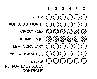
For example, Figure 1 depicts a microarray comprised of cardiovascular tissue
derived
from a single donor with cardiovascular disease. The rows of said microarray
depict four
different tissues derived from the donor (e.g. aorta, circumflex, left-
coronary artery, and a
non-cardiovascular tissue control) arranged in duplicate. The columns of said
microarray
represent said tissues that were isolated from the donor as to correspond with
different states
or stages in the progression of cardiovascular disease (e.g. such as to form a
single-source
chronological microarray). Specifically, the tissues in Column 1 are non-
diseased
cardiovascular tissues. The tissues in Column 2 correspond to an
atherosclerotic stage of
disease. The tissues in Column 3 correspond to a stable angina stage of
disease. The tissues
in Column 4 correspond to an unstable angina stage of disease. The tissues in
Column 5
correspond to non-Q-wave myocardial infarction stage of disease. The tissues
in Column 6
correspond to Q-wave myocardial infarction stage of disease. The present
invention also
contemplates the arraying of additional cardiovascular tissues (e.g. arterial,
venous, and
cardiac) in the same manner as shown in Figure 1 (whether as single samples,
or in duplicate
or triplicate).
It is not intended that the present invention be limited to a cardiovascular
tissue
microarray wherein said tissues are derived from a single donor. In one
embodiment,
cardiovascular tissue selected from the group comprising aorta, circumflex,
and left coronary
artery tissue, is derived from multiple donors at separate times coinciding
with the expression
of symptoms related to atherosclerosis, stable angina, unstable angina, non-Q-
wave myocardial
-24-
CA 02445991 2003-10-28
WO 02/099429 PCT/GBO1/02464
infarction, and Q-wave myocardial infarction. Said tissues are conveniently
arrayed in
paraffin or frozen embedding mold, as described above, and arrayed as
described below.
For example, Figure 2 depicts a microarray comprised of cardiovascular tissue
derived
from a two donors having cardiovascular disease (e.g. such as to form a
comparative
chronological microarray). The rows of said microarray depict four different
tissues derived
from the two donors (e.g. aorta, circumflex, left-coronary artery, and a non-
cardiovascular
tissue control) arranged in duplicate. The columns of said microarray
represent said tissues
that were isolated from each donor as to correspond with different states or
stages in the
progression of cardiovascular disease. Columns 1-6 correspond to the tissues
derived from a
first donor, whereas Columns 7-12 correspond to tissues derived from a second
donor.
Specifically, the tissues in Columns l and 7 are non-diseased cardiovascular
tissues. The
tissues in Columns 2 and 8 correspond to a atherosclerotic stage of disease.
The tissues in
Columns 3 and 9 correspond to a stable angina stage of disease. The tissues in
Columns 4
and 10 correspond to an unstable angina stage of disease. The tissues in
Columns 5 and 11
correspond to non-Q-wave myocardial infarction stage of disease. The tissues
in Columns 6
and 12 correspond to Q-wave myocardial infarction stage of disease. The
present invention
also contemplates the arraying of additional cardiovascular tissues (e.g.
arterial, venous, and
cardiac) in the same manner as shown in Figure 2 (whether as single samples,
or in duplicate
or triplicate).
In another embodiment, a cardiovascular tissue microarray comprised of a
plurality of
tissues from more than two donors is contemplated. For example, Figure 3
depicts a
microarray comprised of cardiovascular tissue derived from a three donors
having
cardiovascular disease. The rows of said microarray depict four different
tissues derived from
the donor (e.g. aorta, circumflex, left-coronary artery, and a non-
cardiovascular tissue control)
arranged in duplicate. The columns of said microarray represent said tissues
that were
isolated from each donor as to correspond with different states or stages in
the progression of
cardiovascular disease. Columns 1-6 correspond to the tissues derived from a
first donor.
Columns 7-12 correspond to tissues derived from a second donor, whereas
Columns 13-18
correspond to said tissues from a third donor. Specifically, the tissues in
Columns 1, 7, and
13 are non-diseased cardiovascular tissues. The tissues in Columns 2, 8, and
14 correspond to
-25-
CA 02445991 2003-10-28
WO 02/099429 PCT/GBO1/02464
an atherosclerotic stage of disease. The tissues in Columns 3, 9 and 15
correspond to a stable
angina stage of disease. The tissues in Columns 4, 10, and 16 correspond to an
unstable
angina stage of disease. The tissues in Columns 5, 11, and 17 correspond to
non-Q-wave
myocardial infarction stage of disease. The tissues in Columns 6, 12, and 18
correspond to
Q-wave myocardial infarction stage of disease. The present invention also
contemplates the
arraying of additional cardiovascular tissues (e.g. arterial, venous, and
cardiac) in the same
manner as shown in Figure 3 (whether as single samples, or in duplicate or
triplicate):
In yet another embodiment, a cardiovascular tissue microarray comprised of a
plurality
of tissues from more than three donors is contemplated. For example, Figure 4
depicts a
microarray comprised of cardiovascular tissue derived from a four donors
having
cardiovascular disease. The rows of said microarray depict four different
tissues derived from
the donor (e.g. aorta, circumflex, left-coronary artery, and a non-
cardiovascular tissue control)
arranged in duplicate. The columns of said microarray represent said tissues
that were
isolated from each donor as to correspond with different states or stages in
the progression of
cardiovascular disease. Columns 1-6 correspond to the tissues derived from a
first donor
having cardiovascular disease. Columns 7-12 correspond to tissues derived from
a second
donor having cardiovascular disease. Columns 13-18 correspond to tissues
derived from a
third donor having cardiovascular disease, whereas Columns 19-24 correspond to
said tissues
from a fourth donor having cardiovascular disease. Specifically, the tissues
in Columns l, 7,
13, and 19 are non-diseased cardiovascular tissues. The tissues in Columns 2,
8, 14, and 20
correspond to an atherosclerotic stage of disease. The tissues in Columns 3,
9, 15, and 21
correspond to a stable angina stage of disease. The tissues in Columns 4, 10,
16, and 22
correspond to an unstable angina stage of disease. The tissues in Columns 5,
11, 17, and 23
correspond to non-Q-wave myocardial infarction stage of disease. The tissues
in Columns 6,
12, 18, and 24 correspond to Q-wave myocardial infarction stage of disease.
The present
invention also contemplates the arraying of additional cardiovascular tissues
(e.g. arterial,
venous, and cardiac) in the same manner as shown in Figure 4 (whether as
single samples, or
in duplicate or triplicate).
It is not intended that the present invention be limited to a microarray
wherein said
microarray is comprised of a plurality of cardiovascular tissues isolated from
a donor as to
-26-
CA 02445991 2003-10-28
WO 02/099429 PCT/GBO1/02464
correspond with multiple states or stages in the progression of cardiovascular
disease. In one
embodiment, the invention contemplates a comparative cardiovascular tissue
microarray
comprising tissues from multiple donors corresponding to a single stage (e.g.
the
atherosclerotic, stable angina, unstable angina, non-Q-wave myocardial
infarction, or Q-wave
myocardial infarction stage) of cardiovascular disease. For example, Figure 5
depicts a
comparative microarray comprised of cardiovascular tissue derived from a five
different
donors (corresponding to Columns B-F, respectively) suffering from
cardiovascular disease.
Column A of said microarray corresponds to non-diseased cardiovascular
tissues. The rows of
said microarray depict four different tissues derived from the donor (e.g.
aorta, circumflex,
left-coronary artery, and a non-cardiovascular tissue control) arranged in
duplicate. In another
embodiment, said comparative microarray comprises tissues from multiple donors
corresponding to more than one disease state (e.g. as seen in Figures 1-4).
The present
invention also contemplates the arraying of additional cardiovascular tissues
(e.g. arterial,
venous, and cardiac) in the same manner as shown in Figure 5 (whether as
single samples, or
in duplicate or triplicate).
It is not intended that the present invention be limited solely to the
arrangement of
diseased cardiovascular tissue on a microarray. In one embodiment, non-
diseased
cardiovascular tissues, selected from the group comprising arterial, venous,
aorta, circumflex,
and left and right coronary artery tissue, is placed on the array, to act as a
negative control.
In a preferred embodiment, both diseased and non-diseased cardiovascular
tissues selected
from the group comprising aorta, circumflex, and left and right coronary
artery tissue, are
placed on the array.
It is not intended that the present invention be limited to having
cardiovascular tissues
arranged on a microarray in singlicate (i.e. a single sample or lane of tissue
samples). In one
embodiment, said cardiovascular tissues are arranged in duplicate. In another
embodiment,
said cardiovascular tissues are arranged in triplicate.
It is not intended that the present invention be limited to arranging
cardiovascular
tissue on a microarray in any specific direction. In one embodiment, said
tissues representing
different states of cardiovascular disease are arranged on the microarray
sequentially from left
_27_
CA 02445991 2003-10-28
WO 02/099429 PCT/GBO1/02464
to right (horizontally). In another embodiment, said tissues are arranged on
the microarray
sequentially from right to left (horizontally). In an alternative embodiment,
said tissues
representing different states of cardiovascular disease are arranged on the
microarray
sequentially from top to bottom (vertically). In a further alternative
embodiment, said tissues
are arranged on the microarray sequentially from bottom to top (vertically).
In preferred embodiments of the invention, functional and topological arterial
tissue
microarrays comprising a plurality of different cardiovascular tissues, and
representing
different cardiovascular disease states, are contemplated. For example, the
present invention
contemplates a cardiovascular tissue microarray based on the topological map
of the coronary
arterial tree as depicted in Figure 8 (i. e. the array is organized so as to
represent samples
taken from particular portions of the heart).
Specifically, Figure 8 depicts a topological map of the coronary arterial tree
as viewed
in one of the projections commonly used in coronary arteriography. The map
divides the
human heart into discrete sections or regions (e.g. A-l, B-2, C-3, etc.) which
correspond with
identically numbered sections of functional and topological arterial tissue
microarrays as
described herein. (See Figures 9-14 for examples of tissue microarrays based
on the
topological map of Figure 8).
The present invention also contemplates a functional and topological
cardiovascular
tissue microarray comprising diseased cardiovascular tissues, from different
portions of the
cardiovascular system (e.g. any of the tissues depicted in Fig. 8), and
representing different
clinical manifestations of atherosclerotic cardiovascular disease (e.g.
coronary heart disease,
stroke, and peripheral arterial vascular disease), as depicted in Figure 9.
Moreover, it is not
intended that the present invention be limited to a functional and topological
cardiovascular
tissue microarray comprising tissue samples from a single donor. In another
embodiment,
said microarray comprises cardiovascular tissue samples from more than one
donor.
For example, as depicted in Figure 10, the present invention contemplates one
embodiment of a cardiovascular tissue microarray comprised of distinct
cardiovascular tissue
types. While only aorta is shown, such tissue types may be selected from the
group
consisting of: aorta, right atrium, and right ventricle (sections A-1 through
A-3 respectively);
_28_
CA 02445991 2003-10-28
WO 02/099429 PCT/GBO1/02464
circumflex, septal wall, and obtuse marginal branch of the left coronary
artery (sections B-1
through B-3 , respectively); and anterior descending branch of the left
coronary artery, left
atrium, and left ventricle (sections C-1 through C-3, respectively). The
tissues depicted in
sections A-C in Figure 10 are those tissues associated with coronary heart
disease. (See also
Figure 9). The cardiovascular tissue microarray of Figure 10 may also comprise
tissues
selected from the group of sinuatrial nodal (sinus node) artery, conus
arteriosus branch of the
right coronary artery, right coronary artery, acute marginal branch of the
right coronary artery,
atrioventricular (A.V.) nodal artery, and posterior descending branch of the
right coronary
artery. Moreover, it is contemplated that a cardiovascular tissue microarray,
as depicted in
IO Figure 10, be comprised of cardiovascular tissues obtained from donors
(living or non-living)
representing different age groups. However, it is not intended that said
tissues be obtained
from donors of any specific age group. For example, in one embodiment,
cardiovascular
tissues are obtained from donors between the ages of 30-39. In another
embodiment, said
donors are between the ages of 40-49. In another embodiment, said donors are
between the
ages of 50-59. In a further embodiment, said donors are between the ages of 60-
69.
Furthermore, the present invention is not limited to donors of any particular
sex, race, or
ethnic background. As shown in Figure 10, donors of several races and
ethnicities such as
"white" (e.g. Caucasian, Arabian, and Middle Eastern/Southwest Asian), "black"
(e.g. African,
African-American, West Indian, and Creole), "yellow" (e.g. Asian, Indian, and
Pacific
Islander) of both sexes, are contemplated. However, the present invention also
comtemplates
donors of many "other" races and ethnicities such as, for example, Hispanic,
Native Hawaiian,
American Indian/Native American, Alaskan Native (e.g. Alaskan Indian, Aleut,
Eskimo),
Aboriginal/Indigenous Peoples, and mufti-racial donors as well.
As depicted in Figure 9, the present invention contemplates cardiovascular
tissue
microarrays in which more than one clinical manifestation of atherosclerotic
cardiovascular
disease is represented. For example, Figure 11 depicts the cardiovascular
tissue microarray of
Figure 10 further comprising cardiovascular tissues implicated in stroke as
indicated by
sections D-1 through D-3 (internal carotid, external carotid, and common
carotid,
respectively). In another embodiment, as depicted in Figure 12, the present
invention
contemplates the cardiovascular tissue microarray of Figure 10 further
comprising
-29-
CA 02445991 2003-10-28
WO 02/099429 PCT/GBO1/02464
cardiovascular tissues implicated in peripheral arterial vascular disease
(PVD) as indicated by
sections E-1 through E-3. As exemplified by Figure 12, the present invention
contemplates
such a cardiovascular tissue microarray wherein section E-1 comprises
abdominal aorta tissue
specimens. As indicated by Figure 13, present invention contemplates a
cardiovascular tissue
microarray wherein section E-2 comprises tissues selected from the group
consisting of
common femoral artery, deep femoral artery, and superior femoral artery.
Moreove, As
indicated by Figure 14, present invention contemplates a cardiovascular tissue
microarray
wherein section E-3 comprises tissues selected from the group consisting of
tibial artery,
popliteal artery, and peroneal artery.
III. Detection of Biomarkers Associated With Cardiovascular Disease
A. General Overview
The present invention relates to an enhanced method of detecting biomarkers
associated with cardiovascular disease through the use of cardiovascular
tissue microarrays as
described above. The methods of the present invention comprise the use of said
cardiovascular tissue microarray such that a plurality of tissues can be
rapidly screened for the
presence or absence of numerous biomarkers associated with cardiovascular
disease.
1. Screening of cell-associated markers of risk of cardiovascular
disease
Certain genetic or biomarkers associated with cardiovascular disease have been
identified. As noted above, the expression of Serpine-1, the gene product of
the plasminogen
activator inhibitor type 1 (PAI-1) gene, has been linked to cardiovascular
diseases. Arterial
serpins regulate central steps in the thrombotic and thrombolytic cascades.
PAI-I is produced
by cells in the vessel wall and acts to inhibit plasminogen activators such as
TPA. (See Lucas
et al., "Transplant Vasculopathy: Viral Anti-inflammatory Serpin Regulation of
atherogenesis," JHeart Lung Transplant, 19(I1): 1029-3~ (2000)). The specific
inhibitors of
plasminogen activators have been classified into 4 immunologically distinct
groups: PAI-I
type PA inhibitor from endothelial cells, PAI-2 type PA inhibitor from
placenta, monocytes,
and macrophages; urinary inhibitor; and protease-nexin-I. PAI-1 cDNA encodes a
protein
containing 402 amino acids with a predicted non-glycosylated molecular mass of
45 kD.
-30-
CA 02445991 2003-10-28
WO 02/099429 PCT/GBO1/02464
Cultured human umbilical vein endothelial cells contain 2 PAI-1 mRNA species,
both encoded
by a single gene, differing by 1 kb in the 3' untranslated region. Plasminogen
activator
inhibitor shows structural similarities to angiotensinogen, alpha 1-
antitrypsin and antithrombin
III. The deduced amino acid sequence showed 30% homology with alpha-1-
antitrypsin and
antithrombin III, indicating that it is a member of the serine proteinase
inhibitor (serpin)
superfamily.
In Carmeliet et al., "Inhibitory role of plasminogen activator inhibitor-1 in
arterial
wound healing and neointima formation: a gene targeting and gene transfer
study in mice,"
Circulation, 96(9): 3180-91 (1997), recombinant PAI-1 expression was
demonstrated in
injured arteries and was found to inhibit neointima formation by inhibiting
smooth muscle cell
migration, suggesting that this may have implications for the treatment of
arterial stenosis in
human following surgical intervention. I~ohler and Grant,
"Plasminogen=activator inhibitor
type 1 and coronary artery disease," N. Engl. J. Med., 342(24): 1792-801
(2000), discussed
the mechanisms regulating the production and action of PAI-1 and the role of
gene-environment interactions in controlling fibrinolysis. They also discussed
how these
factors may affect the risk of artherothrombosis in persons with coronary
artery disease.
Thus, the PAI-1 gene may serve as a biomarker of cardiovascular disease, and
may be
detected as described below.
- Endothelial constitutive nitric oxide synthase
Plasma NOx (nitrate and nitrite) is a stable end product of the vasodilator
Nitric Oxide
(NO). Several polymorphisms in the endothelial constitutive NO synthase
(ecNOS) gene have
been reported, including the 4a/4b VNTR polymorphism in intron 4, the E298D
mutation in
exon 7, and the G10-T polymorphism in intron 23. (See Yoon et al., "Plasma
nitric oxide
concentrations and nitric oxide synthase gene polymorphisms in coronary artery
disease," Clin.
Chena., 46(10): 1626-30 (2000)). The aims of this study were to examine plasma
NOx in
patients with coronary artery disease (CAD) and to assess the association
between plasma
NOx concentrations and three ecNOS gene polymorphisms. Id.
-31-
CA 02445991 2003-10-28
WO 02/099429 PCT/GBO1/02464
Briefly, plasma NOx was measured in samples from I28 healthy controls and from
110 CAD patients at least two months after myocardial infarction. Id. Three
genetic
polymorphisms that are known, or have been suggested, to be associated with
plasma NOx
concentration were also analyzed by PCR-restriction fragment length
polymorphism (PCR-
RFLP). Id. The results of the analysis indicated that median plasma NOx was
significantly
higher (P < 0.001) in CAD patients than in controls. Id. Furthermore, the
median plasma
NOx was significantly higher (P < 0.001) in hypertensive CAD patients than in
controls and
normotensive CAD patients. Id. The G-allele frequency of the G10-T
polymorphism in
intron 23 was was significantly higher in CAD patients than in controls. Id.
Other
polymorphisms showed no differences in allelic frequencies among the control
and CAD
groups. Id. In the controls, individuals with the E298D mutation in exon 7
showed
significantly higher (P = 0.001) median plasma NOx than those without the
mutation. Id.
Thus it was concluded that plasma NOx is higher in hypertensive CAD patients
than in
normotensive CAD patients and controls, and that the E298D polymorphism of the
ecNOS
gene is associated with said increase in plasma NOx. Id.
It has also been reported that a mutation (-786T -~ C) in the promoter region
of the
endothelial nitric oxide synthase gene (eNOS) reduces transcription of the
gene, and is
strongly associated with coronary spastic angina and myocadial infarction.
(See Miyamoto et
al., "Replication protein A1 reduces transcription of the endothelial nitric
oxide synthase gene
containing a -786T ~ C mutation associated with coronary spastic angina," Hum.
Mol. Geuet.,
9(18): 2629-37 (2000)). The functional importance of the diminished eNOS
expression was
revealed by fording that serum nitrate/nitrite levels among individuals
carrying the -786T -~ C
mutation significantly lower than among those individuals without the
mutation. Id.
-32-
CA 02445991 2003-10-28
WO 02/099429 PCT/GBO1/02464
2. Screening of Serum-associated Markers and Screening of Detection
Polymorhism by Specific Genes) of Risk of Cardiovascular Disease
Screening of serum-associated markers of risk of cardiovascular disease
Recent studies indicate that there are several serum-associated markers that
can be
S directly correlated to both the risk of, as well as, the presence of,
cardiovascular disease. It is
believed that specific serum-associated markers can also be correlated to one
or more specific
stages of cardiovascular disease. For example, an increase in the expression
level of the
lipoprotein-associated phospholipase A2 (platelet-activating factor
acetylhydrolase) shows a
strong, positive association with the risk of coronary heart disease. Packard
et al., "
Lipoprotein-associated phospholipase A2 as an independent predictor of
coronary heart
disease," New Ehgl J Med, 343 (16): 1148-1155 (2000). Similarly, it has been
found that
high levels of the plasma-associated proteins Troponin T (a marker of
myocardial damage)
and C-reactive protein (a marker of inflammation) are strongly related to the
long-term risk of
death from cardiac causes in patients suffering from unstable coronary artery
disease. Lindahl
et al., "Markers of Myocardial Damage and Inflammation in Relation to Long-
Term Mortality
in Unstable Coronary Artery Disease," New Engl J Med, 343 (16): 1139-1147
(2000). For
example, a subject who presents with a high level of Troponin T, C-reactive
protein, or
lipoprotein-associated phospholipase A2 as positive indicators of the presence
of
cardiovascular disease can be selected for tissue biopsy and screening for
similar cell and/or
tissue-associated biomarkers. Finally, it has been shown that low density
lipoprotein (LDL)
and oxidized LDL downregulate the level of the key enzyme in endothelin-1 (ET-
1)
generation, endothelin-converting enzyme (ECE), in human internal mammary
artery
endothelial cells. Ruschitzka et al., "Tissue endothelin-converting enzyme
activity correlates
With cardiovascular risk factors in coronary artery disease," Circulation,
102(10): 1086-92
(2000). The vascular ECE activity is inversely correlated with serum LDL
levels and blood
pressure, and positively associated with fibrinogen in human vascular tissue.
Id. Hence, ECE
activity may modulate cardiovascular risk in patients with coronary artery
disease.
-33-
CA 02445991 2003-10-28
WO 02/099429 PCT/GBO1/02464
The present invention contemplates utilizing such serum-associated biomarkers
in order
to determine whether a test subject has, or does not have, cardiovascular
disease. Specifically,
the present invention contemplates that a test subject having a serum Troponin
T level of
greater than or equal to 0.60 p,g/liter is determined to have an increased
risk of death from
cardiac causes. (See Lindahl et al., supra). Similarly, the present invention
also contemplates
that a test subj ect having a serum C-reactive protein level of greater than
or equal to 10
mg/liter is determined to have an increased risk of death from cardiac causes.
Id.
Screening of detection Polymorhism by specific genes) of risk of
cardiovascular disease
The co-existence of multiple alleles at a locus is called genetic
polymorphism. Any
site at which multiple alleles exist as stable components of the population is
by definition
polymorphic. An allele is usually defined as polymorphic when it is present at
a frequency of
>1 % in the population. Multiple versions of the wild-type allele may be
distinguished by
differences in sequence that do not affect their function, and which,
therefore, do not produce
phenotypic variants. As noted by the examples below, many different sequence
variants may
exist at a given locus, including those that change DNA sequence but do not
change protein
sequence, those that change protein sequence without changing function, those
that create
mutant proteins that are nonfunctional.
- Apolipoprotein E
For example, Apolipoprotein E (APOE) is a major protein in lipid metabolism
existing
in three common isoform: APOE2, -3 and -4. The varepsilon4 alelle of the APOE
gene
coding for the APOE4 isoform is associated with an increased risk of
myocardial infarction
(MI) and of Alzheimer's disease. The promoter polymorphism -219 T alelle was
associated
with a significant increased risk of MI and the effect was shown to be
independent of the
presence of the other mutations, including the APOE epsilon2/epsilon3/epsilon
4
polymorphism. Moreover, the -219T allele greatly decreased the APOE plasma
concentrations in a dose-dependent manner Scuteri et al., "Is the apoE4 allele
an independent
predictor of coronary events?," Am J Med 110 (1): 28-32 (2001); Brazier et
al., "Sequence
-34-
CA 02445991 2003-10-28
WO 02/099429 PCT/GBO1/02464
polymorphisms in the apolipoprotein(a) gene and their association with
lipoprotein(a) levels
and myocardial infarction. The ECTIM Study," Atherosclerosis 144(2):323-33
(1999).
- Angiotensin I converting enzyme
Another example is the Angiotensin I converting enzyme (ACE). The emzyme is a
dipeptidyl carboxypeptidase that plays an important role in blood pressure
regulation and
electrolyte balance by hydrolyzing angiotensin I into angiotensin II, a potent
vasopressor, and
aldosterone-stimulating peptide. The enzyme is also able to inactivate
bradykinin, a potent
vasodilator. The ACE gene encodes 2 isozymes. The somatic ACE isozyme is
expressed in
many tissues, including vascular endothelial cells, renal epithelial cells,
and testicular Leydig
cells, whereas the testicular or germinal ACE isozyme is expressed only in
sperm. The
importance of ACE in circulatory homeostasis is well documented. Besides being
present as a
membrane-bound enzyme on the surface of vascular endothelial cells, ACE also
circulates in
Miasma. The plasma enzyme may be synthesized in vascular endothelium. The
inter-
individual variability of plasma ACE concentration is determined by an
insertion (I)/deletion
(D) polymorphism situation in intron 16 of the ACE gene and known as the
ACE/ID
polymorphism.
In one study comparing patients after myocardial infarction (MI) with
controls, an
association was found between coronary heart disease and a polymorphism,
ACE/ID, in the
ACE gene. The insertion(1)/deletion (D) polymorphism involving about 250 by
situated in
intron 16 of the ACE gene, the so-called ACE/ID polymorphism. Cambien et al.,
"Deletion
polymorphism in the gene for angiotensin-converting enzyme is a potent risk
factor for
myocardial infarction," Nature, 359 (6396): 641-4 (1992).
- Factor VII gene polymorphism
Thrombosis underlies most acute manifestations of coronary atherosclerotic
disease,
including, but not limited to, myocardial infarction. Plaque disruption, with
resulting
exposure of tissue factor to blood and binding of tissue factor to circulating
coagulation
Factor VII, is considered a major cause of thrombosis in myocardial
infarction. Several
papers show that a high plasma level of Factor VII was a predictor of death
due to coronary
-35-
CA 02445991 2003-10-28
WO 02/099429 PCT/GBO1/02464
disease. Population studies have suggested that two common polymorphisms in
the Factor VII
gene -the substitution of glutamine for arginine at position 353 in the
catalytic domain
(R353Q) and a 10-by insertion in the promoter region (5'F7) - may be
responsible for up to
one third of the variation in Factor VII levels. The Factor VII gene is also
characterized by a
polymorphism involving a variable number of 37-by repeats in intron 7 (IVS7).
The rare
alleles of each polymorphism are generally associated with decreased levels of
Factor VII.
It is biologically plausible that Factor VII does not influence the
development of coronary
atherosclerosis, but only its thrombotic complication, myocardial infarction.
Russo et al.,
"Polymorphisms in the Factor VII gene and the risk of myocardial infarction in
patients with
coronary artery disease," N. Engl. J. Med. 343 (11): 774-80 (2000).
- Plasma Homocysteine
Elevated plasma homocysteine level is an independent risk factor for
cardiovascular
disease. A common mutation, nucleotide 677C-T, in the gene coding for
methylene
tetrahydrofolate reductase (MTHFR) has been reported to reduce the enzymatic
activity of
MTHFR and is associated with elevated plasma levels of homocysteine,
especially in subjects
with low folate intake. (See Gulec et al., "Methylenetetrahydrofolate
reductase gene
polymorphism and risk of premature myocardial infarction," Clin. Cardiol.,
24(4): 281-84
(2001)).
- E-Selectin Polymorphism
The functional consequences of the single amino acid substitution in E-
selectin that
resulting from a common S 1288 polymorphism in the human population has been
shown.
Wenzel et al., "E-selectin polymorphism and atherosclerosis: an associated
study," Hum. Mol.
Genet., 3: 1935-37 (1994). For example, neutrophils rolling over CHO cells
(expressing wild-
type E-selectin) under shear stress showed twice as high a rate of arrest on
S128R E-selectin.
Id. This difference in leukocyte adhesion in vitro may have relevance in human
atherosclerosis because the S 1288 E-selectin polymorphism has been associated
with an
increased incidence of early severe coronary artery disease. Id.
-36-
CA 02445991 2003-10-28
WO 02/099429 PCT/GBO1/02464
The present invention contemplates utilizing such gene polyrnorphisms in order
to
determine whether a test subject has, or does not have, cardiovascular
disease.
B. Analysis
It is not intended that the present invention be limited to any specific
method for the
S detection of biomarkers associated with cardiovascular disease. In one
embodiment, such a
method comprising: a) providing the cardiovascular tissue microarray of the
present invention;
and b) subjecting said microarray to analysis by a method selected from the
group of
histological analysis, immunological analysis, and nucleic acid hybridization
analysis, such
that the presence or absence of a biomarker associated with cardiovascular
disease is
determined, is contemplated.
1. Histological Analysis
In one embodiment, such a method comprising: a) providing the cardiovascular
tissue
microarray of the present invention; and b) subjecting said microarray
histological analysis
such that the presence or absence of a biornarker associated with
cardiovascular disease is
1 S determined, is contemplated. For example, in one embodiment, said
histological analysis
comprises histological staining with hematoxylin and eosin as follows.
The cardiovascular tissue samples are sectioned and mounted in paraffin as
described
above in Part ILA. The paraffin is removed from the cardiovascular tissue
paraffin sections
prior to staining as described above in Part ILA. The de-paraffinzed tissue
microarray is
stained at room temperature by performing sequential incubations in ethanol,
deionized water
(dH20), Hematoxylin, Bluing Reagent, Eosin, and fixed with ethanol and xylene.
The stained
tissue microarray is air-dried and stored in a desiccator until used.
In another embodiment, said histological analysis is selected from the group
of light
microscopy, phase-contrast microscopy, and osmium tetroxide/glutaraldehyde
treatment
2S followed by electron microscopy, the methods of which are all well-
characterized and known
to those practiced in the field of art.
-37-
CA 02445991 2003-10-28
WO 02/099429 PCT/GBO1/02464
2. Immunological Analysis
In one embodiment, a method comprising: a) providing the cardiovascular tissue
microarray of the present invention; and b) subjecting said microarray to
immunological
analysis such that the presence or absence of a biomarker associated with
cardiovascular
disease is determined, is contemplated. For example, in one embodiment, a
cardiovascular
tissue microarray is analyzed for the presence or absence of the Serpine-1
(Serp-1) protein
(gene product of the PAI-1 gene) following the histological and
immunocytochemical methods
of van Gorder et al., "Cynomolgus Polyoma Virus Infection: A New Member of the
Polyoma
Virus Family Causes Interstitial Nephritis, Ureteritis, and Enteritis in
Immunosuppressed
Cynomolgus Monkeys," Ana J Pathology, 54(4): 1273-84 (1999).
Specifically, a plurality of cardiovascular tissues are fixed and embedded in
paraffin as
described above. Sections of the paraffin embedded tissue are sequentially
stained with
hematoxylin, eosin, and periodic acid Schiff base (PAS). Said sections are
mounted on plain,
uncoated microscope slides, de-paraffmized in xylene, rehydrated in ethanol
and phosphate
buffered saline (PBS), and incubated in peroxide and methanol to block the
activity of
endogenous peroxidase. Said tissue sections are heat-treated, followed by
incubation with
avidin D and biotin to block endogenous biotin. The tissue-mounted slides are
stained
overnight with a primary monoclonal antibody to the Serpine-1 protein (PAI-I
antibody,
Cat.No. AB 6383-ca-I020b, CamBio, Ltd., Cambridge, UK), followed by
biotinylated horse
anti-mouse IgG secondary antibody(Vector Laboratories, Burlingame, CA), then
incubated in
preformed avidin-biotinylated horseradish peroxidase complexes (Elite ABC,
Vector
Laboratories). Said tissue sections are washed and developed with 3-amino-9-
ethyl-carbazole
(Aldrich Chemicals, Milwaukee, WI), counterstained with Gill's hematoxylin,
and mounted in
glycergel (DAI~O, Carpinteria, CA). The binding of the Serpine-1-specific
monoclonal
antibody to tissue samples arranged on a cardiovascular tissue microarray is
visualized by
microscopy, wherein the binding of said antibody indicates the expression of
the PAI-1 gene
product, and thus, the presence of a genetic marker associated with
cardiovascular diseases.
3. Nucleic Acid Analysis
In one embodiment, a method comprising: a) providing the cardiovascular tissue
microarray of the present invention; and b) subjecting said microarray to
nucleic acid
-38-
CA 02445991 2003-10-28
WO 02/099429 PCT/GBO1/02464
hybridization analysis, such that the presence or absence of a biomarker
associated with
cardiovascular disease is determined, is contemplated. In one another
embodiment, said
nucleic acid hybridization analysis comprises a nucleic acid hybridization
method selected
from the group of fluorescence ita situ hybridization (FISH) and ira situ RT-
PCR as described
below.
a. Fluorescence ha Situ Hybridization (FISH)
Of the various DNA mapping techniques currently available, fluorescence in
situ
hybridization (FISH) has proven to be very versatile because of its direct
nature and
sensitivity, its ability to visualize multiple targets in different colors
simultaneously and its
IO potential to cover a wide range of genomic resolutions. See Florijn et al.,
Flumah Mol. Gefiet.
4: 831-36 (1995).
FISH is an analytical technique used to visualize labeled DNA probes in the
fluorescence microscope after binding to essentially complementary DNA
molecules. The
relative location of the bound probes is measured by digital image analysis
techniques on
images recorded from the fluorescence microscope.
It is not intended that the present invention be limited to any specific
method utilizing
the FISH technique to determine the presence or absence of biomarkers
associated with
cardiovascular disease in tissues on a cardiovascular microarray. In one
embodiment, the
present invention contemplates such a method for the detection of the presence
or absence of
the PAI-1 gene on a cardiovascular tissue microarray as follows.
Probe Generation and Nucleic Acid Hybridization
Probes are prepared by labeling DNA containing the nucleic acid sequence
encoding
the human PAI-1 gene using random priming, or iri vitro DNA amplification
using the
polymerase chain reaction (PCR). Plasmid and P1 DNA for probe preparation was
isolated
by standard alkaline lysis procedures and YAC DNA was prepared from yeast
clones using
standard protocols. (See Sherman, et al., Laboratory Course Manual for Methods
in Yeast
Gehetics, Cold Spring Harbor Laboratory Press, NY (1986)). Probes can be
labeled using a
variety of haptens including haptens for non-isotopical labeling such as
biotin, digoxigenin
-39-
CA 02445991 2003-10-28
WO 02/099429 PCT/GBO1/02464
and fluorescein isothiocyanate (FITC). Probes are labeled with the haptens by
incorporation
of commercially available deoxynucleotide derivatives to which the haptens are
bound
covalently (e.g., fluorescein-dUTP, biotin-14-dCTP, digoxigenin-dUTP). The
FITC-labeled
probes can be seen in the fluorescence microscope by eye when bound in
sufficient quantities
or after immunocytochemical signal amplification using antibodies against
FITC. The two
indirect DNA labeling systems (biotin and digoxigenin) require post-
hybridization detection of
bound probe with affinity reagents (e.g., avidin, antibodies) carrying
fluorochromes.
It is not intended that the present invention be limited a FISH method wherein
a DNA
probe is only labeled once. The present invention also contemplates both dual
and triple
probe-labeling schemes. For example, the dual-label probe labeling scheme
contemplates the
preparation of a biotinylated probe that binds to the PAI-1 gene, while
another probe to a
different biomarker of cardiovascular disease is labeled with digoxigenin and
FITC,
respectively. The bound probes are detected after hybridization by incubation
with AMCA-
avidin (blue fluorescence) for biotin-labeled probes, rhodamine-labeled sheep
antibodies
against digoxigenin (rhodamine-anti-digoxigenin; red fluorescence) for
digoxigenin-labeled
probes, or a mouse antibody against FITC followed by incubation with an FITC-
conjugated
horse-anti-mouse antibody (green fluorescence) for fluorescein-labeled probes.
The red
(digoxigenin) signal is typically amplified by incubation of the slide (washed
in three changes
of 2X Sodium Salt Citrate) with a rhodamine-labeled rabbit-anti-sheep
antibody.
Visualization of the AMCA signal involves two signal amplification steps using
a biotinylated
goat-anti-avidin antibody followed by incubation with AMCA-avidin.
The dual label/dual color scheme involves the labeling of probe DNAs with
biotin or
digoxigenin and bound probes are detected with avidin-FITC and rhodamine-anti-
digoxigenin.
Hybridization signals are then amplified once with biotinylated goat-anti-
avidin, followed by a
second layer of avidin-FITC and a Texas Red-labeled antibody against sheep
IgG.
FISH analysis requires the DNA probe as well as the target to be single
stranded for
hybridization. An efficient protocol for denaturation/hybridization is the
application of
approximately 20 ng/p,l of each probe in a solution containing 55% formamide,
10% dextran
sulfate, 100 ng/p,l salmon sperm DNA, 2X Sodium Salt Citrate to the slide,
followed by the
placement of a non-silanated coverslip on top. The cardiovascular tissues
arranged on the
-40-
CA 02445991 2003-10-28
WO 02/099429 PCT/GBO1/02464
microarray and probes) are simultaneously denatured by incubation at 95-
100°C on a hot
plate. The hybridizations are allowed to proceed overnight at 37°C and
the slides are then
washed in 2X Sodium Salt Citrate at 20°C. Bound PAI-1-specific probes
are detected by
conjugation with fluorochrome-labeled avidin and antibodies as describe above.
Final washes
of slides are done in 2X Sodium Salt Citrate, before they are mounted in anti-
fade mounting
medium (Vectashield; Vector Labs, Burlingame, CA)''for microscopic inspection
and subjected
to image analysis as described below.
Image Analysis
A computer-assisted fluorescence microscope is used for mufti-color
visualization of
DNA molecules after FISH. The system consists of a Leica DM IRB research
microscope
equipped with a CCD camera, and Kappa software.
The essential optical feature of.the.microscope is the use of a mufti-band
beam splitter
and emission filter, and a computer-controlled filter wheel to change the
excitation filters.
Each fluorochrome in the specimen is excited by selecting the appropriate
excitation filter.
The band passes in the beam splitter and emission filter are such that all of
the fluorochrome-
specific images can be obtained without moving any elements in the imaging
pathway. The
registration shifts between the red and green images are less that 0.1 p,m
(referred to the
object) at all points in the digital image (Mascio, et al., CytornetYy 19: 51
(1995)). The
current filters are capable of excitation in single bands centered around 360
nm, 405 nm, 490
nm, and 560 nm, and visualization simultaneously in multiple bands. in the
vicinities of 450
nm (blue), 520 nm (green), and 600 nm (red). In addition, dual band excitation
filters for
simultaneous observation of FITC/Texas red are employed (Sakamoto, 1995 #981).
Tissue
samples on a cardiovascular tissue microarray molecules show blue, red, and
green fluorescing
domains wherein they contain regions homologous to the probes used. For
example,
cardiovascular tissues in a microarray that contain regions homologous to the
PAI-1 gene
hybridize to, and therefore are detected by, a complementary biotinylated PAI-
1 DNA probe,
and are indicated by a blue, fluorescent signal. Cardiovascular tissue samples
on a microarray
containing an increase or decrease in the copy number of the PAI-1 gene over
the baseline
number are distinguishable from tissue samples including one copy, or zero
copies, of the
gene.
-41 -
CA 02445991 2003-10-28
WO 02/099429 PCT/GBO1/02464
b. Iu Situ RT-PCR
In situ RT-PCR is an enabling technology for both amplifying and localizing
target
nucleic acid sequences to individual intact cells. The technique involves the
amplification of
mRNA sequences in cells and tissues specimens by firstly creating a
complementary DNA
(cDNA) template using reverse transcriptase (RT) and then amplifying the newly
created
DNA template, using either a labelled (e.g. with Digoxigenin or biotin) primer
or labelled
oligonucleotide (dUTP) within the PCR reaction mix. The labelled product 'is
then detected
using standard detection techniques as for conventional in situ hybridization
or
immunocytochemistry.
Although it is not intended that the present invention be limited to any
specific method
of nucleic acid hybridization analysis using in situ RT-PCR, in one
embodiment, a method for
the in situ RT-PCR of human total RNA to detect the presence, absence, or
variation in the
expression level of the human PAI-1 gene is conducted as follows. (See also,
H. Iwata & J.
Stegeman, "In situ RT-PCR detection of CYP1A mRNA in pharyngeal epithelium and
chondroid cells from chemically untreated fish: involvement in vertebrate
craniofacial skeletal
development?," Biochem Biophys Res Commun, 271(1): 130-37 (2000); K. Iijima et
al.,
"Activation-induced expression of vascular permeability factor by human
peripheral T cells: a
non-radioisotopic semi-quantitative reverse transcription-polyrnerase chain
reaction assay," J
Irnmur2ol Methods, 196: 199-209 (1996)). The PAI-1 primer set utilized is
designed to detect
mRNA encoding the Serpine-1 protein.
Sections of diseases and non-diseased human cardiovascular tissue are mounted
on
uncoated slides and dried at room temperature as described below in Example 1.
In one
embodiment, the following six slides (APES pre-coated) were prepared using
aterial tissue
(e.g. coronary artery and aorta) and cardiac tissue (e.g. ventricle and
auricule):
Slides:
1) USH#I Cardiac tissue (Ventricle and Auricule) stored in RNAlaterTM +
formalin-fixed 2hs
+ Paraffin-embedded
-42-
CA 02445991 2003-10-28
WO 02/099429 PCT/GBO1/02464
2) USH#2 Cardiac tissue (Ventricle and Auricule) stored and fixed in Formol +
Paraffin-embedded
3) USH#3 Cardiac tissue (Ventricle and Auricule) fresh frozen tissue +
cryostat sections
4) USH#4 Artery tissue (Coronary and Aorta) stored in RNAlaterTM + formalin-
fixed 2hs
+Paraffin-embedded
5) USH#5 Artery tissue (Coronary and Aorta) stored and fixed in Formol +
Paraffin-embedded
6) USH#6 Artery tissue (Coronary and Aorta) fresh frozen tissue + cryostat
sections
The slides were deparaffmized by immersion in xylenes at 37°C for
thirty minutes, immersion
in xylenes at room temperature for ten minutes, followed by dehydration in
100% ethanol at
room temperature for ten minutes. The slides were transfered into fresh 100%
Ethanol prior
to rehydration. Rehydration at room temperature was accomplished by incubating
the slides
as follows: 1) 100% Ethanol for 2 minutes; 2) 95% Ethanol for 2 minutes; 3)
70% Ethanol
for 2 minutes; and 4) Distilled Water for 4 minutes. The slides Were air-dried
on a paper
1 S towel (sample side up), immersed in 0.02 M HCL for ten minutes, and washed
twice with
Phosphate Buffered saline (PBS). The slides were extracted with 0.01% Triton X-
100 in PBS
for thirty minutes and washed twice for five minutes in PBS.
The slides were mildly digested for twenty minutes at 37°C with
Proteinase K (0.I
mg/ml in 50 mM Tris-HCl pH 7.6, 5 mM EDTA; Sigma Chemical, St. Louis, MO). The
slides were washed twice for five minutes with PBS containing 2 mg/ml glycine.
The slides
were immersed in aqueous 20% acetic acid for 15 seconds at 4°C in order
to block
endogenous alkaline phosphatase activity. The slides were washed twice for ten
minutes with
PBS and dehydrated by immersion in a graded ethanol series comprising 50%
ethanol, 95%
ethanol, and 100% ethanol respectively.
-43-
CA 02445991 2003-10-28
WO 02/099429 PCT/GBO1/02464
Parallel solution-phase PCR was performed for each "ifa situ RT-PCR assay."
All
reaction set-ups require reference control genes for the target sequence. The
following genes
were amplified as reference controls: 1) Human Protein Phosphatase 1 (Low
number copies);
2) Human Cytoslceletal Gamma Actin (Medium number copies); 3) Human
cytoskeletal
Beta-Actin (High number copies); and 4) Glycerlaldehyde 3-Phosphate
Dehydrogenase
(GAPDH). The procedure was based on the GeneAmp in Situ PCR Core Kit protocol
using
the GeneAmp In Situ PCR System 1000 (AB Cat. Nos. N808-0197 & N804-0001
respectively). :'The results of the solution-phase RT-PCR performed on RNA
isolated and
purified from cardiovascular tissue yielded the following PCR products: 1) the
GAPDH gene
(556 bp); 2) the PAI-1 gene (314 bp); 3) the Beta-actin gene (445 bp); 4) the
Gamma-actin
gene (275 bp); and 5) the Phosphatase 1 gene (394 bp).
A reverse transcriptase solution containing 10 p.1 of SX Superscript First
Strand buffer
(Life Technologies, Inc.), 5 p,1 of 100 mM DTT, 2.5 ~,1 of 10 mM dNTP mixture,
25 pM
Oligo d(T)I6, 80 units RNAsin (Promega Cat. No. N2111), 50 units, Superscript
RNase H-
Reverse Transcriptase (Life Technologies Cat. No. 18053017), and sterile water
was applied
to each slide. The sealed slides are placed on a heat block of the PCR Thermal
Cycler (of the
GeneAmp In Situ PCR System 1000) and incubated according to a temperature
program as
follows: 42°C for 5 minutes; 50°C for 50 minutes; 70°C
for 15 minutes; and held at 4°C until
removed. After incubation, the solution in the incubation chambers is removed,
and the
section is washed in Phosphate-Buffered Saline (PBS).
After reverse transcription, a PCR mixture comprising 0.5 ~M of specific
primers (See
Example 11), 1X PCR buffer II (without MgClz), 3.0 mM MgCl2, 0.2 rnM dNTP
mixture
(0.2 mM each of dATP, dCTP, dGTP, plus 0.13 mM dTTP and 0.07 mM digoxigenin
(DIG)-
11-dUTP [Roche Molecular Cat. No. 1209256]), and 10 units AmpliTaq IS (AB Cat.
No.
N808-0197) is applied to each section in Amplicover Clip/Amplicover Disc
assembly
chambers (AB Cat. Nos. N804-0501 & N8040600, respectively) on the slides. The
sealed
slides were placed on a heat block of the PCR Thermal Cycler and incubated
according to the
following thermal cycling program (denaturation, annealing, and extension
respectively): 1
cycle at 92°C for 1.5 minutes, followed by 35 cycles comprising
92°C for 45 seconds, 46°C
for I.5 minutes, and 72°C for I.5 minutes.
-44-
CA 02445991 2003-10-28
WO 02/099429 PCT/GBO1/02464
After labeling the PCR products, the slides are washed in Tris-NaCI buffer,
and
immersed in Blocking Reagent (Roche Molecular Cat. No. 1096176). The detection
of the
incorporated DIG-labeled dUTP (and thus, the presence, absence, or variation
in the
expression of, the PAI-1 gene) is performed with a highly specific anti-DIG
antibody (Roche
S Molecular Cat. No. 1333062) conjugated with alkaline phosphatase solution
(Roche Molecular
Cat. No. 1464752), which is diluted in blocking reagent. The slides are
incubated with the
antibody/blocking reagent mixture, followed by visualization of the PAI-1-
specific RT-PCR
product by incubation of the slides in nitroblue tetrazolium/5-bromo-4-chloro-
3-
indolylphosphate (Promega Cat. No. 53771).
The results of the ifz situ RT-PCR of the PAI-1 gene from diseased (i.e.
atherosclerotic) aortic tissue gave a strong positive signal after staining
(as described herein).
However, no signal was observed in the results of the in situ RT-PCR of the
PAI-1 gene from
non-diseased (i. e. non-atherosclerotic) aortic tissue after staining.
Moreover, ira situ RT-PCR
analysis of the Phosphatase I mRNA expression in diseased (i. e.
atherosclerotic) ventricular
tissue gave a strong positive signal after staining. However, no such signal
was observed
following RNAse treatment of the tissue.
EXPERIMENTAL
The following examples serve to illustrate certain preferred embodiments and
aspects
of the present invention and are not to be construed as limiting the scope
thereof.
In the experimental disclosure which follows, the following abbreviations
apply:
DAPI (4', 6-diamiidino-2-phenylindole); eq (equivalents); FITC (fluorescein
isothiocyanate);
M (Molar); ~,M (micromolar); N (Normal); mol (moles); mmol (millimoles); ~mol
(micromoles); nmol (nanomoles); g (grams); mg (milligrams); ~,g (micrograms);
ng
(nanogram); L (liters); ml (milliliters); ~,1 (microliters); cm (centimeters);
mm (millimeters);
~,m (micrometers); nm (nanometers); °C (degrees Centigrade); PBS
(phosphate buffered
saline); SDS (sodium dodecylsulfate); SSC (sodium salt citrate); Tris-HCl
(tris[Hydroxymethyl]aminomethane-hydrochloride); rpm (revolutions per minute);
YOYO-1
(YO-YO-1 iodide); LMP (low-melting point); EDTA (ethylenediaminetetracetic
acid); DCTP
(2'-deoxycytidinine S'-triphosphate); dUTP (2'-deoxyuridine S'-triphosphate);
ATCC
- 4S -
CA 02445991 2003-10-28
WO 02/099429 PCT/GBO1/02464
(American Type Culture Collection, Rockville, MD); Roche Molecular (Roche
Molecular
Biochemicals, Indianapolis, IN); Chroma (Chroma Technology, Brattleboro, VT);
DAKO
(DAKOPATTS, Carpintera, CA); Difco (Difco Laboratories, Detroit, MI); Fisher
(Fisher
Scientific, Pittsburg, PA); Gibco-BRL (Gibco-BRL Life Technologies, Inc.,
Rockville, MD);
ICN (ICN Biochemicals, Costa Mesa, CA); Molecular Probes (Molecular Probes,
Eugene,
OR); NEB (New England Biolabs, Beverly, MA); Sigma (Sigma Chemical Co., St.
Louis,
MO); US Biochemical (US Biochemical Corp., Cleveland, OH); Vector (Vector
Laboratories,
Burlingame, CA); Zeiss (Carl Zeiss, Inc., Thornwood, NY); Promega (Promega
Corp.,
Madison, WI); AB (Applied Biosystems, Foster City, CA)
Unless otherwise noted, all restriction enzymes were obtained from Boehringer
Mannheim and restriction digests were performed according to the
manufacturer's instructions.
The following reagents were obtained from Vector: Texas red anti-sheep IgG
(H&L);
fluorescein anti-mouse IgG (H&L); AMCA avidin D; biotinylated anti-avidin D;
and
fluorescein avidin DCS. Anti-digoxigenin-rhodamine, Fab fragments and Anti-
digoxigenin-
fluorescein, Fab fragments were obtained from Roche Molecular. Avidin-CYS was
obtained
from Biological Detection Systems. (Pittsburgh, PA). Avidin Neutralite Cascade
Blue was
obtained from Molecular Probes.
EXAMPLE 1
Preparation of Paraffin-Embedded Tissue
This example provides a standard protocol by which cardiovascular tissues may
be
embedded in paraffin. Paraffin embedding is a process in which the tissue
specimen is fixed
to preserve its cellular structures, and blocked out and embedded in paraffin
to stabilize it for
Long-term storage and easy sectioning. Upon processing, the tissue may be
sectioned,
mounted, and subjected to Laser Capture Microdissection, DNA and/or RNA
extraction, or
analysis by any means contemplated by the invention. Briefly, cardiovascular
tissue
specimens are arrayed in a paraffin embedding block as described above in Part
II. Said
tissues are fixed at separate workstations by sequential incubation in ethanol
and xylene
(concentrations and incubation times are as provided in the table directly
below) at 40°C. The
fixed cardiovascular tissues are then treated with paraffin in order to embed
them in the
-46-
CA 02445991 2003-10-28
WO 02/099429 PCT/GBO1/02464
embedding block (incubation times and temperatures are as provided in the
table directly
below).
Workstation SolutionConcentrationTime (min)Temp.(C)
1. Ethanol 70% 0:30 40
2. Ethanol 80% 0:30 40
3. Ethanol 95% 0:45 40
4. Ethanol 95% 0:45 40
5. Ethanol 100% 0:45 40
6. Ethanol 100% 0:45 40
7 Ethanol 100% 0:45 40
8. Xylenes 100% 0:45 40
9. Xylenes 100% 0:45 40
10. Paraffin 0:30 58
11. Paraffin 0:30 58
After processing as described above, the specimen is now embedded in paraffin.
Sections, or ribbons, of the paraffin embedded cardiovascular tissue are cut
on a clean
microtome with a clean blade. The sectioning of paraffin blocks at 5.5 microns
in thickness
is optimal fox LCM, but the thickness should be dependent on the tissue or
cell (nuclei)
diameter that is being processed. The paraffin ribbons are floated in 43-
44°C deionized water
(no adhesives), and then subsequently mounted upon plain (uncoated) glass
slides (e.g. a
conventional microscope slide).
-47-
CA 02445991 2003-10-28
WO 02/099429 PCT/GBO1/02464
EXAMPLE 2
Hematoxylin and Eosin Staining
This example provides a protocol for the staining of cardiovascular tissues
for
immunological or histological analysis as contemplated by the present
invention. Fresh
staining solutions are prepared and staining vessels are arranged to
facilitate moving through
the staining procedure in a timely manner. This should ensure that the tissue
does not stand at
room temperature for any period of time until it is completely dry (after the
last Xylene
treatment). The cardiovascular tissue samples are sectioned and mounted before
proceeding.
The paraffin is removed from the cardiovascular tissue paraffin sections prior
to continuing
(as described below in Example 3). The de-paraffinzed tissue microarray is
stained by
performing the following sequential incubations at room temperature: 1) 70%
Ethanol for 30
seconds; 2) dH20 for 30 seconds; 3) Hematoxylin for 30 seconds; 4) dH20 for 30
seconds;
and 5) Bluing Reagent for 30 seconds. Then, the microarray is rinsed
sequentially in 70%
and 95% Ethanol, each for 30 seconds, and stained with Eosin for 30 seconds.
The
microarray is sequentially rinsed in 70%, 95%, and 100% Ethanol, each for 30
seconds.
The microarray is fixed in Xylene for 5 minutes, with an optional additional
Xylene
treatment for 5 minutes. The microarray is air dried for 20 minutes in a fume
hood or
vacuum desiccator. The tissue sections are now ready for LCM as described
below. (See
Example 5). The samples are stored in a desiccator when not in use.
EXAMPLE 3
De-Paraffinization of Cardiovascular Tissue Sections
This example provides a protocol by which paraffin-embedded tissues
contemplated
by the invention may be de-paraffinized. Paraffin is removed from
cardiovascular tissue
microanrays (prepared as described above) by the following method.
Cardiovascular tissue
sections that have been mounted onto glass slides to form a microarray, and
air-dried
overnight, are deparaffinized by dipping the slide containing the tissue
section into Copplin
Jars (or other solvent containers) containing the following solutions for the
specified times:
Solution Incubation Time
Xylene 5 minutes
-48-
CA 02445991 2003-10-28
WO 02/099429 PCT/GBO1/02464
Xylene 5 minutes
100%Ethanol30 seconds
95% Ethanol30 seconds
70% Ethanol30 seconds
Distilled Water 30 seconds
After sequential incubation as directed above, the de-paraffmized tissue (i.e.
on the
microarray) is ready for use in the detection of biomarkers associated with
Cardiovascular
Disease as contemplated by the present invention (e.g. by histological
analysis,
immunological analysis, and/or nucleic acid hybridization analysis).
EXAMPLE 4
Tmmunohistochemical Staining (IHC) for Laser Capture Microdissection
This example provides a protocol for a method of immunological analysis as
contemplated by the present invention. For optimal Laser Capture
Microdissection (LCM)
from IHC samples, it is necessary to minimize the amount of time the samples
are incubated
in an aqueous environment. Fewer and shorter incubation steps are recommended.
For the
best results, use charged or poly-L-lysine coated slides for mounting
cardiovascular tissue
sections. Tissues may not adhere through the entire IHC process on a plain
glass slide. Prior
to staining, the concentration of antibody to be used is determined through
titration. For this
protocol, double the normal antibody concentration is used. For RNA work, it
is
recommended to add 1 unit/~l of RNAse inhibitor to the antibody solutions and
to use
RNAse-free glassware and reagents. For paraffin embedded tissue sections, the
sections are
de-paraffinized f rst. For frozen tissue sections the sections are fixed in
70% Ethanol for 30
seconds, or Acetone (4°C) for 4 minutes. If mounted sections were
removed from the -80°C
freezer, allow 30 seconds for the condensation to disappear prior to fixing.
Then, the
following steps are performed.
The tissues are rehydrated in phosphate buffered saline (PBS) for 30 seconds.
The
tissue microarray is blocked with normal serum (1:10 in PBS) from the same
animal species
that the secondary antibody was raised in. (This step is optional depending on
the amount of
background staining that may appear). The excess serum is blotted-off the
microarray by
-49-
CA 02445991 2003-10-28
WO 02/099429 PCT/GBO1/02464
tapping the edge of the slide onto a clean paper towel. The primary antibody
(e.g. goat anti-
human PAI-1 IgG) is applied for 10 minutes at room temperature, and the
microarray is
rinsed in PBS for 10 seconds, 3 times. Then, the secondary antibody (e.g. anti-
goat IgG) is
applied for 10 minutes at room temperature. Note, that the secondary antibody
may either be
conjugated to an enzyme or a fluorescent moiety to aid in detection. A small
amount of
fluorophore (e.g. TRITC) would be added to develop a fluorescently labeled
secondary
antibody, whereas, with an enzyme conjugated antibody, horseradish peroxidase
(HRP) or
alkaline phosphatase (AP) is added. The tissue microarray is rinsed in PBS. If
using an
enzyme conjugated secondary antibody, the reaction is developed under the
microscope.
Substrates that do not result in a black colored product, such as DAB for HRP
and Vector
Red for AP, are used. The tissue microarray is rinsed with dH20. The tissues
are dehydrated
by sequential incubation in: 1) 75% Ethanol for 30 seconds; 2) 95% Ethanol for
30 seconds;
3) 100% Ethanol for 30 seconds; and 4) Xylene for 5 minutes. Finally, the
microarray is
dried in a fume hood for 20-30 minutes. The samples axe now ready for LCM as
described
below. (See Example 5). Samples are stored in a desiccator and protected from
light, when
not being used.
EXAMPLE 5
Laser Capture Microdissection (LCM)
LCM allows precise identification, dissection, and harvesting of pure cell or
tissue
populations that are more representative of the disease process in vivo than
cells in culture
that are distorted by conditions and selection pressures. Ohyama et al.,
"Laser Capture
Microdissection-Generated Target Sample for High-Density Oligonucleotide Array
Hybridization," Biotechuiques, 29: 530-36 (2000). Through the use of LCM,
desirable
cardiovascular tissues placed on microarrays and stained as described above in
Example 3 or 4
may be selectively harvested for nucleic acid isolation. Such nucleic acids
are extracted from
the cardiovascular tissue sections as described below (See Examples 6 & 7) and
utilized
downstream in nucleic acid hybridization analysis.
-50-
CA 02445991 2003-10-28
WO 02/099429 PCT/GBO1/02464
In one embodiment, cardiovascular tissue sections are prepared as described
below
(See Example 8) and mounted on plain microscope slides. A Prep Strip is
applied to flatten
the tissue. Any loose tissue debris is removed prior to LCM. A piece of
transfer film,
CapSure HS (Arcturus Engineering, Mountainview, CA: Cat.# TFHS-SP), is placed
onto the
tissue in the area of interest. The CapSure HS keeps the tissue-transfer film
out of contact
with the tissue. The low power infrared laser is pulsed over the tissues and
cells of interest
(as differentiated by their prior staining) thereby activating the transfer
film which then
expands down onto contact with the tissue. The desired tissues or cells now
adhere to the
CapSure HS transfer film. The CapSure HS transfer film carrier with the
desired cells is
lifted off the slide, leaving the remaining tissue intact. The transfer film
containing the
desired cardiovascular tissues is subsequently processed by DNA and/or RNA
extraction (See
Examples 6 & 7) to yield nucleic acids for downstream analysis.
EXAMPLE 6
DNA Extraction
The following example provides a protocol useful in the extraction of
deoxyribonucleic
acids from tissues as contemplated by the present invention. The following
extraction method
is used for Polymerase Chain Reaction (PCR), measuring loss of heterozygosity
(LOH),
dideoxy fingerprinting (DDF), clonality analysis (chromosome X inactivation),
and direct
sequencing of PCR products for single base mutational analysis. Extractions
are typically
performed on S00-1000 laser captured cells from the LCM procedure as described
above.
(See Example 5).
Pipet 50 ~,1 of proteinase K digestion buffer (1 mg/ml Proteinase K, 1 % Tween
20*
in TE Buffer pH 8.0) into a microfuge tube. Place the tube up-right in a
humidified incubator
at 37-42°C for 5 minutes to pre- heat. Place the CapSure Transfer Film
cap (i.e. the transfer
film containing the desired cardiovascular tissues or cells) onto the
microfuge tube with the
insertion tool. Invert the microfuge tube and flick down the digestion buffer
until all the fluid
is in contact with the surface of the cap containing the sample. Incubate
overnight in the
humidified incubator at 37-42°C. Centrifuge the microfuge tube for 5
minutes at a maximum
of 6400 rpm (2000xg) to collect the fluid and DNA. Remove the transfer film
cap and
visually inspect under the microscope to verify that all cells have been
digested (no cellular
-51 -
CA 02445991 2003-10-28
WO 02/099429 PCT/GBO1/02464
material remains on the transfer film areas of capture). Inactivate the
proteinase K by heating
the microfuge tube at 95°C for 10 minutes. The sample is now ready to
go directly into PCR
amplifications, or into other applications.
EXAMPLE 7
RNA Extraction
The following example provides a protocol useful in the extraction of
deoxyribonucleic
acids from tissues as contemplated by the present invention. The following
extraction method
is used for the purification of ribonucleic acids for use in nucleic acid
hybridization analysis
(e.g. in situ RT-PCR) as contemplated by the present invention. Extractions
are typically
performed on 500-1000 laser captured cells from the LCM procedure as described
above.
(See Example 5).
This RNA extraction utilizes an RNA denaturing buffer (GITC) that is comprised
of
5.25M .GITC (guanidinium isothiocyanate), SOmM Tris-Cl, pH 6.4, 20mM EDTA, and
1%
Triton X-100. Place the CapSure Transfer Film cap (i.e. the transfer film
containing the
desired cardiovascular tissues or cells) onto the microfuge tube (with the
insertion tool)
containing 200 ~,1 RNA denaturing buffer (GITC) and 1.6 p,1 0. 1M [3-
mercaptoethanol.
Invert several times over the course of 2 minutes to digest the tissue off of
cap. Remove the
solution from the microfuge tube and place it in a clean 1.5 ml tube.
Sequentially add 20 ~1
(0. 1 X volume) 2M sodium acetate (pH 4.0), 220 ~1 (1X volume) water saturated
phenol
(bottom layer), and 60 ~,1 (0.3X volume) chloroform-isoamyl alcohol (49:1) to
the microfuge
tube and vortex vigorously. Put the microfuge tube on wet ice for 15 minutes.
Centrifuge
the microfuge tube for 30 minutes at 4°C to separate the aqueous and
organic phases.
Transfer the upper (aqueous) layer to a new tube. Add 1-2 ~,1 glycogen (10
mglml) and 200
p,1 cold isopropanol to the microfuge tube. Put the microfuge tube in a -
80°C freezer for at
least 30 minutes, or overnight. Centrifuge the microfuge tube for 30 minutes
at 4°C with its
cap hinges pointing outward so that the location of the pellet can be better
predicted. Remove
the majority of the supernatant with a 1000 ~.1 pipet tip, and then switch to
a smaller pipet to
remove the rest of the supernatant. Wash the pellet with 400 ~1 cold 70%
Ethanol and spin
the microfuge tube for 5 minutes at 4°C. Remove the supernatant as
explained above. All of
the supernatant should be removed at this point. Let the pellet air dry on ice
to remove any
-52-
CA 02445991 2003-10-28
WO 02/099429 PCT/GBO1/02464
residual ethanol. Pellets can be stored at -80°C until use. When ready
for use, pellets may be
resuspend DEPC-treated water.
If complete removal of DNA from the RNA sample is desired, a DNAse treatment,
followed by re-extraction of the RNA, may be performed as an optional step the
above
protocol. Briefly, to an RNA pellet, add 1 S ~1 DEPC water, 1 ~,l of 20
units/~,1 RNAase
inhibitor (Perkin Elmer), 2~.1 lOX DNAase buffer (Genhunter), and 2 ~.1 10
units/pl DNAase
(20 units total). Incubate the RNA pellet at 37°C for 2 hours in order
to digest any remaining
DNA. After digestion, add 2 ~1 2M NaOAc (pH 4.0), 22 ~l Phenol (water-
saturated), and 6
~1 Chloroform-isoamyl alcohol (49:1) to the microfuge tube in order to re-
extract the RNA.
Place the microfuge tube on ice for 15 minutes, and centrifuge for 10 minutes
at 4°C.
Transfer the upper layer (aqueous phase) to a new tube. Continue with RNA
extraction as
detailed above beginning with the addition of 1-2 ~1 glycogen (10 mg/rril) and
200 ~,l cold
isopropanol to the microfuge tube.
EXAMPLE 8
Preparation of Frozen Tissue for LCM
This example provides a protocol by which tissues contemplated by the present
invention may be processed for the detection of biomarkers associated with
cardiovascular
disease by histological, immunological, or nucleic acid hybridization
analysis. Frozen
embedding is another way to preserve specimens and stabilize them for long-
term storage and
sectioning. Tissue is embedded in a viscous compound, such as O.C.T. (Tissue-
Tek) and
deep-frozen at dry ice, or at a lower temperature (e.g. freezing with liquid
nitrogen). This
method has the benefits of faster processing and excellent molecular recovery.
Up to 800
base pairs for both RNA and DNA have been recovered from O.C.T.-embedded
tissue, and up
to 2 kilobases in cDNA library smears.
Embedding
1. Place an empty, labeled cryomold on dry ice for 1 min. It should remain on
dry ice
during the entire embedding procedure.
2. Cover the bottom of the cryomold with embedding medium (i. e., O.C.T.)
-S3-
CA 02445991 2003-10-28
WO 02/099429 PCT/GBO1/02464
3. Place the frozen tissue against the bottom. To facilitate cutting, the
tissue should be
relatively small (1 cm) and the desired cutting surface should be flush
against the bottom.
4. Fill the cryomold with embedding medium. Cover the dry ice container and
allow the
O.C.T. to harden (it will turn white when frozen).
5. Wrap the block in foil and keep at -80°C until cutting.
Cutting
1. Remove the block from the cryomold (if not already done) and attach it to
the chuck
in the cryostat with O.C.T. The cutting surface should be as parallel as
possible.
2. Allow the block to equilibrate to the cryostat temperature (-20°C )
for about 15
minutes. If the block is too cold during cutting, this time may need to be
extended.
3. Cut 10 ~m (or thinner) sections onto plain uncoated, charged or silanated
glass slides.
If necessary, the sections may be cut thinner or thicker.
4. Keep the slides in the cryostat or on dry ice if LCM is to be performed
that day.
Alternatively, they may be stored in paper slide boxes at -80°C until
needed.
Transfer
For optimal transfer of frozen tissue sections, it is best to keep sections <
10 p,m thick.
Thicker sections are more difficult to visualize. If there are folds in the
tissue, the cap may
not make direct contact with the entire surface at that area. Therefore, it is
advisable to
inspect the tissue before placing down the cap. If any tissue seems to be
mounded or folded,
it is best not to place the cap over that area.
The tissue section must be dry and not coverslipped for effective LCM
transfer. The
staining appears darker and more granular due to light scattered from the
irregular air-tissue
interface. The tissue where the polymer melts and bonds after laser activation
appears lighter
and resembles a coverslipped slide due to the replacement of the air in the
tissue with the
polymer. This phenomenon is called index-matching or polymer wetting.
Poor transfers may result if the slide is not fully dehydrated (i. e. the 100%
ethanol
becomes hydrated after repeated use). The final xylene rinse facilitates the
efficiency of
-54-
CA 02445991 2003-10-28
WO 02/099429 PCT/GBO1/02464
transfer with LCM. While other staining protocols can be used, the slides
should be
dehydrated in a final xylene step.
EXAMPLE 9
IrZ Situ Reverse Transcriptase Polymerase Chain Reaction (IS RT-PCR)
In this example, a method for the ira situ RT-PCR of human total RNA to detect
the
presence, absence, or variation in the expression level of the human PAI-1
gene is described
as follows
a. Section preparation and IS RT-PCR
Sections of human cardiovascular tissue (e.g. 5.5 ~m thick) are mounted on
uncoated
slides and dried at room temperature as described above in Example 1. The
slides are then
rinsed three times for 10 minutes in xylenes, followed by dehydration twice
for 10 minutes
each time in 100% ethanol. The sections are mildly digested by Proteinase I~
(0.1 mg/ml in
50 mM Tris-HCl pH 7.6, S mM EDTA; Sigma Chemical, St. Louis, MO) for 12
minutes at
room temperature. The digestion is stopped by several washes in Tris-buffered
saline (TBS).
A volume of 25 ~,l of a reverse transcriptase solution containing 1X PCR
buffer II, 5
mM MgCl2, 10 mM dNTP mixture, 2.5 p,M Oligo d(T)i6, 20 units RNase inhibitor,
and 100
units MuMLV reverse transcriptase (Perkin-Elmer, Foster City, CA) is applied
to each section
in Frame-Seal Incubation Chambers (MJ Research, Watertown, MA) on slides. The
sealed
slides are placed on a heat block of the PCR Thermal Cycler (GeneAmp In Situ
PCR System
1000, Perkin-Elmer) and incubated according to a temperature program as
follows: 30°C for
minutes; 42°C for one hour followed by S°C for 10 minutes. After
incubation, the
solution in the incubation chambers is carefully removed by pipetting, and the
section is
a
briefly washed in PBS buffer.
PCR amplification of the cDNA generated above is performed using a GeneAmp DNA
25 amplification reagent kit (Perkin-Elmer, Norwalk, CT) with human PAI-1
specific primers
(i. e. primers 1 & 2), and with primers specific to GAPDH (5'-CCC TCC GAC GCC
TGC
TT-3' and 5'-ATC ATC AGC AAT GCC TCC TG-3') as an internal control. Of the
resulting cDNA, 2 q1 is mixed in a PCR reaction containing 1.5 p,1 of 10 mM
dNTP, 2 ~.1
lOX PCR buffer, 0.6 ~.1 50 mM MgClz, 0.2 ~l Taq DNA Polymerase, 10 pmol of
primer 1
30 (5'-GGA ACA AGG ATG AGA TCA GC-3'), 10 pmol of primer 2 (5'-CTG GCC GTT
-55-
CA 02445991 2003-10-28
WO 02/099429 PCT/GBO1/02464
GAA GTA GAG G-3'), 10 pmol of each internal control-specific primers, and 10.~
~,1 of
sterile water. The slide is sealed in an incubation chamber, placed in the PCR
Thermal
Cycler, and subjected to 26 cycles of amplification comprising denaturation at
94°C for 1
minute, annealing at 60°C for 1 minute, polymerization at 72°C
for 3 minutes, and a separate
and final extension cycle at 72°C for 7 minutes. The primary PCR
product is labeled during
a second PCR by including a single cycle of denaturation, annealing, and
extension (94°C for
1 minute, 60°C for 90 seconds, and 72°C for 90 seconds) in 25
~.1 of the PCR mixture
containing 0.2 xnM of dATP, dCTP and dGTP, 0.13 mM of dTTP, and 0.07 mM of
digoxigenin (DIG)-11-dUTP (Roche Molecular Biochemicals, Indianapolis, IN).
b. Digoxigenin detection
After labeling the PCR products, the slides are washed twice in Tris-NaCI (0.1
M Tris,
pH 7.5, 0.15 M NaCI) for 10 minutes, and immersed in blocking buffer (Ruche
Molecular
Biochemicals) for 15 minutes. The detection of the incorporated DIG-labeled
dUTP (and
thus, the presence, absence, or variation in the expression of, the PAI-1
gene) is performed
with a 100 ~.1 of highly specific anti-DIG antibody conjugated with alkaline
phosphatase
(Ruche Molecular Biochemicals) solution, which is diluted 1:500 in blocking
buffer, and then
incubated with the slides for 30 minutes at room temperature. For
visualization of the PAI-1-
specific RT-PCR product, the slides are incubated in 15 ml of freshly prepared
nitroblue
tetrazoliuml5-bromo-4-chloro-3-indolylphosphate (Ruche Molecular Biochemicals)
for 15-30 ,
minutes.
EXAMPLE IO
Specific Primers Utilized and Genes Amplified by the Present Invention
This example provides a description of some of the genes that may be amplified
in
performing nucleic acid hybridization analysis (e.g. ira situ RT-PCR) as
contemplated by the
present invention.
a. Human cytoskeletal Beta-Actin (High number copies)
Human cytoskeletal (3-actin is a cytoskeletal protein that is expressed in
high
abundance (0.3-1%) in the following tissues: adipose tissue; adrenal gland;
bone; brain; breast;
-56-
CA 02445991 2003-10-28
WO 02/099429 PCT/GBO1/02464
colon; embryo; endothelial cells; eye; gall bladder; greater ementum; heart;
kidney; liver;
lung; lymphoid tissue; ovary; pancreas; placenta; prostate; skeletal muscle;
skin; smooth
muscle; spleen; synovial membrane; testis; thymus gland; thyroid gland;
uterus; white blood
cells. The size of (3-actin RNA is 1761 nucleotides in length and its
nucleotide sequence can
be found in GenBank, (Accession Nos. X00351, J00074, M10278, and M10277).
Primers for
in situ RT-PCR were designed based on said nucleotide sequence and have the
following
sequences:
1) sense 5'-GGGAAATCGTGCGTGACATTAAGGA-3' (based on nucleotide numbers 658-
680); and
2) anti-sense 5'-TGTGTTGGCGTACAGGTCTTTGC-3' (based on nucleotide nunribers 932-
911).
Said primers generate a PCR product size of 275 nucleotides in length for RNA,
or 370
nucleotides in lenght for gDNA. (See Ponte et al., "Evolutionary conservation
in the
untranslated regions of actin mRNAs: DNA sequence of a human beta-actin cDNA,"
Nucleic
Acid Res, 12: 1687-1696 (1984)).
b. Glycerlaldehyde 3'-Phosphate Dehydrogenase (GAPDH)
GAPDH is a cytoskeletal protein that is expressed in the following tissues:
adipose
tissue; adrenal gland; bone; brain; breast; colon; embryo; endothelial cells,
epididymis, eye;
gall bladder; heart; kidney; liver; lung; lymphoid tissue; ovary; placenta;
platelet; prostate;
skeletal muscle; skin; smooth muscle; spleen; synovial membrane; thymus gland;
thyroid
gland; uterus; white blood cells; testis. The size of GAPDH RNA is 1268
nucleotides in
length and its nucleotide sequence can be found in GenBank (Accession Nos.
M33197 and
J04038). Primers for ih situ RT-PCR were designed based on said nucleotide
sequence and
have the following sequences:
1) sense 5'-CCCTCCGACGCCTGCTT-3'; and
-57-
CA 02445991 2003-10-28
WO 02/099429 PCT/GBO1/02464
2) anti-sense 5'-ATCATCAGCAATGCCTCCTG-3'.
Said primers generate a PCR product size of 352 nucleotides in length for RNA,
or 556
nucleotides in lenght for gDNA.
c. Human Protein Phosphatase 1 (Low Abundance Genes)
The catalytic subunit of the Human protein phosphatase 1 (HPP-1) is involved
in the
dephosphorylation of proteins and intracellular signaling. HPP-1 is expressed
in low
abundance (0.02%) in the following tissues: adrenal gland; embryo; heart;
placenta; prostate;
skeletal muscle; testis; and uterus. The size of HPP-1 RNA is 1374 nucleotides
in length and
its nucleotide sequence can be found in GenBank (Accession No. X70848).
Primers for in
situ RT-PCR were designed based on said nucleotide sequence and have the
following
sequences:
1) 5'~AAGTACCCCGAGAACTTCTTCCTG- 3' (based on nucleotide nos. 346-387); and
2) S'-TGAGGTCCAAGTCGTGCTTGTG- 3' (based on nucleotide nos. 757-736).
Said primers generate a PCR product size of 394 nucleotides in length for RNA.
(See Song
et al., "Cloning and characterization of a human protein phosphatase 1-
encoding cDNA,"
Gene 129: 291-295 (1993)).
d. Plasminogen Activator Inhibitor 1 (PAI-1)
PAI-1 is part of the class of arterial serpins that regulate steps in the
thrombotic and
thrombolytic cascades. PAI-1 is produced by cells in the vessel wall and acts
to inhibit
plasminogen activators such as TPA, and is expressed in the following tissues:
adipose, aorta,
bone, brain, breast, colon, esophagus, foreskin, gall bladder, heart, kidney,
liver, lymph,
ovary, pancreas, placenta, prostate, skin, stomach, thymus, umbilical vein,
uterus, adrenal
gland, connective tissue, genitourinary tract, and lung. The size of PAI-1 RNA
is 2876
nucleotides in length and its nucleotide sequence can be found in GenBank
(Accession No.
-58-
CA 02445991 2003-10-28
WO 02/099429 PCT/GBO1/02464
M16006.1). Primers for in situ RT-PCR were designed based on said nucleotide
sequence
and have the following sequences:
1) sense 5'-TCCACAAATCAGACGGCAG-3'; and
2) anti-sense 5'-CATAAGGGGCAGCAATGAAC-3'
Said primers generate a PCR product size of 159 nucleotides in length for RNA.
e. Human Cytoskeletal Gamma Actin (Medium number copies)
Human cytoskeletal y-actin is a cytoskeletal protein that is expressed in
medium abundance
(0.3-1%) in the following tissues: adipose tissue; adrenal gland; bone; brain;
breast; colon;
embryo; endothelial cells; epididymis; eye; gall bladder; heart; kidney;
liver; lung; lymphoid
tissue; ovary; placenta; platelet; prostate; skeletal muscle; skin; smooth
muscle; spleen;
synovial membrane; thymus gland; thyroid gland; uterus; white blood cells; and
testis. The
size of y-actin RNA is 1761 nucleotides in length and its nucleotide sequence
can be found in
GenBank (Accession Nos. X00351, J00074, M10278, and M10277). Primers for ira
situ RT-
PCR were designed based on said nucleotide sequence and have the following
sequences:
1) sense 5'-TGTGTTGGCGTACAGGTCTTTGC- 3'; and
2) antisense 5'-ATCATCAGCAATGCCTCCTG- 3'.
Said primers generate a PCR product size of 275 nucleotides in length for RNA,
or 370
nucleotides in length for gDNA.
-59-