Note: Descriptions are shown in the official language in which they were submitted.
CA 02446061 2003-10-31
WO 02/087453 PCT/US02/13604
HELICALLY SHAPED ELECTROPHYSIOLOGY CATHETER
BACKGROUND OF THE INVENTION
This invention generally relates to the treatment of cardiac
arrhythmia and particularly atrial fibrillation and atrial flutter.
Atrial fibrillation is the disorganized depolarization of a
patient's atrium with little or no effective atrial contraction. Prior
methods for treating a patient's arrhythmia include the use of anti-
arrhythmic drugs such as sodium and calcium channel blockers or
drugs which reduce the Beta-adrenergic activity. Other methods
include surgically sectioning the origin of the signals causing the
arrhythmia or the conducting pathway for such signals. However,
the surgical technique is quite traumatic and is unacceptable to a
large number of patients. A more frequently used technique to
terminate the arrhythmia involves destroying the heart tissue which
causes the arrhythmia by ablative energy, e.g., applying a laser
beam or high frequency electrical energy such as RF or microwave
energy, to a desired arrhythmogenic site or pathway on the
patient's endocardium. In the latter method, intravascular
electrophysiological (EP) devices can be used to form lesions within
a patient's atrial chamber to provide results similar to the surgical
segregation techniques in terminating atrial fibrillation, but with
significantly reduced trauma.
Typically, the EP device is advanced within a patient's
vasculature and into a heart chamber, and a lesion is formed on the
endocardium when RF electrical energy is emitted from electrodes
of the device. RF ablation techniques produce lesions of a small
area, so that several lesions are typically formed to completely
1
CA 02446061 2003-10-31
WO 02/087453 PCT/US02/13604
ablate an area. A major problem of RF ablation techniques is
forming a lesion of the requisite size, which completely ablates the
area of interest but does not unnecessarily destroy surrounding
healthy tissue.
What has been needed is an ablation device which allows for
improved creation of lesions of a requisite shape. The present
invention satisfies these and other needs.
SUMMARY OF THE INVENTION
This invention is directed to an electrophysiology (EP) device
for ablating tissue within a patient's body lumen. The EP device of
the invention generally comprises an elongated shaft having a distal
shaft section with a helical shape and at least one electrode on an
exterior portion thereof. One aspect of the invention comprises a
method of performing a medical procedure, such as treating a
patient for atrial arrhythmia, by forming a lesion using an EP device
embodying features of the invention. The terminology helically
shaped should be understood to refer to at least one turn having a
distal portion of the turn longitudinally spaced from a proximal
portion of the turn, at least when the helically shaped section is not
in a reversibly stacked, longitudinally collapsed configuration.
In one embodiment, the helical shape of the distal shaft
section is configured to conform to the inner diameter of a patient's
body lumen, to form one or more lesions which extend around a
wall defining the body lumen. Thus, the turns of the helical distal
shaft section have an outer diameter which is not significantly
smaller or significantly larger than the inner diameter of the body
lumen at the desired site of the lesion. In a presently preferred
embodiment, the diameter of the turns is substantially equal to the
2
CA 02446061 2003-10-31
WO 02/087453 PCT/US02/13604
inner diameter of the body lumen, so that the turns contact the
wall defining the body lumen without significantly expanding and
injuring the body lumen wall.
In another embodiment, the distal shaft section has a
proximal portion with a helical shape and a distal portion with a
noncoiled shape, and at least one electrode on the distal shaft
section. The noncoiled distal portion, which thus is not wound into
circular or helically spiraled configuration, in one presently preferred
embodiment has a substantially straight shape. The terminology
"substantially straight" should be understood to mean a portion
configured to extend in a line, although some minor variations in
the shape of the portion may be present. In a presently preferred
embodiment, electrodes for ablation, and optionally also for sensing
and pacing, are on the helical proximal portion. In one
embodiment, electrodes for sensing and/or pacing are provided on
the noncoiled distal portion of the distal shaft section, which can
be used to map electrical activity in the region of the electrodes, or
to pace the electrical activity of a region of the patient's anatomy
such as the patient's heart.
In a presently preferred embodiment, the EP device has a
core member extending within the elongated shaft. The core
member preferably has a helically shaped distal section to provide
the helical shape to the distal shaft section of the EP catheter. The
core member may be fixed within the shaft, or alternatively,
slidably disposed therein. In the embodiment in which the core
member is slidably disposed within the shaft, a variety of different
core members may be provided allowing the physician to choose a
core member comprising a particularly suitable size, shape or
3
CA 02446061 2003-10-31
WO 02/087453 PCT/US02/13604
material. Thus, an EP device with a distal shaft section having a
desired shape is provided by inserting a core member having the
desired shape therein. The core member may be provided with one
or more jackets, which may be electrically insulating, having a total
thickness of preferably less than about 0.001 inch (0.025 mm).
The distal shaft section of the EP device is preferably
reversibly deformable from the helically shaped configuration to a
lower profile configuration for advancement within the patient's
vasculature. In one embodiment, the EP device of the invention is
slidably disposed in the lumen of a guiding catheter, so that the
radial force of the guiding catheter against the device reversibly
collapses the turns of the helically shaped distal section to smaller
diameter turns which fit within the guiding catheter. In another
embodiment, the turns of the helically shaped distal section are
configured to reversibly collapse completely, so that the guiding
catheter straightens the helically shaped distal section to a straight
configuration. The EP device distal shaft section is thus
constrained from assuming the expanded helical configuration until
the device is displaced out a distal end of the guiding catheter.
The one or more electrodes on the helically shaped distal
shaft section can be used as ablation electrodes to form a lesion
from within a patient's body lumen when electrical energy, and
preferably high frequency energy such as RF energy, is emitted
therefrom. The ablation electrodes) on the helically shaped distal
shaft section may be a combination ablation and sensing electrode,
which is capable of ablation and detection of electrical activity from
within a lumen of the patient's body. In a presently preferred
embodiment, the ablation electrode on the helically shaped distal
4
CA 02446061 2003-10-31
WO 02/087453 PCT/US02/13604
shaft section is a helical coil for improved device flexibility,
although other electrode designs are suitable including cylindrical
bands, arcuate bands, ribbons or the like. A temperature sensor
such as a thermocouple may be provided on the EP device. In one
embodiment, the device includes one or more electrodes for
mapping and/or pacing are provides on the shaft proximal and/or
distal to the helically shaped section in addition to the electrodes on
the helically shaped section. Preferably, the electrodes on the
helically shaped distal shaft section are configured for unipolar use
during ablation, and bipolar use during sensing, by use of a
multiplexing switchbox. The sensing/pacing electrodes proximal
and/or distal to the helically shaped section are preferably
configured for bipolar use, but may be configured for unipolar mode
use. In the unipolar sensing/pacing mode, a separate, return
electrode which is not on the EP device shaft but which is in
contact with the exterior surface of the patient's body is used.
In a method of the invention, the helically shaped distal shaft
section of the EP device is placed at an ostium or within a body
lumen at a desired location. The terminology "body lumen" should
be understood to include a variety of structures in the body,
including a blood vessel and a heart chamber. Typically, an EP
device assembly comprising the EP device of the invention within a
guiding catheter is advanced within a patient's body lumen to a
desired location therein. The EP device distal shaft section is then
deformed from the low profile configuration to the helical
configuration by displacing the EP device relative to the guiding
catheter so that the distal shaft section of the device extends at
least in part outside of the guiding catheter lumen in the body
CA 02446061 2003-10-31
WO 02/087453 PCT/US02/13604
lumen. The helically shaped distal shaft section of the device
contacts a wall defining the body lumen or ostium. The electrodes
are then used to detect electrical activity from within the body
lumen to determine the desired site for forming a lesion. One or
more of the electrodes on the helically shaped distal shaft section
contact the wall defining the ostium or the inner surface of the
body lumen, so that delivery of high frequency energy to the
electrodes forms a lesion extending in whole or in part, one or more
times, around the ostium or the inner surface of the body lumen.
The lesion may be a helically shaped lesion extending spirally along
a length of the body lumen, or may be one or more circular lesions.
The helical shape of the distal shaft section is configured to
provide lesions particularly suitable for treatment of atrial
arrhythmia including atrial fibrillation or flutter. In one embodiment,
a plurality of discontinuous lesions are formed, which thus limits or
avoids the possible disadvantageous results, such as stenosis
formation and spasms in the ablated region, which otherwise occur
from a continuous lesion extending around the full circumference of
the ostium or body lumen.
The EP device of the invention provides for improved lesion
formation due to the ablation electrodes on the helically shaped
distal section having at least one 360° turn. The helically shaped
distal section allows for the formation of lesions extending in whole
or in part around the inner surface of a patient's body lumen. The
turns of the helically shaped distal shaft section can be moved
closer together or further apart within the patient to provide the
desired lesion pattern. Additionally, the device has a low profile
configuration for advancement within the patient which self
6
CA 02446061 2003-10-31
WO 02/087453 PCT/US02/13604
expands into the helically shaped configuration for easy of
deployment within the patient. These and other advantages of the
invention will become more apparent from the following detailed
description and the accompanying exemplary drawings.
BRIEF DESCRIPTION OF THE DRAWINGS
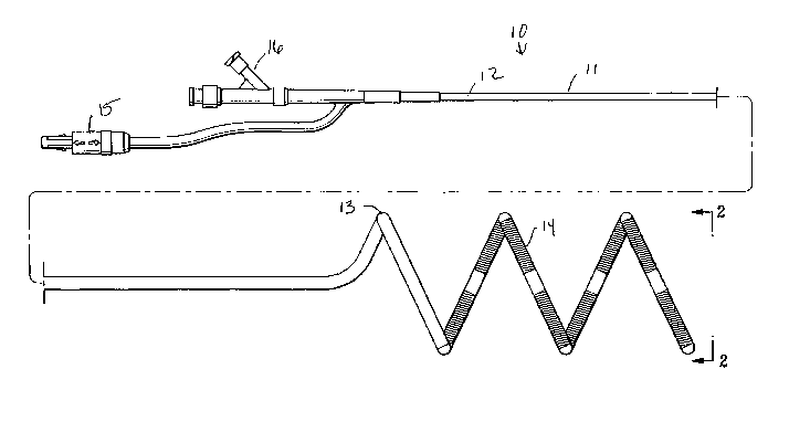
Fig. 1 is an elevational view of an EP device embodying
features of the invention, having a helically shaped distal shaft
section.
Fig. 2 is a transverse cross-sectional view of the EP device
shown in Fig. 1, taken along the lines 2-2.
Fig. 3 is an elevational view, partially in section, of an EP
device assembly embodying features of the invention, illustrating
an EP device in a low profile configuration within a guiding
catheter. .
Fig. 4 is an enlarged longitudinal cross-sectional view of the
EP device assembly shown in Fig. 3 taken along the lines 4-4,
illustrating the EP device distal tip within the guiding catheter.
Fig. 5 is an enlarged longitudinal cross-sectional view of the
EP device assembly shown in Fig. 3 taken along the lines 5-5,
illustrating a portion 'of the EP device distal shaft section within the
guiding catheter.
Fig. 6 is a transverse cross sectional view of the EP device
assembly shown in Fig. 5, taken along lines 6-6.
Fig. 7 is a transverse cross sectional view of an alternative
embodiment of an EP device assembly embodying features of the
invention, having a core wire slidably disposed in a lumen in the
device shaft.
7
CA 02446061 2003-10-31
WO 02/087453 PCT/US02/13604
Fig. 8 is an elevational view, partially in section, of a
patient's heart and an EP device assembly embodying features of
the invention, with the distal end of the EP device transeptally
positioned within a pulmonary vein.
Fig. 9 is an elevational view, partially in section, of the EP
device assembly of Fig. 8, with the turns of the helically shaped
distal shaft section moved closer together in a stacked
configuration.
Fig. 10 is an elevational view of an alternative embodiment of
an EP device embodying features of the invention comprising a
distal shaft section having a proximal portion with a helical shape
and a distal portion with a noncoiled shape with a pair or sensing
and/or pacing electrodes on the distal portion.
Fig. 1 1 is a longitudinal cross sectional view of the distal end
of the EP device of Fig. 10, taken within circle 1 1.
Fig. 12 is an elevational view, partially in section, of the EP
device of Fig. 10, positioned in contact with a wall defining a
pulmonary vein ostium, with the turns of the helically shaped distal
shaft section moved closer together in a stacked configuration.
Fig. 13 is an elevational view of an alternative embodiment of
an EP device embodying features of the invention comprising a
distal shaft section having a proximal portion with a helical shape
with one and one quarter turns, and a distal portion with a
noncoiled shape.
DETAILED DESCRIPTION OF THE INVENTION
Fig. 1 illustrates one embodiment of the EP device 10 of the
invention, generally comprising an elongated shaft 11 having a
proximal shaft section 12, a helically shaped distal shaft section
8
CA 02446061 2003-10-31
WO 02/087453 PCT/US02/13604
13, and a plurality of electrodes 14 on the distal shaft section 13.
An electrical connector 15 and an adapter 16 are on the proximal
end of the device. Fig. 2 illustrates a transverse cross section of
the distal end of the device 10 shown in Fig. 1, taken along lines 2-
2.
Fig. 3 illustrates the EP device 10 within a guiding catheter
20 for introduction and advancement within the patient. The
guiding catheter generally comprises an elongated shaft 21 having
a proximal end 22, a distal end 23, a port 24 in a proximal shaft
section, a port 25 in a distal shaft section, and a lumen 26
extending within the shaft to the port in the distal shaft section.
As illustrated in Fig. 3, the helically shaped distal shaft section of
the EP device 10 is reversibly deformed from the helical
configuration to a low profile configuration within the lumen 26 of
the guiding catheter. In the embodiment illustrated in Fig. 3, with
the EP device slidably disposed within guiding catheter lumen 26,
the radial force of the guiding catheter 20 against the device
reversibly straightens the helically shaped distal section to form a
straight configuration. The helically shaped distal shaft section 13
is preferably self expanding, so that the EP device 10 can be
advanced out the distal end of the guiding catheter 20, or the
guiding catheter 20 proximally retracted, causing the distal shaft
section of the EP device to return to the helically shaped
configuration illustrated in Fig. 1. In alternative embodiments (not
shown), the helically shaped distal shaft section reversibly
collapses to a helical shape with turns having a smaller outer
diameter when the distal shaft section is within the guiding
catheter lumen 26.
9
CA 02446061 2003-10-31
WO 02/087453 PCT/US02/13604
In a presently preferred embodiment, the EP device 10
includes a core member 17 having a helically shaped distal section,
disposed within the shaft 1 1. As best illustrated in Fig. 5, showing
a longitudinal cross section of the of the EP device shown in Fig. 3,
taken along lines 5-5, the shaft 11 comprises a tubular member 18
disposed about the core member 17. The core member 17 extends
within the tubular member to the distal end of the device, and the
tubular member 18 is helically shaped by the core member therein.
Fig. 6 illustrates a transverse cross section of the EP device shown
in Fig. 5, taken along lines 6-6.
The core member 17 is preferably formed of a superelastic
material, such as a NiTi alloy, or stainless steel, and has a
maximum diameter of about 0.01 inch (.25 mm) to about 0.018
inch (.46 mm). The core member 17, and preferably a distal
section thereof, may be tapered as shown in Fig. 4, or optionally
flattened. In a presently preferred embodiment, the core member
has an insulating coating 30, such as a polyester or polyimide
coating. The coating 30 is preferably about 0.0005 inch (0.0127
mm) thick. In the embodiment illustrated in Fig. 4, coating 30
extends distally to a point distal to the shaft 11 distal end and
proximal to the distal end of core member 17. In the embodiment
illustrated in Figs. 5 and 6, the coating 30 on the core member 17
contacts an inner surface of the tubular member 18. The core
member 17 is secured to the tubular member 18 by applying heat
to the device to melt and fuse the tubular member to the core
member coating. However, a variety of suitable means of securing
the core member within the tubular member may be used, such as
an adhesive (not shown) between the core member and the tubular
CA 02446061 2003-10-31
WO 02/087453 PCT/US02/13604
member. In an alternative embodiment of the invention illustrated
in Fig. 7, the core member 17 is slidably disposed within and
removable from a lumen 19 of the tubular member.
As best illustrated in Fig. 4, a flexible coiled tip 27 is provided
on the distal end of the EP device 10. The tip 27 has a closed
distal end, and includes a flexible coil 28 extending beyond the
distal end of the shaft 11 enclosed within a soft coating 29
preferably formed of a polymeric material. In the embodiment
illustrated in Fig. 4, the tip 27 has an open center region for
increased flexibility. A presently preferred polymeric material for
the tip 27 is a fluoropolymer such as THV available from 3M. In
the embodiment illustrated in Fig. 4, the core member 17 is secured
to the distal end of the coil 28, by suitable material such as gold-tin
solder. In another embodiment of the invention, the coil 28 may be
omitted, and the distal end of the EP device preferably provided
with a soft tip to minimize traumatic engagement with a blood
vessel wall.
In the embodiment illustrated in Figs. 5 and 6, the electrodes
14 comprise helical coils which are electrically connected to
insulated electrical conductors 31. In a presently preferred
embodiment, the EP device 10 shaft includes thermocouples 32,
connected to temperature sensor electrical conductors 33 and 34
(i.e., thermocouple wiresl. Thermocouples are preferably located
between adjacent electrodes on an outer surface of the shaft 1 1,
although they may alternatively be at other locations on the EP
device as is conventionally known. A conducting member 35, such
as a gold band, covers the thermocouples, and a polymeric jacket
36, preferably formed from THV, covers the conducting member
11
CA 02446061 2003-10-31
WO 02/087453 PCT/US02/13604
35 and insulates the thermocouple 32 from noise (e.g. RF noise)
present as a result of the energy sent to the electrodes. In the
embodiment illustrated in Fig. 5, the electrical conductors 31 and
thermocouple wires 33, 34 are braided within the tubular member
18. However, the electrical conductors 31 and thermocouple wires
33,34 may have a variety of suitable configurations, including
braided or wound configurations different from that shown in Fig. 5
or a nonbraided configuration. In an alternative embodiment (not
shown), the individually insulated electrical conductors may be
within the tubular member lumen 19 or at least in part within an
outer jacket of the core member in the embodiment in which the
core member is secured to the tubular member. The proximal ends
of the electrical conductors 31 and thermocouple wires 33, 34 are
electrically connected to individual pins of multi-pin connector 15
(Fig. 1 ) on the proximal end of the shaft.
In a method of treating a patient for atrial fibrillation or
flutter, the EP device of the invention is used to form a lesion
extending around an inner surface of the patient's pulmonary vein.
Fig. 8 illustrates an assembly in a patient's heart 40, with the EP
device 10 in a pulmonary vein 41. The device 10 is introduced into
the patient's vascular system, e.g. the femoral vein, percutaneously
or by way of a cut-down, within the guiding catheter 20. The
assembly is preferably advanced into the right atrium 42 from the
inferior vena cava 43, and positioned in the left atrium 44
transeptally, as illustrated in Fig. 8. The EP device 10 distal section
extends out of the port in the distal end of the guiding catheter, so
that the helically shaped distal shaft section of the device is
positioned within the pulmonary vein 41 of the heart. The
12
CA 02446061 2003-10-31
WO 02/087453 PCT/US02/13604
pulmonary vein 41 is mapped using electrodes on the device 10,
and if a pulmonary vein potential is detected, the electrodes on the
distal shaft section are used to form a lesions) extending at least in
part around the wall defining the pulmonary vein lumen or in the
left atrium just outside a pulmonary vein ostium. The position of
the lesion is preferably chosen to interrupt the conduction path to
the atrium. Alternatively, the lesion may be located to ablate the
actual focal origin in the pulmonary vein.
Typically, RF current is delivered to one or two electrodes to
perform a first ablation and then to adjacent electrodes, one or two
electrodes at a time, until an ablation of desired length is obtained
in the body lumen. This reduces the overall power requirements for
the assembly. The temperature sensors can be used to detect the
temperature of the heart wall between the adjacent electrodes, to
control the high frequency energy and determine when the lesions
formed by adjacent electrodes overlap to form continuous lesions
on the wall defining the body lumen. Additionally, feedback of the
temperature data can be used to modulate the power and prevent
thrombus in the preferred use, and cooling fluid can also be used.
After the ablation, the electrodes 14 can be employed to detect
electrical activity to ensure that the ablation has been effective in
terminating the fibrillation or flutter. Typically, the procedure is
performed for the left and right, superior and inferior pulmonary
veins.
The EP device of the invention can be used to form a helical
lesion, a closed circular lesion, or a curvilinear segmental (i.e.,
discontinuous) lesion. For example, in the embodiment illustrated in
Fig. 8, a helical lesion on the body lumen wall can be formed by
13
CA 02446061 2003-10-31
WO 02/087453 PCT/US02/13604
delivering RF energy to the electrodes which as illustrated are
contacting the pulmonary vein wall in a helical array. Typically, the
helical lesion is formed to extend continuously along the body
lumen wall, wherein the individual lesions formed by the
longitudinally adjacent electrodes on the shaft overlap to produce
one continuous lesion. The helical lesion comprises a spiral having
a distal end, and a proximal end longitudinally spaced from the
distal end of the spiral. In an alternative embodiment of the
invention, the lesion formed extends in a closed circle around the
body lumen wall, i.e., a lesion having ends that close together to
form a circle. A closed circle lesion can be formed by displacing
the device distal section to change the electrode position on the
body lumen wall after an initial lesion is formed. For example, in
one embodiment of the method of forming a closed circular lesion,
a helically shaped lesion is first formed on the body lumen wall, and
then the helically shaped distal shaft section of the EP device is
rotated or longitudinally displaced proximally or distally, and a
second lesion which overlaps with the first lesion is formed, to
thereby form at least one closed circular lesion. Alternatively, as
illustrated in Fig. 9, the helically shaped distal shaft section of the
EP device can be provided with closely spaced, stacked adjacent
turns which facilitate the formation of a closed circle lesion.
The spacing between adjacent turns of the helically shaped
distal shaft section can be changed by the physician during
deployment of the EP device within the body luri~en. To increase
the spacing between the helical turns of the device, the distal
extremity of the EP device is displaced out of the distal end of the
guiding catheter so that it is placed in contact with the body lumen
14
CA 02446061 2003-10-31
WO 02/087453 PCT/US02/13604
wall. The guiding catheter is displaced proximally, while a proximal
portion of the EP device is displaced proximally to stretch the turns
of the helically shaped distal shaft section apart, so that the portion
of the EP device distal shaft section that is still inside the guiding
catheter is deployed therefrom with the spacing between the turns
increased. Similarly the spacing between the turns may be
decreased by retracting the guiding catheter proximally while a
proximal portion of the device is displaced distally, to stack the
turns of the helically shaped distal shaft section together.
Fig. 10 illustrates an alternative embodiment of an EP device
1 10 which embodies features of the invention, generally comprising
an elongated shaft 111 having a proximal shaft section 112, a
distal shaft section 113, and a plurality of electrodes 114 on the
distal shaft section 113. An electrical connector 115 is on the
proximal end of the device 110. The distal shaft section 113
comprises a proximal portion 116 with a helical shape having one
or more turns, and a distal portion 1 17 extending from the proximal
portion with a noncoiled shape. In the embodiment illustrated in
Fig. 10, the noncoiled distal portion 1 17 has a straight shape with
an outer surface aligned or parallel with an outer surface of the
proximal shaft section 112. As illustrated in Fig. 10, the noncoiled
distal portion 1 17 has a width about equal to or less than the width
of the proximal shaft section 112. Thus, the noncoiled distal
portion 117 does not have the enlarged outer diameter formed by
the turns of the helically shaped proximal portion 116. The
electrodes 114 on the helically shaped proximal portion preferably
comprise coiled electrodes, and temperature sensors 118 are
located between the coiled electrodes 1 14, preferably on an outer
CA 02446061 2003-10-31
WO 02/087453 PCT/US02/13604
surface of the shaft, as discussed above in relation to the
embodiment of Fig. 1. In a presently preferred embodiment, each
electrode 1 14 has a length of about 3 to about 6 mm. Although 5
electrodes 114 are illustrated in Fig. 10, the number of electrodes
114 may vary, and in a presently preferred embodiment, about 8
electrodes are provided on EP device 110. A pair of sensing
electrodes 1 19 for mapping and/or pacing are on the distal portion
1 17 of the distal shaft section. In an alternative embodiment (not
shown) at least a second pair of sensing and pacing and pacing
electrodes 119 may be provided on the shaft proximal to the
helically shaped proximal portion 116. The sensing and pacing
electrodes are preferably spaced away from the helically coiled
section 116, and in one embodiment are about 1 to about 3 cm,
preferably about 1.5 to about 2 cm from the helically coiled
section. In a presently preferred embodiment, the electrodes 114
on the helically shaped portion 1 16 are configured for unipolar use
during ablation, and bipolar use during sensing. The distal sensing
and pacing electrodes 119 are configured for use a bipolar
electrodes during sensing and pacing. A flexible coiled tip 120 is
secured to the distal end of the distal portion 117, to facilitate
guiding the EP device to a desired location within the patient. In a
presently preferred embodiment, the tip . coil 120 is about 1 to
about 3 cm, most preferably about 2 cm in length, and is formed of
a radiopaque metal such as platinum.
The turns of the helical proximal section 1 16 are illustrated in
a relaxed configuration in Fig. 10. However, the turns of the
helical proximal section 116 can be moved closer together or
further apart within the patient by urging the proximal end of the
16
CA 02446061 2003-10-31
WO 02/087453 PCT/US02/13604
catheter distally or proximally, respectively, with the distal end of
the catheter in a stabilized position within the patient and as
discussed above in relation to the embodiment of Fig. 1.
Each electrode 114 is spaced apart from one or more
adjacent electrodes 1 14 on the shaft 1 11, i.e., the electrodes 1 14
extend discontinuously along the shaft. However, depending on
the duration and power level used during an ablation procedure, the
lesions) formed by electrodes 114 can be discontinuous or
alternatively, can be joined together and thus continuous.
In the embodiment illustrated in Fig. 10, the helical proximal
portion 1 16 forms one full 360° loop and half of a second loop. In
a presently preferred embodiment, about one full 360° loop is
provided, although the number of loops may vary. Because the
proximal portion 1 16 is helical, a proximal section of the 360° loop
is longitudinally spaced apart from the distal section thereof which
completes _ the circumference of the loop in the relaxed
configuration illustrated in Fig. 10. Consequently, the helical
proximal portion forms at least one open or helical 360° loop in the
relaxed configuration, and the electrodes 1 14 thereon form an open
or helical 360°, discontinuous loop. The circumference of one
360°
loop of the helically proximal portion 116 varies depending on the
desired use of the EP device. In a presently preferred embodiment,
the circumference of one 360° loop is about 15 mm to about 40
mm, preferably about 15 mm to about 30 mm. Depending on the
circumference of the loop and the number and length of the
electrodes 114, the electrodes 114 may or may not extend the
length of one or more 360° loops. In a presently preferred
embodiment, the electrodes 1 14 extend along the length of at least
17
CA 02446061 2003-10-31
WO 02/087453 PCT/US02/13604
one 360° loop of the helical proximal portion 116, so that the
electrodes can be used to form a lesion which extends in a
continuous 360° loop, or a discontinuous, partial segment of a
360° loop. However, in alternative embodiments, the helical
proximal portion 116 has a partial loop of less than 360° (not
shown, or the electrode number or length is sufficiently small such
that the electrodes extend along a length of a partial loop of less
than 360° on the helical proximal portion. The circumference of
the helical section, i.e., the length of the helical section if it was
stretched out to a straight, nonhelical shape, is about 5 to about 40
mm, preferably about 5 to about 20 mm.
Fig. 11 illustrates an enlarged, longitudinal cross sectional
view of the distal end of the device 110, taken within circle 1 1.
As illustrated in Fig. 1 1, the shaft 1 1 1 comprises a tubular member
121, having braided electrical conductors 122 in the wall of the
tubular member 121, and having a core member 123 in a lumen of
the tubular member 121. In the embodiment illustrated in Fig. 1 1,
the core member 123 is secured to the flexible coiled tip 120. An
outer layer 124 on an outer surface of the tubular member 121
overlaps the ends of the sensing and pacing electrodes 119.
Fig. 12 illustrates the device 110 with the helically coiled
proximal portion 116 of the distal shaft section 113 in position at
the ostium 46 of a pulmonary artery which forms the junction
between the pulmonary artery and the right atrium of the patient's
heart. As illustrated in Fig. 12, the turns of the helically shaped
proximal portion 116 are in a stacked configuration after having
been moved closer together than the natural relaxed spacing shown
in Fig. 10, by distally forcing the catheter against the wall defining
18
CA 02446061 2003-10-31
WO 02/087453 PCT/US02/13604
the ostium of the pulmonary artery. As a result, the electrodes
114 extend discontinuously, completely around the ostium. High
frequency energy is delivered to one or more of the electrodes 1 14
to form a lesion extending at least in part around the ostium. The
lesion can be caused to be a continuous or a discontinuous circular
lesion depending on the energy level and the length of time of the
ablation, and by rotating the catheter one or more times between
delivery of ablation energy to electrodes 114. As illustrated in Fig.
12, the distal portion 1 17 of the distal shaft section is positioned
within the pulmonary vein 41, to allow for mapping and/or pacing
from within the pulmonary vein. Thus, the sensing electrodes
allow for sensing electrical activity before and after the ablation
energy is delivered to electrodes 1 14, to determine the appropriate
location of the device, and whether the lesions formed therefrom
sufficiently treated the atrial arrhythmia. Although not illustrated,
in one embodiment of performing a medical procedure, the helically
shaped proximal portion 116, and the distal portion 1 17 of the EP
device 110 are both positioned within the pulmonary vein 41,
similar to the embodiment illustrated in Figs. 8 and 9.
Fig. 13 illustrates an alternative embodiment of an EP device
140 embodying features of the invention, similar to catheter 1 10
but with helical proximal portion 116 forming one full 360° loop
and one quarter of a second loop, and with no electrodes 119 on
the noncoiled distal portion 117. A presently preferred method of
using EP device 140 comprises positioning the helical proximal
portion 116 just outside a pulmonary vein at the ostium thereof,
with the noncoiled distal portion 1 17 used as an anchoring section
within and in contact with the pulmonary vein. The helical
19
CA 02446061 2003-10-31
WO 02/087453 PCT/US02/13604
proximal portion 1 16 is pushed against the atrial tissue just outside
the pulmonary vein ostium, thereby ensuring good contact with
atrial tissue for ablation purposes. Pushing the shaft distally with
the helically shaped section braced against the atrial tissue thus
collapses the helix and may advance a distal section of the
proximal shaft section proximal to the helically shaped distal portion
and through the ostium. The electrodes on the helical proximal
portion 1 16 are used to map for pulmonary vein potentials and only
a discontinuous, segmental lesion, rather than an entire
circumference, continuous lesion, is formed by RF ablation, to
barricade the pulmonary vein potentials from exiting the pulmonary
vein.
In one method of the invention, the lesion comprises one or
more closed circles on the endocardium. However, the lesion may
alternatively comprise a discontinuous, partially open circle formed
by a plurality of smaller lesions. Additionally, the lesion may be
formed by the helical distal shaft section in the noncollapsed
configuration to extend helically along a length of the body lumen,
or the lesion may be formed by the helical distal shaft section in the
collapsed configuration to extend only around the circumference of
the body lumen and not helically along a length of the body lumen.
Typically, the lesion formed with the EP device 10/1 10/140 of the
present invention has a width of about 2 to about 7 mm, preferably
about 3 to about 4 mm. The circumference of the lesion (forming a
continuous closed circle, or a discontinuous partially open circle) is
about 5 to about 40 mm, preferably about 5 to about 20 mm. A
lesion extending only circumferentially around the body lumen and
not helically along a length of the body lumen (forming either a
CA 02446061 2003-10-31
WO 02/087453 PCT/US02/13604
continuous closed circle, or a discontinuous partially open circle)
has a length of about the thickness of the EP device shaft. A
helical lesion extending helically along a length of the body lumen
has a length of about to about 5 mm to about 50 mm, preferably
about 5 to about 10 mm. Preferably, in the embodiment in which a
plurality of continuous, closed circle lesions are formed on the body
lumen wall, the lesions are formed near the transition zone between
the left atrial tissue and the pulmonary vein tissue.
The EP device 10/110/140 has a total length, including the
connector 16, of about 100 cm to about 200 cm, and preferably
between 150 and 180, e.g. about 165 cm. The length of the distal
shaft section 13/113 having electrodes 14/114 is about 2 cm to
about 15 cm, and preferably about 4 to about 8 cm, e.g. about 6
cm. The outer diameter of the distal shaft section of the device is
typically about 1.0 mm (3.0 French) to about 2.0 mm (6.0 French),
and preferably about 1.3 mm (4 French) to about 1.7 mm (5
French). The maximum outer dimensions of the electrodes are
generally about 1.0 mm (3 Fr) to about 1.3 mm (4 Fr), and
preferably about 1.22 mm (3.7.Fr). The electrode length is about 2
mm to about 8 mm, and preferably about 4 to about 7 mm, e.g.
about 6 mm. The interelectrode spacing is generally about 1 mm
to about 3 mm, and preferably about 2 mm. In a presently
preferred embodiment, the interelectrode spacing is uniform.
However, the electrode spacing may alternatively be nonuniform.
In a presently preferred embodiment, about 4 to about 12 individual
electrodes are provided on the shaft distal section, however, the
device may have larger number of electrodes if the diameter of the
distal section is increased to greater than 5 Fr.
21
CA 02446061 2003-10-31
WO 02/087453 PCT/US02/13604
Typically, the device is used within the patient's vasculature;
although it may also be used to create lesions within other body
lumens. The device may be advanced retrogradely through the
aorta and left ventricle via a femoral artery access site. As
illustrated in Fig. 8, the guiding catheter may have a bent or
deflectable distal end. Torquing the proximal section 22 of the
guiding catheter, which extends out of the patient during the
procedure, will cause the distal section thereof to be rotatably
displaced within the body lumen and allow the EP device 10 to be
properly positioned.
To the extent not already discussed herein, the EP device
components can be formed of conventional materials. The core
member 17/123 can be formed of a variety of suitable materials
including high spring-back metals, or superelastic metals, or shape
memory metals, such as ELGILOY available from Carpenter
Technology of Pennsylvania, MP35N, available from SPS
Technologies, high tensile strength steel including 304 vacuum-
melted steel, and titanium alloys including Ti-GAI-4V, CP Titanium,
and NiTi.
The electrical connector 14 on the proximal end of the device
may be a commercially available electrical connector such as Part
No. PAB-M08-GLA39J or PAB-M08-TLA39J for an eight pin
connector or Part No. PAB-M08-GLA39A for a connector with a
greater number of pins, e.g. 9-16. The above connectors are
available from Lemo USA, Inc. in Santa Rosa, CA. Suitable
connectors for accessory cables connectable to the above
connectors include PRB-M08-GLL65J for eight pin connectors and
22
CA 02446061 2003-10-31
WO 02/087453 PCT/US02/13604
PRB-M08-G1165A for connectors with more than eight pins. The
latter connectors are also available from the same source.
While the invention has been described herein in terms of
certain preferred embodiments directed to the detection and
treatment of atrial fibrillation and flutter, those skilled in the art will
recognize that the invention may be employed in a wide variety of
procedures. A variety of modifications and improvements may be
made to the present invention without departing from the scope
thereof. Moreover, although individual features of embodiments of
the invention may be shown or discussed in relation to some of the
embodiments and not in others, those skilled in the art will
recognize that individual features of one embodiment of the
invention can be combined with any or all the features of another
embodiment.
23