Note: Descriptions are shown in the official language in which they were submitted.
CA 02448482 2003-11-24
WO 03/006495 PCT/CA02/01058
1
HUMAN PAPILLOMAVIRUS E2 TRANSACTIVATION DOMAIN / INHIBITOR
CO-CRYSTAL AND X-RAY COORDINATES DEFINING THE INHIBITOR-
BINDING POCKET
FIELD OF THE INVENTION
The invention relates to the papillomavirus E2 protein, particularly the
crystalline structure of the human papillomavirus 11 (HPV-1 1) E2 protein
transactivation domain complexed with an inhibitor. Particularly, the
invention
provides crystal structure coordinates that define an inhibitor-binding pocket
and
3-dimension structural model for identifying potential inhibitors that would
fit in this
pocket. Also disclosed are methods for enabling the design and selection of
inhibitors of E2 protein activity involved in papillomavirus DNA replication,
particularly human papillomavirus.
BACKGROUND OF THE INVENTION
Papillomaviruses (PV) are non-enveloped DNA viruses that induce
hyperproliferative lesions of the epithelia. The papillomaviruses are
widespread
in nature and have been recognized in higher vertebrates. Viruses have been
characterized, amongst others, from humans, cattle, rabbits, horses, and dogs.
The first papillomavirus was described in 1933 as cottontail rabbit
papillomavirus
(CRPV). Since then, the cottontail rabbit as well as bovine papillomavirus
type 1
(BPV-1) have served as experimental prototypes for studies on
papillomaviruses.
Most animal papillomaviruses are associated with purely epithelial
proliferative
lesions, and most lesions in animals are cutaneous. In the human there are
more
than 75 types of papillomavirus (HPV) that have been identified and they have
been catalogued by site of infection: cutaneous epithelium and mucosal
epithelium (oral and genital mucosa). The cutaneous-related diseases include
flat
warts, plantar warts, etc. The mucosal-related diseases include laryngeal
papillomas and anogenital diseases comprising cervical carcinomas (Fields,
1996, Virology, 3rd ed. Lippincott - Raven Pub., Philadelphia, N.Y.).
There are more than 25 HPV types that are implicated in anogenital
diseases; these are grouped into "low risk" and "high risk" types. The low
risk
types include HPV type 6, and type 11, which induce mostly benign lesions such
as condyloma acuminata (genital warts) and low grade squamous intraepithelial
lesions (SIL). In the United States, there are approximately 5 million people
with
CA 02448482 2003-11-24
WO 03/006495 PCT/CA02/01058
2
genital warts of which 90% is attributed to HPV-6 and HPV-1 1.
The high-risk types are associated with high grade SIL and cervical cancer
and include most frequently HPV types 16, 18, 31, 33, 35, 45, and 52. The
progression from low-grade SIL to high-grade SIL is much more frequent for
lesions that contain high risk HPV-16 and 18 as compared to those that contain
low risk HPV types. In addition, only four HPV types are detected frequently
in
cervical cancer (types 16, 18, 31 and 45). About 500,000 new cases of invasive
cancer of the cervix are diagnosed annually worldwide (Fields, 1996, supra).
Treatments for genital warts include physical removal such as
cryotherapy, C02 laser, electrosurgery, or surgical excision. Cytotoxic agents
may
also be used such as trichloroacetic acid (TCA), podophyllin or podofilox.
Immunomodulatory agents are also available such as Interferon and Imiquimod
(Aldara , 3M Pharmaceuticals). These treatments are not completely effective
in
eliminating all viral particles and there is either a high cost incurred or
uncomfortable side effects related thereto. Also recurrent warts are common
(Beutner & Ferenczy, 1997, Amer. J. Med., 102(5A):28-37).
The ineffectiveness of the current methods to treat HPV infections has
demonstrated the need to identify new means to control or eliminate such
infections. In recent years, efforts have been directed towards finding
antiviral
compounds, and especially compounds capable of interfering with viral
replication
(Hughes and Romanos, 1993, Nucleic Acids Res. 21:5817-5823; Clark et al.,
Antiviral Res., 1998, 37(2):97-106; Hajduk et al., 1997, J. Med. Chem.,
49(20):3144-3150 and Cowsert et al., 1993, Antimicrob. Agents. Chemother.,
37(2):171-177). To that end, it has therefore become important to study the
genetics of HPVs in order to identify potential chemotherapeutic targets to
contain
and possibly eliminate any diseases caused by HPV infections.
The life cycle of PV is closely coupled to keratinocyte differentiation.
Infection is believed to occur at a site of tissue disruption in the basal
epithelium.
Unlike normal cells, cellular division continues as the cell undergoes
vertical
differentiation. As the infected cells undergo progressive differentiation,
the
cellular replication machinery is maintained which allows viral DNA
replication to
increase, with eventual late gene expression and virion assembly in terminally
differentiated keratinocytes and the release of viral particles (Fields,
supra).
The coding strand for each of the papillomavirus genome contains
approximately ten designated translational open reading frames (ORFs) that
have
CA 02448482 2003-11-24
WO 03/006495 PCT/CA02/01058
3
been classified as either early ORFs or late ORFs. The El to E8 genes are
expressed early in the viral replication cycle. The two late genes (L1 and L2)
code
for the major and minor capsid proteins respectively. The El and E2 gene
products function in viral DNA replication, whereas E5, E6 and E7 modulate
host
cell proliferation. The functions of E3, E4 and E8 gene products are uncertain
at
present.
Studies of HPV have shown that proteins El and E2 are the only viral
proteins required for viral DNA replication (Kuo et al., 1994, J. Biol. Chem.
30:
24058-24065). This requirement is similar to that of bovine papillomavirus
type 1
(BPV-1). Indeed, there is a high degree of similarity between El and E2
proteins
and the ori-sequences of all papillomaviruses (PV) regardless of the viral
species
and type (Kuo et al., 1994, supra).
When viral DNA replication proceeds in vitro, where El protein is present
in excess, replication can proceed in the absence of E2. In vivo, in the
presence
of a vast amount of cellular DNA, replication requires the presence of both El
and
E2. The mechanism for initiating replication in vivo is believed to involve
the
cooperative binding of El and E2 to the origin, leading to the assembly of a
ternary protein-DNA complex (Mohr et al., 1990, Science 250:1694-1699]. The E2
protein is a transcriptional activator that binds to the El protein and, by
doing so
enhances binding of El to the BPV origin of replication (Seo et al., 1993b,
Proc.
Natl. Acad. Sci., 90:2865-2869). Hence, E2 acts as a specificity factor in
directing
El to the origin of replication (Sedman and Stenlund, 1995, Embo. J. 14:6218-
6228). In HPV, Lui et al. suggested that E2 stabilizes binding of El to the
ori
(1995, J. Biol. Chem. 270(45): 27283-27291 and McBride et al., 1991, J. Biol.
Chem 266:18411-18414). These interactions of DNA-protein and protein-protein
occur at the origin of DNA replication (Sverdrup and Myers, supra).
The -45 kD E2 proteins characterized from numerous human and animal
serotypes share a common organization of two domains. The N-terminal
transactivation domain (TAD) is about 220 amino acids and the C-terminal DNA -
binding domain (DBD) is 100 amino acids in length. Both domains are joined by
a
flexible linker region.
E2 activates viral replication through cooperative binding with the viral
initiator protein El to the origin of DNA replication, ultimately resulting in
functional El hexamers. E2 is also a central regulator of viral transcription.
It
interacts with basal transcription factors, including TATA-binding protein,
TFIIIB,
CA 02448482 2003-11-24
WO 03/006495 PCT/CA02/01058
4
and human TAF1170; proximal promoter binding protein such as Spl; and other
cellular factors such as AMF-1, which positively affect E2's transcriptional
activation.
Which of these many interactions are sufficient or necessary to achieve
transcriptional activation is more ambiguous. These details are consistent
with the
idea that enhancer binding proteins function as transcriptional activators by
using
specific protein-protein contacts to link components of the general
transcription
machinery to a promoter, with the goal of recruiting RNA polymerase II. A
third
function of E2 is to aid in the faithful segregation of viral DNA. The bovine
papillomavirus (BPV) genome and E2 protein co-localize with host cell
chromosomes during mitosis, dependent on an intact E2 TAD.
The E2 DBD dimerizes to form a 1i-barrel with flanking recognition helices
positioned in the major grooves of the DNA binding site. In contrast, the
structure
of E2 TAD has remained elusive until Harris and Botchan (1999, Science, 284
(5420); 1673) provided a first model of a proteolytic fragment of HPV-18 E2
TAD
by X-ray crystallography. The model suggests a cashew-shaped protein of 55A x
40A x 30A with a concave cleft on one side of the protein and ridges on the
opposite surface. Harris and Botchan studied whether discrete surfaces
correlated with known E2 activities and particularly identified a prominent
cluster
of residues constituting the inner edge of the main cavity encompassing E175,
L178, Y179, and 173 defining a distinctive surface important for
transcription.
Antson et at (2000, Nature, (403) 805-809) disclose the crystal structure of
the complete E2 TAD from HPV-16, including a second newly identified putative
E2-E2 TAD interface comprising a cluster of 7 conserved residues (R37, A69,
173, E76, L77, T81, and Q80). Anston et al suggested that Q12 and E39 may be
involved in interaction with El.
The E2 protein is considered a potential target for antiviral agents.
However, drug discovery efforts directed towards E2 have been hampered by the
lack of structural information of an E2 complexed with an inhibitor. Neither
the
model of Harris, nor that of Antson provides any information as to the
localization
and/or characterization of a potential inhibitor binding pocket. Structural
information of the apo-E2 TAD has provided some valuable knowledge of the
surface on the'apo-protein but it now appears clear that this is not
representative
of the changes in conformation induced upon binding with an inhibitor.
The lack of specific E2 inhibitors, which is necessary for obtaining co-
CA 02448482 2007-01-24
crystal of E2 and inhibitors, has hampered the search for inhibitor binding
pocket
in E2. Thus, X-ray crystallographic analysis of such protein-inhibitor complex
has
not been possible.
5
SUMMARY OF THE INVENTION
The present invention solves the problems left unanswered by the prior art
by providing a novel composition comprising a human papillomavirus E2 protein
transactivation domain complexed with a small molecule inhibitor of E2 and
methods for making such composition. Advantageously, the present invention
further provides an E2-inhibitor complex that is capable of being crystallized
and
analyzed by X-ray diffraction, thereby providing important information on the
inhibitor-binding pocket of the transactivation domain of the HPV E2 protein.
The
inhibitor provides an invaluable tool to produce a co-crystal allowing
characterization of a previously unknown inhibitor-binding pocket that may be
involved in interaction with El during the replication cycle of HPV.
The invention also provides a method for determining at least a portion of
the three-dimensional structure of molecules or molecular complexes, which
contains at least some structurally similar features to a HPV E2 inhibitor
binding
pocket.
The invention also provides a 3-D model for analyzing and predicting
binding of potential inhibitors to aid in the search for further inhibitors
binding to
the identified pocket. Localization and characterization of this pocket, as
described in the present invention provides a potential new therapeutic target
in
the treatment of PV infections.
The invention also provides a screening method for identifying agents
capable of modulating this new target and a system to select at least one such
agent capable of interfering with PV DNA replication.
The invention also provides a method for producing a drug, which inhibits
interaction of the El-E2 interaction comprising identifying a drug, or
designing a
drug that fits into the pocket as described herein.
According to a first aspect of the invention, there is provided a
crystallizable
composition, comprising an PV E2 TAD-like polypeptide of SEQ ID NO.2
CA 02448482 2003-11-24
WO 03/006495 PCT/CA02/01058
6
complexed with an inhibitor L:
CI
O CI
X. O
O
Me
O HN
O ONa
N~~
L) N
S
According to a second aspect of the invention, there is provided a crystal
comprising an PV E2 TAD-like polypeptide of SEQ ID NO.2 complexed with said
inhibitor L, as defined above.
According to a third aspect of the invention, there is provided a method for
producing a crystallized PV E2 TAD-inhibitor complex (PV E2 TAD-L), as defined
above, comprising:
a) mixing purified PV E2 TAD, contained in a purification buffer, with
solubilized inhibitor L to generate a complex solution containing said
PV E2 TAD-L complex; and
b) crystallizing said complex from a) in a crystallization buffer.
According to a fourth aspect of the invention, there is provided a method for
producing crystallized apo PV E2 TAD, comprising:
a) mixing apo PV E2 TAD, contained in a purification buffer, with a
crystallization buffer.
According to a fifth aspect of the invention, there is provided a method for
producing a crystallized PV E2 TAD-inhibitor complex (PV E2 TAD-L), as defined
above, comprising:
a) solubilizing inhibitor L in a crystallization buffer; and
a) soaking crystallized apo PV E2 TAD, as defined above, into a).
According to a sixth aspect of the invention; there is provided X-ray crystal
CA 02448482 2003-11-24
WO 03/006495 PCT/CA02/01058
7
structure coordinates of PV E2 TAD-inhibitor complex (PV E2 TAD-L), as defined
above.
According to a seventh aspect of the invention there is provided a computer-
readable data storage medium comprising a data storage material encoded with
the X ray crystal structure coordinates, or at least a portion of the
structure
coordinates, set forth in Figure 9.
According to a eighth aspect of the present invention, there is provided a
computer for generating a three dimensional representation of said PV E2 TAD-L
complex, as defined herein, comprising:
a) a computer readable data storage medium having a data storage
material encoded with said structure coordinates set forth in Figure 9;
b) a memory for storing instructions for processing said computer
readable data;
c) a central processing unit coupled to said computer readable data
storage medium for processing said computer readable data into said
three dimensional representation; and
d) a display unit coupled to said central processing unit for displaying
said three dimensional representation.
According to an ninth aspect of the invention, there is provided a method for
producing an E2 protein, said protein being useful for identifying or
characterizing
E2 TAD inhibitors, comprising:
a) using the HPV E2 TAD-L crystal structure, as defined herein, to identify
HPV
inhibitor binding pocket residues;
b) comparing said HPV inhibitor binding pocket residues with analogous
residues in another PV E2;
c) mutating said other PV residues to said HPV residues, to produce a hybrid;
and
d) testing said hybrid for inhibition by an inhibior.
BRIEF DESCRIPTION OF THE FIGURES
Having thus generally described the invention, reference will now be made to
the
accompanying drawings, showing by way of illustration a preferred embodiment
CA 02448482 2003-11-24
WO 03/006495 PCT/CA02/01058
8
thereof, and in which:
Figure IA depicts the amino acid sequence of the HPV-11 E2 transactivation
domain (SEQ ID NO.1) as obtained by Sakai, et al. 1996, J Virol. V70 1602-11;
Figure 1 B depicts the amino acid sequence of the HPV-11 E2 transactivation
domain (SEQ ID NO.2) as obtained according to the procedure of Example 1;
Figure 2 depicts stereo ribbon diagrams of the apo-E2 from HPV-16 as described
in Antson et al. (supra);
Figure 3 depicts stereo ribbon diagrams'of the apo-E2 from HPV-11 as produced
by the Applicant;
Figure 4 depicts stereo ribbon diagrams of the E2 from HPV-11 complexed with
compound L as described herein;
Figure 5 depicts a solvent accessible surface representation of the inhibitor-
binding pocket of the apo-E2 TAD from HPV-16 (Antson et al., supra);
Figure 6A depicts a solvent accessible surface representation of the inhibitor-
binding pocket of the apo-E2 from HPV-11 as produced by the Applicant;
Figure 6B depicts a solvent accessible surface representation of the inhibitor-
binding pocket of the co-crystal;
Figure 7 depicts a schematic representation of the movement of Y19 and H32
occurring in the pocket upon binding with an inhibitor;
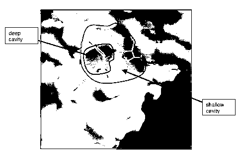
Figure 8 depicts a solvent accessible surface top view of the pocket showing
particularly a deep cavity and a shallow cavity;
Figure 9 lists the atomic structure coordinates for the E2 TAD complexed with
compound L as derived by X-ray diffraction from co-crystals of that complex
CA 02448482 2003-11-24
WO 03/006495 PCT/CA02/01058
9
(hereinafter referred to HPV TAD E2-L). The preparation of the complex is
described in Example 3. The following terms have these meanings: the term A.A.
refers to the amino acid which is identified by each coordinate, in this
column: the
term "CPR" means cis-proline; BLHA= first molecule of inhibitor L; BLHB=
second
molecule of inhibitor L. Information on amino acids 197 to 201 from chain A is
lacking due to the high flexibility of those residues that renders them
invisible to x-
ray. For the same reason, the following amino acids are modeled as Alanine:
E2,
K107, K173, S180, M182, H183 and P196. "X, Y, Z" crystallographically define
the
atomic position determined for each atom in a cartesian coordinate space.
"Occ"
is an occupancy factor that refers to the fraction of the molecules in which
each
atom occupies the position specified by the coordinates. A value of "1"
indicates
that each atom has the same conformation, e.g., the same position, in all
molecules of the crystal. "B" is a thermal factor that measures movement of
the
atom around its atomic center. The coordinates of the residues that form the
deep
cavity are shown in bold; and
Figure 10 depicts the alignment of the amino acid sequence clusters that
define
generally the inhibitor-binding pocket region of the E2 transactivation domain
from
HPV-6A, HPV-1 1, HPV-1 6 and HPV-1 8. The residues in bold indicate that they
define the deep cavity of the inhibitor binding pocket. The single underline
defines
the residues of the bottom of the deep pocket. The double underline indicates
the
shallow pocket residues. Y19 is indicated in italics.
DETAILED DESCRIPTION OF THE INVENTION
Definitions
The following abbreviations are used throughout the specification.
The term "associating with" or "binding" refers to a condition of proximity
between chemical entities or compounds, or portions thereof. The association
may be non-covalent - wherein the juxtaposition is energetically favored by
hydrogen bonding or van der Waals or electrostatic interactions - or it may be
covalent.
The term "binding pocket", as used herein, refers to a region of a molecule
or molecular complex, that, as a result of its shape, favorably associates
with
another molecule, molecular complex, chemical entity or compound. As used
herein, the pocket comprises at least a deep cavity and, optionally a shallow
CA 02448482 2003-11-24
WO 03/006495 PCT/CA02/01058
cavity.
As used herein the term "complex" refers to the combination of a molecule
or a protein, conservative analogs or truncations thereof associated with a
chemical entity.
5 The abbreviations for the a-amino acids used in this application are set
forth as follows:
Amino Acid Symbol Single letter code
Alanine Ala A
Arginine Arg R
Aspartic acid Asp D
Asparagine Asn N
Cysteine Cys C
Glutamic acid Glu E
Glutamine Gln Q
Glycine Gly G
Histidine His H
Isoleucine lie I
Leucine Leu L
Lysine Lys K
Methionine Met M
Phenylalanine Phe F
Proline Pro P
Serine Ser S
Threonine Thr T
Tryptophan Trp W
Tyrosine Tyr Y
Valine Val V
10 The term "analog" as used herein denotes, in the context of this invention,
a sequence of amino acid that retains a biological activity (either functional
or
structural) that is substantially similar to that of the original sequence.
This analog
may be from the same or different species and may be a natural analog or be
CA 02448482 2003-11-24
WO 03/006495 PCT/CA02/01058
11
prepared synthetically. Such analogs include amino acid sequences having
substitutions, deletions, or additions of one or more amino acids, provided
that the
biological activity of the protein is conserved. Particularly, the term
"conservative
analog" denotes an analog having amino acid substituted by another amino acid
having strong or weak similarity (see, for example, Dayhoff, M.O., (1978),
Atlas of
Protein Sequence and Structure, 5, suppl. 3, National Biomedical Research
Foundation, Washington, D.C.) as defined according to the following Table:
Table of amino acid similarity
Amino acid Strong Weak
A G, S C, T, V
C A, S
D E G, H, K, N, Q, R, S
E D H,K,N,Q,R,S
F W, Y H, I, L, M
G A D, N, S
H Y D, E, F, K, N, Q, R
I L,M,V F
K R D,E,H,N,Q,S,T
L I, M, V F
M I, L, V F
N Q D, E, G, H, K, R, S, T
P S, T
Q N D, E, H, K, R, S
R K D,E,H,N,Q
S A,T C, D, E, G, K, N,P,Q
T S A,K,N,P,V
V I,L,M A,T
W F, Y
Y F, H, W
The term "side chain" with reference to an amino acid or amino acid
residue means a group attached to the a-carbon atom of the a-amino acid. For
example, the R-group side chain for glycine is hydrogen, for alanine it is
methyl,
for valine it is isopropyl. For the specific R-groups or side chains of the a-
amino
CA 02448482 2003-11-24
WO 03/006495 PCT/CA02/01058
12
acids reference is made to A.L. Lehninger's text on Biochemistry (see chapter
4).
The term "truncation" refers to any segment of the E2 TAD amino acid
sequence and/or any segment of any of the analogs described herein above that
comprise the amino acids sufficient to define the deep cavity of the inhibitor-
binding pocket of the present invention in the same spatial relationship as
the one
defined by the coordinates of Figure 9.
The term "root mean square deviation" or "rms deviation" or "rmsd" means
the square root of the arithmetic mean of the square of the deviations from
the
mean. In the context of atomic objects, the numbers are given in angstroms
(A). It
is a way to express the deviation or variation from a trend or object. For the
purpose of the present invention, all rmsd comparison were obtained by
comparing structures that had been superimposed using the main chain atoms of
H32, W33 and L94 only, to the minimum overlap rms, by rigid body movement
only. The main chain atom rmsd for this action between our apo structure and
the
complex disclosed herein is 0.078A.
PREFERRED EMBODIMENTS
1. Composition
According to a first embodiment, there is provided a crystallizable
composition,
comprising an HPV E2 TAD-like polypeptide of SEQ ID NO.2 complexed with an
inhibitor L:
CI
0 CI
N. O
Me
0 HN
0 ONa
,
L) ,N
S
Preferably, the composition comprises amino acids 1-220 of the HPV E2 protein
(SEQ ID NO.1) as defined according to the numbering of Swiss Prot: locus
VE2_HPV11 accession P04015; unique ID: g137671, conservative analogs or
CA 02448482 2003-11-24
WO 03/006495 PCT/CA02/01058
13
truncations thereof. More preferably, the trans-activation domain (TAD) of E2
comprises amino acids 1-218, particularly 1-215 and even more preferably 1-
201.
Still, most preferably, the E2 TAD used for the present invention comprises
amino
acids 2-201 and still most particularly 2-196. Even most preferably, the
composition comprises amino acids 15-104 of the E2 TAD.
In another aspect of the first embodiment, the HPV E2 TAD used for the present
invention is obtained from the HPV-11 strain and is complexed with the small
molecule inhibitor L. Other types of papillomavirus (PV) are also contemplated
by
the present invention, including BPV (bovine paplillomavirus) or CRPV (Cotton
Tail Rabbit Virus).
According to a second embodiment, there is provided a crystal comprising an
HPV E2 TAD-like polypeptide of SEQ ID NO.2 complexed with the inhibitor L.
2. Method of crystallizing
According to a third embodiment of the invention, there is provided a method
for
producing a crystallized HPV E2 TAD-inhibitor complex (HPV E2 TAD-L), as
defined above, comprising:
a) mixing purified HPV E2 TAD, contained in a purification buffer, with
solublized inhibitor L to generate a complex solution containing said
HPV E2 TAD-L complex; and
b) crystallizing said complex from a) in a crystallization buffer.
In a preferred aspect of the third embodiment step a), the inhibitor L is
solublized
in 100% DMSO at a concentration of 60mM.
In a preferred aspect of the third embodiment step a), the purification buffer
contains a reducing agent that may be selected from TCEP or DTT. More
preferably the reducing agent is TCEP. Preferably, the reducing agent is TCEP
at
a concentration of about 1 mM to about 10mM. More preferably, the reducing
agent is TCEP at a concentration of 5mM.
Preferably, the purification buffer is used at a pH of between 7 and 9. More
preferably, the purification buffer is used at pH of 8.
CA 02448482 2003-11-24
WO 03/006495 PCT/CA02/01058
14
Further to the reducing agent, a salt can be added to aid stability of the HPV
E2
TAD. Preferably, the salt may be selected from NaCl, NH4SO4, or KCI. More
preferably, the salt is NaCl at a concentration of about 200mM to about 800mM.
More preferably, the salt is NaCI at a concentration of 500mM.
Further to the reducing agent, a buffer can be added to further aid the
stability of
the HPV E2 TAD. Preferably, the buffer may be selected from Tris-HCI, HEPES
or bis-Tris. More preferably, the buffer is Tris-HCI at a concentration of
between
OnM and 50mM. Most preferably, the buffer is Tris-HCI at a concentration of
25nM.
Further to the reducing agent, a chelating agent may be added to reduce
degradation of HPV E2 TAD by proteases. Preferably, the chelating agent may
be EDTA or EGTA. More preferably, the chelating agent is EDTA at a
concentration of between 0mM and 1 mM. Even more preferably, the chelating
agent is EDTA at a concentration of between 0mM and 0.5mM. Most preferably,
the chelating agent is EDTA at a concentration of 0.1 mM.
In a preferred aspect of the third embodiment step a), preferably the HPV E2
TAD
protein solution is used at a concentration of about 5mg/ml to about 15 mg/ml
in
the purification buffer. More preferably, the HPV E2 TAD is used at a
concentration of about 10mg/ml HPV E2 TAD in the purification buffer.
In a preferred aspect of the third embodiment step b), preferably the
crystallization
buffer may be selected from MES, sodium phosphate, potassium phosphate,
sodium acetate or sodium succinate. More preferably, the crystallization
buffer is
MES at a concentration of about 50mM to about 0.2M. Most preferably, the
crystallization buffer is MES at a concentration of 0.1 M.
Preferably, the crystallization buffer further contains a precipitating agent,
which
aids crystallization of the HPV E2 TAD. Preferably, the precipitating agent
may be
selected from MPD, isopropanol, ethanol, or tertiary butanol. More preferably,
the
precipitating agent is MPD at a concentration of 30% to about 40%. Most
preferably, the precipitating agent is MPD at a concentration of 35%.
CA 02448482 2007-01-24
Preferably, the crystallization buffer is used at a pH of between 4.5 and 6.5.
Most
preferably, the crystallization buffer is used at a pH of 5.5
5 In a preferred aspect of the third embodiment step b), the crystallization
is carried
out at between 0 C and 10 C. More preferably, the crystallization is carried
out at
4 C.
In a preferred aspect of the third embodiment, crystallization of the HPV E2
TAD-
10 L complex was carried out using the hanging drop vapor diffusion technique.
In an important aspect of the third embodiment, the crystallized HPV E2 TAD-L
complex invention is amenable to X-ray crystallography. Using X-ray
crystallography analysis, the HPV E2 TAD-inhibitor complex crystals obtained
15 belong to space group P4(1) with unit cell dimension of a=b=60.7A and
c=82.5A
and contain one molecule per asymmetric unit. Initial diffraction data were
measured using a home source TM x-ray generator (Rigaku, Japan) equipped with
an R-axis IITM image plate area detector (Molecular Structure Corp, Texas).
Preferably, data to a resolution of 3.15A were collected on a single crystal
of the
complex cooled at 100K.
According to a fourth embodiment of the invention, there is provided a method
for
producing crystallized apo HPV E2 TAD, comprising:
a) mixing apo HPV E2 TAD, contained in a purification buffer, with a
crystallization buffer.
In a preferred aspect of the fourth embodiment, the apo HPV E2 TAD is apo
HPV-11 E2 TAD. More preferably, the apo HPV E2 TAD is apo Se-HPV-1 1 E2
TAD.
In a preferred aspect of the fourth embodiment, the purification buffer
contains is
as described herein. Preferably, the apo HPV E2 TAD protein solution is used
at
a concentration of about 1 mg/ml to about 15 mg/ml in the purification buffer.
More preferably, the apo HPV E2 TAD is used at a concentration of about 1
mg/ml
to about 10mg/ml E2 TAD in the purification buffer. Most preferably, the apo
HPV
CA 02448482 2007-01-24
16
E2 TAD is used at a concentration of 5mg/ml in the purification buffer.
In a preferred aspect of the fourth embodiment, the crystallization buffer may
be
selected from MES, sodium phosphate, potassium phosphate, sodium acetate or
sodium succinate. More preferably, the crystallization buffer is sodium
succinate
at a concentration of about 50mM to about 0.2M. Most preferably, the
crystallization buffer is sodium succinate at a concentration of 0.1 M.
Preferably, the crystallization buffer further contains PEG8K, PEG4K or PEG5K
mono methyl ether. More preferably, the crystallization buffer further
contains
PEG5K mono methyl ether at a concentration of about 10% to about 25%. Most
preferably, the crystallization buffer further contains PEG5K mono methyl
ether at
a concentration of 18%.
Preferably, the crystallization buffer is used at a pH of between 4.5 and 6.5.
Most
preferably, the crystallization buffer is used at a pH of 5.0
Preferably, the crystallization buffer further contains ammonium sulfate at a
concentration of about O.1 M to about 0.4M. Most preferably, the
crystallization
buffer further contains ammonium sulfate at a concentration of 0.2M.
In a preferred aspect of the fourth embodiment step, the crystallization is
carried
out at between 0 C and 10 C. More preferably, the crystallization is carried
out at
4 C.
The apo HPV-1 1 E2 TAD crystals belong to space group C222 with unit cell
dimension of a=54.9A, b=169.9A and c=46.1A and contained one molecule per
asymmetric unit. Diffraction data were collected on beamline X4aTM (NSLS,
Brookhaven National Laboratory, New York). Four data sets were collected form
a single crystal cooled at 100K, at four different x-ray wavelengths near the
selenium absorption edge (0.9790A, 0.9794A, 0.9743A, and 0.9879A). Images
were collected on a ADSC Q4 CCD. Preferably, the maximum resolution was
2.4A.
According to a fifth embodiment of the invention, there is provided a method
for
CA 02448482 2003-11-24
WO 03/006495 PCT/CA02/01058
17
producing a crystallized HPV E2 TAD-inhibitor complex (HPV E2 TAD-L), as
defined above, comprising:
a) solubilizing inhibitor L in a crystallization buffer; and
b) soaking crystallized apo HPV E2 TAD, as defined above, into a).
In an alternative aspect of the fifth embodiment of the invention, there is
provided
a method for producing a crystallized HPV E2 TAD-inhibitor complex (HPV E2
TAD-L), as defined above, comprising:
a) adding inhibitor L into a crystallization buffer containing crystallized
HPV
E2 TAD.
3. X-ray coordinates
According to a sixth embodiment, there is provided X-ray crystal structure
coordinates of the HPV E2 TAD-inhibitor complex (HPV E2 TAD-L), as defined
above. More preferably, the coordinates are of the inhibitor-binding pocket.
Even
more preferably, the set of coordinates for the HPV E2 TAD-inhibitor complex
are
defined according to Figure 9.
Preferably, the inhibitor-binding pocket comprises a deep cavity which is
delimited
by the side chains of amino acids H32, W33 and L94, wherein the side chain of
Y19 of the HPV E2 TAD is moved away from its native position to form a deep
cavity of such dimensions as to allow entry of a small molecule inhibitor.
More
preferably, the deep cavity is lined at its bottom by amino acids H29 and T97.
Most preferably, the pocket further comprises a shallow cavity that is
delimited by
one or more of amino acids L15, 5,136, E39, K68, N71 and A72.
Preferably, the inhibitor-binding pocket is defined according to the
coordinates
assigned to the following clusters of amino acids:
15 21.....28 39....68 72 ...... 90
104
LLELYEE.....KHIMHWKCIRLE.... KGHNA...... EPWTLQDTSYEMWLT
(SEQ ID NO.9) (SEQ ID NO.10) (SEQ ID NO.11) (SEQ ID NO. 18)
More preferably, the inhibitor-binding pocket and particularly its deep cavity
is
CA 02448482 2007-01-24
18
defined by the coordinates of H32, W33 and L94 according to Figure 9. More
preferably, the coordinates of the side chains of H32, W33 and L94.
Alternatively, one may consider changing the side chain of Y19 from a protein
construct that would reproduce a similar deep cavity without the hindrance of
the
Y19 side chain.
Even more preferably, the bottom of the deep pocket is defined by the
coordinates of amino acids H29 and T97. Even most preferably, the shallow
cavity of the inhibitor-binding pocket is defined by the coordinates of one or
more
of amino acids L15, 136, E39, K68, N71 and A72.
The three-dimensional structure of the HPV E2 TAD-L complex of this
invention is defined by a set of structure coordinates as set forth in Figure
9. The
term "structure coordinates" refers to cartesian coordinates derived from
mathematical operations related to the patterns obtained on diffraction of a
monochromatic beam of X-rays by the atoms (scattering centers) of an E2-L
complex in crystal form. The diffraction data are used to calculate an
electron
density map of the repeating unit of the crystal. The electron density maps
are
then used to establish the positions of the individual atoms of the E2 TAD
inhibitor
pocket.
Those of skill in the art will understand that a set of structure coordinates
for a protein or protein-inhibitor complex or a portion thereof, is a relative
set of
points that define a shape in three dimensions. Thus, it is possible that an
entirely
different set of coordinates could define a similar or identical shape.
The variations in coordinates may be generated by mathematical
manipulations of the structure coordinates. For example, the structure
coordinates set forth in Figure 9 could be manipulated by crystallographic
permutations of the structure coordinates, fractionalization or matrix
operations to
sets of the structure coordinates or any combination of the above.
Various computational analyses are necessary to determine whether a
molecule or molecular complex or a portion thereof is sufficiently similar to
all or
parts of the HPV E2 protein or HPV E2 TAD described above as to be considered
the same. Such analyses may be carried out in current software applications,
such as the Molecular SimilarityTM application of QUANTA (Molecular
Simulations
CA 02448482 2003-11-24
WO 03/006495 PCT/CA02/01058
19
Inc., San Diego, CA) version 4.1.
The Molecular Similarity application permits comparisons between
different structures, different conformations of the same structure, and
different
parts of the same structure. The procedure used in Molecular Similarity to
compare structures is divided into four steps: 1) load the structures to be
compared; 2) define the atom equivalence in these structures; 3) perform a
fitting
(superposition) operation; and 4) analyze the results.
Each structure is identified by a name. One structure is then identified as
the target (i.e., the fixed structure); all remaining structures are working
structures
(i.e., moving structures). Since atom equivalency within QUANTA is defined by
user input, for the purpose of this invention rmsd values were determined
using
main chain atoms for amino acids H32, W33 and L94 between the two structures
being compared.
When a rigid fitting method is used, the working structure is translated and
rotated to obtain an optimum fit with the target structure. The fitting
operation
uses an algorithm that computes the optimum translation and rotation to be
applied to the moving structure, such that the root mean square difference of
the
fit over the specified pairs of equivalent atom is an absolute minimum. After
superposition of the two structures, a rmsd value can be calculated for
specific
sets of equivalent atoms.
4. Coordinates stored on machine readable medium
In a seventh embodiment, there is provided a computer-readable data storage
medium comprising a data storage material encoded with the structure
coordinates, or at least a portion of the structure coordinates set forth in
Figure 9.
Examples of such computer readable data storage media are well known to
those skilled in the art and include, for example CD-ROM and diskette ("floppy
disks").
Thus, in accordance with the present invention, the structure coordinates of a
HPV E2-inhibitor complex, and in particular a HPV E2 TAD-L complex, and
portions thereof can be stored in a machine-readable storage medium. Such
data may be used for a variety of purposes, such as drug discovery and X-ray
crystallographic analysis of protein crystal.
CA 02448482 2003-11-24
WO 03/006495 PCT/CA02/01058
Accordingly, in an eighth embodiment, there is provided a computer for
generating a three dimensional representation of the HPV E2 TAD-L complex,
comprising:
a) a computer readable data storage medium comprising a data storage
5 material encoded with the structure coordinates set forth in Figure 9;
b) a memory for storing instructions for processing said computer
readable data;
c) a central processing unit coupled to said computer readable data
storage medium for processing said computer readable data into said
10 three dimensional representation; and
d) a display unit coupled to said central processing unit for displaying
said three dimensional representation.
5. 3-dimensional structure of pocket
15 The invention also provides a 3-dimensional structure of at least a portion
of the molecular complex, which contains features structurally similar to a
HPV E2
TAD inhibitor binding pocket.
The shape of the inhibitor binding pocket, according to the present
invention, can be viewed as comprising a deep pocket and, optionally, a
shallower
20 pocket (see Figure 7). The shape of the deep cavity is defined by the
relative
positions of the side chains of amino acids H32, W33 and L94 and not their
absolute coordinates according to Figure 9. Similar coordinates or three-
dimensional model may be obtained from different techniques (e.g. NMR,
modeling, etc.) and are considered to fall within the scope of the present
invention.
Thus, this invention also provides the three-dimensional structure of an
HPV E2-inhibitor complex, specifically an HPV E2 TAD-L complex. Importantly,
this. has provided for the first time, information about the shape and
structure of
this HPV E2 TAD inhibitor-binding pocket.
6. Using the three-dimensional model for screening
In a ninth embodiment, there is provided a method for evaluating the potential
of a
chemical entity to associate with a papillomavirus E2 transactivation domain
comprising a binding pocket defined by the structure coordinates of an HPV-11
E2 protein transactivation domain comprising amino acids H32, W33 and L94, or
CA 02448482 2003-11-24
WO 03/006495 PCT/CA02/01058
21
a three-dimensional model thereof.
Optionally, the invention further provides for the same method where the
binding pocket further comprises the structure coordinates of one or both of
H29
and T97 that define the bottom of the deep pocket..
Optionally, the invention further provides for the same method where the
binding pocket further comprises the structure coordinate of at least one
amino
acid selected from the group consisting of: L15, 5,136, E39, K68, N71 and A72.
For the first time, the present invention permits the use of structure-based
or rational drug design techniques to design, select, and synthesize chemical
entities, including inhibitory compounds that are capable of fitting and/or
binding
to HPV E2 TAD inhibitor binding pocket, or any portion thereof.
One particularly useful drug design technique enabled by this invention is
iterative drug design. Iterative drug design is a method for optimizing
associations between a protein and a compound by determining and evaluating
the three-dimensional structures of successive sets of protein/compound
complexes.
Those of skill in the art will realize that association of natural ligands or
substrates with the binding pocket of their corresponding receptors or enzymes
is
the basis of many biological mechanisms of action. Similarly, many drugs exert
their biological effects through association with the binding cavities of
receptors
and enzymes. Such associations may occur with all or any parts of the binding
pocket. An understanding of such associations will help lead to the design of
drugs having more favorable associations with their target receptor or enzyme,
and thus, improved biological effects. Therefore, this information is valuable
in
designing potential ligands or inhibitors of receptors or enzymes, such as
inhibitors of HPV E2-like polypeptides, and more importantly HPV E2 TAD.
In iterative drug design, crystals of a series of protein/compound
complexes are obtained and then the three-dimensional structure of each
complex is solved. Such an approach provides insight into the association
between the proteins and compounds of each complex. This is accomplished by
selecting compounds with inhibitory activity, obtaining crystals of this new
protein/compound complex, solving the three-dimensional structure of the
complex, and comparing the associations between the new protein/compound
complex and previously solved protein/compound complexes. By observing how
changes in the compound affected the' protein/compound associations, these
CA 02448482 2003-11-24
WO 03/006495 PCT/CA02/01058
22
associations may be optimized.
In some cases, iterative drug design is carried out by forming successive
protein-compound complexes and then crystallizing each new complex.
Alternatively, a pre-formed protein crystal is soaked in the presence of an
inhibitor, as described above, thereby forming a protein/compound complex and
obviating the need to crystallize each individual protein/compound complex.
Advantageously, the HPV E2 protein,crystals, and in particular the E2 TAD
crystals, provided by this invention may be soaked in the presence of an
inhibitor
or in particular an E2 inhibitor, such as compound L, to provide E2-inhibitor
crystal
complexes, as described above.
7. Using the pocket for screening
In certain instances, one may be able to engineer an E2 TAD lacking the
side chain of Y19 to reproduce the inhibitor-binding pocket as defined herein.
Such modifications of the primary sequence to achieve a similar binding pocket
is
intended to be within the scope of the present invention. Also covered is the
use
of such a modified E2 TAD for screening purposes (either by NMR, MS, probe
displacement assays, etc.) to screen for potential inhibitor of the newly
defined
pocket.
8. Alteration of cottontail rabbit papillomavirus (CRPV) E2 for efficient
binding of
inhibitors
In tenth embodiment, there is provided a method for producing an E2 protein,
said
protein being useful for identifying or characterizing E2 TAD inhibitors,
comprising:
a) using the HPV E2 TAD-L crystal structure, as defined above, to identify HPV
inhibitor binding pocket residues;
b) comparing said HPV inhibitor binding pocket residues with Cottontail Rabbit
Papilloma Virus (CRPV) protein residues;
c) mutating said CRPV residues to said HPV residues, to produce a hybrid; and
d) testing said hybrid for inhibition by an inhibitor.
Infection of laboratory rabbits with cottontail rabbit papillomavirus (CRPV)
or
introduction of the CRPV genome into the skin of these rabbits results in the
growth of large warts. The CRPV model system has been used to evaluate
CA 02448482 2008-10-01
23
potential anti-HPV treatments (Kreider, J.W., et al. (1992) "Preclinical
system for
evaluating topical podofilox treatment of papillomas: dose response and
duration
of growth prior to treatment" J. Invest. Dermatol. 99, 813-818.). One can
envisage that this would constitute a convenient system for testing the in
vivo
efficacy of E2-binding HPV DNA replication inhibitors. However, the CRPV and
HPV E2 proteins share only 39% sequence identity and inhibitors which bind to
the HPV protein may not bind to CRPV E2.
The HPV E2 TAD-inhibitor crystal structure, as described herein, can be
used to identify residues, which are members of the HPV inhibitor binding
pocket
and which differ in the CRPV protein. The corresponding CRPV residues can
then be mutated to the HPV counterpart. The resulting hybrid can be tested by
in
vitro translation of the hybrid gene to produce an E2 protein which could be
tested
in vitro assays, such as the E2-dependent E1-DNA binding assay (see Example
6). If the hybrid protein is functional in the assay, and proves to be
sensitive to
HPV inhibitors, the corresponding gene can be used to induce the growth of
warts
on rabbits. Warts resulting from this procedure should be treatable by
inhibitors
originally targeted to HPV E2. Thus use of this hybrid model, generated by
analysis of the HPV TAD inhibitor complex, could be used to test HPV
compounds in an animal model. This technique may also be applicable to other
papilloma viruses such as, but not limited to, bovine papilloma virus (BPV).
In order that this invention be more fully understood, the following examples
are
set forth. These examples are for the illustrative purposes only and are not
to be
construed as limiting the scope of this invention in any way.
EXAMPLES
Example 1
Expression and Purification of HPV-1 1 E2 TAD.
Expression of His-tagged HPV-1 I E2 transactivation domain. Amino acids 2-201
of HPV-11 E2 (SEQ ID NO.2) were amplified by pcr from plasmid pCR3-E2
(Titolo, 1999) using the primers 5'-CAA GAC GTG CGC TAG ACC ATG GGA
CAT CAC CAT CAC CAT CAC GAA GCA ATA GCC AAG-3'(sense) LSEQ ID
NO.3) and 5'-CAC CAA GTG GAT CCG CTA GCT TAG CTA GAT ACA GAT
GCA GGA-3' (antisense) (SEQ ID NO.4). The pcr product was digested using
Ncol and BamHI and ligated into plasmid pET-28b, which had been similarly
CA 02448482 2007-01-24
24
digested. The ligation product was transformed into MAX Efficiency competent
DH5a E. coli (Life Technologies). Recombinant plasmid encoding His-tagged
HPV1 1 E2 TAD (His-TAD) was isolated from a culture of the transformed DH5a,
and the DNA sequence of the E2 TAD was verified to be correct. The isolated
plasmid was then transformed into E. coli strain BL21(DE3)pLysS (Novagen).
A second construct encoding an additional four lysines placed at the C-
terminus of the E2 transactivation domain (Lys-tailed TAD) was generated by
pcr
using the sense primer 5'-GGG CGC TAG ACC ATG GGA CAT CAC CAT CAC
CAT CAC GAA GCA ATA GCC AAG CGT TTA G-3' (SEQ ID NO.5) and the anti
sense primer 5'-CCC CGG ATC CTC ATT ACT TTT TCT TTT TGC TAG ATA
CAG ATG CAG GAG AAC-3' (SEQ ID NO.6). This pcr product was digested as
above and ligated into plasmid pET1 5b. The DNA sequence encoding for HPV1 1
E2 amino acids 2-201 was verified to be correct, and the plasmid was
transformed into E. coli strain BL21 (DE3)pLysS as described above.
For protein expression, CircleGrowTM medium (Biol01) containing 34
g/mL chloramphenicol and 50 g/mL kanamycin (His-TAD) or 100 g/mL
ampicillin (Lys-tailed TAD) was inoculated with one-twenty fifth volume of a
fresh
overnight culture and cells were grown at 37 C until an O.D.(600 nm) of
approximately 1.0 was reached. The culture was then shifted to 22 C and
protein
expression was induced at O.D.(600 nm) = 1.4 with 0.5 mM IPTG. After six
hours, cells were harvested by centrifugation and frozen on dry ice, then
stored at
-80 C.
Purification of His-tagged HPV1I TAD proteins. The purification procedure was
identical for the His-tagged TAD and Lys-tailed TAD proteins; all steps were
performed at 4 C. Cells were resuspended at 5 mL per gram in purification
buffer
(25 mM Tris-HCI pH 8.0, 500 mM NaCl, 5 mM TCEP) plus protease inhibitors
pepstatin, leupeptin, and antipain (each at 5 g/ml), phenylmethylsulfonyl
fluoride
(1 mM), and Pefabloc (Roche, 0.4 mM). The suspension was sonicated, and
the crude lysate was centrifuged for 30 min at 26,000 g. The supernatant was
injected onto a 5 mL Hi-Trap chelating column (APB) equilibrated with nickel
sulfate. After washing with purification buffer plus 0 mM and 25 mM imidazole,
TAD was eluted with purification buffer containing 100 mM imidazole. TAD-
containing fractions were pooled and concentrated to less than 5 mL, then
loaded
onto a Superdex-75 TM gel filtration column (APB) equilibrated with
purification
buffer plus 0.1 mM EDTA. Fractions containing pure TAD were pooled and
CA 02448482 2003-11-24
WO 03/006495 PCT/CA02/01058
concentrated to approximately 5 mg/mL (His-tagged TAD) or 12 mg/mL (Lys-
tailed TAD). Concentrated protein was aliquoted, frozen on dry ice, and stored
at
-80 C.
5 Expression and purification of His-TAD containing selenomethionine. The
plasmid encoding His-TAD was transformed into E. coli strain B834 (auxotrophic
for methionine). A single bacterial colony was used to inoculate an overnight
culture in LB medium containing 34 g/mL chloramphenicol and 50 g/mL
kanamycin. A portion of this culture was diluted 4000-fold in DL30 medium
(D.M.
10 LeMaster and R.M. Richards, Biochemistry (1985) v24, 7263-68), lacking
methionine and supplemented with 2 g/mL biotin and thiamin and 50 g/mL d,l-
selenomethionine and the same antibiotics. After 26 hours at 37 C, the culture
had reached a density of 0.8 (O.D. 600 nm), and expression was induced at 23 C
with 0.5 mM IPTG. After approximately seven hours, cells were harvested and
15 stored as described above. Purification was performed as described above
for
His-TAD, except that purification buffers were sparged with helium before use,
and His-TAD was eluted with 200 mM imidazole after washes at 50 and 100 mM.
Example 2
20 Synthesis and Purification of compound L
CA 02448482 2003-11-24
WO 03/006495 PCT/CA02/01058
26
0 0 0 0
O +H / CI a / O
Me Me \ CI Me \
O (A) 0 (B) O CI
CI (D) CI
O CI
b O o
M
C O
N
O N
cis/cis (IF G) 0
translcis (H/1)
/ (E)
Separation of isomers
/ iti ii
SAN S-N
d CI
O CI
b O
Me O
C
O HN
NaO O
ID-TLPCI racemic cis/cis (J/K) N
S
CI
O
Me
O HN \
Na 0
L N
S
5-Methyl 1,3-indanedione (A)
To a suspension of 4-methyl phthalic anhydride (25.65 g , 158.2 mmol) in MeOH
(79 ml-) at room temperature, was added sodium methoxide (69 mL of 25 % wt
solution , 316 mmol). After 30 min. the reaction mixture was diluted with
water and
the aqueous layer was washed with Et2O. The aqueous layer was acidified with
HCI (4N) and extracted with Et2O. The organic layer was rinsed with brine,
dried
(MgSO4), filtered and concentrated under reduced pressure.
The crude residue was dissolved in acetonitrile (79 ml-) and cooled to 0 C.
To
CA 02448482 2003-11-24
WO 03/006495 PCT/CA02/01058
27
the resulting solution was added successively DBU (31.3 g, 206 mmol), and
iodomethane (33.7 g, 237.3 mmol). After 1 hour at 0 C, iodomethane (33.7 g,
237.3 mmol) was added and the reaction was warmed to room temperature and
stirred for a further hour. The reaction mixture was concentrated under
reduced
pressure, and the residue was diluted with Et2O (300 mL). The ethereal
solution
was washed successively with aqueous HCI (4N, 100 mL), NaOH (10%) and
Brine, dried (MgSO4), filtered and concentrated to dryness. The resulting
residue
was treated with an ethereal solution of diazomethane to complete the
esterification, after which was concentrated to give the 4-methyl dimethyl
phtalate
(22.2 g, 67% yield) as a pale yellow oil.
To a solution of crude 4-methyl dimethyl phthalate (22.20 g, 106.6 mmol) in
ethyl
acetate (107 mL), was added sodium hydride (97 %, 3.84 g, 160 mmol). The
resulting suspension was heated to reflux for 4.5 hours followed by cooling to
room temperature and Et2O (100 mL) addition to give a yellow precipitate. The
yellow solid was filtered and washed twice with a mixture of ethyl alcohol /
diethyl
ether (1/1).
This yellow solid was then dissolved in HCI (4N, 100 mL) and heated to reflux
for
30 min. After cooling EtOAc was added and the organic phase separated and
washed with brine, dried (MgSO4), filtered and concentrated to give 5-methyl
1,3-
indanedione as a yellow solid (3.7 g, 22% yield)
Step a:
To a solution of 5-methyl indan-1, 3-dione (A) (410 mg, 2.6 mmol) in EtOH (13
mL) was added 3, 4-dichlorobenzaldehyde (B) (493 mg, 2.8 mmol) followed by
piperidine (1 drops). The reaction mixture was heated at reflux for 30 min.
After
cooling, to the reaction mixture was added aqueous hydrogen peroxide (30%,
0.87 mL, 7.7 mmol) and DBU (97 mg, 0.6 mmol). Stirring was continued for 30
min. then hexane (5 mL) was added and the precipitate was filtered. The
resulting
solid was triturated twice with a mixture of propanol/hexane (1/1) and dried
under
high vacuum to give 3-(3,4-dichlorophenyl)-spiro (oxirane-2, 2'-[5-Methyl-
indan])-
1', 3'-dione (D) (701 mg, 82 % yield).
Step c:
A mixture of 3-(3,4-dichlorophenyl)-spiro (oxirane-2, 2'-[5-Methyl-indan])-1',
3'-
dione (D) (200 mg, 0.8 mmol) and 1-(4-[1,2,3}thiazol-4yl-phenyl)-pyrrole-2,5-
dione
CA 02448482 2003-11-24
WO 03/006495 PCT/CA02/01058
28
(e) (155 mg, 0.6 mmol) in toluene (4.6 mL) was heated to reflux for 16 h.
After
cooling and concentration, the residue was triturated with EtOAc to give a
mixture
of two compounds F/G (racemic cis/cis isomers, 228 mg, 60 % yield)
Step d:
To a solution of compounds F/G (210 mg, 0.36 mmol) in CH3CN (36 mL) was
added NaOH (0.02N, 17.8 mL, 0.36 mmol) using a syringe pump over 1 h. After
the addition was completed, the reaction mixture was stirred for an extra I h.
The
solution was then lyophilized to give a mixture of racemic compounds J/K
(227mg, quantitative yield). Pure enantiomer L was obtained via separation on
preparative HPLC using a chiral column (Chiracel OD, isocratic eluent 65%
CH3CN / H2O containing 0.06% TFA; UV lamp at 205 nm; flow 7 mL/min.). The
desired fractions were combined and lyophilized. The corresponding` sodium
salt
was prepared by treatment with NaOH (0.02N, 1 equiv.) in acetonitrile followed
by
lyophilization to give the sodium salts (15 mg) as white solid.
L: 1H-NMR (400 MHz, DMSO-d6) S 10.35 (s, 1 H), 8.40 (d, J = 8.6 Hz, 2H), 7.89-
7.80 (m, 3H), 7.64 (m, 3H), 7.52 (d, J = 8.3 Hz, 1 H), 7.51 -7.34 (m, 1 H),
5.75 (s,
1 H), 4.19 (m,1 H), 3.78 (m, 1 H), 2.57 (s, 3H); ES MS m/z 606 (MH+).
The inhibitory activity of the compound was assessed according to the
enzymatic assays described in Example 6 and was determined to have an IC50 of
. 180nM. Selectivity of the inhibitor was verified by lack of activity (or
lower potency)
in the SV40 large T antigen assay as described in Example 7.
Example 3
E2 TAD-inhibitor complex formation
Inhibitor L powder was solubilized in 100% DMSO at a concentration of
60mM. The protein solution consisted of 10mg/ml E2TAD in purification buffer
(25mM Tris-HCI pH to 8.0, 500mM NaCI, 5mM TCEP, 0.1mM EDTA). The
complex of E2TAD-L was made by mixing 1 l of inhibitor L in 74 L of protein
solution. The solution was kept at 4 C for 2-3 hours before the
crystallization
experiments were performed.
Example 4
Crystallization and Data Collection
Crystallization of the apo-E2 TAD and complex E2TAD-L were carried out
CA 02448482 2007-01-24
29
using the hanging drop vapor diffusion technique (A. McPherson, Preparation
and
Analysis of Protein Crystals, Krieger Pub. 1989) in VDXTM crystallization
plates
(Hamton Research, Laguna Niguel, California).
In particular for the apo HPV-1 1 E2 TAD: 1 L of the Se-E2 TAD solution
(5mg/ml in purification buffer) was mixed with 1 L of a solution made of 0.1
M Na
succinate pH 5.0, 18% PEG5000mme and 0.2M ammonium sulfate. The resulting
2 L drop was suspended above a 1 ml reservoir solution made of 0.1 M Na
succinate pH5.0, 18% PEG5000mme and 0.2M ammonium sulfate. The crystals
obtained at 4 C belong to space group C222 with unit cell dimension of
a=54.9A,
b=169.9A and c=46.1A and contained one molecule per asymmetric unit
Diffraction data were collected on beamline X4a (NSLS, Brookhaven
National Laboratory, New York). Four data sets were collected form a single
crystal cooled at 100K, at four different x-ray wavelengths near the selenium
absorption edge (0.9790A, 0.9794A, 0.9743A, and 0.9879A). Images were
collected on a ADSC Q4 CCD, the maximum resolution was 2.4A.
For crystallization of the complex: 1 L of the complex solution, as
described in example 3, was mixed with 1 L of a solution made of 0.1 M MES pH
5.5 and 35% MPD (methyl pentane diol). The resulting drop was suspended
above a 1 mL reservoir solution made of 0.1 M MES pH 5.5, 35% MPD. Plates
were then stored at 4C. The crystals obtained belong to space group P4(1) with
unit cell dimension of a=b=60.7A and c=82.5A and contain one molecule per
asymmetric unit.
Initial diffraction data were measured using a home source x-ray generator
(Rigaku, Japan) equipped with an R-axis II image plate area detector
(Molecular
Structure Corp, Texas). Data to a resolution of 3.15A were collected on a
single
crystal of the complex cooled at 100K.
High resolution diffraction data were then collected on beamline X25
(NSLS, Brookhaven National Laboratory, New York). Diffraction image were
collected on a Brandeis B4TM detector (Brandeis University) mounted on a kappa-
axisTM goniometer (Enraf-Nonius, The Netherlands). A full data set to a
resolution
of 2.4A was collected on a single crystal of the complex cooled at 100K
(presented in Figure 9).
Example 5
Phasing, Model Building and Refinement
CA 02448482 2003-11-24
WO 03/006495 PCT/CA02/01058
Phasing of the apo crystal data was done by MAD (Multi wavelength
Anomalous Dispersion) using the program MLPHARE (Collaborative
Computational Project, number4, 1994, the CCP4 suite: programs for Protein
Crystallography, Acta Cryst. D50, 760-763).
5 For the complex crystal, Molecular Replacement (MR) method was used
for initial estimation of diffraction data phases. The apo structure of Se-
E2TAD
was used as a model. A rotation and translation search were done using the
program AMORE (Collaborative Computational Project, number4, 1994, the
CCP4 suite: programs for Protein Crystallography, Acta Cryst. D50, 760-763).
10 Model building into electron density map was carried out with the software
0 (Alwyn Jones, Upsala University, Sweden) and model refinement was done
with software CNX (Molecular Simulation Inc, San Diego, California). The new
model was then improved by a cycling procedure including electron-density map
calculation, model rebuilding and model refinement steps. The final model
15 included residues 2 to 196 of E2 TAD and two inhibitor L molecules. The
latest
crystallographic R factor was 24.6% and R free factor is 29.3%.
Example 6
E2-dependent El origin-binding assay.
20 This assay was modeled- on a similar assay for SV40 T Antigen described
by McKay (J. Mol. Biol., 1981,145:471). A 400bp radiolabeled DNA probe,
containing the HPV-11 origin of replication (Chiang et al., 1992, Proc. Natl.
Acad.
Sci. USA 89:5799) was produced by pcr, using plasmid pBluescriptTM SK
encoding the origin (nucleotides 7886-61 of the HPV-11 genome in unique
25 BAMH1 site) as template and primers flanking the origin. Radiolabel was
incorporated as [33P]dCTP. Binding assay buffer consisted of: 20 mM Tris pH
7.6,
100 mM NaCl, 1 mM DTT, 1 mM EDTA.
Other reagents used were protein A-SPA beads (type II, Amersham) and
K72 rabbit polyclonal antiserum, raised against a peptide corresponding to the
C-
30 terminal 14 amino acids of HPV-11 El. Following the protocol from Amersham,
one bottle of beads was mixed with 25 mL of binding assay buffer. For the
assay,
a saturating amount of K72 antiserum was added to the beads and the mixture
was incubated for 1 h, washed with one volume of binding assay buffer, and
then
resuspended in the same volume of fresh binding assay buffer. Binding
reactions
contained 8 ng of E2, approximately 100-200 ng of El-containing nuclear
extract
CA 02448482 2003-11-24
WO 03/006495 PCT/CA02/01058
31
expressed from baculovirus-infected cells (as reported in WO 99/57283), and
0.4
ng of radiolabeled probe in a total of 80 L of binding assay buffer. After I
h at
room temperature, 25 L of K72 antibody-SPA bead suspension was added to
the binding reaction and mixed. After an additional hour of incubation at room
temperature, the reactions were centrifuged briefly to pellet the beads and
the
extent of complex formation was determined by scintillation counting on a
Packard TopCountTM. Typically, the signal for reactions containing El and E2
was
20-30 fold higher than the background observed when either El, E2, or both was
omitted.
Example 7
SV40 T Antigen-DNA Binding Assay
This assay measures the formation of an SV40 T Antigen (TAg)-origin
complex. The assay was developed by R. D. G. McKay (J. Mol. Biol. (1981) 145,
471-488). In principle, it is very similar to the E2-dependent E1-DNA binding
assay (Example 6), with TAg replacing El and E2, and a radiolabeled SV40 on
probe replacing the HPV on probe. The assay is used as a counterscreen for the
assay of Example 6, since TAg shares functional homology to El and E2, but has
very low sequence similarity.
The radiolabeled ori-containing DNA probe was made by PCR using
pCH110 plasmid (Pharmacia) as a template. This template encodes the SV40
minimal origin of replication at nucleotides 7098-7023. Primers were "sv4O-
6958sens" = 5'-GCC CCT AAC TCC GCC CAT CCC GC (SEQ ID NO.7), and
"sv40-206anti" = 5'-ACC AGA CCG CCA CGG CTT ACG GC (SEQ ID NO.8).
The PCR product was approximately 370 base pairs long and was radiolabeled
using 50 Ci/100 L PCR reaction of dCTP (a-33P). Subsequent to the PCR
reaction, the product was purified using either the Qiagen PCR purification
kit,
or a phenol extraction/ethanol precipitation procedure. The purified product
was
diluted to 1.5 ng/ L (estimated by gel electrophoresis) in TE. Fresh
preparations
had approximately 150,000 cpm/ L.
Binding reactions were performed by mixing 30 I of TAg solution (100
ng/well, 200 ng of a 33P-radiolabeled DNA probe, and 7.5 l of 10 x DNA
binding
buffer (200 mM Tris-HCI pH 7.6, 100 mM NaCl, 1 mM EDTA, 10 mM DTT) in a
final volume of 75 p1. Binding reactions were allowed to proceed at room
CA 02448482 2003-11-24
WO 03/006495 PCT/CA02/01058
32
temperature for 60 min. The Large T Antigen: Purchased from Chimerx, at 2.0
mg/mL.
The protein-DNA complexes were immunocaptured using an a-TAg
monoclonal antibody (PAb 101, subtype IgG2a, hybridoma obtained from ATCC
and antibody purified in-house) bound to protein A-SPA beads.
Immunoprecipitation of protein-DNA complexes was carried out for 1 hr at room
temperature. The plates were spun briefly and the precipitated radiolabeled
DNA
fragments were counted on a TopCount counter.
DISCUSSION
Figure 2 shows a model of the crystal structure of E2 TAD from HPV-1 6
(Antson et al., 2000, Nature, (403) 805-809). A zoom view on the binding
pocket
region in this model, as shown in Figure 5, reveals that amino acids Y32, W33
and L94 define a cavity that is too small to define a suitable pocket that
will enable
a small molecule inhibitor to bind therein, without comparable adjustments of
the
amino acid side chains to accommodate the inhibitor.
Even when the corresponding HPV-11 E2 TAD domain is crystallized and
modeled, the corresponding amino acids again reveal a cavity too small to
define
any sort of pocket that could be viewed as a target suitable for inhibitor-
binding
(Figure 6A). As shown in Figure 6B, the present invention for the first time,
now
shows that the crystal structure of the new E2 TAD-inhibitor complex provides
a
novel and unexpected inhibitor-binding pocket that constitutes a unique tool
for
identifying potential inhibitors of the HPV DNA replication process.
Surprisingly, the structure of the E2 TAD-inhibitor complex reveals that
binding of inhibitor L induces a movement of the side chain of tyrosine at
position
19 (Figure 7) where the aromatic ring rotates in a significant manner out of
the
small cavity seen in the apo-structure, resulting in the formation of a deep
cavity.
The movement of the tyrosine 19 side chain gives an rms deviation for all
atoms
of 1.959A. One skilled in the art will understand that this deviation
constitutes a
huge movement, which could not have been predicted to occur on its own or in
the presence of a small molecule inhibitor.
In addition, the imidazole ring of histidine 32 rotates by 90 degrees to
accommodate the inhibitor but still remains part of the deep cavity. The
movement
of the histidine 32 main chain gives an rms deviation for all atoms of 0.704A.
Neither of these two rotational movements could have been predicted to occur
CA 02448482 2003-11-24
WO 03/006495 PCT/CA02/01058
33
and result in the formation of this deep cavity within the binding pocket.
As shown in Figure 6A, the deep cavity is defined by amino acids histidine
32, tryptophan 33, and leucine 94. The "all atoms" rmsd displacement of these
three amino acids residues is 0.515A. Such rms can not be accounted for by the
native flexibility of these residues within the context of the binding pocket.
Indeed,
a rms deviation of 1.0 A is considered within normal limits in the context of
a
whole protein of 200 amino acids. In the present case, the rms variation for
all
atoms of H32, W33 and L94 between HPV-16 apo E2TAD of Antson supra and
Applicant's HPV-11 apo E2TAD, is 0.212 A. This defines the predictable (upper)
limit by which these 3 residues can move in concert. The present invention is
outside that range of predictable movement for these three residues.
Serine 98 is not on the same plane as H32, W33 and L94 and forms part
of a shallower portion that may also be used for generating models of a larger
pocket comprising a deep cavity formed by the H32, W33 and L94 and a shallow
cavity defined by one or more amino acids selected from: L15, 136, E39, K68,
N71, A72, S98 and Y99, (see Figure 8).
Figure 9 lists the X-ray coordinates of the protein-inhibitor complex which
can be used for modeling purposes. Apparent from these coordinates is the fact
that the complex obtained by the Applicant contains two molecules of
inhibitor,
however the model revealed that the second inhibitor resides outside the deep
cavity and does not interact with the protein in a significant manner. Also,
the
following amino acids are modeled as Alanine due to their high flexibility
that
renders them invisible to x-ray: E2, K107, K173, 5180, M182, H183 and P196.
According to Harris & Botchan, 1999 (Science, 284 (5420); 1673), various
E2 proteins average only 30% amino acid sequence identity. However, mutational
analysis suggest that various E2 TADs share a common fold and mechanism of
action. In keeping with this last statement, the amino acid clusters defining
the
inhibitor-binding pocket identified by the Applicant possess a surprising
amount of
identity/similarity, even between low-risk and high-risk HPVs (Figure 10). The
first
cluster identified comprises the side chain of amino acid Y19 that moves away
from the pocket region thereby opening up the deep cavity. This amino acid is
highly conserved among various types of HPV having 100% identity between
HPV-6 , 11, 16, and 18. The second cluster comprises histidine 32 and
tryptophan 33 that define the deep cavity of the pocket. Histidine 32 is
identical
between HPV-6 and -11 and has strong similarity between low-risk and high-risk
CA 02448482 2003-11-24
WO 03/006495 PCT/CA02/01058
34
HPV, whereas tryptophan 33 is 100% identical amongst the four types. Finally,
the fourth cluster comprises Leucine 94 that also define the deep cavity of
the
pocket and is 100% conserved between the 4 HPV types.
When defining the bottom of the deep pocket, H29 is identical among
HPV-6, -11 and -16 and is similar in HPV-1 8. Similarly, T97 is identical
among
HPV-6, -11 and -18 and is similar in HPV-16.
When defining the shallow cavity of the pocket, amino acid L15 is part of
the first cluster identified and is highly similar between the low risk and
high risk
HPV. Within the second cluster, 136 is also highly similar whereas E39 is
highly
conserved amongst all 4 types. A third cluster is identified that lines the
shallow
cavity of the binding pocket wherein K68 and N72 are both highly conserved
throughout the types. Finally, N71 is identical between HPV-6 and 11 and is
similar with the high risk types. The shallow pocket further comprises amino
acids
of the fourth cluster such as S98 and Y99 that are also highly similar among
the
different types of HPV.
The high degree of identity / similarity strongly indicates that this pocket
as
defined according to the HPV-11 E2 TAD of the invention will also be found in
other types of HPV, either low risk or high risk. Presumably, inhibitors
binding to
this pocket, particularly the deep cavity, as modeled using the data of Figure
9
have a strong likelihood of binding / inhibiting the E2 protein from a wide
range of
papilloma viruses.