Note: Descriptions are shown in the official language in which they were submitted.
CA 02449087 2003-11-27
WO 03/020196 PCT/US02/27796
ANTIPROTON PRODUCTION AND DELIVERY FOR IMAGING
AND TERMINATION OF UNDESIRABLE CELLS
FIELD OF THE INVENTION
The invention relates to the use of radiation to treat medical conditions and,
more
specifically, to devices, procedures, and systems that controllably deliver
antiprotons into a patient for
the targeted termination of undesirable cells, such as cancerous cells, within
the patient.
BACKGROUND OF THE INVENTION
Numerous medical conditions are caused by the existence and/or proliferation
of unwanted
or undesirable cells within a patient. Such conditions include cardiovascular
ailments, such as atrial
fibrillation and in-stent restenosis of coronary arteries, arteriovenous
vascular malformations (AVMs),
cardiac arrhythmias, Parkinson's disease, orthopedic ailments, such as post-op
ossification,
degenerative and inflammatory arthritis and bone spurs, wet macular
degeneration, endocrine
disorders, such as insulinomas and pituitary adenomas, herniated or inflamed
discs, ovary-related
conditions, Graves opthalmoplegia, dermatological ailments, such as
furunclosis, telangiectasia,
Kaposi's sarcoma, genito-urinary conditions, and cancer.
More specifically, cancer is caused by the altered regulation of cell
proliferation, resulting in
the abnormal and deadly formation of cancer cells and spread of tumors. Cells
are the basic building
blocks and fundamental functioning units of animals, such as human beings.
Each cell is composed
of a nucleus, which contains chromosomes, surrounded by cytoplasm contained
within a cell
membrane. Most cells divide by a process called mitosis. While normal cells
have functioning
restraints that limit the timing and extent of cell division, cancerous cells
do not have such functioning
restraints and keep dividing to an extent beyond that which is necessary for
proper cell repair or
replacement. This cell proliferation eventually produces a detectable lump or
mass herein referred to
as a tumor. If not successfully treated, it can kill the animal host.
Cancer that initiates in a single cell, and causes a tumor localized in a
specific region, can
spread to other parts of the body by direct extension or through the blood
stream or lymphatic
vessels, which drain the tumor-bearing areas of the body and converge into
regional sites containing
nests of lymph nodes. The ability of cancer cells to invade into adjacent
tissue and spread to distant
sites (metastasize) is dependent upon having access to a blood supply. As
such, tumors larger than
2 mm have a network of blood vessels growing into them, which can be highly
fragile and susceptible
to breakage.
CA 02449087 2003-11-27
WO 03/020196 PCT/US02/27796
Several general categories of cancer exist. Carcinomas are cancers arising
from epithelial
(squamous cell carcinoma) or secretory surfaces (adenocarcinomas); sarcomas
are cancers arising
within supporting structures such as bone, muscle, cartilage, fat or fibrous
tissue; hematological
malignancies are cancers arising from blood cell elements such as leukemia
lymphoma and
myeloma. Other cancers include brain cancers, nerve cancers, melanomas, and
germ cell cancers
(testicular and ovarian cancers). Carcinomas are the most common types of
cancers and include
lung, breast, prostate, gastro-intestinal, skin, cervix, oral, kidney and
bladder cancer. The most
frequently diagnosed cancer in men is prostate cancer; in women it is breast
cancer. The lifetime risk
of a person developing cancer is about 2 in 5 with the risk of death from
cancer being about 1 in 5.
Diagnosing cancer often involves the detection of an unusual mass within the
body, usually
through some imaging process such as X-ray, Magnetic Resonance Imaging (MRI),
or Computed
Tomography (CT) scanning, followed by the surgical removal of a specimen of
that mass (biopsy)
and examination by a pathologist who examines the specimen to determine if it
is cancer and, if so,
the type of cancer. Positron Emission Tomography (PET) can be used to non-
invasively detect
abnormally high glucose metabolic activity in tissue areas and thereby assist
in the detection of some
cancers. The cancer is then assigned a stage that refers to the extent of the
cancer. Each cancer
has a staging protocol designated by organ. Conventionally, Stage I indicates
the existence of a
detectable tumor under a specified size, depending on cancer type. Stage II
indicates that the
cancer has spread into adjacent tissue or lymph nodes. Stage III indicates
that the cancer has
spread beyond its own region or has grown to a minimum size qualifying it for
Stage III status, and
Stage IV indicates that the cancer involves another organs) at a distant site.
Stages are typically
assigned by physical examination, radiographic imaging, clinical laboratory
data, or sometimes by
exploratory surgery.
Once diagnosed and identified in terms of characteristics, location, and
stage, the cancer is
treated using one, or a combination of several, methods, including surgery,
chemotherapy, and
radiation. Other less commonly used treatment approaches do exist, including
immunotherapy. The
cancer is treated with one or several basic goals in mind: cure, prevention of
spread, prolongation of
survival, andlor palliation (symptom relief).
Surgery is currently a preferred treatment approach where the cancer is
localized, in an early
stage, and present in only one place. Preferably, the cancer is within a
substantial margin of normal
tissue and can be excised without unacceptable morbidity or incurring the risk
of death. Moreover,
for surgery to be successful, the cancer should have little potential to
spread to other parts of the
-2-
CA 02449087 2003-11-27
WO 03/020196 PCT/US02/27796
body. Surgery needs to be followed up by diagnostic imaging to determine if
the cancer has been
removed and, in many cases, subsequent adjuvant radiation and/or chemotherapy
is administered.
Chemotherapy, usually employing medicines that are toxic to cancer cells, is
given by
injection into the blood stream or by pill. With certain limitations, the
chemotherapeutic agents travel
to all parts of the body and can treat cancer in any location by interfering
with cell division. Although
affecting cancer cells to a greater extent, chemotherapeutic agents do
interfere with normal cell
division as well, causing severe side effects and adverse health consequences
to patients, such as
kidney failure, severe diarrheas, or respiratory problems. Certain agents are
highly toxic to the heart,
reproductive organs, andlor nerves. Almost all are toxic to the bone marrow,
which is responsible for
the production of the white and the red blood cells and platelets. Because
white blood cells such as
granulocytes, monocytes and lymphocytes, are primarily responsible for
fighting infections and
platelets are essential for clotting, chemotherapeutic agents often cause
patients to be highly
susceptible to infections and spontaneous bleeding. Other side effects include
nausea and
ulcerations. The course of chemotherapy requires a number of dosage cycles to
attack cancer cells,
permit healthy cells to recover, and then again attack the target cancer
cells. Depending on the
patient's response, a decision is made to either stop treatment or continue
with some sort of
maintenance dosage.
Radiation therapy is the exposing of cancerous cells to ionizing radiation
with the objective of
terminating those cells over one or several divisionrcycles. Conventionally,
radiation is delivered by
sending an energy beam, typically x-rays, through a pathway containing healthy
tissue and into the
target cancerous region. Because energy is being driven through healthy
tissue, medical
practitioners must determine the best way to deliver sufficient energy to kill
a plurality of cancerous
cells without generating unacceptable levels of collateral damage to adjacent
normal tissue. Several
factors should be taken into account, including, for example: 1) the energy
deposition profile, which
determines what amount of energy a particular radiation beam, having a
particular energy level, will
deliver to the pathway relative to the target cancer cells, 2) the amount of
energy needed to terminate
cancerous cells, which determines the threshold level of energy that needs to
be delivered to the
target site and, consequently, what amount of collateral damage may have to be
tolerated in order to
do so, and 3) the size, shape, and location of the tumor, which is used to
calculate the requisite
radiation dosage and determine the appropriate configurations by which
radiation beams can be
delivered to the target site.
Conventional radiation therapies are frequently unable to deliver sufficiently
high levels of
radiation to a target region without generating unacceptably high levels of
collateral damage. The
-3-
CA 02449087 2003-11-27
WO 03/020196 PCT/US02/27796
most common radiation therapy, x-ray (or photon), has a linear energy transfer
(LET) profile that
varies with depth. The LET of photon radiation increases initially and then
decreases with depth,
often depositing more energy in intervening tissue than in the target tumor
site for deeply buried
targets. Photons also continue traveling through the body, once they pass the
target region, further
depositing energy in healthy tissue. Photons are therefore unable to precisely
target a tumor region
without endangering surrounding normal tissues.
As such, x-ray radiation treatment sequentially delivers small doses of
radiation (fractions)
capable of terminating cancerous cells without inflicting too much damage on
normal cells. Dividing
cells are more susceptible to radiation damage; non-dividing (i.e. resting
cells) are less susceptible.
X-ray radiation is very often delivered using multiple fields which are
required to avoid repeatedly
exposing a single healthy tissue pathway to lethal radiation. For example, a
typical treatment regimen
may require 20-25 exposures in which 200 RADS (Radiation Adsorbed Dose) are
delivered per day,
5 days per week for 5 weeks, resulting in a total dose of 5,000 RADS, or 50
Grays, where several of
those exposures occur through different pathways having the same target
region, an isocenter, in
common. Frequent radiation treatments (fractionation of dose) need to occur
over a large portion of
the replication cycle of a particular cancer, explaining the basis for why a
series of treatments over
several weeks is required to treat cancer with photon radiation therapy. It
should be noted that, even
with treatment fractionation and using multiple dose delivery pathways, the
collateral damage causes
substantial adverse health consequences, from nausea and pain to the permanent
disruption of
mucosal linings surfaces and adjacent supporting structures.
Proton therapy is another form of radiation therapy currently being ,used to
treat cancer.
Relative to other conventional approaches, protons have improved physical
properties for radiation
therapy because, as a radiation source, they are amenable to control, and thus
the radiation
oncologist can more precisely shape dose distribution inside a patient's body.
Therefore, the dose
delivered by a proton beam may be better localized in space relative to
conventional radiation
therapies, both in the lateral direction and in depth, causing more
destruction at a target site with
correspondingly less collateral damage.
As shown in Figure 1, where the target tumor site is at a depth of 25 cm, a
mono-energetic
proton beam 110 deposits the same energy dosage as a beam of photon energy 105
at the target
point. However, the collateral damage, represented by the difference 115, 120
in the areas under the
curves between the energy dosages of the two respective beams 110, 105
(measured in areas
outside the target region 125), is far greater for the photon beam 105. As a
result, the proton beam
-4-
CA 02449087 2003-11-27
WO 03/020196 PCT/US02/27796
110 delivers the same termination power at the tumor site with correspondingly
less collateral
damage.
A substantial amount of investment has been made in researching proton
therapies and
building and deploying a proton therapy infrastructure, including proton
accelerators, proton delivery
devices, such as proton gantries, and specialized medical facilities. Despite
this substantial
investment, proton therapy still has several significant disadvantages. Most
significantly, while the
energy deposition profile in proton radiation represents an improvement over
conventional
approaches, it still does not deliver sufficient amount of termination power
at a tumor site relative to
the collateral damage it causes.
Another cancer therapy, heavy ion therapy, uses a heavy ion, namely an atom
(e.g., a
carbon atom) that has been stripped of its electrons, to deliver cancer cell
terminating energy to a
target region. Like proton beam therapy, heavy ion therapy has the ability to
deposit energy directly
into the cancerous tumor in three dimensions, hence the dose delivered by the
heavy ion beam may
also be better localized in space relative to conventional radiation therapies
both in lateral direction
and in depth. Heavy ions deposit more energy into a tumor than do protons and
hence have more
cancer cell killing capability than do protons. Heavy ions do have the
capability of killing resting cells,
but while the killing power deposited on the tumor for ion therapy is
dramatically greater, the
collateral damage to healthy intervening tissue (that tissue between the skin
surface and the tumor)
is likewise greater - even greater collateral damage than for conventional
radiation. In fact, collateral
damage inflicted by heavy ion therapy can be even greater than the direct
damage to the tumor with
proton therapy. Additionally, in certain heavy ion therapy applications,
treatment imaging is enabled
by the fragmentation of the heavy ion, such as ~ZC, as it approaches a patient
in-beam and as it
strikes cells while traveling through a patient. The heavy ion fragments into
isotopes that may be
imaged through conventional PET detection, that being ~~C in the case of ~zC
heavy ion therapy.
This imaging process is not, however, real-time in that imaging is delayed
until the radioisotope
decays and is substantially complicated by the migration of the isotope within
the tumor
SUMMARY OF THE INVENTION
It will be highly beneficial to treat cancer by delivering radiation to a
tumor region that is
sufficient enough to kill both dividing and non-dividing (resting) cancerous
cells while not terminating
an unacceptably high number of healthy cells in the radiation delivery
pathway, thereby minimizing
the number of treatments required and substantially eliminating fractionation
requirements.
Additionally, neither proton therapy nor heavy ion therapy permits any real-
time imaging of the
treatment as it occurs. It will be very beneficial to deploy a radiation
source that can enable the real
-5
CA 02449087 2003-11-27
WO 03/020196 PCT/US02/27796
time imaging of the treatment, where the images are generated at the point of
the radiation delivery
as by-products of the cancer cell termination process.
Preferred methods and systems disclosed herein are directed toward the use of
antiprotons
for the termination of cells. The cells thus terminated are preferably
unwanted or undesirable due to
any of a great number of reasons, including, but not limited to, being
malformed or simply being
present in an unwanted or undesirable location. The methods and systems, in
preferred
embodiments, are directed toward treatment of conditions caused by the
existence and/or
proliferation of undesirable cells. Such conditions include cardiovascular
ailments, such as atrial
fibrillation and in-stent restenosis of coronary arteries, arteriovenous
vascular malformations (AVMs),
cardiac arrhythmias, Parkinson's disease, orthopedic ailments, such as post-op
ossification,
degenerative and inflammatory arthritis and bone spurs, wet macular
degeneration, endocrine
disorders, such as insulinomas and pituitary adenomas, herniated or inflamed
discs, ovary-related
conditions, Graves opthalmoplegia, dermatological ailments, such as
furunclosis, telangiectasia,
Kaposi's sarcoma, genito-urinary conditions, and cancer.
In one embodiment, there is provided a method for treating a patient having a
plurality of
undesirable target cells, such as cancer cells, comprising receiving a
plurality of antiprotons in a
trapped state, inserting the antiprotons into an accelerator, accelerating the
antiprotons to a
predetermined, therapeutic energy level, forming a beam of antiprotons, and
exposing at least a
portion of the plurality of undesirable target cells to the beam, thereby
causing the termination of one
or more of said cells.
In another embodiment, there is provided a method for treating a patient
having a plurality of
undesirable target cells, such as cancer cells, in an area comprising imaging
the area, determining a
dose of antiproton radiation to be delivered to the area wherein the
determination is a function of the
destructive effect of antiprotons annihilating in the area and the destructive
effect of alpha particles
released from the annihilations, and delivering the determined dose of
antiprotons to the area.
In another embodiment, there is provided a system for treating a patient
having a plurality of
undesirable target cells, such as cancer cells, comprising an accelerator
having a receptor port for
receiving a plurality of antiprotons wherein the accelerator accelerates the
antiprotons from a trapped
state to a predetermined, therapeutic energy level, an antiproton delivery
device for directing the
antiprotons as a beam at the plurality of undesirable target cells in a
patient; and a patient station for
supporting the patient in a position allowing the plurality of undesirable
target cells be radiated by the
beam of antiprotons.
-6-
CA 02449087 2003-11-27
WO 03/020196 PCT/US02/27796
In another embodiment, there is provided a system for treating a patient
having a plurality of
undesirable cells, such as cancer cells, comprising an accelerator for
accelerating a plurality of
antiprotons to a predetermined, therapeutic energy level, an antiproton
delivery device for directing
the antiprotons as a beam at the plurality of undesirable target cells in a
patient, a beam monitoring
system, structurally integrated with the antiproton delivery device, for
monitoring the beam; and a
patient station for supporting the patient in a position allowing the
plurality of undesirable target cells
be radiated by the beam of antiprotons. In a preferred embodiment, the system
further comprises a
processor operative to process an instruction set that determines a dose of
antiproton radiation to be
delivered to said area wherein the determination is a function of the
destructive affect of antiprotons
annihilating in said area and the destructive affect of alpha particles
released from said annihilations;
and an output device in data communication with the processor.
In another embodiment, there is provided a method for activating a patient's
immune
response to counter cancerous cell growth comprising receiving a plurality of
antiprotons into an
accelerator, accelerating the antiprotons to a predetermined, therapeutic
energy level, forming a
beam of antiprotons, and exposing a tumor in the patient to the beam, wherein
the activation is
achieved by minimizing injury to tumor-adjacent antigen serving macrophage
dendritic cells and
minimizing injury to lymphokine activated killer T-cells in the tumor
microenvironment.
In another embodiment, there is provided a system for activating a patient's
immune
response to counter cancerous cell growth comprising an accelerator for
accelerating a plurality of
antiprotons to a predetermined, therapeutic energy level, an antiproton
delivery device for directing
the antiprotons as a beam at a tumor in a patient; and a patient station for
supporting the patient in a
position to have said tumor be radiated by the beam, wherein the activation is
achieved by
minimizing injury to tumor-adjacent antigen serving macrophage dendritic cells
and minimizing injury
to lymphokine activated killer T-cells in the tumor microenvironment.
Certain other embodiments also include novel embodiments of antiproton
delivery devices,
including a retrofitted proton gantry and a fixed beam antiproton delivery
system, and an antiproton
medical facility integrating existing cancer diagnostic stations with
antiproton therapy, as described
herein. Because of the unique nature of antiprotons and their annihilation
characteristics, some
preferred antiproton delivery device embodiments further incorporate detector
arrays, capable of
detecting characteristic emissions in the course of treatment.
_7_
CA 02449087 2003-11-27
WO 03/020196 PCT/US02/27796
BRIEF DESCRIPTION OF THE DRAWINGS
These and other features and advantages of the described embodiments of the
invention will
be appreciated, as they become better understood by reference to the following
Detailed Description
when considered in connection with the accompanying drawings, wherein:
FIG 1 is a graph of energy deposition as compared to depth for conventional
radiation
therapies;
FIG 1A is a diagram of a typical antiproton annihilation event;
FIG 2 is a graph of energy deposition as compared to depth for conventional
radiation
therapies and antiproton therapy;
FIG 3 is a schematic flowchart representation of one preferred embodiment;
FIG 4 is a schematic flowchart representation of another preferred embodiment;
FIG 5 is a diagram of an antiproton production facility;
FIG 6 is a diagrammatic representation of antiproton generation;
FIG 7 is a schematic representation of one embodiment of an antiproton
delivery device;
FIG 8 is a schematic representation of another embodiment of an antiproton
gantry;
FIG 9 is a schematic representation of another embodiment of an antiproton
delivery device;
FIG 10A is a schematic representation of an embodiment of an antiproton
delivery device
combined with a detector array;
FIG 10B is a schematic representation of an embodiment of an antiproton
delivery device
combined with a detector array;
FIG 10C is a schematic representation of a detector array, taken from a beam
pipe
perspective, using PbWOa as a calorimeter element and applied to brain
imaging;
FIG 10D is a side view schematic representation of a detector array using
PbWOa as a
calorimeter element and applied to brain imaging;
FIG 10E is a schematic representation of a detector array, taken from a beam
pipe
perspective, using PbWOa as a calorimeter element and applied to torso
imaging;
FIG 10F is a side view schematic representation of a detector array using
PbWOa as a
calorimeter element and applied to torso imaging;
FIG 10G is a schematic representation of a detector array, taken from a beam
pipe
perspective, using Csl(TI) as a calorimeter element and applied to brain
imaging;
FIG 10H is a side view schematic representation of a detector array using
Csl(TI) as a
calorimeter element and applied to brain imaging;
_g_
CA 02449087 2003-11-27
WO 03/020196 PCT/US02/27796
FIG 101 is a schematic representation of a detector array, taken from a beam
pipe
perspective, using Csl(TI) as a calorimeter element and applied to torso
imaging;
FIG 10J is a side view schematic representation of a detector array using
Csl(TI) as a
calorimeter element and applied to torso imaging;
FIG 10K is a schematic representation of a detector array, taken from a beam
pipe
perspective, using Ir as a calorimeter element and applied to brain imaging;
FIG 10L is a side view schematic representation of a detector array using Ir
as a calorimeter
element and applied to brain imaging;
FIG 10M is a schematic representation of a detector array, taken from a beam
pipe
perspective, using Ir as a calorimeter element and applied to torso imaging;
FIG 10N is a side view schematic representation of a detector array using Ir
as a calorimeter
element and applied to torso imaging;
FIG 100 is a schematic representation of a detector array, taken from a beam
pipe
perspective, using W as a calorimeter element and applied to brain imaging;
FIG 10P is a side view schematic representation of a detector array using W as
a calorimeter
element and applied to brain imaging;
FIG 10Q is a schematic representation of a detector array, taken from a beam
pipe
perspective, using W as a calorimeter element and applied to torso imaging;
FIG 10R is a side view schematic representation of a detector array using W as
a calorimeter
element and applied to torso imaging;
FIG 11 is a layout of an exemplary antiproton radiation medical facility;
FIG 11a is a schematic representation of a beam line integrated into a medical
facility;
FIG 12 is a schematic flowchart of an existing therapy station integrated with
an antiproton
treatment protocol station; and
FIG 13 is an exemplary output graphic combining antiproton dosage ranges with
tumor
location.
DETAILED DESCRIPTION OF THE PREFERRED EMBODIMENTS
Preferred embodiments disclosed herein are related toward preferred methods
and systems
for the use of antiprotons for the termination of cells, including, but not
limited to use for the treatment
of medical conditions caused by existing or proliferating unwanted or
undesirable cells, such as
cancer, and the accompanying devices, systems, and processes to conduct such
treatments. Such
conditions include cardiovascular ailments, such as atrial fibrillation and in-
stent restenosis of
_g_
CA 02449087 2003-11-27
WO 03/020196 PCT/US02/27796
coronary arteries, arteriovenous vascular malformations (AVMs), cardiac
arrhythmias, Parkinson's
disease, orthopedic ailments, such as post-op ossification, degenerative and
inflammatory arthritis
and bone spurs, wet macular degeneration, endocrine disorders, such as
insulinomas and pituitary
adenomas, herniated or inflamed discs, ovary-related conditions, Graves
opthalmoplegia,
dermatological ailments, such as furunclosis, telangiectasia, Kaposi's
sarcoma, genito-urinary
conditions, and cancer. While the detailed description provided herein
primarily discusses the
application of the preferred methods and systems to the termination of
cancerous cells, one of
ordinary skill in the art will appreciate that the methods and systems can be
applied to the termination
of any type of unwanted or undesirable cell. The specific use of cancer in the
present description
should not be interpreted to limit the application of the methods and systems
to the treatment of
cancer. Furthermore, unwanted and undesirable shall be used interchangeably to
describe the cells
which are the preferred targets of the antiprotons as described herein.
Antiprotons have been identified as a preferential radiation source for the
treatment of cancer
for several reasons. First, as discussed herein, antiproton production and
distribution are now
technically and economically feasible, making antiprotons a viable radiation
source for medical
treatments.
Second, as antiprotons travel through a substance, such as human tissue, they
transfer
energy in a manner similar to other charged particles. As with protons,
antiprotons lose kinetic
energy as they pass through a substance, causing collateral damage to the
healthy tissue pathway.
The theory of energy loss for a charged particle can be described by the
following equation, where
the stopping power (dEldx) in MeV is approximated using p (g/cm3) as the
density of the medium, ~i
is the velocity (v/c) of the moving particle,
f(~i) = In(2mc2(32/(1- ~z))- az, m is the mass of the electron (0.51 MeV/c2),
and Z~, A~, C;, and I;, (MeV)
are the atomic number, weight, concentration, and excitation potential of the
i~" element, respectively.
1 _d~ C.30708 Z; - G';
_ ~ ~ ~f~~~ - In 1: ~
P a!x - ~ ~ . ~ t1:
As the velocity of a charged particle decreases, the stopping power increases
rapidly
because of the inverse proportional dependence on particle velocity ((i2). The
result is a very large
energy deposition toward the end point which, in the case of cancer therapy,
is in the tumor itself.
The large final energy deposition causes a sharp Bragg Peak, as shown in
Figure 1 for proton
therapy.
Unlike protons, however, antiprotons undergo a highly energetic annihilation
event, releasing
a plurality of charged and neutral particles and causing a much greater amount
of damage in the
-10-
CA 02449087 2003-11-27
WO 03/020196 PCT/US02/27796
target region, once they slow down in the target area and become captured in a
nucleus or as they
pass through the target area. Referring to Figure 1A, when an antiproton 105A
comes to rest with a
nucleus, it generates an annihilation event 110A, in which several by-products
are generated,
including gamma radiation 115A, mesons (both charged and neutral pions) 120A,
and heavy charged
particles 125A. The heavy charged particles are highly destructive to nuclei
adjacent to the
annihilation site and, therefore, propagate the damage incurred from the
initial antiproton annihilation
to adjacent cells, thereby terminating more cells in the course of a single
antiproton exposure. This
unique annihilation event allows for the targeted, localized delivery of
larger amounts of cell-
terminating radiation with substantially similar amounts of collateral damage,
thereby permitting
cancer treatment regimens that do not require fractionated treatment
protocols. The nature of this
annihilation event is an important element in the proper determination of
dosage and to the real-time
imaging process, as later discussed herein.
Referring to Figure 2, the relative doses (arbitrary units) of various
radiation sources are
shown in relation to depth of energy deposition in tissue. A target tumor site
203 is identified at a
particular depth, such as 11-12 cm. A mono-energetic proton beam 210 delivers
a relative biological
dose of 1, as compared to a beam of photon energy 205, which delivers a
relative biological dose of
approximately 0.65. An antiproton beam 220 substantially overlays with the
proton beam 210, but
has a greater relative dose at greater than 1.2, the difference being
represented by 225. Despite the
greater relative dose, the antiproton beam 220 has substantially similar
amounts of collateral damage
compared to the proton beam 210 and far less collateral damage compared to the
photon beam 205,
the collateral damage being caused by the deposition of energy over the region
230 between the skin
surface and tumor site. As a result, the antiproton beam 220 delivers the
greater termination power
at the tumor site 203 with correspondingly less collateral damage (the
difference in collateral damage
determined by taking the difference between the integrated areas under beam
curve 210 and beam
curve 220 calculated over region 230). From a different perspective, for the
same collateral damage,
the antiproton beam can deliver far greater termination power at the tumor
site relative to proton and
photon radiation sources.
One preferred embodiment, as diagrammed in Figure 3, comprises the production
of
antiprotons 305, the collection and then deceleration of antiprotons to a
desired energy level 310, the
storage and cooling of antiprotons 312, the storage of antiprotons in a
cooling ring or delivery
synchrotron 313, the formation of antiprotons into an administrable beam 315,
the measurement of
antiprotons to determine the actual number being delivered 320, the delivery
of that measured beam
via an antiproton delivery and imaging device to a prepared patient 325,
optionally though preferably
CA 02449087 2003-11-27
WO 03/020196 PCT/US02/27796
the dose measurement and imaging of the resultant radiation event and
comparison of that image to
previously recorded images of the target area 330, and, optionally, though
preferably, the adjustment
of dosage characteristics to insure the impacted area, as imaged, aligns with
the desired target area
335. Prior to the delivery step, a patient had been prepared, optionally,
though preferably, by
imaging the target area 340 using imaging technologies, to confirm the size,
location, and
configurational characteristics of the target tumor, and determining an
appropriate treatment regimen
in light of the tumor characteristics 345. A patient is then securely
positioned relative to the
antiproton delivery and image device. The treatment regimen data informs the
extent of deceleration
310 (i.e. the predetermined delivery energy of the antiprotons useful for
treatment), antiproton
delivery methodology 325, and the dose measurement and imaging of the
resultant radiation event
and comparison of that image to previously recorded images of the target area
330.
Another embodiment, as diagrammed in Figure 4, comprises the production of
antiprotons
405, the collection, deceleration and cooling of antiprotons 410, the trapping
of cooled and slowed
antiprotons into a trap device 412, the transport of the trap device to a
medical facility 413, the
reception and acceleration (i.e. to a suitable energy) of antiprotons at the
medical facility 414, the
cooling and formation of antiprotons into an administrable beam 415, the
measurement of antiprotons
to determine the actual number being delivered 420, the delivery of that
measured beam via an
antiproton delivery and imaging device to a prepared patient 425, optionally
though preferably the
dose measurement and imaging of the resultant radiation event and comparison
of that image to
previously recorded images of the target area 430, and, optionally though
preferably, the adjustment
of dosage characteristics to insure the impacted area, as imaged, aligns with
the desired target area
435. Prior to the delivery step, a patient had been prepared, optionally
though preferably, by imaging
the target area 440 using imaging technologies, to confirm the size, location,
and configurational
characteristics of the target tumor, and determining an appropriate treatment
regimen in light of the
tumor characteristics 445. A patient is then securely positioned relative to
the antiproton delivery and
image device. The treatment regimen data informs the extent of antiproton
acceleration (i.e. the
delivery energy of the antiprotons needed for treatment) 414, antiproton
delivery methodology 425,
and the dose measurement and imaging of the resultant radiation event and
comparison of that
image to previously recorded images of the target area 430.
These two preferred embodiments, along with other embodiments, shall be
discussed in
greater detail in each of the subsequent sections.
-12-
CA 02449087 2003-11-27
WO 03/020196 PCT/US02/27796
Antiproton Production
Antiprotons for use in the preferred methods and systems disclosed herein can
be generated
by any method. The antiproton generation process is described herein using a
circular accelerator,
such as the one found at Fermi National Laboratory in Batavia, Illinois. It
should be noted, however,
that the Fermi accelerator has been designed to generate antiprotons having
far greater energies
than that which are generally preferred for use in connection with the
preferred methods and systems
disclosed herein. Although such antiprotons may be effectively altered to suit
the methods, as
discussed below. Different accelerators, such as a circular accelerator that
accelerates particles to
energies lower than those achieved by Fermi National Accelerator Laboratory,
can also be effectively
used in the context of the methods and systems described herein.
In a preferred embodiment, antiproton production comprises a six-stage
process, culminating
in the deceleration of antiprotons for medical application or storage and
trapping, as discussed in the
subsequent sections. Referring now to Figure 5, a device [not shown], an
exemplary embodiment of
which is a Cockroft-Walton, is used to add electrons to hydrogen atoms
delivered from a source 510,
resulting negative ions consisting of two electrons and one proton. The device
applies a positive
voltage to the negative ions, thereby accelerating them. In one embodiment,
the negative ions are
accelerated to an energy of approximately 750 keV.
The negative ions are transferred from the Cockroft-Walton device and enter
into a linear
accelerator (or a Linear Injector) 505, referred to as a Linac, which
comprises a plurality of tanks filled
with tubes spaced varying distances apart. An electric field is applied to the
tubes, repeatedly
reversing in direction, causing the negative ions to alternately hide in tubes
when the electric field, as
applied, will slow them down, and emerge into gaps between the tubes when the
field is of a direction
that accelerates them. The Linac 505 further increases the energy of the ions
to approximately 400
MeV. The negative ions are passed through a carbon foil, thereby removing the
electrons and
leaving protons, which are then passed into a booster synchrotron 515. The
booster synchrotron 515
is a circular accelerator, a rapid cycling synchrotron that forces the
positively charged particles to
travel in a circular path through the application of magnetic fields. Through
each revolution, the
protons experience the repeated application of accelerating electric fields
and therefore increase in
energy. In one embodiment, the booster 515 raises the energy level of protons
to about 8 GeV,
cycles approximately 12 times in rapid succession, and introduces about 12
proton packets (pulses)
into the main accelerator ring 520, which is a synchrotron that further
accelerates the protons to
about 150 GeV. In the embodiment, the accelerator 520 is approximately four
miles in circumference
with a tunnel ten feet in diameter and housing approximately 1,000 copper-
coiled magnets to bend
-13-
CA 02449087 2003-11-27
WO 03/020196 PCT/US02/27796
and focus the protons. In another embodiment, the booster 515 introduces
proton packets into a 14
GeV main accelerator ring 520.
In this embodiment, antiprotons are produced by extracting bunches of
approximately 120
GeV protons from this synchrotron ring 520, transporting them via a beamline
523 to a production
target 525, and focusing them on the target 525. In other related embodiments,
the protons may be
at other energies as would be recognized by those skilled in the art. The
proton collisions with the
target 525 produce a number of particles, including antiprotons. The produced
antiprotons are
selected, as shown in Figure 6, and transported to a ring 530 where they are
debunched and then
cooled, preferably by a process referred to as stochastic cooling. In this
context, beam cooling is the
technique where both the physical size and energy spread of a particle beam
circulating in a
cooling/storage ring are reduced with little accompanying beam loss, as
further discussed below.
Subsequently, the antiprotons are transferred to another ring 535 for
deceleration or acceleration to
appropriate energies for delivery to a specialized antiproton trap 540, to a
treatment system 545 or
for accumulation and/or storage.
Antiprotons are created by the interaction of high-energy protons with nuclei
in the target
area. Referring now to Figure 6, a schematic diagram of antiproton production
is provided. Protons
605 having an energy level are focused on, and impact, a target 610. The
target is preferably
comprised of a metallic material that is relatively easy to remove heat from,
such as copper, nickel, or
iridium. In approximately one collision per million, an antiproton-proton pair
is formed. In one
operation, approximately 10 trillion protons impinge on the target per minute,
generating 10 million
antiprotons. Using magnets 615, antiprotons are separated from the positively
charged protons and
directed toward a system and process for cooling the antiproton beam.
As previously stated, antiprotons can be created in a number of different
ways. In another
embodiment, protons are accelerated in a linear accelerator, a booster, and
then a synchrotron up to
about 27 GeV. The protons are focused onto a target, such as the materials
mentioned above, and,
in the interaction of the protons with the target nuclei, produce many
particle-antiparticle pairs,
including proton-antiproton pairs.
One of ordinary skill in the art will appreciate that the present invention is
not limited to the
above-described antiproton generation methods. For example, other methods and
systems for
generating negative hydrogen ions, not simply a Cockroft-Walton device may be
used. Additionally,
while specific energy levels have been described, preferred methods can be
effectively performed by
generating antiprotons from protons accelerated to any appropriate range, such
as approximately 12
GeV/c, 11 GeVlc, 10 GeV/c, 13 GeVlc, among other values. In a preferred
embodiment, a circular
-14-
CA 02449087 2003-11-27
WO 03/020196 PCT/US02/27796
accelerator with a smaller circumference is used to generate protons and
antiprotons at lower energy
levels, thereby allowing for a more cost-effective antiproton production
method.
The process of producing antiprotons results in a plurality of antiprotons
moving at high
momentum, with varying energies (referred to as energy spreads) and directions
(referred to as
transverse oscillations). To commercially deploy antiprotons, however, such
energy spreads and
transverse oscillations are preferably reduced. The term "cooling" refers to
the reduction of the
beam's transverse dimensions and energy spread.
Electric fields are preferably applied to antiprotons, as they travel through
a vacuum pipe ring
structure. Within the radio frequency cavities, as antiprotons decelerate, the
size of their transverse
oscillations increase. If not managed properly, a substantial number of
antiprotons can be lost in this
process. Among the cooling methods that may be used to avoid excessive
antiproton loss are
stochastic cooling and electron cooling. Electron cooling uses an electron
beam merged with the
antiproton beam to act as a heat exchanger and is more effective at low
energy. In stochastic
cooling, the beam is positionally sampled by a monitor and an error signal
generated in a monitor is
fed back, via a corrector, to the beam sample that created it. This process
eventually centers the
sample's characteristics towards an average value, after a large number of
passages through the
apparatus.
In preferred embodiments, generated antiprotons are decelerated to an energy
level suitable
for the particular medical treatment methodology being employed. More
specifically, where a
medical facility is located proximate to the antiproton generation location,
generated antiprotons are
preferably slowed from their generation energies to a medically beneficial
energy level, such as
between 1 MeV and 300 MeV, preferably around 250 MeV, and delivered directly
to a patient, as
further discussed below. To do so, a deceleration, cooling, and collection
process is performed.
Antiprotons are decelerated to a low energy level, for example between 1.5 and
3 GeVlc, or
alternatively, they are generated at that energy. In one embodiment, the
deceleration process is
performed using the aforementioned cooling techniques in a separate, dedicated
deceleration ring.
In another embodiment, this first deceleration step is unnecessary because a
low-energy antiproton
production method is used and consequently generates low energy antiprotons,
such as in the 1.5-3
GeV/c range. It should be noted that the 1.5-3 GeVlc energy range is not meant
to be restrictive of
the low energy range.
Once in the 1.5 GeV/c range, antiprotons are collected and further decelerated
to a medically
beneficial energy level, such as about 250 MeV. In a preferred embodiment,
this collection and
second deceleration stage is conducted by employing the aforementioned cooling
and deceleration
-15-
CA 02449087 2003-11-27
WO 03/020196 PCT/US02/27796
techniques in a dedicated cooling and deceleration ring. The antiprotons can
be stored either in the
cooling ring or in the delivery synchrotron. As discussed below, the
antiprotons, once a medically
beneficial energy level, are introduced via a beam line to a patient, a
controlled, adjustable energy
level, through a number of alternative antiproton delivery devices.
Alternatively, where a medical facility is not proximate to an antiproton
production location,
preferably antiprotons are produced, stored, and transported to facility
sites. Antiprotons are
therefore similarly decelerated down to an appropriate level, after which the
antiprotons are
squeezed out in groups, referred to as bunches, and ejected through the
application of a kicker
magnet which leads the ejected antiprotons through a separate line into an
accumulator, collector, or
some other storage device. A person familiar with high-energy physics will
understand how to
produce, collect, cool, decelerate and extract antiprotons through the
application of vacuums pumps,
magnets, radio-frequency cavities, high voltage instruments and electronic
circuits. Antiprotons
circulate inside vacuum pipes in order to avoid contact with matter with which
they annihilate. The
vacuum should be as high as possible and therefore several vacuum pumps, which
extract air, are
placed around the pipe. The magnets used include dipoles, which serve to
change the direction of
antiproton movement and insure they stay within the circular track, and
quadrupoles, which are used
as lenses or focusing magnets to insure that antiproton beam size is smaller
than the vacuum pipe
size. Electric fields are used to modify antiproton energy levels and are
provided for by radio-
frequency cavities that produce high voltages synchronized with the rotation
of antiprotons around
the ring.
Antiprotons may either be stored in a ring for future use or in traps for
distribution to
antiproton medical facilities. In one embodiment, antiprotons are stored in
traps, such as those
disclosed in U.S. Patent Nos. 6,160,263 and 5,977,554 which are incorporated
herein by reference.
The trapped antiprotons are inserted into a linear accelerator or synchrotron,
accelerated to
appropriate energy levels, and then formed into a beam for use in treatment.
Operationally, the trap
is attached to an inlet port that interfaces with a Linac or RFQ. The electric
field used to trap the
voltage is decreased while an attracting field is generated in the
accelerator, causing the antiprotons
to drift into the accelerator structure. Antiprotons therefore drift from the
trap at very low energies, on
the order of about 10-20 KeV. Once the antiprotons are positioned inside the
accelerator, they are
accelerated to an appropriate energy level. The delivery synchrotron is
preferably designed to be
stable at 1 MeV - 300 MeV energy levels and will result in antiprotons being
delivered at certain
minimum energies, which can be accelerated to using a small Linac or an RFQ.
An exemplary
-16-
CA 02449087 2003-11-27
WO 03/020196 PCT/US02/27796
cyclotron will preferably be designed for the production of an antiproton
beam, i.e. 1.5 mA proton
current at 590 MeV.
Whether obtaining the antiprotons from a decelerator attached to the main
antiproton
production source or obtaining antiprotons from a trapped state and
accelerating them, a main
antiproton beam is generated. The beam is stored and conditioned in a delivery
synchrotron. The
stored antiprotons can then be adjusted to an appropriate energy level while
in the delivery
synchrotron. Adjustment of the energy can be readily achieved such as by using
the rapid-cycling
energy characteristic of the delivery synchrotron or by using a set of carbon
or copper degrader
blocks, or a combination of the two methods. In a combination mode, the energy
of the beam can be
adjusted by changing the arrangement of the degrader blocks to provide
variable degrader
thicknesses to the beam and by tuning the beam line to the appropriate
delivery momentum. In a
preferred embodiment, no degrader blocks are used to adjust the beam energy,
as the degrader
processes may produce spurious particle emissions such as undesired neutrons.
Spurious particle
emission is generally avoided if the delivery synchrotron is adjusted to
provide particles of the desired
target energy level directly. A calculated number of antiprotons at the
correct energy is then split off
the stored beam using an electrostatic splitter for delivery to a patient.
For medical applications, the target energy level may vary between about 1 MeV
and 300
MeV, preferably about 250 MeV and including 5 MeV, 10 MeV, 15 MeV, 20 MeV, 25
MeV, 30 MeV,
35 MeV, 40 MeV, 45 MeV, 50 MeV, 55 MeV, 60 MeV, 65 MeV, 70 MeV, 75 MeV, 80
MeV, 85 MeV,
90 MeV, 95 MeV, 100 MeV, 105 MeV, 110 MeV, 115 MeV, 120 MeV, 125 MeV, 130 MeV,
135 MeV,
140 MeV, 145 MeV, 150 MeV, 155 MeV, 160 MeV, 165 MeV, 170 MeV, 175 MeV, 180
MeV, 185
MeV, 190 MeV, 195 MeV, 200 MeV, 205 MeV, 210 MeV, 220 MeV, 225 MeV, 230 MeV,
235 MeV,
240 MeV, 245 MeV, 250 MeV, 255 MeV, 260 MeV, 265 MeV, 270 MeV, 275 MeV, 280
MeV, 285
MeV, 290 MeV, 295 MeV, and 300 MeV. The specific energy used at any time
depends upon the
particle penetration depth for the specific treatment being performed. The
particle beam is preferably
analyzed in momentum and phase space using beam profile monitors to insure the
resultant beam is
appropriately shaped and is substantially monochromatic in order to couple the
beam into the
delivery device. The delivery synchrotron provides substantially monochromatic
particles directly by
the intrinsic nature of the synchrotron acceleration process. The shape
characteristic of the particle
beam is adjustable by means of a pair of magnetic quadrupole focusing elements
positioned along
the delivery beam pipe. In treatments requiring high spatial resolution, the
beam will be focused into
a small spot size using the magnetic quadrupole focusing elements. Other
treatments may utilize a
broader, less highly focused beam. A continuous range of beam geometries
between broad and
-17-
CA 02449087 2003-11-27
WO 03/020196 PCT/US02/27796
sharply focused can be achieved using the magnetic quadrupole focusing
elements, without affecting
the monochromatic nature of the beam. The beam is then introduced into a beam
line, a vacuum
pipe, that is directed into the antiproton radiating and imaging device.
Antiproton RadiatinQand Imagina Device
The beam line is directed through an antiproton radiating and imaging device
in order to
administer antiproton radiation to a patient. In one embodiment, a gantry is
used to deliver
antiprotons to a patient, or a proton therapy gantry is retrofitted to accept
and deliver antiprotons
instead of protons. Referring to Figure 7, an antiproton gantry is shown. The
antiproton gantry
comprises a delivery pipe 1005 passing through a shielded support structure
1010 and into a gantry
head 1015 through which the antiprotons are directed into a patient 1020.
Although not required, the
delivery pipe 1005 bends as it extends out from an accelerator [not shown],
through the structure
1010, and into the gantry head 1015 through the application of magnets 1030.
More specifically, in
the illustrated embodiment, the antiproton beam [not shown] enters into the
structure 1010 via the
vacuum pipe 1005 and is deflected by two 35 degrees bending magnets 1030 that
are parallel to the
rotation axis of the gantry head 1015. Once in the gantry head 1015, the beam
is directed, through
the use of a magnet 1030, through a nozzle 1035 having a monitor and range
shifter system [not
shown], and into the patient 1020. In addition to the plurality of magnets
1030, there are preferably
also focusing quadrupole magnets [not shown].
Preferably the support structure 1010 is designed to provide maximum rigidity
to the beam
line. The weight of the entire gantry generally is dominated by the bending
magnets 1030 and
appropriate balancing weights should be provided in the structure 1010 to
insure the gantry does not
fall, tip, or otherwise become unstable.
Operationally, the antiproton beam is deposited in the patient as a sequence
of sequential,
directed applications. Referring to Figure 8, the number of antiprotons
delivered in a single, directed
application is measured by the beam monitor system 1140 positioned in the
nozzle 1135. In one
embodiment, the beam monitoring system comprises two monitoring subsystems
providing two
independent beam flux measurements. The first subsystem comprises two parallel
plane ionization
chambers. The first chamber covers the size of the full swept beam. The
external high-voltage
planes are preferably made of thin Mylar foils, approximately 25 microns,
coated with aluminum. The
signal plane in the middle of the chamber is generally open to air and
operates at about 2 kV. The
gap between the signal and high voltage foils is approximately 5mm on each
side of the signal plane,
allowing for a fast collection time of less than 100 microseconds. The second
chamber is a similar
-18-
CA 02449087 2003-11-27
WO 03/020196 PCT/US02/27796
ionization chamber with a larger gap, i.e. 1 cm, and a lower electric field,
i.e. 2 kV of applied voltage.
The reaction time of the second monitor is slower. The second subsystem
comprises of a position
sensitive monitor made of kapton foils coated with 4mm wide aluminum strips.
The ionization charge
created in the gap of the chamber is collected on the different strips,
providing the information on the
position and shape of the antiproton beam. In preferred embodiments, This
information is monitored
continuously during treatment by reading the content of scalers at the end of
each spot. Preferably,
two strip planes are used, one for the direction perpendicular to the sweeper
displacement and the
other parallel to it. It should be further noted that other methods and
systems can be used to monitor
the beam. For example, measuring antiproton delivery rates can be achieved by
calculating the
difference between how many antiprotons are left in a storage device, cooling
ring, or other source
after a pulse of antiprotons has been delivered to the synchrotron relative to
how many antiprotons
were present in the source prior to the pulse.
Once the target number of antiprotons has been reached, the beam is switched
oft using a
fast kicker-magnet [not shown] located in the beam line ahead of the gantry
head 1115. In one
embodiment, the fast kicker magnet is a 20 cm long, laminated C-magnet with a
5 cm pole gap, and
the vacuum chamber is an elliptical pipe comprised of a material capable of
enabling the generation
and maintenance of a sufficiently high vacuum level. The lamination of the
magnet and the material
of the beam pipe are chosen to avoid eddy current effects during switching of
the kicker magnet. In
one embodiment, Ferrite Philips 8C11 may be used for the yoke of the kicker
magnet to minimize
eddy currents and aid compatibility with the ultra-high vacuum environment.
The kicker magnet is
operated at 50 amps to deflect the beam in the vertical direction. With this
device, the beam can be
switched on and off in less than 50 microseconds.
The depth of the dose deposition is measured by a range shifter system 1145.
The range
shifter is placed in the nozzle, behind the monitoring system, and, in one
embodiment, consists of 40
degrader plates, which cover the full swept beam. Pneumatic valves can be used
to move individual
plates into the beam path. The mechanical movement of the beam takes
approximately 30 ms per
plate. Using a single command, removing all plates from the beam path can
occur in approximately
200 ms. Of the 40 plates, 36 are made of polyethylene and have a thickness
equal to an antiproton
range of 4.7 mm in water. One plate has only half that thickness to allow for
a more precise depth
scanning at low energy. Three plates are made of thin lead foil and can be
used to enlarge the spot
size, if desired. The projected dead time contribution from the range shifter
system is 35-40 seconds,
30 seconds to move plates into the beam path and 5-10 seconds to remove the
full stack. Additional
devices can be used to contour the beam, including specially designed metal
alloys. These devices
-19-
CA 02449087 2003-11-27
WO 03/020196 PCT/US02/27796
may be used at the outlet of the nozzle [not shown] and can conform the beam
to the cross-sectional
size and shape of the target area within the patient.
In preferred embodiments, a beam is formed and delivered without the use of
degraders or
other devices to physically contour the beam. The inclusion of barriers,
structures, or other materials
within the beam line can cause the unwanted generation of particles, such as
pions, neutrons and
gamma rays, that will dose the patient without any beneficial medical purpose.
To vary dosage
levels, it is preferred to use a variable energy synchrotron whose energy
level can be modified as
needed to deliver antiprotons to the requisite depth.
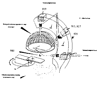
In another embodiment, shown in Figure 9, a delivery pipe 940 is directed
through a series of
magnets 919, 915, 917, 910 and positioned relative to a patient table 930. The
delivery pipe 940
bends as it extends out from an accelerator [not shown], through a shielded
support structure 905,
and into the plurality of delivery heads 935 through the application of
magnets 919, 910, 917, 915.
Operationally, the fixed delivery mechanism can deliver an antiproton beam 920
from multiple
directions without requiring a rotatable gantry. The present embodiment can
therefore direct multiple
antiproton beams 920 to target a single isocenter without requiring the more
complex gantry
structure. While the present embodiment is shown having three delivery points
from which fixed
beams 920 are emitted in the direction of the patient table 930, one of
ordinary skill in the art will
appreciate that, using the appropriate number and type of bending magnets, the
beam line can be
designed to deliver any number of fixed beam configurations directed toward
the patient table.
More specifically, in the illustrated embodiment, the antiproton beam [not
shown] enters into
the structure 905 via the vacuum pipe 940. The vacuum pipe extends through one
135 degree
bending magnet 910, present in line with the delivery pipe 940, and into a
nozzle head 935. When
activated by a control system [not shown], the bending magnet 910 operates to
redirect the
antiproton beam into a second vacuum pipe section 940a, into a first 90 degree
bending magnet 915,
and through a second nozzle head 935, if the 90 degree bending magnet 915 is
activated by a
control system [not shown]. If the 90 degree bending magnet 915 is
unactivated, a first 45 degree
bending magnet 917 is activated to redirect the antiproton beam into and
through a third vacuum pipe
section 940b, into a second 135 degree bending magnet 919, and through a third
nozzle head 935.
The first 45 degree bending magnet 917 and first 90 degree bending magnet 915
are shown in
Figure 9 as being co-located in the same area. Preferably the support
structure 905 is designed to
provide maximum rigidity to the beam line. The weight of the entire gantry is
generally dominated by
the bending magnets 919, 910, 915, 917 and appropriate balancing weights
should be provided in
the structure 905 to insure the gantry does not fall, tip, or otherwise become
unstable.
-20-
CA 02449087 2003-11-27
WO 03/020196 PCT/US02/27796
Operationally, the antiproton beam is deposited in the patient preferably as a
sequence of
sequential pulses, directed from one, or a combination of several, delivery
points defined by nozzles
935. For example, in operation, the 135 degree bending magnet 910 can be
inactivated by a control
system [not shown] to allow an antiproton beam to travel into and through a
nozzle head 935 having
a monitor and range shifter system [not shown], and into the patient [not
shown]. Where a second
beam impingement path is desired, e.g. through a second delivery point, the
135 degree bending
magnet 910 can be activated by a control system [not shown] to allow an
antiproton beam to be
redirected into the first 90 degree bending magnet and, if activated, through
a nozzle head 935
having a monitor and range shifter system [not shown) and into the patient
[not shown]. Where a
third beam impingement path is desired, the first 45 degree bending magnet 917
can be activated by
a control system [not shown] to allow an antiproton beam to be redirected into
the second 135
degree bending magnet and, if activated, through a nozzle head 935 having a
monitor and range
shifter system [not shown] and into the patient [not shown]. A beam
impingement path is the
pathway through the patient that is traveled by an antiproton beam to reach a
target region.
As previously discussed, the number of antiprotons delivered in a single,
directed application
is preferably measured by a beam monitor system positioned in the nozzle 935.
In one embodiment,
the beam monitoring system comprises two monitoring subsystems providing two
independent beam
flux measurements. The two monitoring subsystems are substantially similar to
those described in
relation to the gantry configuration. Similarly, other methods and systems can
be used to monitor the
beam. Once the target number of antiprotons has been delivered into a patient
through a delivery
point, the beam is switched off preferably using a fast kicker-magnet [not
shown] located in the beam
line 940. The fast kicker magnet and associated support structures are
substantially similar to those
described in relation to the gantry configuration.
While a range shifter system and other additional devices can be used to
control and contour
the beam, as discussed in relation to the gantry configuration, in preferred
embodiments a beam is
formed and delivered without the use of degraders or other devices to
physically contour the beam.
The inclusion of barriers, structures, or other materials within the beam line
can cause the unwanted
generation of particles, such as pions, neutrons and gamma rays, that will
dose the patient without
any beneficial medical purpose. To vary dosage levels, it is preferred to use
a variable energy
synchrotron whose energy level can be modified as needed to deliver
antiprotons to the requisite
depth.
In both the gantry and fixed beam configurations, the patient table can be
fixed or moveable.
Where moveable, the patient table can be moved linearly along all three
coordinate planes, x, y, and
-21-
CA 02449087 2003-11-27
WO 03/020196 PCT/US02/27796
z, and rotationally across one or more coordinate planes, as needed. In a
preferred embodiment, the
patient table comprises an elongated rectangular bedding, preferably of
sufficient firmness to
maintain the patient on an even plane surface, that is affixed to a table
frame that preferably has at
least four legs connected, at their bases, to wheels. The frame is preferably
a metallic structure
capable of being tilted to modify the planar position of the bedding without
requiring the concurrent
repositioning of the patient. One of ordinary skill in the art will appreciate
that numerous table
designs can be with in various embodiments, including the one described by
U.S. Patent No.
6,152,599 incorporated herein by reference, without departing from the scope
of the invention.
As further discussed below, a plurality of variables are monitored and
modified to insure that
the proper dosage is being delivered to the proper area within the patient.
The position and quantity
of each dose is determined by the application of an antiproton treatment
protocol and cancer
diagnostic procedure pursuant to one preferred embodiment. Through the
diagnosis and protocol
procedures, dose distributions of various shapes, from uniform to complex, can
be constructed and
delivered by modifying the beam impingement path and location on the patient,
the number of
antiprotons delivered, and the energy of the antiprotons. The antiproton beam,
as delivered, is
rapidly focused on the target area using magnetic fields in the form of a
highly directed pencil beam
positioned in three-dimensional space to insure the dose distribution
substantially matches the
distribution determined theoretically by Bragg Peak calculations.
In one embodiment, the gantry head can be rotated circumferentially relative
to the patient to
allow for the radial movement of the nozzle around the patient. The radial
movement preferably
covers a 180 degree arc above the patient table. Additionally, the patient
table can preferably be
rotated, both vertically and horizontally, to establish an appropriate beam
delivery angle relative to
the gantry head. In operation, singular doses can be delivered, through
specific tissue pathways,
and then terminated. If necessary, the gantry head andlor patient table can
then be moved to
position the patient for a subsequent exposure to an antiproton beam via a
different tissue pathway.
The patient table is preferably not repeatedly rotated in the course of a
treatment to reposition a
patient in order to avoid creating discomfort to the patient and because such
table adjustments often
use far greater time and technician assistance.
Where a target volume is being treated for which multiple doses delivered
adjacent to one
another may be needed it is preferred to use a sweeper magnet to move the
beam, thereby speeding
up adjustment time and obtaining greater precision relative to mechanical
reconfigurations. One
preferred sweeper magnet is a 40 cm long H-type laminated magnet with a 5 cm
pole gap having a
vacuum pipe made of insulator material to avoid eddy effects. Using this type
of sweeper effect, the
-22-
CA 02449087 2003-11-27
WO 03/020196 PCT/US02/27796
beam spot can be moved by about 10 cm. The current in the coils can be chosen
at any desired
value, preferably in the range of +I- 500 amps, which corresponds to a
magnetic field range of +I- 0.8
Tesla. The sweeper magnet is used to perform the most frequent displacements
of the antiproton
beam. For adjacent irradiations requiring only a small change of current in
the sweeper magnet, the
time required to switch the beam off and adjust position should be below about
5 ms. For example,
where a treatment requires 10,000 adjacent spots delivered to a single target
area, total dead time
may be limited to under one minute.
In another embodiment, the dose distributions of various shapes, from uniform
to complex,
can be constructed and delivered by transmitting a beam of antiprotons from a
plurality of different
delivery points fixed in space. Referring back to Figure 9, a single isocenter
980, for example a
tumor located in the brain of a patient, can be targeted via three different
beam pathways using the
three delivery points. Additionally, the patient table can be preferably
rotated, both vertically and
horizontally, to establish an appropriate beam delivery angle relative to the
delivery points. In
operation, singular doses can be delivered, through specific tissue pathways,
and then terminated.
The patient table is preferably not repeatedly rotated in the course of a
treatment.
In preferred antiproton device configurations, an operator workstation
comprising a data
processor, data storage device, and display is in data communication with the
delivery synchrotron,
magnets, and delivery structures, such as the motorized drive gears attached
to the gantry head
and/or to the base of the patient table. The workstation is programmed to
implement the antiproton
treatment protocol developed for the patient. An operator initiates the
workstation and indicates,
through an interface, that the patient is positioned in an initial reference
position. By positioning the
patient in an initial reference position, the workstation can be informed as
to where the patient sits in
space and, therefore, move the gantry head andlor patient table into the
proper position relative to
the patient, for delivering the antiproton beam. Several methods may be used
for positioning,
including, but not limited to those which follow. The initial reference
position can be established, for
example, by placing the patient in a specific position relative to the table
utilizing spine implanted
radio-opaque fiducials which may be implanted in the patient's spinal column
permitting accurate
repositioning of the patient to +/- 1.7 mm. The initial reference position can
also be established by
placing the patient in a specific position relative to the patient table or by
covering the patient with a
sheet comprised of a grid of electronic contacts, each of said contacts being
placed in a specific
position relative to the patient's body. More specifically, in one embodiment,
the grid of electronic
contacts is interconnected by a conductive material and culminates in a single
wire contact extending
into a grid reader. The grid reader sends a signal into and through the
contacts, receives responses
-23-
CA 02449087 2003-11-27
WO 03/020196 PCT/US02/27796
from the contacts, reconstructs the grid structure in space, and transmits the
grid configuration to the
workstation. Operating on assumptions as to how that grid structure is
positioned relative to the
patient's body, the workstation can identify specific points on the patient's
body.
Beginning with the patient in an initial reference position, the workstation
transmits a signal to
the motorized drive gears of the gantry head and/or patient table informing
the drive gears to move
the gantry head and/or patient table into a specific position based upon the
angle and path by which
an initial antiproton dose will be delivered into the patient. Where a fixed
beam line configuration is
being used, only the patient table is manipulated to achieve a specific
position based upon the angle
and path by which an initial antiproton dose will be delivered into the
patient.
With the patient position positioned, the workstation transmits a signal to
the beam monitor
system informing it what amount of antiprotons are to be delivered and also
transmits a signal to the
range shifter system informing it of the dosage depth prior to activating the
delivery synchrotron to
accelerate (or decelerate) and deliver antiprotons of the desired energy level
to the system. In one
embodiment, the delivery system is activated and antiprotons are delivered to
an appropriate depth
and in an appropriate number, as measured and monitored by the range shifter
and beam monitoring
systems respectively. Preferably a plurality of procedures are used in
parallel to monitor the quantity
and depth of dose delivery. For example, a first procedure can comprise the
workstation actively
communicating scanning parameters to the ranger shifter and beam monitoring
systems.
Concurrently, a second procedure can be implemented in which the workstation
passively monitors
the activities of the ranger shifter and beam monitoring systems. Passive
monitoring can be
achieved by detecting the number and location of antiproton annihilations
within the patient, as
further discussed below, and deriving the associated energy level and number
of antiprotons
delivered. The data generated from the second procedure can be compared to the
parameters of the
first procedure to cross check the accuracy of the monitoring and shut down
systems. If a
discrepancy is identified, an automatic shutdown procedure can be effectuated,
where the antiproton
source is turned off, the fast kicker magnet is activated, and/or a solid beam
shutter is deployed.
In a second preferred embodiment, the workstation transmits a signal to the
beam monitoring
system informing it what amount of antiprotons are to be delivered and also
transmits a signal to the
delivery synchrotron to accelerate and deliver antiprotons at a specific,
predefined energy level,
thereby eliminating the need for degraders, range-shifters or other such
mechanism that may
generate unwanted particles, such as pions, neutrons and gamma rays. In one
embodiment, the
delivery system is activated and antiprotons are delivered to an appropriate
depth and in an
-24-
CA 02449087 2003-11-27
WO 03/020196 PCT/US02/27796
appropriate number, as measured and monitored by the beam monitoring system.
Preferably, similar
parallel procedures as discussed above are used to monitor the quantity and
depth of dose delivery.
After the initial antiproton irradiation is completed, the parameters for the
position of beam
scan are preferably modified to enable the irradiation of an entire target
area. Beam repositioning is
preferably performed with the beam switched off. As previously discussed, beam
repositioning can
be effectuated by gantry head movement, table movement, or the use of a
deflecting magnet (such
as a sweeper magnet), depending on the antiproton delivery device being used.
In a gantry configuration, to insure beam focus on the designated target area,
referred to as
the isocenter, it is preferred that the shape of the poles of the 90 degrees
bending magnet and of the
sweeper magnet are designed to produce a displacement of the swept beam which
is substantially
exactly parallel to its direction and to maintain the focusing of the beam at
the isocentric plane
independently of the setting of the sweeper magnet. The shape of the scanned
beam is preferably
sweeper invariant. The precision of the beam is measured at better than 1 mm
for beam parallelism
during scanning (independent of sweeper position), change of beam shape during
scanning
(independent of sweeper position), isocenter stability (independent of gantry
angle), and beam
position reproducibility after a change of the beam energy.
In addition to the above controls, for both the fixed beam and gantry
configurations, a
preferred embodiment additionally has a plurality of backup controls to shut
down or otherwise block
the undesired antiproton irradiation of a patient. Antiproton beams are
automatically controlled by a
fast kicker magnet. In case the kicker magnet fails to activate, another form
of beam shut down
should be immediately deployed, such as the switching off of the antiproton
accelerator. Alternatively
or in combination, a mechanical beam shutter can be used to block the patient
from antiproton
exposure.
In a preferred embodiment of the antiproton gantry device, shown in Figure
10a, the gantry
1050a is combined with a plurality of detectors 1060a that enable the imaging
of certain patient tissue
areas subjected to antiproton radiation. A patient [not shown] is positioned
on a patient table 1065a.
Antiproton beam 1070a enters gantry 1050a and is directed toward a target
volume 1075a. As
previously discussed, a plurality of different configurations can be used to
direct beam 1070a toward
volume 1075a, and the configuration shown in Figure 10a is merely an exemplary
embodiment. The
detectors 1060a are arrayed in a configuration that avoids obstructing beam
1070a while
concurrently exposing the detector array 1060a to antiproton annihilation
emissions that can be used
to conduct real-time imaging, as further discussed below.
-25-
CA 02449087 2003-11-27
WO 03/020196 PCT/US02/27796
Similarly, in a preferred embodiment of the fixed beam device, shown in Figure
10b, the fixed
beam system 1050b is combined with a plurality of detectors 1060b, 1062b that
enable the imaging
of certain patient tissue areas subjected to antiproton radiation. A patient
[not shown] is positioned on
a rotatable patient table 1065b. An antiproton beam line [not shown] enters
gantry 1050b and is
directed by action of a plurality of beam magnets toward a target volume
1075b. As previously
discussed, a plurality of different configurations 1070b can be used to direct
an incoming beam
toward volume 1075b, and the configuration shown in Figure 10b is merely an
exemplary
embodiment. The detectors 1060b, 1062b are arrayed in a substantially
spherical upper detector
configuration 1060b and substantially spherical lower detector configuration
1062b that avoids
obstructing the plurality of beams 1070b while concurrently exposing the
detector array 1060b, 1062b
to antiproton annihilation emissions that can be used to conduct real-time
imaging, as further
discussed below.
The detectors are preferably made of a high atomic number, high-density
material capable of
interacting with gamma rays to create an electromagnetic shower. The shower
energy is
substantially contained inside a volume, each having a radius of two times the
Moliere radius and
having a length of approximately 20 Xo radiation lengths. In one embodiment,
the detector assembly
is supported by a carriage, which can be rotated around the target axis
running on a bent, nearly
semicircular track. The detector may also be moved radially by a screw
arrangement to a specified
range of distance from the target to the crystal face.
In specialized high-energy physics experimentation, energetic charged
particles and gamma
rays, which are produced when an antiproton annihilates at rest on a proton
and which then move
radially away from that annihilation site, are detected and tracked back to a
common point, referred
to as the vertex. The process of tracking the energetic charged particles and
gamma rays back to
their common point of origination is referred to as vertex reconstruction. To
effectively perform vertex
reconstruction, the detectors used are preferably, designed to detect
particles and/or radiation that
have the highest likelihood of escaping a patient's body with the least amount
of scattering or other
perturbations that can complicate determinations of where the particle andlor
radiation had
originated.
Assuming an antiproton beam penetrates and stops at the center of a sphere of
water having
a 15 centimeter (cm) radius and annihilates, only those particles having
energy greater than given by
the stopping range of 15 cm of water will escape the sphere and be capable of
being detected.
Relative to charged kaons, neutral kaons (short), and neutral kaons (long),
muons and charged pious
have the highest probability of escaping the 15 cm sphere. Neutral pious decay
in less than 0.025
-26-
CA 02449087 2003-11-27
WO 03/020196 PCT/US02/27796
microns from the point of annihilation into a pair of gamma rays that escape
with energy carried by
the pion.
Charged pious escaping material undergo substantial amounts of scattering,
thereby
increasing the complexity of vertex reconstruction. When being emitted out of
15 cm of water,
charged pious having moments less than about 160 MeVlc stop in the water and
are not detected,
while charged pious having moments in excess of about 150 MeV/c scatter
laterally, relative to the
direction of the linearly formed track, by a root mean square of approximately
7 millimeters. The
change of direction is dependent upon the particle's momentum, the particle's
charge, and the
material through which the particle is passing.
Although lateral displacement improves as particle momentum increases, even at
the higher
moments, pion lateral scattering is at or around 1.5 mm, thereby limiting
imaging precision to plus or
minus 1.5 mm. This limitation decreases as the site of annihilation approaches
the surface.
In a preferred embodiment, vertex reconstruction is performed using neutral
pion decay
gamma radiation. Unlike charged pious, gamma rays have a high probability of
escaping a material
body without undergoing substantial interactions which cause scattering and
skew vertex
reconstruction calculations. Further, the pair of gamma rays emitted can be
traced back to the point
where the neutral pion decayed and, because neutral pious decay within 0.025
microns of the
annihilation point, can provide a more accurate representation of where the
annihilation occurred. In
a typical annihilation event, the mean number of gammas emitted for each
antiproton annihilation
event is four (two for each neutral pion), and can be as high as 10.
Operationally, vertex reconstruction is performed by relying on the detection
of multiple
points along the shower axis and the use of those multiple points to generate
a vector localizing a
common origination area. It is preferred that any heavy inorganic
scintillators used to detect gamma
rays have one or more of certain desired characteristics, including, high
stopping power to maximize
the probability of complete absorption of the incident energy, high timing
resolution, high energy
resolution, minimum dead time, wavelengths of emission that match with the
spectral response of the
photodetectors, mechanical ruggedness, radiation hardness, chemical stability
in normal atmospheric
conditions, and reasonable cost. Existing heavy scintillators meet certain of
these criteria, including
high luminous efficiency measured in photonsIMeV (Nal(TI) and Csl(TI)), high
density/high atomic
number (BGO), short Moliere radius (BGO and CeF3), high initial photon
intensity measured in
photons/MeVlns with high timing resolution (BaF2), and high luminous
efficiency and wavelength
suitable for silicon photodiodes (Csl(TI) and CdW04). Proper selection of a
detector provides for a
further benefit of gamma ray shower detection over charged particle detection
is speed. Shower
-27-
CA 02449087 2003-11-27
WO 03/020196 PCT/US02/27796
detection can be done using a fast scintillator, less than 15 nanoseconds,
thereby allowing a faster
response than charged particle tracking.
Tungsten (W), sodium iodide doped with thallium (Nal(TI)), and lead tungstate
(PbW04) are
three materials which can preferably be used for shower detection. Sodium
iodide activated by
thallium is a well-known material used for scintillation applications. Nal(TI)
has a high luminescence
efficiency and. spectroscopic performance with minimal significant self
absorption of the scintillated
light. Lead tungstate is a highly efficient and fast scintillator with one of
the shortest radiation lengths
and Moliere radii among the known scintillators, satisfactory light yield for
this energy range, and high
radiation stability. Radiation lengths for Nal(TI), Csl(TI), PbWOa, BGO,
Tungsten (W), and Iridium (1r)
are 2.59 cm, 1.86 cm, 0.89 cm, 1.12 cm, 0.323 cm, and 0.27 cm, respectively.
The Moliere Radius
for Nal(TI), PbWOa, and W are 4.5 cm, 2.2 cm, and 0.8 cm respectively. The
density for Nal(TI),
Csl(TI), PbWOa, BGO, Tungsten (W), and Iridium (1r) are 3.67, 4.53, 8.28,
7.13, 19.4, and 22.4,
respectively. The decay time for Nal(TI) is 250 ns while for PbWOa it is
between 5 and 15 ns. For
BGO and Csl(TI), the decay times are 300 ns and 0.917.0 Ns. The light output
for Nal(TI), Csl(TI),
PbWOa, and BGO are 1.0, 0.85, 0.01, and 0.15, respectively. Another material
usable for the present
application includes uranium, which has the requisite Moliere radius and
material density. However,
because it is not actively sensitive to the shower, it will have to be
combined with an active
scintillator. Using layers of tungsten, for example, in combination with
uranium can provide a
satisfactory detector device.
When using tungsten, one can employ the sensing element in a matrix, such as a
3.23x3.23x3.23 mm3 matrix, or in a crossed 3.23x3.23x200 mm3 hodoscope plastic
scintillator array
to sample the shower's charged particles passing between sandwich plates.
Locating the vertex with
a precision on the order of 500 microns is possible using these techniques.
When using lead
tungstate, one can use a 9x9x9 mm3 matrix or a crossed 9x9x200 mm3 hodoscope
sensor array. The
radiation length of lead tungstate is 2.7 times greater than tungsten.
Although the preferred
approach is dependent upon a plurality of technical, as well as economic
considerations, one
consideration favoring the smaller shower localization of tungsten over lead
tungstate is its ability to
separate the gamma pair of the neutral pion decay. As the moments of neutral
pions increase, lead
tungstate loses efficiency at separating decay gammas relative to a tungsten
shower detector.
Preferably, the detectors are surrounded by a shielding structure to isolate
the detectors from
the surrounding environment. In one implementation, Nal(TI) crystals are
surrounded by an active
plastic shield, a passive LiH shield, and a low activity thick lead shield
which, in combination, have a
-28-
CA 02449087 2003-11-27
WO 03/020196 PCT/US02/27796
cosmic rejection efficiency around 98%. Further, the detectors are preferably
supported by a
carriage structure to enable efficient rotation around a target axis.
Referring to Figures 10c through 10r, a plurality of detector configurations
is shown particular
to each calorimeter imager material used. Imagers for Nal and BGO are not
shown because Nal is
similar to Csl and BGO is similar to PbWOa. Figures 10c and 104 show preferred
detector
configurations for brain imaging using PbWOa as the calorimeter imager
material. In Figures 10c and
104, a beam pipe 10834 delivers an antiproton beam to a predesignated area
within the patient's
brain 10854. The beam direction and imager 10864 are fixed relative to each
other. The patient is
positioned on a table 10844. In use, a patient will first be positioned
between detector elements, as
shown in Figures 10a and 10b, and then the elements are assembled into a
portion of a spherical
shell sharing the center with the annihilation region, appropriately
accounting for straggling and
multiple straggling limits. Sufficient resolution can be achieved by having
the calorimeter elements
point approximately to the annihilation site. When annihilation occurs, a
plurality of gamma rays
10874 are emitted, due to the decay of neutral pious generated in the course
of the annihilation,
which extend from the target region 10854 with an opening angle of
approximately 30 degrees taken
from the point of annihilation. The gamma radiation may have an opening angle
less than 30
degrees, but not more than 30 degrees relative to each other. In one
embodiment, the imager length
(20 radiation lengths) is 17.8 cm. The inner radius size is 0.89 cm x 0.89 cm
with the outer radius
size being 1.8 cm x 1.8 cm. The maximum mass is approximately 1556 kg.
Figures 10g and 10h, 10k and 101, and 10o and 10p show a similar detector
configuration
using Csl(TI), Ir, and W as calorimeter elements, respectively. A beam pipe
1083h, 10831, 1083p
delivers an antiproton beam to a predesignated area within the patient's brain
1085h, 10851, 1085p.
The beam direction and imager 1086h, 10861, 1086p are fixed relative to each
other at least during
detection. The patient is positioned on a table 1084h, 10841, 1084p. In use, a
patient will first be
positioned between detector elements, as shown in Figures 10a and 10b, and
then the elements
assembled into a portion of a spherical shell sharing the center with the
annihilation region,
appropriately accounting for straggling and multiple straggling limits.
Sufficient resolution can be
achieved by having the calorimeter elements point approximately to the
annihilation site. When
annihilation occurs, a plurality of gamma rays 1087h, 10871, 1087p are
emitted, due to the decay of
neutral pious generated in the course of the annihilation, which extend from
the target region 1085h,
10851, 1085p with an opening angle of approximately 30 degrees taken from the
point of annihilation.
The gamma radiation may have an opening angle less than 30 degrees, but not
more than 30
degrees relative to each other. In one embodiment, or Csl(TI), the imager
length (20 radiation
_29_
CA 02449087 2003-11-27
WO 03/020196 PCT/US02/27796
lengths) is 37.2 cm, the inner radius size is 1.86 cm x 1.86 cm, the outer
radius size is 5.7 cm x 5.7
cm and the maximum mass is approximately 3172 kg; for Ir, the imager length
(20 radiation lengths)
is 10.8 cm, the inner radius size is 2.7 cm x 2.7 cm, and the maximum mass is
approximately 1073
kg; for W, the imager length (20 radiation lengths) is 12.92 cm, the inner
radius size is 0.32 cm x
0.32 cm, the outer radius size is 5.7 cm x 5.7 cm, and the maximum mass is
approximately 1137 kg;
and for BGO (not shown), the imager length (20 radiation lengths) is 22.4 cm
and the inner radius
size is 1.12 cm x 1.12 cm. For non scintillators, such as Ir and W, preferably
approximately 50% of
the space in the length is dedicated to a plastic scintillator read out of the
shower in a hodoscope's
geometry.
Figures 10e and 10f show detector configurations for torso imaging using PbW04
as the
calorimeter imager material. A beam pipe 1083f delivers an antiproton beam to
a predesignated area
within the patient's torso 1085f. The beam direction and imager 1086f are
preferably fixed relative to
each other. The patient is positioned on a table 1084f. As previously stated,
in use, a patient will first
be positioned between detector elements, as shown in Figures 10a and 10b, and
then the elements
are assembled into a portion of a spherical shell sharing the center with the
annihilation region,
appropriately accounting for straggling and multiple straggling limits.
Sufficient resolution may be
achieved when the calorimeter elements point approximately to the annihilation
site. When
annihilation occurs, a plurality of gamma rays 1087f are emitted, due to the
decay of neutral pions
generated in the course of the annihilation, which extend from the target
region 1085f with an
opening angle of approximately 30 degrees taken from the point of
annihilation. The gamma
radiation may have an opening angle less than 30 degrees, but not more than 30
degrees relative to
each other. In one embodiment, the imager length (20 radiation lengths) is
17.8 cm, the inner radius
size is 0.89 cm x 0.89 cm, the outer radius size is 1.8 cm x 1.8 cm, and the
maximum mass is
approximately 3618 kg.
Figures 10i and 10j, 10m and 10n, and 10q and 10r show a similar detector
configuration
using Csl(TI), Ir, and W as calorimeter elements, respectively. A beam pipe
1083j, 1083n, 1083r
delivers an antiproton beam to a predesignated area within the patient's torso
1085j, 1085n, 1085r.
The beam direction and imager 1086j, 1086n, 1086r are fixed relative to each
other. The patient is
positioned on a table 1084j, 1084n, 1084r. In use, a patient will first be
positioned between detector
elements, as shown in Figures 10a and 10b, and then the elements are assembled
into a portion of a
spherical shell sharing the center with the annihilation region, appropriately
accounting for straggling
and multiple straggling limits. Sufficient resolution may be achieved when the
calorimeter elements
point approximately to the annihilation site. When annihilation occurs, a
plurality of gamma rays
-30-
CA 02449087 2003-11-27
WO 03/020196 PCT/US02/27796
1087j, 1087n, 1087r are emitted, due to the decay of neutral pions generated
in the course of the
annihilation, which extend from the target region 1085j, 1085n, 1085r with an
opening angle of
approximately 30 degrees taken from the point of annihilation. The gamma
radiation may have an
opening angle less than 30 degrees, but not more than 30 degrees relative to
each other. In one
embodiment, for Csl(TI), the imager length (20 radiation lengths) is 37.2 cm,
the inner radius size is
1.86 cm x 1.86 cm, the outer radius size is 3.8 cm x 3.8 cm and the maximum
mass is approximately
6328 kg; for Ir, the imager length (20 radiation lengths) is 10.8 cm, the
inner radius size is 2.7 cm x
2.7 cm, and the maximum mass is approximately 2500 kg; for W, the imager
length (20 radiation
lengths) is 12.92 cm, the inner radius size is 0.32 cm x 0.32 cm, and the
maximum mass is
approximately 2618 kg. For non scintillators, such as Ir and W, approximately
50% of the space in
the length is dedicated to a plastic scintillator read out of the shower in a
hodoscope's geometry.
With respect to performance, the angular acceptance achieved in the crystal
barrel
spectrometer is approximately 6 degrees (100mrad). Without adjacent cell
interpolation, the
calorimeter imager materials, operating in the aforementioned brain imager and
torso imager
configurations, have the following degrees of angular acceptance: for brain
imager configurations, the
angular acceptance of Csl(TI), PbWOa, BGO, W, and Ir is 103 mrad, 49 mrad, 62
mrad, 18 mrad, and
15 mrad respectively. For torso imager configurations, the angular acceptance
of Csl(TI), PbWOa,
BGO, W, and Ir is 53 mrad, 25 mrad, 32 mrad, 9.2 mrad, and 7.2 mrad
respectively. If interpolation
is implemented, a 300% gain in resolution may be achieved for certain
calorimeter imager materials
operating in certain configurations, upwards of a 1000% gain in resolution for
materials such as
PbWOa. The highest angular resolution can be achieved with W or Ir, although
Ir may be expensive
to use.
Dia4nosis and Treatment Strategy
Cancer is diagnosed using a variety of methods, a few of which are discussed
herein. A
patient suspected to have cancer maybe imaged using x-ray, CT, MRI,
radioactively labeled tracer
uptake, thermography, ultra sound and PET scanning. A medical practitioner
skilled in the art of
cancer diagnosis will understand how to use these technologies to yield an
image that can indicate
the presence of an unusual mass, and possibly, cancer.
In one preferred embodiment, a patient is treated in a medical facility in
which antiproton
radiation therapy can be delivered. A schematic plan layout of an exemplary
medical facility is
provided in Figure 11. The exemplary medical facility 1100 comprises a
plurality of areas dedicated
to standard medical facility functions, including examination rooms,
maintenance areas, reception
-31-
CA 02449087 2003-11-27
WO 03/020196 PCT/US02/27796
areas, waiting rooms, janitorial rooms, utilities, staircases 1187, elevators
1180, a lobby 1190, and
staff areas, such as staff offices, meeting rooms, lunch areas, patient record
keeping. Preferably,
sizeable rooms internal to the facility 1185 are used for staff offices and/or
examination rooms, rooms
adjacent to the treatment area 1160 are used for patient preparation and
changing, larger rooms
1170 are used for meeting or waiting areas, the smaller rooms 1175 are used
for utilities or janitorial
purposes, and the other rooms 1165 are used for storing patient records,
secretarial functions, lunch
rooms, smaller staff offices, and at least one dosimetry room and health
physics room.
The illustrated medical facility 1100 further comprises areas specialized for
the delivery of
antiproton therapy. A plurality of treatment rooms 1103 surrounded by heavy
shielding 1135 is
located in the back of the facility 1100. A control room 1130 is integrally
provided with each
treatment room 1103. In one room 1003, a MRI 1145 is provided proximate to a
CT simulator 1155.
In a set of second rooms 1103, a patient table 1120 is situated proximate to a
delivery point 1115
integrally attached to a delivery device, such as a fixed beam or gantry
device. Additionally, a
treatment chair 1140 and an array of detectors [not shown] can also be
situated proximate to the
delivery point 1115 and patient table 1120. In a third room 1103, a
calibration system 1125 is
provided that enables an operator to calibrate the operation of the beam
transport system 1105 and
delivery synchrotron [not shown]. Operationally, an antiproton beam is caused
to travel through the
beam transport system 1105 and bend by force of a plurality of bending magnets
1110, which are
housed in a support structure.
Depending upon a centralized schedule of operation, one of the plurality of
beam lines
directed into specific treatment rooms 1103 will be active and delivering a
pre-designated dose of
antiprotons to a patient 1124 positioned on a patient table 1120. Antiprotons
traveling through the
beam transport 1105 will be directed into the appropriate beam pipe that feeds
a particular treatment
room 1103. The beam pipe as shown terminates in a gantry or
verticallhorizontal beamline.
Referring to Figure 11a, an exemplary beam line 1105a integrated with a
medical facility 1100a is
shown in the context of delivering an antiproton beam to a fixed beam
antiproton delivery device.
Two fixed beams 1125a are generated, focused on a target volume 1130a, by
action of a plurality of
bending magnets selectively bending antiprotons traveling through a beam pipe
1140a. A person
familiar with high-energy physics will understand how to produce, collect,
cool, decelerate and extract
antiprotons through the application of vacuums pumps, magnets, radio-frequency
cavities, high
voltage instruments and electronic circuits. Antiprotons circulate inside
vacuum pipes in order to
avoid contact with matter with which they annihilate. The vacuum should be
optimal, therefore
several vacuum pumps, which extract air, are placed around the pipe. The
magnets used include
-32-
CA 02449087 2003-11-27
WO 03/020196 PCT/US02/27796
dipoles, which serve to change the direction of antiproton movement and insure
they stay within the
circular track, and quadrupoles, which are used as lenses or focusing magnets
to insure that
antiproton beam size is smaller than the vacuum pipe size. Electric fields are
used to modify
antiproton energy levels and are provided for by radio-frequency cavities that
produce high voltages
synchronized with the rotation of antiprotons around the ring. While the
medical facility 1100, 1100a
has been described in relation to a specific design and layout, one of
ordinary skill in the art will
appreciate~that other space configurations can be used, depending upon the
particular conditions of
the location and the needs of the facility.
A patient is positioned in a diagnosis area that can have one of, or a
combination of, several
diagnostic devices. One diagnostic device can include a magnetic resonance
imaging (MRI) scan in
which a patient is subjected to an external, uniform magnetic field and
radiofrequency energy that
excites protons in the patient's body and subsequently produces signals with
amplitudes dependent
on relaxation characteristics and spin density. Abnormalities can be detected
by identifying unusual
signals that indicate a particular region has a different proton density than
normally expected.
Another diagnostic device that can reveal tissue structure and therefore
identify unusual masses is
computer tomography (CT) scanning. CT scans are performed by passing x-rays
through a patient,
at a large number of angles, by rotating the x-ray source around the patient.
A plurality of detector
arrays, located opposite the x-ray source, collect the transmission projection
data in the form of
various data points. The data points are synthesized into a tomographic image,
or imaged slice, of a
patient. The variation in transmission data is indicative of tissue density
and can be used to identify
unusual masses in the body.
A third possible diagnostic device is a positron emission tomography (PET)
scan in which the
patient is administered, through an intravenous injection, a positron-emitting
radioactive substance
comprising a form of glucose that reacts with tissues in the body, in
proportion to metabolic activity.
By measuring the different amounts of positrons released by healthy and
cancerous tissues, a
computer creates an image reflective of the biological activity occurring
within the patient. Because
cells from many cancers have a higher affinity for certain positron-emitting
radioactive substances,
such as F~8 labeled glucose, the tumor area may be imaged. PET scans can be
combined with x-ray
based scans and MRI scans to confirm that an unusual structure may, in fact,
be cancerous. More
specifically, PET scans can be overlaid onto, or combined with, MRI or CT
images to generate an
integrated image that shows tissue structure associated with metabolic
activity.
Output from one or more of the aforementioned diagnostic devices can be used
by a medical
practitioner, including technicians, nurses, radiologists, oncologists, and
other medical professionals,
-33-
CA 02449087 2003-11-27
WO 03/020196 PCT/US02/27796
to determine whether the patient has cancer and, if so, the location, extent,
and stage of the cancer.
In a preferred embodiment, shown in Figure 12, at least one of the diagnostic
scans from the PET
scan 1305, MRI scan 1310, andlor CT scan 1315, is stored in an operator
workstation 1320,
transmitted to an antiproton treatment protocol station 1325, and used to
assist in the development of
an antiproton based treatment regimen. Alternatively, only the data
representing key treatment
parameters may be transmitted to the antiproton treatment protocol station.
Referring back to Figure 11, the patient, once imaged, is taken to an
antiproton treatment
protocol station. The station can be co-located with the diagnostic machinery,
placed in a separate
office within the same building, or located in a completely separate facility.
The schematic
representation in Figure 11 is provided for example purposes only.
Having identified and quantified the tumor location, a treatment protocol
using antiproton
radiation is developed. In a preferred approach, data representing the tumor
size and location is
transmitted from imaging technologies, as previously described, to an
antiproton treatment protocol
station. The treatment protocol station applies a set of analyses to determine
the amount of
antiprotons, antiproton energy sufficient for treatment, and preferred
delivery pathways and
communicates that protocol to an antiproton radiating and imaging station, as
previously described.
The antiproton treatment protocol station is in data communication with the
imaging station used or,
alternatively, is capable of receiving data stored on media, such as a disk or
CD-ROM.
In one embodiment, shown in Figure 12, the treatment protocol station
comprises a display
1350, print-out device 1355, storage device 1360, modem or network control
card 1365, and
processor 1370 capable of communicating with the display (any type of
monitor), print out device
(any type of printer), storage device, and modem/network controller and of
implementing a plurality of
instruction sets for determining the amount of antiprotons, antiproton energy
sufficient for treatment,
and preferred delivery pathways given a tumor size and location. The amount of
antiproton radiation
needed to terminate a mass is calculated, along with the amount of energy
needed to deliver an
antiproton to the mass depth. Using equations to determine the amount of
energy deposited in
collateral tissue and the residual energy plus annihilation event radiation
effects, such as caused by
the emission of particles like alpha particles, the energy deposited in the
mass, along with the lateral
spreads and Bragg Peak contours, can be determined. Once done, an energy
deposition profile can
be generated that covers the entire mass with sufficient antiproton induced
radiation by summing
multiple Bragg Peaks, assuming a plurality of spot scans performed at varying
depths within the
tumor region. The amount and energy level of antiprotons, for each location to
be irradiated, defines
the protocol, which is then sent to the antiproton radiation and imaging
device, as previously
-34-
CA 02449087 2003-11-27
WO 03/020196 PCT/US02/27796
discussed. During operation, a preferred embodiment monitors, through a beam
monitoring system
and range shifter or a delivery synchrotron, the actual dosage being delivered
to insure it correlates
with the desired calculated dosage. To the extent a range shifter is used,
antiproton losses, caused
by the degradation process, need to be calculated and incorporated into all
beam monitoring
calculations to insure accurate determination of actual antiprotons delivered
to the patient.
As an example, a patient is diagnosed with a 1 cubic centimeter (cc) tumor
located 10
centimeters below the skin surface. The diagnosis occurs through a combination
of MRI and PET
scans, which indicates a mass having a high metabolic rate in the patient's
chest cavity. Using the
location and tumor size data, the amount of antiprotons to be used to
annihilate the target region is
determined. One preferred method of determining the amount of antiprotons
needed is by assuming
the density of tissue to be around 1 gram per cc, assuming 500 rads will be
sufficient to terminate the
cancerous cells, and equating the relative biological effect (RBE) of
antiproton radiation in the target
volume to that of heavy ions having a 30 MeV recoil (RBE=5). This reflects the
fact that at least one
30 MeV recoil heavy ion is produced for each antiproton annihilation event.
Because 500 rads is
approximately equivalent to 30 x 109 MeV per gram, the total number of
antiprotons needed to deliver
500 rads is 109. It should be noted that, if the RBE of the chosen radiation
were lower, as with
photons, a greater amount of radiation, as measured in rads, will have to be
delivered to the same
target region in order to terminate the cancerous cells. For example, photon
radiation has a RBE of
1, thereby requiring 2500 rads to have the same cell terminating effect as
antiprotons, which, when
equated to heavy ions, has a RBE of 5.
To determine the amount of energy 109 antiprotons should have in order to
reach 10 cm
below a surface, one can use a TRIM calculation, as found in Zeigler J.F.,
Biersack J.P., and Littmark
U., "Stopping and Range of Ions in Solids,", Vol.1, 1985 (Pergamon Press, NY).
Applying a TRIM
calculation demonstrates that an antiproton beam energy of approximately 108
MeV will achieve an
end-of-range position that is 10cm below the surface in a patient. Given that,
like protons,
antiprotons are low linear energy transfer particles and that only a small
portion of antiprotons
annihilate prior to reaching the target region, approximately 30 MeV of the
108 MeV is deposited in
the target region, while 78 MeV is deposited in collateral tissue. Assuming
the volume of collateral
tissue between the skin and target area is 9 cc (1 cm x 1 cm x 9 cm), the
damage inflicted by
traveling antiprotons can be defined by a RBE of 1.2 (20% greater than
protons), and damage is
uniformly spread across the collateral tissue, the collateral damage is equal
to approximately 168
rads ((1.2 x 78 MeVlantiproton x 109 antiprotons)19 cc), which is tolerable
and therefore does not
require a multiple pathway dosage profile (although it may be done if
desired). Therefore, combined
-35-
CA 02449087 2003-11-27
WO 03/020196 PCT/US02/27796
the protocol produces a recommended treatment plan: one exposure of 109
antiprotons having an
energy of 108 MeV.
In dealing with tumors having a volume greater than 1 cc, multiple doses,
spread across a
region, may be preferred to minimize collateral tissue damage in any single
location. For example,
some lung and prostate cancers are intermediate sized tumors and can range, on
average, around
150 cc and 35 cc with average surface depths of 12 and 6 cm, respectively.
Head and neck tumors
may be irregularly shaped and in some embodiments, multiple doses may be
utilized to cover the
target region.
In either case, the high degree of localization provided by antiproton
radiation therapy
preferably allows for one or more of the following: (1) the termination of
cancer cells with minimal
fractionation requirements; (2) producing tumor cell injury by causing
numerous double strand DNA
breaks and by inducing cell membrane injury of trans-membranal surface
proteins, i.e. by interfering
with EGFR ( epidermal growth factor receptor ) and VEGF (vascular endothelial
growth factor
receptor) transduction signaling; (3) sparing injury to tumor-adjacent antigen
serving macrophage
dendritic cells, which facilitate tumor lysis by T-cells in the tumor
microenvironment; (4) avoiding
injury in the tumor microenvironment to lymphokine activated killer (LAK) T-
cells, which become
effector cells causing tumor lysis when served with tumor antigens by
dendritic cells, an important
immunologic activity in facilitating the body's natural defenses against tumor
growth; (5) permitting
the progeny of tumor sensitized effector LAK T-cells to provide cell lysis of
distant microscopic tumor
metastatic implants; and (6) causing less hematopoietic injury, which is
common in photon regimens,
since the bone marrow will be spared the effects of radiation exit dose and
dose fall off. This is of
particular importance with the increasing use of simultaneous chemotherapy-
photon radiation therapy
protocols in a variety of cancers, which often lead to blood count depressions
that necessitate
interruption of treatment. The highly conformal nature of antiproton radiation
will avoid this adverse
result.
In one preferred embodiment, the treatment protocol station will have a
computer-
implemented software program capable of taking the requisite input data,
namely tumor size and
location, and, as shown in Figure 13, outputting impact graphs superimposed on
the scanned images
of the patient's tumor, as generated from conventional therapies. A tumor body
1305 is identified and
located relative to a patient's anatomy. The tumor body 1305 is positioned in
an area within the
patient's brain 1310. A plurality of delineated antiproton dosage regions 1315
are defined relative to
the tumor body 1305. The dose regions 1315 can be defined in numerous ways,
including by
percent dosage relative to calculated dose requirements or by absolute dosage
amounts, with higher
-36-
CA 02449087 2003-11-27
WO 03/020196 PCT/US02/27796
dose regions generally being centered within the plurality of dose regions
1315 and lower dose
regions extending to the periphery.
It should be noted that, because of the ability to precisely deliver high
amounts of energy into
an area without high accompanying collateral damage, a medical practitioner
does not need to cover
an entire mass with antiproton radiation, but rather, can selectively target
highly sensitive areas
within a tumor volume to achieve tumor mass destruction with minimal
radiation. For example,
because tumors rely on fragile blood vessel networks to fuel their rapid
growth, it may be possible to
kill an entire tumor mass through the directed application of antiproton
radiation on areas responsible
for providing primary blood supply. By irradiating critical blood vessels one
can induce angiolysis,
thereby shutting down essential blood supply to a tumor. Similarly, tumors may
be killed using
antiproton radiation by causing blood vessel swelling such as in AVM's
(arterio-venous
malformations) which will result in the eventual cut off of a tumor's blood
supply. Tumors may also be
killed by biologically isolating them through the application of antiproton
radiation circumferentially
and sparing normal structures interior to the tumor, such as the urethra
coursing through a malignant
prostate gland. Circumferential antiproton radiation may also induce fibrosis
around a tumor mass
isolating the tumor and causing it to necrose.
It should further be noted that a substantial number of repeated treatments is
not required.
Treatment fractionation is required in conventional therapies because of the
inability to drive high
enough radiation levels to target tissue without causing high collateral
damage. Lower target
radiation levels, though sufficient to kill dividing cells, are not sufficient
to kill resting cells. As a
result, multiple treatments have to be applied in order to kill the target
cancer cells, and because of
the rapid dividing nature of cancer cells, they are more impacted than the
collateral cells which have
time to repair after radiation exposure. The preferred methods and systems
disclosed herein enable
the delivery of high radiation levels in target tissue, thereby killing both
resting and dividing cancer
cells, without causing unacceptable levels of damage to healthy tissue.
Optionally, a patient may also be imaged using a PET scan after the antiproton
radiation
exposure is completed. Typically, to perform PET scanning, a patient is
administered a glucose-
tagged radioactive substance that decays inside the body and, in the process,
releases positrons
which, when detected, can be used to generate an image. Conventional PET scans
are limited by
the need to have the patient, PET imaging station, and radioactive isotope
source (the
radiopharmaceutical of appropriate activity) all proximate to each other.
Specifically, PET applications rely on the use of biologically active
radiopharmaceuticals
where radioactive isotopes in the radiopharmaceutical emit positrons. These
isotopes are typically
-37-
CA 02449087 2003-11-27
WO 03/020196 PCT/US02/27796
generated through the use of synchrotrons, such as the RDS cyclotron,
manufactured by Siemens,
which is a frequently used PET device. It incorporates a computer terminal to
control the flow of
production, and a biosynthesizer unit to carry out the chemical synthesis of
radiopharmaceuticals.
Using the synchrotron, a stream of charged particles, such as protons or
deuterons, bombard a
collection of stable, sometimes enriched, isotopes and interact with a subset
of those isotopes.
Three nuclear reactions are commonly used for the production of C-11 and F-18,
the most common
PET isotopes. These reactions are: ~4N(p,a)~~C, in which the interaction of
~4N with a proton is then
followed by the emission of an alpha particle, resulting in ~~C, ~80(p,n)~BF,
in which the interaction of
yap with a proton is then followed by the emission of a neutron, resulting in
~$F, and 2~Ne(d, a)~BF, in
which the interaction of 2~Ne with a deuteron is then followed by the emission
of an alpha particle,
resulting in ~BF. Radiopharmaceuticals, made from these radioactive isotopes,
are then introduced
into a patient's body where the decay of the isotope is monitored.
While many radioactive isotopes can be produced in the cyclotron, the isotopes
produced
are preferably amenable to human PET use and, therefore; (1 ) are capable of
emitting positrons
when they undergo radioactive decay and transform from an unstable isotope
into a stable one. (2)
Because such isotopes tend to emit positrons relatively quickly, the isotope
half-life is preferably long
enough to allow for a patient to be administered the substance and placed in a
position to be
scanned,. Furthermore, it is preferred that the isotopes are readily
incorporated into a useful radio-
pharmaceutical by chemical synthesis. The most commonly generated isotopes
include carbon-11
(half-life 20 minutes), nitrogen-13 (half-life 10 minutes), oxygen-15 (half-
life 2 minutes), and fluorine-
18 (half-life 110 minutes). Because of these short half-lives, some PET
installations have cyclotrons
proximate to the PET machine. For example, at the University of Iowa, a
compact medical cyclotron
is used to generate high energy protons or deuterons by forcing the particles
to traverse the cyclotron
several hundred times and, during each orbit, receive about 90 keV of energy.
When the energies
are high enough, the particles are removed through electrostatic deflection
and are made to impinge
upon small volume hollow metallic cylinders filled with a non-radioactive gas
~or liquid, causing
nuclear reactions to take place within the cylinder and generating the
appropriate isotopes.
For certain applications, some of the preferred methods and systems disclosed
herein
complement the use of PET-specific cyclotron and biosynthesizing stations to
perform a PET scan.
Conventional PET systems are used to measure and study biological functions,
such as glucose
uptake. In one embodiment, PET administration is used in combination with
certain preferred
methods and systems disclosed herein to conduct PET scans. A patient is
administered a PET-
isotope labeled glucose molecule in order to identify enhanced glucose uptake
areas in the body.
-38-
CA 02449087 2003-11-27
WO 03/020196 PCT/US02/27796
The detector array incorporated into the antiproton delivery device can be
used to monitor resulting
decay, thereby repurposing detectors used for antiproton annihilation tracking
and measurement for
PET scanning. When treating with antiprotons, a medical practitioner can then
directly compare PET
scanning results with antiproton treatment results. One of ordinary skill in
the art will appreciate that,
in addition to the aforementioned characteristics, the antiproton delivery
detector system for this
embodiment should be sufficiently sensitive to differentiate between the decay
generated by
increased uptake areas of the radiopharmaceutical and the decay generated by
the general uptake of
the radiopharmaceutical throughout the body.
Additionally, one preferred embodiment enables the in-situ generation of PET
isotopes. The
exposure of human tissue to antiproton radiation generates a plurality of
unstable isotopes, including,
for example, oxygen-15, that are radioactive and emit a positron as a decay
product. More
specifically, when introduced into a target region, antiproton interactions
generate oxygen-15,
nitrogen-13, and carbon-11 as by-products. After the appropriate period of
time (depending on the
half life of the isotope), the generated isotopes decay, emitting positrons.
The positrons travel a short
distance in the target area before striking an electron. When this collision
occurs, two gamma rays
are simultaneously produced and travel away from each other at 180 degrees,
toward the detector
assembly already present for tracking gamma radiation generated from neutral
pion decay. Each
time two detectors detect a gamma ray simultaneously, the annihilation is
recorded and the vertex, or
point of gamma production, is determined. One of ordinary skill in the art
will appreciate that, by
reconstructing the location of the plurality of vertices, one can determine
where the highest
concentrations of isotope generation occurred, where the highest
concentrations of tissue existed,
and, by extrapolation, where cancerous tissue was located, assuming
correlations between isotope
generation, tissue density and cancerous tissue.
Further, because those radioactive by-products are generated through
antiproton
annihilations in the region of interest, they better image only the region of
interest. A difference
between conventional PET imaging and the PET imaging aspect of preferred
embodiments is that
the conventional PET image reveals regions of enhanced glucose uptake whereas
the image in a
preferred embodiment reveals a region where antiproton annihilations have
occurred. Conventional
PET scanning is dependent upon the uptake, by tissue, of radioactively tagged
glucose, which may
or may not be confined to a particular region of interest. As a result, a
substantial amount of gamma
radiation is emitted by positron-electron annihilations that are outside the
region of interest and the
result of the uptake of tagged glucose by healthy tissue elsewhere in the
patient. These various
emissions represent noise in the form of an undesired background signal
relative to the gamma
-39-
CA 02449087 2003-11-27
WO 03/020196 PCT/US02/27796
emissions from the areas of interest. In one preferred embodiment, the signal
to noise ratio is greatly
enhanced by the elimination of extraneous radiation emissions from areas
outside the target region.
It should be noted, however, that the intrinsic resolution of the conventional
PET image and the
resolution of the PET image produced by preferred embodiments are similar and
that both images
are degraded in absolute resolution due to diffusion and migration of the PET
isotopes in tissue
before the radioactive decay occurs that emits the positrons.
Another advantage of the PET imaging aspect of a preferred embodiment is that
standard
PET cameras can be used to collect the image and the same detectors used for
conventional PET
imaging can be used to detect antiproton-generated PET. As the radioactive
decay of the PET
isotope does not occur promptly with respect to the antiproton annihilations,
computer modeling of
the diffusion and migration of the PET isotopes in tissue should be done in
order to reconstruct where
the annihilation took place. A higher resolution image, relative to PET
images, is obtainable by
imaging the higher-energy gamma ray emissions that are associated with the
decay of the neutral
pious that are created in the antiproton annihilation event, as the neutral
pious decay nearly
instantaneously after the neutral pion is created. It should be noted that, as
previously discussed, the
detection of the gamma rays from neutral pion decays generally uses a
different type of detector than
the gamma ray detectors used in a standard PET camera.
A further aspect of a preferred embodiment is the use of the low background
noise
characteristic of antiproton-produced radioisotopes, coupled with the short
half lives of the
radioisotopes, to image flow andlor diffusion characteristics within vessels
or through tissue.
Antiproton annihilations in blood or other fluids create short-lived
radioisotopes within the blood or
fluids. The most common radioisotopes that are produced in human fluids are
~~C, ~3N, and X50,
which have half lives of 20, 10, and 2 minutes, respectively. Circulatory
blockages or hemorrhages
can be imaged using standard PET imaging equipment to follow the diffusion of
small volumes of
blood or fluid that is initially irradiated with a low-intensity, highly
localized pulse of antiprotons. A
low-intensity pulse of antiprotons creates a small volume of radioisotopes
that will flow with the blood
or fluid in the local region. The path of the flow is readily imaged from the
emitted radiation because
the background intensity is negligible, as described above, and the resulting
signal-to-noise is high.
The short half lives of the radioisotope species result in large signals
relative to background levels for
ease of detection and short total lifetimes for low residual effects.
The various methods and systems described above provide a number of ways to
carry out
the invention. The present invention contemplates the coverage of numerous
variations of the
disclosed embodiments, which, although not specifically detailed herein, are
variants, examples, or
-40-
CA 02449087 2003-11-27
WO 03/020196 PCT/US02/27796
species of the disclosed systems, devices, and processes. Of course, it is to
be understood that not
necessarily all objectives or advantages described may be achieved in
accordance with any
particular embodiment of the invention. Thus, for example, those skilled in
the art will recognize that
the methods may be performed and/or the systems built in a manner that
achieves or optimizes one
advantage or group of advantages as taught herein without necessarily
achieving other objectives or
advantages as may be taught or suggested herein.
Furthermore, the skilled artisan will recognize the interchangeability of
various features from
different embodiments. Similarly, the various features and steps discussed
above, as well as other
known equivalents for each such feature or step, can be mixed and matched by
one of ordinary skill
in this art to perform methods and build systems in accordance with principles
described herein.
Although the invention has been disclosed in the context of certain
embodiments and
examples, it will be understood by those skilled in the art that the invention
extends beyond the
specifically disclosed embodiments to other alternative embodiments and/or
uses and obvious
modifications and equivalents thereof. Accordingly, the invention is not
intended to be limited by the
specific disclosures of preferred embodiments herein, but instead by reference
to claims attached
hereto.
-41-