Note: Descriptions are shown in the official language in which they were submitted.
CA 02449950 2003-12-12
1
DESCRIPTION
AGENTS FOR TREATING DISEASES CAUSED BY NONSENSE MUTATIONS
Technical Field
The present invention relates to therapeutic agents for
treating diseases caused by nonsense mutations, in particular, agents
that induce the readthrough of nonsense mutations.
Background Art
Many genetic diseases are caused by premature stop mutations
in human genes , which result in premature termination of translation
and the generation of truncated, inactive, and unstable products
(Atkinson, J., and Martin, R. (1994) Mutations to nonsense codons
in human genetic disease: implications for gene therapy by nonsense
suppressor tRNAs. Nucleic Acid. Res. 22, 1327-1334) . An example is
Duchenne muscular dystrophy (DMD), an X-linked recessive disorder
characterized by a lack of dystrophin protein in sarcolemma (plasma
membranes of striated muscle fibres), which affects one in 3,500
males.
The mdx mouse is an animal model for DMD used for identifying
diseases caused by stop mutations, and for developing methods for
treating such diseases . The mdx mouse has a nonsense mutation (CAA
to TAA) at the 3,185th nucleotide of the dystrophin gene. This
nonsense mutation produces a stop codon at exon 23 (Bulfiled, G.,
Siller, W. G., Weight, PA., Moore, K. J. (1989) X chromosome-linked
muscular dystrophy (mdx) in the mouse. Proc. Natl. Acad. Sci. USA
81, 1189-1192; Sicinski, P., Geng, Y., Ryder-Cook, A. S., Barnard,
E. A., Darlison, M. G., Barnard, P. J. (1989) The molecular basis
of muscular dystrophy in the mdx mouse . Science 244 , 1578-1582 ) . The
stop codon causes premature termination of protein synthesis and thus
inhibits the expression of dystrophin and dystrophin-associated
glycoprotein complex. This results in a deficiency of these proteins
in the muscle cell membrane.
Explicit translocations (mainly, deletions or duplications) of
the dystrophin gene are found in 65g of young male patients affected
CA 02449950 2003-12-12
2
by DMD. However, the remaining 35~ have nonsense mutations or other
point mutations which affect mRNA splicing. So far, pharmacological
therapy for DMD patients and mdx mice has consisted of corticosteroids
such as prednisone and deflazacort, or azathioprine, an agent used
to reduce corticosteroid use. However, the use of corticosteroids
is associated with side effects, and thus they can be used to advantage
only for a short time (Granchelli, J. A., Pollina, C., Hudecki, M.
S. (2000) Pre-clinical screening of drugs using the mdx mouse.
Neuromuscular Disorders. 10, 235-239; Griggs, R. G., Moxley, R. T
3rd.,~Mendell, J. R., Fenichel, G. M., Brooke, M. H., Pestronk, A.,
Miller, J. P., Cwik, V. A., Pandya, S., and Robinsan, J. (1993)
Duchenne dystrophy: randomized, controlled trial of prednisone (18
months) and azathioprine. Neurology 43, 520-527). Thus, identifying
a clinically useful method for suppressing premature stop mutations
in the dystrophin gene will benefit a significant number of DMD
patients.
In recent years, the possibility of chemotherapy which targets
nonsense mutations has been gaining strength. Gentamicin (GM) is an
aminoglycoside antibiotic that decreases the fidelity of translation
and provides a readily accessible treatment for diseases caused by
nonsense mutations. GM induces the suppression of stop codons during
translation in both prokaryotic and eukaryotic cells . GM is already
being used in clinical trials using cells from patients with cystic
fibrosis,Hurler'sdisease,andinfant neuronalceroidlipofuscinosis,
all caused by nonsense mutations . Moreover, it is reported that GM
restores dystrophin function in drug-treated mdx mice (Barton-Davis,
E.R., Cordiner, L., Shoturma, D.I., Leiland, S.E., Sweeney, H.L.
(1999) Aminoglycoside antibiotics restore dystrophin function to
skeletal muscles of mdx mice. J. Clin. Invest. 104, 375-381) . Thus,
GM is currently undergoing clinical trials for Duchenne and limb
girdle muscular dystrophy.
However, like other aminoglycoside antibiotics, GM tends to
cause many side effects such as kidney disorders and hearing loss.
Furthermore, the long-term use of a single agent promotes the
emergence of bacteria resistant to that agent.
CA 02449950 2003-12-12
3
Disclosure of the Invention
As described above, an aminoglycoside antibiotic such as GM is
a drug candidate for treating diseases caused by a nonsense mutation .
However, the side effects of GM led the inventors to search for other
possible drug candidates distinct from GM, but with a similar stop
codon-readthrough activity, Scientific literature reports that
negamycin (8-hydroxy-(3-lysine linked with methylhydrazinoacetic
acid: NM) , a dipeptide antibiotic discovered in Japan in 1970, also
- induces readthrough of the stop codon in a prokaryotic translation
system (Hamada, M., Takeuchi, T., Kondo, S., Ikeda, Y., Naganawa,
H., Maeda, K., Okami, Y., and Umezawa, H. (1970) A new antibiotic,
negamycin. J. Antibiot. Tokyo 23, 170-171; Uehara, Y., Hori, M.,
Umezawa, H. (1974) Negamycin inhibits termination of protein
synthesis directed by phage f2 RNA in vitro. Biochem. Biophys. Acta.
374 , 82-95) . The present inventors therefore used mdx mice to analyze
whether or not NM can be used as a therapeutic agent for diseases
caused by a nonsense mutation. They found that NM restored dystrophin
in skeletal muscles both in vivo and in vitro. NM was found to have
an equal or higher readthrough-inducing activity compared to that
of GM. In addition to this effect, it also exhibited considerably
less toxicity than GM. The present invention is based on these
findings made by the present inventors. Specifically, the present
invention relates to:
(1) a composition comprising a dipeptide antibiotic for
treating a disease caused by a nonsense mutation;
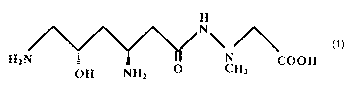
(2) the composition of (1), wherein the dipeptide antibiotic
is the compound of Formula (I) shown below, or an analog of said
compound that can promote readthrough of the nonsense mutation:
Formula (I)
H
N'
HZN : ~ « 'N COON
OH NHZ O CH3
and
(3) the composition of (1) or (2) , wherein the disease caused by the
CA 02449950 2003-12-12
4
nonsense mutation is selected from the group consisting of muscular
dystrophy, cystic fibrosis, Hurler's disease, and infantile neuronal
ceroid lipofuscinosis.
Such compositions of the present invention for treating
diseases caused by nonsense mutations include dipeptide antibiotics.
Unlike conventionally used aminoglycoside antibiotics such as
gentamicin, dipeptide antibiotics generate no serious side effects,
and can promote the readthrough of nonsense mutations. Thus,
- dipeptide antibiotics can be used instead of or in combination with
gentamicin or such, to treat diseases caused by nonsense mutations .
A preferable example of a "dipeptide antibiotic" is negamycin
(methyl hydrazinoacetic acid-linked 8-hydroxy-(3-lysine: NM)
represented by the above formula (I), which has a higher
readthrough-promoting activity than gentamicin. The
above-mentioned "dipeptide antibiotics" also include compounds
structurally similar to negamycin (hereinafter referred to as
"negamycin analogs" ) . As long as a negamycin analog has a structure
similar to that of negamycin and has a readthrough-promoting activity
similar to that of negamycin, its antimicrobial activity is not
relevant.
Negamycin can be prepared, for example, from the culture
supernatant of the Actinomyces strain M890-C2 or MF752-NF9 (Hamada,
M . , Takeuchi , T . , Kondo , S . , I keda , Y . , Naganawa , H . , Maeda , K
. , Okami ,
Y. , and Umezawa, H. (1970) A new antibiotic, negamycin. J. Antibiot.
Tokyo 23, 170-171). Negamycin analogs can be prepared from the
culture supernatants of the above-mentioned Actinomyces strains or
other Actinomyces strains using, as an index, the activity of
promoting the readthrough of nonsense mutations. The negamycin
analogs may be naturally-occurring compounds or may be prepared by
artificially modifying the above-mentioned negamycin. Such
artificial modifications include those introduced for the purposes
of: regulating the activity to promote the readthrough of nonsense
mutations; formulating drugs; or delivering drugs to target cells
(drug delivery); etc.
Further, there is no limitation on the type of "disease caused
by a nonsense mutation" , as long as it is a disease caused by a genetic
CA 02449950 2003-12-12
deficiency due to a nonsense mutation. Such diseases include, for
example, cystic fibrosis (CFTR) , thalassemia ((3-globin) , gastric
cancer (APC), hemophilia (Factor VIII, IX), lung cancer, ovarian
cancer (p53) or such, Duchenne muscular dystrophy (dystrophin) and
5 limb girdle muscular dystrophy (y-sarcoglycan), obesity (insulin
receptor), phenylketonuria,(phenylalanine hydroxylase), and such
(Atkinson, J., and Martin, R. (1994) Mutations to nonsense codons
in human genetic disease: implications for gene therapy by nonsense
- suppressor tRNAs. Nucleic Acid Res 22: 1327-34) . Among the diseases
listed above, cystic fibrosis (Howard et a1. Biochem. Soc. Trans,
21: 846-851 (1996) ; Wilschanski et a1. Am. J. Respir. Crit. Care Med.
161: 860-865 (2000) ; Clancy, J. P. et a1. Am. J. Respir. Crit. Care
Med., 163: 1683 (2001)), muscular dystrophy (Barton-Davis et a1. J.
Clin. Invest. , 104: 375-381 (1999) ; Wagner, K. R. et a1. , Ann. Neurol .
49: 706-711 (2001) ) , Hurler's syndrome (Kim M. Keeling, et al. Human
Mol. Gene. 10: 291-299 (2001)), and late infantile neural ceroid
lipofucinosis (2001 lysosomal tripeptidyl-peptidase 1: Sleat, D. E.
et al. Europ. J. Paediatr. Neurol. 5 Suppl. A: 57-62 (2001)) have
been studied and treated by using the readthrough activity of
gentamicin. Therefore, these diseases may be treated with negamycin
instead of gentamicin.
The readthrough of nonsense mutations can be induced by
administering the above-mentioned negamycin at a daily dose of lx
10-' to 1x 10⁻² mol/kg weight, preferably lx 10-6 to 1x 10⁻³ mol/kg weight,
over a period appropriate to ensure an effect. Without limitation,
powder, granules, tablets, capsules, solutions, injections, and such
are used as the dosage form for administration to patients. Thus,
a therapeutic composition that comprises a dipeptide antibiotic as
an active ingredient, such as the above-mentioned negamycin, can be
formulated with adjuvants such as pharmaceutically acceptable
excipients, binders, disintegrants, lubricants, flavoring agents,
solubilizers, suspending agents, coating agents, and such, as
required.
Brief Description of the Drawings
Fig. 1 depicts photographs showing the presence of dystrophin
CA 02449950 2003-12-12
6
expression and rates of muscle degeneration in Negamycin-treated and
untreated mdx TA muscles . Immunofluorescence and EBD stainings were
carried out as described in Materials and Methods . Panels A, C, and
E are the results of immunofluorescent staining using B10 control
mice, untreated mdx mice, and NM-treated mdx mice respectively.
Panels B, D, and F are EBD staining patterns of B10 control mice,
untreated mdx mice, and NM-treated mdx mice respectively. The bar=
0 dun .
- Fig. 2 is a graph that shows the ratio of the expression level
10 of dystrophin (immunofluorescence-positive fiber) and degenerated
muscle fiber (EBD dye-positive fiber) in TA muscle fibers of
antibiotic-treated mice. The black bar (referred to as "dys" in this
figure) indicates the ratio of dystrophin positive fibers; the gray
bar (referred to as "EB" in this figure) indicates the ratio of Evans
Blue-positive fibers. "NM" indicates mdx mice (seven week-old, six
individuals) that were injected with NM in PBS at a dose of 1.2x 10⁻⁵
mol/kg/day subcutaneously for two weeks; "GM" indicates mdx mice
(seven week-old, six individuals) that were injected with GM in PBS
at a dose of 1.2x 10⁻⁵ mol/kg/day subcutaneously for two weeks. PBS
(0.1 ml) alone was injected to control "mdx" mice (n= 6) and C57BL/10
( "B10") (n= 6 ) mice . About 350-550 muscle fibers were counted in each
mouse (n= 6). The bar indicates the mean ~ standard deviation.
Fig. 3 depicts photographs showing the result of immunoblotting
analysis for dystrophin expression . Panel ( 1 ) shows immunoblotting
results for dystrophin expressed in B10 control mice (lanes A, B,
and C) ; control mdx mice (lane D, E, and F) ; arid NM-treated mdx mice
(lane G, H, and I). Panel (1) shows, from the left, Dystrophin
expression in hind leg muscles (lanes A, D, and G), the diaphragm
(lanes B, E, and H) , and cardiac muscles (lanes C, F, and I) of each
mouse. Panel (2) shows the immunoblotting results for dystrophin
expressed in hind leg muscles . Lane A shows NM-treated mdx mice; lane
B, a sample buffer; lane C, NM-treated mdx mice (x100 of lane A);
lane D, NM-untreated control mdx mice; and lane E, B10 control mice.
Fig. 4 depicts photographs showing the expression of dystrophin
in cultured mdx skeletal muscle cells (mdx-sk) . Panels C and D show
the expression of dystrophin in the cells presented in panels A and
CA 02449950 2003-12-12
7
B, respectively. Panel A shows NM (50 ~,g/ml negamycin)-treated
myotubes observed under a phase contrast microscope; panel B,
NM-untreated myotubes observed under a phase contrast microscope;
panel C, NM (50 ~g/ml negamycin)-treated myotubes stained with
dystrophin; panel D, NM-untreated myotubes stained with dystrophin.
The bar= 40 Eun.
Fig. 5 depicts photographs showing the results of
immunoblotting for dystrophin (427 kDa) in cultured mdx skeletal
muscle cells (mdx-sk) . Lanes A and B show results for myotubes treated
with NM (100 ~tg/ml) for seven days; lane C shows results for untreated
mdx myotubes; and lane D for C2C12 myotubes.
Fig. 6 is a graph that shows weight changes of mdx mice during
NM administration . Negamycin-treated mdx mice at, a dose of 1 . 2x 10⁻⁵
mol/kg (NM1: solid diamond); 1.2x 10⁻⁴ mol/kg (NM10: solid square);
6.0x 10⁻⁴ mol/kg (NM50: solid triangle) ; 1.2x 10⁻³ mol/kg (NM100: X) ;
and NM-untreated control mdx mice (solid line and solid square).
Fig. 7 shows the result of a hearing test based on auditory brain
stem response in antibiotic-treated mice . A, B, and C show the results
for antibiotic-untreated mice, NM-treated mice and GM-treated mice
respectively.
Best Mode for Carrying out the Invention
The present invention is specifically illustrated below with
reference to Examples, but it is not to be construed as being limited
thereto.
[Example 1] Preparation of negamycin
Culture in flasks:
Cells of the Actinomyces M890-C2 strain were inoculated into
fifty 500-ml flasks containing 60 ml of C medium (2 . 0% glucose, 2 . 0
starch, 2.0% soybean extract, 0.5% dry yeast, 35% CaC03, 0.0005%
CuS04~5H20, 0.0005% MnCl2-4H20, 0.005% ZnS04-7H20) and incubated while
shaking (at 220 rpm) at 28°C for four days. The culture media were
filtered through (4%) pearlite. The filtrate was collected and
subj ected to anion-exchange column chromatography (Diaion SAlOA, 1 . 5
liters, OH form). Elution was then carried out with a 0.2 N HCl
CA 02449950 2003-12-12
8
solution. Fractions retaining antimicrobial activity against the
Escherichia coli K-12 strain were collected (500-ml fractions;
fraction numbers 12-16). The active fractions were neutralized with
ammonia water, and then concentrated to 500 ml under reduced pressure.
The resulting concentrated solution was then loaded onto a column
of anion-exchange resin (Amb~rlite CG50, 250 ml, NH4 form) , and eluted
with 0.1% ammonia water. Fractions exhibiting antimicrobial
activity against the K-12 strain were collected (18-g fractions,
fraction numbers 31-70). The active fractions were freeze-dried to
give 32.8 mg of a light brown powder. The powder was resuspended in
the buffer, and the resulting solution was further loaded onto a column
of anion-exchange resins (Amberlite CG50, 250 ml, NH4 form) . Elution
was then carried out with 0.1% ammonia water by the same procedure
as describe above. Fractions exhibiting antimicrobial activity
against the K-12 strain were collected (200-g fractions, fraction
number 6) . The active fraction was freeze-dried to give 13.8 mg of
a white powder.
Culture in jars:
Cells of the Actinomyces M890-C2 strain were inoculated into
a jar fermenter containing five liters of C medium. The cells were
then cultured while shaking (at 300 rpm) under aerobic conditions
of five liters/min at 28°C for five days. Similar to the
above-described flask culture, the culture medium was filtered
through (4%) pearlite. The filtrate was subjected to anion-exchange
column chromatography (Diaion SAlOA, 1.5 liters, ~H form) , and then
active fractions were collected (500-ml fractions; fraction numbers
9-12) . The active fractions were neutralized with ammonia water, and
concentrated to 500 ml under reduced pressure. The resulting
concentrated solution was loaded onto a column of anion-exchange
resins. The column was washed with one liter of distilled water.
Elution was then carried out with 0.1% ammonia water, and active
fractions were collected (200-g fractions, a water-eluted fraction,
and ammonia water-eluted fractions 1 and 2). The active fractions
were freeze-dried to give 4.8 g of a brown powder. This powder was
resuspended in a buffer. The resulting solution was further loaded
CA 02449950 2003-12-12
9
onto a column of anion-exchange resins (Amberlite CG50, 250 ml, NH4
form) , and then eluted with 0. 1$ ammonia water. The active fractions
were collected (200-g fractions, fraction numbers 15-16), and
freeze-dried to give 27.7 mg of a brown powder. In the second-round
purification with anion-exchange resins, fractions 6-9 were found
to retain antimicrobial activity.
[Example 2] Restoration of the expression of dystrophin by the
- administration of negamycin to a muscular dystrophy mouse model
Mdx mice were used as a dystrophy mouse model . NM was dissolved
in PBS and Millipore-filtered just before injection, in order to avoid
degradation during storage in solution. Mdx mice (male, seven
week-old, six for each dose) were injected subcutaneously with an
NM solution (137 mM NaCl, 2.68 mM KC1, 8.10 mM Na2HS04, and 1.47 mM
KH2P04) prepared using PBS, at half the concentration (1 .2x 10-5 mol/kg)
of the solution in the GM experiment by Barton-Davis et al.
(Barton-Davis, E. R., Cordiner, L. Shoturma, D. I., Leiland, S. E.,
Sweeney, H. L. (1999) Aminoglycoside antibiotics restore dystrophin
function to skeletal muscles of mdx mice. J. Clin. Invest. 104,
375-381) every day for two weeks . Control mdx mice (n= 6) and C57BL/10
(B10, n= 6) mice were injected with PBS (0.1 ml) alone, every day
for two weeks.
After injection, immunofluorescence and Evans Blue staining
were performed to detect dystrophin. Immunofluoresce staining was
carried out as follows using antibodies against the C-terminus of
dystrophin. Animals were sacrificed using an overdose of ether gas.
The tibialis anterior (TA) muscles were removed and frozen in melting
isopentane for immunohistochemistry. Then, 7 Etm transverse
cryosections were prepared. After pre-incubation in a blocking
solution for 15 minutes with 20~ horse serum in PBS, the cryosections
were washed with PBS three times for ten minutes and incubated for
one hour at room temperature with the primary antibody (rabbit
anti-dystrophin polyclonal antibody, a gift from Dr. Y. Nonomura,
University of Tokyo, 1:200 diluted in PBS containing 2~ bovine serum
albumin [BSA] ) . After washing with PBS three times for ten minutes,
the sections that reacted with the primary antibody were labeled for
CA 02449950 2003-12-12
one hour with 1:100 diluted fluorescein-labeled anti-rabbit IgG
(Amersham Pharmacia Biotech, Tokyo, Japan). After labeling with
secondary antibodies, dystrophin was detected by fluorescence
microscopy, based on fluorescence emission.
5 Evans Blue staining was carried out by intraperitoneal
inj ection of Evans Blue Dye (BBD : 2 ~ EBD in PBS , 0 . 1 ml ) to all animals
twelve hours before sacrificing. EBD staining visualizes
degenerating muscle fibers that have permeable membranes (Matsuda,
R. , Nishikawa, A. , Tanaka, H. et al . (1995) Visualization of dystrophic
10 muscle fibers in mdx mice by vital staining with Evans Blue: Evidence
of apoptosis in dystrophin-deficient muscle. J. Biochem. 118,
959-964).
As a result of immunofluorescence staining,
dystrophin-positive fibers were detected in the NM-treated mdx mice
(Fig, 1E) as well as in the dystrophin-positive control B10 mice (Fig.
1A) . In contrast, muscle fibers in the NM-untreated mdx mice (Fig.
1C) were all negative for dystrophin (Fig. 1C).
The percentage of dystrophin-positive TA muscle fibers in
drug-treated mice was higher than in untreated mdx mice. Among
drug-treated mice, the percentage of dystrophin-positive TA muscle
fibers in NM-treated mice was higher than in GM-treated mdx mice
reported previously (Arakawa, M., Nakayama, Y., Hara, T., Shiozuka,
M. , Takeda, S. , Kaga, K. , Konda, S. , Morita, S. , Kitamura, T. , Matsuda,
R. (2001) Negamycin can restore dystrophin in mdx skeletal muscle.
Acta. Myologica. XX, 154-158). The percentage of fibers that
exhibited increased membrane permeability (EBD-positive fibers) was
lower in drug-treated animals than in untreated animals , and further,
among drug-treated animals, the percentage was lower in NM-treated
mice than in GM-treated mice (Fig. 2).
These results indicated that administration of NM could lead
to the restoration of dystrophin expression and effectivesuppression
of the formation of membrane-permeable, degenerated muscle fibers.
As described above, the experiment results obtained by
immunofluorescence staining and such showed that NM could restore
dystrophin expression. Thus, the expression of dystrophin was
confirmed in more detail by immunoprecipitation and immunoblotting.
CA 02449950 2003-12-12
11
The left hind leg muscle (600 mg) , the diaphragm (100 mg) , and
the cardiac muscle {100 mg) were collected from NM-treated mdx mice,
NM-untreated mdx mice, and B10 mice as described above. Each of the
specimens was homogenized in 15 ml of a homogenizing solution (pH
7 . 2 ) (pyrophosphate mixture , 20 mM Na4P20~ , 20 mM NaH2P04 , and 1mM MgCl2
,
pH 7.1) containing 10~ sucrose and 0.5 mM EDTA. The homogenization
was carried out with a Teflon homogenizer at maximal speed for one
minute (Mitchell, R. D., Palade, P., and Fleicher, S. (1983)
Purification of morphologically intact triadstructuresfromskeletal
muscle. J. Cell Biol. 96, 1008-1016; Yoshida, M. , Suzuki, A. , Shimizu,
T. , and Ozawa, E. (1992) Proteinase-sensitive sites on isolated rabbit
dystrophin. J. Biochem. 112, 433-439). The homogenate was
centrifuged in an RA rotor (KUBOTA) at 9 , 000 rpm for 15 minutes . The
resulting supernatant was recovered, and then centrifuged again in
an RA rotor at 14 , 000 rpm for 30 minutes to prepare a microsome fraction .
The pellet was dissolved in 1 ml of 1~ digitonin solution (0.5 M NaCl,
0.5 M sucrose, 0.1 mM PMSF, 50 mM Tris-HCl, and lUlml aprotinin,
pH7.2) .
Immunoprecipitation was performed as described by Abe et a1.
(Abe, M., Saitoh, 0., Nakata, H., Yoda, A., and Matsuda, R. (1996)
Expression of neurofilament proteins in proliferating C2C12 mouse
skeletal muscle cells. Exp. Cell Res. 229, 48-59) using muscle protein
sample preparations prepared from mdx mice and B10 mice as described
above. The protein sample preparations described above were each
incubated with anti-dystrophin monoclonal antibody DYS2 (10 ~1)
[Novocastra, Newcastle, UK] at 4°C overnight. Then, wheat germ
agglutinin-Sepharose CL-6B (30 ~tl) (Sigma, Tokyo, Japan) was further
added, and the solution was incubated at 4 ° C for another 60 minutes .
After incubation, the solution was centrifuged at 14,000 rpm at
4°C
for five minutes. Resulting pellets were washed with 0.2~ NP-40 in
PBS (500 ~.l) three times and boiled in sodium dodecyl sulfate (SDS)
sample buffer (62.5 mM Tris-HC1, 2~ SDS, 5 mM EDTA, 5°s
2-mercaptoethanol, 0.1 mM PMSF, and 10~ glycerol, pH 6.8) for four
minutes. After boiling, the supernatant was collected, and the
protein concentration thereof was determined by microburette method.
The samples corresponding to 25 ~,g of total protein that
CA 02449950 2003-12-12
12
resulted from the immunoprecipitation were subjected to
SDS-polyacrylamide gel electrophoresis (SDS-PAGE) with a 4%-8%
gradient gel. After SDS-PAGE, proteins were transferred from the gel
onto a nitrocellulose membrane (Gelman Sciences) for immunoblotting.
The membrane was immersed in a blocking solution (5% skim milk in
25 mM Tris-HC1 [pH 7 . 4] , 137 mMNaCl, 2 . 68 mM KC1, [TBS] , 0. 05% Tween20:
5% skim milk TBST or PEST) at 4°C overnight. After blocking, the
membrane was incubated with a dystrophin-specific antibody (1:100
diluted anti-dystrophin monoclonalantibody DYS3or DYS2, [Novocastra,
Newcastle, UK], or 1:500 diluted rabbit anti-dystrophin polyclonal
antibody; dilutions done using the blocking solution) at room
temperature for one hour. After being washed with 5% skim milk TBST
or PBST three times for ten minutes , the membrane was incubated with
a horseradish peroxidase-conjugated secondary antibody that had been
diluted 1:1000 or 1:3000 in 5% skim milk TBST and then washed with
TBST or PBST three times for 30 minutes. The washed membrane was
treated with an enzyme chemiluminescenee (ECL) kit (Amersham
Pharmacia Biotech, Tokyo, Japan) , and then exposed to an X-ray film,
Hyper-film ECL (Amersham Pharmacia Biotech, Tokyo, Japan), for
visualizing the electrophoresis pattern of dystrophin.
As a result of immunoblotting, dystrophin bands were detected
in hind leg muscles (Fig. 3 (1) A, B, and C) of positive control B10
mice as expected. Also, dystrophin bands were detected in hind leg
muscles of NM-treated mdx mice (Fig. 3 (1) G, H, and I) although the
band intensity thereof was lower than that of the positive control.
From the results of dystrophin expression analysis in hind legs, the
dystrophin expression level restored by NM administration (Fig. 3
(2) A) was estimated to be about 10% of the dystrophin expression
level in normal B10 hind leg muscle (Fig. 3 (2) E).
[Example 3] Dystrophin restoring effect of NM in immortalized cells
(mdx-sk) derived from mdx skeletal muscle cell
The SV40T immortalized mdx satellite cell line, mdx-sk, was
newly established from the mdx mouse to test the dystrophin restoring
level in cultured mdx skeletal muscle cells. The mdx-sk line was
established by introducing a retroviral vector carrying a cDNA for
CA 02449950 2003-12-12
13
the temperature-sensitive form of the Simian Virus 40 large T antigen
(a kind gift from Dr. Drinkwater) to a primary culture of mdx myoblasts
obtained from the mdx mice. The retrovirus was produced using the
packaging cell line, Plat-E, as previously described (Morita, S.,
Kojima, T. , Kitamura, T. (2000) Plat-E: an efficient and stable system
for transient packaging of r~troviruses. Gene Therapy 7, 1063-1066) .
The cell line was cultured and maintained in Dulbecco's Modified Eagle
Medium (DMEM) -high glucose (4 , 500 mg/1) supplemented with 20% fetal
- calf serum at 32.5°C in a COZ incubator. To induce muscle
differentiation, the culture medium was changed to a differentiation
medium (10% horse serum in Eagle MEM) and the culture temperature
was shifted to 39.5°C.
After induction of differentiation, the cells were cultured,
and NM (50 ~,g/ml (2.0x 10⁻⁴ mol/kg) or 100 ~glml (4.0x 10⁻⁴ mol/kg)
in a differentiation medium solution) was added to the culture. Then,
the cells were allowed to differentiate by culturing in the medium
without an antibiotic for seven days. C2C12 cells were maintained
in a growth medium, and then cultured in a differentiation medium
at 38°C in a C02 incubator.
In order to examine whether NM can restore dystrophin expression
in the mdx-sk cells established as described above, NM was added at
a concentration of 50 ~,g/ml or 100 ~ug/ml to a differentiation medium
of myotubes cultured for seven days, and then culture was continued
for another seven days. After culturing (in the presence of 50 ~g/ml
NM), the restoration of dystrophin expression was investigated by
immunofluorescence staining. After being collected from the medium,
the mdx-sk cells were washed twice with PBS. The cells were then fixed
with 100% ethanol for 15 minutes, and treated with a PBS solution
containing 0.5% Triton X-100 for ten minutes. The cells were then
washed three times with PBS for ten minutes, and pre-incubated in
a blocking solution with PBS containing 20% horse serum for 15 minutes .
After being washed three times with PBS for ten minutes, the cells
were incubated with a primary antibody (rabbit anti-dystrophin
polyclonal antibody (a kind gift from Dr. Nonomura, Tokyo University)
diluted 1:200 in PBS containing 2% bovine serum albumin (BSA)) at
room temperature for one hour . After being washed three times with
CA 02449950 2003-12-12
14
PBS for ten minutes, the cells were incubated for labeling with 1:100
diluted fluorescein-labeled anti-rabbit IgG (Amersham Pharmacia
Biotech, Tokyo, Japan) at room temperature for one hour. The cells
were then observed under a fluorescence microscope.
According to the results of immunofluorescence staining, there
was no significant fluorescence signal, and therefore, dystrophin
was not expressed in NM-untreated myotubes (Fig. 4B and 4D) . On the
other hand, strong fluorescence signals were detected in NM-treated
- myotubes, and therefore, dystrophin was confirmed to be expressed
therein (Fig. 4A and 4C).
Furthermore, dystrophin expression in the above-mentioned
cultured cells (in the presence of 100 ~tg/ml (4.0x 10⁻⁴ mol/kg) NM)
was also examined by immunoblotting. First, proteins were prepared
by homogenizing Mdx-sk myotubes and C2C12 myotubes pooled on ice in
10-cm gelatin-coated plates containing 500 ~tl of a homogenization
solution (HS: 20 mM Tris-HC1 (pH 7.6) , 150 mM NaCl, 1$ Nonidet P-40
(NP-40) , 100 ~tg/ml DNase, 1mM phenylmethyl sulfonyl fluoride (PMSF) ,
1 ~g/ml N-tosyl-L-phenylalanyl chloromethyl ketone (TPCK), 1 ~,g/ml
N-tosyl-L-lysyl chloromethyl ketone (TLCK) , 200 U/ml aprotinin, and
5 mM ethylenediamine tetra-acetic acid (EDTA)).
Immunoprecipitation was performed according to the method
described by Abe et a1. (supra) as Example 2, using mds-sk myotube
protein preparations as above. After centrifugation of mdx-sk and
C2C12 myotube protein preparations at 9 , 000 rpm, the supernatant was
collected and incubated with the anti-dystrophin monoclonal antibody
DYS2 (3 ~,1) or the anti-dystrophin monoclonal antibody MANDRA1 (3
~1) (Sigma, Tokyo, Japan) at room temperature (RT) for 60 minutes.
Protein A=Sepharose CL-4B (30 ~1) (Sigma, Tokyo, Japan) was added,
and the solution was incubated at room temperature for another 60
minutes and centrifuged at 14, 000 rpm for five minutes . Pellets were
washed with 0.2~ NP-40 in PBS (500 ~tl) three times and boiled in sodium
dodecyl sulfate (SDS) buffer for five minutes.
A sample corresponding to the total protein amount after
immunoprecipitation from each of the 10-cm plates was subjected to
SDS-polyacrylamide gel electrophoresis (PAGE) on a 4~-8~ gradient
gel. After PAGE, the proteins were transferred onto a nitrocellulose
CA 02449950 2003-12-12
membrane (Gelman Sciences) by the same method as described in Example
2. Then, the membrane was subjected to immunoblotting with specific
antibodies (1:100 diluted anti-dystrophin monoclonalantibodiesDYS3
and DYS2 (Novocastra, Newcastle, UK); 1:500 diluted rabbit
5 anti-dystrophin polyclonalantibody. The electrophoretic pattern of
dystrophin was finally visualized by exposing the membrane to X-ray
film.
According to the pattern of dystrophin expression visualized
by immunoblotting, no band was detected at the position corresponding
10 to 427 kDa in the NM-untreated mdx-sk myotube sample, but a clear
band was detected at the position of 427 kDa in the NM-treated mdx-sk
myotube sample (Fig. 5A and 5B) , as well as in the dystrophin-positive
C2C12 myotube sample (Fig. 5D).
15 [Example 4) NM toxicity test
Generally, aminoglycoside antibiotics like GM are accompanied
by strong side effects. Although these antibiotics are routinely
used for the treatment of bacterial infections, they often cause
nephrotoxicity and ototoxicity. The present inventors examined NM
toxicity by measuring changes in body weight and hearing activity
in drug-treated mice.
In order to measure changes in body weight due to drug
administration, male mdx mice (seven weeks old, four for each dose)
were inj ected daily with NM at 1 . 2x 10⁻⁵ mol/kg (lx the effective dose) ,
1.2x 10⁻⁴ mol/kg (10x) , 6.0x 10⁻⁴ mol/kg (50x) , or 1.2x 10⁻³ mol/kg
(100x) for 14 days. The body weight of each mouse was measured
everyday. Other mdx mice (seven weeks old, two for each dose) were
injected with GM (1.2x 10⁻³ mol/kg [100x]) as a control test. Body
weight was also measured every day.
For the ototoxicity assay, each Mdx mouse to be tested was
injected with 1.2x 10⁻⁴ mol/kg NM or GM daily for seven days. One
day after the final injection, the hearing threshold was measured
based on auditory brain stem response, according to the method of
Shapiro et a1. (Shapiro, S. M. , Moller, A. R. , Shiu, G. K. Brain-stem
auditory evoked potentials in rats with high-dose pentbarbital.
(1984) Electroencephalogr. Clin. Neurophysiol. 58, 266-276).
CA 02449950 2003-12-12
16
According to evaluation of the auditory brain stem response,
a hearing loss of 80 dN or lower was detected only in GM-treated mice,
and not in NM-treated mice (Fig. 7). The measurement of change in
body weight revealed that body weight of mice treated with lower doses
of NM (lx and 10x minimal effective dose) increased sequentially (Fig.
6, NM1 and NM10) , in a manned similar to control mice. However, the
body weight of mice treated with higher doses of NM (50x and 100x)
decreased (Fig. 6, NM50 and NM100) . It is noteworthy that the highest
dose of NM treatment was not lethally toxic. In contrast, the high
dose of GM used conventionally (1.2x 10⁻³ mol/kg/day [100x] ) , killed
all of the tested mice within four hours (data not shown). These
results suggest that NM has a significantly lower toxicity than GM.
Industrial Applicability
I5 As described above, the present invention revealed that it is
possible to restore dystrophin expression deficiencies that occur
due to nonsense mutations . The level to which dystrophin expression
was restored by the composition of the present invention was revealed
to be higher than with the previously used gentamicin. In addition,
unlike gentamicin, the composition of the present invention generated
no serious side effects. Thus, the composition of the present
invention can be used effectively, either in place of or in combination
with gentamicin, as a therapeutic agent for diseases caused by
nonsense mutations, such as muscular dystrophy, cystic fibrosis, and
Hurler's syndrome.